Evacuated Blood Collection Tube For Erythrocyte ... - DocCheck Shop
Evacuated Blood Collection Tube For Erythrocyte ... - DocCheck Shop
Evacuated Blood Collection Tube For Erythrocyte ... - DocCheck Shop
- No tags were found...

You also want an ePaper? Increase the reach of your titles
YUMPU automatically turns print PDFs into web optimized ePapers that Google loves.
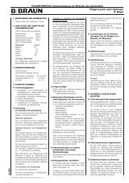
* <strong>Evacuated</strong> <strong>Blood</strong> <strong>Collection</strong> <strong>Tube</strong><strong>For</strong> <strong>Erythrocyte</strong> Sedimentation Rate DeterminationBlack Cap. Labelled <strong>Tube</strong>sReorder No. 366065 (U.S.A. Only)Black Cap. Labelled <strong>Tube</strong>sReorder No. 366674 (Not available in U.S.A.)Black Stopper. Labelled <strong>Tube</strong>sReorder No. 606666 (Not available in U.S.A.)FOR IN VITRO DIAGNOSTIC USE.INTENDED USEThe SEDITAINER <strong>Erythrocyte</strong> Sedimentation Rate (ESR) System is for collection of bloodspecimens for the measurement of ESR within the blood collection tube, using theSEDITAINER ESR Stand.SUMMARY AND PRINCIPLES OF SYSTEMThe SEDITAINER <strong>Tube</strong> provides a means of collecting and testing blood in a closed evacuatedsystem. The tube contains 1.25 mL of 0.105 Molar (M) buff e red sodium citrate solution,giving a 4:1 blood to additive ratio when the evacuated tube is correctly filled to its 5 mL drawvolume. The SEDITAINER <strong>Tube</strong> provides a means to measure the ESR (mm/h) without theneed to access or transfer the specimen. The filled tube has a 100 mm column height andthe ESR is read from a scale on the SEDITAINER Stand. The scale was determined usinga mathematical algorithm that relates the distance the erythrocytes have fallen to a valuethat would be obtained by using the Classic Westergren Method (Berg, 1985).The use of a 100 mm column results in a measured value that differs from the Westergrenmethod. The general relationship is non-linear. A mathematical approximation of this relationshipwas determined using the results of evaluations across the full range of ESR values.This mathematical approximation is described by the following equation:y = 0.87x–0.0027x 2Where:x = Westergren Valuey = SEDITAINER Valueand was used to design the scale on the SEDITAINER Stand to approximate the WestergrenESR.PRINCIPLES OF THE ESR<strong>Erythrocyte</strong>s sediment when anticoagulated blood is alllowed to sit undisturbed. Sedimentationoccurs in three stages during the 1 h test. During the first 10 minutes rouleaux form,with slow sedimentation. <strong>For</strong> the next 40 minutes cells settle rapidly, with most of the settlingoccurring during this period. Finally, during the last 10 minutes, packing occurs. Iftubes are allowed to sit undisturbed for more than 1 h, additional settling may occur.COMPONENTS OF SYSTEMThe SEDITAINER <strong>Tube</strong> is a sterile glass evacuated tube measuring 120 mm long by 10.25mm wide, with a black rubber stopper. The stopper is covered by a plastic shield thatassists in the proper placement of the SEDITAINER <strong>Tube</strong> in the standard size (13 and16 mm) needle holder. The tube contains 1.25 mL of a 0.105 M solution of buff e red sodiumcitrate and has a vacuum level to draw 5.0 mL of blood. Only the interior of the tube issterile. (See “Summary and Principles of System” section.)PRODUCT USE PRECAUTIONS:CAUTION:1. All glass has the potential for breakage; take precautionary measures during handling.2. Handle all biologic samples and blood collection “sharps” (lancets, needles, luer adapters,and blood collection sets) according to the policies and procedures of your facility.3. Obtain appropriate medical attention in the event of any exposure to biologic samples(for example, through a puncture injury), since this may transmit viral hepatitis, HIV(AIDS), or other infectious disease.4. Utilize any built-in used needle protector, if the blood collection device provides one.Becton Dickinson does not recommend reshielding used needles. However, the policiesand procedures of your facility may differ and must always be followed.5. D i s c a rd all blood collection “sharps” in biohazard containers approved for their disposal.6. Transferring a sample from a syringe to a tube is not recommended. Additional manipulationof sharps increases the potential for needlestick injury. In addition, depressingthe syringe plunger during transfer can create a positive pressure, forcefully displacingthe stopper and sample and causing a potential blood exposure. Using a syringe forblood transfer may also cause over or underfilling of tubes, resulting in an incorrectblood-to-additive ratio and potentially incorrect analytic results. <strong>Tube</strong>s with draw volumesmaller than apparent dimensions may not fill to their stated volume when filled from asyringe. The laboratory should be consulted regarding the use of these samples.7. If blood is collected through an intravenous (I.V.) line, ensure that line has been clearedof I.V. solution before beginning to fill blood collection tubes. This is critical to avoiderroneous laboratory data from I.V. fluid contamination.8. Store unused inventory as indicated on carton and case before use.SEDITAINER StandReorder No. 3660169. Use tube before expiration date shown on carton, case and tube labels.10. Take precautions to prevent possible backflow of additive from the tube during blooddrawing. (See “Prevention of Backflow” section.)11. DO NOT REMOVE THE TUBE CLOSURE UNDER ANY CIRCUMSTANCES.SPECIMEN COLLECTION AND PROCESSINGRequired Equipment Not Provided for Specimen <strong>Collection</strong>:1. Practice Universal Precautions, using gloves and appropriate apparel for protection fromexposure to bloodborne pathogens.2. Any VACUTAINER ® Brand Needle Holder of the standard size may be used.3. Alcohol swab for cleansing site.4. Dry sterile gauze.5. Tourniquet.6. Needle disposal container for used needle or needle/holder combination.Required Equipment Not Provided for Specimen Processing:1. Gloves and other personal protective equipment as necessary for protection from exposureto bloodborne pathogens.2. SEDITAINER Stand.Preparation for Specimen <strong>Collection</strong>:Be sure the following materials are readily accessible before performing venipuncture:1. See required equipment above.2. All necessary tubes, identified for size, draw, and additive.3. Labels for positive patient’s identification of samples.Recommended Order of Draw:1. <strong>Tube</strong>s for sterile samples2. <strong>Tube</strong>s without additives3. <strong>Tube</strong>s for coagulation studies4. <strong>Tube</strong>s with additives (e.g., heparin, EDTA)SST ® <strong>Tube</strong>s, VACUTAINER ® Clot Activator <strong>Tube</strong>s, and VACUTAINER ® PLUS <strong>Tube</strong>s are consi d e red additive tubes. SEDITAINER <strong>Tube</strong>s may be drawn after coagulation tubes or includedwith other additive tubes.Prevention of Backflow:Since some evacuated blood collection tubes contain chemical additives, it is important toavoid possible backflow from the tube, with the possibility of adverse patient reactions. Toguard against backflow, observe the following precautions:1. Place patient’s arm in a downward position.2. Hold tube with the stopper uppermost.3. Release tourniquet as soon as blood starts to flow into tube.4. Make sure tube contents do not touch stopper or end of the needle during venipuncture .Venipuncture Technique and Specimen <strong>Collection</strong>:General InstructionsWEAR GLOVES DURING VENIPUNCTURE AND WHEN HANDLING BLOOD COLLECTIONTUBES TO MINIMIZE EXPOSURE HAZARD.1. Select tube or tubes appropriate for required specimen. <strong>For</strong> sterile collections, see thespecific instructions noted in the collection device product circular.2. Assemble needle in holder. Be sure needle is firmly seated to ensure needle does notunthread during use. If drawing sterile specimen, use a sterile holder.3. Gently tap tubes containing additives to dislodge any material that may be adhering tothe stopper.4. Place tube into holder. Do not puncture stopper.5. Select site for venipuncture.6. Apply tourniquet. Prepare venipuncture site with an appropriate antiseptic. DO NOTPALPATE VENIPUNCTURE AREA AFTER CLEANSING.7. Place patient’s arm in a downward position.8. Remove needle shield. Perform venipuncture WITH ARMDOWNWARD AND TUBE STOPPER UPPER-MOST.9. Push tube onto needle, puncturing stopper diaphragm.Center tubes in holder when penetrating the stopper to preventsidewall penetration and resultant premature vacuumloss.10. REMOVE TOURNIQUET AS SOON AS BLOOD APPEARS IN TUBE. DO NOT ALLOWCONTENTS OF TUBE TO CONTACT THE STOPPER OR END OF THE NEEDLE DURINGPROCEDURE.Note: <strong>Blood</strong> may occasionally leak from the needle sleeve. Practice Universal Precautionsto minimize exposure hazard.
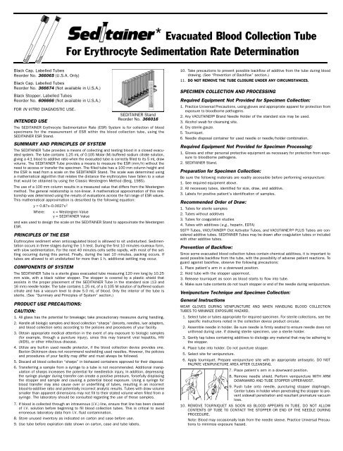
If no blood flows into tube or if blood ceases to flow before an adequate specimen iscollected, the following steps are suggested to complete satisfactory collection:a. Push tube forward until tube stopper has been penetrated. If necessary, hold inplace to ensure complete vacuum draw.b. Confirm correct position of needle cannula in vein.c. If the multiple sample needle is used, remove tube and place new tube onto theholder.d. If second tube does not draw, remove needle and discard. Repeat procedure fromStep 1.11. When first tube has filled to its stated volume and blood flow ceases, remove it fromholder.12. Place succeeding tubes in holder, puncturing diaphragm to begin flow. Draw tubeswithout additives before tubes with additives. See Recommended Order of Draw.13. While each successive tube is filling, turn the filled tube upside-down and return it toupright position. This is one complete inversion.<strong>For</strong> proper additive performance, SEDITAINER <strong>Tube</strong>s must be inverted and returned toupright 8 - 10 times. If other tubes are collected during the same venipuncture, referto appropriate product circular for handling instructions.14. As soon as blood stops flowing in the last tube, remove needle from vein, applyingpressure to puncture site with dry sterile swab until bleeding stops.15. Once clotting has occur red, apply bandage if desired.16. After venipuncture, the top of the stopper may contain residual blood. Take proper precautionswhen handling tubes to avoid contact with this blood. Any needle holder thatbecomes contaminated with blood is considered hazardous and should be decontaminatedwith bleach or disposed of.17. Dispose of the used needle using an appropriate disposal device. DO NOT RESHIELD.Reshielding of needles increases the risk of needlestick injury and blood exposure.PERFORMANCE OF THE SEDITAINER ESR1. Collect venous blood into SEDITAINER <strong>Tube</strong>.2. Just before performing the test, invert the tube 8 - 10 times to mix thoroughly. Use ofa rotating mixer is recommended.3. Place SEDITAINER <strong>Tube</strong> in SEDITAINER Stand. Align 0 mark at top of scale with thebottom of the meniscus of the blood at the blood-air interface.4. Place rack on table or counter where it will not be moved or disturbed for the durationof the test. Stand must be level.5. Set timer for 60 minutes.6. When timer indicates, read level of interface between the settled erythrocytes and thesupernatant plasma from scale on SEDITAINER Stand.RESULTSANALYTIC EQUIVALENCEStudies have been perf o rmed showing the analytic equivalence of the results pro v i d e dby the SEDITAINER <strong>Tube</strong> and Stand to that of the Classic Westergren Method. Usingthe median Seditainer values for each 5 mm/h Classic Westergren interval, for 300patients in the range of 0-130 mm/h, parabolic regression determined the relation ofthe Seditainer ESR System to Classic Westergren to be y = 0.87x–0.0027x 2 , wherex = Classic Westergren Value (mm/h) and y = Seditainer Value for correspondingWestergren Value. Using this equation for the 300 patients, a correlation coefficientof 0.998 was determined. 1 The following diagram demonstrates this relation: Data areavailable upon request.Hemolysis, resulting in decreased red cell concentration, may affect ESR. The stand must belevel. An angle change of as little as 3° from vertical may increase ESR by as much as 30%.The surface on which the stand is placed must not vibrate, as this accelerates sedimentation.TECHNICAL SERVICETechnical Service may be reached at 800-631-0174. You may write to Becton DickinsonVACUTAINER Systems for information at:Technical ServiceBecton Dickinson VACUTAINER Systems1 Becton Drive, Franklin Lakes, NJ 07417-1885REFERENCES1. Berg B. Considerations in sedimentation rate measurements. Clin Lab <strong>For</strong>um. 1985.2. Bridgen ML, Page NE. Three closed-tube methods for determining erythrocyte sedimentation rate. LabMed. 1993;24:97-102.3. H u rd C, Knight T. Laboratory evaluation of the Seditainer ESR System. Med Tech Sci. Sep 1986:53-55.4. I n t e rnational Committee for Standardization in Hematology. Recommendation for measurement oferythrocyte sedimentation rate of human blood. Am J Clin Pathol. 1977;68:505-507.5. Marstein S, Korneliussen R. Seditainer. Måling av senkningsreaksjonen erfaringer med et lukkets y s t e m . Tidsskrift Norske Lœgeforen. 1986;31:2645-2647.6. Patton WN, Meyer PJ, Stuart J. Evaluation of sealed vacuum extraction method (Seditainer) for measurementof erythrocyte sedimentation rate. J Clin Pathol. 1989;42:313-317.7. Sandberg S, Sonstabo K, Eriksen AG, Daae L, Finess L. Ny metode for måling av senkningsreaksjonen.Velegnet for almenpakis og sykehus. Tidsskrift Norske Lœgeforen. 1986;27.8. We s t e rg ren A. Studies of the suspension stability of the blood in pulmonary tuberc u l o s i s . Acta MedScand. 1921;54:247-282.9. Henry JB. Todd-Sanford-Davidsohn clinical diagnosis and management by laboratory methods, 16thedition. St. Louis: CV Mosby, 1979.1. Collect blood, using acceptedvenipuncture technique.PROCEDURETHE SEDITAINER* ESR SYSTEM2. Gently invert SEDITAINER <strong>Tube</strong> at least8 - 10 times.3. Repeat before inserting <strong>Tube</strong> into Stand.70605040302010It is the laboratory’s ultimate responsibility to verify that a change from one tube typeto another does not significantly affect analytic results obtained from patient samples.CONVERSION SCALEThe conversion scale becomes highly compressed above We s t e rg ren values of 100 mmand ESR readings above this level should be repeated using the Classic WestergrenMethod if precise values are required.LIMITATIONS OF SYSTEMThe ESR is a non-specific test. No specific diagnostic information is produced by this test.Elevations result from a multitude of conditions from pregnancy to malignancy.The quantity of blood drawn varies with altitude, ambient temperature, barometric pre s s u re,tube age, venous pressure, and filling technique. Store samples at room temperaturebefore testing. Test should be performed within 6 hours of collection.Possible Sources of Error00 20 40 60 80 100 120 140WESTERGREN (mm/h)I n c o rrect additive to blood ratio will affect results. <strong>Tube</strong>s should be filled to the stated volume.Failure to mix sample thoroughly by inverting 8 - 10 times, may result in erroneous results4. Insert SEDITAINER <strong>Tube</strong> into Stand.6. Set timer and read erythrocyte levelafter 1 hour.5. Align zero level of scale to bottom ofmeniscus.7. Discard SEDITAINER <strong>Tube</strong>s withoutopening.* S E D I TAINER, VA C U TAINER, SST, and PLUS are trademarks of Becton Dickinson and Company.*S E D I TAINER Stand Reorder Number 366016 – U.S. Pat. No. 4,801,428.Becton Dickinson VACUTAINER Systems Europe Becton Dickinson VACUTAINER SystemsB.P. No. 37-38241 MEYLAN CEDEX - France 1 Becton Drive, Franklin Lakes, NJ 07417-18854000102 ( )7/96