Influence of inorganic filler content on the radiopacity of dental resin ...
Influence of inorganic filler content on the radiopacity of dental resin ...
Influence of inorganic filler content on the radiopacity of dental resin ...

You also want an ePaper? Increase the reach of your titles
YUMPU automatically turns print PDFs into web optimized ePapers that Google loves.
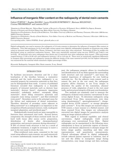
Dental Materials Journal 2012; 31(2): 266–272<br />
<str<strong>on</strong>g>Influence</str<strong>on</strong>g> <str<strong>on</strong>g>of</str<strong>on</strong>g> <str<strong>on</strong>g>inorganic</str<strong>on</strong>g> <str<strong>on</strong>g>filler</str<strong>on</strong>g> <str<strong>on</strong>g>c<strong>on</strong>tent</str<strong>on</strong>g> <strong>on</strong> <strong>the</strong> <strong>radiopacity</strong> <str<strong>on</strong>g>of</str<strong>on</strong>g> <strong>dental</strong> <strong>resin</strong> cements<br />
Gabriel FURTOS 1,2 , Bogdan BALDEA 3 , Laura SILAGHI-DUMITRESCU 1 , Marioara MOLDOVAN 1 ,<br />
Cristina PREJMEREAN 1 and Luminita NICA 4<br />
1Department <str<strong>on</strong>g>of</str<strong>on</strong>g> Dental Materials, "Raluca Ripan" Institute <str<strong>on</strong>g>of</str<strong>on</strong>g> Research in Chemistry, 30 Fantanele Street, 400294 Cluj-Napoca, Romania<br />
2Faculty <str<strong>on</strong>g>of</str<strong>on</strong>g> Chemistry and Chemical Engineering, Babes-Bolyai University, Cluj-Napoca, Romania<br />
3Department <str<strong>on</strong>g>of</str<strong>on</strong>g> Prosthod<strong>on</strong>tics, Faculty <str<strong>on</strong>g>of</str<strong>on</strong>g> Dental Medicine, Victor Babes University <str<strong>on</strong>g>of</str<strong>on</strong>g> Medicine and Pharmacy Timisoara, 9 Revolutiei 1989 Avenue,<br />
300070 Timisoara, Romania<br />
4Department <str<strong>on</strong>g>of</str<strong>on</strong>g> Endod<strong>on</strong>tics, Faculty <str<strong>on</strong>g>of</str<strong>on</strong>g> Dental Medicine, Victor Babes University <str<strong>on</strong>g>of</str<strong>on</strong>g> Medicine and Pharmacy Timisoara, 9 Revolutiei 1989 Avenue,<br />
300070 Timisoara, Romania<br />
Corresp<strong>on</strong>ding author, Gabriel FURTOS; E-mail: gfurtos@yahoo.co.uk<br />
Digital radiography was used to measure <strong>the</strong> <strong>radiopacity</strong> <str<strong>on</strong>g>of</str<strong>on</strong>g> 18 <strong>resin</strong> cements to determine <strong>the</strong> influence <str<strong>on</strong>g>of</str<strong>on</strong>g> <str<strong>on</strong>g>inorganic</str<strong>on</strong>g> <str<strong>on</strong>g>filler</str<strong>on</strong>g> <str<strong>on</strong>g>c<strong>on</strong>tent</str<strong>on</strong>g> <strong>on</strong><br />
<strong>radiopacity</strong>. Four disk specimens (n=4) <str<strong>on</strong>g>of</str<strong>on</strong>g> each light-curing cement were digitally radiographed al<strong>on</strong>gside an aluminum step wedge<br />
using an intraoral sensor (XIOS Plus, Sir<strong>on</strong>a, Germany), and <strong>the</strong>ir mean gray value measured. Percentage <str<strong>on</strong>g>of</str<strong>on</strong>g> <str<strong>on</strong>g>filler</str<strong>on</strong>g> by weight was<br />
determined using an analytical combusti<strong>on</strong> furnace. Data were statistically analyzed using <strong>on</strong>e-way ANOVA and Tukey’s test<br />
(�=0.05). All materials were more radiopaque than dentin and 12 materials were more radiopaque than enamel. Filler percentage<br />
ranged between 17.36 to 53.56 vol% and <strong>radiopacity</strong> between 1.02 to 3.40 mm Al. There were no statistically significant differences<br />
in <str<strong>on</strong>g>inorganic</str<strong>on</strong>g> <str<strong>on</strong>g>filler</str<strong>on</strong>g> percentage and <strong>radiopacity</strong> am<strong>on</strong>g <strong>the</strong> different shades <str<strong>on</strong>g>of</str<strong>on</strong>g> <strong>the</strong> same material (p>0.05), but <strong>the</strong> highest <strong>radiopacity</strong><br />
was measured for <strong>the</strong> material which c<strong>on</strong>tained a higher percentage <str<strong>on</strong>g>of</str<strong>on</strong>g> <str<strong>on</strong>g>filler</str<strong>on</strong>g>.<br />
Keywords: Radiopacity, Inorganic <str<strong>on</strong>g>filler</str<strong>on</strong>g>, Resin cements, X-ray, Dental<br />
INTRODUCTION<br />
To facilitate n<strong>on</strong>-invasive detecti<strong>on</strong> and for a clear<br />
localizati<strong>on</strong> <str<strong>on</strong>g>of</str<strong>on</strong>g> <strong>the</strong> interface between a restorative<br />
material and <strong>the</strong> tooth structure, <strong>radiopacity</strong> is an<br />
essential property for restorative materials so that <strong>the</strong>y<br />
can be distinguished from tooth. A survey <str<strong>on</strong>g>of</str<strong>on</strong>g> published<br />
literature revealed that <strong>radiopacity</strong> is a desirable<br />
property <str<strong>on</strong>g>of</str<strong>on</strong>g> intraoral materials such as denture base<br />
materials 1) , denture liners 2) , elastomeric impressi<strong>on</strong><br />
materials 3) , endod<strong>on</strong>tic sealers 4) , retrograde filling<br />
materials 5) , direct filling restorative materials and<br />
composite <strong>resin</strong> luting cements 6,7) , and fiber-reinforced<br />
<strong>resin</strong> posts 8) . Sec<strong>on</strong>dary caries is <strong>the</strong> principal cause for<br />
<strong>the</strong> failure and replacement <str<strong>on</strong>g>of</str<strong>on</strong>g> <strong>dental</strong> restorati<strong>on</strong>s.<br />
Therefore, detecti<strong>on</strong> <str<strong>on</strong>g>of</str<strong>on</strong>g> sec<strong>on</strong>dary caries adjacent to<br />
filling materials 9) <strong>on</strong> a radiograph is as important as <strong>the</strong><br />
recogniti<strong>on</strong> <str<strong>on</strong>g>of</str<strong>on</strong>g> faulty proximal c<strong>on</strong>tours, voids, inadequate<br />
marginal adaptati<strong>on</strong>, and interfacial gaps 10) .<br />
Endod<strong>on</strong>tically treated teeth are more susceptible to<br />
fractures than unrestored vital teeth. Several factors<br />
compromise <strong>the</strong> strength <str<strong>on</strong>g>of</str<strong>on</strong>g> <strong>the</strong>se restored teeth: loss <str<strong>on</strong>g>of</str<strong>on</strong>g><br />
hard tooth tissue after access cavity preparati<strong>on</strong>,<br />
dehydrati<strong>on</strong> <str<strong>on</strong>g>of</str<strong>on</strong>g> dentin after chemomechanical<br />
preparati<strong>on</strong>, and excessive pressure during obturati<strong>on</strong> 11) .<br />
If 50% <str<strong>on</strong>g>of</str<strong>on</strong>g> cor<strong>on</strong>al tooth structure is lost as a result <str<strong>on</strong>g>of</str<strong>on</strong>g><br />
endod<strong>on</strong>tic treatment, it might be necessary to insert a<br />
post in <strong>the</strong> root canal to retain <strong>the</strong> core.<br />
Fiber-reinforced <strong>resin</strong> posts, cemented in <strong>the</strong> root<br />
canal using adhesive <strong>resin</strong> cements, are used extensively<br />
to restore endod<strong>on</strong>tically treated teeth 11) . For <strong>the</strong> fiber<br />
Color figures can be viewed in <strong>the</strong> <strong>on</strong>line issue, which is avail-<br />
able at J-STAGE.<br />
Received Oct 24, 2011: Accepted Nov 30, 2011<br />
doi:10.4012/dmj.2011-225 JOI JST.JSTAGE/dmj/2011-225<br />
post, <strong>the</strong> radiopaque property allows its visualizati<strong>on</strong><br />
and identificati<strong>on</strong> <strong>on</strong> X-ray films against <strong>the</strong> surrounding<br />
tooth structure and core material 8,11) , and hence <strong>the</strong><br />
implied importance <str<strong>on</strong>g>of</str<strong>on</strong>g> <strong>radiopacity</strong> for core build-up<br />
materials too. For <strong>the</strong> adhesive <strong>resin</strong> cement used to<br />
cement <strong>the</strong> post in <strong>the</strong> root canal, <strong>the</strong> radiopaque<br />
property allows clinicians to use X-ray images to<br />
determine <strong>the</strong> quality <str<strong>on</strong>g>of</str<strong>on</strong>g> <strong>the</strong> restorati<strong>on</strong>, absence/<br />
presence <str<strong>on</strong>g>of</str<strong>on</strong>g> voids, adaptati<strong>on</strong> <str<strong>on</strong>g>of</str<strong>on</strong>g> post to <strong>the</strong> root canal<br />
walls, and structural integrity <str<strong>on</strong>g>of</str<strong>on</strong>g> <strong>the</strong> post-core foundati<strong>on</strong>.<br />
Two methods are comm<strong>on</strong>ly used to measure <strong>the</strong><br />
<strong>radiopacity</strong> <str<strong>on</strong>g>of</str<strong>on</strong>g> <strong>dental</strong> <strong>resin</strong> cements: c<strong>on</strong>venti<strong>on</strong>al X-rays<br />
versus digital X-rays. To determine <strong>radiopacity</strong> using<br />
c<strong>on</strong>venti<strong>on</strong>al X-ray films, measurements were d<strong>on</strong>e<br />
using densitometers 10) , spectrophotometers 12) , chargecoupled<br />
device (CCD) sensors 13) , or storage phosphor<br />
plate systems 13) . X-ray film images are scanned using a<br />
CCD camera, a laser scanner or a flatbed scanner, and<br />
<strong>the</strong>se scanned X-ray film images are subsequently<br />
c<strong>on</strong>verted into digital images for quantitative analysis<br />
using an image analysis s<str<strong>on</strong>g>of</str<strong>on</strong>g>tware 14) .<br />
With regard to technical capability and clinical<br />
c<strong>on</strong>venience, digital radiographs wield several<br />
advantages over c<strong>on</strong>venti<strong>on</strong>al X-ray films. Digital<br />
radiographs can be manipulated in <strong>the</strong> following ways:<br />
enlargement to focus <strong>on</strong> particular regi<strong>on</strong>s <str<strong>on</strong>g>of</str<strong>on</strong>g> interest,<br />
c<strong>on</strong>trast and density adjustments to sharpen images for<br />
better image quality, smoothing and false color<br />
representati<strong>on</strong>, and quantitative measurements.<br />
Clinical advantages <str<strong>on</strong>g>of</str<strong>on</strong>g>fered by digital radiographs<br />
include reduced patient exposure to radiati<strong>on</strong>, ease <str<strong>on</strong>g>of</str<strong>on</strong>g><br />
use and <strong>the</strong> ability to manipulate image during<br />
interpretati<strong>on</strong>, easy image storage and data exchange
for referral, reduced potential operator exposure to<br />
radiati<strong>on</strong> 15) , and no need for film development chemicals.<br />
Traditi<strong>on</strong>al film development, unless performed<br />
carefully, can produce significant variati<strong>on</strong>s in <strong>the</strong> final<br />
radiograph. In c<strong>on</strong>trast, a digital method provides more<br />
c<strong>on</strong>sistent results 16) .<br />
The aim <str<strong>on</strong>g>of</str<strong>on</strong>g> this study was to use digital radiography<br />
to measure <strong>the</strong> <strong>radiopacity</strong> <str<strong>on</strong>g>of</str<strong>on</strong>g> 18 <strong>dental</strong> <strong>resin</strong> cements<br />
and compare <strong>the</strong>m against <strong>the</strong> <strong>radiopacity</strong> <str<strong>on</strong>g>of</str<strong>on</strong>g> human<br />
enamel and dentin. All <strong>the</strong> materials examined in this<br />
study are clinically used in <strong>the</strong> restorati<strong>on</strong> <str<strong>on</strong>g>of</str<strong>on</strong>g><br />
endod<strong>on</strong>tically treated teeth as core build-up materials<br />
and/or as adhesive cements for fiber-reinforced <strong>resin</strong><br />
posts. Percentage <str<strong>on</strong>g>of</str<strong>on</strong>g> <str<strong>on</strong>g>inorganic</str<strong>on</strong>g> <str<strong>on</strong>g>filler</str<strong>on</strong>g> <str<strong>on</strong>g>c<strong>on</strong>tent</str<strong>on</strong>g>, by weight<br />
and volume, was calculated for each <strong>dental</strong> <strong>resin</strong> cement<br />
to determine its influence <strong>on</strong> <strong>the</strong> latter’s <strong>radiopacity</strong>.<br />
Dent Mater J 2012; 31(2): 266–272 267<br />
MATERIALS AND METHODS<br />
Dental <strong>resin</strong> cement specimens<br />
Eighteen commercially available <strong>dental</strong> <strong>resin</strong> cements<br />
were selected for investigati<strong>on</strong> in this study. Their<br />
details, such as indicati<strong>on</strong>s (core build-up material or<br />
luting cement) and chemical compositi<strong>on</strong>s, are listed in<br />
Table 1.<br />
For each <strong>dental</strong> <strong>resin</strong> cement, four disks (n=4)<br />
measuring 8 mm diameter and 1 mm thickness were<br />
prepared and polymerized by a photocuring source<br />
(XL3000, 3M Dental Products, St Paul, MN, USA) for<br />
40 s. As per <strong>the</strong> manufacturers’ recommendati<strong>on</strong>s,<br />
Super-B<strong>on</strong>d C&B (J Morita Europe, Dietzenbach,<br />
Hessen, Germany) was prepared at a polymer/m<strong>on</strong>omer<br />
ratio <str<strong>on</strong>g>of</str<strong>on</strong>g> 1.2 and HybridCem (J Morita Europe) at a<br />
powder/liquid ratio <str<strong>on</strong>g>of</str<strong>on</strong>g> 1/1. Specimen thickness was<br />
Table 1 Details <str<strong>on</strong>g>of</str<strong>on</strong>g> <strong>the</strong> <strong>dental</strong> <strong>resin</strong> cements investigated in this study, as provided by <strong>the</strong> manufacturers<br />
No. Type <str<strong>on</strong>g>of</str<strong>on</strong>g> material Materials (Brand) Shade Compositi<strong>on</strong><br />
1 Core build-up<br />
material<br />
2 Core build-up<br />
material<br />
3 Core build-up<br />
material<br />
4 Core build-up<br />
material<br />
5 Core build-up<br />
material<br />
6 Core build-up<br />
material<br />
7 Core build-up<br />
material<br />
8 Core build-up<br />
material<br />
9 Luting cement<br />
material<br />
Build-It FR<br />
(Pentr<strong>on</strong> Clinical, Wallingford, CT,<br />
USA)<br />
CromaCore<br />
(J Morita Europe, Dietzenbach,<br />
Germany)<br />
Core Paste XP<br />
(DenMat, Santa Maria, CA, USA)<br />
Mirafit Core<br />
(Hager & Werken, Duisburg,<br />
Germany)<br />
ParaPost ParaCore Automix<br />
(Coltene Whaledent, Altstatten,<br />
Switzerland)<br />
ParaPost ParaCore Automix<br />
(Coltene Whaledent)<br />
Rock Core<br />
(Danville Materials Inc., San<br />
Ram<strong>on</strong>, CA, USA)<br />
Sealacore Core Composite<br />
(PDSA Produits Dentaires, Vevey,<br />
Switzerland)<br />
Duo Cement Plus<br />
(Coltene Whaledent)<br />
Gold Bis-GMA, UDMA, HDDMA, mixture <str<strong>on</strong>g>of</str<strong>on</strong>g><br />
bariumborosilicate, calcium alumin<str<strong>on</strong>g>of</str<strong>on</strong>g>luro-silicate,<br />
silica and chopped glass<br />
fiber. 68% wt.; 52% vol. <str<strong>on</strong>g>inorganic</str<strong>on</strong>g> <str<strong>on</strong>g>filler</str<strong>on</strong>g>.<br />
Enamel<br />
w/Fluoride<br />
NC<br />
Glass <str<strong>on</strong>g>filler</str<strong>on</strong>g>s in methacrylate <strong>resin</strong><br />
NC% wt.; vol. <str<strong>on</strong>g>inorganic</str<strong>on</strong>g> <str<strong>on</strong>g>filler</str<strong>on</strong>g><br />
A3 Bis-GMA, UDMA, HDDMA, silane<br />
treated bariumborosilicate, glass fibers,<br />
fluoride, pigments with initiators,<br />
stabilizers and UV absorber. NC% wt.;<br />
vol. <str<strong>on</strong>g>inorganic</str<strong>on</strong>g> <str<strong>on</strong>g>filler</str<strong>on</strong>g><br />
Dentin Methacrylate, fluoride barium glass,<br />
amorphous silica<br />
≈ 68% wt; 50% vol. <str<strong>on</strong>g>inorganic</str<strong>on</strong>g> <str<strong>on</strong>g>filler</str<strong>on</strong>g><br />
White Methacrylate, fluoride barium glass,<br />
amorphous silica.<br />
68% wt; 50% vol. <str<strong>on</strong>g>inorganic</str<strong>on</strong>g> <str<strong>on</strong>g>filler</str<strong>on</strong>g><br />
C2 Bis-GMA, barium glass 69%, silica 3%,<br />
NC% wt.; 49% vol. <str<strong>on</strong>g>inorganic</str<strong>on</strong>g> <str<strong>on</strong>g>filler</str<strong>on</strong>g><br />
Universal Mixture <str<strong>on</strong>g>of</str<strong>on</strong>g> <strong>resin</strong>s based <strong>on</strong> Bis-GMA<br />
(methacrylates), catalysts, stabilizers,<br />
pigments. NC% wt.; NC% vol. <str<strong>on</strong>g>inorganic</str<strong>on</strong>g><br />
<str<strong>on</strong>g>filler</str<strong>on</strong>g><br />
Base<br />
Catalyst<br />
Bis-EMA, Bis-GMA, TEGDMA, barium<br />
glass silanized, amorphous silicic acid,<br />
hydrophobed. 71% wt.; 54.5% vol.<br />
<str<strong>on</strong>g>inorganic</str<strong>on</strong>g> <str<strong>on</strong>g>filler</str<strong>on</strong>g>
268<br />
Table 1 (c<strong>on</strong>tinued)<br />
Dent Mater J 2012; 31(2): 266–272<br />
No. Type <str<strong>on</strong>g>of</str<strong>on</strong>g> material Materials (Brand) Shade Compositi<strong>on</strong><br />
10 Luting cement<br />
material<br />
11 Luting cement<br />
material<br />
12 Luting cement<br />
material<br />
13 Luting cement<br />
material<br />
14 Luting cement<br />
material<br />
15 Luting cement<br />
material<br />
16 Luting cement<br />
material<br />
17 Luting cement<br />
material<br />
18 Luting cement<br />
material<br />
Hybrid Cem<br />
(J Morita Europe)<br />
Maxcem<br />
(Kerr Corp.,<br />
Orange, CA, USA)<br />
Microcem duo<br />
(Saremco Dental AG, Rebstein,<br />
Switzerland)<br />
Multilink Sprint<br />
(Ivoclar Vivadent, Schaan,<br />
Liechtenstein)<br />
Multilink Sprint<br />
(Ivoclar Vivadent, Schaan,<br />
Liechtenstein)<br />
ParaPost Cement<br />
(Coltene Whaledent)<br />
ParaCem Universal DC<br />
(Coltene Whaledent)<br />
RelyX ARC<br />
(3M ESPE, St. Paul, MN, USA)<br />
Super-B<strong>on</strong>d C&B<br />
(J Morita Europe)<br />
4-META, MMA, HEMA, acet<strong>on</strong>e, water<br />
Powder/Liquid=1/1,<br />
NC% wt.; vol. <str<strong>on</strong>g>inorganic</str<strong>on</strong>g> <str<strong>on</strong>g>filler</str<strong>on</strong>g><br />
Clear GPDM, - self-etching/self-adhering<br />
acidic m<strong>on</strong>omer; com<strong>on</strong>omers including<br />
m<strong>on</strong>o-, di-, and tri-functi<strong>on</strong>al<br />
methacrylate m<strong>on</strong>omers, initiator,<br />
photoinitiator, stabilizer, barium glass<br />
<str<strong>on</strong>g>filler</str<strong>on</strong>g>, fluoroaluminosilicate glass <str<strong>on</strong>g>filler</str<strong>on</strong>g>,<br />
fumed silica, average <str<strong>on</strong>g>filler</str<strong>on</strong>g> particle size<br />
<str<strong>on</strong>g>of</str<strong>on</strong>g> 3.6 µm. 67% wt.; 46% vol. <str<strong>on</strong>g>inorganic</str<strong>on</strong>g><br />
<str<strong>on</strong>g>filler</str<strong>on</strong>g><br />
Barium glass, silanized with silane<br />
A-174, Bis-GMA, Bis-EMA, TEGDMA,<br />
higly dispersed silicium, dioxide,<br />
silanized, catalysts, inhibitors,<br />
pigments. NC% wt.; vol. <str<strong>on</strong>g>inorganic</str<strong>on</strong>g><br />
<str<strong>on</strong>g>filler</str<strong>on</strong>g>.<br />
Opaque Dimethacrylates, acidic m<strong>on</strong>omers,<br />
barium glass, YbF3, silic<strong>on</strong> oxide.<br />
NC % wt.; 48% vol. <str<strong>on</strong>g>inorganic</str<strong>on</strong>g> <str<strong>on</strong>g>filler</str<strong>on</strong>g><br />
Yellow Dimethacrylates, acidic m<strong>on</strong>omers,<br />
barium glass, YbF3, silic<strong>on</strong> oxide<br />
NC % wt.; 48% vol. <str<strong>on</strong>g>inorganic</str<strong>on</strong>g> <str<strong>on</strong>g>filler</str<strong>on</strong>g><br />
Base: Methacrylates, barium glass,<br />
silanized, amorphous silica,<br />
Catalyst: Methacrylates, barium glass,<br />
silanized, amorphous silica, BPO<br />
NC% wt.; vol. <str<strong>on</strong>g>inorganic</str<strong>on</strong>g> <str<strong>on</strong>g>filler</str<strong>on</strong>g><br />
B3 Base: Bis-EMA, Bis-GMA, TEGDMA,<br />
barium glass silanized, amorphous<br />
silica, Initiators.<br />
Catalyst: Bis-EMA, Bis-GMA,<br />
TEGDMA, Barium glass silanized,<br />
amorphous silica, BPO.<br />
NC% wt.; vol. <str<strong>on</strong>g>inorganic</str<strong>on</strong>g> <str<strong>on</strong>g>filler</str<strong>on</strong>g><br />
A3 Bis-GMA, TEGDMA, zirc<strong>on</strong>ia/silica<br />
<str<strong>on</strong>g>filler</str<strong>on</strong>g>, pigments, amine and<br />
photoinitiator system (Paste A), BPO<br />
(Paste B).<br />
67.5% wt.; NC % vol. <str<strong>on</strong>g>inorganic</str<strong>on</strong>g> <str<strong>on</strong>g>filler</str<strong>on</strong>g><br />
L-Type<br />
Radiopaque<br />
4-META/MMA-TBB, metal oxide<br />
Polymer/M<strong>on</strong>omer=1.2<br />
NC% wt.; NC% vol. <str<strong>on</strong>g>inorganic</str<strong>on</strong>g> <str<strong>on</strong>g>filler</str<strong>on</strong>g><br />
Bis-EMA: Bisphenol A diethoxymethacrylate; Bis-GMA: Bisphenol A diglycidyl methacrylate; BPO: Benzoyl peroxide; CQ:<br />
Camphorquin<strong>on</strong>e; GPDM: Glyceroldimethacrylate dihydrogen phosphate; HEMA: 2-hydroxyethyl methacrylate; HDDMA:<br />
1,6-hexanediol dimethacrylate; MMA: methyl methacrylate; TBB: tri-n-butylborane (initiator); TEGDMA: Triethyleneglycol<br />
dimethacrylate; UDMA: Urethane dimethacrylate; 4-META: 4-methacryloxyethyl trimellitic anhydride; NC: Informati<strong>on</strong><br />
not collected; % wt: Percentage <str<strong>on</strong>g>of</str<strong>on</strong>g> <str<strong>on</strong>g>filler</str<strong>on</strong>g> by weight; % vol.: Percentage <str<strong>on</strong>g>of</str<strong>on</strong>g> <str<strong>on</strong>g>filler</str<strong>on</strong>g> by volume.
measured using a micrometer. Specimens with thickness<br />
greater than 1 mm (±0.01) were sanded using 800-grit<br />
sandpaper until <strong>the</strong>ir thickness was 1 mm (±0.01).<br />
Specimens with voids were excluded from this study.<br />
Human enamel and dentin specimens<br />
This study was approved by <strong>the</strong> Commissi<strong>on</strong> <strong>on</strong> Bioethics<br />
<str<strong>on</strong>g>of</str<strong>on</strong>g> <strong>the</strong> Victor Babes University <str<strong>on</strong>g>of</str<strong>on</strong>g> Medicine and Pharmacy,<br />
Timisoara. Two freshly extracted human molars and<br />
<strong>on</strong>e premolar tooth —free from caries, hypoplastic<br />
defects or cracks and extracted for orthod<strong>on</strong>tic reas<strong>on</strong><br />
from <strong>the</strong> same subject— were selected for use in this<br />
study. The subject/patient signed an informed c<strong>on</strong>sent<br />
before any clinical procedure was performed.<br />
The extracted teeth were stored in buffered formalin<br />
for 24 h after extracti<strong>on</strong> and <strong>the</strong>n in water at room<br />
temperature (23±1°C). After embedding <strong>the</strong> teeth in a<br />
methyl-methacrylate casting <strong>resin</strong> (Dentacryl,<br />
Sp<str<strong>on</strong>g>of</str<strong>on</strong>g>aDental Western Europe, Bioggio Ticino,<br />
Switzerland), <strong>on</strong>e mesiodistal secti<strong>on</strong> <str<strong>on</strong>g>of</str<strong>on</strong>g> 1 mm (±0.01)<br />
thickness was cut from each tooth using a rotary cutting<br />
machine (IsoMet, Buehler, Lake Bluff, IL, USA).<br />
Digital radiography<br />
An aluminum step wedge c<strong>on</strong>sisting <str<strong>on</strong>g>of</str<strong>on</strong>g> 1- to 5-mm steps<br />
was prepared to be used as a radiographic reference.<br />
The <strong>radiopacity</strong> <str<strong>on</strong>g>of</str<strong>on</strong>g> 18 commercially available <strong>dental</strong><br />
<strong>resin</strong> cements were determined with reference to <strong>the</strong><br />
aluminum step wedge and human enamel and dentin<br />
slices <str<strong>on</strong>g>of</str<strong>on</strong>g> equivalent thickness.<br />
Dental <strong>resin</strong> cement specimens and human enamel<br />
and dentin specimens were placed al<strong>on</strong>gside <strong>the</strong><br />
aluminum step wedge <strong>on</strong> an intraoral sensor (XIOS Plus,<br />
Sir<strong>on</strong>a Dental Systems, Bensheim, Germany). Imaging<br />
was d<strong>on</strong>e using an intraoral X-ray machine (MinRay,<br />
Soredex, Tuusula, Finland) at 70 kV, 7 mA, and 0.04 s<br />
with a target-sensor distance <str<strong>on</strong>g>of</str<strong>on</strong>g> 30 cm.<br />
The mean gray values <str<strong>on</strong>g>of</str<strong>on</strong>g> each step <str<strong>on</strong>g>of</str<strong>on</strong>g> <strong>the</strong> aluminum<br />
step wedge and <strong>the</strong> specimens were measured by<br />
outlining a regi<strong>on</strong> <str<strong>on</strong>g>of</str<strong>on</strong>g> interest using <strong>the</strong> equal-density<br />
area tool <str<strong>on</strong>g>of</str<strong>on</strong>g> a <strong>dental</strong> imaging s<str<strong>on</strong>g>of</str<strong>on</strong>g>tware (Image J 1.37v,<br />
Wayne Rasband, Nati<strong>on</strong>al Institutes <str<strong>on</strong>g>of</str<strong>on</strong>g> Health, Be<strong>the</strong>sda,<br />
MD, USA). With each specimen, five regi<strong>on</strong>s were<br />
selected for measurement: <strong>on</strong>e in <strong>the</strong> central area and<br />
four in <strong>the</strong> different quadrants <str<strong>on</strong>g>of</str<strong>on</strong>g> each disk specimen.<br />
Each regi<strong>on</strong> selected was devoid <str<strong>on</strong>g>of</str<strong>on</strong>g> any air bubbles inside<br />
<strong>the</strong> material, and <strong>the</strong> average gray value was recorded<br />
for every specimen. For each radiographic image, its<br />
gray scale value was c<strong>on</strong>verted into equivalent aluminum<br />
thickness using a generated calibrati<strong>on</strong> curve correlating<br />
gray scale values to aluminum thickness. The <strong>radiopacity</strong><br />
value <str<strong>on</strong>g>of</str<strong>on</strong>g> each specimen was expressed in terms <str<strong>on</strong>g>of</str<strong>on</strong>g> <strong>the</strong><br />
equivalent thickness <str<strong>on</strong>g>of</str<strong>on</strong>g> aluminum per 1 mm unit<br />
thickness <str<strong>on</strong>g>of</str<strong>on</strong>g> material.<br />
Percentage <str<strong>on</strong>g>of</str<strong>on</strong>g> <str<strong>on</strong>g>filler</str<strong>on</strong>g> <str<strong>on</strong>g>c<strong>on</strong>tent</str<strong>on</strong>g> by weight and volume<br />
For each <strong>dental</strong> <strong>resin</strong> cement, its <str<strong>on</strong>g>inorganic</str<strong>on</strong>g> <str<strong>on</strong>g>filler</str<strong>on</strong>g> <str<strong>on</strong>g>c<strong>on</strong>tent</str<strong>on</strong>g><br />
was measured by combusti<strong>on</strong> analysis in a furnace (n=5).<br />
Percentage <str<strong>on</strong>g>of</str<strong>on</strong>g> <str<strong>on</strong>g>filler</str<strong>on</strong>g> by weight was determined by<br />
calculating <strong>the</strong> difference in <strong>the</strong> weight <str<strong>on</strong>g>of</str<strong>on</strong>g> <strong>the</strong> crucible<br />
Dent Mater J 2012; 31(2): 266–272 269<br />
before and after ashing in air. Percentage <str<strong>on</strong>g>of</str<strong>on</strong>g> <str<strong>on</strong>g>filler</str<strong>on</strong>g> weight<br />
was c<strong>on</strong>verted to volume percentage using <strong>the</strong> following<br />
formula:<br />
wf/df<br />
Filler volume fracti<strong>on</strong> = ×100%<br />
wf/df + wr/dr<br />
where wf and wr are <strong>the</strong> weight fracti<strong>on</strong>s <str<strong>on</strong>g>of</str<strong>on</strong>g> <str<strong>on</strong>g>filler</str<strong>on</strong>g> and<br />
<strong>resin</strong> respectively, and df and dr are <strong>the</strong> densities <str<strong>on</strong>g>of</str<strong>on</strong>g> <str<strong>on</strong>g>filler</str<strong>on</strong>g><br />
and <strong>resin</strong> at 2.4 g/cm 3 and 1.2 g/cm 3 respectively.<br />
Statistical analysis<br />
Data were statistically analyzed using <strong>on</strong>e-way analysis<br />
<str<strong>on</strong>g>of</str<strong>on</strong>g> variance ANOVA (SPSS versi<strong>on</strong> 11.5, SPSS Inc., USA)<br />
and Tukey’s test with level <str<strong>on</strong>g>of</str<strong>on</strong>g> significance set at 0.05 to<br />
determine <strong>the</strong> presence <str<strong>on</strong>g>of</str<strong>on</strong>g> statistically significant<br />
differences between <strong>the</strong> mean values <str<strong>on</strong>g>of</str<strong>on</strong>g> <strong>the</strong> tested<br />
materials.<br />
RESULTS<br />
Radiopacity <str<strong>on</strong>g>of</str<strong>on</strong>g> <strong>dental</strong> <strong>resin</strong> cements in comparis<strong>on</strong> to<br />
enamel and dentin<br />
The mean <strong>radiopacity</strong> values and standard deviati<strong>on</strong>s <str<strong>on</strong>g>of</str<strong>on</strong>g><br />
<strong>the</strong> investigated materials are shown in Fig. 1. Am<strong>on</strong>g<br />
<strong>the</strong> tested <strong>dental</strong> <strong>resin</strong> cements, <strong>the</strong>re were no<br />
statistically significant differences (p>0.05) according to<br />
<strong>on</strong>e-way ANOVA. However, <strong>the</strong> <strong>radiopacity</strong> values <str<strong>on</strong>g>of</str<strong>on</strong>g><br />
some tested materials were statistically different from<br />
those <str<strong>on</strong>g>of</str<strong>on</strong>g> enamel or dentin.<br />
Multilink Sprint <str<strong>on</strong>g>of</str<strong>on</strong>g> Opaque and Yellow shades<br />
(Ivoclar Vivadent, Schaan, Liechtenstein) had <strong>the</strong><br />
highest <strong>radiopacity</strong> values <str<strong>on</strong>g>of</str<strong>on</strong>g> 3.40 mm Al and 3.36 mm Al<br />
respectively, which were statistically different from that<br />
<str<strong>on</strong>g>of</str<strong>on</strong>g> enamel (p
270<br />
Percentage <str<strong>on</strong>g>of</str<strong>on</strong>g> <str<strong>on</strong>g>filler</str<strong>on</strong>g> <str<strong>on</strong>g>c<strong>on</strong>tent</str<strong>on</strong>g> by weight and volume<br />
Combusti<strong>on</strong> analysis revealed that <strong>the</strong> <str<strong>on</strong>g>filler</str<strong>on</strong>g> <str<strong>on</strong>g>c<strong>on</strong>tent</str<strong>on</strong>g> <str<strong>on</strong>g>of</str<strong>on</strong>g><br />
<strong>the</strong> tested <strong>dental</strong> <strong>resin</strong> cements ranged between 29.56<br />
wt% (17.36 vol%) for Super-B<strong>on</strong>d C&B (J Morita Europe)<br />
and 69.75 wt% (53.56 vol%) for Multilink Sprint <str<strong>on</strong>g>of</str<strong>on</strong>g><br />
Yellow shade (Ivoclar Vivadent) (Table 2).<br />
DISCUSSION<br />
The <strong>radiopacity</strong> <str<strong>on</strong>g>of</str<strong>on</strong>g> a material increases with a higher<br />
<str<strong>on</strong>g>filler</str<strong>on</strong>g> percentage and a higher amount <str<strong>on</strong>g>of</str<strong>on</strong>g> high atomic<br />
number elements in <str<strong>on</strong>g>filler</str<strong>on</strong>g> particles 10,17) . This explained<br />
why <strong>the</strong> chemical compositi<strong>on</strong>s <str<strong>on</strong>g>of</str<strong>on</strong>g> some <strong>dental</strong> composites<br />
available <strong>on</strong> <strong>the</strong> market included high atomic number<br />
<str<strong>on</strong>g>filler</str<strong>on</strong>g> elements such as barium, str<strong>on</strong>tium, zirc<strong>on</strong>ium,<br />
zinc, ytterbium, titanium, tantalum, lanthanum, or<br />
indium (Table 1) 17-21) . When <str<strong>on</strong>g>filler</str<strong>on</strong>g> volume is increased to<br />
70% or bey<strong>on</strong>d and <strong>the</strong> amount <str<strong>on</strong>g>of</str<strong>on</strong>g> radiopaque oxide in<br />
<str<strong>on</strong>g>filler</str<strong>on</strong>g> particles is above 20%, <strong>the</strong> <strong>radiopacity</strong> <str<strong>on</strong>g>of</str<strong>on</strong>g> a <strong>dental</strong><br />
composite would exceed that <str<strong>on</strong>g>of</str<strong>on</strong>g> human enamel 17) .<br />
In <strong>the</strong> present study, all <strong>the</strong> <strong>dental</strong> <strong>resin</strong> cements<br />
tested met <strong>the</strong> ISO 4049 standard 19) in that <strong>the</strong>ir<br />
<strong>radiopacity</strong> values exceeded 1 mm Al and were higher<br />
than that <str<strong>on</strong>g>of</str<strong>on</strong>g> human dentin. For different shades <str<strong>on</strong>g>of</str<strong>on</strong>g> <strong>the</strong><br />
same material, namely Multilink Sprint (Opaque,<br />
Yellow) and ParaPost ParaCore Automix (White,<br />
Dentin), <strong>the</strong>re were no statistically significant differences<br />
in <strong>radiopacity</strong> and percentage <str<strong>on</strong>g>of</str<strong>on</strong>g> <str<strong>on</strong>g>filler</str<strong>on</strong>g> am<strong>on</strong>g <strong>the</strong> two<br />
Dent Mater J 2012; 31(2): 266–272<br />
Fig. 1 Mean (SD) <strong>radiopacity</strong> values <str<strong>on</strong>g>of</str<strong>on</strong>g> 18 commercially available composite <strong>resin</strong> cements, in comparis<strong>on</strong> to dentin and<br />
enamel. (Horiz<strong>on</strong>tal bars indicate mean values are not statistically significant different from each o<strong>the</strong>r when<br />
analyzed using <strong>the</strong> Tukey’s test, p>0.05.)<br />
shades <str<strong>on</strong>g>of</str<strong>on</strong>g> <strong>the</strong> same material.<br />
For Multilink Sprint Opaque and Multilink Sprint<br />
Yellow, not <strong>on</strong>ly did <strong>the</strong>y have <strong>the</strong> highest <str<strong>on</strong>g>filler</str<strong>on</strong>g> <str<strong>on</strong>g>c<strong>on</strong>tent</str<strong>on</strong>g>s<br />
as revealed by combusti<strong>on</strong> analysis, <strong>the</strong>y had radiopaque<br />
<str<strong>on</strong>g>filler</str<strong>on</strong>g>s such as barium glass and YbF3 in <strong>the</strong>ir <str<strong>on</strong>g>filler</str<strong>on</strong>g><br />
compositi<strong>on</strong>s. These dual <str<strong>on</strong>g>filler</str<strong>on</strong>g>-related factors caused<br />
<strong>the</strong>m to have <strong>the</strong> highest <strong>radiopacity</strong> am<strong>on</strong>g <strong>the</strong> tested<br />
materials and which was significantly higher than that<br />
<str<strong>on</strong>g>of</str<strong>on</strong>g> enamel. Ranking just below Multilink Sprint Opaque<br />
and Multilink Sprint Yellow in terms <str<strong>on</strong>g>of</str<strong>on</strong>g> <str<strong>on</strong>g>filler</str<strong>on</strong>g> <str<strong>on</strong>g>c<strong>on</strong>tent</str<strong>on</strong>g><br />
were ParaPost ParaCore Automix White and ParaPost<br />
ParaCore Automix Dentin. Apart from <strong>the</strong>ir high <str<strong>on</strong>g>filler</str<strong>on</strong>g><br />
percentages, <strong>the</strong>y also had barium glass in <strong>the</strong>ir <str<strong>on</strong>g>filler</str<strong>on</strong>g><br />
compositi<strong>on</strong>s, <strong>the</strong>reby rendering <strong>the</strong>m with <strong>radiopacity</strong><br />
values significantly higher than that <str<strong>on</strong>g>of</str<strong>on</strong>g> enamel.<br />
Maxcem, ParaCem Universal DC, Duo Cement Plus,<br />
Build-It FR, Rock Core, Mirafit Core, and Microcem duo<br />
also c<strong>on</strong>tained barium in <strong>the</strong>ir <str<strong>on</strong>g>filler</str<strong>on</strong>g> compositi<strong>on</strong>s. It was<br />
highly probable that <strong>the</strong>ir <strong>radiopacity</strong> values were<br />
influenced by <strong>the</strong> amount <str<strong>on</strong>g>of</str<strong>on</strong>g> barium in <strong>the</strong>ir <str<strong>on</strong>g>filler</str<strong>on</strong>g><br />
particles. Microcem duo had a low <strong>radiopacity</strong> value<br />
which was not significantly different from that <str<strong>on</strong>g>of</str<strong>on</strong>g> dentin<br />
(p>0.05), and this could be due to a low barium <str<strong>on</strong>g>c<strong>on</strong>tent</str<strong>on</strong>g><br />
in its <str<strong>on</strong>g>filler</str<strong>on</strong>g> particles.<br />
Super-B<strong>on</strong>d C&B had <strong>the</strong> lowest <str<strong>on</strong>g>filler</str<strong>on</strong>g> percentage,<br />
but a <strong>radiopacity</strong> <str<strong>on</strong>g>of</str<strong>on</strong>g> 2.56 mm Al which was significantly<br />
higher than that <str<strong>on</strong>g>of</str<strong>on</strong>g> dentin. This <strong>radiopacity</strong> value could<br />
be caused by <strong>the</strong> presence <str<strong>on</strong>g>of</str<strong>on</strong>g> high atomic number
elements in <strong>the</strong> <str<strong>on</strong>g>filler</str<strong>on</strong>g> particles. RelyX ARC had zirc<strong>on</strong>ium<br />
oxide/zirc<strong>on</strong>ia in its <str<strong>on</strong>g>filler</str<strong>on</strong>g> compositi<strong>on</strong>, a radiopaque<br />
oxide which was composed <str<strong>on</strong>g>of</str<strong>on</strong>g> a high atomic number<br />
element. For this reas<strong>on</strong>, its <strong>radiopacity</strong> was statistically<br />
higher than that <str<strong>on</strong>g>of</str<strong>on</strong>g> dentin.<br />
There were small differences between <strong>the</strong> <str<strong>on</strong>g>filler</str<strong>on</strong>g><br />
Dent Mater J 2012; 31(2): 266–272 271<br />
Table 2 Percentages <str<strong>on</strong>g>of</str<strong>on</strong>g> <strong>the</strong> <str<strong>on</strong>g>inorganic</str<strong>on</strong>g> <str<strong>on</strong>g>filler</str<strong>on</strong>g> <str<strong>on</strong>g>c<strong>on</strong>tent</str<strong>on</strong>g> <str<strong>on</strong>g>of</str<strong>on</strong>g> <strong>dental</strong> <strong>resin</strong> cements, as revealed by combusti<strong>on</strong> analysis<br />
No Product_Shade (Brand)<br />
Mean (SD)wt%<br />
by combusti<strong>on</strong><br />
Mean (SD) vol%<br />
by combusti<strong>on</strong><br />
1 Multilink Sprint_Yellow (Ivoclar Vivadent) 69.75 (0.17) 53.56 (0.20)<br />
2 Multilink Sprint_Opaque (Ivoclar Vivadent) 69.37 (0.42) 53.11 (0.49)<br />
3 ParaPost ParaCore Automix_White (Coltene Whaledent) 67.27 (0.16) 50.69 (0.18)<br />
4 ParaPost ParaCore Automix_Dentin (Coltene Whaledent) 67.19 (0.26) 50.59 (0.29)<br />
5 Build-it_Gold (Pentr<strong>on</strong>) 67.04 (1.51) 50.44 (1.73)<br />
6 Maxcem Clear_Clear (Kerr) 66.75 (0.42) 50.09 (0.48)<br />
7 Duo Cement Plus (Coltene Whaledent) 66.25 (0.12) 49.53 (0.14)<br />
8 RelyX ARC_A3 (3M ESPE) 65.88 (0.96) 49.12 (1.08)<br />
9 Core Paste XP Enamel w/Fluoride (DenMat) 65.71 (0.29) 48.93 (0.32)<br />
10 Mirafit Core_A3 (Hager & Werken) 64.43 (0.24) 47.53 (0.26)<br />
11 ParaCem Universal DC (Coltene Whaledent) 64.13 (0.28) 47.19 (0.30)<br />
12 CromaCore (J Morita Europe) 60.11 (0.32) 42.97 (0.33)<br />
13 Rock Core_C2 (Danville Materials Inc.) 59.98 (0.33) 42.84 (0.34)<br />
14 Sealacore Core Composite_Universal (PDSA Produits<br />
Dentaires)<br />
59.56 (0.11) 42.41 (0.11)<br />
15 ParaPost Cement (Coltene Whaledent) 58.86 (1.42) 41.72 (1.44)<br />
16 Microcem duo (Saremco Dental AG) 43.34 (2.46) 27.69 (2.04)<br />
17 HybridCem (J Morita Europe) 32.38 (0.91) 19.32 (0.64)<br />
18 Super-B<strong>on</strong>d C&B (J Morita Europe) 29.56 (1.69) 17.36 (1.18)<br />
SD: Standard deviati<strong>on</strong>;<br />
Vertical bars indicate that mean values are not statistically significant different from each o<strong>the</strong>r when compared using<br />
Tukey’s test.<br />
fracti<strong>on</strong> values provided by <strong>the</strong> manufacturers and those<br />
obtained by combusti<strong>on</strong> analysis in this study. A<br />
reas<strong>on</strong>able explanati<strong>on</strong> was that <strong>the</strong> manufacturers<br />
used <strong>the</strong>ir own original methods to calculate <strong>the</strong> <str<strong>on</strong>g>filler</str<strong>on</strong>g><br />
fracti<strong>on</strong>s, or that <strong>the</strong>y had included <strong>the</strong> percentage <str<strong>on</strong>g>of</str<strong>on</strong>g><br />
silane coating in <strong>the</strong>ir calculati<strong>on</strong>s.
272<br />
In <strong>the</strong> case <str<strong>on</strong>g>of</str<strong>on</strong>g> endod<strong>on</strong>tically treated teeth restored<br />
with carb<strong>on</strong> fiber or glass fiber posts which are <str<strong>on</strong>g>of</str<strong>on</strong>g> low<br />
<strong>radiopacity</strong> 8) , <strong>dental</strong> cements <str<strong>on</strong>g>of</str<strong>on</strong>g> higher <strong>radiopacity</strong> are<br />
<strong>the</strong> better and preferred choice. For commercially<br />
available <strong>dental</strong> cements <str<strong>on</strong>g>of</str<strong>on</strong>g> low <strong>radiopacity</strong> 6,18,22) , <strong>the</strong>y<br />
will show up as a separate layer 7) especially if <strong>the</strong><br />
<strong>radiopacity</strong> is lower than that <str<strong>on</strong>g>of</str<strong>on</strong>g> dentin and <strong>the</strong><br />
restorative material.<br />
CONCLUSIONS<br />
Within <strong>the</strong> limitati<strong>on</strong>s <str<strong>on</strong>g>of</str<strong>on</strong>g> <strong>the</strong> present study, <strong>the</strong> following<br />
c<strong>on</strong>clusi<strong>on</strong>s were arrived at:<br />
1. Some commercially available <strong>dental</strong> <strong>resin</strong> cements<br />
showed a direct correlati<strong>on</strong> between <str<strong>on</strong>g>filler</str<strong>on</strong>g> <str<strong>on</strong>g>c<strong>on</strong>tent</str<strong>on</strong>g><br />
and <strong>radiopacity</strong>. Between <strong>the</strong> different shades <str<strong>on</strong>g>of</str<strong>on</strong>g><br />
<strong>the</strong> same material, <strong>the</strong>re were no statistically<br />
significant differences in <str<strong>on</strong>g>filler</str<strong>on</strong>g> <str<strong>on</strong>g>c<strong>on</strong>tent</str<strong>on</strong>g> and<br />
<strong>radiopacity</strong>.<br />
2. Use <str<strong>on</strong>g>of</str<strong>on</strong>g> low-<strong>radiopacity</strong> materials should be avoided<br />
because it may lead to incorrect diagnosis.<br />
3. Dental <strong>resin</strong> cements are recommended to have<br />
higher <strong>radiopacity</strong> than dentin, and ideally similar<br />
to or even slightly higher <strong>radiopacity</strong> than enamel<br />
to improve <strong>the</strong>ir clinical detecti<strong>on</strong>.<br />
4. Digital image analysis might be a suitable<br />
alternative to transmissi<strong>on</strong> densitometry for <strong>the</strong><br />
evaluati<strong>on</strong> <str<strong>on</strong>g>of</str<strong>on</strong>g> <strong>the</strong> <strong>radiopacity</strong> <str<strong>on</strong>g>of</str<strong>on</strong>g> <strong>dental</strong> <strong>resin</strong><br />
cements.<br />
ACKNOWLEDGMENTS<br />
The authors are grateful to Coltene Whaledent, DenMat,<br />
Hager & Werken, Ivoclar Vivadent, J Morita Europe,<br />
Saremco Dental AG, PDSA Produits Dentaires, and 3M<br />
ESPE for <strong>the</strong>ir generous d<strong>on</strong>ati<strong>on</strong>s <str<strong>on</strong>g>of</str<strong>on</strong>g> <strong>the</strong> <strong>dental</strong><br />
composite materials.<br />
The authors would like to thank <strong>the</strong> Romanian<br />
Ministry <str<strong>on</strong>g>of</str<strong>on</strong>g> Educati<strong>on</strong>, Research and Youth for <strong>the</strong>ir<br />
support with <strong>the</strong> grant, IDEI 1047. One <str<strong>on</strong>g>of</str<strong>on</strong>g> <strong>the</strong> authors<br />
(G.F.) wishes to thank <strong>the</strong> Babes-Bolyai University <str<strong>on</strong>g>of</str<strong>on</strong>g><br />
Cluj-Napoca that this work was made possible with<br />
financial support provided through <strong>the</strong> Sectoral<br />
Operati<strong>on</strong>al Program for Human Resources Development<br />
2007–2013, co-financed by <strong>the</strong> European Social Fund,<br />
under <strong>the</strong> project number POSDRU 89/1.5/S/60189 with<br />
<strong>the</strong> title “Postdoctoral Programs for Sustainable<br />
Development in a Knowledge Based Society”.<br />
REFERENCES<br />
1) Bloodworth KE, Render PJ. Dental acrylic <strong>resin</strong> <strong>radiopacity</strong>:<br />
literature review and survey <str<strong>on</strong>g>of</str<strong>on</strong>g> practiti<strong>on</strong>ers’ attitudes. J<br />
Pros<strong>the</strong>t Dent 1992; 67: 121-123.<br />
2) Goshima T, Gettleman L, Goshima Y, Yamamoto A.<br />
Evaluati<strong>on</strong> <str<strong>on</strong>g>of</str<strong>on</strong>g> radiopaque denture liner. Oral Surg Oral Med<br />
Dent Mater J 2012; 31(2): 266–272<br />
Oral Pathol 1992; 74: 379-382.<br />
3) Parissis N, Iakovidis D, Chirakis S, Tsirlis A. Radiopacity <str<strong>on</strong>g>of</str<strong>on</strong>g><br />
elastomeric impressi<strong>on</strong> materials. Aust Dent J 1994; 39:<br />
184-187.<br />
4) Bodrumlu E, Sumer AP, Gungor K. Radiopacity <str<strong>on</strong>g>of</str<strong>on</strong>g> a new root<br />
canal sealer, Epiphany. Oral Surg Oral Med Oral Pathol Oral<br />
Radiol Endod 2007; 104: 59-61.<br />
5) Rud J, Rud V, Munksgaard EC. Retrograde root filling with<br />
dentin-b<strong>on</strong>ded modified <strong>resin</strong> composite. J Endod 1996; 22:<br />
477-480.<br />
6) Rubo MH, el-Mowafy O. Radiopacity <str<strong>on</strong>g>of</str<strong>on</strong>g> dual-cured and<br />
chemical-cured <strong>resin</strong>-based cements. Int J Prosthod<strong>on</strong>t 1998;<br />
11: 70-74.<br />
7) Akerboom HB, Kreulen CM, van Amer<strong>on</strong>gen WE, Mol A.<br />
Radiopacity <str<strong>on</strong>g>of</str<strong>on</strong>g> posterior composite <strong>resin</strong>s, composite <strong>resin</strong><br />
luting cements, and glass i<strong>on</strong>omer lining cements. J Pros<strong>the</strong>t<br />
Dent 1993; 70: 351-355.<br />
8) Finger WJ, Ahlstrand WM, Fritz UB. Radiopacity <str<strong>on</strong>g>of</str<strong>on</strong>g> fiberreinforced<br />
<strong>resin</strong> posts. Am J Dent 2002; 15: 81-84.<br />
9) Espelid I, Tveit AB, Ericks<strong>on</strong> RL, Keck SC, Glasspoole EA.<br />
Radiopacity <str<strong>on</strong>g>of</str<strong>on</strong>g> restorati<strong>on</strong>s and detecti<strong>on</strong> <str<strong>on</strong>g>of</str<strong>on</strong>g> sec<strong>on</strong>dary caries.<br />
Dent Mater 1991; 7: 114-147.<br />
10) Toyooka H, Taira M, Wakasa K, Yamaki M, Fujita M, Wada<br />
T. Radiopacity <str<strong>on</strong>g>of</str<strong>on</strong>g> 12 visible-light-cured <strong>dental</strong> composite<br />
<strong>resin</strong>s. J Oral Rehabil 1993; 20: 615-622.<br />
11) El Guindy J, Fouda MY. Effect <str<strong>on</strong>g>of</str<strong>on</strong>g> obturating systems, dowel<br />
materials, and adhesive luting technique <strong>on</strong> <strong>the</strong> resistance to<br />
fracture <str<strong>on</strong>g>of</str<strong>on</strong>g> endod<strong>on</strong>tically treated teeth. J Prosthod<strong>on</strong>t 2010;<br />
19: 544-552.<br />
12) Williams JA, Billingt<strong>on</strong> RW. The <strong>radiopacity</strong> <str<strong>on</strong>g>of</str<strong>on</strong>g> glass i<strong>on</strong>omer<br />
<strong>dental</strong> materials. J Oral Rehabil 1990; 17: 245-248.<br />
13) Farman TT, Farman AG, Scarfe WC, Goldsmith LJ. Optical<br />
densities <str<strong>on</strong>g>of</str<strong>on</strong>g> <strong>dental</strong> <strong>resin</strong> composites: a comparis<strong>on</strong> <str<strong>on</strong>g>of</str<strong>on</strong>g> CCD,<br />
storage phosphor, and Ektaspeed plus radiographic film. Gen<br />
Dent 1996; 44: 532-537.<br />
14) Okuda Y, Noda M, K<strong>on</strong>o H, Miyamoto M, Sato H, Ban S.<br />
Radio-opacity <str<strong>on</strong>g>of</str<strong>on</strong>g> core materials for all-ceramic restorati<strong>on</strong>s.<br />
Dent Mater J 2010; 29: 35-40.<br />
15) Wenzel A, Gröndahl HG. Direct digital radiography in <strong>the</strong><br />
<strong>dental</strong> <str<strong>on</strong>g>of</str<strong>on</strong>g>fice. Int Dent J 1995; 45: 27-34.<br />
16) Rasimick BJ, Gu S, Deutsch AS, Musikant BL. Measuring<br />
<strong>the</strong> <strong>radiopacity</strong> <str<strong>on</strong>g>of</str<strong>on</strong>g> luting cements, dowels, and core build-up<br />
materials with a digital radiography system using a CCD<br />
sensor. J Prosthod<strong>on</strong>t 2007; 16: 357-364.<br />
17) Watts DC. Radiopacity vs. compositi<strong>on</strong> <str<strong>on</strong>g>of</str<strong>on</strong>g> some barium and<br />
str<strong>on</strong>tium glass composites. J Dent 1987; 15: 38-43.<br />
18) Schulz H, Schimmoeller B, Pratsinis SE, Salz U, Bock T.<br />
Radiopaque <strong>dental</strong> adhesives: dispersi<strong>on</strong> <str<strong>on</strong>g>of</str<strong>on</strong>g> flame-made<br />
Ta2O5/SiO2 nanoparticles in methacrylic matrices. J Dent<br />
2008; 36: 579-587.<br />
19) Internati<strong>on</strong>al Standards Organizati<strong>on</strong>. ISO 4049:1988,<br />
Dentistry polymer-based filling, restorative and luting<br />
materials. Internati<strong>on</strong>al Standards Organizati<strong>on</strong>, Geneva,<br />
1988; pp. 1-27.<br />
20) Matsumura H, Sueyoshi M, Atsuta M. Radiopacity and<br />
physical properties <str<strong>on</strong>g>of</str<strong>on</strong>g> titanium-polymethacrylate composite.<br />
J Dent Res 1992; 71: 2-6.<br />
21) Shah PM, Sidhu SK, Ch<strong>on</strong>g BS, Ford TR. Radiopacity <str<strong>on</strong>g>of</str<strong>on</strong>g><br />
<strong>resin</strong>-modified glass i<strong>on</strong>omer liners and bases. J Pros<strong>the</strong>t<br />
Dent 1997; 77: 239-242.<br />
22) Goshima T, Goshima Y. The optimum level <str<strong>on</strong>g>of</str<strong>on</strong>g> <strong>radiopacity</strong> in<br />
posterior composite <strong>resin</strong>s. Dentomaxill<str<strong>on</strong>g>of</str<strong>on</strong>g>ac Radiol 1989; 18:<br />
19-21.