Structure and functions of lampbrush chromosomes - BioTechnologia
Structure and functions of lampbrush chromosomes - BioTechnologia
Structure and functions of lampbrush chromosomes - BioTechnologia

You also want an ePaper? Increase the reach of your titles
YUMPU automatically turns print PDFs into web optimized ePapers that Google loves.
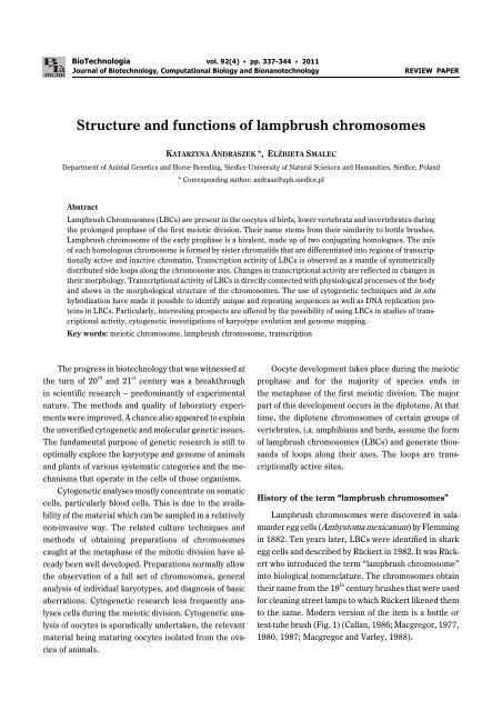
<strong>BioTechnologia</strong> vol. 92(4) C pp. 337-344 C 2011Journal <strong>of</strong> Biotechnology, Computational Biology <strong>and</strong> BionanotechnologyREVIEW PAPER<strong>Structure</strong> <strong>and</strong> <strong>functions</strong> <strong>of</strong> <strong>lampbrush</strong> <strong>chromosomes</strong>KATARZYNA ANDRASZEK *, ELŻBIETA SMALECDepartment <strong>of</strong> Animal Genetics <strong>and</strong> Horse Breeding, Siedlce University <strong>of</strong> Natural Sciences <strong>and</strong> Humanities, Siedlce, Pol<strong>and</strong>* Corresponding author: <strong>and</strong>rasz@uph.siedlce.plAbstractLampbrush Chromosomes (LBCs) are present in the oocytes <strong>of</strong> birds, lower vertebrata <strong>and</strong> invertebrates duringthe prolonged prophase <strong>of</strong> the first meiotic division. Their name stems from their similarity to bottle brushes.Lampbrush chromosome <strong>of</strong> the early prophase is a bivalent, made up <strong>of</strong> two conjugating homologues. The axis<strong>of</strong> each homologous chromosome is formed by sister chromatids that are differentiated into regions <strong>of</strong> transcriptionallyactive <strong>and</strong> inactive chromatin. Transcription activity <strong>of</strong> LBCs is observed as a mantle <strong>of</strong> symmetricallydistributed side loops along the chromosome axis. Changes in transcriptional activity are reflected in changes intheir morphology. Transcriptional activity <strong>of</strong> LBCs is directly connected with physiological processes <strong>of</strong> the body<strong>and</strong> shows in the morphological structure <strong>of</strong> the <strong>chromosomes</strong>. The use <strong>of</strong> cytogenetic techniques <strong>and</strong> in situhybridization have made it possible to identify unique <strong>and</strong> repeating sequences as well as DNA replication proteinsin LBCs. Particularly, interesting prospects are <strong>of</strong>fered by the possibility <strong>of</strong> using LBCs in studies <strong>of</strong> transcriptionalactivity, cytogenetic investigations <strong>of</strong> karyotype evolution <strong>and</strong> genome mapping.Key words: meiotic chromosome, <strong>lampbrush</strong> chromosome, transcriptionThe progress in biotechnology that was witnessed atthe turn <strong>of</strong> 20 th <strong>and</strong> 21 st century was a breakthroughin scientific research – predominantly <strong>of</strong> experimentalnature. The methods <strong>and</strong> quality <strong>of</strong> laboratory experimentswere improved. A chance also appeared to explainthe unverified cytogenetic <strong>and</strong> molecular genetic issues.The fundamental purpose <strong>of</strong> genetic research is still tooptimally explore the karyotype <strong>and</strong> genome <strong>of</strong> animals<strong>and</strong> plants <strong>of</strong> various systematic categories <strong>and</strong> the mechanismsthat operate in the cells <strong>of</strong> those organisms.Cytogenetic analyses mostly concentrate on somaticcells, particularly blood cells. This is due to the availability<strong>of</strong> the material which can be sampled in a relativelynon-invasive way. The related culture techniques <strong>and</strong>methods <strong>of</strong> obtaining preparations <strong>of</strong> <strong>chromosomes</strong>caught at the metaphase <strong>of</strong> the mitotic division have alreadybeen well developed. Preparations normally allowthe observation <strong>of</strong> a full set <strong>of</strong> <strong>chromosomes</strong>, generalanalysis <strong>of</strong> individual karyotypes, <strong>and</strong> diagnosis <strong>of</strong> basicaberrations. Cytogenetic research less frequently analysescells during the meiotic division. Cytogenetic analysis<strong>of</strong> oocytes is sporadically undertaken, the relevantmaterial being maturing oocytes isolated from the ovaries<strong>of</strong> animals.Oocyte development takes place during the meioticprophase <strong>and</strong> for the majority <strong>of</strong> species ends inthe metaphase <strong>of</strong> the first meiotic division. The majorpart <strong>of</strong> this development occurs in the diplotene. At thattime, the diplotene <strong>chromosomes</strong> <strong>of</strong> certain groups <strong>of</strong>vertebrates, i.a. amphibians <strong>and</strong> birds, assume the form<strong>of</strong> <strong>lampbrush</strong> <strong>chromosomes</strong> (LBCs) <strong>and</strong> generate thous<strong>and</strong>s<strong>of</strong> loops along their axes. The loops are transcriptionallyactive sites.History <strong>of</strong> the term “<strong>lampbrush</strong> <strong>chromosomes</strong>”Lampbrush <strong>chromosomes</strong> were discovered in salam<strong>and</strong>eregg cells (Ambystoma mexicanum) by Flemmingin 1882. Ten years later, LBCs were identified in sharkegg cells <strong>and</strong> described by Rückert in 1982. It was Rückertwho introduced the term “<strong>lampbrush</strong> chromosome”into biological nomenclature. The <strong>chromosomes</strong> obtaintheir name from the 19 th century brushes that were usedfor cleaning street lamps to which Rückert likened themto the same. Modern version <strong>of</strong> the item is a bottle ortest-tube brush (Fig. 1) (Callan, 1986; Macgregor, 1977,1980, 1987; Macgregor <strong>and</strong> Varley, 1988).
338K. Andraszek, E. SmalecThe basic pr<strong>of</strong>ile <strong>of</strong> LBCs was performed with a 20×zoom <strong>of</strong> the microscope. In the case <strong>of</strong> avian mitotic<strong>chromosomes</strong>, a 20× zoom only makes it possible toidentify the metaphase plate, not always enablingthe determination <strong>of</strong> the number <strong>of</strong> <strong>chromosomes</strong>. Figure2 shows a comparison <strong>of</strong> the proportions <strong>of</strong> a metaphaseplate at a 20× zoom, the metaphase second-pairchromosome at a 100× microscopic zoom <strong>and</strong> the second<strong>lampbrush</strong> bivalent at a 20× zoom. The mitotic<strong>chromosomes</strong> were isolated from peripheral blood <strong>of</strong>domestic geese. The LBCs, in turn, were sampled fromoocytes <strong>of</strong> domestic geese.Lampbrush chromosome structureFig. 1. A <strong>lampbrush</strong> chromosome <strong>and</strong> the “original item”. The arrowsindicate analogous structures; a – telomeric loop, b – sideloops, c – a chromatid without loops (K. Andraszek, unpublished)Lampbrush <strong>chromosomes</strong> are intermediate structurespresent during the first meiotic division. In a prolongeddiplotene stage, they undergo decondensation thatresults in the production <strong>of</strong> very large chromosomalstructures. LBCs’ length ranges (depending on the species)from 400 to 800 mm, which makes them up to 30times larger than their mitotic counterparts (Callan,1986; Callan, et al. 1987; Rodionov, 1996).Fig. 2. A comparison <strong>of</strong> the size <strong>of</strong> LBCs <strong>and</strong> mitotic <strong>chromosomes</strong>.A 20-fold microscopic magnification <strong>of</strong> the metaphaseplate (a), 100-fold magnification <strong>of</strong> the second-pair mitoticchromosome (b), a 20-fold magnification <strong>of</strong> the second <strong>lampbrush</strong>bivalent (c). The arrow points at the second-pair mitoticchromosome on the metaphase plate (K. Andraszek)In the early prophase, a LBC is a bivalent that consists<strong>of</strong> two pairs <strong>of</strong> conjugating homologues, eventuallyforming a tetrad. Each chromatid is composed <strong>of</strong> alternatelypositioned regions <strong>of</strong> condensed inactive chromatin(chromomeres visible as dark irregular structures <strong>and</strong>also observed in the interphase nucleus) <strong>and</strong> side loops<strong>of</strong> decondensed chromatin. In the homologous sections<strong>of</strong> the bivalent, chromatin is condensed (spirally twisted)or decondensed in the form <strong>of</strong> side loops – two per eachchromosome <strong>and</strong> four at the level <strong>of</strong> the bivalent. The loopconstitutes a part <strong>of</strong> the chromosome axis. It is extensibleas well as contractible. The contractibility <strong>of</strong> the loop resultsin the contraction <strong>and</strong> dilation <strong>of</strong> the chromomere(Angelier, et al. 1984, 1990; Macgregor, 1987; Chelysheva,et al. 1990; Morgan, 2002). A diagram <strong>of</strong> LBCstructure is presented in Figure 3.Employment <strong>of</strong> a 100× zoom to analyse LBC structurehas made it possible to observe chromomeres,chiasmata <strong>and</strong> sister chromatids <strong>of</strong> each bivalent homologue.An identical zoom used for the analysis <strong>of</strong> avianmitotic <strong>chromosomes</strong> enables only the identification <strong>of</strong>their morphological structure in relation to the firstcouple <strong>of</strong> macrochromosome pairs. In the early prophase,the LBC is a bivalent that consists <strong>of</strong> two conjugatinghomologues ultimately becoming a tetrad.Figure 4 shows a 20-fold magnification <strong>of</strong> the secondgoose bivalent <strong>and</strong> its distinctive structures visible witha 100× zoom. Letters next to the arrows correspondwith the respective magnifications in Figure 4. In thecase <strong>of</strong> a structural analysis <strong>of</strong> male meiotic <strong>chromosomes</strong>,it is not possible to observe these crucial meioticcytogenetic features.
<strong>Structure</strong> <strong>and</strong> <strong>functions</strong> <strong>of</strong> <strong>lampbrush</strong> <strong>chromosomes</strong> 339Fig. 3. Schematic (a) <strong>and</strong> detailed (b) <strong>lampbrush</strong> <strong>chromosomes</strong>tructure (Katarzyna Andraszek)Numerous morphological types <strong>of</strong> LBC loops havebeen identified. Such differentiation is determined bythe type <strong>and</strong> the number <strong>of</strong> proteins that are directlybound to the emergent transcripts. In terms <strong>of</strong> transcriptionalactivity, there are two basic loop types. “Complex”loops have a matrix with a very complicated morphologicalstructure (loop-formed or fibriform). “Complex”loops are classified as marker loops that enable chromosomeidentification or side loops. Another type <strong>of</strong> loopsare the “plain” loops. They constitute the majority <strong>of</strong>chromosome loops <strong>and</strong> have a delicate fibrous matrix,with occasionally well-visible asymmetry. They are alwaysloop-shaped (Angelier et al., 1984; Leòn <strong>and</strong> Kezer,1990; Morgan, 2002, 2007).Lampbrush <strong>chromosomes</strong> include domains <strong>of</strong> openchromatin in which the genes can be potentially transcriptive<strong>and</strong> domains <strong>of</strong> locked chromatin without expression(Roy et al., 2002; Gaginskaya et al., 2009).Lampbrush chromosome loops are considered an example<strong>of</strong> open chromatin. Their analogues are thought to bethe “puffs” <strong>of</strong> polytenic <strong>chromosomes</strong>. They differ betweeneach other. Polytenic <strong>chromosomes</strong> are made up<strong>of</strong> parallel chromatids, whereas <strong>lampbrush</strong> chromosomechromatin constitutes <strong>of</strong> a single DNA helix. Observationunder an electron microscope have demonstrated thatthe diameter <strong>of</strong> the loop thread corresponds with theDNA helix diameter, i.e. 1.9 nm (Olins <strong>and</strong> Olins, 2003;Gaginskaya et al., 2009).Fig. 4. The second goose <strong>lampbrush</strong> chromosome with a magnification <strong>of</strong> its distinctive structures. A 20-fold magnification <strong>of</strong>the second goose bivalent (a) <strong>and</strong> its distinctive structures visible with a 100× zoom (b, f – telomeres, c – centromere, d – chiasm,e – sister chromatids). The particular bivalent structures in the 100-fold blow-ups are marked with blue arrows (K. Andraszek)
340K. Andraszek, E. SmalecAvian <strong>lampbrush</strong> <strong>chromosomes</strong> are associated withprotein bodies/structures (PBs). These non-typical structuresare present in cells only in association with LBCs.They have a regular connection with the chromosomeaxis <strong>of</strong> each LBC in the heterochromatin region (Soloveiet al., 1996, Krasikova et al., 2004). In terms <strong>of</strong> morphology,PBs resemble Cajal bodies (CBs) present in amphibiansin association with LBCs. However, immunocytochemicalresearch has shown that PBs neither containp80 coilin, nor any other CB matrix indices, such asfibrillarin or splicing- <strong>and</strong> U7snRNPs-specific trimethyloguanosineepitopes. The distinctive composition <strong>of</strong> PBssuggests a completely different function from that <strong>of</strong>CBs. PBs may be involved in the coordination <strong>of</strong> spatiallayout <strong>of</strong> <strong>chromosomes</strong>. The location <strong>of</strong> PBs is frequentlyassociated with repetitive sequences surroundingthe centromere. Exploration <strong>of</strong> the potential role <strong>of</strong> centromere-related<strong>and</strong> centromeric heterochromatin-relatedproteins in the biogenesis <strong>and</strong> location <strong>of</strong> PBs <strong>and</strong>CBs constitutes a new trend in the research on <strong>lampbrush</strong>chromosome structure (Gall, 2000; Morgan et al.,2000; Morgan, 2002, 2005; Muphy et al., 2002).Lampbrush chromosome transcriptionA routine mitotic chromosome analysis can only providethe description <strong>of</strong> their morphology. Transcriptionalactivity <strong>of</strong> genes can only be assessed using molecularmethods that consist detecting the amount <strong>of</strong> the transcriptionproduct. Transcriptional activity <strong>of</strong> LBCs may beobserved even under a light microscope <strong>and</strong> can bedetermined for morphological changes.Therefore, LBCs are used as a model in studies <strong>of</strong>transcriptional regulation. Changes in transcriptional activityresult in a different morphological structure <strong>of</strong><strong>lampbrush</strong> chromosome loops (Gall, 1983; Morgan,2002). Moreover, a higher transcriptional activity <strong>of</strong>micro<strong>chromosomes</strong> is observed due to a greater density<strong>of</strong> genes (Rodionov, 1996; Angelier et al., 1984; Morgan,2002). Transcriptional activity analyses are performedon the basis <strong>of</strong> assumption that the side loops <strong>of</strong> LBCsare the transcriptionally active sites. A decrease intranscriptional activity is observed as a shrinking <strong>of</strong> theside loops (Varley et al., 1980; Gall, 1983; Callan et al.,1987; Gaginskaya <strong>and</strong> Tsvetkov, 1988; Morgan, 2002,2007; Galkina et al., 2006; Gaginskaya et al., 2009).The morphology <strong>and</strong> transcriptional activity <strong>of</strong> LBCsvary depending on the reproductive cycle (Andraszeket al., 2009). They can also undergo seasonal changes(Tsvetkov <strong>and</strong> Parfenov, 1994). This is particularly evidentin hibernating amphibians. During the summer,when the animals are the most active, the transcriptionalactivity <strong>of</strong> LBCs is the highest, as well. In the autumn,LBCs’ activity abates. Nevertheless, this is not associatedwith morphological changes. At that time <strong>of</strong> the year,each transcription unit contains approximately 10 RNP(ribonucleoproteinic) filaments, while in the summer,this number is twice as high, the change correspondingwith morphological transformation. During the winter,transcription substantially declines. Both in <strong>chromosomes</strong><strong>and</strong> in nucleoli, numerous <strong>and</strong> very characteristicmorphological changes take place (Tsvetkov <strong>and</strong> Parfenov,1994). Not more than 70% <strong>of</strong> the nuclear DNA issubject to transcription at the time. Although in the case<strong>of</strong> physical factors, such as radiation or numerous chemicalfactors, a similar effect on the structure <strong>and</strong> activity<strong>of</strong> LBCs in various groups <strong>of</strong> animals can be expected,seasonal changes predominantly affect polikilotherms(Morgan, 2002, 2007).The degree <strong>of</strong> DNA compaction in LBCs is regulatedby changes in the distance between nucleosomes, especiallythe non-adjacent ones. The compaction ratio <strong>of</strong>DNA (number <strong>of</strong> DNA μm in a 1 μm chromatin fiber)in non-transcribed fibrils is equal to 2.1, in transcriptionalunits with moderate <strong>and</strong> weak activity it is 1.7, <strong>and</strong>in transcriptional units with intensive transcription it isclose to 1. (Franke <strong>and</strong> Scheer, 1978; Gaginskaya <strong>and</strong>Tsvetkov, 1988; Morgan, 2002, 2007). The nucleosomes<strong>of</strong> transcriptionally inactive chromatin are evenly spaced,the gaps between the nucleosomes corresponding withlinker DNA length. In transcription units with insignificantor declining transcription, nucleosomes are identifiedin the axial part <strong>of</strong> the chromosome, between sets<strong>of</strong> polymerase units. The gaps between the polymeraseblocks are not even. After polymerase has passed alongthe DNA matrix <strong>and</strong> the regulatory proteins have becomedissociated, nucleosome reconstruction follows(Spring <strong>and</strong> Franke, 1981; Gaginskaya <strong>and</strong> Tsvetkov,1988; Solovei et al., 1992).In transcriptionally active regions <strong>of</strong> <strong>chromosomes</strong>,histone proteins give way to non-histone proteins, inducingthe loss <strong>of</strong> the nucleosomal structure <strong>of</strong> the codingchromosome segments that assume the shape <strong>of</strong> a loop.However, the exposure <strong>of</strong> nucleosomes, which enablesDNA transcription, does not entail histone dissociation
<strong>Structure</strong> <strong>and</strong> <strong>functions</strong> <strong>of</strong> <strong>lampbrush</strong> <strong>chromosomes</strong> 341but leads to a spatial rearrangement <strong>of</strong> the transcribedregions, allowing access <strong>of</strong> RNA polymerase to the adjacentpromotor sequence. This occurs through the fixation<strong>of</strong> regulatory proteins at the site <strong>of</strong> a remote activatingsequence. What is particularly the characteristic <strong>of</strong><strong>lampbrush</strong> <strong>chromosomes</strong> is that non-histone HMG (HighMobility Group) proteins become incorporated in thestructure <strong>of</strong> the chromatin. HMGs are structural proteins<strong>of</strong> chromatin that reduce chromatin condensation(Di Mario et al., 1989; Korner et al., 2003).The assumption <strong>of</strong> appropriate spatial conformationby chromatin enables the commencement <strong>of</strong> transcription<strong>of</strong> LBC DNA in the presence <strong>of</strong> RNA polymerasecompounds bound with LBC loops. The transcriptionallyactive loops represent 5-10% <strong>of</strong> DNA. The remainder isinactive chromatin compacted in the chromomeres.The result <strong>of</strong> transcription is visible under an electronmicroscope as a ribonucleoproteinic mantle. The mantletends to be asymmetrical, corresponding with risingelectron density from the base towards the middle <strong>of</strong>the loop (Angelier et al., 1990, 1996).The average length <strong>of</strong> a typical <strong>lampbrush</strong> chromosomeloop is 10-15 μm, though some can be as long as50 or even 100 μm. The rate <strong>of</strong> transcription in <strong>lampbrush</strong>chromosome loops determined with a radioactiveRNA precursor is 5 μm per hour. Thus, one loop istranscribed within two to a dozen or so hours. DNAcompaction degree in LBCs is not well known. However,it is estimated that 1 μm <strong>of</strong> loop length contains around3 thous<strong>and</strong> base pairs. Thus, an average loop containsabout 30-40 thous<strong>and</strong> base pairs, which correspondswith the average length <strong>of</strong> RNA transcribed in the oocytes.Nevertheless, it is much longer than transcriptionunits <strong>of</strong> somatic cells. This is the result <strong>of</strong> skipping transcriptiontermination signals (Kropotova <strong>and</strong> Gaginskaya,1984; Hutchison, 1987; Gaginskaya <strong>and</strong> Tsvetkov, 1988;Morgan, 2002, 2007).The loops can be classified according to the type <strong>of</strong>the transcriptional polymerase. The largest loops includethose transcribed by polymerase II. The smallest loopsare transcribed by polymerase III. They contain 5S RNAcoding units (Kay <strong>and</strong> Gall 1981), tRNA (Müller et al.1987) or short replication sequences (Kroll et al. 1987).Since 5S RNA sequences are short <strong>and</strong> divided by noncodingelements, transcription being basically limited tocoding sequences, the transcripts <strong>of</strong> these sequencesare also short <strong>and</strong>, consequently, do not have the distinctivematrix made up <strong>of</strong> RNP filaments. That is whythey are so well visible in the microscopic phase contrast(Murphy et al., 2002). LBCs can be divided into thosewith one transcriptional unit <strong>and</strong> those with two ormore. Over the length <strong>of</strong> 1 μm, one transcriptional unitis transcribed by a densely compacted package <strong>of</strong> around13-20 polymerase molecules (Leòn <strong>and</strong> Kezer, 1990;Macgregor <strong>and</strong> Varley, 1988; Morgan, 2002).Regulation <strong>of</strong> LBC transcription is performed bymeans <strong>of</strong> modifications <strong>of</strong> chromosome structure <strong>and</strong>the activity <strong>of</strong> a number <strong>of</strong> post-transcription factors.The process <strong>of</strong> transcriptional activity modification consists<strong>of</strong> a set <strong>of</strong> interrelated reactions in which numerousinterconnected, both structural <strong>and</strong> enzymatic, factorstake part. The first stage is the loosening <strong>of</strong> chromatin.This is the element that differentiates LBCs from mitotic<strong>chromosomes</strong>. While somatic cell <strong>chromosomes</strong> completelylose their structure during transcription, LBCs retainit. The preservation <strong>of</strong> the structure by LBCs duringtranscriptional activity is connected with the presence <strong>of</strong>so-called “constitutive” nucleosomes (Scheer et al.,1984; Scheer, 1987; Gaginskaya <strong>and</strong> Tsvetkov, 1988;Olins <strong>and</strong> Olins, 2003). The transcription <strong>of</strong> the oocytespecifictopoisomerase I (topo-I) variant is activatedduring the formation <strong>of</strong> LBC structures. This topoisomeraseis present in LBC loops <strong>and</strong> participates in the spatialconformation <strong>of</strong> these structures. The inhibition <strong>of</strong> topo-Iactivity causes the <strong>lampbrush</strong> loops to recede <strong>and</strong> stimulatesthe condensation <strong>of</strong> nuclear chromatin (Gebaueret al. 1996). Observations <strong>of</strong> LBC loops with an electronmicroscope revealed that the twisting loops containtranscriptive polymerases that are less closely compactedthan during active transcription. Additionally, anaccompanying condensation <strong>of</strong> the loops between thosepolymerases into the form <strong>of</strong> nucleosomes was observed(Scheer, 1987; Morgan, 2002).In the condensed segments, the chromomeres buildup compact chromatin in which genes or gene-containingtranscriptional units are not transcribed. During oogenesis,loops <strong>of</strong> approximately 50 μm in length correspondwith active transcriptional units. They constitute 5-10%<strong>of</strong> the total length <strong>of</strong> the chromosome. Chromosomemaps <strong>of</strong> different oocytes at various ages are identical<strong>and</strong> remain constant for a given oocyte, which suggestsa species-specific nature <strong>of</strong> sequences transcribed duringoogenesis. It was possible to identify RNA transcribed insome <strong>of</strong> the loops <strong>and</strong> thus initiate the mapping <strong>of</strong>
342K. Andraszek, E. Smalec<strong>lampbrush</strong> <strong>chromosomes</strong> (Callan, 1986; Chelyshevaet al., 1990; Morgan, 2002, 2007; Galkina et al., 2006;Saifitdinova et al., 2003; Gaginskaya et al., 2009).Lampbrush <strong>chromosomes</strong> in cytogenetics<strong>and</strong> genomicsLampbrush <strong>chromosomes</strong> <strong>of</strong> various species havea very similar structure <strong>and</strong> perform the same function.Comparative studies <strong>of</strong> LBCs in various species haveshown that the side loops seem to be much longer inspecies with higher C-values (genome size refers to thehaploid set <strong>of</strong> <strong>chromosomes</strong>). This regularity explicitlyreflects differences in the organisation <strong>of</strong> genome sequences.One explanation <strong>of</strong> the effect <strong>of</strong> genome size onthe loop length is based on the existence <strong>of</strong> substantialdifferences in the length <strong>and</strong> distribution <strong>of</strong> transcribedsequences in relation to chromomere sequences in variouslysized genomes (Macgregor, 1980; Gregory,2002). Another theory suggests that the total increase inthe length <strong>of</strong> loop transcription units results from socalled“over-transcription” <strong>of</strong> longer intergenic segmentspresent in larger genomes. On the other h<strong>and</strong>, researchby Gall <strong>and</strong> Murphy (1998) has proved that loop length isspecies-specific. What is certain, however, is that someorganisational characteristics <strong>of</strong> sequences <strong>of</strong> large genomescan, to a certain extent, affect the length <strong>of</strong> transcriptionalunits <strong>of</strong> LBCs.The sequencing <strong>of</strong> part <strong>of</strong> the myosin gene <strong>of</strong> the tritonwas a pro<strong>of</strong> that much longer introns are present inthe triton gene than in the genome <strong>of</strong> mammals withC-DNA values similar to those <strong>of</strong> Xenopus (Casimir etal., 1988). However, the data on DNA sequences in largegenomes <strong>of</strong> tailed amphibians are so few that, at present,it is not entirely known how universally any <strong>of</strong> the sequencing-basedexplanations can be used to account forthe correlation between high C-DNA values <strong>and</strong> looplength. Uncertain data relating to the loop length pointto another important question concerning the structure<strong>and</strong> function <strong>of</strong> LBCs – namely: the affinity, organisation<strong>and</strong> control <strong>of</strong> transcription loops. The “overtranscription”model initially provided a basis for underst<strong>and</strong>ingwhy in transcriptional units <strong>of</strong> LBCs there appearedhighly repetitive sequences along with pol II complexescontaining complexes that initiate transcriptional elongation<strong>of</strong> the loops. These complexes are assumed to failto react to termination signals <strong>and</strong> keep transcribingflanking regions saturated with repetitions (Morgan,2002, 2007).Particular interest in recent years has been devotedto possibilities <strong>of</strong> using LBCs in genome mapping. Thisstrategy can combine chromosome marker mapping <strong>and</strong>physical gene mapping using the in situ hybridisationtechnique with genetic maps constructed on the basis <strong>of</strong>chiasm incidence in the analysed bivalents. Equally importantis the possibility <strong>of</strong> using <strong>lampbrush</strong> <strong>chromosomes</strong>in the analyses <strong>of</strong> the interaction <strong>of</strong> genes withother cellular structures. Particularly promising seem tobe the possibilities <strong>of</strong> using <strong>lampbrush</strong> <strong>chromosomes</strong> inthe mapping <strong>of</strong> avian genomes (Griffin et al., 2088; Penrad-Mobayedet al., 2009; Bi <strong>and</strong> Bogart, 2010; Dakset al., 2010; Solinhac et al., 2010).Lampbrush <strong>chromosomes</strong> were first used as the objectsfor cytogenetic analyses <strong>of</strong> poultry by Kropotova<strong>and</strong> Gaginskaya (1984) <strong>and</strong> Hutchison (1987). The authorsbelieve that LBCs provide valuable information onthe expression <strong>of</strong> avian genes. They also claim that the<strong>chromosomes</strong> are indispensable for the cytogenetic study<strong>of</strong> animals with small genomes, where the large numbers<strong>of</strong> mitotic <strong>chromosomes</strong> <strong>and</strong> their small sizes precludethe analysis <strong>of</strong> micro<strong>chromosomes</strong>. As in the case<strong>of</strong> b<strong>and</strong>ing patterns <strong>of</strong> mitotic <strong>chromosomes</strong>, LBCs havea specific configuration <strong>of</strong> active <strong>and</strong> non-active chromomeresobserved as a pattern <strong>of</strong> side loops <strong>and</strong> looplessareas (Andraszek <strong>and</strong> Smalec, 2011). In a report onthe genome <strong>and</strong> <strong>chromosomes</strong> <strong>of</strong> Gallus domesticus,<strong>lampbrush</strong> <strong>chromosomes</strong> were recognised as a new modelin avian cytogenetics (Schmid et al., 2005).Epigenetic mechanisms acting at the level <strong>of</strong> DNAmethylation <strong>and</strong> histone modification change the structure<strong>of</strong> LBC chromatin <strong>and</strong> control the interaction <strong>of</strong> active<strong>and</strong> non-active genes. The open conformation <strong>of</strong>chromatin is transcriptionally active, whereas the “closed”conformation is associated with so-called transcriptiondecline (Grummt <strong>and</strong> Pikaard, 2003). LBCs are dividedinto domains containing open chromatin in whichthe genes can be potentially transcriptive, <strong>and</strong> domains<strong>of</strong> locked chromatin without detectable sequence expression.The loops <strong>of</strong> LBCs are a classic example <strong>of</strong> an openchromatin. Moreover, oocyte transcription is a complexprocess in which, apart from LBCs, other nuclear structuresare involved as well (Gall, 1983, 2000; Gall et al.,1999; Saifitdinova et al., 2003).
<strong>Structure</strong> <strong>and</strong> <strong>functions</strong> <strong>of</strong> <strong>lampbrush</strong> <strong>chromosomes</strong> 343In conclusion, studies on <strong>lampbrush</strong> <strong>chromosomes</strong>have been conducted for over a hundred years. And yet,only a general idea <strong>of</strong> LBC structure has been developedthus far. What is not known are the factors that initiatethe changes that transform condensed <strong>chromosomes</strong>into decondensed <strong>lampbrush</strong> structures. LBCs are consideredas model structures in the studies <strong>of</strong> transcriptioncontrol. Changes in their transcriptional activityare reflected as modifications <strong>of</strong> the LBC morphologicalstructure <strong>and</strong> are associated with the physiological processes<strong>of</strong> the organism. Moreover, due to their decondensedstructure, <strong>lampbrush</strong> <strong>chromosomes</strong> are increasinglymore <strong>of</strong>ten used as objects <strong>of</strong> cytogenetic analyses,in basic cytogenetic experiments, <strong>and</strong> as modelstructures <strong>of</strong> the epigenetic chromatin control.ReferencesAndraszek K., Smalec E. (2011) Comparison <strong>of</strong> active transcriptionregions <strong>of</strong> <strong>lampbrush</strong> <strong>chromosomes</strong> with the mitoticchromosome G pattern in the European domestic gooseAnser anser. Arch. Tierz. 54: 69-82.Andraszek K., Smalec E., Tokarska W. (2009) Identification<strong>and</strong> structure <strong>of</strong> <strong>lampbrush</strong> sex bivalents prior to <strong>and</strong> afterthe reproduction period <strong>of</strong> the European domestic gooseAnser anser. Folia Biol. (Krakow) 57: 143-148.Angelier N., Paintraud M., Lavaud A., Lechaire J.P. (1984)Scanning electron microscopy <strong>of</strong> amphibian <strong>lampbrush</strong><strong>chromosomes</strong>. Chromosoma 89: 243-253.Angelier N., Bonnanfant-Jais M.L., Herberts C., Lautredou N.,Moreau N., N'Da E., Penrad-Mobayed M., Rodriguez-Martin M.L., Sourrouille P. (1990) Chromosomes <strong>of</strong> amphibianoocytes as a model for gene expression: significance<strong>of</strong> <strong>lampbrush</strong> loops. Int. J. Dev. Biol. 34: 69-80.Angelier N., Penrad-Mobayed M., Billoud B., Bonnanfant-Jais M.L., Coumailleau P. (1996) What role might <strong>lampbrush</strong><strong>chromosomes</strong> play in maternal gene expression?Int. J. Dev. Biol. 40: 645-652.Bi K., Bogart J. (2010) Probing the meiotic mechanism <strong>of</strong>intergenomic exchanges by genomic in situ hybridizationon <strong>lampbrush</strong> <strong>chromosomes</strong> <strong>of</strong> unisexual Ambystoma(Amphibia: Caudata). Chromosome Res. 18: 371-382.Callan H.G. (1986) Lampbrush <strong>chromosomes</strong>. SpringerVerlag, Berlin, Heidelberg, New York, Toronto, 1986.Callan H.G., Gall J.G., Berg C.A. (1987) The <strong>lampbrush</strong> <strong>chromosomes</strong><strong>of</strong> Xenopus laevis: preparation, identification, <strong>and</strong> distribution<strong>of</strong> 5S DNA sequences. Chromosoma 95: 236-250.Casimir C.M., Gates P.B., Ross-Macdonald P.B., Jackson J.F.,Patient R.K., Brockes J.P. (1988) <strong>Structure</strong> <strong>and</strong> expression<strong>of</strong> a new cardio-skeletal myosin gene. Implications forthe C value paradox. J. Mol. Biol. 202: 287-296.Chelysheva L.A., Solovei I.V., Rodionov A.V., Yakovlev A.F.,Gaginskaya E.R. (1990) Lampbrush chromosoms <strong>of</strong>the chicken: the cytological map <strong>of</strong> the macrobivalents.Cytology 32: 303-316.Daks A., Derjusheva S., Krasikova A., Zlotina A., GaginskayaE., Galkina S. (2010) Lampbrush <strong>chromosomes</strong> <strong>of</strong> theJapanese quail (Coturnix coturnix japonica): a new version<strong>of</strong> cytogenetic maps. Rus. J. Genet. 46: 1335-1338.Di Mario P.J., Bromley S.E., Gall J.G. (1989) DNA-binding proteinson <strong>lampbrush</strong> chromosome loops. Chromosoma 97:413-420.Franke W.W., Scheer U. (1978) Morphology <strong>of</strong> transcriptionalunits at different states <strong>of</strong> activity. Philosoph. Trans.Royal Soc. London 283: 333-342.Gaginskaya E.R., Tsvetkov A.G. (1988) Electron microscopyresearch on the chromatin structure <strong>of</strong> dispersed <strong>lampbrush</strong><strong>chromosomes</strong> in the hen. Tsitologiia 30: 142-150.Gaginskaya E., Kulikova T., Krasikova A. (2009) Avian <strong>lampbrush</strong><strong>chromosomes</strong>: a powerful tool for exploration <strong>of</strong> genomeexpression. Cytogenet. Genome Res. 124: 251-267.Galkina S., Deryusheva S., Fillon V., Vignal A., Crooijmans R.,Groenen M., Rodionov A., Gaginskaya E. (2006) FISHon avian <strong>lampbrush</strong> <strong>chromosomes</strong> produces higher resolutiongene mapping. Genetica 128: 241-251.Gall J.M. (1983) Transcription <strong>of</strong> repetetive sequences onXenopus lampbrusch <strong>chromosomes</strong>. Proc. Natl. Acad. Sci.USA 80: 3364-3367.Gall J.G., Murphy C. (1998) Assembly <strong>of</strong> <strong>lampbrush</strong> <strong>chromosomes</strong>from sperm chromatin. Mol. Biol. 9: 733-747.Gall J.G., Bellini M., Zheng’an W., Murphy C. (1999) Assembly<strong>of</strong> the nuclear transcription <strong>and</strong> processing machinery:Cajal bodies (coiled bodies) <strong>and</strong> transcriptosomes.Mol. Biol. Cell 10: 4385-4402.Gall J.G. (2000) Cajal bodies: the first 100 years. Annu. Rev.Cell. Dev. Biol. 6: 273-300.Gebauer D., Mais C., Zinger K., Hock R., Lieb B., Scheer U.(1996) Localization <strong>of</strong> a high molecular weight form <strong>of</strong>DNA topoisomerase I in amphibian oocytes. Int. J. Dev.Biol. 40: 239-244.Gregory T.R. (2002) A bird's-eye view <strong>of</strong> the C-value enigma:genome size, cell size, <strong>and</strong> metabolic rate in the classAves. Int. J. Org. Evolution 56: 121-130.Griffin D., Robertson L.B., Tempest H.G., Vignal A., Fillon V.,Crooijmans R.P.M.A., Groenen M.A.M., Deryusheva S., GaginskayaE., Carré W., Waddington D., Talbot R., Völker M.,Masab<strong>and</strong>a J.S., Burt D.W. (2008) Whole genome comparativestudies between chicken <strong>and</strong> turkey <strong>and</strong> their implicationsfor avian genome evolution. BMC Genomics 9: 168.Grummt I., Pikaard C.S. (2003) Epigenetic silencing <strong>of</strong> RNApolymerase I transcription. Nat. Rev. Mol. Cell Biol. 4:641-649.Hutchison N. (1987) Lampbrush <strong>chromosomes</strong> <strong>of</strong> the chickenGallus domesticus. J. Cell Sci. 105: 1493-1500.Kay B.K., Gall J.G. (1981) 5S ribosomal RNA genes <strong>of</strong> the newNotophthalmus viridescens. Nucl. Acid Res. 9: 6457-6469.Korner U., Bustin M., Scheer U., Hock R. (2003) Developmentalrole <strong>of</strong> HMGN proteins in Xenopus laevis. Mech.Dev. 120: 1177-1192.
344K. Andraszek, E. SmalecKrasikova A., Kulikova T., Saifitdinova A., Derjusheva S., GaginskayaE. (2004) Centromeric protein bodies on avian <strong>lampbrush</strong><strong>chromosomes</strong> contain a protein detectable with an antibodyagainst DNA topoisomerase II. Chromosoma 113:316-323.Kroll A., Carbon P., Ebel J.P., Appel B. (1987) Xenopus tropicalisU6 snRNA genes transcribed by Pol III contain theupstream promoter elements used by Pol II dependentU-snRNA genes. Nucl. Acids Res. 15: 2463-2478.Kropotova E.V., Gaginskaya E.R. (1984) Lampbrush <strong>chromosomes</strong>from the Japanese quail oocytes. Tsitologiia 26:1008-1015.Le Moigne A. (1999) Biologia rozwoju. Wydawnictwo NaukowePWN, Warszawa.Leòn P., Kezer J. (1990) Loop size in newt <strong>lampbrush</strong> <strong>chromosomes</strong>.Chromosoma 99: 83-86.Macgregor H.C. (1977) Chromatin <strong>and</strong> chromosome structure.Academic Press.Macgregor H.C. (1980) Recent developments in the study <strong>of</strong><strong>lampbrush</strong> <strong>chromosomes</strong>. Heredity 44: 3-35.Macgregor H.C. (1984) Chromosome structure <strong>and</strong> function.Van Rostr<strong>and</strong> i Reinhold Publishing Corp, New York.Macgregor H.C. (1987) Lampbrush <strong>chromosomes</strong>. J. Cell Sci.88: 7-9.Macgregor H.C., Varley J. (1988) Working with animal <strong>chromosomes</strong>.John Wiley & Sons. London, New York, Brisbane,Toronto, Singapore.Morgan G.T. (2002) Lampbrush <strong>chromosomes</strong> <strong>and</strong> associatedbodies: new insights into principles <strong>of</strong> nuclear structure<strong>and</strong> function. Chromosome Res. 10: 177-200.Morgan G.T. (2007) Localized co-transcriptional recruitment<strong>of</strong> the multifunctional RNA-binding protein CELF1 by<strong>lampbrush</strong> chromosome transcription units. ChromosomeRes. 15: 985-1000.Morgan G.T., Doyle O., Murphy C., Gall J.G. (2000) RNA polymeraseII in Cajal bodies <strong>of</strong> amphibian oocytes. J. Struct.Biol. 129: 258-268.Murphy C., Wang Z., Roeder R.G., Gall J.G. (2002) RNA polymeraseIII in Cajal bodies <strong>and</strong> <strong>lampbrush</strong> <strong>chromosomes</strong> <strong>of</strong>the Xenopus oocyte nucleus. Mol. Biol. Cell 13: 3466-3476.Müller F., Clarkson S.G., Galas D.J. (1987) Sequence <strong>of</strong>a 3.18 kb t<strong>and</strong>em repeat <strong>of</strong> Xenopus laevis DNA containing8 tRNA genes. Nucl. Acid Res. 15: 7191.Olins D.E., Olins A.L. (2003) Chromatin history: our viewfrom the bridge. Nature Rev. Mol. Cell Biol. 4: 809-814.Penrad-Mobayed M., El Jamil A., Kanhoush R., Perrin C.(2009) Working map <strong>of</strong> the <strong>lampbrush</strong> <strong>chromosomes</strong> orXenopus tropicalis: a new tool for cytogenetic analysis.Dev. Dyn. 238: 1492-1501.Rodionov A.V. (1996) Micro versus macro: a review <strong>of</strong> structure<strong>and</strong> <strong>functions</strong> <strong>of</strong> avian micro- <strong>and</strong> macro<strong>chromosomes</strong>.Genetika 32: 597-608.Roy J.P., Stuart J.M., Lund J., Kim S.K. (2002) Chromosomalclustering <strong>of</strong> muscle-expressed genes in Caenorhabditiselegans. Nature 418: 975-979.Saifitdinova A., Derjusheva S., Krasikova A., Gaginskaya E.(2003) Lampbrush <strong>chromosomes</strong> <strong>of</strong> the chaffinch (Fringillacoelebs L.). Chromosome Res. 11: 99-113.Scheer U. (1987) <strong>Structure</strong> <strong>of</strong> <strong>lampbrush</strong> chromosome loopsduring different states <strong>of</strong> transcriptional activity as visualizedin the presence <strong>of</strong> physiological salt concentrations.Biol. Cell 59: 33-42.Scheer U., Hinssen H., Franke W.W., Jockusch B.M. (1984)Microinjection <strong>of</strong> actinbinding proteins <strong>and</strong> actin antibodiesdemonstrates involvement <strong>of</strong> nuclear actin in transcription<strong>of</strong> <strong>lampbrush</strong> <strong>chromosomes</strong>. Cell 39: 111-122.Schmid M., N<strong>and</strong>a I., Hoehn H., Schartl M., Haaf T., BuersteddeJ.M., Arakawa H., Caldwell R.B., Weigend S., Burt D.W.et al. (2005) Second report on chicken genes <strong>and</strong> <strong>chromosomes</strong>2005. Cytogenet. Genome Res. 109: 415-479.Solinhac R., Leroux S., Galkina S., Chazara O., Feve K., VignolesF., Morisson M., Derjusheva S., Bed’hom B., VignalA., Fillon V., Pitel F. (2010) Integrative mapping analysis<strong>of</strong> chicken microchromosome 16 organization. BMCGenomics 11: 616.Solovei I.V., Gaginskaya E., Allen T., Macgregor H.C. (1992)A novel structure associated with <strong>lampbrush</strong> <strong>chromosomes</strong>in the chicken Gallus domesticus. J. Cell Sci. 101: 759-772.Solovei I.V., J<strong>of</strong>fe B.I., Gaginskaya E.R., Macgregor H.C.(1996) Transcription <strong>of</strong> <strong>lampbrush</strong> <strong>chromosomes</strong> <strong>of</strong> acentromerically localized highly repeated DNA in pigeon(Columba) relates to sequence arrangement. ChromosomeRes. 4: 588-603.Spring H., Franke W.W. (1981) Transcriptionally active chromatinin loops <strong>of</strong> <strong>lampbrush</strong> <strong>chromosomes</strong> at physiologicalsalt concentrations as revealed by electron microscopy<strong>of</strong> sections. Eur. J. Cell Biol. 24: 298-308.Tsvetkov A.G., Parfenov V.N. (1994) Seasonal transformationsin the <strong>lampbrush</strong> <strong>chromosomes</strong> <strong>and</strong> the morphogenesis <strong>of</strong>the karyosphere capsule in Rana temporaria oocytes detectableby an analysis <strong>of</strong> the isolated nuclear structures.Tsitologiia 36: 64-70.Varley J.M., Macgregor H.C., Erba H.P. (1980) Satelite DNAis transcribed on <strong>lampbrush</strong> <strong>chromosomes</strong>. Nature 283:686-688.