FAO Fisheries Technical Paper 402/2 - Library - Network of ...
FAO Fisheries Technical Paper 402/2 - Library - Network of ...
FAO Fisheries Technical Paper 402/2 - Library - Network of ...

Create successful ePaper yourself
Turn your PDF publications into a flip-book with our unique Google optimized e-Paper software.
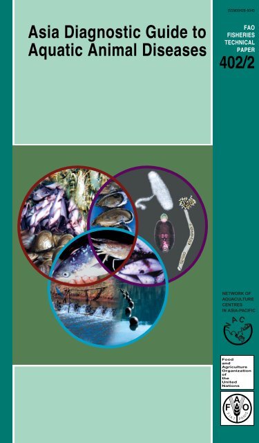
ISSNO0428-9345Asia Diagnostic Guide toAquatic Animal Diseases<strong>FAO</strong>FISHERIESTECHNICALPAPER<strong>402</strong>/2NETWORK OFAQUACULTURECENTRESIN ASIA-PACIFICN AC AFoodandAgricultureOrganization<strong>of</strong>theUnitedNationsF A OFI ATI SP A N
ISSNO0428-9345Asia Diagnostic Guide toAquatic Animal Diseases<strong>FAO</strong>FISHERIESTECHNICALPAPER<strong>402</strong>/2Edited byMelba G. Bondad-ReantasoNACA, Bangkok, Thailand(E-mail: Melba.Reantaso@enaca.org)Sharon E. McGladderyDFO-Canada, Moncton, New Brunswick(E-mail: McGladderyS@dfo-mpo.gc.ca)Iain EastAFFA, Canberra, Australia(E-mail: Iain.East@affa.gov.au)andRohana P. Subasinghe<strong>FAO</strong>, Rome(E-mail: Rohana.Subasinghe@fao.org)NETWORK OFAQUACULTURECENTRESIN ASIA-PACIFICN AC AFoodandAgricultureOrganization<strong>of</strong>theUnitedNationsF A OFI ATI SP A N
The designations employed and the presentation <strong>of</strong> material in thispublication do not imply the expression <strong>of</strong> any opinion whatsoeveron the part <strong>of</strong> the Food and Agriculture Organization <strong>of</strong> the UnitedNations (<strong>FAO</strong>) or <strong>of</strong> the <strong>Network</strong> <strong>of</strong> Aquaculture Centres in Asia-Pacific(NACA) concerning the legal status <strong>of</strong> any country, territory, cityor area or <strong>of</strong> its authorities, or concerning the delimitation <strong>of</strong> its frontiersor boundaries.ISBN 92-5-104620-4All rights reserved. No part <strong>of</strong> this publication may be reproduced,stored in a retrieval system, or transmitted in any form or by any means,electronic, mechanical, photocopying or otherwise, without the priorpermission <strong>of</strong> the copyright owner. Applications for such permission,with a statement <strong>of</strong> the purpose and extent <strong>of</strong> the reproduction,should be addressed to the Co-ordinator, <strong>Network</strong> <strong>of</strong> AquacultureCentres in Asia-Pacific (NACA), Suraswadi Building, Department <strong>of</strong><strong>Fisheries</strong>, Kasetsart University Campus, Ladyao, Jatujak, Bangkok10900, Thailand, or the Chief, Publishing and Multimedia Service,Information Division, <strong>FAO</strong>, Viale delle Terme di Caracalla, 00100 Rome,Italy or by e-mail to copyright@fao.org .© <strong>FAO</strong> and NACA 2001iii 4
PREPARATION OF THIS DOCUMENTThe Asia Diagnostic Guide to Aquatic Animal Diseases or ‘Asia Diagnostic Guide’ is a comprehensive,up-datable diagnostic guide in support <strong>of</strong> the implementation <strong>of</strong> the Asia Regional<strong>Technical</strong> Guidelines on Health Management for the Responsible Movement <strong>of</strong> Live AquaticAnimals or ‘<strong>Technical</strong> Guidelines’. It was developed from technical contributions <strong>of</strong> members<strong>of</strong> the Regional Working Group (RWG) and <strong>Technical</strong> Support Services (TSS) and other aquaticanimal health scientists in the Asia-Pacific region and outside who supported the Asia-PacificRegional Aquatic Animal Health Management Programme. The Asia Diagnostic Guide is a third <strong>of</strong>a series <strong>of</strong> <strong>FAO</strong> <strong>Fisheries</strong> <strong>Technical</strong> <strong>Paper</strong>s developed as part <strong>of</strong> an <strong>FAO</strong> <strong>Technical</strong> Co-operationProject – Assistance for the Responsible Movement <strong>of</strong> Live Aquatic Animals – implementedby NACA, in collaboration with OIE and several other national and regional agencies and organizations.The <strong>Technical</strong> Guidelines and the associated Beijing Consensus and ImplementationStrategy (BCIS) was published as first (<strong>FAO</strong> <strong>Fisheries</strong> <strong>Technical</strong> <strong>Paper</strong> <strong>402</strong>) <strong>of</strong> the series. TheManual <strong>of</strong> Procedures for the Implementation <strong>of</strong> the Asia Regional <strong>Technical</strong> Guidelines onHealth Management for the Responsible Movement <strong>of</strong> Live Aquatic Animals or ‘Manual <strong>of</strong>Procedures’, which provides background material and detailed technical procedures to assistcountries and territories in the Asia-Pacific region in implementing the <strong>Technical</strong> Guidelines wasthe second <strong>of</strong> the series (<strong>FAO</strong> <strong>Fisheries</strong> <strong>Technical</strong> <strong>Paper</strong> <strong>402</strong>, Supplement 1). The Asia DiagnosticGuide (<strong>FAO</strong> <strong>Fisheries</strong> <strong>Technical</strong> <strong>Paper</strong> <strong>402</strong>, Supplement 2) is published as the third document <strong>of</strong>the series. All <strong>of</strong> the above-mentioned documents, developed in a highly consultative processover a period <strong>of</strong> three years (1998-2001) <strong>of</strong> consensus building and awareness raising, are inconcordance with the OIE International Aquatic Animal Code (Third Edition) and the OIE DiagnosticManual for Aquatic Animal Diseases (Third Edition) and the WTO’s Sanitary andPhytosanitary Agreement (SPS) and in support <strong>of</strong> relevant provisions <strong>of</strong> <strong>FAO</strong>’s Code <strong>of</strong> Conductfor Responsible <strong>Fisheries</strong> (CCRF).DistributionAquatic animal health personnel<strong>FAO</strong> Fishery Regional and Sub-Regional Officers<strong>FAO</strong> <strong>Fisheries</strong> DepartmentNACACover page: Representation <strong>of</strong> relationship between host, pathogen and the environment indisease development.iv 5
PREFACEThe Food and Agriculture Organization <strong>of</strong> theUnited Nations (<strong>FAO</strong>) and the <strong>Network</strong> <strong>of</strong>Aquaculture Centres in Asia-Pacific (NACA) arepleased to present this document entitled AsiaDiagnostic Guide to Aquatic Animal Diseasesor ‘Asia Diagnostic Guide’. The Asia DiagnosticGuide is the third and last <strong>of</strong> a series <strong>of</strong> <strong>FAO</strong><strong>Fisheries</strong> <strong>Technical</strong> <strong>Paper</strong>s (<strong>FAO</strong> Fish. Tech.Pap. No. <strong>402</strong> and <strong>402</strong> Supplement 1), whichwas developed by representatives from 21Asian governments, scientists and experts onaquatic animal health, as well as by representativesfrom several national, regional and internationalagencies and organizations. TheAsia Diagnostic Guide provides valuable diagnosticguidance for implementing the Asia Regional<strong>Technical</strong> Guidelines on Health Managementfor the Responsible Movement <strong>of</strong> LiveAquatic Animals and their associated implementationplan, the Beijing Consensus andImplementation Strategy (BCIS) (see <strong>FAO</strong> Fish.Tech. Pap. No. <strong>402</strong>). It also complements theManual <strong>of</strong> Procedures for implementing the<strong>Technical</strong> Guidelines (see <strong>FAO</strong> Fish. Tech. Pap.No. <strong>402</strong>, Supplement 1). The entire series ismeant for assisting national and regional effortsin reducing the risks <strong>of</strong> diseases due totrans-boundary movement (introduction andtransfer) <strong>of</strong> live aquatic animals. The implementation<strong>of</strong> the <strong>Technical</strong> Guidelines will contributeto securing and increasing income <strong>of</strong>aquaculturists in Asia by minimizing the diseaserisks associated with trans-boundarymovement <strong>of</strong> aquatic animal pathogens. Inmany countries in Asia, aquaculture and capturefisheries provide a mainstay <strong>of</strong> rural foodsecurity and livelihoods, and effective implementation<strong>of</strong> the <strong>Technical</strong> Guidelines will contributeto regional efforts to improve rural livelihoods,within the broader framework <strong>of</strong> responsiblemanagement, environmentalsustainability and protection <strong>of</strong> aquaticbiodiversity.An <strong>FAO</strong> <strong>Technical</strong> Co-operation Programme(TCP) Project (TCP/RAS 6714 (A) and 9065 (A)- “Assistance for the Responsible Movement<strong>of</strong> Live Aquatic Animals”) was launched byNACA in 1998, with the participation <strong>of</strong> 21countries from throughout the region. This programcomplemented <strong>FAO</strong>’s efforts in assistingmember countries to implement the relevantprovisions in Article 9 - Aquaculture Development- <strong>of</strong> the Code <strong>of</strong> Conduct for Responsible<strong>Fisheries</strong> (CCRF), at both the national andregional levels. A set <strong>of</strong> Guiding Principles, formulatedby a group <strong>of</strong> aquatic animal healthexperts at the Regional Workshop held in 1996in Bangkok, formed the basis for an extensiveconsultative process, between 1998-2000, involvinginput from government-designated NationalCo-ordinators (NCs), NACA, <strong>FAO</strong>, OIE,and regional and international specialists.Based on reports from these workshops, aswell as inter-sessional activities co-ordinatedby <strong>FAO</strong> and NACA, the final <strong>Technical</strong> Guidelineswere presented and discussed at the FinalProject Workshop on Asia Regional HealthManagement for the Responsible Trans-boundaryMovement <strong>of</strong> Live Aquatic Animals, held inBeijing, China, 27 th -30 th June 2000.The <strong>Technical</strong> Guidelines were reviewed anddiscussed by the participants <strong>of</strong> this meeting,which included the NCs, <strong>FAO</strong>, NACA, OIE (Representatives<strong>of</strong> the Fish Disease Commissionand Regional Representation in Tokyo), andmany regional and international aquatic animalhealth management specialists. The NCs gaveunanimous agreement and endorsement <strong>of</strong> the<strong>Technical</strong> Guidelines, in principle, as providingvaluable guidance for national and regional effortsin reducing the risks <strong>of</strong> disease due to thetrans-boundary movement <strong>of</strong> live aquatic animals.Recognizing the crucial importance <strong>of</strong> implementation<strong>of</strong> the <strong>Technical</strong> Guidelines, the participantsprepared a detailed implementationstrategy, the Beijing Consensus and ImplementationStrategy (BCIS), focussing on NationalStrategies and with support through regionaland international co-operation. This comprehensiveimplementation strategy was unanimouslyadopted by the workshop participants.The countries that participated in the development<strong>of</strong> the <strong>Technical</strong> Guidelines and BCIS, andthe associated Manual <strong>of</strong> Procedures and AsiaDiagnostic Guide are Australia, Bangladesh,Cambodia, China P.R., Hong Kong China, India,Indonesia, Iran, Japan, Korea (D.P.R.), Korea(R.O.), Lao (P.D.R.), Malaysia, Myanmar,Nepal, Pakistan, the Philippines, Singapore, SriLanka, Thailand and Vietnam.vi 7
PREFACE<strong>FAO</strong> and NACA extend special thanks to all thegovernments, agencies, and organizations thattook part in this significant, and sometimesdaunting endeavor, as well as to all the individualswho generously contributed time, effortand expertise to the compilation <strong>of</strong> thisdocument and other information producedduring the process.Ichiro NomuraAssistant Director General<strong>Fisheries</strong> DepartmentFood and Agriculture Organization <strong>of</strong> the UnitedNationsViale delle Terme di Caracalla00100 Rome, ItalyFax: + 39 06 570-53020E-mail: ichiro.nomura@fao.org orfi-enquires@fao.orgWebsite: http://www.fao.org/fi/default.aspPedro BuenoCo-ordinator<strong>Network</strong> <strong>of</strong> Aquaculture Centres in Asia-Pacific(NACA)Department <strong>of</strong> <strong>Fisheries</strong>,Kasetsart University Campus, Ladyao, JatujakBangkok 10900, ThailandFax: (662) 561-1727E-mail: Pedro.Bueno@enaca.orgWebsite: http://www.enaca.orgvii 8
FOREWORDMovement <strong>of</strong> live aquatic animals is a necessityfor development <strong>of</strong> aquaculture on bothsubsistence and commercial levels. However,such movements increase the probability <strong>of</strong> introducingnew pathogens, which can have direconsequences on aquaculture, capture fisheriesand related resources, as well as the livelihoodswhich depend on them. In order to minimizeor avoid the risk <strong>of</strong> pathogen transfer viaaquatic animal movements, it is essential thatthe individuals and organizations involved insuch activities appreciate, and participate in,the overall health management process.The adverse social, economic and environmentalimpacts that have resulted from the irresponsibleor ill-considered movement <strong>of</strong> live aquaticanimals and their products have led to globalrecognition <strong>of</strong> the need for health managementprotocols to protect aquaculture, fisheries resourcesand the aquatic environment. In manycases, these impacts have been a direct result<strong>of</strong> the absence <strong>of</strong> effective national and regionalhealth management strategies. However, formulation<strong>of</strong> effective quarantine measures,health certification and guidelines applicable onan international scale is complicated. A widerange <strong>of</strong> social, economic and environmentalcircumstances have to be considered, alongwith the range <strong>of</strong> aquatic animal species involvedand their pathogens and diseases. Inaddition, differing reasons for moving liveaquatic animals and products impose a furtherset <strong>of</strong> variables to the process. Nevertheless,the serious impacts <strong>of</strong> unrestricted regional andinternational movement <strong>of</strong> aquatic animals meritinternational recognition - a fact clearly reflectedin the International Aquatic Animal Health Codeand the Diagnostic Manual <strong>of</strong> Aquatic AnimalDiseases <strong>of</strong> the Office International desÉpizooties 1 , which provide guidelines and recommendationsfor reducing the risk <strong>of</strong> spreadingspecific pathogens considered relevant tointernational trade <strong>of</strong> aquatic animals.Since present international protocols are notalways applicable to the disease concerns <strong>of</strong>aquatic food production and trade in the AsiaRegion, the need for effective health managementprotocols that focus on the species anddisease problems <strong>of</strong> this region has been recognizedfor many years. A regional, as opposedto national, approach is considered appropri-ate, since many countries in the region sharesocial, economic, industrial, environmental, biologicaland geographical characteristics. Manycountries also share waterbodies withneighbours and the watersheds <strong>of</strong> several majorAsian rivers transcend national boundaries.A regionally adopted health management programwill facilitate trade, and protect aquaticproduction (subsistence and commercial) andthe environment upon which they depend, frompreventable disease incursions.A joint <strong>FAO</strong>/NACA Asia-Regional Programmeon Aquatic Animal Health Management was undertakento review the need for better healthmanagement to support safe movement <strong>of</strong> liveaquatic animals and the applicability <strong>of</strong> existinginternational codes on aquatic animal healthmanagement, quarantine and health certification,including those <strong>of</strong> the OIE, the EuropeanInland <strong>Fisheries</strong> Advisory Commission (EIFAC),and the International Council for Exploration <strong>of</strong>the Sea (ICES) to Asian circumstances. Thisreview 2 highlighted the fact that the diseaserisks associated with pathogen transfer in theAsia Region can only be reduced through abroader approach to aquatic animal healthmanagement than currently outlined in diseasespecificcodes <strong>of</strong> practice (e.g., the OIE code)or in codes and protocols developed specificallyfor northern hemisphere countries (e.g.,the ICES and EIFAC codes). In addition, it underlinedthe need for pre-border (exporter), borderand post-border (importer) involvement inthe program, to ensure co-operative healthmanagement <strong>of</strong> aquatic animal movement. Withthe support <strong>of</strong> an <strong>FAO</strong> <strong>Technical</strong> Co-operationProgramme (TCP) implemented by NACA, theAsia Regional <strong>Technical</strong> Guidelines on HealthManagement for the Responsible Movement <strong>of</strong>Live Aquatic Animals is a document that wascompiled by a group <strong>of</strong> aquatic animal healthexperts within and outside the region to assistthe development <strong>of</strong> effective health managementprocedures for safe movement <strong>of</strong> liveaquatic animals within and between countriesin the region. The first companion document,the Manual <strong>of</strong> Procedures for the Implementation<strong>of</strong> the Asia Regional <strong>Technical</strong> Guidelineson Health Management for the ResponsibleMovement <strong>of</strong> Live Aquatic Animals, providesbackground material and detailed technical proceduresto assist countries and territories in the1see OIE. 2000a. International Aquatic Animal Health Code. 3rd edn. Office International des Epizooties, Paris, 153 p.; and OIE.2000b. Diagnostic Manual for Aquatic Animal Diseases. 3rd edn, Office International des Epizooties, Paris, 237 p.2see Humphrey, J.D., J.R. Arthur, R.P. Subasinghe and M.J. Phillips. 1997. Aquatic Animal Quarantine and Health Certificationin Asia. Proceedings <strong>of</strong> the Regional Workshop on Health and Quarantine Guidelines for the Responsible Movement (Introductionand Transfer <strong>of</strong> Aquatic Organisms), Bangkok Thailand, 28 January 1996. <strong>FAO</strong> Fish. Techn. Pap. No. 373, 153 p.viii 9
FOREWORDAsia Region in implementing the <strong>Technical</strong>Guidelines. This second companion document,Asia Diagnostic Guide, provides valuable diagnosticguidance for implementing the <strong>Technical</strong>Guidelines and also complementary to theManual <strong>of</strong> Procedures.10 ix
ACKNOWLEDGEMENTSVery special thanks go to Dr. Michael J. Phillips<strong>of</strong> NACA for his vision and constant encouragement;NACA Co-ordinators, Mr. HassanaiKongkeo (1996-2001) and Mr. Pedro Bueno(2001 to present) for their strong support to theAsia regional program on aquatic animal health;and the team from Multimedia Asia for their creativeideas and friendly cooperation and quickresponse to the sometimes untimely demandsto complete the Asia Diagnostic Guide.The Editors12 xi
TABLE OF CONTENTSTitle PageDisclaimer and Copyright StatementsPreparation <strong>of</strong> This DocumentAbstractPrefaceForewordAcknowledgementsTable <strong>of</strong> ContentsGlossaryAbbreviationsScientific and Common NamesSECTION 1- INTRODUCTIONI. INTRODUCTIONI.1 BackgroundI.2 Objectives and ScopeI.3 Guide for UsersI.4 Health and Aquatic AnimalsI.5 Role <strong>of</strong> Diagnostics in Aquatic Animal HealthI.6 Levels <strong>of</strong> DiagnosticsI.7 ReferencesBasic Anatomy <strong>of</strong> a Typical Bony FishSECTION 2 - FINFISH DISEASESF.1 GENERAL TECHNIQUESF.1.1F.1.1.1F.1.1.2F.1.1.2.1F.1.1.2.2F.1.1.2.3F.1.1.3F.1.1.3.1F.1.1.3.2F.1.2F.1.3F.1.3.1F.1.3.2F.1.3.3F.1.3.4F.1.3.5F.1.3.6F.1.3.7F.1.3.8F.1.4F.1.4.1F.1.4.2F.1.4.3F.1.5Gross ObservationsBehaviourSurface ObservationsSkin and FinsGillsBodyInternal ObservationsBody Cavity and MuscleOrgansEnvironmental ParametersGeneral ProceduresPre-Collection PreparationBackground InformationSample Collection for Health SurveillanceSample Collection for Disease DiagnosisLive Specimen Collection for ShippingDead or Tissue Specimen Collection for ShippingPreservation <strong>of</strong> Tissue SamplesShipping Preserved SamplesRecord-KeepingGross ObservationsEnvironmental ObservationsStocking RecordsReferencesiiiiiivvviviiix131733353940404042434346485050505050515252525253535354545455555656575757575713
TABLE OF CONTENTSVIRAL DISEASES OF FINFISHF.2 Epizootic Haematopoietic Necrosis (EHN)F.3 Infectious Haematopoietic Necrosis (IHN)F.4 Oncorhynchus masou Virus (OMV)F.5 Infectious Pancreatic Necrosis (IPN)F.6 Viral Encephalopathy and Retinopathy (VER)F.7 Spring Viraemia <strong>of</strong> Carp (SVC)F.8 Viral Haemorrhagic Septicaemia (VHS)F.9 LymphocystisBACTERIAL DISEASE OF FINFISHF.10 Bacterial Kidney Disease (BKD)FUNGUS ASSOCIATED DISEASE FINFISHF.11 Epizootic Ulcerative Syndrome (EUS)59626568727679828690F.AIF.AIIF.AIIIANNEXESOIE Reference Laboratories for Finfish DiseasesList <strong>of</strong> Regional Resource Experts for FinfishDiseases in Asia-PacificList <strong>of</strong> Useful Diagnostic Manuals/Guides toFinfish Diseases in Asia-Pacific9598105Basic Anatomy <strong>of</strong> an OysterSECTION 3 - MOLLUSCAN DISEASESM.1 GENERAL TECHNIQUESM.1.1M.1.1.1M.1.1.2M.1.1.3M.1.1.4M.1.2M.1.3M.1.3.1M.1.3.2M.1.3.3M.1.3.4M.1.3.5M.1.3.6M.1.3.7M.1.4M.1.4.1M.1.4.2M.1.4.3M.1.5Gross ObservationsBehaviourShell Surface ObservationsInner Shell ObservationsS<strong>of</strong>t-Tissue SurfacesEnvironmental ParametersGeneral ProceduresPre-Collection PreparationBackground InformationSample Collection for Health SurveillanceSample Collection for Disease DiagnosisLive Specimen Collection for ShippingPreservation <strong>of</strong> Tissue SamplesShipping Preserved SamplesRecord KeepingGross ObservationsEnvironmental ObservationsStocking RecordsReferencesDISEASES OF MOLLUSCSM.2 Bonamiosis (Bonamia sp., B. ostreae)M.3 Marteiliosis (Marteilia refringens, M. sydneyi)M.4 Mikrocytosis (Mikrocytos mackini, M. roughleyi)10811011111111111111411411611611611611611611711811811811911911912112512914
TABLE OF CONTENTSM.5 Perkinsosis (Perkinsus marinus, P. olseni)M.6 Haplosporidiosis (Haplosporidium costale,H. nelsoni)M.7 Marteilioidosis (Marteilioides chungmuensis,M. branchialis)M.8 Iridovirosis (Oyster Velar Virus Disease)133138144147M.AIM.AIIM.AIIIANNEXESOIE Reference Laboratories forMolluscan DiseasesList <strong>of</strong> Regional Resource Experts forMolluscan Diseases in Asia-PacificList <strong>of</strong> Useful Diagnostic Guides/Manuals toMolluscan Health149150152Internal and External Anatomy <strong>of</strong> a Penaeid ShrimpSECTION 4 - CRUSTACEAN DISEASESC.1 GENERAL TECHNIQUESC.1.1 Gross ObservationsC.1.1.1 BehaviourC.1.1.1.1 GeneralC.1.1.1.2 MortalitiesC.1.1.1.3 FeedingC.1.1.2 Surface ObservationsC.1.1.2.1 Colonisation and ErosionC.1.1.2.2 Cuticle S<strong>of</strong>tening, Spots and DamageC.1.1.2.3 ColourC.1.1.2.4 Environmental ObservationsC.1.1.3 S<strong>of</strong>t-Tissue SurfacesC.1.2 Environmental ParametersC.1.3 General ProceduresC.1.3.1 Pre-collection PreparationC.1.3.2 Background InformationC.1.3.3 Sample Collection for Health SurveillanceC.1.3.4 Sample Collection for Disease DiagnosisC.1.3.5 Live Specimen Collection for ShippingC.1.3.6 Preservation <strong>of</strong> Tissue SamplesC.1.3.7 Shipping Preserved SamplesC.1.4 Record-KeepingC.1.4.1 Gross ObservationsC.1.4.2 Environmental ObservationsC.1.4.3 Stocking RecordsC.1.5 ReferencesVIRAL DISEASES OF SHRIMPC.2 Yellowhead Disease (YHD)C.3 Infectious Hepatopancreas and HaematopoieticNecrosis (IHHN)C.4 White Spot Disease (WSD)C.4a Bacterial White Spot Syndrome (BWSS)15615715715715715715815815815815816016016016016016216216216216416516516516516616616717317818315
TABLE OF CONTENTSC.5 Baculoviral Midgut Gland Necrosis (BMN)C.6 Gill-Associated Virus (GAV)C.7 Spawner Mortality Syndrome("Midcrop mortality syndrome")C.8 Taura Syndrome (TS)C.9 Nuclear Polyhedrosis Baculovirosis (NPD)BACTERIAL DISEASE OF SHRIMPC.10 Necrotising Hepatopancreatitis (NH)FUNGAL DISEASE OF CRAYFISHC.11 Crayfish Plague186189192194201207211C.AIC.AIIC.AIIIANNEXESOIE Reference Laboratories forCrustacean DiseasesList <strong>of</strong> Regional Resource Experts for CrustaceanDiseases in the Asia-PacificList <strong>of</strong> Useful Manuals/Guide to CrustaceanDiseases in Asia-Pacific215216219List <strong>of</strong> National Coordinators(NCs)Members <strong>of</strong> Regional Working Group (RWG) and<strong>Technical</strong> Support Services (TSS)List <strong>of</strong> Figures22122523016
GLOSSARY 1AbscessAbiotic factorsan aggregation <strong>of</strong> haemocytes (blood cells) associated with necrotic(decaying) host cells. Abscesses may or may not contain debris frominvasive organisms which have been killed by host defences. In advancedabscesses there is a decrease in cell definition (especially the nuclei)towards the centre <strong>of</strong> the lesion, compared to cells around the periphery.Abscesses frequently involve breakdown <strong>of</strong> epithelial linings and may besurrounded by phagocytic and/or fibrocytic haemocytes.physical factors which affect the development/survival <strong>of</strong> an organismAcquired immunity defence response developed following recovery from an infection (orvaccination) to a specific infectious agent (or group <strong>of</strong> agents)AcuteAdhesionAetiologic Agent(Etiologic)infection or clinical manifestation <strong>of</strong> disease which occurs over a shortperiod <strong>of</strong> time (cf 'Chronic')(Crustacea) binding <strong>of</strong> subcuticular tissues to the cuticle due to destruction<strong>of</strong> the cuticle by chitinolytic bacteria or fungi. This may impede moulting.the primary organism responsible for changes in host animal, leading todiseaseAetiology (Etiology) the study <strong>of</strong> the cause <strong>of</strong> disease, including the factors which enhancetransmission and infectivity <strong>of</strong> the aetiologic agent.AlevinsAnaemiaAnorexiaAntennal glandAntibody (Ab)AntigenAquatic animalsAquacultureAscitesAsepticfry <strong>of</strong> certain species <strong>of</strong> fish, particularly trout and salmonids that still havethe yolk-sac attached(Vertebrate) a deficiency in blood or <strong>of</strong> red blood cellsloss <strong>of</strong> appetite(Crustacea) excretory pores at the base <strong>of</strong> the antennae (also known askidney gland, excretory organ and green gland)a protein capable <strong>of</strong> cross-reacting with an antigen. In vertebrates,antibody is produced by lymphoid cells in response to antigens. Themechanism <strong>of</strong> antibody production in shellfish is not known.a substance or cell that elicits an immune reaction. An antigen may haveseveral epitopes (surface molecules) to which antibody can bind (cfMonoclonal and Polyclonal Antibodies).live fish, molluscs and crustaceans, including their reproductive products,fertilised eggs, embryos and juvenile stages, whether from aquaculturesites or from the wildcommonly termed "fish farming", it refers more broadly to the commercialhatching and rearing <strong>of</strong> marine and freshwater aquatic animals and plantsaccumulation <strong>of</strong> serous fluid in the abdominal cavity; dropsyfree from infection; sterile1Definitions <strong>of</strong> words with * were adopted from OIE International Aquatic Animal Health Code. 3rd Edition. 2000. All otherdefinitions were taken from the following references: <strong>FAO</strong>/NACA (2000); Dorland's Illustrated Medical Dictionary (27th Edition);"Virology Glossary" copyright 1995 by Carlton Hogan and University <strong>of</strong> Minnesota (permission to copy and distribute granted toindividuals and non-pr<strong>of</strong>it groups http://www.virology.net/ATVG;ossary.html); On-line Medical Dictionary athttp://www.graylab.ac.uk/omd/index.html.17
GLOSSARYAtrophyAutolysis(-lytic)AvirulentAxenic cultureBacteriologyBacteriophageBacteriumBasophilicBioassayBroodstock*CalcareousCannibalismCarrierCeroidChelating agentdecrease in amount <strong>of</strong> tissue, or size <strong>of</strong> an organ, after normal growth hasbeen achievedenzyme induced rupture <strong>of</strong> cell membranes, either as a normal function <strong>of</strong>cell replacement or due to infectionan infection which causes negligible or no pathology (cf Virulent).culture containing cells <strong>of</strong> a single species (bacterial culture) or cell-type(tissue culture) (uncontaminated or purified)science that deals with the study <strong>of</strong> bacteria(abbreviation - Phage) any virus that infects bacteria(bacteria) unicellular prokaryotic (nuclear material not contained within anucleus) microorganisms that multiply by cell division (fission), typicallyhave a cell wall; may be aerobic or anaerobic, motile or non-motile, freeliving,saprophytic or pathogenicacidic cell and tissue components staining readily with basic dyes (i.e.hematoxylin); chromatin and some secretory products in stained cellsappear blue to purplea quantitative procedure that uses susceptible organisms to detect toxicsubstances or pathogens.sexually mature fish, molluscs or crustaceanspertaining to or containing lime or calciumthe eating <strong>of</strong> a species <strong>of</strong> animal by the same species <strong>of</strong> animalan individual who harbors the specific organisms <strong>of</strong> a disease withoutmanifest symptoms and is capable <strong>of</strong> transmitting the infection; thecondition <strong>of</strong> such an individual is referred to as carrier statenon-staining metabolic by-product found in many bivalves. Abnormallyhigh concentrations indicate possible environmental or pathogen-inducedphysiological stress.chemical agent used to decalcify calcium carbonate in mollusc shells orpearls, e.g., ethylenediaminetetracetic acid (EDTA)Chemotherapeutant chemical used to treat an infection or non-infectious disorderChitinChitinolytic(chitinoclastic)ChronicClinicalChromatinlinear polysaccharide in the exoskeletons <strong>of</strong> arthropods, cell walls <strong>of</strong> mostfungi and the cyst walls <strong>of</strong> ciliates(Mycology and Bacteriology) chitin degrading organisms with enzymescapable <strong>of</strong> breaking down the chitin component <strong>of</strong> arthropod exoskeletonslong-term infection which may or may not manifest clinical signspertaining to or founded on actual observationnucleoprotein complex containing genomic DNA and RNA in the nucleus<strong>of</strong> most eukaryotic cells18
GLOSSARYChromatophoresCiliostaticCloneCoagulationConchiolinConcretionsContagiousCrustaceans*CuticleCystCytologyCytopathic effectDecalcificationDecapitationDeoxyribovirusDFATDiapedesisDiseaseDisease agentDiagnosis*Disinfection*motile, pigment-containing epidermal cells responsible for colourexotoxin toxin secreted by some bacteria that inhibits ciliary functionsa population derived from a single organismclotting (adhesion <strong>of</strong> haemocytes)nitrogenous albuminoid substance, dark brown in colour, that forms theorganic base <strong>of</strong> molluscan shellsnon-staining inclusions in the tubule and kidney cells <strong>of</strong> scallops and pearloysters, produced during the digestive cycle. Similar inclusions are als<strong>of</strong>ound in the gut epithelia <strong>of</strong> other bivalves.a disease normally transmitted only by direct contact between infectedand uninfected organismsaquatic animals belonging to the phylum Arthropoda, a large class <strong>of</strong>aquatic animals characterized by their chitinous exoskeleton and jointedappendages, e.g. crabs, lobsters, crayfish, shrimps, prawns, isopods,ostracods and amphipods(Crustacea) the protein structure <strong>of</strong> arthropods consisting <strong>of</strong> an outer layer(epicuticle), an underlying exocuticle (pigmented), endocuticle (calcified)and membranous uncalcified layer. Chitin is in all layers except theepicuticle.(a) a resilient dormant stage <strong>of</strong> a free-living or parasitic organism, or(b) a host-response walling <strong>of</strong>f a tissue irritant or infectionthe study <strong>of</strong> cells, their origin, structure, function and pathologypertaining to or characterized by pathological changes in cellsthe process <strong>of</strong> removing calcareous mattercutting <strong>of</strong> the head portion(DNA-virus) virus with a deoxyribonucleic acid genome (cf Ribovirus)Direct Fluorescent Antibody Test/Technique; an immunoassay techniqueusing antibody labelled to indicate binding to a specific antigenmigration <strong>of</strong> haemocytes across any epithelium to remove metabolic byproduct,dead cells and microbial infectionsany deviation from or interruption <strong>of</strong> the normal structure or function <strong>of</strong> anypart, organ, or system (or combination there<strong>of</strong>) <strong>of</strong> the body that ismanifested by a characteristic set <strong>of</strong> symptoms and signs and whoseaetiology, pathology and prognosis may be known or unknownan organism that causes or contributes to the development <strong>of</strong> a diseasedetermination <strong>of</strong> the nature <strong>of</strong> a diseasethe application, after thorough cleansing, <strong>of</strong> procedures intended todestroy the infectious or parasitic agents <strong>of</strong> diseases <strong>of</strong> aquatic animals;this applies to aquaculture establishments (i.e. hatcheries, fish farms,19
GLOSSARYobjects that may have been directly or indirectly contaminatedDNA (ssDNA,dsDNA)DNA probesDropsyEcdysal glandEctoparasiteELISAEmaciationEndemicEndothelialEndotheliumEndosymbiosisEnvelopeEnzooticEosinophilicEpibiontEpipoditeEpitopeEpizooticEpidemiologydeoxyribonucelic acid. Nucleic acid comprised <strong>of</strong> deoxyribonucleotidescontaining the bases adenine, guanine, cytosine and thymine.Single strand DNA (ssDNA) occurs in some viruses (usually as a closedcircle). In eukaryotes and many viruses, DNA is double-stranded (dsDNA).segments <strong>of</strong> DNA labelled to indicate detection <strong>of</strong> homologous segments<strong>of</strong> DNA in samples <strong>of</strong> tissues or cultures (see RNA probes)the abnormal accumulation <strong>of</strong> serous fluid in the cellular tissues or in abody cavity(Crustacea) see Y-organa parasite that lives on the outside <strong>of</strong> the body <strong>of</strong> the hostEnzyme Linked Immunosorbent Assay, used to detect antigen (antigencapture ELISA) or antibody (antibody capture ELISA)a wasted condition <strong>of</strong> the bodypresent or usually prevalent in a population or geographical area at alltimespertaining to or made up <strong>of</strong> endotheliumthe layer <strong>of</strong> epithelial cells that lines the cavities <strong>of</strong> the heart and <strong>of</strong> theblood and lymph vessels, and the serous cavities <strong>of</strong> the body originatingfrom the mesoderman association between two organisms (one living within the other) whereboth derive benefit or suffer no obvious adverse effect(Virology) lipoprotein membrane composed <strong>of</strong> host lipids and viralproteins (non-enveloped viruses are composed solely <strong>of</strong> the capsid andnucleoprotein core)present in a population at all times but, occurring only in small numbers<strong>of</strong> casesbasic cell and tissue components staining readily with acidic dyes (i.e.eosin); stained cells appear pink to redorganisms (bacteria, fungi, algae, etc.) which live on the surfaces (cffouling) <strong>of</strong> other living organisms(Crustacea) cuticular extension <strong>of</strong> the base (protopodite) <strong>of</strong> the walkinglegs (pereiopods)the component <strong>of</strong> an antigen which stimulates an immune response andwhich binds with antibodyaffecting many animals within a given are at the same time; widely diffusedand rapidly spreading (syn. Epidemic - used for human disease)science concerned with the study <strong>of</strong> the factors determining and influencing the frequency and distribution <strong>of</strong> disease or other health related events20
GLOSSARYand their causes in a defined population for the purpose <strong>of</strong> establishingprograms to prevent and control their development and spreadEpizootiologyEpitheliumEpitopeErosionEukaryoteanExoenzymeExopthalmiaExoskeletonExudateEuthanasiaFiltrationFinfish*FryFingerlingFixationFixativeForeign bodiesFormalinFoulingFungusthe study <strong>of</strong> factors influencing infection by a pathogenic agentthe layer <strong>of</strong> cells covering the surface <strong>of</strong> the body and all gastrointestinallinings. Epithelia are usually one cell thick and supported by a basalmembrane.structural component <strong>of</strong> an antigen which stimulates an immune responseand which binds with antibody.destruction <strong>of</strong> the surface <strong>of</strong> a tissue, material or structureorganism that contains the chromosones within a membrane-boundnucleus (cf Prokaryote)extracellular enzyme released by a cell or microorganismabnormal protrusion <strong>of</strong> the eyeballs(Crustacea) the chitin and calcified outer covering <strong>of</strong> crustaceans (andother arthropods) which protects the s<strong>of</strong>t-inner tissuesmaterial, such as fluid, cells, or cellular debris, which has escaped fromblood vessels and has been deposited in tissues or on tissue surfaces,usually as a result <strong>of</strong> inflammationan easy or painless deathpassage <strong>of</strong> a liquid through a filter, accomplished by gravity, pressure orvacuum (suction)fresh or saltwater fish <strong>of</strong> any agenewly hatched fish larvaea young or small fishpreservation <strong>of</strong> tissues in a liquid that prevents protein and lipidbreakdown and necrosis; the specimen is hardened to withstand furtherprocessing; and the cellular and sub-cellular contents are preserved in amanner close to that <strong>of</strong> the living statea fluid (e.g. aldehyde or ethanol-based solutions)) that prevents denaturation and autolysis by cross-linking <strong>of</strong> proteinsany organism or abiotic particle not formed from host tissuea 37% solution <strong>of</strong> formaldehyde gasthe mass colonisation <strong>of</strong> hard substrates by free-living organisms. Extremefouling <strong>of</strong> living organisms, such as molluscs or shrimp, can impede theirnormal body-functions leading to weakening and deathany member <strong>of</strong> the Kingdom Fungi, comprising single-celled or multinucleate organisms that live by decomposing and absorbing the organicmaterial in which they growoyster farms, shrimp farms, nurseries), vehicles, and different equipment/21
GLOSSARYGapingGram's StainGranulomasGranulomatosisGranulosis virusGross signsHaematopoieticHaematopoietictissueHaemocytesHaemolymphHaemocyteinfiltrationHaemocytopeniaHaemocytosisHaemorrhageHatcheries*HepatopancreasHistologyHistolysisHistopathologyweakened molluscs that cannot close their shells when removed fromwater; this rapidly lead to desiccation or predation <strong>of</strong> the s<strong>of</strong>t-tissues andis indicative <strong>of</strong> molluscs in poor condition (including possible infection)stain used to differentiate bacteria with permeable cells walls (Gramnegative)and less permeable cell walls (Gram-positive)any small nodular delimited aggregation <strong>of</strong> granular haemocytes, ormodified macrophages resembling epithelial cells (epithelioid cells)any condition characterized by the formation <strong>of</strong> multiple granulomasBaculoviridae belonging to subgroup (B), characterised by a singlenucleocapsid within an envelope. Granulosis viruses form intra-nuclearellipsoid or rounded occlusion bodies (granules or capsules) containingone or two virions.signs <strong>of</strong> disease visible to the naked eyepertaining to or effecting the formation <strong>of</strong> blood cells(Decapoda) a sheet <strong>of</strong> tissue composed <strong>of</strong> small lobulessurrounded by fibrous connective tissue which lies along the dorso-lateralsurfaces <strong>of</strong> the posterior portion <strong>of</strong> the cardiac stomach (Brachyura) orsurrounding the lateral arterial vessels, secondary maxillipeds andepigastric tissues (Penaeidae and Nephropidae); (Bivalves) unknown;(Vertebrates) spleenblood-cellscell-free fraction <strong>of</strong> the blood containing a solution <strong>of</strong> protein and nonproteinaceousdefensive moleculesaccumulation <strong>of</strong> haemocytes around damaged or infected tissues; sincethe type <strong>of</strong> haemocytes most commonly responsible for phagocytosis aregranulocytes, focal infiltration is <strong>of</strong>ten referred to as a "granuloma"a reduction in the number <strong>of</strong> cells in the circulatory system, usuallyassociated with a reduction in blood-clotting capabilitysystemic destruction <strong>of</strong> blood cells (syn. Haemolysis)(Vertebrate) escape <strong>of</strong> blood from the vessels; bleeding(Invertebrate) uncontrolled loss <strong>of</strong> haemocytes due to tissue trauma,epithelial rupture, chronic diapedesisaquaculture establishments raising aquatic animals from fertilized eggsdigestive organ composed <strong>of</strong> ciliated ducts and blind-ending tubules,which secrete digestive enzymes for uptake across the digestive tubuleepithelium; also responsible for release <strong>of</strong> metabolic by-products and othermolecular or microbial wastes (cf Metaplasia, Diapedesis)the study that deals with the minute structure, composition and function <strong>of</strong>tissuesbreakdown <strong>of</strong> tissue by disintegration <strong>of</strong> the plasma membranesstructural and functional changes in tissues and organs <strong>of</strong> the body which22
GLOSSARYcause or are caused by a disease seen in samples processed byhistologyHomogenate tissue ground into a liquid state in which all cell structure is disintegratedHost individual organism infected by another organismHusbandry management <strong>of</strong> captive animals to enhance reproduction, growth andhealthHyperplasia abnormal increase in size <strong>of</strong> a tissue or organ due to an increase innumber <strong>of</strong> cellsHypertrophy abnormal enlargement <strong>of</strong> cells due to irritation or infection by anintracellu lar organism.Hyphae (Mycology) tubular cells <strong>of</strong> filamentous fungi; may be divided by crosswalls(septae) into multicellular hyphae, may be branched. Interconnectinghyphae are called mycelia.Icosahedral shape <strong>of</strong> viruses with a 5-3-2 symmetry and 20, approximatelyequilateral, triangular facesIFAT Indirect Fluorescent Antibody Test/Technique; a technique usingunlabelled antibody and a labelled anti-immunoglobulin to form a'sandwich' with any antigen-bound antibodyImmunity protection against infectious disease conferred either by the immuneresponse generated by immunization or previous infection or by othernon-immunologic factorsImmunization protection against disease by deliberate exposure to pathogenantigens to induce defence system recognition and enhance subsequent responses to exposure to the same antigens (syn Vaccination)Immunoassay any technique using the antigen-antibody reaction to detect andquantify the antigens, antibodies or related substances (see ELISA,IFAT, DFAT)ImmunodepressionImmun<strong>of</strong>luorescencedecrease in immune system response to antigens due to an infection(same or different agent) or exposure to an immunosupressantchemical.(syn. Immunosupression)any immuno-histochemical method using antibody labeled with afluorescent dyeDirect - if a specific antibody or antiserum with a fluorochrome andused as a specific fluorescent stainIndirect - if the fluorochrome is attached to an antiglobulin, and atissue constituent is stained using an unlabeled specific antibody andthe labeled antiglobulin, which binds the unlabeled antibodyImmunoglobulin (Ig)family <strong>of</strong> proteins constructed <strong>of</strong> light and heavy molecular weightchains linked by disulphide bonds; usually produced in response toantigenic stimulationImmunohistochemistry application <strong>of</strong> antigen-antibody interactions to histochemical tech23
GLOSSARYniques, as in the use <strong>of</strong> immun<strong>of</strong>luorescenceImmunologybranch <strong>of</strong> biomedical science concerned with the response <strong>of</strong> theorganisms to antigenic challenge, the recognition <strong>of</strong> self and not self, andall the biological (in vivo), serological (in vitro), and physical chemicalaspects <strong>of</strong> immune phenomenaImmunostimulation enhancement <strong>of</strong> defense responses, e.g., with vaccinationImmunizationInclusion bodyInfectiousInfectionInfiltrationInflammationinduction <strong>of</strong> immunitynon-specific discrete bodies found within the cytoplasm or nucleus <strong>of</strong> acell. Frequently viral (cf Cowdry body, Polyhedrin Inclusion /OcclusionBodies), or bacterial microcolonies (cf RLOs) (syn. Inclusions)capable <strong>of</strong> being transmitted or <strong>of</strong> causing infectioninvasion and multiplication <strong>of</strong> an infectious organism within host tissues.May be clinically benign (cf sub-clinical or 'carrier') or result in cell or tissuedamage. The infection may remain localized, subclinical, and temporary ifthe host defensive mechanisms are effective or it may spread an acute,sub-acute or chronic clinical infection (disease).(Invertebrates) haemocyte migration to a site <strong>of</strong> tissue damage or infectionby a foreign body/organism ('inflammation'). Infiltration may also occur forroutine absorption and transport <strong>of</strong> nutrients and disposal <strong>of</strong> wasteproducts.(Vertebrate) initial response to tissue injury characterised by the release <strong>of</strong>amines which cause vasodilation, infiltration <strong>of</strong> blood cells, proteins andredness that may be associated with heat generation(Invertebrates) infiltration response to tissue damage or a foreign body. Theinfiltration may be focal, diffuse or systemic (syn. Infiltration).Innate immunityIntensity <strong>of</strong>infectionIntercellularInterstitial tissueIntracellularIntrapallialKaryolysisKaryorrhexicLesionhost defence mechanism that does not require prior exposure to thepathogenthe number <strong>of</strong> infectious agents in an individual organism or specimen;"mean" intensity is the average number <strong>of</strong> infectious agents present in allinfected individuals in a samplesituated or occurring between the cells in a tissuetissue or cells between epithelial bound organ systems; also known as (cells)Leydig tissue (molluscs) or connective tissuesituated or occurring within a cell(Bivalves) space between the mantle, gills and other s<strong>of</strong>t-tissues; thespace between the mantle and inner shell is the extrapallial spacea form <strong>of</strong> necrosis where the chromatin leaches out <strong>of</strong> the nucleus withoutdisrupting the nuclear membrane, leaving an 'empty' appearing nucleusrupture <strong>of</strong> the nucleus and nuclear membrane, releasing chromatingranules into the cytoplasmany pathological or traumatic change in tissue form or function24
GLOSSARYLethargyLiquefactionLuminescentLymphoid organLymphoid organabnormal drowsiness or stupor (response only to vigorous stimulation); acondition <strong>of</strong> indifferenceconversion <strong>of</strong> a tissue into a semi-solid or fluid mass due to necrosismarine or euryhaline bacteria which contain luciferase (a fluorescent, bacteriaenzyme)e.g., Vibrio harveyi and V. splendidus(Crustacea) an organ situated between the anterior and posterior stomachchambers which connects the sub-gastric artery to the anterior aorta, via amass <strong>of</strong> interconnected tubulesspherical cellular masses composed <strong>of</strong> presumed phagocytic haemocytes,spheres which sequester Taura Syndrome Virus (TSV) and aggregatewithin intertubular spaces <strong>of</strong> the lymphoid organsMacrophages (Vertebrates) large (10-20 mm) amoeboid blood cells, responsible for phagocytosis, inflammation, antibody and cytotoxin production.Mandibular organMantle retraction/recessionMelaninMelanisationMelanophoresMetaplasiaMicrocoloniesMicroorganismMolecular probesMolluscs*Monoclonalantibody(MaB)MoribundMortality(Crustacea) large glandular organ close to the ventral epidermis betweenthe mandibles; believed to be related to the moulting cycle, although itdoes not produce a known moult-inducing hormoneduring periods <strong>of</strong> no growth in molluscs, the mantle retracts away fromthe edge <strong>of</strong> the shell. Prolonged mantle retraction leaves the inner shelledge open to erosion and fouling.dark brown-black polymer (pigment) <strong>of</strong> indole quinone which has enzymeinhibiting properties. It forms part <strong>of</strong> the primary defence mechanismagainst cuticle and epidermal damage in many crustaceansabnormal deposits <strong>of</strong> dark pigment in various organs or tissues(Crustacea) dermal cells containing melanin (syn. melanocytes)the change in shape <strong>of</strong> any epithelial cell, e.g., from columnar to cuboidalor squamous (flattened)membrane-bound populations <strong>of</strong> Chlamydia bacteria or non-membranebound Rickettsial colonies (cf Inclusion bodies)principally, viruses, bacteria and fungi (microscopic species, and taxonomically-related macroscopic species). Microscopic protistans (Protozoa)and algae may also be referred to as microoorganisms.see DNA probesaquatic organism belonging to the Phylum Mollusca in the KingdomMetazoa characterized by s<strong>of</strong>t unsegmented bodies. Most forms areenclosed in a calcareous shell. The different developmental stages <strong>of</strong>molluscs are termed larvae, postlarvae, spat, juvenile and adult.identical antibody molecules produced by clonage <strong>of</strong> the antibodyproducing cell and responsive to a single antigen epitope (cf Epitope)diseased; near deathdeath25
GLOSSARYMoultingMucousMucusMultiple aetiologyMycelial coloniesMyceliumMycologyMycosisMyodegenerationMysis larvaeNacreNauplius(-plii)NecrosisNotifiableDiseases*NuclearPolyhedrosisVirus (NPV)NucleocapsidOcclusionOcclusion body(Crustacea) the shedding <strong>of</strong> the exoskeleton to permit growth (increase insize) <strong>of</strong> internal s<strong>of</strong>t-tissues (syn. Ecdysis)pertaining or relating to, or resembling mucusthe free slime <strong>of</strong> the mucous membrane, composed <strong>of</strong> secretion <strong>of</strong> theglands, along with various inorganic salts, desquamated cells andleukocytesdisease associated with more than one infectious agent; may be directlyattributed to one or more infectious organism (cf Syndrome)(Bacteriology) colony growth <strong>of</strong> Gram-positive Actinomycete bacteria withbranched mycelia which may fragment into rods or coccoid forms(Mycology) network formed by interconnecting hyphae (syn. Mycelialnetwork)the study <strong>of</strong> fungi (Mycota)any disease resulting from infection by a fungusbreakdown <strong>of</strong> muscle fibres(Crustacea) pelagic larval stage between protozoea (zoeal) and post larvainner layer <strong>of</strong> molluscan shells; may have an iridescent crystal matrix(mother-<strong>of</strong>-pearl)(Crustacea) earliest larval stage; with three pairs <strong>of</strong> appendages,uniramous first antennae, biramous second antennae and mandiblessum <strong>of</strong> the morphological changes indicative <strong>of</strong> cell death and caused bythe progressive and irreversible degradative action <strong>of</strong> enzymes; it mayaffect groups <strong>of</strong> cells or part <strong>of</strong> a structure or an organ; necrosis may takedifferent forms and be associated with saprobionts (bacterial, fungal orprotistan) proliferation.'diseases notifiable to the OIE' means the list <strong>of</strong> transmissible diseasesthat are considered to be <strong>of</strong> socio-economic and/or public health importance within countries and that are significant in the international trade inaquatic animals and aquatic animal products (see also OIE 1997, OIE2000a, b)Baculoviruses (Type A) which produce intranuclear polyhedral proteinmatrices (see Polyhedral Occlusion/Inclusion Bodies)protein-nucleic acid complex which may form the core, capsid and/orhelical nucleoprotein <strong>of</strong> the virion(vascular) filling or blocking <strong>of</strong> vascular sinuses by haemocytes; (perivascular) infiltration <strong>of</strong> haemocytes, several cells deep into the tissues surrounding vascular sinuses; (luminal) filling or blocking <strong>of</strong> gonoducts, renal ducts,digestive tubules or ducts by haemocytes or other cell debris(see Polyhedrin Inclusion/Occlusion Body)26
GLOSSARYOedema (edema)OpportunisticOsmoregulationOther SignificantDiseases*OutbreakOvertParasiteParasitologyPassagePatent infectionPathogenPathogenicityPathognomonicPathologyPCRPereiopodsPeriostracumPhagesPhagocytosispresence <strong>of</strong> abnormally large amounts <strong>of</strong> fluid in the intercellular spaces <strong>of</strong>the bodyorganism capable <strong>of</strong> causing disease only when a host's resistance ispathogen lowered by other factors (another disease, adverse growingconditions, drugs, etc.)maintenance <strong>of</strong> osmolarity by a simple organism or body cell with respectto the surrounding mediumdiseases that are <strong>of</strong> current or potential international significance inaquaculture, but that have not been included in the list <strong>of</strong> diseasesnotifiable to the OIE because they are less important than the 'notifiablediseases', or because their geographical distribution is limited, or is toowide for notification to be meaningful,or it is not yet sufficiently defined, orbecause the aetiology <strong>of</strong> the disease is not well enough understood, arapproved diagnostic methods are not available (see also OIE 1997, OIE2000a, b)the sudden onset <strong>of</strong> disease in epizootic proportionsopen to view; not concealedan organism which lives upon or within another living organism (host) atwhose expense it obtains some advantage, generally nourishmentscience that deals with the study <strong>of</strong> parasites(Virology) the successive transfer <strong>of</strong> a virus or other infectious agentthrough a series <strong>of</strong> experimental animals, tissue culture, or syntheticmedia with growth occurring in each mediumperiod when clinical signs and/or the infectious organism can be detected(cf Prepatent)an infectious agent capable <strong>of</strong> causing diseasethe ability to produce pathologic changes or diseasesign or symptom that is distinctive for a specific disease or pathologic conditiondeals with the essential nature <strong>of</strong> disease, especially <strong>of</strong> the structural andfunctional changes in tissues and organs <strong>of</strong> the body which cause or arecaused by a diseasePolymerase Chain Reaction, a process by which nucleic acid sequencescan be replicated ('nucleic acid amplification')(Crustacea) thoracic appendages ('walking legs') (cf Pleopods andUropods)(Molluscs) calcareous layers <strong>of</strong> shell which may contain quinine-tannedprotein(see Bacteriophage)uptake by a cell <strong>of</strong> material from the environment by invagination <strong>of</strong> itsplasma membrane27
GLOSSARYPlasma membranetrilaminar membrane enclosing the cytoplasm and organelles <strong>of</strong> a cellPleiopod small legs <strong>of</strong> some crustaceansPleomorphic organism demonstrating more than one body form within a life-cyclePolyadenalated messenger RNA (mRNA) which has a polyadenylate sequence bound toRNA the 3' end <strong>of</strong> the molecule. This is common in most eukaryote mRNAand is present in some riboviruses. The function <strong>of</strong> this addition isunknown.Polyclonal (more correctly, but rarely, termed 'Polyclonal antiserum') anantibodies antiserum prepared from an organism exposed to an antigen. The PAb(PAb) contains several different antibodies, each specific to a different epitope<strong>of</strong> the same antigen. (see Monoclonal antibody).Polyhedral Inclusion/ protein-based crystalline matrix made up <strong>of</strong>Occlusion Body Polyhedrin (Baculovirus group A - Nuclear Polyhedrosis Viruses(POB, PIB) (NPV)) or Granulin (Baculovirus group B - Granulosis Viruses (GV)).Baculovirus group C do not form occlusion bodies.Polymorphic (a) capability <strong>of</strong> molecules, such as enzymes, to exist in several forms;(b) ability <strong>of</strong> nuclei <strong>of</strong> certain cells (e.g., haemocytes) to change shape;and (c) ability <strong>of</strong> microorganisms to change shape (e.g., in different hostspecies or tissues)Pop-eye abnormal protrusion <strong>of</strong> the eyes from the eye socketsPostlarvae the stage following metamorphosis from larvae to juvenile in the(PL) life cycle <strong>of</strong> Crustacea. In penaeid shrimp, this is commonly counted indays after appearance <strong>of</strong> postlarval features, e.g., PL12 indicates apost-larvae that has lived 12 days since its metamorphosis from thezoea stage <strong>of</strong> development.Predator an organism that derives elements essential for its existence fromorganisms <strong>of</strong> other species, which it consumes and destroysPredispose to make susceptible to a disease which may be activated by certainconditions, as by stressPreening (Crustacea) cleaning surface tissues or eggs exposed to fouling (cfEpibionts and Fouling); some crustaceans have modified appendages toenhance preening (e.g., the gill-rakers <strong>of</strong> Brachyura)Prepatent period period between infection and the manifestation <strong>of</strong> clinical or detectablesigns <strong>of</strong> diseasePrevalence percentage <strong>of</strong> individuals in a sample infected by a specific disease,parasite or other organismProkaryote (syn. Bacteria) cellular micro-organisms in which the chromsones are notenclosed within a nucleusProphylactic (-axis):action or chemotherpeutant administered to healthy animals in order toprevent infection (see Treatment)Pustule a sub-epidermal swelling containing necrotic cell debris as a result <strong>of</strong>inflammation (haemocyte infiltration) in response to a focal infection28
GLOSSARYPutativePyknosis/PyknoticQuarantinesignifies that which is commonly thought, reputed or believedcontraction <strong>of</strong> nuclear contents to a deep staining (basophilic) irregularmass, sign <strong>of</strong> death cell (cf Karryorhexis and Karyolysis)holding or rearing <strong>of</strong> aquatic animals under conditions which prevent theirescape, and the escape <strong>of</strong> any pathogens they may be carrying, into thesurrounding environment. This usually involves sterelisation/disinfection <strong>of</strong>all effluent and quarantine materials.Quarantine measures are measures developed as a result <strong>of</strong> risk analysisto prevent the transfer <strong>of</strong> disease agents with live aquatic animal movements, with pre-border, border and post-border health managementprocesses, however, such activities are equally applicable to intra-nationalmovements <strong>of</strong> live aquatic animal.RepairReservoirResistanceResistanceRibosomesRibovirus(RNA-virus)RiskRNARNA probesrRNASaprobiontsSchizontsprocess to re-establish anatomical and functional integrity <strong>of</strong> tissues afteran injury or infection(host or infection) an alternate or passive host or carrier that harborspathogenic organisms, without injury to itself, and serves as a source fromwhich other individuals can be infected(to Disease) (cf Acquired immunity and Innate immunity) the capacity <strong>of</strong> anorganism to control the pathogenic effects <strong>of</strong> an infection. Resistance doesnot necessarily negate infection ('Refraction') and varying degrees <strong>of</strong>tolerance to the infection may be manifest. Heavy sub-clinical infectionsare indicative <strong>of</strong> resistance (syn. Tolerance; opp. Susceptible)(Antibiotic or 'drug' resistance) the capability <strong>of</strong> a microbe to evadedestruction by an antibiotic. This may arise from changes in the antigenicproperties <strong>of</strong> the microbe. Survival and multiplication leads to development<strong>of</strong> drug resistant strains <strong>of</strong> the pathogen. This may confer resistance torelated (heteroresistance) or non-related antibiotics (multiple drugresistance).intracytoplasmic granules which are rich in RNA and function in proteinsynthesisvirus with a ribonucleic acid (see RNA) genome (see Deoxyribovirus)the probability <strong>of</strong> negative impact(s) on aquatic animal health, environmental biodiversity and habitat and/or socio-economic investment(s)ribonucleic acid consisting <strong>of</strong> ribonucleotides made up <strong>of</strong> the bases (ssRNA,dsRNA) adenine, guanine, cytosine and uracilsegments <strong>of</strong> RNA which are labelled to detect homologous segments <strong>of</strong>RNA or DNA in tissue or culture samples (cf DNA probes)(Ribosomal RNA) RNA component <strong>of</strong> the ribonucleoprotein organelleresponsible for protein synthesis within a cell(syn. Saprotroph) organisms which obtain nutrition from dead organicmatterthe multinucleated stage or form <strong>of</strong> development during schizogony29
GLOSSARYSecondarySepticaemiaSerologySerumShipment*SporangiumSporangiumSporeSporogenesisSterilizationStressSub-clinicalSurveillance*SusceptibleSyndromeSynergisticSystemicSystemic infectionTail rotTelsoninfection infection resulting from a reduction in the host's resistance as aconsequence <strong>of</strong> an earlier infectionsystemic disease associated with the presence and persistence <strong>of</strong>pathogenic microorganisms or their toxins in the blood; blood poisoningterm now used to refer to the use <strong>of</strong> such reactions to measure serumantibody titers in infectious disease (serologic tests), to the clinicalcorrelations <strong>of</strong> the antibody titer (the 'serology' <strong>of</strong> a disease) and the use <strong>of</strong>serologic reactions to detect antigensfluid component <strong>of</strong> coagulated haemolympha group <strong>of</strong> aquatic animals or products there<strong>of</strong> destined for transportation(Mycology) hyphal swelling which contains motile or non-motile zoospores;release is via a pore or breakdown <strong>of</strong> the sporangial wall. (syn. Zoosporangium)(Bacteriology) the cell, or part <strong>of</strong> a cell, which subsequently develops intoan endospore (intracellularly formed spore)infective stage <strong>of</strong> an organism that is usually protected from the environment by one or more protective membranes (syn. Zoospores)formation <strong>of</strong> or reproduction by spores; sporulationany process (physical or chemical) which kills or destroys all contaminatingorganisms, irrespective <strong>of</strong> type; a sterile environment (aquatic or solid) isfree <strong>of</strong> any living organismthe sum <strong>of</strong> biological reactions to any adverse stimuli (physical, internal orexternal) that disturb the organism's optimum operating status(asymptomatic) an infection with no evident symptoms or clinical signs <strong>of</strong>disease, or a period <strong>of</strong> infection preceding the onset <strong>of</strong> clinical signs (cfPrepatent)a systematic series <strong>of</strong> investigations <strong>of</strong> a given population <strong>of</strong> aquaticanimals to detect the occurrence <strong>of</strong> disease for control purposes, andwhich may involve testing <strong>of</strong> samples <strong>of</strong> a populationan organism which has no immunity or resistance to infection by a anotherorganisman assembly <strong>of</strong> clinical signs which when manifest together are indicative<strong>of</strong> a distinct disease or abnormality (syn. Pathognomic/ Pathognomonic)(infection) pathology increased by two or more infections by differentagents, compared with the effect from individual effects (opp. to 'antagonistic' or 'suppressive', where one infection counteracts the other)pertaining to or affecting the body as a wholean infection involving the whole bodydisintegration <strong>of</strong> tail and fin tissue(Crustacea) terminal segment <strong>of</strong> the abdomen which overlies the uropods30
GLOSSARYTomontTransmissionthe non-feeding, dividing stage or form in the life cycle <strong>of</strong> certain protozoathat typically encysts and produces tomites by fissiontransfer <strong>of</strong> an infectious agent from one organism to anotherHorizontal - direct from environment (e.g., via ingestion, skin and gills)Vertical - prenatal transmission (i.e., passed from parent to egg); may beeither inside the egg (intra-ovum) or through external exposure to pathogens from the parent generationTransportTraumaTreatmentactionTrophozoitesTumourUbiquitousUlcerUropodsVaccineVacuolatedVeligerVelum (Velar)ViableVirionmovement <strong>of</strong> stocks between locations by human influencean effect <strong>of</strong> physical shock or injurytaken to eradicate an infection (cf Prophylaxis)the active, motile, feeding stage <strong>of</strong> a protozoan organism, as contrastedwith the non-motile encysted stageabnormal growth as a result <strong>of</strong> uncontrolled cell division <strong>of</strong> a localisedgroup <strong>of</strong> cellsexisting or being everywhereexcavation <strong>of</strong> the surface <strong>of</strong> an organ or tissue, involving sloughing <strong>of</strong>necrotic inflammatory tissue.(Crustacea) the terminal appendages underlying the telson that form the'tail fan' (see Pereiopods and Pleopods)an antigen preparation from whole or extracted parts <strong>of</strong> an infectiousorganism, which is used to enhance the specific immune response <strong>of</strong> asusceptible hostcontaining spaces or cavities within the cytoplasm <strong>of</strong> a cell(Mollusc) ciliated planktonic larval stage(Mollusc) ciliated feeding surface <strong>of</strong> veliger larvaecapable <strong>of</strong> living or causing a diseaseindividual viral particlecontaining nucleic acid (the nucleoid), DNA or RNA(but not both) and a protein shell, or capsidVirogenic stroma(e) site <strong>of</strong> viral replication or assembly (syn. Viroplasm)VirogensisVirologyVirulenceproduction <strong>of</strong> virionsbranch <strong>of</strong> microbiology which is concerned with the study <strong>of</strong> viruses andviral diseasesthe degree <strong>of</strong> pathogenicity caused by an infectious organism, as indicatedby the severity <strong>of</strong> the disease produced and its ability to invade the tissues<strong>of</strong> the host; the competence <strong>of</strong> any infectious agent to producepathologic effects; virulence is measured experimentally by the medianlethal dose (LD 50) or median infective dose (ID 50)31
GLOSSARYVirusone <strong>of</strong> a group <strong>of</strong> minute infectious agents, characterized by a lack <strong>of</strong>independent metabolism and by the ability to replicate only within livinghost cellsY-organ (Crustacea) (syn. Ecdysal gland) gland resonsible for production <strong>of</strong> the moultinghormone ecdysone. Production <strong>of</strong> the moulting hormone is controlled by amoult inhibiting hormone synthesised in the eye-stalkZoea larvaeZoospores(Crustacea) stage following metamorphosis from the nauplius larva,characterised by four pairs <strong>of</strong> thoracic appendages; may be referred to asprotozoea where differentiation between the nauplius and mysis orpostlarva stage <strong>of</strong> development is difficultmotile, flagellated and asexual spores32
ABBREVIATIONSBF-2 Bluegill-Fin 2BKDBacterial kidney diseaseBMNBaculoviral Midgut Gland NecrosisBMNVBaculoviral Midgut Gland Necrosis VirusBPBaculovirus penaeiBWSSBacterial white spot syndromeCAIsCowdry type A inclusion bodiesCHSE-214Chinook salmon embryo-214CPECytopathic effectCSHVCoho Salmon HerpesvirusCSTVCoho Salmon Tumour VirusCTABcetyltrimethylammonium bromideDFATDirect fluorescent antibody testDNADeoxyribonucleic aciddddouble distilleddsDNAdouble stranded DNADTABdodecyltrimethylammonium bromideEHNEpizootic Haematopoietic NecrosisEHNVEpizootic Haematopoeitic Necrosis VirusELISAEnzyme-linked Immunosorbent AssayEPCEpithelioma papulosum cyprinaeERAEUS-related AphanomycesEUSEpizootic Ulcerative SyndromeFBSfetal bovine serumFEVFish Encephalitis VirusFHMFathead MinnowGAVGill Associated VirusGPglucose peptoneGPYglucose peptone yeastH&EHaematoxylin & EosinHHNBVBaculoviral Hypodermal and Haematopoietic Necrosis1G4F1% Glutaraldehyde : 4% FormaldehydeICTVInternational Committee on Taxonomy <strong>of</strong> VirusesIFATIndirect Fluorescent Antibody TestIgGprimary antibody (IgG)IHHNInfectious Hypodermal and Hematopoietic NecrosisIHHNVInfectious Hypodermal and Hematopoietic Necrosis VirusIHNInfectious Haematopoietic NecrosisIHNVInfectious Haematopoietic Necrosis VirusIPNInfectious Pancreatic NecrosisIPNVInfectious Pancreatic Necrosis VirusISHin situ hybridizationkDakilodaltonKDM2Kidney Disease MediumKDMCKidney Disease Medium CharcoalLDVLymphocystis Disease VirusLOS‘lymphoid organ spheroids’LOVLymphoid Organ VirusLOVVLymphoid Organ Vacuolisation VirusLPVLymphoidal Parvo-like VirusMabMonoclonal antibodyMCMSMid-crop Mortality SyndromeMEMMinimal Essential MediumMGMycotic Granuloma-fungus“MSX”multinucleate sphere XNeVTANerka virus Towada Lake, Akita and Amori prefectureNHPNecrotising HepatopancreatitisNPBNuclear Polyhedrosis BaculovirosisOKVOncorhynchus kisutch virus33
ABBREVIATIONSOTCOMVOVVDPCRPBSPKDPLPNHPRDSRHVRKVRNARSDRTG-2RT-PCRRV-PJRVCSDS-PAGESEEDSEMBVSJNNVSKDMSMVSMVDSPFssDNAssRNA“SSO”SSN-1SVCSVCVTEMTNHPTPMSTSTSVUVVERVHSVHSVVIMSVNNYBVYHDYHVYHDBVYHDLVYTVWSBVWSDWSSWSSVoxytetracyclineOncorhynchus masou virusOyster Velar Virus DiseasePolymerase Chain ReactionPhosphate Buffered SalineProliferative Kidney DiseasePostlarvaePeru Necrotizing Hepatopancreatitis“runt deformity syndrome”Rainbow Trout HerpesvirusRainbow Trout Kidney VirusRibonucleic AcidRed spot diseaseRainbow Trout Gonad-2Reverse Transcriptase-Polymerase Chain ReactionRod-shaped Nuclear Virus <strong>of</strong> Penaeus japonicusRhabdovirus carpioSodium dodecyl sulfate polyacrylamide gel electophoresisShrimp Explosive Epidemic DiseaseSystemic Ectodermal and Mesodermal BaculovirusStriped Jack Nervous Necrosis VirusSelective Kidney Disease MediumSpawner-isolated Mortality VirusSpawner-isolated Mortality Virus DiseaseSpecific Pathogen Freesingle stranded DNAsingle stranded RNAseaside organismStriped Snakehead (Channa striatus) cell-lineSpring Viraemia <strong>of</strong> CarpSpring Viraemia <strong>of</strong> Carp VirusTransmission Electron MicroscopyTexas Necrotizing HepatopancreatitisTexas Pond Mortality SyndromeTaura SyndromeTaura Syndrome VirusultravioletViral Encephalopathy and RetinopathyViral Haemorrhagic SepticaemiaViral Haemorrhagic Septicaemia VirusVirginia Institute <strong>of</strong> Marine ScienceViral Nervous Necrosis)Yellowhead BaculovirusYellowhead diseaseYellowhead VirusYellowhead Disease BaculovirusYellow-Head-Disease-Like virusYamame tumour virusWhite Spot BaculovirusWhite Spot DiseaseWhite Spot SyndromeWhite Spot Syndrome Virus34
SCIENTIFIC AND COMMON NAMESA. FINFISH (Hosts)Scientific NameArgentina sphyraenaAristichthys nobilisBidyanus bidyanusCarassius auratusCarassius carassiusChanna striatusChanos chanosClupea harengusClupea pallasiCoregonus spp.Ctenopharyngodon idellusCyprinus carpioDicentrarchus labraxEpinepheles akaaraEpinephelus malabaricusEpinephelus moaraEsox luciusGadus macrocephalusGadus morhuaGalaxias olidusGambussia affinisHippoglossus hippoglossusHypophthalmichthys molitrixIctalurus melasLabroides dimidatusLates calcariferMacquaria australasicaMelanogrammus aeglefinusMerlangius merlangiusMicromesistius poutassouMugil cephalusOncorhynchus ketaOncorhynchus kisutchOncorhynchus masouOncorhynchus mykissOncorhynchus nerkaOncorhynchus rhodurusOncorhynchus tshawytschaOplegnathus fasciatusOplegnathus punctatusOreochromis spp.Paralichthys olivaceusPerca fluviatilisPlecoglossus altivelisPoecilia reticulataPseudocaranx dentexRhinonemus cimbriusSalmo salarSalmo truttaSalvelinus fontinalisScophthalmus maximusSeriola quinqueradiataSilurus glanisSparus aurataCommon Namelesser argentinebighead carpsilver perchgoldfishcrucian carpstriped snakeheadmilkfishherringPacific herringwhite fishgrass carpcommon carpEuropean sea bassred-spotted grouperbrown spotted grouperkelp grouperpikePacific codAtlantic codmountain galaxiasmosquito fishhalibutsilver carpcatfishdoctor fishsea bass, Australian barramundiMacquarie perchhaddockwhitingblue whitinggrey mulletchum salmoncoho salmonsockeye salmon/Yamame salmon/masousalmonrainbow or steelhead troutKokanee (non-anadromous sockeye) salmonamago salmonchinook salmonJapanese parrotfishrock porgyTilapiaJapanese flounderredfin perchayuguppystriped jackrocklingAtlantic salmonbrown troutbrook troutturbotJapanese yellowtail floundersheatfishgilt-head sea bream35
SCIENTIFIC AND COMMON NAMESSprattus sprattusTakifugu rubripesTinca tincaThymallus thymallusTrisopterus esmarkiiUmbrina cirrosasprattiger puffertenchgraylingNorway poutshi drumB. MOLLUSCS (Hosts)Scientific NameAcanthogobius flavimanusArca sp.Argopecten gibbusAustrovenus stutchburyiBarbatia novae-zelandiae (Family Arcidae)Cerastoderma (= Cardium) eduleCrassostrea angulataCrassostrea ariakensisCrassostrea commercialisCrassostrea gigasCrassostrea virginicaCrassostrea angulataHaliotis cyclobatesHaliotis laevigataHaliotis roeiHaliotis rubraHaliotis scalarisMacomona liliana (Family Tellinidae)Mercenaria mercenariaMytilus edulisMytilus galloprovincialisOstrea angasiOstrea conchaphila (O. lurida)Ostrea edulisOstrea lutaria (Tiostrea lutaria)Ostrea puelchanaPatinopecten yessoensisPinctada albicansPinctada maximaPteria penguinRuditapes decussatusRuditapes philippinarumSaccostrea commercialisSaccostrea (Crassostrea) cucullataSaccostrea echinataSaccostrea glomerataScrobicularia planaTiostrea chilensis (Ostrea chilensis)Tiostrea lutariaTridacna maximaCommon NameJapanese yellow gobyclamsCalico scallopNew Zealand cockles(not available)Common European cocklePortuguese oystersAriake cupped oysterSydney rock oysterPacific oysterAmerican oystersPortugese oystersabalonegreenlip abaloneabaloneblacklip abaloneabalone(bivalve, not available)hard shell clamedible musseledible musselflat oyster (southern mud oyster)Olympia oysterEuropean oysterNew Zealand oyster(not available)Japanese (Yesso) scallopspearl oysterMother <strong>of</strong> pearlwinged pearl oysterEuropean clamManila clamSydney rock oysterMangrove oysterNorthern black lip oysterSydney rock oystersPeppery furrow shellSouth American oyster(not available)giant clamC. CRUSTACEANS (Hosts)Scientific NameAcetes spp. (Crustacea:Sergestidae)Cherax quadricarinatusEuphausia spp.Common Namekrill, small shrimpfreshwater crayfish, red clawkrill36
SCIENTIFIC AND COMMON NAMESMarsupenaeus (Penaeus) japonicusMetapenaeus ensisPalaemon styliferusPenaeus aztecusPenaeus californiensisPenaeus chinensisPenaeus duodarumPenaeus esculentusPenaeus indicusPenaeus japonicusPenaeus marginatusPenaeus merguiensisPenaeus monodonPenaeus occidentalisPenaeus paulensisPenaeus penicillatusPenaeus plebejusPenaeus schmittiPenaeus semisulcatusPenaeus setiferusPenaeus stylirostrisPenaeus subtilisPenaeus vannameiKuruma prawnred endeavour or greasy back shrimp/prawn(not available)Northern brown shrimpyellowleg shrimpChinese white shrimpcaged pink shrimpbrown tiger shrimp/prawnIndian or red legged banana shrimp/prawnJapanese king or Kuruma shrimp/prawnAloha prawncommon or Gulf banana shrimp/prawngiant black tiger shrimp/prawnWestern white shrimppink shrimpredtail prawn, beige colored shrimpEastern king shrimp/prawnwhite shrimpgrooved tiger or green tiger shrimp/prawnNative white shrimpblue shrimpSouthern brown shrimpwhite shrimpD. Pathogens/Disease AgentsAeromonas hydrophilaArgulus foliaceusArgulus sppAphanomyces astaciAphanomyces invadansAphanomyces invaderisAphanomyces piscicidaBaculovirus penaeiBonamia ostreaeBonamia spDermocystidium marinumHaplosporidium costaleHaplosporidium. NelsoniHerpervirusHexamita inflataHexamita salmonisMytilicola sp.Labyrinthomyxa marinusLerneae cyprinaceaMarteilia mauriniMarteilia refringensMarteilia sydneyiMarteilioides branchialisMarteilioides christenseniMarteilioides chungmuensisMarteilioides lengehiMikrocytos mackiniMikrocytos roughleyiMinchinia costaleMinchinia nelsoniMyxobolus artusLigula sp.Perkinsus atlanticus37
SCIENTIFIC AND COMMON NAMESPerkinsus marinusPerkinsus olseniPerkinsus qugwadiPiscicola geometraPolydora sp.Posthodiplostomum cuticolaRanavirusRenibacterium salmoninarumRhabdovirus carpioSalmincola salmoneusStaphylococcus aureusVibrio harveyiVibrio splendidusVibrio spp38
SECTION 1- INTRODUCTIONI. INTRODUCTIONI.1 BackgroundI.2 Objectives and ScopeI.3 Guide for UsersI.4 Health and Aquatic AnimalsI.5 Role <strong>of</strong> Diagnostics in Aquatic Animal HealthI.6 Levels <strong>of</strong> DiagnosticsI.7 References3940404042434346I. INTRODUCTIONI.1 BackgroundThe <strong>FAO</strong> Regional <strong>Technical</strong> CooperationProgramme (TCP) Project “Assistance for ResponsibleMovement <strong>of</strong> Live Aquatic Animals”(TCP/RAS/6714-A and 9605-A), wasimplemented in January 1998 by NACA , in cooperationwith the OIE 1 , regional and internationalagencies (e.g. AAHRI 2 , AusAID/APEC 3 ,AFFA 4 , and others), representatives (designatedNational Coordinators and focal points for diseasereporting) <strong>of</strong> 21 governments/territoriesin the Asia-Pacific region (Australia,Bangladesh, Cambodia, China PR, Hong KongSAR China, India, Indonesia, Iran, Japan, Korea(DPR), Korea (RO), Lao PDR, Malaysia,Myanmar, Nepal, Pakistan, Philippines,Singapore, Sri Lanka, Thailand and Vietnam)and many regional and international aquaticanimal disease experts. The over-all objective<strong>of</strong> the program was to provide guidance tocountries in undertaking responsible movement(introductions and transfers) <strong>of</strong> live aquatic animalsthrough appropriate strategies that minimizepotential health risks associated with liveaquatic animal movements. The program tookinto account the need for concordance with existinginternational agreements/treaties (e.g.WTO’s SPS Agreement and OIE health standards)along with the need for the strategies tobe practically applicable to the Asia region andin support for <strong>FAO</strong>’s Code <strong>of</strong> Conduct for Responsible<strong>Fisheries</strong> (CCRF). This TCP becamethe focal point for the development <strong>of</strong> a strong,multidisciplinary Asia-Pacific RegionalProgramme on Aquatic Animal Health Managementwhich is a major element <strong>of</strong> NACA’s FiveYear Work Programme (2001-2005). The “AsiaRegional <strong>Technical</strong> Guidelines on HealthManagement for the Responsible Movement<strong>of</strong> Live Aquatic Animals and the Beijing Consensusand Implementation Strategy(TGBCIS)” or ‘<strong>Technical</strong> Guidelines’ (<strong>FAO</strong>/NACA 2000) and the corresponding “Manual<strong>of</strong> Procedures (MOP)” (<strong>FAO</strong>/NACA 2001) weredeveloped over a period <strong>of</strong> three years (from1998-2001) <strong>of</strong> awareness and consensus buildingin consultation (through various nationallevel and regional workshops, <strong>FAO</strong>/NACA/OIE1998) with government representatives, representatives<strong>of</strong> collaborating organizations andaquatic animal health experts. The ‘<strong>Technical</strong>Guidelines’ was finally adopted in principle duringa Final Workshop <strong>of</strong> the TCP held in Beijing,China PR in June 2000 (<strong>FAO</strong>/NACA 2000). TheAsia Diagnostic Guide to Aquatic Animal Diseasesor ‘Asia Diagnostic Guide’ is a third <strong>of</strong>a series <strong>of</strong> documents produced under the TCPthat will support the implementation <strong>of</strong> the‘<strong>Technical</strong> Guidelines’ particularly with respectto the component on disease diagnosis, surveillanceand reporting.The ‘Asia Diagnostic Guide’ is a comprehensivediagnostic manual for the pathogens anddiseases listed in the NACA/<strong>FAO</strong>/OIE QuarterlyAquatic Animal Disease Reporting System 5 . Itwas developed from technical contributionsfrom members <strong>of</strong> the Regional Working Group(RWG) and <strong>Technical</strong> Support Services (TSS)<strong>of</strong> the TCP and other aquatic animal health scientistsin the Asia-Pacific region and outsidewho supported the regional programme.Many useful aquatic animal health diagnosticguides and manuals and others in CD-ROM formatalready exist in the literature. Some are in1Office International des Epizooties2Aquatic Animal Health Research Institute <strong>of</strong> the Thai Department <strong>of</strong> <strong>Fisheries</strong>3Australian Agency for International Development/Asia-Pacific Economic Cooperation4Agriculture, <strong>Fisheries</strong> and Forestry <strong>of</strong> Australia5The quarterly reporting system was developed as one <strong>of</strong> the four major components <strong>of</strong> the TCP, developed based on the OIEInternational Aquatic Animal Health Code – 1997, in cooperation with the OIE Regional Representation for Asia and the Pacific.39
INTRODUCTIONthe language <strong>of</strong> individual countries. In the Asia-Pacific region, more recent ones includeIndonesia’s Manual for Fish Disease Diagnosis- II (Koesharyani et al. 2001, GRIM 6 /JICA 7 ); thePhilippines’ Diseases <strong>of</strong> Penaeid Shrimps in thePhilippines (Lavilla-Pitogo et al. 2000,SEAFDEC-AQD 8 ); Thailand’s (a) Diagnostic Proceduresfor Finfish Diseases (Tonguthai et al.1999, AAHRI), (b) Health Management in ShrimpPonds, Third Edition (Chanratchakool et al.1998, AAHRI), and (c) Epizootic Ulcerative Syndrome(EUS) <strong>Technical</strong> Handbook (Lilley et al.1998, ACIAR 9 /DFID 10 /AAHRI/NSW 11 -<strong>Fisheries</strong>/NACA);Australia’s Australian Aquatic AnimalDisease – Identification Field Guide (Herfortand Rawlin 1999, AFFA); and a CD-ROM onDiagnosis <strong>of</strong> Shrimp Diseases (Alday deGraindorge and Flegel 1999). Some more arelisted and appear as an Annex in the differentsections <strong>of</strong> the Asia Diagnostic Guide.The ‘Asia Diagnostic Guide’ supplementsthese existing manuals/guides and providesrelevant information on diseases in the NACA/<strong>FAO</strong> and OIE Asia-Pacific Quarterly AquaticAnimal Disease Reporting System, whichcommenced during 3 rd quarter <strong>of</strong> 1998 (NACA/<strong>FAO</strong> 1999, OIE 1999). The information in theAsia Diagnostic Guide is presented in a formatthat spans from gross observations at the pondor farm site (Level 1), to guidance for informationon technologically advanced molecular orultrastructural diagnostics and laboratory analyses(Levels II and III, and OIE 2000a, b), thus,taking into account international, regional, andnational variations in disease concerns, as wellas varying levels <strong>of</strong> diagnostic capability betweencountries <strong>of</strong> the Asia-Pacific region.I.2 Objectives and ScopeThe objective <strong>of</strong> the “Asia Diagnostic Guide”is to produce a manual/guide <strong>of</strong> specific usefor both farm and laboratory level aquatic animaldisease diagnostics in the Asia region thatcomplements the ‘Manual <strong>of</strong> Procedures’ andthat which will serve as a supplement to theimplementation <strong>of</strong> the ‘<strong>Technical</strong> Guidelines’.The Asia Diagnostic Guide is aimed at providinga tool that can be used to expand nationaland regional aquatic animal diagnostic capacitiesand the infrastructure required to meet theOIE aquatic animal health standards (OIE2000a, b). This guide aims to improve aquaticanimal health awareness as well as provideknowledge on how to access the diagnosticresources required to help prevent or controldisease impacts.The Asia Diagnostic Guide focuses on theNACA/<strong>FAO</strong> and OIE listed diseases, but alsoincludes some which are significant in parts <strong>of</strong>the Asia-Pacific region.I.3 Guide for UsersThe Asia Diagnostic Guide is divided into foursections: Section 1 on Introduction, Background,Scope and Purpose, Guide for Users,Health and Aquatic Animals, Role <strong>of</strong> Diagnosticsand Levels <strong>of</strong> Diagnostics; Sections 2 to4, divided into host groups, i.e. Finfish Diseases(Section 2), Molluscan Diseases (Section3) and Crustacean Diseases (Section 4),each commences with a chapter on “GeneralTechniques” which covers the essential “startingpoints” that will enable prompt and effectiveresponse(s) to disease situations in aquaticanimal production. This chapter is not diseasespecific,providing information applicable to awide range <strong>of</strong> both infectious and noninfectiousdisease situations. It emphasizes the importance<strong>of</strong> gross observations (Level 1), and howand when they should be made. It also describesenvironmental parameters worth recording,general procedures for sampling andfixation and the importance <strong>of</strong> record-keeping.Each General Techniques section is dividedas follows:Gross ObservationsBehaviourSurface ObservationsEnvironmental ParametersGeneral ProceduresPre-collection PreparationBackground InformationSample Collection for Health ScreeningSample Collection for Disease DiagnosisLive Specimen Collection and ShippingDead or Tissue Specimen Collection andShippingPreservation <strong>of</strong> TissuesShipping Preserved Specimens6Gondol Research Institute for Mariculture <strong>of</strong> the Central Research Institute for Sea Exploration and <strong>Fisheries</strong>, Indonesia’sDepartment <strong>of</strong> Marine Affairs and <strong>Fisheries</strong>7Japan International Cooperation Agency8Aquaculture Department <strong>of</strong> the Southeast Asian <strong>Fisheries</strong> Development Center9Australian Centre for International Agricultural Research10Department for International Development <strong>of</strong> the United Kingdom11New South Wales (Australia)40
INTRODUCTIONTable I.2.1. NACA/<strong>FAO</strong> and OIE listed diseases and other diseases 12 covered in the Asia DiagnosticGuide.DISEASES PREVALENT IN SOME PARTS OF THE REGIONFinfish diseases1. Epizootic haematopoietic necrosis* (EHN)2. Infectious haematopoietic necrosis* (IHN)3. Oncorhynchus masou virus disease* (OMVD)4. Infectious pancreatic necrosis (IPN)5. Viral encephalopathy and retinopathy (VER)6. Epizootic ulcerative syndrome (EUS)7. Bacterial kidney disease (BKD)Mollusc diseases1. Bonamiosis * (Bonamia sp., B. ostreae)2. Marteiliosis * (Marteilia refringens, M. sydneyi)3. Microcytosis * (Mikrocytos mackini, M. roughleyi)4. Perkinsosis * (Perkinsus marinus, P. olseni)Crustacean diseases1. Yellowhead disease (YHD)2. Infectious hypodermal and haematopoietic necrosis (IHHN)3. White spot disease (WSD)4. Baculoviral midgut gland necrosis (BMN)5. Gill associated virus (GAV)6. Spawner mortality syndrome (‘Midcrop mortality syndrome’) (SMVD)DISEASES PRESUMED EXOTIC TO THE REGION, BUT REPORTABLE TO THE OIEFinfish diseases1. Spring viraemia <strong>of</strong> carp* (SVC)2. Viral haemorrhagic septicaemia* (VHS)Mollusc diseases1. Haplosporidiosis* (Haplosporidium costale, H. nelsoni)OTHER DISEASES WITHIN THE REGION, NOT CURRENTLY LISTEDFinfish diseases1. LymphocystisMollusc diseases1. Marteilioidosis (Marteilioides chungmuensis, M. branchialis)2. Iridovirosis (Oyster velar virus disease)Crustacean diseases (the following diseases are so far presumed, but not proven,to be exotic to this region)1. Taura Syndrome (TS)2. Nuclear Polyhedrosis Baculovirosis (Baculovirus penaei)3. Necrotising hepatopancreatitis4. Crayfish plague*OIE Notifiable Diseases (OIE 1997)12The diseases listed in the Asia-Pacific Quarterly Aquatic Animal Disease Reporting System were agreed through a process <strong>of</strong>consultation with the National Coordinators, members <strong>of</strong> the Regional Working Group (RWG) and <strong>Technical</strong> Support Services(TSS), <strong>FAO</strong>, NACA and OIE based on the OIE International Aquatic Animal Health Code – 1997, including some diseases whichare deemed important to the Asia-Pacific region.41
INTRODUCTIONRecord-keepingGross ObservationsEnvironmental ObservationsStocking RecordsReferencesThe “General Techniques” section is followed,under each host group, by a number <strong>of</strong> chaptersaimed at specific diseases (e.g. viral, bacterial,fungal) listed on the current NACA/<strong>FAO</strong>and OIE quarterly reporting list (Table 1.2.1).These are recognised <strong>of</strong> being <strong>of</strong> regional importance,as well as <strong>of</strong> international trade significance.Those diseases listed as “Notifiable”or “Other Significant Diseases” by the OIE arecross-referenced to the most up-to-date version<strong>of</strong> the OIE Diagnostic Manual for AquaticAnimal Diseases (OIE 2000b, also available athttp://www.oie.int). The diagnostic techniquesdescribed in the Asia Diagnostic Guide are consistentwith those recommended by OIE. Sinceit is recognised that disease diagnostics is adynamic field, it is highly recommended thatanyone using this manual for the purpose <strong>of</strong>health certification for international transfers<strong>of</strong> live aquatic organisms refer to the OIEdiagnostic manual prior to performing diagnostics(screening) for this purpose. In addition,other diseases/infectious agents not presentlyincluded on the Regional Quarterly ReportingList are included in the Asia DiagnosticGuide, since they are <strong>of</strong> interest to the regionand infect commercially significant species.Each chapter on specific diseases is presentedwith information on the following:• Causative Agent(s)- an introductory paragraphon the causative agent(s) responsiblefor the disease.• Host Range – the range <strong>of</strong> hosts that can beinfected (both naturally and experimentally).• Geographic Distribution - known/recordedgeographic range <strong>of</strong> the disease (updated,where applicable, using the Asia-PacificQuarterly Aquatic Animal Disease Reports for1999-2000 (OIE, NACA/<strong>FAO</strong> quarterly reports).• Clinical Aspects - the effects <strong>of</strong> the diseaseare described, ranging from gross observationsand behavioural changes, lesions andother external clues, to gross and microscopicinternal pathology.• Screening Methods – are the examinationmethods applied to check healthy appearingaquatic animals to determine whether ornot they are infected by a potentially significantinfectious agent.• Diagnostic Methods – are the examinationprocedures used to try and determine thecause <strong>of</strong> a disease or clinical infection.Screening and diagnostic methods aredivided into two types:Presumptive Diagnosis – preliminary diagnosisbased on gross observations and circumstantialevidence. Where more than one infectiousagent may be responsible, confirmatorydiagnosis (usually laboratory Level II and/or III)may be required; andConfirmatory Diagnosis – positive identification<strong>of</strong> the causative agent, with a high degree<strong>of</strong> diagnostic confidence.• Modes <strong>of</strong> Transmission - deals with theknown modes <strong>of</strong> transmission (spread) <strong>of</strong> thedisease and the factors associated with itsspread (environmental, handling, life-historystage, reservoirs <strong>of</strong> infection, etc.). This area<strong>of</strong> diagnostics is known as epidemiology (orepizootiology) and available observationsfrom this field <strong>of</strong> study are also included.• Control Measures – describes any controlmeasures which are known to work, shouldthe disease appear.• References – recent relevant publicationsabout the disease.The chapters for each host group also includethree Annexes providing information <strong>of</strong> the (a)list <strong>of</strong> OIE Reference Laboratories, (b) a list <strong>of</strong>regional disease experts who can provide informationand valuable health advise, and (c)useful guides/manuals. A Glossary is also included.I.4 Health and Aquatic AnimalsUnlike other farm and harvesting situations,where the animals and plants are visible, aquaticanimals require more attention in order to monitortheir health. They are not readily visible, exceptunder tank-holding conditions, and theylive in a complex and dynamic environment.Likewise, feed consumption and mortalitiesmay be equally well hidden under water. Unlikethe livestock sector, aquaculture has a widerange <strong>of</strong> diversity in species cultured, farmingenvironment, nature <strong>of</strong> containment, intensity<strong>of</strong> practice and culture system used. The range<strong>of</strong> diseases found in aquaculture is also varied,some with low or unknown host specificitiesand many with non-specific symptoms. Diseaseis now recognized as one <strong>of</strong> the most importantchallenges facing the aquaculture sector.The complexity <strong>of</strong> the aquatic ecosystemmakes the distinction between health, sub-optimalperformance and disease obscure. Diseasesin aquaculture are not caused by a single42
INTRODUCTIONevent but are the end result <strong>of</strong> a series <strong>of</strong> linkedevents involving the interactions between thehost (including physiological, reproductive anddevelopmental stage conditions), the environmentand the presence <strong>of</strong> a pathogen (Snieszko1974). Under aquaculture conditions, three factorsare particularly important affecting host’ssusceptibility: stocking density, innate susceptibilityand immunity (natural/acquired). Environmentincludes not only the water and its components(such as oxygen, pH, temperature, toxins,wastes) but also the kind <strong>of</strong> managementpractices (e.g. handling, drug treatments, transportprocedures, etc.). Pathogens may includeviruses, bacteria, parasites and fungi; diseasesmay be caused by a single species or a mixture<strong>of</strong> different pathogens. The introduction <strong>of</strong>infectious diseases is another major concernin aquaculture. As in livestock, the aquacultureand fisheries sector will continue to face increasingglobal exposure to disease agents asit intensifies trade in live aquatic organisms andtheir products (Subasinghe et al. 2001).The first and most important defenses againstpreventable disease losses under such complexsituations are:monitoring as regularly as possible andappropriate action at the first sign(s) <strong>of</strong> suspiciousbehaviour, lesions or mortalities.These fundamental approaches – despite havingbeen long instilled in human and agriculturalproduction – still require reinforcement inmany aquatic animal production sectors. Somefarmers and harvesters still hesitate to act atthe first sign <strong>of</strong> health problems, due to concernthat it may reflect on their production capability,or that it will result in failure in the competitivemarket place. Hiding or denying healthproblems, however, can be as destructive toaquatic animals as it is elsewhere. It is importantto recognise that disease is a challengethat everyone has to face, and having the resourcesthat can effectively deal with it, are theprimary weapons against misplaced ignoranceand fear.I.5 Role <strong>of</strong> Diagnostics in Aquatic AnimalHealth Management and DiseaseControlDiagnostics play two significant roles in aquaticanimal health management and disease control.As described above, some diagnostic techniquesare used to screen healthy animals toensure that they are not carrying infection atsub-clinical levels by specific pathogens. Thisis most commonly conducted on stocks orpopulations <strong>of</strong> aquatic animals destined for livetransfer from one area or country to another.Such screening provides protection on tw<strong>of</strong>ronts: (a) it reduces the risk that animals arecarrying few, if any, opportunistic agents whichmight proliferate during shipping, handling orchange <strong>of</strong> environment; and (b) it reduces therisk <strong>of</strong> resistant or tolerant animals transferringa significant pathogen to a population whichmay be susceptible to infection. The secondrole <strong>of</strong> diagnostics is to determine the cause<strong>of</strong> unfavourable health or other abnormality(such as spawning failure, growth or behaviour)in order to recommend mitigating measures applicableto the particular condition. This is themost immediate, and clearly recognised, role<strong>of</strong> diagnostics in aquatic animal health.Accurate diagnosis <strong>of</strong> a disease is <strong>of</strong>ten incorrectlydescribed as complicated and costly. Thismay be the case for some <strong>of</strong> the more difficultto diagnose diseases or newly emerging diseases.Disease diagnosis is not solely a laboratorytest. A laboratory test may confirm thepresence <strong>of</strong> a specific disease agent, or it mayexclude its presence with a certain level <strong>of</strong> certainty.Incorrect diagnosis can lead to ineffectiveor inappropriate control measures (whichmay be even more costly). For example, a “new”disease agent may get introduced to a majoraquaculture producing area, or the animals mayall die in shipment/during handling. Disease diagnosticsshould be made as a continuum <strong>of</strong>observations starting on the farm and, in fact,commencing prior to the disease event. Thedifferent levels <strong>of</strong> disease diagnostics which canbe undertaken when investigating a diseasesituation are discussed in the section below.I.6 Levels <strong>of</strong> DiagnosticsThe Asia Diagnostic Guide is built on a framework<strong>of</strong> “three levels” <strong>of</strong> diagnostics, agreedupon during the Second Regional Workshop <strong>of</strong>the TCP held in Bangkok in February 1999 (see<strong>FAO</strong>/NACA 2000). Table I.6.1 below outlines thediagnostic activities at each level, who is responsible,and the equipment and training required.It should be noted that none <strong>of</strong> the levelsfunction in isolation, but build on each other,each contributing valuable data and informationfor optimum diagnoses. Level 1 providesthe foundation and is the basis <strong>of</strong> Levels II andIII since findings using higher level(s) can onlybe meaningfully interpreted only in conjunctionwith observations and results obtained fromlower levels.43
INTRODUCTIONLevel I (farm/production site observations,record-keeping and health management) isstrongly emphasized throughout the Asia DiagnosticGuide as this forms the basis for triggeringthe other diagnostic levels (II and III).Level II includes the specialisations <strong>of</strong> parasitology,histopathology, bacteriology and mycology,which require moderate capital and traininginvestment, and which, generally-speaking,cannot be conducted at the farm or culture site.Level III comprises the types <strong>of</strong> advanced diagnosticspecialisation which requires significantcapital and training investment. As thereader will note, immunology and biomoleculartechniques are included in Level III, althoughfield kits are now being developed for farm orpond-side use (Level I) as well as use in microbiologyor histology laboratories (Level II). Theseefforts are good indication that technologytransfer is now enhancing diagnostics and, withsolid quality control and field validation, it iscertain that more Level III technology will becomefield accessible in the near future (Walkerand Subasinghe 2000).One <strong>of</strong> the most important aspects <strong>of</strong>maximising the effectiveness <strong>of</strong> the three diagnosticlevels is ensuring that Level I diagnosticianshave access to, and know how to contactLevels II and III support (and at what cost),and vice versa. Level III diagnostic support isusually based on referrals, so has little contactwith field growing conditions. They, therefore,need feedback to ensure any diagnosis (andactions recommended) are relevant to aquaticanimal production situation being investigated.Thus, the baseline aim for initiating diagnosticcapability is Level I. Confirmatory diagnostics,or second opinion, where required, can be obtainedby referral until such capabilities are developedlocally. The period required to developLevel II and/or Level III diagnostic infrastructures,usually depends on the disease situationsbeing faced and tackled by Level I diagnosticiansin the area/country and the resourcesavailable. Where there are few problems, thereis little incentive to build diagnostic capability.This is a vulnerable position, and strong linkswith Level II and/or III diagnostics are good precautionarymeasures and strongly promotedunder the regional program – especially for introductions<strong>of</strong> live aquatic animals into a relativelydisease-free area.44
IINTRODUCTION<strong>Technical</strong> requirements to support activitiesField keys.Farm record keeping formats.Eqiopment listsModel clinical observation sheets.Pond/Site record sheets.Preservation/transportation guidelines for Levels II/IIIdiagnoses.Model job descriptions/skill requirements.Asia Diagnostic Guide for Aquatic Animal DiseasesModel laboratory record-keeping systemProtocols for preservation/ transport <strong>of</strong> samples to Level IIIModel laboratory requirements/ equipment/ consumables listsModel job descriptions/ skill listsAccess to Level II and Level III specialist expertiseAsia Diagnostic Guide for Aquatic Animal DiseasesOIE Diagnostic Manual for Aquatic Animal DiseasesRegional General Diagnostics ManualsModel laboratory requirements/ equipment/ consumables listsModel job descriptions/ skill requirementsContact information for reference laboratoriesProtocols for preservation <strong>of</strong> samples for consultation/ validationAsia Diagnostic Guide for Aquatic Animal DiseasesOIE Diagnostic Manual for Aquatic Animal DiseasesGeneral molecular and microbiology diagnostic referencesTable I.6.1 Diagnostic Levels, Associated Requirements and Responsibilities.LevelActivityObservation <strong>of</strong> animaland environmentGross clinicalexaminationWork requirementsKnowledge <strong>of</strong> normal (feeding,behaviour, growth) <strong>of</strong> stock.Frequent / regular observation <strong>of</strong> stock.Regular, consistent record-keeping andassistance (Levels II, III).maintenance <strong>of</strong> records – includingfundamental environmental information.ResponsibilityFarm worker/manager.Fishery extension<strong>of</strong>ficers.On-site veterinarysupport.Local fishery biologists.Knowledge contacts for health diagnosisAbility to submit and/or preserverepresentative specimens for optimaldiagnosis (Levels II, III).IIParasitologyBacteriologyLaboratories with basic equipment andpersonnel trained/experienced in aquaticanimal pathology.Fish biologists/ technicians.Aquatic Veterinarians.MycologyHistopathologyKeep and maintain accurate diagnostic andlaboratory case records.Ability to preserve and storage specimens foroptimal Level III diagnoses.Parasitologists/ technicians.Mycologists/ techniciansBacteriologists/ technicians.Knowledge <strong>of</strong>/ contact with different areas <strong>of</strong>specialisation within Level II.Histopathologists/ technicians.Knowledge <strong>of</strong> who to contact for Level IIIdiagnostic assistance.IIIVirologyElectron microscopyMolecular biologyImmunologyHighly equipped laboratory with highlyspecialised and trained personnel.Keep and maintain accurate diagnosticand laboratory case records.Preserve and store specimens.Virologist/ technician.Ultrastructural histopathologist/technicians.Molecular biology scientists/technicians.Maintenance <strong>of</strong> contact with peopleresponsible for sample submission.45
INTRODUCTIONI.7 ReferencesAlday de Graindorge, V. and T.W. Flegel. 1999.Diagnosis <strong>of</strong> shrimp diseases with emphasison blacktiger prawn, Penaeus monodon.Food and Agriculture Organization <strong>of</strong> theUnited Nations (<strong>FAO</strong>),Multimedia Asia Co.,Ltd, BIOTEC, <strong>Network</strong> <strong>of</strong> Aquaculture Centresin Asia Pacific (NACA)and SoutheastAsian Chapter <strong>of</strong> the World Aquaculture Society(WAS). Bangkok, Thailand. (Inter-activeCD-ROM format).Chanratchakool, P., J.F. Turnbull, S.J. Funge-Smith, I.H. MacRae and C. Limsuan. 1998.Health management in shrimp ponds. ThirdEdition. Aquatic Animal Health Research Institute(AAHRI), Bangkok, Thailand. 152p.<strong>FAO</strong>/NACA. 2000. Asia Regional <strong>Technical</strong>Guidelines on Health Management for theResponsible Movement <strong>of</strong> Live Aquatic Animalsand the Beijing Consensus and ImplementationStrategy. <strong>FAO</strong> <strong>Fisheries</strong> <strong>Technical</strong><strong>Paper</strong>. No. <strong>402</strong>. Rome, <strong>FAO</strong>. 2000. 53p.<strong>FAO</strong>/NACA. 2001. Manual <strong>of</strong> Procedures for theImplementation <strong>of</strong> the Asia Regional <strong>Technical</strong>Guidelines on Health Management for theResponsible Movement <strong>of</strong> Live Aquatic Animals.<strong>FAO</strong> <strong>Fisheries</strong> <strong>Technical</strong> <strong>Paper</strong>. No.<strong>402</strong>,Suppl. 1. Rome, <strong>FAO</strong>. 2001. 106p.<strong>FAO</strong>/NACA/OIE. 1998. Report <strong>of</strong> the First TrainingWorkshop <strong>of</strong> the Regional Programme forthe Development <strong>of</strong> <strong>Technical</strong> Guidelines onQuarantine and Health Certification and Establishment<strong>of</strong> Informations Systems, for theResponsible Movement <strong>of</strong> Live Aquatic Animalsin Asia. Bangkok, Thailand, 16-20 January1998. TCP/RAS/6714 Field DocumentNo. 1. Food and Agriculture Organization <strong>of</strong>the United Nations (<strong>FAO</strong>), <strong>Network</strong> <strong>of</strong> AquacultureCentres in Asia Pacific (NACA) and theOffice International des Epizooties (OIE).Bangkok, Thailand. 142p.Herfort, A. and G.T. Rawlin. 1999. AustralianAquatic Animal Disease Identification FieldGuide. Agriculture, <strong>Fisheries</strong> and Forestry –Australia (AFFA), Canberra. 90p.Koesharyani, I., D. Roza, K. Mahardika, F.Johnny, Zafran and K. Yuasa. 2001. Manualfor Fish Disease Diagnosis – II. Marfine Fishand Crustacean Diseases in Indonesia.Gondol Research Institute for Mariculture andJapan International Cooperation Agency.Bali, Indonesia. 49p.Lavilla-Pitogo, C.R. G.D. Lio-Po, E.R Cruz-Lacierda, E.V. Alapide-Tendencia and L.D. dela Pena. 2000. Diseases <strong>of</strong> penaeid shrimpsin the Philippines. Aquaculture ExtensionManual No. 16. Second edition. AquacultureDepartment <strong>of</strong> the Southeast Asian <strong>Fisheries</strong>Development Center (SEAFDEC-AQD),Tigbauan, Iloilo, Philippines. 83p.Lilley, J.H., R.B. Callinan, S. Chinabut, S.Kanchanakhan, I.H. MacRae and M.J.Phillips. 1998. Epizootic Ulcerative Syndrome(EUS) <strong>Technical</strong> Handbook. AquaticAnimal Health Research Institute (AAHRI),Bangkok, Thailand. 88p.NACA/<strong>FAO</strong>. 1999. Quarterly Aquatic AnimalDisease Report (Asia and Pacific Region),July-September 1998. <strong>FAO</strong> Project TCP/RAS/6714. <strong>Network</strong> <strong>of</strong> Aquaculture Centresin Asia Pacific (NACA), Bangkok, Thailand.37p.OIE. 1997. OIE International Aquatic AnimalHealth Code, Second Edition. 1997. OfficeInternational des Epizooties (OIE), Paris,France. 192p.OIE. 1998. Quarterly Aquatic Animal DiseaseReport, October-December 1998 (Asian andPacific Region). 1998(2). The OIE Representationfor Asia and the Pacific, Tokyo, Japan.33p.OIE. 2000a. OIE International Aquatic AnimalHealth Code, Third Edition, 2000. Office Internationaldes Epizooties (OIE), Paris,France. 153p.OIE. 2000b. OIE Diagnostic Manual for AquaticAnimal Diseases, Third Edition, 2000. OfficeInternational des Epizooties (OIE), Paris,France. 237p.Snieszko, S.F. 1974. The effects <strong>of</strong> environmentalstress on outbreaks <strong>of</strong> infectious diseases<strong>of</strong> fishes. J. Fish. Biol. 6:197-208.Subasinghe, R.P., M.G. Bondad-Reantaso andS.E. McGladdery. 2001. Aquaculture development,health and wealth. In: Subasinghe,R.P., P. Bueno, M.J. Phillips, C. Hough, S.E.McGladdery and J.R. Arthur (eds.). Aquaculturein the Third Millennium. <strong>Technical</strong> Proceedings<strong>of</strong> the Conference on Aquaculturein the Third Millennium, Bangkok, Thailand,20-25 February 2000. (in press).46
INTRODUCTIONTonguthai, K. S. Chinabut, T. Somsiri, P.Chanratchakool and S. Kanchanakhan.1999. Diagnostic Procedures for Finfish Diseases.Aquatic Animal Health Research Institute,Bangkok, Thailand.Walker, P. and R.P. Subasinghe (eds.) 2000.DNA-based molecular diagnostic techniques:research needs for standardization and validation<strong>of</strong> the detection <strong>of</strong> aquatic animalpathogens and diseases. Report and proceedings<strong>of</strong> the Expert Workshop on DNAbasedMolecular Diagnostic Techniques: ResearchNeeds for Standardization and Validation<strong>of</strong> the Detection <strong>of</strong> Aquatic AnimalPathogens and Diseases. Bangkok, Thailand,7-9 February 1999. <strong>FAO</strong> <strong>Fisheries</strong> <strong>Technical</strong><strong>Paper</strong>. No. 395. Rome, <strong>FAO</strong>. 2000. 93p.47
Basic Anatomy <strong>of</strong> a Typical Bony Fishspinal cordswim bladderbrainheartliverpelvic findorsal finstomachkidney caudal peduncleBasic anatomy <strong>of</strong> a typical bony fish.intestineanal finspleencaudal fin48
SECTION 2 - FINFISH DISEASESBasic Anatomy <strong>of</strong> a Typical Bony FishSECTION 2 - FINFISH DISEASESF.1 GENERAL TECHNIQUESF.1.1F.1.1.1F.1.1.2F.1.1.2.1F.1.1.2.2F.1.1.2.3F.1.1.3F.1.1.3.1F.1.1.3.2F.1.2F.1.3F.1.3.1F.1.3.2F.1.3.3F.1.3.4F.1.3.5F.1.3.6F.1.3.7F.1.3.8F.1.4F.1.4.1F.1.4.2F.1.4.3F.1.5Gross ObservationsBehaviourSurface ObservationsSkin and FinsGillsBodyInternal ObservationsBody Cavity and MuscleOrgansEnvironmental ParametersGeneral ProceduresPre-Collection PreparationBackground InformationSample Collection for Health SurveillanceSample Collection for Disease DiagnosisLive Specimen Collection for ShippingDead or Tissue Specimen Collection for ShippingPreservation <strong>of</strong> Tissue SamplesShipping Preserved SamplesRecord-KeepingGross ObservationsEnvironmental ObservationsStocking RecordsReferencesVIRAL DISEASES OF FINFISHF.2 Epizootic Haematopoietic Necrosis (EHN)F.3 Infectious Haematopoietic Necrosis (IHN)F.4 Oncorhynchus masou Virus (OMV)F.5 Infectious Pancreatic Necrosis (IPN)F.6 Viral Encephalopathy and Retinopathy (VER)F.7 Spring Viraemia <strong>of</strong> Carp (SVC)F.8 Viral Haemorrhagic Septicaemia (VHS)F.9 LymphocystisBACTERIAL DISEASE OF FINFISHF.10 Bacterial Kidney Disease (BKD)FUNGUS ASSOCIATED DISEASE FINFISHF.11 Epizootic Ulcerative Syndrome (EUS)485050505050515252525253535354545455555656575757575759626568727679828690F.AIF.AIIF.AIIIANNEXESOIE Reference Laboratories for Finfish DiseasesList <strong>of</strong> Regional Resource Experts for FinfishDiseases in Asia-PacificList <strong>of</strong> Useful Diagnostic Manuals/Guides toFinfish Diseases in Asia-Pacific959810549
F.1 GENERAL TECHNIQUESGeneral fish health advice and other valuableinformation are available from the OIE ReferenceLaboratories, Regional Resource Expertsin the Asia-Pacific, <strong>FAO</strong> and NACA. Alist is provided in Annexes F.AI and AII, andup-to-date contact information may be obtainedfrom the NACA Secretariat in Bangkok(e-mail: naca@enaca.org). Other useful guidesto diagnostic procedures which provide valuablereferences for regional parasites, pestsand diseases are listed in Annex F.AIII.F.1.1 Gross ObservationsF.1.1.1 Behaviour (Level I)At a time when there are no problems on the farm,“normal behaviour” <strong>of</strong> the animals should be observedto establish and describe the “normal”situation. Any change from normal behaviourshould be a cause for concern and warrants investigation.Prior to the clinical expression <strong>of</strong> diseasesigns, individual finfish may exhibit increasedfeed consumption followed by cessation<strong>of</strong> feeding, or the fish may simply go <strong>of</strong>f feedalone. Taking note <strong>of</strong> normal feed conversionratios, length/weight ratios or other body-shapesigns described below, is essential in order todetect impending disease.Abnormal behaviour includes fish swimming nearthe surface, sinking to the bottom, loss <strong>of</strong> balance,flashing, cork-screwing or air gulping (nonair-breathers) or any sign which deviates fromnormal behaviour. Bursts <strong>of</strong> abnormal activity are<strong>of</strong>ten associated with a generalised lethargy.Behavioural changes <strong>of</strong>ten occur when a fish isunder stress. Oxygen deprivation leads to gulping,listlessness, belly-up or rolling motion. Thiscan be due to blood or gill impairment. Flashingcan indicate surface irritation, e.g., superficialsecondary infections <strong>of</strong> surface lesions. Corkscrewand other bizarre behaviour may also indicateneurological problems that may be diseaserelated (see F.6 - Viral Encephalopathy and Retinopathy).Patterns <strong>of</strong> mortalities should be closely monitored,as well as levels <strong>of</strong> mortality. If losses persistor increase, samples should be sent for laboratoryanalysis (Level II and/or III). Mortalities thatseem to have a uniform or random distributionshould be examined immediately and environmentalfactors during, pre- and post-mortalityrecorded. Mortalities that spread from one areato another may suggest the presence <strong>of</strong> aninfectious disease agent and should besampled immediately. Affected animals shouldbe kept (isolated) as far away as possible fromunaffected animals until the cause <strong>of</strong> the mortalitiescan be established.F.1.1.2 Surface Observations (Level I)Generally speaking, no surface observations canbe linked to a single disease problem, however,quick detection <strong>of</strong> any <strong>of</strong> the following clinicalsigns, plus follow-up action (e.g., removal or isolationfrom healthy fish, submission <strong>of</strong> samplesfor laboratory examination), can significantly reducepotential losses.F.1.1.2.1 Skin and Fins (Level I)Damage to the skin and fins can be the consequence<strong>of</strong> an infectious disease (e.g., carperythrodermatitis). However, pre-existing lesionsdue to mechanical damage from contact withrough surfaces, such as concrete raceways, orpredator attack (e.g., birds, seals, etc., or chemicaltrauma) can also provide an opportunity forprimary pathogens or secondary pathogens (e.g.,motile aeromonads) to invade and establish. Thisfurther compromises the health <strong>of</strong> the fish.Common skin changes associated with disease,which should encourage further actioninclude red spots (Fig. F.1.1.2.1a), which maybe pin-point size (petechiae) or larger patches.These tend to occur around the fins, operculum,vent and caudal area <strong>of</strong> the tail, but maysometimes be distributed over the entire surface.Indications <strong>of</strong> deeper haemorrhaging orosmotic imbalance problem saredarkenedcolouration. Haemorrhagic lesions may precedeskin erosion, which seriously affect osmoregulationand defense against secondary infections.Erosion is commonly found on the dorsalsurfaces (head and back) and may be causedby disease, sunburn or mechanical damage. Insome species, surface irritation may be indicatedby a build up <strong>of</strong> mucous or scale loss.Surface parasites, such as copepods, ciliates orflatworms, should also be noted. As with the gills,these may not be a problem under most circumstances,however, if they proliferate to noticeablyhigher than normal numbers (Fig. F.1.1.2.1b),this may lead to secondary infections or indicatean underlying disease (or other stress)problem. The parasites may be attached superficiallyor be larval stages encysted in thefins, or skin. Such encysted larvae (e.g., flatwormdigenean metacercariae) may be detectedas white or black spots (Fig. F.1.1.2.1c)50
F.1 General Techniquesin the skin (or deeper muscle tissue).(K Ogawa)Abnormal growths are associated withtumourous diseases, which can be caused bydisease, such as Oncorhynchus masou virus(see F.4 - Oncorhynchus masou Virus Disease)and Lymphocystis (see F.9 - Lymphocystis), orother environmental problems.The eyes should also be observed closely fordisease indications. Shape, colour, cloudiness,gas bubbles and small haemorrhagic lesions (redspots) can all indicate emerging or actual diseaseproblems. For example, eye enlargementand distension, known as “Popeye”, is associatedwith several diseases (Fig. F.1.1.2.1d).Fig.F.1.1.2.1c. Ayu, Plecoglossus altivelis, infectedwith Posthodiplostomum cuticola (?)metacercariae appearing as black spots onskin.(R Chong)F.1.1.2.2 Gills (Level I)The most readily observable change to s<strong>of</strong>t tissuesis paleness and erosion <strong>of</strong> the gills(Fig.F.1.1.2.2a). This is <strong>of</strong>ten associated withdisease and should be <strong>of</strong> major concern. Redspots may also be indicative <strong>of</strong> haemorrhagicproblems, which reduce the critical functioningability <strong>of</strong> the gills. Fouling, mucous build-up orparasites (ciliate protistans, monogeneans,copepods, fungi, etc.) may also reduce functionalsurface area and may be indicative <strong>of</strong>other health problems (Fig.F.1.1.2.2b).Thesemay affect the fish directly or render it moresusceptible to secondary infections.(MG Bondad-Reantaso)Fig. F.1.1.2.1d. Typical ulcerative, popeye, finand tail rot caused by Vibrio spp.(SE McGladdery)Fig.F.1.1.2.2a.Example <strong>of</strong> gillerosion on Atlanticsalmon, Salmosalar, due to intenseinfestationby the copepodparasite Salmin -cola salmoneus.(MG Bondad-Reantaso)Fig.F.1.1.2.1a. Red spot disease <strong>of</strong> grass carp.(JR Arthur)Fig.F.1.1.2.2b. Fish gills infected with monogeneanparasites.>Fig.F.1.1.2.1b. Surface parasites, Lerneaecyprinacea infection <strong>of</strong> giant gouramy.51
F.1 General TechniquesF.1.1.2.3 Body (Level I)Any deviation from normal body shape in a fishis a sign <strong>of</strong> a health problem. Common changesinclude “pinhead” which usually affects young fishindicating developmental problems; lateral ordorso-vental bends in the spine (i.e., lordosis andscoliosis) can reveal nutritional or environmentalwater quality problems. Another common, andeasily detected, change in body shape is“dropsy”. Dropsy is a distention <strong>of</strong> the abdomen,giving the fish a “pot belly” appearance. This is astrong indicator <strong>of</strong> disease problems which mayinclude swelling <strong>of</strong> internal organs (liver, spleenor kidney), build up <strong>of</strong> body fluids (clear =oedema; bloody fluids = ascites), parasite problems,or other unknown cause. Dropsy is a commonelement in many <strong>of</strong> the serious diseaseslisted in the Asia Diagnostic Guide since it is commonlyassociated with systemic disruption <strong>of</strong> osmoregulationdue to blood-cell or kidney damage.F.1.1.3 Internal Observations (Level I)As a follow up to behavioural changes, samples<strong>of</strong> sick fish should be examined and cut openalong the ventral surface (throat to anus). Thiswill allow gross observation <strong>of</strong> the internal organsand body cavity. A healthy-appearing fish shouldalso be opened up the same way, if the personhas little experience with the normal internal workings<strong>of</strong> the fish they are examining. Organ arrangementand appearance can vary betweenspecies.Normal tissues should have no evidence <strong>of</strong> freefluid in the body cavity, firm musculature, creamwhitefat deposits (where present) around thepyloric caecae, intestine and stomach, a deepred kidney lying flat along the top <strong>of</strong> the bodycavity (between the spinal cord and swim-bladder),a red liver, a deep red spleen and pancreas.The stomach and intestine may contain food.Gonadal development will vary depending onseason. The heart (behind the gill chamber andwalled <strong>of</strong>f from the body cavity) and associatedbulbous arteriosus should be distinct and shiny.F.1.1.3.1 Body Cavity and Muscle(Level I/II)Clues to disease in a body cavity most commonlyconsist <strong>of</strong> haemorrhaging and a build up <strong>of</strong> bloodyfluids. Blood spots in the muscle <strong>of</strong> the body cavitywall, may also be present. Body cavity wallswhich disintegrate during dissection may indicatea fish that has been dead for a while and whichis, therefore, <strong>of</strong> little use for accurate diagnosis,due to rapid invasion <strong>of</strong> secondary saprobionts(i.e., microbes that live on dead and decayingtissues).Necrotic musculature may also indicate amuscle infection, e.g., by myxosporean parasites.This can be rapidly investigated bysquashing a piece <strong>of</strong> the affected muscle betweentwo glass slides or between a Petri dishlid and base, and examining it under a compoundor dissection microscope. If spore-likeinclusions are present, a parasite problem canbe reasonably suspected. Some microsporidianand myxosporean parasites can form cysts inthe muscle (Fig.F.1.1.3.1a), peritoneal tissues(the membranous network which hold the organsin place in the body cavity), and organsthat easily visible to the naked eye as clumpsor masses <strong>of</strong> white spheres. These too, requireparasitology identification. Worms may also bepresent, coiled up in and around the organs andperitoneal tissues. None <strong>of</strong> these parasites(though unsightly) are usually a disease-problem,except where present in massive numberswhich compress or displace the organs(Fig.F.1.1.3.1b).F.1.1.3.2 Organs (Levels I-III)Any white-grey patches present in the liver, kidney,spleen or pancreas, suggest a disease problem,since these normally represent patches <strong>of</strong>necrosis or other tissue damage. In organs suchas kidney and spleen, this can indicate disruption<strong>of</strong> blood cell production. Kidney lesions canalso directly affect osmoregulation and liver lesionscan affect toxin and microbial defensemechanisms. Swelling <strong>of</strong> any <strong>of</strong> these organs toabove normal size is equally indicative <strong>of</strong> a diseaseproblem which should be identified, as soonas possible.Swollen intestines (Fig.F.1.1.3.2a andFig.F.1.1.3.2b) should be checked to see if thisis due to food or a build up <strong>of</strong> mucous. The latteris indicative <strong>of</strong> feed and waste disposal disruption,as well as intestinal irritation, and is commonlyfound in association with several seriousdiseases. This may also occur due to opportunisticinvasion <strong>of</strong> bowels that have been irritatedby rapid changes in feed, e.g., by the flagellateprotistan Hexamita salmonis. Mucous filled intestinescan be spotted externally via the presence<strong>of</strong> trailing, flocculent or mucous faeces (casts).52
F.1 General Techniques(H Yokoyama)(MG Bondad-Reantaso)(K Ogawa)Fig.F.1.1.3.1a. Myxobolus artus infection in theskeletal muscle <strong>of</strong> 0+ carp.(K Ogawa)Fig.F.1.3.1b. Ligula sp. (Cestoda) larvae infectionin the body cavity <strong>of</strong> Japanese yellow goby,Acanthogobius flavimanus.(H Yokoyama)Fig.F.1.3.2a. Distended abdomen <strong>of</strong> goldfish.F.1.2 Environmental Parameters(Level I)Water quality and fluctuating environmentalconditions, although not <strong>of</strong> contagious concern,can have a significant effect on finfish health,both directly (within the ranges <strong>of</strong> physiologicaltolerances) and indirectly (enhancing susceptibilityto infections). This is especiallyimportant for species grown in conditions thatbear little resemblance to the wild situation. Watertemperature, salinity, turbidity, fouling andFig.F.1.3.2b. Japanese Yamame salmon(Onchorynchus masou) fingerlings showingswollen belly due to yeast infection.plankton blooms are all important factors. Highstocking rates, common in intensive aquaculture,predispose individuals to stress as well asminor changes in environmental conditions thatcan precipitate disease. Accumulation <strong>of</strong> wastefeed indicates either overfeeding or a decreasein feeding activity. In either situation, the breakdownproducts can have a direct toxic effector act as a medium for microbial proliferationand secondary infections. Likewise, other pollutantscan also have a significant effect on fishhealth.F.1.3 General ProceduresF.1.3.1 Pre-Collection Preparation(Level I)Wherever possible, the number <strong>of</strong> specimensrequired for laboratory examination should beconfirmed before the samples are collected.Larger numbers are generally required forscreening purposes than for diagnosis <strong>of</strong> mortalities,or other abnormalities. The diagnosticlaboratory which will be receiving the sampleshould also be consulted to ascertain the bestmethod <strong>of</strong> transportation (e.g., on ice, preservedin fixative, whole or tissue samples). The laboratorywill also indicate if both clinically affected,as well as apparently healthy individuals, arerequired for comparative purposes.Inform the laboratory <strong>of</strong> exactly what is goingto be sent (i.e., numbers, size-classes or tissuesand intended date <strong>of</strong> collection and delivery)so the laboratory can be prepared prior tosample arrival. Such preparation can speed upprocessing <strong>of</strong> a sample (fixative preparation,labeling <strong>of</strong> slides, jars, cassettes, test-tubes,Petri-plates, data-sheets, etc.) by as much asa day.53
F.1 General TechniquesF.1.3.2 Background Information (Level I)All samples submitted for diagnosis should include as much supporting information as possibleincluding:• reason(s) for submitting the sample (i.e. health screening, certification)• gross observations,feed records,and envronmental parameters• history and origin <strong>of</strong> the fish population date <strong>of</strong> transfer and source location(s) if the stock doesnot originate from on-site.These information will help clarify whether handling stress, change <strong>of</strong> environment or infectiousagents are causes for concern. It will also help speed up diagnosis, risk assessment, and husbandrymanagement and treatment recommendations.F.1.3.3 Sample Collection for Health SurveillanceThe most important factors associated with collection <strong>of</strong> specimens for surveillance are:• sample numbers that are high enough (see Table F.1.3.3 below)• susceptible species are sampled• sampling includes age-groups and seasons that are most likely to manifest detectable infections.Such information is given under the specific disease sections.Prevalence (%)Population Size 0.5 1.0 2.0 3.0 4.0 5.0 10.050 46 46 46 37 37 29 20100 93 93 76 61 50 43 23250 192 156 110 75 62 49 25500 314 223 127 88 67 54 261000 448 256 136 92 69 55 272500 512 279 142 95 71 56 275000 562 288 145 96 71 57 2710000 579 292 146 96 72 29 27100000 594 296 147 97 72 57 271000000 596 297 147 97 72 57 27>1000000 600 300 150 100 75 60 30Table F.1.3.3 1 . Sample sizes needed to detect at least one infected host in a population <strong>of</strong> a givensize, at a given prevalence <strong>of</strong> infection. Assumptions <strong>of</strong> 2% and 5% prevalences are most commonlyused for surveillance <strong>of</strong> presumed exotic pathogens, with a 95% confidence limit.F.1.3.4 Sample Collection for Disease Diagnosis (Level I)All samples submitted for disease diagnosis should include as much supporting information aspossible including:• reason(s) for submitting the sample (mortalities, abnormal growth, etc.)• handling activities (net/cage de-fouling, size sorting/grading, site changes, predators, new species/stockintroduction, etc.)1Ossiander, F.J. and G. Wedermeyer. 1973. Journal <strong>Fisheries</strong> Research Board <strong>of</strong> Canada 30:1383-1384.54
F.1 General Techniques• environmental changes (rapid water qualitychanges, such as turbidity fluxes, saltwaterincursion into freshwater ponds, unusualweather events, etc.).These information will help clarify whether handlingstress, change <strong>of</strong> environment or infectiousagents may be a factor in the observedabnormalities/mortalities. Such information isnecessary for both rapid and accurate diagnosis,since it helps focus the investigative proceduresrequired.F.1.3.5 Live Specimen Collection for Shipping(Level I)Collection should take place as close to shippingtime as possible, to reduce mortalitiesduring transportation. This is especially importantfor moribund or diseased fish.The laboratory should be informed <strong>of</strong> the estimatedtime <strong>of</strong> arrival <strong>of</strong> the sample, in order toensure that the laboratory has the materials requiredfor processing prepared before the fisharrive. This shortens the time between removal<strong>of</strong> the fish from water and preparation <strong>of</strong> thespecimens for examination (see F.1.3.1).The fish should be packed in double plasticbags, filled with water to one third <strong>of</strong> theircapacity with the remaining 2/3 volume inflatedwith air/oxygen. The bags should be tightlysealed (rubber bands or tape) and packed insidea styr<strong>of</strong>oam box or cardboard box linedwith styr<strong>of</strong>oam. A plastic bag measuring 60 x180 cm is suitable for a maximum <strong>of</strong> four 200-300 g fish. The volume <strong>of</strong> water to fish volume/biomass is particularly important for live fishbeing shipped for ectoparasite examination, soadvance checking with the diagnostic laboratoryis recommended. The box should be sealedsecurely to prevent spillage and may be doublepacked inside a cardboard carton. The laboratoryshould be consulted about the packagingrequired.Containers should be clearly labeled as follows:“LIVE SPECIMENS, STORE AT ___ to ___°C,DO NOT FREEZE”(Insert temperature tolerance range <strong>of</strong> fish beingshipped)If being shipped by air also indicate“HOLD AT AIRPORT AND CALL FOR PICK-UP”(Clearly indicate the name and telephone number<strong>of</strong> the person responsible for picking up thepackage, or receiving it at the laboratory).Where possible, ship early in the week to avoiddelivery during the weekend which may lead toimproper storage and loss <strong>of</strong> samples.Inform the contact person(s) as soon as the shipmenthas been sent and provide the name <strong>of</strong> thecarrier, flight number, waybill number and estimatedtime <strong>of</strong> arrival, as appropriate.F.1.3.6 Dead or Tissue Specimen Collectionfor Shipping (Level I)In some cases, samples may be unable to bedelivered live to a diagnostic laboratory due todistance or slow transport connections. In suchcases, diagnostic requirements should be discussedwith laboratory personnel prior to samplecollection. Shipping <strong>of</strong> non-preserved tissuesor dead specimens may require precautions toprevent contamination or decay. In addition,precautions should be taken to protect ectoparasites,if these are <strong>of</strong> probable significance.For bacteriology, mycology or virology:• Small fish may be bagged, sealed and transportedwhole on ice/frozen gel-packs.• For larger fish, the viscera can be asepticallyremoved, placed in sterile containers andshipped on ice/frozen gel-packs.• For bacteriology or mycology examinations– ship fish individually bagged and sealed,on ice/frozen gel-packs.• For virology examination - bag fish with fivevolumes <strong>of</strong> Hanks’ basal salt solution containingeither gentamycin (1,000 mg/ml) or penicillin(800 IU/ml) + dihydrostreptamycin (800mg/ml). Anti-fungal agents such as Mycostatinor Fungizone may also be incorporated at alevel <strong>of</strong> 400 IU/ml.Note: Intact or live specimens are ideally bestsince dissected tissues rapidly start autolysiseven under ice, making them useless for steriletechnique and bacteriology, particularly for tropicalclimates. Fish destined for bacteriologicalexamination can be kept on ice for a limited period.The icing should be done to ensure that theorgans/tissues destined for examination usingsterile technique are kept at temperatures belowambient water (down to 4°C is a standard low)but not freezing. Individual bagging is also recommendedin order to prevent contamination by2Further details are available in “Recommendations for euthanasia <strong>of</strong> experimental animals” Laboratory Animals 31:1-32 (1997).55
F.1 General Techniquesone individual within a sample.F.1.3.7 Preservation (Fixation) <strong>of</strong> TissueSamples (Level I)Fish should be killed prior to fixation. With smallfish, this can be done by decapitation, however,this causes mechanical damage to the tissuesand is unsuitable for larger fish. Alternatively,euthanasia with an overdose <strong>of</strong> anaestheticis a better (unless examination is for ectoparasites,which may be lost) option. Benzocaineor Etomidate, administered at triple therecommended dose is usually effective foranaesthesizing fish. Injection <strong>of</strong> anaestheticshould be avoided, wherever possible, due tohandling induced tissue trauma 2 . Putting fishin iced water is also recommended prior to killing<strong>of</strong> fish.Very small fish, such as fry or alevins, shouldbe immersed directly in a minimum <strong>of</strong> 10:1(fixative:tissue) volume ratio.For large fish (>6 cm), the full length <strong>of</strong> the bodycavity should be slit open (normally along themid-ventral line) and the viscera and swim bladdergently displaced to permit incision <strong>of</strong> eachmajor organ, at least once, to allow maximumpenetration <strong>of</strong> the fixative. Ideally, the organ, orany lesions under investigation, should be removed,cut into blocks (
F.1 General Techniquesestimated time <strong>of</strong> arrival, as appropriate.F.1.4 Record-Keeping (Level I)It is critical to establish, and record, normalbehaviour and appearance to compare with observationsmade during disease events. Recordkeepingis, therefore, an essential component <strong>of</strong>effective disease management. For fish, many<strong>of</strong> the factors that should be recorded on a regularbasis are outlined in sections F.1.4.1, F.1.4.2and F.1.4.3.F.1.4.1 Gross Observations (Level I)These can be included in routine records <strong>of</strong> fishgrowth that, ideally would be monitored on aregular basis, either by sub-sampling from tanksor ponds, or by estimates made from surfaceobservations.For hatcheries, critical information that should berecorded include:• feeding activity• growth• mortalitiesThese observations should be recorded daily, forall stages, including date, time, tank #, broodstock(where there are more than one) and food source.Dates and times <strong>of</strong> tank and water changes, pipeflushing/back-flushing and/or disinfection,should also be recorded. Ideally, these recordsshould be checked (signed <strong>of</strong>f) regularly by theperson responsible for maintaining the facility.For pond or net/cage sites, observations whichneed to be recorded include:• growth• fouling• mortalitiesThese should be recorded with date, site locationand any relevant activities (e.g., sample collectionfor laboratory examination). As elsewhere,these records should be checked regularly bythe person responsible for the facility.F.1.4.2 Environmental Observations(Level I)Environmental observations are most applicableto open water, ponds, cage and net culture systems.Information that should be recorded include:• weather• water temperature• oxygen• salinity• turbidity (qualitative evaluation or Secchi disc)• algal blooms• human activity (handling, neighbouring landuse/water activities)• pHThe frequency <strong>of</strong> these observations will vary withsite and fish species. Where salinity or turbidityrarely vary, records may only be required duringrainy seasons or exceptional weather conditions.Temperate climates may require more frequentwater temperature monitoring than tropicclimates. Human activity(ies) should also be recordedon an “as it happens” basis, since theremay be time-lag effects. In all cases, date andtime should be recorded, as parameters suchas temperature and pH can vary markedly duringthe day, particularly in open ponds and inter-tidalsites.It may not always be possible to monitor oxygenlevels in the pond. However, the farmer shouldbe aware that in open non-aerated ponds, oxygenlevels are lowest in the early morning whenplants (including algae) have used oxygen overnight.Photosynthesis and associated oxygenproduction will only commence after sunrise.F.1.4.3 Stocking Records (Level I)All movements <strong>of</strong> fish into and out <strong>of</strong> a hatcheryor site should be recorded, including:• the source <strong>of</strong> the broodstock/eggs/larvae/juvenilesand their health certification• the volume or number <strong>of</strong> fish• condition on arrival• date and time <strong>of</strong> delivery and name <strong>of</strong> personresponsible for receiving the fish• date, time and destination <strong>of</strong> stock shippedoutfrom a hatchery or site.Such records are also applicable (but less critical)to movements between tanks, ponds, cageswithin a site. Where possible, animals from differentsources should not be mixed. If mixing isunavoidable, keep strict records <strong>of</strong> which sourcesare mixed and dates <strong>of</strong> new introductions intothe holding site or system.F.1.5 ReferencesChinabut, S. and R.J. Roberts. 1999. Pathologyand histopathology <strong>of</strong> epizootic ulcerative syndrome(EUS). Aquatic Animal Health ResearchInstitute. Department <strong>of</strong> <strong>Fisheries</strong>, Royal ThaiGovernment. Bangkok, Thailand. 33p.57
F.1 General TechniquesClose, B., K. Banister, V. Baumans, E. Bernoth,N. Bromage, J. Bunyan, W. Erhardt, P.Flecknell, N. Gregory, H. Hackbarth, D.Morton and C. Warwick. 1997. Recommendationsfor euthanasia <strong>of</strong> experimental animals:Part 2. Lab. Anim.31:1-32.Ossiander, F.J. and G. Wedermeyer. 1973.Computer program for sample size requiredto determine disease incidence in fishpopulations. J. Fish. Res. Bd. Can. 30: 1383-1384.Tonguthai, K., S. Chinabut, T. Somsiri, P.Chanratchakool, and S. Kanchanakhan.1999. Diagnostic Procedures for Finfish Diseases.Aquatic Animal Health Research Institute,Department <strong>of</strong> <strong>Fisheries</strong>, Bangkok,Thailand.58
VIRAL DISEASES OF FINFISHESF.2 EPIZOOTIC HAEMATOPOIETICNECROSIS (EHN)F.2.1 Background InformationF.2.1.1 Causative AgentEpizootic Haematopoietic Necrosis (EHN) iscaused by a double-stranded DNA, non-envelopedIridovirus known as EpizooticHaematopoeitic Necrosis Virus (EHNV). Thisvirus shares at least one antigen with iridovirusesinfecting sheatfish (Silurus glanis) and the catfish(Ictalurus melas) in Europe and with amphibianiridoviruses from North America (frog virus3) and Australia (Bohle iridovirus). Recently, theOIE included the two agents, European catfishvirus and European sheatfish virus, as causativeagents <strong>of</strong> EHN (OIE 2000a; http://www.oie.int).Current classification in the genus Ranavirus isunder review (see http://www.ncbi.nlm.nih.gov/ICTV). More detailed information about the diseasecan be found in the OIE Diagnostic Manualfor Aquatic Animal Diseases (OIE 2000a).F.2.1.2 Host RangeEHNV infects redfin perch (Perca fluviatilis) andrainbow trout (Oncorhynchus mykiss). Other fishspecies found to be susceptible to EHNV afterbath exposure are Macquarie perch (Macquariaaustralasica), mosquito fish (Gambussia affinis),silver perch (Bidyanus bidyanus) and mountaingalaxias (Galaxias olidus).F.2.1.3 Geographic DistributionHistorically, the geographic range <strong>of</strong> EHNVinfections has been restricted to mainlandAustralia. However, a recent OIE decision toinclude sheatfish and catfish iridoviruses ascauses <strong>of</strong> EHN, increased the geographicdistribution to include Europe. A related virusrecently isolated from pike-perch in Finland, wasfound to be immunologically cross-reactive butnon-pathogenic to rainbow trout.F.2.1.4 Asia-Pacific Quarterly AquaticAnimal Disease Reporting System(1999-2000)Australia reported the occurrence <strong>of</strong> EHN inVictoria (last year 1996), New South Wales (lastyear 1996) and South Australia (1992). It wasalso known to have occurred in New South Walesduring first quarter <strong>of</strong> 2000, with annualoccurrence in the Australian Capital Territory(without laboratory confirmation) (OIE 1999,2000b).India reported EHN during last quarter <strong>of</strong> 1999affecting murrels and catfishes (OIE 1999).F.2.2 Clinical AspectsThere are no specific clinical signs associatedwith EHN. Mortalities are characterised bynecrosis <strong>of</strong> liver (with or without white spots),spleen, haematopoietic tissue <strong>of</strong> the kidney andother tissues. Disruption <strong>of</strong> blood function leadsto osmotic imbalance, haemorrhagic lesions,build up <strong>of</strong> body fluids in the body cavity. Thebody cavity fluids (ascites) plus enlarged spleenand kidney may cause abdominal distension(dropsy).Clinical disease appears to be associated withpoor water quality, as well as water temperature.In rainbow trout, disease occurs at temperaturesfrom 11 to 17˚C (in nature) and 8 - 21˚C(experimental conditions). No disease is foundin redfin perch at temperatures below 12˚Cunder natural conditions. Both juvenile and adultredfin perch can be affected, but juvenilesappear more susceptible (Fig.F.2.2a). EHNV hasbeen detected in rainbow trout ranging from fryto market size, although mortality occurs mostfrequently in 0+ - 125 mm fork-length fish.F.2.3 Screening MethodsMore detailed information on methods forscreening EHN can be found in the OIEDiagnostic Manual for Aquatic Animal Diseases(OIE 2000a), at http://www.oie.int, or selectedreferences.As with other disease agents, screening for thepresence <strong>of</strong> an infectious agent in a sub-clinicalpopulation requires larger sample numbers thanfor a disease diagnosis. Numbers will varyaccording to the confidence level required (seeF.1.3.3).F.2.3.1 PresumptiveF.2.3.1.1 Gross Observations (Level 1)and Histopathology (Level II)It is not possible to detect infections in sub-clinicalfish, using gross observations (Level I) orhistopathology (Level II).F.2.3.1.2 Virology (Level III)EHNV can be isolated on Bluegill Fin 2 (BF-2) orFathead Minnow (FHM) cell lines. This requiressurveillance <strong>of</strong> large numbers (see Table F.1.3.3)59
F.2 Epizootic HaematopoieticNecrosis (EHN)<strong>of</strong> sub-clinical fish to detect low percentagecarriers.F.2.3.2 ConfirmatoryF.2.3.2.1 Immunoassays (Level III)Suspect cytopathic effects (CPE) in BF-2 or FHMcell-lines require confirmation <strong>of</strong> EHNV as thecause through immunoassay (indirect fluorescentantibody test (IFAT) or enzyme linkedimmunosorbent assay (ELISA) or PolymeraseChain Reaction (PCR) (Level III).F.2.4 Diagnostic MethodsMore detailed information on methods fordiagnosis <strong>of</strong> EHN can be found in the OIEDiagnostic Manual for Aquatic Animal Diseases(OIE 2000a), at http://www.oie.int, or selectedreferences.EHNV is a highly resistant virus that canwithstand freezing for prolonged periods, thus,fish may be stored and or/transported frozenwithout affecting the diagnosis.F.2.4.1 PresumptiveF.2.4.1.1 Gross Observation (Level I)As described under F.2.2, mass mortalities <strong>of</strong>small redfin perch under cool water conditions(< 11 °C), which include cessation <strong>of</strong> feeding,abdominal distension, focal gill and finhaemorrhage, as well as overall skin darkening,should be considered suspect for EHNV infection.Similar observations in rainbow trout fingerlings(11-17 °C) may also be considered suspect,but the conditions are not specific to EHNin either host.Necropsy may reveal liver and spleenenlargement or focal pale spots on the liver, butthese, again, are non-specific.F.2.4.1.2 Histopathology (Level II)Histopathology in haematopoietic kidney, liver,spleen and heart tissues are similar in bothinfected redfin perch and rainbow trout, althoughperch livers tend to have larger focal or locallyextensive areas <strong>of</strong> necrosis. Gills <strong>of</strong> infectedperch show focal blood clots, haemorrhage andfibrinous exudate. Focal necrosis occurs in thepancreas and intestinal wall. In the former tissuesite necrosis can become extensive.F.2.4.1.3 Virology (Level III)Whole alevin or juvenile perch (
F.2 Epizootic HaematopoieticNecrosis (EHN)F.2.6Control Measures(AAHL)Prevention <strong>of</strong> movement <strong>of</strong> infected fishbetween watersheds, and minimising contactbetween trout farms and surrounding perchpopulations is recommended. In addition,reducing bird activity at farm sites may beeffective in reducing the chances <strong>of</strong> exposureand spread. Precautionary advice andinformation for recreational fishermen usinginfected and uninfected areas may also reduceinadvertent spread <strong>of</strong> EHN.F.2.7Selected ReferencesGould, A.R., A.D. Hyatt, S.H. Hengstberger, R.J.Whittington, and B.E.H. Couper. 1995. A polymerasechain reaction (PCR) to detect epizooticnecrosis virus and Bohle iridovirus. Dis.Aquat. Org. 22: 211-215.Hyatt, A.D., B.T. Eaton, S. Hengstberger,G.Russel. 1991. Epizootic haematopoieticnecrosis virus: detection by ELISA, immunohistochemistryand electron microscopy. J.Fish Dis.14: 605-618.Fig.F.2.2a. Mass mortality <strong>of</strong> single species <strong>of</strong>redfin perch. Note the small size <strong>of</strong> fish affectedand swollen stomach <strong>of</strong> the individual to thecentre <strong>of</strong> the photograph. Note thecharacteristic haemorrhagic gills in the fish onthe left in the inset.(EAFP)OIE. 1999. Regional Aquatic Animal DiseaseYearbook 1999 (Asian and Pacific Region).OIE Representation for Asia and the Pacific.Tokyo, Japan. 35p.OIE. 2000a. Diagnostic Manual for Aquatic AnimalDiseases, Third Edition, 2000. Office Internationaldes Epizooties, Paris, France.237p.OIE. 2000b. Regional Aquatic Animal DiseaseYearbook 1999 (Asian and Pacific Region).OIE Representation for Asia and the Pacific.Tokyo, Japan. 40p.Whittington, R.J. and K.A. Steiner. 1993. Epizootichaematopoietic necrosis virus (EHNV):improved ELISA for detection in fish tissuesand cell cultures and an efficient method forrelease <strong>of</strong> antigen from tissues. J. Vir.Meth.43: 205-220.Whittington, R.J., L.A. Reddacliff, I. Marsh, C.Kearns, Z. Zupanovic, Z. and R.B.Callinan.1999. Further observations on theepidemiology and spread <strong>of</strong> epizootichaematopoietic necrosis virus (EHNV) infarmed rainbow trout Oncorhynchus mykiss insoutheastern Australia and a recommendedsampling strategy for surveillance. Dis.Aquat. Org. 35: 125-130.F.3Fig.F.3.2a. IHN infected fry showing yolk sachaemorrhages.(EAFP)Fig.F.3.2b. Clinical signs <strong>of</strong> IHN infected fishinclude darkening <strong>of</strong> skin, haemorrhages on theabdomen and in the eye around the pupil.61
F.3 INFECTIOUS HAEMATOPOIETICNECROSIS (IHN)F.3.1 Background InformationF.3.1.1 Causative AgentInfectious Haematopoietic Necrosis (IHN) iscaused by an enveloped single stranded RNA(ssRNA) Rhabdovirus, known as InfectiousHaematopoietic Necrosis Virus (IHNV). It is currentlyunassigned to genus, but the InternationalCommittee on Taxonomy <strong>of</strong> Viruses (ICTV) iscurrently reviewing a new genus – Novirhabdovirus– which is proposed to include VHSV andIHNV (see http://www.ncbi.nlm.nih.gov/ICTV).More detailed information about the disease canbe found in the OIE Diagnostic Manual for AquaticAnimal Diseases (OIE 2000a).F.3.1.2 Host RangeIHNV infects rainbow or steelhead trout(Oncorhynchus mykiss), sockeye salmon(O. nerka), chinook (O. tshawytscha), chum(O.keta), yamame (O. masou), amago(O. rhodurus), coho (O. kisutch), and Atlanticsalmon (Salmo salar). Pike fry (Esox lucius),seabream and turbot can also be infected underexperimental conditions.F.3.1.3 Geographic DistributionHistorically, the geographic range <strong>of</strong> IHN waslimited to the Pacific Rim <strong>of</strong> North America but,more recently, the disease has spread tocontinental Europe and Asia.F.3.1.4 Asia-Pacific Quarterly AquaticAnimal Disease Reporting System(1999- 2000)India reported occurrence <strong>of</strong> IHN during lastquarter <strong>of</strong> 1999 affecting murrels and catfishes;Korea RO reported IHN among rainbow troutduring 3 rd and 4 th (September) quarters <strong>of</strong> 2000while Japan reported occurrence <strong>of</strong> IHN everymonth during 1999 and 2000 (OIE 1999, 2000b).F.3.2 Clinical AspectsAmong individuals <strong>of</strong> each fish species, there isa high degree <strong>of</strong> variation in susceptibility toIHNV. Yolk-sac fry (Fig.F.3.2a) are particularlysusceptible and can suffer 90-100% mortality. Inrainbow trout, such mortalities are correlated withwater temperatures
F.3 Infectious HaematopoieticNecrosis (IHN)F.3.4.1 PresumptiveF.3.4.1.1 Gross Observations (Level I)Behavioural changes are not specific to IHN butmay include lethargy, aggregation in still areas<strong>of</strong> the pond with periodic bursts <strong>of</strong> erraticswimming (see F.3.2) and loss <strong>of</strong> equilibrium.Changes in appearance include darkdiscolouration <strong>of</strong> the body (especially the dorsalsurface and tail fin regions), especially in yolksacfry stages (90-100% mortality). Theabdomen can be distended due toaccumulation <strong>of</strong> fluids in the body cavity(dropsy) and haemorrhaging may be visible atthe base <strong>of</strong> the fins, on the operculum andaround the eyes. The eyes may also show signs<strong>of</strong> water imbalance in the tissues by bulging(“pop-eye”). There may be vent protrusion andtrailing white/mucoid casts.F.3.4.1.2 Histopathology (Level II)Tissue sections show varying degrees <strong>of</strong>necrosis <strong>of</strong> the kidney and spleen(haematopoietic) tissues, as well as in the brainand digestive tract.F.3.4.1.3 Virology (Level III)Whole alevins (body length ≤4 cm), visceraincluding kidney (fish 4 – 6 cm in length) or kidney,spleen and brain tissues from larger fish, arerequired for isolating the virus on EPC or BF-2cell lines. Confirmation <strong>of</strong> IHNV being the cause<strong>of</strong> any resultant CPE requires immunoassayinvestigation, as described below.F.3.4.2 ConfirmatoryF.3.4.2.1 Immunoassays (IFAT or ELISA)(Level III)Diagnosis <strong>of</strong> IHNV is achieved via immunoassay<strong>of</strong> isolates from cell culture using IFAT or ELISA,or immunological demonstration <strong>of</strong> IHNV antigenin infected fish tissues.F.3.4.2.2 Transmission Electron Microscopy(TEM) (Level III)TEM <strong>of</strong> cells infected in cell-culture reveals enveloped,slightly pleomorphic, bullet shaped virions,45-100 nm in diameter and 100-430 nm long.Distinct spikes are evenly dispersed over most<strong>of</strong> the surface <strong>of</strong> the envelope (although thesemay be less evident under some cell-cultureconditions). The nucleocapsids are coiled andshow cross-banding (4.5 – 5.0 nm apart) innegative stain and TEM. Viral replication takesplace in the cytoplasm with particle maturationat the cell membrane or the Golgi cisternae.F.3.5 Modes <strong>of</strong> TransmissionIHNV is usually spread by survivors <strong>of</strong> infections,which carry sub-clinical infections. When suchfish mature, they may shed the virus duringspawning. Clinically infected fish can also spreadthe disease by shedding IHNV with faeces, urine,spawning fluids and mucus secretions. Othersources <strong>of</strong> infection include contaminatedequipment, eggs from infected fish, and bloodsucking parasites (e.g., leeches, Argulus spp.).Fish-eating birds are believed to be anothermechanism <strong>of</strong> spread from one site to another.The most prominent environmental factoraffecting IHN is water temperature. Clinicaldisease occurs between 8˚C and 15˚C undernatural conditions. Outbreaks rarely occur above15°C.F.3.6 Control MeasuresControl methods currently rely on avoidancethrough thorough disinfection <strong>of</strong> fertilised eggs.Eggs, alevins and fry should be reared on virusfreewater supplies in premises completelyseparated from possible IHNV-positive carriers.Broodstock from sources with a history <strong>of</strong> IHNoutbreaks should also be avoided whereverpossible. At present, vaccination is only at anexperimental stage.As with viral haemorrhagic septicaemia virus(VHSV, see F.8), good over-all fish healthcondition seems to decrease the susceptibilityto overt IHN, while handling and other types <strong>of</strong>stress frequently cause sub-clinical infection tobecome overt.F.3.7 Selected ReferencesEnzmann, P.J., D. Fichtner, H. Schuetze, andG. Walliser. 1998. Development <strong>of</strong> vaccinesagainst VHS and IHN: Oral application,molecular marker and discrimination <strong>of</strong>vaccinated fish from infected populations. J.Appl. Ichth.14: 179-183.Gastric, J., J. Jeffrey. 1991. Experimentallyinduced diseases in marine fish with IHHNVand a rhabdovirus <strong>of</strong> eel. CNEVA –Laboratoirede Pathologie des Animaux Aquatiques B.P.63
F.3 Infectious HaematopoieticNecrosis (IHN)70 – 29289 Plouzane, France. EAS Spec.Publ. No. 14.Hattenberger-Baudouy, A.M., M. Dabton, G.Merle, and P. de Kinkelin. 1995. Epidemiology<strong>of</strong> infectious haematopoietic necrosis (IHN) <strong>of</strong>salmonid fish in France: Study <strong>of</strong> the course<strong>of</strong> natural infection by combined use <strong>of</strong> viralexamination and seroneutralisation test anderadication attempts. Vet. Res. 26: 256-275 (inFrench).OIE. 1999. Regional Aquatic Animal DiseaseYearbook 1999 (Asian and Pacific Region).OIE Representation for Asia and the Pacific.Tokyo, Japan. 35p.OIE. 2000a. Diagnostic Manual for Aquatic AnimalDiseases, Third Edition, 2000. Office Internationaldes Epizooties, Paris, France.237p.OIE. 2000b. Regional Aquatic Animal DiseaseYearbook 1999 (Asian and Pacific Region).OIE Representation for Asia and the Pacific.Tokyo, Japan. 40p.Park, M.S., S.G. Sohn, S.D. Lee, S.K. Chun,J.W. Park, J.L Fryer, and Y.C. Hah. 1993.Infectious haematopoietic necrosis virus insalmonids cultured in Korea. J. Fish Dis. 16:471-478.Schlotfeldt, H.-J. and D.J. Alderman. 1995.What Should I Do? A Practical Guide for theFreshwater Fish Farmer. Suppl. Bull. Eur.Assoc. Fish Pathol. 15(4). 60p.64
F.4 ONCORHYNCHUS MASOUVIRUS (OMV)F.4.1 Background InformationF.4.1.1 Causative AgentOncorhynchus masou virus disease (OMVD) iscaused by Oncorhynchus masou virus (OMV)is believed to belong to the FamilyHerpesviridae, based on an icosahedraldiameter <strong>of</strong> 120-200 nm, and enveloped,dsDNA properties. OMV is also known asYamame tumour virus (YTV), Nerka virusTowada Lake, Akita and Amori prefecture(NeVTA), coho salmon tumour virus (CSTV),Oncorhynchus kisutch virus (OKV), coho salmonherpesvirus (CSHV), rainbow trout kidney virus(RKV), or rainbow trout herpesvirus (RHV). OMVdiffers from the herpesvirus <strong>of</strong> Salmonidae type1, present in the western USA. Currently thissalmonid herpesvirus has not beentaxonomically assigned (see http://www.ncbi.nlm.nih.gov/ICTV). More detailedinformation about the disease can be found inthe OIE Diagnostic Manual for Aquatic AnimalDiseases (OIE 2000a).F.4.1.2 Host RangeKokanee (non-anadromous sockeye) salmon(Oncorhynchus nerka) is most susceptible,followed, in decreasing order <strong>of</strong> susceptibility, bymasou salmon (O. masou), chum salmon(O. keta), coho salmon (O. kisutch) and rainbowtrout (O. mykiss).F.4.1.3 Geographic DistributionOMVD is found in Japan and, probably (as yetundocumented) the coastal rivers <strong>of</strong> eastern Asiathat harbour Pacific salmon.F.4.1.4 Asia-Pacific Quarterly AquaticAnimal Disease Reporting System (1999-2000)Japan reported OMVD during all months <strong>of</strong> 1999and 2000; and suspected by Korea RO for 1999,and during first two quarters <strong>of</strong> 2000 (OIE 1999,2000b).F.4.2 Clinical AspectsOMV infects and multiplies in endothelial cells <strong>of</strong>blood capillaries, spleen and liver, causingsystemic oedema and haemorrhaging. Onemonth-oldalevins are the most susceptibledevelopment stage. Kidney, spleen, liver andtumours are the sites where OMV is mostabundant during the course <strong>of</strong> overt infection.Four months after the appearance <strong>of</strong> clinicalsigns, some surviving fish may developepitheliomas (grossly visible tumours) around themouth (upper and lower jaw) and, to a lesserextent, on the caudal fin operculum and bodysurface. These may persist for up to 1 year. In 1yr-old coho salmon, chronic infections manifestthemselves as skin ulcers, white spots on theliver and papillomas on the mouth and bodysurface. In rainbow trout, however, there are few(if any) external symptoms, but intestinalhaemorrhage and white spots on the liver areobserved.Survivors <strong>of</strong> OMVD develop neutralisingantibodies which prevent re-infection, however,they can remain carriers <strong>of</strong> viable virus.F.4.3 Screening MethodsMore detailed information on screening methodsfor OMV can be found in the OIE DiagnosticManual for Aquatic Animal Diseases (OIE 2000a),at http://www.oie.int or selected references.F.4.3.1 PresumptiveF.4.3.1.1 Gross Observations (Level I)Persistent superficial tumours are rare, butindicative <strong>of</strong> a potential carrier <strong>of</strong> viable OMV.Species, such as rainbow trout show no suchlesions. Sub-clinical carriage cannot normally bedetected using histology.F.4.3.1.2 Virology (Level III)OMV can be isolated from reproductive fluids,kidney, brain and spleen tissue samples onChinook salmon embryo-214 (CHSE-214) orrainbow trout gonad-2 (RTG-2) cell lines. Anyresultant CPE requires further immunological andPCR analyses to confirm the identity <strong>of</strong> the virusresponsible (see F.4.3.2.1).F.4.3.2 ConfirmatoryF.4.3.2.1 Immunoassays and Nucleic AcidAssays (Level III)Cytopathic effect (CPE) from cell cultures, as wellas analyses <strong>of</strong> reproductive fluids, kidney, brainand spleen tissue samples from suspect fish canbe screened using specific neutralisation antibodytests, indirect immun<strong>of</strong>luorescent antibodytests (IFAT) with immunoperoxidase staining,ELISA or Southern Blot DNA probe assays.65
F.4 Oncorhynchus Masou Virus (OMV)F.4.4 Diagnostic Methods(M Yoshimizu)More detailed information on diagnosticmethods for OMV can be found in the OIEDiagnostic Manual for Aquatic Animal Diseases(OIE 2000b), at http://www.oie.int or selectedreferences.F.4.4.1 PresumptiveF.4.4.1.1 Gross Observation (Level I)Behavioural changes include lethargy andaggregation around the water inflow by youngsalmonids <strong>of</strong> susceptible species. Pin-pointhaemorrhaging or ulcers may be visible on theskin, along with darkened colouration. Popeyemay also be present. Internally, white spots maybe present on the liver (Fig.F.4.4.1.1a). Afterapproximately 4 months, surviving fish may showsigns <strong>of</strong> skin growths around the mouth(Fig.F.4.4.1.1b) or, less commonly, on theoperculum, body surface or caudal fin area.Fig.F.4.4.1.1a. OMV-infected chum salmonshowing white spots on the liver.(M Yoshimizu)F.4.4.1.2 Histopathology (Level II)Tissue sections from suspect fish may showlesions with enlarged nuclei in the epithelialtissues <strong>of</strong> the jaw, inner operculum and kidney.F.4.4.1.3 Virology (Level III)Whole alevin (body length ≤ 4 cm), viscera includingkidney (4 – 6 cm length) or, for larger fish,skin ulcerative lesions, neoplastic (tumourous tissues),kidney, spleen and brain are required fortissue culture using CHSE-214 or RTG-2 celllines.The cause <strong>of</strong> resultant CPE should be confirmedas viral using the procedures outlined inF.4.3.2.1.Fig.F.4.4.1.1b. OMV-induced tumourdeveloping around the mouth <strong>of</strong> chum salmonfingerling.(M Yoshimizu)F.4.4.1.4 Transmission Electron Microscopy(TEM) (Level III)Detection <strong>of</strong> virions in the nuclei <strong>of</strong> affected tissuesand tumours by TEM. The dsDNA virionsare enveloped and icosahedral, measuring 120-200 nm in diameter (Fig.F.4.4.1.3).F.4.4.2ConfirmatoryF.4.4.2.1 Gross Observations (Level I)Gross behaviour and clinical signs at the onset<strong>of</strong> OMVD are not disease specific. Thus, confirmatorydiagnosis requires additional diagnosticexamination or occurrence with a docu-Fig.F.4.4.1.3. OMV particles isolated frommasou salmon, size <strong>of</strong> nucleocapsid is 100 to110 nm.66
F.4 Oncorhynchus Masou Virus (OMV)mented history <strong>of</strong> OMVD on-site or mortalitiesseveral months preceding the appearance <strong>of</strong>epithelial lesions and tumours.F.4.4.2.2 Virology (Level III)As described for F.4.3.1.2F.4.4.2.3 Immunoassays and Nucleic AcidAssays (Level III)As described for F.4.3.2.1.OIE. 2000b. Regional Aquatic Animal DiseaseYearbook 1999 (Asian and Pacific Region).OIE Representation for Asia and the Pacific.Tokyo, Japan. 40p.Yoshimizu, M., T. Nomura, Y. Ezura, Y. and T.Kimura. 1993. Surveillance and control <strong>of</strong> infectioushaematopoieticnecrosis virus (IHNV)and Oncorhynchus masou virus (OMV) <strong>of</strong> wildsalmonid fish returning to the northern part <strong>of</strong>Japan 1976-1991. Fish. Res.17: 163-173.F.4.5 Modes <strong>of</strong> TransmissionVirus is shed with faeces, urine, external andinternal tumours, and, possibly, with skinmucus. Reservoirs <strong>of</strong> OMV are clinically infectedfish as well as wild or cultured sub-clinicalcarriers. Maturation <strong>of</strong> survivors <strong>of</strong> early lifehistoryinfections may shed virus with theirreproductive fluids (“egg associated”, ratherthan true vertical transmission). Egg-associatedtransmission, although less frequent than othermechanisms <strong>of</strong> virus release, is the most likelysource <strong>of</strong> infection in alevins.F.4.6Control MeasuresThorough disinfection <strong>of</strong> fertilised eggs, in additionto rearing <strong>of</strong> fry and alevins, in water free <strong>of</strong>contact with contaminated materials or fish, hasproven effective in reducing outbreaks <strong>of</strong>OMVD.Water temperatures
F.5 INFECTIOUS PANCREATICNECROSIS (IPN)F.5.1 Background InformationF.5.1.1 Causative AgentInfectious pancreatic necrosis (IPN) is causedby a highly contagious virus, Infectious pancreaticnecrosis virus (IPNV) belonging to theBirnaviridae. It is a bi-segmented dsRNA viruswhich occurs primarily in freshwater, but appearsto be saltwater tolerant. More detailed informationabout the disease can be found in the OIEDiagnostic Manual for Aquatic Animal Diseases(OIE 2000a).F.5.1.2 Host RangeIPN most commonly affects rainbow trout(Oncorhynchus mykiss), brook trout (Salvelinusfontinalis), brown trout (Salmo trutta), Atlanticsalmon (Salmo salar) and several Pacific salmonspecies (Oncorhynchus spp.). Serologicallyrelated are reported from Japanese yellowtailflounder (Seriola quinqueradiata), turbot(Scophthalmus maximus), and halibut(Hippoglossus hippoglossus). Sub-clinicalinfections have also been detected in a widerange <strong>of</strong> estuarine and freshwater fish speciesin the families Anguillidae, Atherinidae, Bothidae,Carangidae, Cotostomidae, Cichlidae, Clupeidae,Cobitidae, Coregonidae, Cyprinidae, Esocidae,Moronidae, Paralichthydae, Percidae, Poecilidae,Sciaenidae, Soleidae and Thymallidae.F.5.1.3 Geographic DistributionThe disease has a wide geographical distribution,occurring in most, if not all, major salmonidfarming countries <strong>of</strong> North and South America,Europe and Asia.F.5.1.4 Asia-Pacific Quarterly AquaticAnimal Disease Reporting System(1999-2000)IPN was reported by Japan and suspected inKorea RO for 1999; in 2000, reported by Japanfor the whole year except for the month <strong>of</strong>February, and by Korea RO in April (OIE 1999,2000b).F.5.2 Clinical AspectsThe first sign <strong>of</strong> IPN in salmonid fry is the suddenonset <strong>of</strong> mortality. This shows a progressiveincrease in severity, especially following introduction<strong>of</strong> feed to post-yolk-sac fry. IPN also affectsAmerican salmon smolt shortly after transferto sea-cages. Clinical signs include darkening<strong>of</strong> the lower third <strong>of</strong> the body and smallswellings on the head (Fig.F.5.2.a) and a pronounceddistended abdomen (Fig.F.5.2b andFig.F.5.2c) and a corkscrewing/spiral swimmingmotion. Some fish may also show ‘pop-eye’ deformities.Cumulative mortalities may vary fromless than 10% to more than 90% dependingon the combination <strong>of</strong> several factors such asvirus strain, host and environment. Survivors<strong>of</strong> the disease, at early or late juvenile stages,are believed to be carriers <strong>of</strong> viable IPNV forlife. Mortality is higher when water temperaturesare warm, but there is no distinct seasonalcycle.The pancreas, oesophagus and stomach becomeulcerated and haemorrhagic. The intestinesempty or become filled with clear mucous (thismay lead to white fecal casts).F.5.3 Screening MethodsMore detailed information on screening methodsfor IPN can be found in the OIE DiagnosticManual for Aquatic Animal Diseases (OIE 2000b),at http://www.oie.int or selected references.As with other disease agents, screening for thepresence <strong>of</strong> an infectious agent in a sub-clinicalpopulation requires larger sample numbers thanfor a disease diagnosis. Numbers will varyaccording to the confidence level required (seeF.1.3.3).F.5.3.1 PresumptiveF.5.3.1.1 Gross Observations (Level I)and Histopathology (Level II)Carriers <strong>of</strong> sub-clinical infections show noexternal or internal evidence <strong>of</strong> infection at thelight microscope level.F.5.3.1.2 Virology (Level III)Screening procedures use viral isolation onChinook Salmon Embryo (CHSE-214) or BluegillFin (BF-2) cell lines. The cause <strong>of</strong> any CPE,however, has to be verified using confirmatorytechniques (F.5.3.2.2). Fish material suitable forvirological examination include whole alevin(body length ≤ 4 cm), viscera including kidney(fish 4 – 6 cm in length) or, liver, kidney and spleenfrom larger fish.68
F.5 Infectious Pancreatic Necrosis(IPN)F.5.3.2 Confirmatory(J Yulin)F.5.3.2.1 Immunoassays and MolecularProbe Assays (Level III)The viral cause <strong>of</strong> any CPE on CHSE-214 orBF-2 cell lines has to be confirmed by either animmunoassay (Neutralisation test or ELISA) or(EAFP)Fig.F.5.4.1.3. CPE <strong>of</strong> IHNV.(J Yulin)Fig.F.5.2a. IPN infected fish showing darkcolouration <strong>of</strong> the lower third <strong>of</strong> the body andsmall swellings on the head.(J Yulin)Fig.F.5.4.1.4. IPN Virus isolated from rainbowtrout fimported from Japan in1987. Virusparticles are 55 nm in diameter.Fig.F.5.2b. Rainbow trout fry showing distendedabdomen characteristic <strong>of</strong> IPN infection.Eyed-eggs <strong>of</strong> this species were importedfrom Japan into China in 1987.(EAFP)PCR techniques, including reversetranscriptasePCR (RT-PCR) and in situhybridization (ISH).F.5.4 Diagnostic MethodsMore detailed information on diagnosticmethods for IPN can be found in the OIEDiagnostic Manual for Aquatic Animal Diseases(OIE 2000b), at http://www.oie.int or selectedreferences.F.5.4.1 PresumptiveF.5.4.1.1 Gross Observations (Level I)Fig.F.5.2c. Top: normal rainbow trout fry, below:diseased fry.Clinical signs in salmonid fry and parr includelying on the bottom <strong>of</strong> tanks/ponds, or showingcork-screw swimming behaviour. High mortalitiesmay occur when fry are first fed or in smolt shortlyafter transfer to seawater. Chronic low mortalitiesmay persist at other times. Dark discolouration(especially <strong>of</strong> the dorsal and tail surfaces) maybe accompanied by swollen abdomens, pop-eyeand/or pale faecal casts.69
F.5 Infectious Pancreatic Necrosis(IPN)F.5.4.1.2 Histopathology (Level II)Tissue pathology is characterised by necroticlesions and ulcers in the pancreas, oesophagusand stomach. The intestines may be empty orfilled with clear mucus (NB difference fromparasite infection by Hexamita inflata(Hexamitiasis), where there is a yellowish mucusplug).F.5.4.1.3 Virology (Level III)As described for screening (F.5.3.1.2), fish materialsuitable for virological examination includewhole alevin (body length ≤ 4 cm), viscera includingkidney (fish 4 – 6 cm in length) or, liver,kidney and spleen for larger fish. The virus(Fig.F.5.4.1.3) can be isolated on CHSE-214 orBF-2 cell lines, but the cause <strong>of</strong> resultant CPEhas to be verified using confirmatory techniques(F.5.3.2).F.5.4.1.4 Transmission Electron Microscopy(TEM) (Level III)The ultrastructural characteristics <strong>of</strong> IPNV areshared by most aquatic birnavididae, thus,immunoassay or nucleic acid assays are requiredfor confirmation <strong>of</strong> identity. Birnaviruses are nonenveloped,icosahedral viruses, measuringapproximately 60 nm in diameter (Fig.F.5.4.1.4).The nucleic acid component is bi-segmented,dsRNA, which can be distinguished usingstandard histochemistry.F.5.4.2 ConfirmatoryF.5.4.2.1 Virology and Immunoassay(Level III)As described for screening (F.5.3.2.1), the viralcause <strong>of</strong> any CPE on CHSE-214 or BF-2 celllines has to be confirmed by either animmunoassay (Neutralisation test or ELISA) orPCR techniques, including RT-PCR and ISH.F.5.5Modes <strong>of</strong> TransmissionThe disease is transmitted both horizontallythrough the water route and vertically via the egg.Horizontal transmission is achieved by viral uptakeacross the gills and by ingestion. The virusshows strong survival in open water conditionsand can survive a wide range <strong>of</strong> environmentalparameters. This, in addition to its lack <strong>of</strong> hostspecificity, provides gives IPNV the ability to persistand spread very easily in the open-waterenvironment.F.5.6 Control MeasuresPrevention methods include avoidance <strong>of</strong>fertilised eggs from IPNV carrier broodstock anduse <strong>of</strong> a spring or borehole water supply (free<strong>of</strong> potential reservoir fish). Surface disinfection<strong>of</strong> eggs has not been entirely effective inpreventing vertical transmission.Control <strong>of</strong> losses during outbreaks involves reducingstocking densities and dropping watertemperatures (in situations where temperaturecan be controlled).Vaccines are now available for IPN and theseshould be considered for fish being grown in IPNVendemic areas.F.5.7 Selected ReferencesFrost, P. and A. Ness. 1997. Vaccination <strong>of</strong>Atlantic salmon with recombinant VP2 <strong>of</strong>infectious pancreatic necrosis virus (IPNV),added to a multivalent vaccine, suppressesviral replication following IPNV challenge. FishShellf. Immunol. 7: 609-619.Granzow, H., F. Weiland, D. Fichtner, and P.J.Enzmann. 1997. Studies on the ultrastructureand morphogenesis <strong>of</strong> fish pathogenic virusesgrown in cell culture. J. Fish Dis. 20: 1-10.Lee, K.K., T.I. Yang, P.C. Liu, J.L. Wu, and Y.L.Hsu. 1999. Dual challenges <strong>of</strong> infectiouspancreatic necrosis virus and Vibrio carchariaein the grouper Epinephelus sp.. Vir. Res. 63:131-134.OIE. 1999. Regional Aquatic Animal DiseaseYearbook 1999 (Asian and Pacific Region).OIE Representation for Asia and the Pacific.Tokyo, Japan. 35p.OIE. 2000a. Diagnostic Manual for Aquatic AnimalDiseases, Third Edition, 2000. Office Internationaldes Epizooties, Paris, France.237p.OIE. 2000b. Regional Aquatic Animal DiseaseYearbook 1999 (Asian and Pacific Region).OIE Representation for Asia and the Pacific.Tokyo, Japan. 40p.Seo, J-J., G. J. Heo, and C.H. Lee. 1998.Characterisation <strong>of</strong> aquatic Birnavirusesisolated from Rockfish (Sebastes schlegeli)cultured in Korea. Bull. Eur. Assoc. Fish Pathol.18: 87-92.70
F.5 Infectious Pancreatic Necrosis(IPN)Schlotfeldt, H.-J. and D.J. Alderman. 1995.What Should I Do? A Practical Guide for theFreshwater Fish Farmer. Suppl. Bull. Eur.Assoc. Fish Pathol. 15(4). 60p.Wang, W.S., Y.L. Wi, and J.S. Lee. 1997. Singletube, non interrupted reverse transcriptasePCR for detection <strong>of</strong> infectious pancreatic necrosisvirus. Dis. Aquat. Org. 28: 229-233.Yoshinaka, T., M. Yoshimizu, and Y. Ezura. 1998.Simultaneous detection <strong>of</strong> infectioushaematopoietic necrosis virus (IHNV) and infectiouspancreatic necrosis virus(IPNV) by reverse transcriptase (RT) polymerasechain reaction (PCR). Fish. Sci. 64:650-651.71
F.6 VIRAL ENCEPHALOPATHY ANDRETINOPATHY (VER)F.6.1F.6.1.1Background InformationCausative AgentsViral Encephalopathy and Retinopathy (VER) iscaused by icosahedral, non-envelopednodaviruses, 25-30 nm in diameter. Theseagents are also known as Striped Jack NervousNecrosis Virus (SJNNV), Viral Nervous Necrosis(VNN) and Fish Encephalitis Virus (FEV). Allshare serological similarities with the exception<strong>of</strong> those affecting turbot (F.6.1.2). More detailedinformation about the disease can be found inthe OIE Diagnostic Manual for Aquatic AnimalDiseases (OIE 2000a).F.6.1.2Host RangeThe pathology <strong>of</strong> VER occurs in larval and,sometimes, juvenile barramundi (sea bass, Latescalcarifer), European sea bass (Dicentrarchuslabrax), turbot (Scophthalmus maximus), halibut(Hippoglossus hippoglossus), Japaneseparrotfish (Oplegnathus fasciatus), red-spottedgrouper (Epinepheles akaara), and striped jack(Pseudocaranx dentex). Disease outbreaks withsimilar/identical clinical signs have been reportedin tiger puffer (Takifugu rubripes), Japaneseflounder (Paralichthys olivaceus), kelp grouper(Epinephelus moara), brown spotted grouper(Epinephelus malabaricus), rock porgy(Oplegnathus punctatus), as well as othercultured marine fish species.F.6.1.3 Geographic DistributionVER occurs in Asia, the Mediterranean and thePacific.F.6.1.4 Asia-Pacific Quarterly AquaticAnimal Disease Reporting System(1999-2000)Australia reported VER occurrence during 8 <strong>of</strong>12 months in 1999, and 7 <strong>of</strong> 12 months in 2000.Japan reported VER in 6 <strong>of</strong> 12 months in 2000,and 3 <strong>of</strong> 12 months in 1999. Last major outbreakreported by Singapore was in 1997 and recentlyin April 1999 and November 2000 amongseabass. Korea RO suspected VER occurrencefor whole year <strong>of</strong> 1999 and half year <strong>of</strong> 2000(OIE 1999, OIE 2000b).F.6.2 Clinical AspectsVER affects the nervous system. All affectedspecies show abnormal swimming behaviour(cork-screwing, whirling, darting and belly-upmotion) accompanied by variable swim bladderhyperinflation, cessation <strong>of</strong> feeding, changes incolouration, and mortality (Fig.F.6.2). Differencesbetween species are most apparent with relationto age <strong>of</strong> onset and clinical severity. Earlierclinical onset is associated with greater mortality,thus onset at one day post-hatch in striped jackresults in more severe losses than suffered byturbot, where onset is up to three weeks posthatch.Mortalities range from 10-100%.Two forms <strong>of</strong> VER have been induced withexperimental challenges (Peducasse et al.1999):i) acute – induced by intramuscular inoculation,andii) sub-acute – by intraperitoneal inoculation, bath,cohabitation and oral routes.F.6.3 Screening MethodsMore detailed information on screening methodsfor VER can be found in the OIE DiagnosticManual for Aquatic Animal Diseases (OIE 2000b),at http://www.oie.int or selected references.F.6.3.1 PresumptiveThere are no obvious diagnostic lesions that canbe detected in sub-clinical carriers.F.6.3.2 ConfirmatoryF.6.3.2.1 Virology (Level III)The nodavirus from barramundi has beencultured on a striped snakehead (Channastriatus) cell line (SSN-1) (Frerichs et al. 1996).The applicability <strong>of</strong> this cell line to othernodaviruses in this group is unknown.F.6.3.2.2 Nucleic Acid Assays (Level III)A newly developed polymerase chain reaction(PCR) method has shown potential for screeningpotential carrier striped jack and other fishspecies (O. fasciatus, E. akaara, T. rubripes, P.olivaceus, E. moara, O. punctatus and D. labrax).F.6.4 Diagnostic MethodsMore detailed information on diagnosticmethods for VER can be found in the OIEDiagnostic Manual for Aquatic Animal Diseases(OIE 2000b), at http://www.oie.int or selectedreferences.72
F.6 Viral Encephalopathy andRetinopathy (VER)(J Yulin)(S Chi Chi)Fig.F.6.2. Fish mortalities caused by VER.F.6.4.1PresumptiveF.6.4.1.1 Gross ObservationsAbnormal swimming behaviour and swim-bladderinflation in post-hatch larvae and juvenilestages <strong>of</strong> the host. Species described above,along with associated mortalities are indicative<strong>of</strong> VER. Different species show different grossclinical signs (Table F.6.4.1.1). Non-feeding,wasting and colour changes in association withbehavioural abnormalities, should also be consideredsuspect.F.6.4.1.2 Histopathology (Level II)Normal histological methods may reveal varyingdegrees <strong>of</strong> vacuolisation in the brain or retinaltissues (Fig.F.6.4.1.2a and Fig.F.6.4.1.2b). Smalllarvae can be embedded whole in paraffinblocks and serially sectioned to providesections <strong>of</strong> brain and eyeballs. Larger fish(juvenile) usually require removal and fixation<strong>of</strong> eyes and brain.All the diseases described/named under F.6.1.1demonstrate vacuolisation <strong>of</strong> the brain, althoughsome species (e.g., shi drum, Umbrina cirrosa)may show fewer, obvious, vacuolar lesions. Inaddition, vacuolisation <strong>of</strong> the nuclear layers <strong>of</strong>the retina may not be present in Japaneseparrotfish or turbot. Intracytoplasmic inclusions(≤ 5 µm diameter) have been described insections <strong>of</strong> European sea bass and Australianbarramundi, Japanese parrotfish and brownspottedgrouper nerve tissue. Neuronal necrosishas been described in most species.Vacuolisation <strong>of</strong> the gut is not caused by VERnodaviruses, but is typical.Figs.F.6.4.1.2a, b. Vacuolation in brain (Br) andretina (Re) <strong>of</strong> GNNV-infected grouper in ChineseTaipei (bar = 100 mm).F.6.4.2 ConfirmatoryF.6.4.2.1 Virology (Level III)As described under F.6.3.2.1.F.6.4.2.2 Immunoassays (Level III)Immunohistochemistry protocols for tissue sectionsfixed in Bouin’s or 10% buffered formalinand direct fluorescent antibody test (DFAT) techniquesuse antibodies sufficiently broad in specificityto be able to detect at least four other virusesin this group. An ELISA test is only applicableto SJNNV from diseased larvae <strong>of</strong> stripedjack.F.6.4.2.3 Transmission Electron Microscopy(TEM) (Level III)Virus particles are found in affected brain andretina by both TEM and negative staining.Positive stain TEM reveals non-enveloped,icosahedral, virus particles associated withvacuolated cells and inclusion bodies. Theparticles vary from 22-25 nm (European seabass) to 34 nm (Japanese parrotfish) and formintracytoplasmic crystalline arrays, aggregatesor single particles (both intra- and extracellular).In negative stain preparations, non-enveloped,73
F.6 Viral Encephalopathy andRetinopathy (VER)SpeciesBehaviourChangesAppearanceChangesOnset <strong>of</strong> Clinical SignsBarramundiUncoordinateddarting andPale colouration,anorexia andEarliest onset at 9 days post-hatch.Usual onset at15-18 days post-corkscrewswimming; <strong>of</strong>f feedwastinghatchEuropean Sea BassWhirling swimming;<strong>of</strong>f feedSwim-bladderhyperinflationEarliest onset at 10 days posthatch.Usual onset 25-40 dayspost-hatchJapanese ParrotfishSpiral swimmingDarkened colourFirst onset anywhere between 6-25 mm total lengthRed-spotted GrouperWhirling swimming-First onset at 14 days post-hatch(7-8 mm total length). Usual onsetat 9-10 mm total lengthBrown-spotted GrouperStriped Jack-Abnormal-Swim-bladder20-50 mm total length1-4 days post-hatchTurbotswimmingSpiral and/orhyperinflationDarkened colour< 21 days post-hatchlooping swimpattern, belly-up atrestTable F.6.4.1.1 – adapted from OIE (1997)round to icosahedral particles, 25-30 nm, aredetectable. These are consistent with VERnodaviruses.F.6.4.2.4 Nucleic Acid Assay (Level III)Reverse transcriptase PCR assays have beendeveloped for VER nodavirus detection andidentification.F.6.5 Modes <strong>of</strong> TransmissionVertical transmission <strong>of</strong> VER virus occurs instriped jack, and ovarian infection has beenreported in European sea bass. Other modes <strong>of</strong>transmission have not been clearlydemonstrated, but horizontal passage fromjuvenile fish held at the same site, andcontamination <strong>of</strong> equipment cannot yet be ruledout. Experimental infections have been achievedin larval stripe jack and red-spotted grouper usingimmersion in water containing VER virus.Juvenile European sea bass have also beeninfected by inoculation with brain homogenatesfrom infected individuals.F.6.6 Control MeasuresControl <strong>of</strong> VNN in striped jack and other affectedspecies is complicated by the verticaltransmission <strong>of</strong> the virus(es). Strict hygiene inhatcheries may assist in controlling VNNinfection. Culling <strong>of</strong> detected carrier broodstockis one control option used for striped bass,however, there is some evidence that reducedhandling at spawning can reduce ovarianinfections and vertical transmission in somecarrier fish. Control <strong>of</strong> clinical disease in stripedbass using the following techniques has alsoshown some success:• no recycling <strong>of</strong> culture water• chemical disinfection <strong>of</strong> influent water and larvaltanks between batches, and• reduction <strong>of</strong> larval density from 15-30 larvae/litre to
F.6 Viral Encephalopathy andRetinopathy (VER)F.6.7 Selected ReferencesAnderson, I., C. Barlow, S. Fielder, D. Hallam,M. Heasman and M. Rimmer. 1993. Occurrence<strong>of</strong> the picorna-like virus infecting barramundi.Austasia Aquacult. 7:42-44.Arimoto, M., J. Sato, K. Maruyama, G. Mimuraand I. Furusawa. 1996. Effect <strong>of</strong> chemical andphysical treatments on the inactivation <strong>of</strong>striped jack nervous necrosis virus (SJNNV).Aquac. 143:15-22.Boonyaratpalin, S., K. Supamattaya, J.Kasornchandra, and R.W. H<strong>of</strong>fman.1996.Picorna-like virus associated with mortalityand a spongious encephalopathy in grouper,Epinephelus malabaricus. Dis. Aquat. Org. 26:75-80.Bovo, G., T. Nishizawa, C. Maltese, F.Borghesan, F. Mutinelli, F. Montesi, and S.De Mas. 1999. Viral encephalopathy andretinopathy <strong>of</strong> farmed marine fish species inItaly. Vir. Res. 63: 143-146.Chi, S.C., W.W. Hu, and B.L. Lo. 1999.Establishment and characterization <strong>of</strong> acontinuous cell line (GF-1) derived fromgrouper, Epinephelus coioides (Hamilton): acell line susceptible to grouper nervousnecrosis virus (GNNV). J. Fish Dis. 22: 172-182.Comps, M., M. Trindade, and C. Delsert. 1996.Investigation <strong>of</strong> fish encephalitis virus (FEV)expression in marine fishes using DIG-labelledprobes. Aquac. 143:113-121.jack Pseudocaranx dentex selected asspawners. J. Aquat. Anim. Health 8: 332-334.OIE. 1997. OIE Diagnostic Manual for AquaticAnimal Diseases. Second Edition.OfficeInternational des Epizooties, Paris, France.252p.OIE. 1999. Regional Aquatic Animal DiseaseYearbook 1999 (Asian and Pacific Region).OIE Representation for Asia and the Pacific.Tokyo, Japan. 35p.OIE. 2000a. Diagnostic Manual for Aquatic AnimalDiseases, Third Edition, 2000. Office Internationaldes Epizooties, Paris, France.237p.OIE. 2000b. Regional Aquatic Animal DiseaseYearbook 1999 (Asian and Pacific Region).OIE Representation for Asia and the Pacific.Tokyo, Japan. 40p.Peducasse, S., J. Castric, R. Thiery, J.Jeffroy,A. Le Ven, and F. Baudin-Laurencin,. 1999.Comparative study <strong>of</strong> viral encephalopathy andretinopathy in juvenile sea bass Dicentrarchuslabrax infected in different ways. Dis. Aquat.Org. 36: 11-20.Thiery, R., R.C. Raymond, and J. Castric. 1999.Natural outbreak <strong>of</strong> viral encephalopathy andretinopathy in juvenile sea bass,Dicentrarchus labrax: study by nested reversetranscriptase-polymerase chainreaction. Vir. Res. 63: 11-17.Frerichs, G.N., H.D. Rodger, and Z. Peric.1996.Cell culture isolation <strong>of</strong> piscine neuropathynodavirus from juvenile sea bass,Dicentrarchus labrax. J. Gen. Vir. 77: 2067-2071.Munday, B.L. and T. Nakai. 1997. Special topicreview: Nodaviruses as pathogens in larvaland juvenile marine finfish. World J. Microbiol.Biotechnol. 13:375-381.Nguyen, H.D., K. Mushiake, T. Nakai, and K.Muroga. 1997. Tissue distribution <strong>of</strong> stripedjack nervous necrosis virus (SJNNV) in adultstriped jack. Dis. Aquat. Org. 28: 87-91.Nishizawa, T., K. Muroga, K. and M.Arimoto.1996. Failure <strong>of</strong> polymerase chainReaction (PCR) method to detect striped jacknervous necrosis virus (SJNNV) in Striped75
F.7 SPRING VIRAEMIA OF CARP(SVC)F.7.1 Background InformationF.7.1.1 Causative AgentsSpring viraemia <strong>of</strong> carp (SVC) is caused byssRNA Vesiculovirus (Rhabdoviridae), known asSpring viraemia <strong>of</strong> carp Virus (SVCV) or Rhabdoviruscarpio (RVC) (Fijan 1999). More detailedinformation about the disease can be found inthe OIE Diagnostic Manual for Aquatic AnimalDiseases (OIE 2000a).F.7.1.2 Host RangeSVCV infects several carp and cyprinid species,including common carp (Cyprinus carpio), grasscarp (Ctenopharyngodon idellus), silver carp(Hypophthalmichthys molitrix), bighead carp(Aristichthys nobilis), crucian carp (Carassiuscarassius), goldfish (C. auratus), tench (Tincatinca) and sheatfish (Silurus glanis).F.7.1.3 Geographic DistributionSVC is currently limited to the parts <strong>of</strong> continentalEurope that experience low water temperaturesover winter.F.7.1.4 Asia-Pacific Quarterly AquaticAnimal Disease Reporting System(1999 – 2000)No reported case in any country during reportingperiod for 1999 and 2000 (OIE 1999, 2000b).F.7.2 Clinical AspectsYoung carp and other susceptible cyprinids(F.7.1.2), up to 1 year old, are most severelyaffected. Overt infections are manifest in springwhen water temperatures reach 11-17 °C. Poorphysical condition <strong>of</strong> overwintering fish appearsto be a significant contributing factor. Mortalitiesrange from 30-70%.Viral multiplication in the endothelial cells <strong>of</strong> bloodcapillaries, haematopoietic tissue and nephroncells, results in oedema and haemorrhage andimpairs tissue osmoregulation. Kidney, spleen,gill and brain are the organs in which SVCV ismost abundant during overt infection. Survivorsdemonstrate a strong protective immunity,associated with circulating antibodies, however,this results in a covert carrier state.F.7.3 Screening MethodsMore detailed information on methods forscreening SVC can be found in the OIE DiagnosticManual for Aquatic Animal Diseases (OIE2000a), at http://www.oie.int or selected references.F.7.3.1 PresumptiveThere are no methods for detection <strong>of</strong> sub-clinicalinfections using gross observations or routinehistology.F.7.3.1.1 Virology (Level III)Screening for sub-clinical carriers uses tissuehomogenates from the brain <strong>of</strong> any size fish orthe ovarian fluids from suspect broodstock fish.Cell lines susceptible to SVCV are EPC andFHM. Any resultant CPE requires molecularbasedassays as described under F.7.3.2.F.7.3.2 ConfirmatoryF.7.3.2.1 Immunoassays (Level III)CPE products can be checked for SVCV using avirus neutralisation (VN) test, indirect fluorescentantibody tests (IFAT), and ELISA. IFAT can alsobe used on direct tissue preparations.F.7.4 Diagnostic MethodsMore detailed information on methods fordiagnosis <strong>of</strong> SVC can be found in the OIEDiagnostic Manual for Aquatic Animal Diseases(OIE 2000a), at http://www.oie.intor selected references.F.7.4.1 PresumptiveF.7.4.1.1 Gross Observations (Level I)Sudden mortalities may occur with no otherclinical signs. Behavioural clues are non-specificto SVC and include lethargy, separation from theshoal, gathering at water inlets or the sides <strong>of</strong>ponds and apparent loss <strong>of</strong> equilibrium.External signs <strong>of</strong> infection are also non-specific,with fish showing varying degrees <strong>of</strong> abdominaldistension (dropsy), protruding vents and trailingmucoid faecal casts. Haemorrhaging at the bases<strong>of</strong> the fins and vent, bulging eye(s) (pop-eye orexophthalmia), overall darkening and pale gillsmay also be present (Figs.F.7.4.1.1a, b, c andd).Internal macroscopic signs <strong>of</strong> infection includean accumulation <strong>of</strong> body cavity fluids (ascites)76
F.7 Spring Viraemia <strong>of</strong> Carp (SVC)which may lead to the dropsy visible asabdominal distension, bloody and mucousfilledintestines, swim-bladder haemorrhageand gill degeneration.F.7.4.1.2 Transmission Electron Microscopy(TEM) (Level III)Detection <strong>of</strong> enveloped, bullet-shaped, viralparticles measuring 90-180 nm in length and witha regular array <strong>of</strong> spicules on the surface inspleen, kidney and brain tissues, or in isolatesfrom CPE in the cell-lines described underF.7.4.1.3, should be considered indicative <strong>of</strong> SVCin susceptible carp species showing other clinicalsigns <strong>of</strong> the disease. Viral replication takes placein the cytoplasm with maturation in associationwith the plasma membrane and Golgi vesicles.F.7.4.1.3 Virology (Level III)Whole fish (body length ≤4 cm), or visceraincluding kidney (fish 4 - 6 cm in length) or kidney,spleen and brain <strong>of</strong> larger fish, can be preparedfor tissue culture using Epithelioma papulosumcyprinae (EPC) or FTM cell lines. Resultant CPEshould be examined using the diagnostictechniques outlined below and under F.7.3.2.1to confirm SVCV as the cause.F.7.4.2ConfirmatoryF.7.4.2.1 Immunoassays (Level III)As described under F.7.4.1.3, SVCV can beconfirmed in CPE products using a virusneutralisation (VN) test, indirect fluorescentantibody tests (IFAT), and ELISA. IFAT can alsobe used on direct tissue preparations.F.7.4.2.2 Nucleic Acid Assay (Level III)RT-PCR techniques are under development.F.7.5 Modes <strong>of</strong> TransmissionHorizontal transmission can be direct (contactwith virus shed into the water by faeces, urine,reproductive fluids and, probably, skin mucous)or indirectly via vectors (fish-eating birds, the carplouse Argulus foliaceus or the leech Piscicolageometra). Vertical transmission is also possiblevia SVCV in the ovarian fluids (however, the rarity<strong>of</strong> SVC in fry and fingerling carp indicatesthat this may be a minor transmission pathway).SVCV is hardy and can retain infectivity afterexposure to mud at 4°C for 42 days, streamwater at 10°C for 14 days, and after drying at4-21°C for 21 days. This means that avenuesfor establishing and maintaining reservoirs <strong>of</strong>infection are relatively unrestricted. This, plusthe broad direct and indirect mechanisms fortransmission, makes this disease highly contagiousand difficult to control.F.7.6 Control MeasuresNo treatments are currently available althoughsome vaccines have been developed. Most effortis applied to optimising the overwinteringcondition <strong>of</strong> the fish by reducing stocking density,reduced handling and strict maintenance <strong>of</strong>hygiene. New stocks are quarantined for at leasttwo weeks before release into ponds for growout.Control <strong>of</strong> spread means rapid removal anddestruction <strong>of</strong> infected and contaminated fishimmediately on detection <strong>of</strong> SVC. Repeatoutbreaks may allow action based onpresumptive diagnosis. First time outbreaksshould undertake complete isolation <strong>of</strong> affectedfish until SVC can be confirmed.F.7.7 Selected ReferencesDixon, P.F., A.M. Hattenberger-Baudouy, andK. Way. 1994. Detection <strong>of</strong> carp antibodies tospring viraemia <strong>of</strong> carp virus by competitiveimmunoassay. Dis. Aquat. Org. 19: 181-186.Fijan, N. 1999. Spring viraemia <strong>of</strong> carp and otherviral diseases and agents <strong>of</strong> warm-water fish,pp 177-244. In: Woo, P.T.K and Bruno, D.W.(eds).Fish Diseases and Disorders. Vol 3.Viral, Bacterial and Fungal Infections. CABIPublishing, Oxon, UK.OIE. 1999. Regional Aquatic Animal DiseaseYearbook 1999 (Asian and Pacific Region).OIE Representation for Asia and the Pacific.Tokyo, Japan. 35p.OIE. 2000a. Diagnostic Manual for Aquatic AnimalDiseases, Third Edition, 2000. Office Internationaldes Epizooties, Paris, France.237p.OIE. 2000b. Regional Aquatic Animal DiseaseYearbook 1999 (Asian and Pacific Region).OIE Representation for Asia and the Pacific.Tokyo, Japan. 40p.Oreshkova, S.F., I.S. Shchelkunov, N.V.Tikunova, T.I. Shchelkunova, A.T. Puzyrev,and A.A. Ilyichev. 1999. Detection <strong>of</strong> spring77
F.7 Spring Viraemia <strong>of</strong> Carp (SVC)viraemia <strong>of</strong> carp virus isolates byhybridisation with non-radioactive probesand amplification by polymerase chainreaction. Vir. Res. 63: 3-10.(EAFP)Rodak, L., Z. Pospisil, J. Tomanek, T. Vesley, T.Obr, and L. Valicek. 1993. Enzyme-linkedimmunosorbent assay (ELISA) for thedetection <strong>of</strong> spring viraemia <strong>of</strong> carp virus(SVCV) in tissue homogenates <strong>of</strong> the carpCyprinus carpio L. J. Fish Dis. 16: 101-111.Schlotfeldt, H.-J. and D.J. Alderman. 1995. WhatShould I Do? A Practical Guide for the FreshwaterFish Farmer. Suppl.Bull. Eur. Assoc. FishPathol. 15(4). 60p.Fig.F.8.4.1.1. Non-specific internal sign(petechial haemorrhage on muscle) <strong>of</strong> VHSinfected fish.(EAFP)abcdFigs.F.7.4.1.1a, b, c, d. Non-specific clinical signs <strong>of</strong> SVC infected fish, which may include swollenabdomen, haemorrhages on the skin, abdominal fat tissue, swim bladder and other.78
F.8 VIRAL HAEMORRHAGICSEPTICAEMIA (VHS)F.8.1 Background InformationF.8.1.1 Causative AgentViral haemorrhagic septicaemia (VHS) is causedby ssRNA enveloped rhabdovirus, known as viralhaemorrhagic septicaemia virus (VHSV).VHSV is synonymous with Egtved virus. Althoughpreviously considered to fall within theLyssavirus genus (Rabies virus), the ICTV haveremoved it to “unassigned” status, pending evaluation<strong>of</strong> a proposed new genus – Novirhabdovirus– to include VHSV and IHNV (see http://www.ncbi.nlm.nih.gov/ICTV). Several strains <strong>of</strong>VHSV are recognised. More detailed informationabout the disease can be found in the OIE DiagnosticManual for Aquatic Animal Diseases (OIE2000a).F.8.1.2 Host RangeVHS has been reported from rainbow trout(Oncorhynchus mykiss), brown trout (Salmotrutta), grayling (Thymallus thymallus), white fish(Coregonus spp.), pike (Esox lucius) and turbot(Scophthalmus maximus). Genetically distinctstrains <strong>of</strong> VHSV have also been associated withdisease in Pacific salmon (Oncorhynchus spp.),Pacific cod (Gadus macrocephalus) and Pacificherring (Clupea pallasi). These strains show littlevirulence in rainbow trout challenges (OIE2000a). VHSV has also been isolated from Atlanticcod (Gadus morhua), European sea bass(Dicentrarchus labrax), haddock (Melanogrammusaeglefinus), rockling (Rhinonemuscimbrius), sprat (Sprattus sprattus), herring(Clupea harengus), Norway pout (Trisopterusesmarkii), blue whiting (Micromesistiuspoutassou), whiting (Merlangius merlangius) andlesser argentine (Argentina sphyraena)(Mortensen 1999), as well as turbot(Scophthalmus maximus) (Stone et al. 1997).Among each species, there is a high degree <strong>of</strong>variability in susceptibility with younger fish showingmore overt pathology.F.8.1.3 Geographic DistributionVHSV is found in continental Europe, the AtlanticOcean and Baltic Sea. Although VHSV-likeinfections are emerging in wild marine fish inNorth America, VHS continues to be considereda European-based disease, until the phylogeneticidentities <strong>of</strong> the VHSV-like viruseswhich do not cause pathology in rainbow troutcan be clearly established.F.8.1.4 Asia-Pacific Quarterly Aquatic Animal Disease Reporting System(1999-2000)Japan reported the disease during second quarter<strong>of</strong> 2000, no other reports from other countries(OIE 1999, 2000b).F.8.2 Clinical AspectsThe virus infects blood cells (leucocytes), theendothelial cells <strong>of</strong> the blood capillaries,haematopoietic cells <strong>of</strong> the spleen, heart, nephroncells <strong>of</strong> the kidney, parenchyma <strong>of</strong> the brainand the pillar cells <strong>of</strong> the gills. Spread <strong>of</strong> the viruscauses haemorrhage and impairment <strong>of</strong> osmoregulation.This is particularly severe in juvenilefish, especially during periods when water temperaturesranging between 4 – 14°C.F.8.3 Screening MethodsMore detailed information on methods for screeningVHS can be found in the OIE DiagnosticManual for Aquatic Animal Diseases (OIE 2000a),at http://www.oie.int or selected references.F.8.3.1 PresumptiveF.8.3.1.1 Gross Observations (Level I)and Histopathology (Level II)There are no gross visible clues (Level I) or histopathologyclues (Level II) to allow presumptivediagnosis <strong>of</strong> sub-clinical VHS infections. Subclinicalcarriers should be suspected, however,in populations or stocks which originate fromsurvivors <strong>of</strong> clinical infections or from confirmedcarrier broodstock.F.8.3.1.2 Virology (Level III)VHSV can be isolated from sub-clinical fish onBluegill Fry (BF-2), Epithelioma papulosumcyprinae (EPC) or rainbow trout gonad (RTG-2).Any resultant CPE requires further immunoassayor nucleic acid assay to confirm VHSV asthe cause (F.8.3.2).F.8.3.1.3 Immunoassay (Level III)Immunohistochemistry can be used to highlightVHSV in histological tissue samples (which ontheir own cannot be used to screen sub-clinicalinfections). Due to the wide range <strong>of</strong> hosts andserotypes, however, any cross-reactions needto be confirmed via tissue culture and subsequentviral isolation as described under F.8.3.1.2.79
F.8 Viral Haemorrhagic Septicaemia(VHS)F.8.3.2 ConfirmatoryF.8.3.2.1 Immunoassay (Level III)Identification <strong>of</strong> VHSV from cell-line culture canbe achieved using a virus neutralisation test,indirect fluorescent antibody test (IFAT) or ELISA.F.8.3.2.2 Nucleic Acid Assay (Level III)RT-PCR techniques have been developed.F.8.4 Diagnostic ProceduresMore detailed information on methods for diagnosis<strong>of</strong> VHS can be found in the OIE DiagnosticManual for Aquatic Animal Diseases (OIE 2000a),at http://www.oie.int or selected references.F.8.4.1 PresumptiveF.8.4.1.1 Gross Observation (Level I)There are no VHS-specific gross clinical signs.General signs are shared with bacterialsepticaemias, IHN, osmotic stress, handlingtrauma, etc., and include increased mortality,lethargy, separation from the shoal, gatheringaround the sides <strong>of</strong> ponds, nets or water inlets.The skin may become darkened andhaemorrhagic patches may be visible at the case<strong>of</strong> the fins, the vent and over the body surface.Gill may also be pale. Internal organ changes mayor may not be present depending on the speed<strong>of</strong> onset <strong>of</strong> mortalities (stressed fish die quicker).Where present these include an accumulation <strong>of</strong>bloody body cavity fluids (ascites), mucous-filledintestines and pale rectal tissues. Pin-pointhaemorrhages may also be present throughoutthe muscle (Fig.F.8.4.1.1), fat (adipose) tissueand swim-bladder.F.8.4.1.2 Virology (Level III)VHSV can be isolated from whole alevin (bodylength ≤ 4 cm), viscera including kidney (fish 4– 6 cm in length) or kidney, spleen and braintissue samples from larger fish, using BF-2, EPCor RTG-2 (as described under F.8.3.1.2). Anyresultant CPE requires further immunoassay ornucleic acid assay to confirm VHSV as the cause(F.8.3.2.1/2).F.8.4.1.3 Immunoassay (Level III)Immunohistochemistry can be used to highlightVHSV in histopathological lesions (however, histologyis not a normal method <strong>of</strong> diagnosingVHS). Due to the wide range <strong>of</strong> hosts and serotypes,however, any cross-reactions need to beconfirmed via tissue culture and subsequent viralisolation as described under F.8.3.1.2.F.8.4.2 ConfirmatoryAs described under F.8.3.2.F.8.5 Modes <strong>of</strong> TransmissionVHSV is shed in the faeces, urine and sexualfluids <strong>of</strong> clinically infected and sub-clinical carrierfish (wild and cultured). Once established at asite or in a water catchment system, the diseasebecomes enzootic because <strong>of</strong> the virus carrierfish. Water-borne VHSV can be carried 10-26 kmdownstream and remain infective. Mechanicaltransfer by fish-eating birds, transport equipmentand non-disinfected eggs from infectedbroodstock, have all been demonstrated as viableroutes <strong>of</strong> transmission (Olesen 1998).F.8.6 Control MeasuresNo treatments are currently available, althoughDNA-based vaccines have shown some successunder experimental conditions. Most controlmethods aim towards breaking the transmissioncycle and exposure to carriers, as well as reducingstress. Pathogenic proliferation occurs at temperatures
F.8 Viral Haemorrhagic Septicaemia(VHS)Heppell, J., N. Lorenzen, N.K. Armstrong, T. Wu,E. Lorenzen, K. Einer-Jensen, J. Schorr, andH.L. Davis. 1998. Development <strong>of</strong> DNA vaccinesfor fish: Vector design, intramuscularinjection and antigen expression using viralhaemorrhagic septicaemia virus genes as amodel. Fish and Shellf. Immunol. 8: 271-286.Lorenzen, N., E. Lorenzen, K. Einer-Jensen, J.Heppell, T. Wu, and H. Davis. 1998. Protectiveimmunity to VHS in rainbow trout(Oncorhynchus mykiss, Walbaum) followingDNA vaccination. Fish and Shellf. Immunol. 8:261-270.Mortensen, H.F. 1999. Isolation <strong>of</strong> viralhaemorrhagic septicaemia virus (VHSV) fromwild marine fish species in the Baltic Sea,Kattegat, Skagerrak and the North Sea. Vir.Res. 63: 95-106.OIE. 1999. Regional Aquatic Animal DiseaseYearbook 1999 (Asian and Pacific Region).OIE Representation for Asia and the Pacific.Tokyo, Japan. 35p.OIE. 2000a. Diagnostic Manual for Aquatic AnimalDiseases, Third Edition, 2000. Office Internationaldes Epizooties, Paris, France.237p.OIE. 2000b. Regional Aquatic Animal DiseaseYearbook 1999 (Asian and Pacific Region).OIE Representation for Asia and the Pacific.Tokyo, Japan. 40p.Olesen, N.J. 1998. Sanitation <strong>of</strong> viralhaemorrhagic septicaemia (VHS). J. Appl.Ichth. 14: 173-177.Schlotfeldt, H.-J. and D.J. Alderman. 1995. WhatShould I Do? A Practical Guide for the FreshwaterFish Farmer. Suppl. Bull. Eur. Assoc.Fish Pathol. 15(4). 60p.Stone, D.M., K. Way, and P.F. Dixon. 1997.Nucleotide sequence <strong>of</strong> the glycoprotein gene<strong>of</strong> viral haemorrhagic septicaemia (VHS) virusesfrom different geographical areas: A linkbetween VHS in farmed fish species and virusesisolated from North Sea cod (Gadusmorhua L.). J. Gen. Vir. 78: 1319-1326.81
F.9 LYMPHOCYSTISF.9.1 Background InformationF.9.1.1 Causative AgentLymphocystis is caused by dsDNA, non-enveloped,iridoviruses, with particles measuring200±50 nm, making them among the largest <strong>of</strong>the Iridoviridae. The iridovirus associated with giltheadsea bream (Sparus aurata) is known asLymphocystis Disease Virus (LDV).F.9.1.2 Host RangeLymphocystis occurs in many marine and somefreshwater fish families, including, herring(Clupeidae), smelt (Osmeridae), sea bass(Serranidae), flounder (Paralichthidae), snappers(Lutjanidae), perch (Percidae), drum(Sciaenidae), butterfly fishes (Chaetodontidae),cichlids (Cichlidae), gobies (Gobiidae) and sole(Soleidae).F.9.1.3 Geographic DistributionThe geographic range <strong>of</strong> lymphocystis is probablyglobal. The disease has been reported fromEurope, North and Central America, Australia,Africa, Hawaii, the South Pacific and Asia.F.9.2 Clinical AspectsLymphocystis is a common chronic and benigninfection by an iridovirus that results in uniquelyhypertrophied cells, typically in the skin and fins<strong>of</strong> fishes. The main clinical signs are white(occasionally pale red), paraffin-like nodulescovering the skin and fins <strong>of</strong> sick fish (Fig.F.9.2a). Some particulate inclusions maybe observed in the lymphoma lesion.At maturity the lesions are irregularly elevatedmasses <strong>of</strong> pebbled texture. The colour is lightcream to grayish, but covering epithelial tissuemay be normally pigmented. Vascularitysometimes gives large clusters <strong>of</strong> cells a reddishhue. Considerable variation occurs in size,location, and distribution <strong>of</strong> the masses. Infectedcells may also occur singly.Although the infection is rarely associated withovert disease, mortalities can occur under cultureconditions, possibly due to impaired gill,swimming or feeding capability with mechanicallyintrusive lesions. The primary effect, however, iseconomical, as fish with such grossly visible lesionsare difficult to market.F.9.3 Screening MethodsCurrently there are no detection techniques thatare sensitive enough to detect or isolate thisgroup <strong>of</strong> iridoviruses from sub-clinically infectedfish. To date, cell-culture techniques have beenlimited to isolation <strong>of</strong> virus from evident lymphomalesions.F.9.4Diagnostic MethodsF.9.4.1 PresumptiveF.9.4.1.1 Gross Observations (Level I)The main external signs associated withlymphocystis are white (or occasionally palepink), paraffin wax-like nodules or growths overthe skin and fins. Such growths may containsmall granular-like particles, and some mayshow signs <strong>of</strong> vascularisation (extension <strong>of</strong>blood capillaries into the tissue growth) (Fig.F.9.4.1.1a and Fig. F.9.4.1.1b). The presence <strong>of</strong>the granular inclusions is an important for distinguishinglymphocystis from Carp Pox disease(caused by a Herpesvirus) (Fig.F.9.4.1.1c).The wax-like appearance is also an importantfeature which distinguishes Lymphocystis fromfungal (mycotic) skin growths (Fig.F.9.4.1.1d).F.9.4.2 ConfirmatoryF.9.4.2.1 Histopathology (Level II)Light microscopy <strong>of</strong> tissue sections <strong>of</strong> lymphomareveal that the particulate inclusions arevirus-induced giant cells <strong>of</strong> connective tissueorigin, enveloped with a thick capsule. The diameter<strong>of</strong> each giant cell is about 500 Îm, amagnification <strong>of</strong> normal cell volume <strong>of</strong> 50,000to 100,000-fold (Fig.F.9.4.2.1a). The distinctivecapsule (Fig.F.9.4.2.1b), enlarged and centrallylocated nucleus and nucleolus, and cytoplasmicinclusions, are unique. No such cell alterationsoccur with Carp Pox Herpesvirus infections.In addition, there may be some eosinophilic,reticulate (branching) inclusion bodies incytoplasm <strong>of</strong> the giant cell. These correspondto the viral replicating bodies which have a lightrefractive density which renders them “cytoplasmic-like”and demonstrate one <strong>of</strong> the fewinstances when a viral aetiology can be diagnosedat the light microscope level with a highdegree <strong>of</strong> confidence. Identification <strong>of</strong> the exactvirus(es) involved requires further investigation,however, this level <strong>of</strong> diagnosis is sufficientto allow control advice to be made (F.9.6).82
F.9 Lymphocystis(MG Bondad-Reantaso)(J Yulin)Fig.F.9.2a. Wild snakehead infected withlymphocystis showing irregularly elevatedmasses <strong>of</strong> pebbled structure.(J Yulin)Fig.F.9.4.1.1d. Goldfish with fungal (mycotic)skin lesions(J Yulin)Fig.F.9.4.1.1a. Flounder with severelymphocystis.(J Yulin)Fig.F.9.4.2.1a. Giant (hypertrophied lymphomacells with reticulate or branching inclusion bodiesaround the nuclei.(J Yulin)Fig.F.9.4.1.1b. Lymphocystis lesions showinggranular particle inclusions.(J Yulin)Fig.F.9.4.2.1b. Impression smear <strong>of</strong>lymphocystis showing some giant cells, andhgaline capsules (membrane).Fig.F.9.4.1.1c. Carp Pox Disease caused byHerpesvirus.83
F.9 LymphocystisF.9.4.2.2 Transmission Electron Microscopy(TEM) (Level III)TEM <strong>of</strong> ultra-thin section <strong>of</strong> lymphocystis tissueis the principal method <strong>of</strong> further confirming grossand light microscope observations. The viral particlesshow up as large icosahedral (roughly hexagonal-spherical)particles measuring 150-300nm within the encapsulated cell cytoplasm. Ultrastructuralfeatures <strong>of</strong> lymphocystis iridovirusparticles include a dense core within two unitmembranes making up the capsid(Fig.F.9.4.2.2aand Fig.F.9.4.2.2b). Note the difference fromHerpesvirus in Carp Pox, which are envelopedand smaller virions (Fig.F.9.4.2.2c).F.9.4.2.3 Virology (Level III)Due to the relative ease <strong>of</strong> finding and identifyingthe viruses associated with lymphocystis lesions(compared with the viral agents <strong>of</strong> other fish diseases),there has been little emphasis on cellculture as a means <strong>of</strong> confirming diagnosis <strong>of</strong>the disease. However, the growing impact <strong>of</strong> thisdisease in aquaculture situations around theworld has increased interest in differentiatingbetween the iridoviral agents involved and enhancingapparent acquired immunity to infection.A new cell-line from gilt-head sea bream is currentlyunder investigation and has shown promisefor isolating lymphocystis iridoviruses.F.9.5 Modes <strong>of</strong> TransmissionHorizontal contact and water-borne transmissionappear to be the principal mechanism forlymphocystis virus spread. This is reinforced byproliferation <strong>of</strong> the problem under intensive cultureconditions. High population density and externaltrauma enhance transmission. External surfacesincluding the gills appear to be the chief portal <strong>of</strong>epidermal entry. The oral route seems not to beinvolved, and there is no evidence <strong>of</strong> verticaltransmission.(J Yulin)Fig.F.9.4.2.2a. Electron micrograph showingnumerous viral particles in cytoplasm.(J Yulin)Fig.F.9.4.2.2b. Enlarged viral particles showingtypical morphology <strong>of</strong> iridovirus (100 mm =bar).(J Yulin)F.9.6 Control MethodsAt present, there is no known method <strong>of</strong> therapyor <strong>of</strong> immunization. There is some evidence <strong>of</strong>antibodies in at least one flatfish species, however,this remains to be investigated further.Avoidance <strong>of</strong> stocking with clinically infected fish,early detection through monitoring and sterile(land-fill or chemical) disposal, along withminimising stocking densities and handling skintrauma,have proven to be effective controls.Fig.F.9.4.2.2c. Herpervirus in Carp Pox showingenveloped and smaller virions compared tolymphocystis virus.84
F.9 LymphocystisF.9.7 Selected ReferencesBowden, R.A., D.J. Oestmann, D. H. Lewis, andM.S. Frey. 1995. Lymphocystis in red drum.J. Aquat. Anim. Health 7: 231-235.Bowser, P.R., G.A. Wooster, and R.G. Getchell.1999. Transmission <strong>of</strong> walleye dermal sarcomaand lymphocystis via waterborne exposure.J. Aquat. Anim. Health 11: 158-161.Yulin, J., Z. Chen, H. Liu, J. Pen, and Y. HuangY. 1999. Histopathological and electron microscopicobservation <strong>of</strong> Lymphocystic diseasevirus in flounder (Paralichthys), PP30. In: Book<strong>of</strong> Abstracts. Fourth Symposium on Diseasesin Asian Aquaculture. Fish Health Section <strong>of</strong>the Asian <strong>Fisheries</strong> Society, Philippines.Chao, T.M. 1984. Studies on the transmissibility <strong>of</strong> lymphocystis disease occurring inseabass (Lates calcarifer Bloch). Sing. J.Prim. Ind. 12: 11-16.Dixon, P., D. Vethaak, D. Bucke, and M.Nicholson, M. 1996. Preliminary study <strong>of</strong> thedetection <strong>of</strong> antibodies to lymphocystis diseasevirus in flounder, Platichthys flesus L., exposedto contaminated harbour sludge. Fishand Shellf. Immunol. 6: 123-133.Garcia-Rosado, E., D. Castro, S. Rodriguez, S.I.Perez-Prieto, and J.J. Borrego. 1999. Isolationand characterization <strong>of</strong> lymphocystis virus(FLDV) from gilt-edged sea bream (Sparusaurata L.) using a new homologous cell line.Bul. Europ. Assoc. Fish Pathol. 19: 53-56.Limsuan, C., S. Chinabut and Y. Danayadol.1983. Lymphocystis disease in seabass (Latescalcarifer). National Inland <strong>Fisheries</strong> Institute,<strong>Fisheries</strong> Division, Department <strong>of</strong> <strong>Fisheries</strong>.Tech. Pap. No. 21. 6p. (In Thai, with Englishabstract).Perez-Prieto, S.I., S. Rodrigues-Saint-Jean, E.Garcia-Rosado, D. Castro, D., M..C. Alvarez,and J.J. Borrego. 1999. Virus susceptibility <strong>of</strong>the fish cell line SAF-1 derived from gilt-headseabream. Dis. Aquat. Org. 35:149-153.Williams, T. 1996. The iridoviruses. Adv. Vir.Res. 46: 345-412.Wolf, K. 1988. Fish viruses and fish viral diseases.Cornell University Press, Ithaca, NY.Xue, L., G. Wang, X. Xu, and M. Li. 1998. Preliminarystudy on lymphocystis disease <strong>of</strong> marinecage cultured Lateolabrax japonicus. MarineSciences/Haiyang Kexue. Qingdao No. 2:54-57.Yulin, J., Y. Li, and Z. Li. 1991. Electron microscopicobservation <strong>of</strong> pathogen <strong>of</strong> carp-poxdisease. Acta Hydrobiologica Sinica/Shuisheng Shengwu Xuebao.15: 193-195.85
BACTERIAL DISEASE OF FINFISHF.10 BACTERIAL KIDNEY DISEASE(BKD)F.10.1 Background InformationF.10.1.1 Causative AgentBacterial kidney disease (BKD) is caused byRenibacterium salmoninarum, a coryneform, rodshaped,Gram-positive bacterium that is the solespecies belonging to the genus Renibacterium.More detailed information about the diseases canbe found in the OIE Manual for Aquatic AnimalDiseases (OIE 2000a).F.10.1.2 Host RangeFish <strong>of</strong> the Salmonidae family are clinicallysusceptible, in particular the Oncorhynchusspecies (Pacific salmon and rainbow trout).F.10.1.3 Geographic DistributionBKD occurs in North America, Japan, WesternEurope and Chile.F.10.1.4 Asia-Pacific Quarterly AquaticAnimal Disease Reporting System(1999-2000)Japan reported BKD occurrence for whole yearexcept for the month <strong>of</strong> December for both 1999and 2000 reporting period. Pakistan suspectedthe disease from July to December <strong>of</strong> 1999 (OIE1999, 2000b).F.10.2 Clinical AspectsRenibacterium salmoninarum infections can buildup over a long period <strong>of</strong> time, with clinical diseaseonly appearing in advanced infections, usuallywhen the fish have completed their first year <strong>of</strong>life. Virulence <strong>of</strong> R. salmoninarum varies with:• the strain <strong>of</strong> bacterium• the salmon species infected• environmental and holding conditions.The bacteria can evade lysosomal breakdownby the blood cells that engulf them, thus avoiddestruction by the fishes’ primary defencemechanism. Nutrition and seawater transfers canalso affect the pathogenicity <strong>of</strong> R. salmoninaruminfections and broodstock infection levels arebelieved to have a direct correlation tosusceptibility in their <strong>of</strong>fspring. Progeny <strong>of</strong> parentstock with low levels or no infection with R.salmoninarum show better survival than <strong>of</strong>fspringfrom BKD compromised fish. This may reflectgreater transmission titres by the latter (F.10.5).F.10.3 Screening MethodsDetailed information on methods for screeningBKD can be found at the OIE Diagnostic Manualfor Aquatic Animal Diseases (OIE 2000a), at http://www.oie.int, or selected references.F.10.3.1 PresumptiveF.10.3.1.1 Gross Observations (Level I)and Histopathology (Level II)There are no gross signs or histological lesionsthat can be detected in sub-clinical carriers <strong>of</strong>Renibacterium salmoninarum.F.10.3.1.2 Bacteriology (Level II)When no lesions are present, the kidney shouldbe selected for culture. In mature females,coelomic fluids may also be used. Specialisedgrowth media, such as, kidney disease mediumenriched with serum (KDM2) or charcoal(KDMC), or selective kidney disease medium(SKDM) are required due to the fastidious nature<strong>of</strong> Renibacterium salmoninarum.Growth requires 2-3 weeks, but may take up to12 weeks. Colonies are pinpoint to 2 mm indiameter, white-creamy, shiny, smooth, raisedand entire (Fig.F.10.3.1.2a). The rods(Fig.F.10.3.1.2b) are 0.3-1.5 x 0.1-1.0 mm,Gram-positive, PAS-positive, non-motile, nonacid-fast, frequently arranged in pairs or chainsor in pleiomorphic forms (“Chinese letters”). Oldcultures may achieve a granular or crystallineappearance. Transverse sections through suchcolonies will reveal the presence <strong>of</strong> Gram-positiverods in a crystalline matrix. Although few otherbacteria have these growth characteristics,identification <strong>of</strong> the bacteria should be confirmedby immunoassay (F.10.3.2.1) or nucleic acidassay (F.10.3.2.2).F.10.3.2 ConfirmatoryF.10.3.2.1Immunoassays (Level II/III)Agglutination tests, direct and indirectfluorescent antibody tests (DFAT, IFAT) andELISA kits are now available that can be usedto detect R. salmoninarum antigen in fishtissues, as well as from bacterial cultures. TheELISA tests are believed to be the mostsensitive to low-titre infections, hence they arerecommended for screening for sub-clinicalcarriers (such as ovarian fluids from broodstocksalmonids). Commercially produced kits are86
F.10 Bacterial Kidney Disease (BKD)(M Yoshimizu)(EAFP)Fig.F.10.3.1.2a. Pinpoint colonies up to 2 mmin diameter <strong>of</strong> Renibacteriium salmonimarum,white-creamy, shiny, smooth, raised and entire;three weeks after incubation at 15°C on KDM-2 medium.(M Yoshimizu)Fig.F.10.4.1.1b. Enlargement <strong>of</strong> spleen is alsoobserved from BKD infected fish.especially for sub-clinical cases, or first timeisolations (Griffiths et al. 1996).F.10.3.2.2 Nucleic Acid Assays (Level III)Renibacterium salmoninarum primers have beendeveloped for PCR-probes. These can detect R.salmoninarum DNA in tissue homogenates. Theprimers have been published and some kits arenow commercially available.F.10.4 Diagnostic MethodsDetailed information on methods for diagnosis <strong>of</strong>BKD can be found at the OIE Diagnostic Manualfor Aquatic Animal Diseases (OIE 2000a), at http://www.oie.int, or selected references.Fig.F.10.3.1.2b. Renibacterium salmoninarumrods, isolated from masou salmon.(M Yoshimizu)Fig.F.10.4.1.1a. Kidney <strong>of</strong> masou salmonshowing swelling with irregular grayish.also available, which contain specificinstructions. Positive ELISA results, using eitherpolyclonal or monoclonal antibodies, should becorroborated with other diagnostic tests,F.10.4.1 PresumptiveF.10.4.1.1 Gross Observations (Level I)Gross clinical signs are not usually evident untilinfections have become well-advanced (usuallyafter at least 1 year). These include exophthalmia(pop-eye), varying degrees <strong>of</strong> abdominaldistension (dropsy) due to disruption <strong>of</strong> thekidney excretory function, skin lesions andhaemorrhaging.Internally, there is evidence <strong>of</strong> grey/white lesions(granulomas) in all the organs, but especially thekidney (Fig.F.10.4.1.1a); enlargement <strong>of</strong> spleen(Fig.F.10.4.1.1b) is also observed. The greyishspots may show signs <strong>of</strong> multiplication andcoalescence until the whole kidney appearsswollen and bloated with irregular greyishpatches. BKD can be distinguished fromproliferative kidney disease (PKD) insalmonids, where the kidney becomes enlargedbut there is no associated grey discolouration.Another salmonid kidney disease –nephrocalcinosis – only affects the urinary87
F.10 Bacterial Kidney Disease (BKD)ducts, which develop a white porcelain textureand colour.F.10.4.1.2 Smears (Level I)Smears from tissue lesions <strong>of</strong> susceptible hostsstained with Gram’s stain or other metachromaticstain may reveal large numbers <strong>of</strong> small Grampositive,rod-shaped, bacteria. Care should betaken not to confuse these with the melaningranules commonly present in kidney tissues.Other Gram-positive bacteria, such as Lacticspecies, may also be present, so furtherbacteriological identification methods arerequired.F.10.4.1.3 Bacteriology (Level II)Whenever possible, culturing should be used forconfirmation despite the difficulties imposed bythe slow, fastidious growth <strong>of</strong> Renibacteriumsalmoninarum. Presumptive diagnosis is alsopossible from bacterial culture due its slow growth(2-3 weeks) at 15°C. Kidney and other organswith suspicious lesions should be sampled. Protocolsfor culture are as described underF.10.3.1.2. Although few other bacteria havethese growth characteristics, identification <strong>of</strong> thebacteria should be confirmed by immunoassay(F.10.3.2.1) or nucleic acid assay (F.10.3.2.2)F.10.4.2 ConfirmatoryF.10.4.2.1 Immunoassay (Level II/III)Slide agglutination tests can be used for rapididentification <strong>of</strong> culture colonies. Bacterialagglutination is determined by comparison withduplicate suspension containing rabbit serum, asa control. Co-agglutination with Staphylococcusaureus (Cowan I strain) sensitised with specificimmunoglobulins is also effective at enhancingthe agglutination process (Kimura and Yoshimizu1981).For immun<strong>of</strong>luorescence (direct and indirect) andELISA tests, MAbs against specific determinantsare recommended to avoid cross-reactions withother bacteria. As noted under F.10.3.2.1, positiveresults, using either polyclonal or monoclonalantibodies, should be corroborated with otherdiagnostic tests, especially for first time isolations(Griffiths et al. 1996).F.10.4.2.2 Nucleic Acid Assays (Level III)As described under F.10.3.2.2, Renibacteriumsalmoninarum PCR-probes are now available.Cross-checking positive samples with other diagnosticmethods (bacteriology, immunoassay),however, is highly recommended, especially forfirst time isolations (Hiney and Smith 1999).F.10.5 Modes <strong>of</strong> TransmissionRenibacterium salmoninarum is widely distributedin both freshwater and marine environments. Itcan be transmitted horizontally by water-bornerelease and faecal contamination, as well as viareservoir hosts which span all salinity ranges.Indirect vertical transmission via reproductivefluids and spawning products is also possiblefor sub-clinical carriers <strong>of</strong> the bacteria.F.10.6 Control MeasuresDue to its intracellular location in the host fish,BKD is difficult to treat with antibiotics. Injection<strong>of</strong> female broodstock with erythromycin at regularintervals prior to spawning appears to have somesuccess in preventing vertical transmission toeggs. Vaccination and medicated feeds have alsoshown some success in reducing the occurrence<strong>of</strong> BKD, however, results have varied with strain<strong>of</strong> R. salmoninarum and host species.Most emphasis is placed on breaking vertical andhorizontal transmission routes (F.10.5). Culling<strong>of</strong> high BKD-titre broodstock, reducing stockingdensity, avoiding contact with sub-clinicalcarriers/reservoirs, reducing handling stress andavoiding unacclimatised transfer from fresh tosaltwater, have all proven effective in reducingBKD pathogenicity.F.10.7 Selected ReferencesAustin, B., T.M. Embley, and M. Goodfellow.1983. Selective isolation <strong>of</strong> Renibacteriumsalmoninarum. FEMS Micro. Let. 17: 111-114.Brown, L.L., G.K. Iwama, T.P.T. Evelyn, W.S.Nelson, and R.P. Levine. 1994. Use <strong>of</strong> thepolymerase chain reaction (PCR) to detectDNA from Renibacterium salmoninarumwithin individual salmon eggs. Dis. Aquat.Org. 18: 165-171.Daly, J.G. and R.M.W. Stephenson. 1985.Charcoal agar, a new growth medium for thefish disease bacterium Renibacteriumsalmoninarum. Appl. Environ. Micro. 50: 868-871.Evelyn, T.P.T. 1977. An improved growthmedium for the kidney disease bacterium and88
F.10 Bacterial Kidney Disease (BKD)some notes on using the medium. Bull. <strong>of</strong>the OIE. 87: 511-513.Griffiths, S.G., K. Liska, and W.H. Lynch. 1996.Comparison <strong>of</strong> kidney tissueand ovarian fluidfrom broodstock Atlantic salmon for detection<strong>of</strong> Renibacterium salmoninarum, and use <strong>of</strong>SKDM broth culture with western blotting toincrease detection in ovarian fluid. Dis. Aquat.Org. 24: 3-9.Hiney, M.P. and P.R. Smith. 1999. Validation <strong>of</strong>polymerase chain reaction-based techniquesfor proxy detection <strong>of</strong> bacterial fish pathogens:Framework, problems and possible solutionsfor environmental applications. Aquac. 162:41-68.Kimura, T. and M. Yoshimizu. 1981. Acoagglutination test with antibody-sensitisedstaphylococci for rapid and simple diagnosis<strong>of</strong> bacterial kidney disease (BKD). Dev. Biol.Standard. 49: 135-148.Leon, G., M.A. Martinez, J.P. Etchegaray, M.I.Vera, Figueroa and M. Krauskopf. 1994.Specific DNA probes for the identification <strong>of</strong>the fish pathogen Renibacterium salmoninarum.Wor. J. Micro.Biotech. 10: 149-153.Meyers, T.J., S. Short, C. Farrington, K. Lipson,H.J. Geiger, and R. Gates. 1993. Establishment<strong>of</strong> a positive-negative threshold opticaldensity value for the enzyme-linked immunosorbentassay (ELISA) to detect solubleantigen <strong>of</strong> Renibacterium salmoninarum inAlaskan Pacific salmon. Dis. Aquat. Org.16:191-197.OIE. 1999. Regional Aquatic Animal DiseaseYearbook 1999 (Asian and Pacific Region).OIE Representation for Asia and the Pacific.Tokyo, Japan. 35p.OIE. 2000a. Diagnostic Manual for Aquatic AnimalDiseases, Third Edition, 2000. Office Internationaldes Epizooties, Paris, France.237p.OIE. 2000b. Regional Aquatic Animal DiseaseYearbook 1999 (Asian and Pacific Region).OIE Representation for Asia and the Pacific.Tokyo, Japan. 40p.Schlotfeldt, H.-J. and D.J. Alderman. 1995.What Should I Do? A Practical Guide for theFreshwater Fish Farmer. Suppl. Bull. Eur.Assoc. Fish Pathol. 15(4). 60p.89
FUNGUS ASSOCIATED DISEASEF.11 EPIZOOTIC ULCERATIVESYNDROME (EUS)F.11.1Background InformationF.11.2 Clinical AspectsF.11.1.1 Causative FactorsThe mycotic granulomas in EUS-affected tissuesare caused by the Oomycete fungusAphanomyces invadans (also known as A.invaderis, A. piscicida, Mycotic Granuloma-fungus(MG) and ERA [EUS-relatedAphanomyces]). It is also known as Red spotdisease (RSD). More detailed information aboutthe disease can be found in the OIE DiagnosticManual for Aquatic Animal Diseases (OIE 2000a).F.11.1.2 Host RangeEUS affects freshwater and estuarine warm waterfish and was first reported in farmed ayu(Plecoglossus altivelis) in Japan (Fig.F.11.1.2a).Severe outbreaks occurred in Eastern Australiaaffecting estuarine fish, particularly grey mullet(Mugil cephalus). Region-wide, over 50 species(Fig.F.11.1.2b) have been confirmed affected byhistopathological diagnosis (Lilley et al., 1998),but some important culture species includingtilapia, milkfish and Chinese carps have beenshown to be resistant.F.11.1.3 Geographic DistributionEUS was first reported in Japan and subsequentlyin Australia. Outbreaks have shown awestward pattern <strong>of</strong> spread through Southeastand South Asia. EUS has also spread westwardwith major outbreaks reported in Papua NewGuinea, Malaysia, Indonesia, Thailand, Philippines,Sri Lanka, Bangladesh and India. EUS hasmost recently been confirmed in Pakistan. Thepathology demonstrated by ulcerative mycosis(UM)-affected estuarine fish along the Atlanticcoast <strong>of</strong> USA is indistinguishable from EUS, butfurther work is required to compare the causalagents involved in each case.F.11.1.4 Asia-Pacific Quarterly AquaticAnimal Disease Reporting System(1999-2000)Australia, Bangladesh, India, Japan, Lao PDR,Nepal, Philippines, Sri Lanka, and Thailandreported the disease on various months for thereporting year 1999; for the year 2000, Australia,Bangladesh, India, Japan, Lao PDR, Nepal,Pakistan, Philippines and Thailand reportedpositive occurrence <strong>of</strong> EUS (OIE 1999, OIE2000b).Affected fish typically show necrotic dermal ulcers,characterised histologically by the presence<strong>of</strong> distinctive mycotic granulomas in underlyingtissues. The mycotic granulomas inEUS-affected tissues are caused by theOomycete fungus Aphanomyces invadans. Initiallesions may appear as red spots(Fig.F.11.2a), which become deeper as the infectionprogresses and penetrate underlyingmusculature (Fig.F.11.2b). Some advanced lesionsmay have a raised whitish border. Highmortalities are usually associated with EUS outbreaksbut, in certain cases, where fish do notsuccumb to secondary invasion <strong>of</strong> these gapingwounds, ulcers may be resolved.F.11.3 Screening MethodsThere are no screening methods for sub-clinicalanimals available.F.11.4 Diagnostic MethodsMore detailed information on methods for diagnosis<strong>of</strong> EUS can be found in the OIE Manualfor Aquatic Animal Diseases (OIE 2000a), athttp://www.oie.int, or selected references..F.11.4.1 PresumptiveF.11.4.1.1Gross Observations (Level I)The gross appearance <strong>of</strong> lesions varies betweenspecies, habitat and stage <strong>of</strong> lesion development(Fig.F.11.4.1.1a). The most distinctive EUSlesion is the open dermal ulcer. However, otherdiseases may also result in similar clinical lesions(Fig.11.4.1.1b) and it is, therefore, importantto confirm the presence <strong>of</strong> A. invadans toensure accurate diagnosis.F.11.4.1.2Rapid Squash Muscle Preparation(Level I)Presumptive diagnosis <strong>of</strong> EUS in susceptiblefish showing dermal lesions can be made bydemonstrating aseptate hyphae (12-30 ?m indiameter) in squash preparations <strong>of</strong> the muscleunderlying the visible lesion (Fig.11.4.1.2). Thiscan be achieved using a thin piece <strong>of</strong> musclesquashed between two glass plates or microscopeslides, examined using a light or dissectingmicroscope under field conditions.90
F.11 Epizootic Ulcerative Syndrome(EUS)(K Hatai)(MG Bondad-Reantaso)Fig.F.11.1.2a. Ayu, Plecoglatus altivelis, infectedwith mycotic granulomatosis.(RB Callinan)Fig.F.11.2b. Snakehead in Philippines (1985)showing typical EUS lesions.(MG Bondad-Reantaso)Fig.F.11.1.2b. EUS affected farmed silver perchBidyanus bidyanus from Eastern Australia.(MG Bondad-Reantaso)Fig.F.11.4.1.1b. Red spot disease <strong>of</strong> grass carpin Vietnam showing ulcerative lesions.F.11.4.2 ConfirmatoryFig.F.11.2a. Catfish showing initial EUS redspots.(MG Bondad-Reantaso)Fig.F.11.4.1.1a. Wild mullet in Philippines (1989)with EUS.F.11.4.2.1 Histopathology (Level II)Confirmatory diagnosis requires histologicaldemonstration <strong>of</strong> typical granulomas and invasivehyphae using haematoxylin and eosin(Fig.F.11.4.2.1a) or a general fungus stain (e.g.Grocott’s) (Fig.F.11.4.2.1b). Early EUS lesionsshow shallow haemorrhagic dermatitis with noobvious fungal involvement. Later lesions demonstrateA. invadans hyphae penetrating the skeletalmuscle tissues and increasing inflammation.The fungus elicits a strong inflammatory responseand granulomas are formed around the penetratinghyphae, a typical characteristic <strong>of</strong> EUS. Thelesion progresses from a mild chronic dermatitis,to a severe, locally pervasive, necrotisingdermatitis, with severe degeneration <strong>of</strong> themuscle. The most typical lesions are large, open,haemorrhagic ulcers about 1-4 cm in diameter.These commonly show secondary infections withbacteria, and pathogenic strains <strong>of</strong> Aeromonashydrophila have been isolated from lesions.F.11.4.2.2 Mycology (Level II)Moderate, pale, raised, dermal lesions are mostsuitable for fungal isolation attempts. Remove91
F.11 Epizootic Ulcerative Syndrome(EUS)the scales around the periphery <strong>of</strong> the lesionand sear the underlying skin with a red-hotspatula to sterilise the surface. Using a sterilescalpel blade and sterile, fine pointed, forceps,cut through skin underlying the seared area andcut horizontally to lift the superficial tissues andexpose the underlying muscle. Ensure the instrumentsdo not contact the external surfaceand contaminate the underlying muscle. Asepticallyexcise 2 mm 3 pieces <strong>of</strong> muscle, approximately2 mm 3 , and place on a Petri dish containingCzapek Dox agar with penicillin G (100units/ml) and oxolinic acid (100 mg/ml). Sealplates and incubate at room temperature examiningdaily. Transfer emerging hyphal tipsonto fresh plates <strong>of</strong> Czapek Dox agar until culturesare free <strong>of</strong> contamination.The fungus can be identified to genus by inducingsporogenesis (Fig.F.11.4.2.2a) and demonstratingthe asexual characteristics <strong>of</strong>Aphanomyces as described in Lilley et al. (1998).A. invadans is characteristically slow growing inculture (Fig.F.11.4.2.2b) and fails to grow at 37 o Con GPY agar (GP broth with 0.5 g/l yeast extractand 12 g/l technical agar). Detailed temperaturegrowthpr<strong>of</strong>iles are given in Lilley and Roberts(1997). Confirmation that the isolate is A.invadans can be made by injecting a 0.1 ml suspension<strong>of</strong> 100+ motile zoospores intramuscularlyin EUS-susceptible fish (preferablyChanna striata) at 20 o C, and demonstrating histologicallygrowth <strong>of</strong> aseptate hyphae 12-30 µmin diameter in muscle <strong>of</strong> fish sampled after 7days, and typical mycotic granulomas in muscle<strong>of</strong> fish sampled after 14 days.F.11.5 Modes <strong>of</strong> TransmissionThe spread <strong>of</strong> EUS is thought to be due to floodingand movement <strong>of</strong> affected and/or carrierfish. Aphanomyces invadans is considered tobe the “necessary cause” <strong>of</strong> EUS, and ispresent in all cases, however, an initial skin lesionis required for the fungus to attach andinvade underlying tissues. This lesion may beinduced by biotic or abiotic factors. In Australiaand Philippines, outbreaks have been associatedwith acidified water (due to acid sulfatesoil run<strong>of</strong>f), along with low temperatures, presence<strong>of</strong> susceptible fish and A. invadanspropagules. In other areas, where acid waterdoes not occur, it is possible that other biological(e.g., rhabdovirus infection) or environmentalfactors (e.g., temperature) may initiate lesions.F.11.6 Control MeasuresControl in wild populations is impossible in mostcases. Selection <strong>of</strong> resistant species for culturepurposes currently appears to be the mosteffective means <strong>of</strong> farm-level control. Wherechanging culture species is not an option, measuresshould be taken to eradicate or excludethe fungus through:• drying and liming <strong>of</strong> ponds prior to stocking• exclusion <strong>of</strong> wild fish• use <strong>of</strong> prophylactically-treated, hatcheryrearedfry• use <strong>of</strong> well-water• salt bath treatments• disinfection <strong>of</strong> contaminated nets and equipment.F.11.7 Selected ReferencesBlazer, V.S., W.K. Vogelbein, C.L. Densmore,E.B. May, J.H. Lilley and D.E. Zwerner. 1999.Aphanomyces as a cause <strong>of</strong> ulcerative skinlesions <strong>of</strong> menhaden from Chesapeake Baytributaries. J. Aquat. Anim. Health 11:340-349.Bondad-Reantaso, M,G., S.C. Lumanlan, J.M.Natividad and M.J. Phillips. 1992. Environmentalmonitoring <strong>of</strong> the epizootic ulcerativesyndrome (EUS) in fish from Munoz, NuevaEcija in the Philippines, pp. 475-490. In: Diseasesin Asian Aquaculture 1. M. Shariff, R.P.Subasinghe and J.R. Arthur (eds). Fish HealthSection, Asian <strong>Fisheries</strong> Society, Manila, Philippines.Callinan, R.B., J.O. Paclibare, M.G. Bondad-Reantaso, J.C. Chin and R.P. Gogolewsky.1995. Aphanomyces species associated withepizootic ulcerative syndrome (EUS) in thePhilippines and red spot disease (RSD) inAustralia: preliminary comparative studies. Dis.Aquat. Org. 21:233-238.Callinan, R.B., J.O. Paclibare, M.B. Reantaso,S.C. Lumanlan-Mayo, G.C. Fraser andJ.Sammut. 1995. EUS outbreaks in estuarinefish in Australia and the Philippines: associationswith acid sulphate soils, rainfall andAphanomyces, pp. 291-298. In:Diseases inAsian Aquaculture 1. M. Shariff, J.R. Arthurand R.P. Subasinghe (eds). Fish Health Section,Asian <strong>Fisheries</strong> Society, Manila, Philippines.92
F.11 Epizootic Ulcerative Syndrome(EUS)(MG Bondad-Reantaso)(K Hatai)Fig.F.11.4.1.2. Granuloma from squash preparation<strong>of</strong> muscle <strong>of</strong> EUS fish.Fig.F.11.4.2.2a.Typical characteristic<strong>of</strong> Aphanomycessporangium.(MG Bondad-Reantaso)(MG Bondad-Reantaso)Fig.F.11.4.2.1a. Typical severe mycotic granulomasfrom muscle section <strong>of</strong> EUS fish (H & E).(MG Bondad-Reantaso)Fig.F.11.4.2.2b. Growth <strong>of</strong> Aphanomycesinvadans on GP agar.Fig.F.11.4.2.1b. Mycotic granulomas showingfungal hyphae (stained black) using Grocottsstain.Chinabut, S. and R.J. Roberts. 1999. Pathologyand Histopathology <strong>of</strong>EpizooticUlcerative Syndrome (EUS). AquaticAnimal Health Research Institute,Department<strong>of</strong> <strong>Fisheries</strong>, Royal Thai Government,Bangkok, Thailand, 33p. ISBN 974-7604-55-8.Fraser, G.C., R.B. Callinan, and L.M. Calder.1992. Aphanomyces species associatedwithred spot disease: an ulcerative disease <strong>of</strong> estuarinefish from eastern Australia. J. Fish Dis.15:173-181.Hatai, K. and S. Egusa. 1978. Studies on thepathogenic fungus <strong>of</strong> mycotic granulomatosis-II.Some <strong>of</strong> the note on the MG-fungus.Fish Pathol. 13(2):85-89 in Japanese, withEnglish abstract).Hatai, K., S. Egusa, S. Takahashi and K. Ooe.1977. Study on the pathogenic fungus <strong>of</strong>mycotic granulomatosis-I. Isolation andpathogenicity <strong>of</strong> the fungus from cultured ayuinfected with the disease. Fish Pathol. 11(2):129-133.93
F.11 Epizootic Ulcerative Syndrome(EUS)Lilley, J.H., Callinan, R.B., Chinabut, S.,Kanchanakhan, S., MacRae, I.H., andPhillips,M.J. 1998. Epizootic Ulcerative Syndrome(EUS) <strong>Technical</strong> Handbook.TheAquatic Animal Health Research Institute,Bangkok. 88p.Lilley, J.H. and Roberts, R.J. 1997. Pathogenicityand culture studies comparingAphanomyces involved in epizootic ulcerativesyndrome (EUS) with other similar fungi. J.Fish Dis. 20: 135-144.OIE. 1999. Regional Aquatic Animal DiseaseYearbook 1999 (Asian and Pacific Region).OIE Representation for Asia and the Pacific.Tokyo, Japan. 35p.OIE. 2000a. Diagnostic Manual for Aquatic AnimalDiseases, Third Edition, 2000. Office Internationaldes Epizooties, Paris, France.237p.OIE. 2000b. Regional Aquatic Animal DiseaseYearbook 1999 (Asian and Pacific Region).OIE Representation for Asia and the Pacific.Tokyo, Japan. 40p.Roberts, R.J., B. Campbell and I.H. MacRae(eds). 1994. Proceedings <strong>of</strong> the RegionalSeminar on Epizootic Ulcerative Syndrome,25-27 January 1994. The Aquatic AnimalHealth Research Institute. Bangkok, Thailand.Tonguthai, K. 1985. A preliminary account <strong>of</strong> ulcerativefish diseases in the Indo-Pacific region(a comprehensive study based on Thaiexperiences). National Inland <strong>Fisheries</strong> Institute,Bangkok, Thailand. 39p.94
ANNEX F.AI. OIE REFERENCE LABORATORIESFOR FINFISH DISEASESDiseaseEpizootic haematopoietic necrosisvirus (EHNV)Infectious haematopoietic necrosisvirus (Rhabdoviruses)Onchorhynchus masou virusSpring viremia <strong>of</strong> carp virusViral haemorrhagic septicaemia virusExpert/LaboratoryDr. A. HyattAustralian Animal Health LaboratoryGeelong, Victoria 3213, AUSTRALIATel: 61-3-52275000Fax: 61-3-52275555E-mail: alex.hyatt@dah.csiro.auDr. R. WhittingtonElizabeth MacArthur Agricultural InstitutePMB 8, CamdenNSW 2570, AUSTRALIATel: 61-2-46293333Fax: 61-2-46293343E-mail: Richard.Whittington@smtpgwy.agric.nsw.gov.auDr. J. A. LeongOregon State UniversityDepartment <strong>of</strong> MicrobiologyNash Hall 220, Corvallis, Oregon 93331-3804UNITED STATES <strong>of</strong> AMERICATel: 1-541-7371834Fax: 1-541-7370496E-mail: leongj@orst.eduDr. J. WintonWestern <strong>Fisheries</strong> Research Center6505 N.E. 65th StreetSeattle, Washington 98115UNITED STATES <strong>of</strong> AMERICAE-mail: jim_winton@nbs.govDr. M. YoshimizuLaboratory <strong>of</strong> MicrobiologyFaculty <strong>of</strong> <strong>Fisheries</strong>Hokkaido University3-1-1, Minato-cho, HakodateHokkaido 041-0821JAPANTel./Fax: 81-138-408810E-mail: yosimizu@pop.fish.hokudai.ac.jpDr. B.J. HillThe Centre for Environment, <strong>Fisheries</strong> and AquacultureSciences (CEFAS)Barack Road, the Nothe, Weymouth, DorsetDT4 8UB UNITED KINGDOMTel: 44-1305-206626Fax:44-1305-206627E-mail: b.j.hill@cefas.co.ukDr. N.J. OllesenDanish Veterinary LaboratoryHangovej 2, DK-8200 Aarhus NDENMARKTel: 45-89372431Fax:45-89372470E-mail: njo@svs.dk95
Annex F.AI. OIE Reference Laboratoriesfor Finfish DiseasesChannel catfish virusViral encephalopathy and retinopathyInfectious pancreatic necrosisInfectious salmon anaemiaEpizootic ulcerative syndromeBacterial kidney diseaseDr. L.A. HansonFish Diagnostic LaboratoryCollege <strong>of</strong> Veterinary MedicineMississippi State UniversityBox 9825, Spring StreetMississippi 39762UNITED STATES <strong>of</strong> AMERICATel: 1-662-3251202Fax: 1-662-3251031E-mail: hanson@cvm.msstate.eduDr. G. BovoInstituto Zoopr<strong>of</strong>ilaticco Sperimentale delle VenezieDipartimento di Ittiopatologia, Via Romea 14/A35020 Legnaro PD ITALYTel: 39-049-8830380Fax: 39-049-8830046E-mail: bovo.izs@interbusiness.itDr. T. NakaiFish Pathology LaboratoryFaculty <strong>of</strong> Applied Biological SciencesHiroshima UniversityHigashihiroshima 739-8528JAPANTel: 81-824-247947Fax: 81-824-227059E-mail: nakaitt@ipc.hiroshima-u.ac.jpDr. B.J. HillThe Centre for Environment, <strong>Fisheries</strong> and AquacultureSciences (CEFAS)Barack Road, the Nothe, Weymouth, DorsetDT4 8UB UNITED KINGDOMTel: 44-1305-206626Fax:44-1305-206627E-mail: b.j.hill@cefas.co.ukDr. B. DannevigNational Veterinary InstituteUllevalsveien 68P.O. Box 8156Dep., 0033 OsloNORWAYTel: 47-22-964663Fax: 47-22-600981E-mail: birgit.dannevig@vetinst.noDr. Kamonporn TonguthaiAquatic Animal Health Research InstituteDepartment <strong>of</strong> <strong>Fisheries</strong>Kasetsart University CampusJatujak, Ladyao, Bangkok 10900THAILANDTel: 662-5794122Fax: 662-5613993E-mail: kamonpot@fisheries.go.thDr. R.J. PaschoWestern <strong>Fisheries</strong> Research CenterU.S. Geological SurveyBiological Resources Division6505 N.E. 65th StreetSeattle, Washington 9811596
Annex F.AI. OIE Reference Laboratoriesfor Finfish DiseasesEnteric septicaemia <strong>of</strong> catfishPiscirikettsiosisUNITED STATES <strong>of</strong> AMERICATel: 1-206-5266282Fax: 1-206-5266654E-mail: ron_pascho@usgs.govDr. L.A. HansonFish Diagnostic LaboratoryCollege <strong>of</strong> Veterinary MedicineMississippi State UniversityBox 9825, Spring StreetMississippi 39762UNITED STATES <strong>of</strong> AMERICATel: 1-662-3251202Fax: 1-662-3251031E-mail: hanson@cvm.msstate.eduDr. J.L. FryerDistinguished Pr<strong>of</strong>essor EmeritusDepartment <strong>of</strong> Biology220 Nash HallOregon State UniversityCorvallis, Oregon 97331-3804Tel: 1-541-7374753Fax:1-541-7372166E-mail: fryerj@bcc.orst.eduGyrodactylosis (Gyrodactylus salaris) Dr. T. Atle MoNational Veterinary InstituteUllevalsvein 68P.O. Box 8156Dep., 0033 OsloNORWAYTel: 47-22-964722Fax: 47-22-463877E-mail: tor-atle.mo@vetinst.noRed sea bream iridoviral diseaseDr. K. NakajimaVirology Section, Fish Pathology DivisionNational Research Institute <strong>of</strong> Aquaculture<strong>Fisheries</strong> Agency422-1 Nakatsuhama, Nansei-choWatarai-gun Mie 516-0913JAPANTel: 81-599661830Fax: 81-599661962E-mail: kazuhiro@nria.affrc.go.jp97
ANNEX F.AII. LIST OF REGIONAL RESOURCEEXPERTS FOR FINFISH DISEASES INASIA PACIFIC 1DiseaseEpizootic ulcerativesyndrome (EUS)ExpertDr. Richard CallinanNSW <strong>Fisheries</strong>, Regional Veterinary LaboratoryWollongbar NSW 2477AUSTRALIATel (61) 2 6626 1294Mob 0427492027Fax (61) 2 6626 1276E-mail: richard.callinan@agric.nsw.gov.auDr. C.V. MohanDepartment <strong>of</strong> AquacultureCollege <strong>of</strong> <strong>Fisheries</strong>, UASMangalore-575002INDIATel: 91 824 439256 (College); 434356 (Dept), 439412(Res)Fax: 91 824 438366E-mail: cv_mohan@yahoo.comPr<strong>of</strong>. Kishio HataiDivison <strong>of</strong> Fish DiseasesNippon Veterinary and Animal Science University1-7-1 Kyonan-cho, Musashino, Tokyo 180JAPANTel: 81-0422-31-4151Fax: 81-0422-33-2094E-mail: hatai@scan-net.ne.jpMs. Susan Lumanlan-MayoFish Health SectionBureau <strong>of</strong> <strong>Fisheries</strong> and Aquatic ResourcesArcadia Building, 860 Quezon AvenueQuezon City, Metro ManilaPHILIPPINESTel/Fax: 632-372-5055E-mail: slmayo99@yahoo.comMr. Jose O. PaclibareFish Health SectionBureau <strong>of</strong> <strong>Fisheries</strong> and Aquatic ResourcesArcadia Building, 860 Quezon AvenueQuezon City, Metro ManilaPHILIPPINESTel/Fax: 632-372-5055E-mail: jopac@edsamail.com.phDr. Erlinda LacierdaFish Health SectionAquaculture DepartmentSoutheast Asian <strong>Fisheries</strong> Development CenterTigbauan, Iloilo 5021PHILIPPINESTel: 63 33 335 1009Fax: 63 33 335 1008E-mail: eclacier@aqd.seafdec.org.phDr. Somkiat KanchanakhanAquatic Animal Health Research InstituteDepartment <strong>of</strong> <strong>Fisheries</strong>Kasetsart University Campus1The experts included in this list have previously been consulted and agreed to provide valuable information and health adviseconcerning their particular expertise.98
Annex F.AII.List <strong>of</strong> Regional Resource Experts forFinfish Diseases in Asia PacificViral nervous necrosis (VNN)Viral encephalopathy andretinopathy (VER)Jatujak, Ladyao, Bangkok 10900THAILANDTel: 662-5794122Fax: 662-5613993E-mail: somkiatkc@fisheries.go.thDr. Supranee ChinabutAquatic Animal Health Research InstituteDepartment <strong>of</strong> <strong>Fisheries</strong>Kasetsart University CampusJatujak, Ladyao, Bangkok 10900THAILANDTel: 662-5794122Fax: 662-5613993E-mail: supranee@fisheries.go.thDr. Melba B. Reantaso<strong>Network</strong> <strong>of</strong> Aquaculture Centres in Asia PacificDepartment <strong>of</strong> <strong>Fisheries</strong> CompoundKasetsart University CampusJatujak, Ladyao, Bangkok 10900THAILANDTel: 662- 561-1728 to 9 ext. 113Fax: 662-561-1727E-mail: Melba.Reantaso@enaca.orgDr. Kei Yuasa<strong>Fisheries</strong> and Aquaculture International Co., Ltd.No. 7 Khoji-machi Bldg., Room B1054-5 Khoji-machi, Chiyoda-kuTokyo 102-0083JAPANTel: 81-3-3234-8847Fax:81-3-3239-8695E-mail: fai@faiaqua.com; yuasakei@hotmail.comDr Myoung-Ae ParkPathology DivisionNational <strong>Fisheries</strong> Research and Development InstituteSouth Sea Regional <strong>Fisheries</strong> Research Institute347 Anpo-ri, Hwayang-myun, Yeosu-CityChullanam-do, 556-820KOREA ROTel: 82-662-690-8989Fax: 82-662-685-9073E-mail : mapark@haema.nfrda.re.krDr. Lin LiGuangdong Daya Wan <strong>Fisheries</strong> Development CenterAotou Town Huizhou City, Guangdong ProvincePEOPLE’s REPUBLIC OF CHINATel: 86-752-5574225Fax: 86-752-5578672E-mail: hzgdfdc@pub.huizhou.gd.cn; linli39@hotmail.comDr. Qiwei QinTropical Marine Science InstituteNational University <strong>of</strong> Singapore10 Kent Ridge Crescent 119260SINGAPORETel: +65-7749656Fax: +65-7749654Email: tmsqinqw@nus.edu.sg99
Annex F.AII.List <strong>of</strong> Regional Resource Experts forFinfish Diseases in Asia PacificLymphocystisDiseases <strong>of</strong> Grass CarpParasitic DiseasesDr Shau Chi ChiDepartment <strong>of</strong> ZoologyNational Taiwan UniversityTAIWAN PROVINCE <strong>of</strong> CHINAFax : 886 2 2367 3852E-mail : shauchi@ccms.ntu.edu.twDr Nguyen Huu DungCenter for Bio-Tech and Environment ResearchUniversity <strong>of</strong> <strong>Fisheries</strong>02 Nguyen Dinh Chieu St.Nha Trang CityVIETNAMTel: 84 58 83 2065Fax: 84 58 83 1147E-mail : huudung@dng.vnn.vnPr<strong>of</strong>. Jiang YulinShenzhen Exit and Entry Inspection and Quarantine Bureau40 Heping Road, Shenzhen 518010PEOPLE’s REPUBLIC OF CHINATel: 86-755-5592980Fax:86-755-5588630E-mail: szapqbxi@public.szptt.net.cnPr<strong>of</strong>. Jiang YulinShenzhen Exit and Entry Inspection and Quarantine Bureau40 Heping Road, Shenzhen 518010PEOPLE’s REPUBLIC OF CHINATel: 86-755-5592980Fax:86-755-5588630E-mail: szapqbxi@public.szptt.net.cnDr. Robert D. AdlardQueensland MuseumPO Box 3300, South Brisbane,Queensland 4101AUSTRALIATel: +61 7 38407723Fax: +61 7 38461226E-mail: RobertAd@qm.qld.gov.au;http://www.qmuseum.qld.gov.auPr<strong>of</strong>essor R.J.G. LesterDepartment <strong>of</strong> Microbiology and ParasitologyThe University <strong>of</strong> Queensland, Brisbane 4072AUSTRALIATel: +61-7-3365-3305Fax:+61-7-3365-4620E-mail:R.Lester@mailbox.uq.edu.au; http://www.biosci.uq.edu.au/micro/academic/lester/lester.htmDr. Ian D. WhittingtonDepartment <strong>of</strong> Microbiology and ParasitologyThe University <strong>of</strong> QueenslandBrisbane, Queensland 4072AUSTRALIATel: 61-7-3365-3302Fax:61-7-3365-4620E-mail: i.Whittington@mailbox.uq.edu.auPr<strong>of</strong>. Abu Tweb Abu AhmedDepartment <strong>of</strong> ZoologyDhaka – 100BANGLADESHTel: 880-2-9666120100
Annex F.AII.List <strong>of</strong> Regional Resource Experts forFinfish Diseases in Asia PacificFax:880-2-8615583E-mail: zooldu@citechno.netPr<strong>of</strong>. Kazuo OgawaDepartment <strong>of</strong> Aquatic BioscienceGraduate School <strong>of</strong> Agricultural and Life SciencesThe University <strong>of</strong> TokyoYayoi, Bunkyo-ku Tokyo 113-8657JAPANTel: 81-3-5841-5282Fax:81-3-5841-5283E-mail: aogawak@mail.ec-u-tokyo.ac.jpDr. Bo Young JeePathology DivisionNational <strong>Fisheries</strong> Research and Development Institute408-1,Shirang-ri, Kitang-upKitang-gun, PusanKOREA ROTel: 82-51-720-2498Fax:82-51-720-2498E-mail: protjee@nfrdi.re.krDr. Kim Jeong-HoLaboratory <strong>of</strong> Aquatic Animal DiseasesCollege <strong>of</strong> Veterinary MedicineChungbuk National UniversityCheongju, Chungbuk 361-763KOREA ROTel: +82-43-261-3318Fax: +82-43-267-3150E-mail: kimjh89@trut.chungbuk.ac.kr, kimjh89@yahoo.comDr. Craig J. HaywardLaboratory <strong>of</strong> Aquatic Animal DiseasesCollege <strong>of</strong> Veterinary MedicineChungbuk National UniversityCheongju, Chungbuk 361-763,KOREA ROTel: +82-43-261-2617Fax: +82-43-267-3150E-mail: chayward@trut.chungbuk.ac.kr, dameunagi@hotmail.comDr. Susan Lim Lee-HongInstitute <strong>of</strong> Biological SciencesInstitute <strong>of</strong> Postgraduate Studies and ResearchUniversiti <strong>of</strong> Malaya50603 Kuala LumpurMALAYSIATel: 603-7594502Fax: 603-7568940E-mail: susan@umcsd.um.edu.myPr<strong>of</strong>. Mohammed ShariffFaculty <strong>of</strong> Veterinary MedicineUniversiti Putra Malaysia43400 Serdang, SelangorMALAYSIATel: 603-9431064; 9488246Fax: 603-9488246; 9430626E-mail: shariff@vet.upm.edu.my101
Annex F.AII.List <strong>of</strong> Regional Resource Experts forFinfish Diseases in Asia PacificBacterial DiseasesDr. Leong Tak SengNo. 3 Cangkat Minden, Lorong 1311700 Glugor, Pulau PinangMALAYSIAE-mail: mhpg@pc.jaring.myDr. Tin TunDepartment <strong>of</strong> ZoologyUniversity <strong>of</strong> MandalayMYANMARE-mail: phys.mdy@mptmail.net.mmDr. Erlinda LacierdaFish Health SectionAquaculture DepartmentSoutheast Asian <strong>Fisheries</strong> Development CenterTigbauan, Iloilo 5021PHILIPPINESTel: 63 33 335 1009Fax: 63 33 335 1008E-mail: eclacier@aqd.seafdec.org.phDr. Supranee ChinabutAquatic Animal Health Research InstituteDepartment <strong>of</strong> <strong>Fisheries</strong>Kasetsart University CampusJatujak, Ladyao, Bangkok 10900THAILANDTel: 662-5794122Fax: 662-5613993E-mail: supranee@fisheries.go.thDr. Melba B. Reantaso<strong>Network</strong> <strong>of</strong> Aquaculture Centres in Asia PacificDepartment <strong>of</strong> <strong>Fisheries</strong> CompoundKasetsart University CampusJatujak, Ladyao, Bangkok 10900THAILANDTel: 662- 561-1728 to 9 ext. 113Fax: 662-561-1727E-mail: Melba.Reantaso@enaca.orgMr. Bui Quang TeResearch Institute for Aquaculture No. 1Dinh Bang, Tu Son, Bac NinhVIETNAMDr. Indrani KarunasagarDepartment <strong>of</strong> Fishery MicrobiologyUniversity <strong>of</strong> Agricultural SciencesMangalore – 575 002INDIATel: 91-824 436384Fax: 91-824 436384E-mail: mircen@giasbg01.vsnl.net.inPr<strong>of</strong>. Kiyokuni MurogaFish Pathology LaboratoryFaculty <strong>of</strong> Applied Biological ScienceHiroshima UniversityHigashi-hiroshima 739JAPANE-mail: fpath@hiroshima-u.ac.jpPr<strong>of</strong>. Mohammed ShariffFaculty <strong>of</strong> Veterinary MedicineUniversiti Putra Malaysia102
Annex F.AII.List <strong>of</strong> Regional Resource Experts forFinfish Diseases in Asia PacificViral Diseases43400 Serdang, SelangorMALAYSIATel: 603-9431064; 9488246Fax: 603-9488246; 9430626E-mail: shariff@vet.upm.edu.myMr. Jose O. PaclibareFish Health SectionBureau <strong>of</strong> <strong>Fisheries</strong> and Aquatic ResourcesArcadia Building, 860 Quezon AvenueQuezon City, Metro Manila, PHILIPPINESTel/Fax: 632-372-5055E-mail: jopac@edsamail.com.phMrs. Celia Lavilla-TorresFish Health SectionAquaculture DepartmentSoutheast Asian <strong>Fisheries</strong> Development CenterTigbauan, Iloilo 5021PHILIPPINESTel: 63 33 335 1009Fax: 63 33 335 1008E-mail: celiap@aqd.seafdec.org.phDr. Temdoung SomsiriAquatic Animal Health Research InstituteDepartment <strong>of</strong> <strong>Fisheries</strong>Kasetsart University CampusJatujak, Ladyao, Bangkok 10900THAILANDTel: 662-5794122Fax: 662-5613993E-mail: temdouns@fisheries.go.thMr. Jeong Wan DoPathology DivisionNational <strong>Fisheries</strong> Research and Development Institute408-1, Shirang-ri, Kitang-upKitang-gun, PusanKOREA ROTel: 82-51-720-2481Fax:82-51-720-2498E-mail: jwdo@nfrdi.re.krPr<strong>of</strong>. Jiang YulinShenzhen Exit and Entry Inspection and Quarantine Bureau40 Heping Road, Shenzhen 518010PEOPLE’s REPUBLIC OF CHINATel: 86-755-5592980Fax:86-755-5588630E-mail: szapqbxi@public.szptt.net.cnDr. Gilda Lio-PoFish Health SectionAquaculture DepartmentSoutheast Asian <strong>Fisheries</strong> Development CenterTigbauan, Iloilo 5021PHILIPPINESTel: 63 33 335 1009Fax: 63 33 335 1008E-mail: liopo@aqd.seafdec.org.ph103
Annex F.AII.List <strong>of</strong> Regional Resource Experts forFinfish Diseases in Asia PacificFungal DiseasesFinfish DiseasesDr. Somkiat KanchanakhanAquatic Animal Health Research InstituteDepartment <strong>of</strong> <strong>Fisheries</strong>Kasetsart University CampusJatujak, Ladyao, Bangkok 10900THAILANDTel: 662-5794122Fax: 662-5613993E-mail: somkiatkc@fisheries.go.thPr<strong>of</strong>. Kishio HataiDivison <strong>of</strong> Fish DiseasesNippon Veterinary and Animal Science University1-7-1 Kyonan-cho, Musashino, Tokyo 180JAPANTel: 81-0422-31-4151Fax: 81-0422-33-2094E-mail: hatai@scan-net.ne.jpDr. Kei Yuasa<strong>Fisheries</strong> and Aquaculture International Co., Ltd.No. 7 Khoji-machi Bldg., Room B1054-5 Khoji-machi, Chiyoda-kuTokyo 102-0083JAPANTel: 81-3-3234-8847Fax:81-3-3239-8695E-mail: fai@faiaqua.com; yuasakei@hotmail.comDr. Mark CraneAAHL Fish Diseases LaboratoryAustralian Animal Health LaboratoryCSIRO Livestock IndustriesPrivate Bag 24, Geelong Vic 3220AUSTRALIATel : +61 3 52 275118Fax: +61 3 52 275555E-mail: mark.crane@li.csiro.auDr. Shuqin WuPearl River <strong>Fisheries</strong> Research InstituteChinese Academy <strong>of</strong> Fishery SciencesBaihedong, Guangzhou, Guangdong 510380PEOPLE’s REPUBLIC OF CHINATel: +86 (20) 81517825 ; +86 (20) 81501543Fax: +86 (20) 81504162E-mail: sqwxm@163.netDr. Jian-Guo HeSchool <strong>of</strong> Life SciencesZhongshan UniversityGuangzhou 510275PEOPLE’s REPUBLIC OF CHINATel: +86-20-84110976Fax: +86-20-84036215Email: lsbrc05@zsu.edu.cnDr. N. NilakarawasamDepartment <strong>of</strong> ZoologyThe Open UniversityNawala, NugegodaSRI LANKATel.: 094-1-853777 ext. 270E-mail: nnila@ou.ac.lk104
F.AIII. LIST OF USEFUL GUIDES/MANUAL OFFINISH DISEASES IN ASIA-PACIFIC• Atlas <strong>of</strong> Fish Diseases (1989) by Kishio Hatai, Kazuo Ogawa and Hitomi Hirose (eds.)Midori Shobo, Tokyo, 267 p. (in Japanese)Information: Pr<strong>of</strong>. Kazuo OgawaDepartment <strong>of</strong> Aquatic BioscienceGraduate School <strong>of</strong> Agricultural and Life SciencesThe University <strong>of</strong> TokyoYayoi, Bunkyo, Tokyo 113-8657Tel: +81-3-5841-5282/5284Fax: +81-3-5841-5283E-mail: aogawak@mail.ecc.u-tokyo.ac.jp• Parasites and Diseases <strong>of</strong> Culture Marine Finfishes in Southeast Asia (1994) by LeongTak SengInformation: Dr. Leong Tak SengNo. 3 Cangkat Minden, Lorong 1311700 Glugor, Pulau PinangMalaysiaE-mail: mhpg@pc.jaring.my• Asian Fish Health Bibliography III Japan by Wakabayashi H (editor). Fish Health SpecialPublication No. 3. Japanese Society <strong>of</strong> Fish Pathology, Japan and Fish Health Section <strong>of</strong> Asian<strong>Fisheries</strong> Society, Manila, PhilippinesInformation: Japanese Society <strong>of</strong> Fish Pathology• Checklist <strong>of</strong> the Parasites <strong>of</strong> Fishes <strong>of</strong> the Philippines by J. Richard Arthur and S. Lumanlan-Mayo. 1997. <strong>FAO</strong> <strong>Fisheries</strong> <strong>Technical</strong> <strong>Paper</strong> 369. 102p.Information: Dr. Rohana P. Subasinghe<strong>FAO</strong> <strong>of</strong> the United NationsViale delle Terme di CaracallaRome 00100 ItalyE-mail: Rohana.Subasinghe@fao.org• Manual for Fish Diseases Diagnosis: Marine Fish and Crustacean Diseases in Indonesia(1998) by Zafran, Des Roza, Isti Koesharyani, Fris Johnny and Kei YuasaInformation: Gondol Research Station for Coastal <strong>Fisheries</strong>P.O. Box 140 Singaraja, Bali, IndonesiaTel: (62) 362 92278Fax: (62) 362 92272• Diagnostic Procedures for Finfish Diseases (1999) by Kamonporn Tonguthai, SupraneeChinabut, Temdoung Somsiri, Pornlerd Chanratchakool and Somkiat KanchanakhanInformation: Aquatic Animal Health Research InstituteDepartment <strong>of</strong> <strong>Fisheries</strong>Kasetsart University CampusJatujak, Ladyao, Bangkok 10900THAILANDTel: (66.2) 579.41.22Fax: (66.2) 561.39.93E-mail: aahri@fisheries.go.th• Pathology and Histopathology <strong>of</strong> Epizootic Ulcerative Syndrome (EUS) by SupraneeChinabut and RJ RobertsInformation: Aquatic Animal Health Research InstituteDepartment <strong>of</strong> <strong>Fisheries</strong>Kasetsart University CampusJatujak, Ladyao, Bangkok 10900THAILANDTel: (66.2) 579.41.22Fax: (66.2) 561.39.93105
F.AIII. List <strong>of</strong> Useful Guides/Manual <strong>of</strong> FinfishDiseases in Asia-PacificE-mail: aahri@fisheries.go.th• Fish Health for Fisfarmers (1999) by Tina ThorneInformation: <strong>Fisheries</strong> Western Australia3rd Floor, SGIO Atrium186 St. Georges Terrace, Perth WA 6000Tel: (08) 9482 7333Fax: (08) 9482 7389Web: http://www.gov.au.westfish• Australian Aquatic Animal Disease – Identification Field Guide (1999) by Alistair Herfortand Grant RawlinInformation: AFFA Shopfront – Agriculture, <strong>Fisheries</strong> and Forestry – AustraliaGPO Box 858, Canberra, ACT 2601Tel: (02) 6272 5550 or free call: 1800 020 157Fax: (02) 6272 5771E-mail: shopfront@affa.gov.au• Manual for Fish Disease Diagnosis - II: Marine Fish and Crustacean Diseases in Indonesia (2001) by Isti Koesharyani, Des Roza, Ketut Mahardika, Fris Johnny, Zafran and KeiYuasa, edited by K. Sugama, K. Hatai, and T NakaiInformation: Gondol Research Station for Coastal <strong>Fisheries</strong>P.O. Box 140 Singaraja, Bali, IndonesiaTel: (62) 362 92278Fax: (62) 362 92272106
107
Basic Anatomy <strong>of</strong> an Oysterdigestive glanddenticlemouthstomachrectumpericardial cavitymantleanusgilladductor muscleBasic anatomy <strong>of</strong> an oyster.108
SECTION 3 - MOLLUSCAN DISEASESBasic Anatomy <strong>of</strong> an OysterSECTION 3 - MOLLUSCAN DISEASESM.1 GENERAL TECHNIQUESM.1.1M.1.1.1M.1.1.2M.1.1.3M.1.1.4M.1.2M.1.3M.1.3.1M.1.3.2M.1.3.3M.1.3.4M.1.3.5M.1.3.6M.1.3.7M.1.4M.1.4.1M.1.4.2M.1.4.3M.1.5Gross ObservationsBehaviourShell Surface ObservationsInner Shell ObservationsS<strong>of</strong>t-Tissue SurfacesEnvironmental ParametersGeneral ProceduresPre-Collection PreparationBackground InformationSample Collection for Health SurveillanceSample Collection for Disease DiagnosisLive Specimen Collection for ShippingPreservation <strong>of</strong> Tissue SamplesShipping Preserved SamplesRecord KeepingGross ObservationsEnvironmental ObservationsStocking RecordsReferencesDISEASES OF MOLLUSCSM.2 Bonamiosis (Bonamia sp., B. ostreae)M.3 Marteiliosis (Marteilia refringens, M. sydneyi)M.4 Mikrocytosis (Mikrocytos mackini, M. roughleyi)M.5 Perkinsosis (Perkinsus marinus, P. olseni)M.6 Haplosporidiosis (Haplosporidium costale,H. nelsoni)M.7 Marteilioidosis (Marteilioides chungmuensis, M.branchialis)M.8 Iridovirosis (Oyster Velar Virus Disease)108110111111111111114114116116116116116116117118118118119119119121125129133138144147M.AIM.AIIM.AIIIANNEXESOIE Reference Laboratories forMolluscan DiseasesList <strong>of</strong> Regional Resource Experts for MolluscanDiseases in Asia-PacificList <strong>of</strong> Useful Diagnostic Guides/Manuals toMolluscan Health149150152109
M.1 GENERAL TECHNIQUES(SE McGladdery)(MG Bondad- Reantaso)cFig.M.1.1.1. Gaping hard shell clam,Mercenaria mercenaria, despite air exposureand mechnical tapping.(MG Bondad-Reantaso)dFig.M.1.1.2c,d. Pteria penguin shell with densemulti-taxa fouling, Guian Pearl Farm, EasternPhilippines (1996).(SE McGladdery)Fig.M.1.1.2a. Mollusc encrustment (arrows) <strong>of</strong>winged oyster, Pteria penguin, Guian PearlFarm, Eastern Samar, Philippines (1996).(D Ladra)Fig.M.1.1.2e. Polydora sp. tunnels and shelldamage at hinge <strong>of</strong> American oyster,Crassostrea virginica, plus barnacle encrusting<strong>of</strong> other shell surfaces.(MG Bondad-Reantaso)Fig.M.1.1.2b. Pteria penguin cultured at GuianPearl Farm, Eastern Samar, Philippines with extensiveshell damage due to clionid (boring)sponge (1992).>Fig.M.1.1.2f. Winged oyster, Pteria penguin,shell with clionid sponge damage. Guian PearlFarm, Eastern, Philippines (1996).110
M.1 General TechniquesGeneral molluscan health advice and othervaluable information are available from the OIEReference Laboratories, Regional ResourceExperts in the Asia-Pacific, <strong>FAO</strong> and NACA.A list is provided in Annexes M.A1 and M.AII,and up-to-date contact information may beobtained from the NACA Secretariat inBangkok (e-mail: naca@enaca.org). Otheruseful guides to diagnostic procedures whichprovide valuable references for molluscan diseasesare listed in Annex M.AIII.M.1.1 Gross ObservationsM.1.1.1 Behaviour (Level I)It is difficult to observe behavioural changes inmolluscs in open-water, however, close attentioncan be made <strong>of</strong> behaviour <strong>of</strong> both broodstockand larvae in hatcheries. Since disease situationscan erupt very quickly under hatchery conditions,regular and close monitoring is worthLevel I efforts (see Iridovirus - M.8).Feeding behaviour <strong>of</strong> larval molluscs is also agood indicator <strong>of</strong> general health. Food accumulationin larval tanks should be noted and samples<strong>of</strong> larvae examined, live, under a dissecting microscopefor saprobiotic fungi and protists (e.g.ciliates) and/or bacterial swarms. Pre-settlementstages may settle to the bottom prematurely orshow passive circulation with the water flow currentsin the holding tanks.Juvenile and adult molluscs may also cease feeding,and this should be cause for concern undernormal holding conditions. If feeding does notresume and molluscs show signs <strong>of</strong> weakening(days to weeks depending on water temperature)samples should be collected for laboratory examination.Signs <strong>of</strong> weakening include gaping (i.e.bivalve shells do not close when the mollusc istouched or removed from the water) (Fig.M.1.1.1), accumulation <strong>of</strong> sand and debris inthe mantle and on the gills, mantle retractionaway from the edge <strong>of</strong> the shell, and decreasedmovement in mobile species (e.g. scallop swimming,clam burrowing, abalone grazing, etc.).Open-water mortalities that assume levels <strong>of</strong>concern to the grower should be monitored todetermine if there are any patterns to the losses.Sporadic moralities following periods <strong>of</strong> intensehandling should be monitored with minimal additionalhandling if at all possible. If the mortalitiespersist, or increase, samples should be collectedfor laboratory analysis. Mortalities that appear tohave a uniform distribution should be examinedimmediately and environmental factors pre- andpost-mortality recorded. Mortalities that appearto spread from one area to another suggest thepresence <strong>of</strong> an infectious disease agent andshould be sampled immediately. Affected animalsshould be kept as far away as possible from unaffectedanimals until the cause <strong>of</strong> the mortalitiescan be determined.M.1.1.2 Shell Surface Observations(Level I)Fouling organisms (barnacles, limpets, sponges,polychaete worms, bivalve larvae, tunicates,bryozoans, etc.) are common colonists <strong>of</strong> molluscshell surfaces and do not normally presenta threat to the health <strong>of</strong> the mollusc (Fig.M.1.1.2a,b). Suspension and shallow water culture,however, can increase exposure to foulingand shells may become covered by other animalsand plants (Fig. M.1.1.2c,d). This can affecthealth directly by impeding shell opening andclosing (smothering) or indirectly through competitionfor food resources. Both circumstancescan weaken the mollusc so cleaning may be required.Such defouling should be undertaken asrapidly as possible, to minimise the period <strong>of</strong> removalfrom the water, during cooler periods <strong>of</strong>the day. Rapid cleaning is usually achieved usinghigh pressure water or mechanical scapers.Defouled molluscs should be returned to cleanwater. Fouling organisms should not be discardedin the same area as the molluscs, since this willaccelerate recolonisation. Signs <strong>of</strong> weakeningthat persist or increase after cleaning, should beinvestigated further by laboratory examination.Shell damage by boring organisms, such assponges and polychaete worms (Fig. M.1.1.2e,f) is normal in open-water growing conditions.Although usually benign, under certain conditions(especially in older molluscs) shells may be renderedbrittle or even become perforated. Suchdamage can weaken the mollusc and render itsusceptible to pathogen infections.Shell deformities (shape, holes in the surface),fragility, breakage or repair should be noted, butare not usually indicative <strong>of</strong> a disease condition(Fig.M.1.1.2g, h). Abnormal colouration andsmell, however, may indicate a possible s<strong>of</strong>t-tissueinfection which may require laboratory examination.M.1.1.3 Inner Shell Observations (Level I)The presence <strong>of</strong> fouling organisms (barnacles,sponges, polychaete worms, etc.) on the innershell surface is a clear indication <strong>of</strong> a weak/111
M.1 General Techniques(MG Bondad-Reantaso)(B Jones)gFig.M.1.1.3a1. Abalone (Haliotis roei) from abatch killed by polydoriid worms.hFig.M.1.1.2g,h. Pinctada maxima, shell withclionid sponge damage due to excavation <strong>of</strong>tunnels exhalent-inhalent openings (holes) tothe surface (arrows). Other holes (small arrows)are also present that may have been causedby polychaetes, gastropod molluscs or otherfouling organisms. Guian Pearl Farm, EasternPhilippines (1996).(MG Bondad-Reantaso)(D Ladra)bFig.M.1.1.3a. Winged oyster, Pteria penguin,shell showing clionid sponge damage throughto the inner shell surfaces, Guian Pearl Farm,Eastern Philippines (1996).cFig.M.1.1.3b,c. b. Shells <strong>of</strong> Pinctada maximashowing a erosion <strong>of</strong> the nacreous inner surfaces(arrows), probably related to chronicmantle retraction; c. Inner surface <strong>of</strong> shellshowing complete penetration by boringsponges (thin arrows).112
M.1 General Techniques(MG Bondad-Reantaso)(MG Bondad-Reantaso)dFig.M.1.1.3g. Inner shell <strong>of</strong> winged pearl oystershowing: tunnels at edge <strong>of</strong> the shell (straightthick arrow); light sponge tunnel excavation(transparent arrow); and blisters (small thickarrow) at the adductor muscle attachment site.Guian Pearl Farm, Eastern Philippines (1996).(SE McGladdery)eFig.M.1.1.3.h. Extensive shell penetration bypolychaetes and sponges causing weakeningand retraction <strong>of</strong> s<strong>of</strong>t-tissues away from theshell margin <strong>of</strong> an American oyster Crassostreavirginica.fFig.M.1.1.3d,e,f. Pinctada maxima (d), Pteriapenguin (e) and edible oyster (Crassostrea sp.)(f) shells showing Polydora-related tunnel damagethat has led to the formation <strong>of</strong> mud-filledblisters.113
M.1 General Techniquessick mollusc (Fig.M.1.1.3a and Fig.M.1.1.3a1).The inner surfaces are usually kept cleanthrough mantle and gill action. Perforation <strong>of</strong>the inner surface can be sealed <strong>of</strong>f by deposition<strong>of</strong> additional conchiolin and nacre(Fig.M.1.1.3b,c). This may result in the formation<strong>of</strong> a mud- or water-filled “blister”(Fig.M.1.1.3d,e,f). Shell coverage can also occurover irritants attached to, or lying up againstthe inner shell, a process that may result in a“blister pearl” (Fig.M.1.1.3g).Where perforation or other irritants exceed repair,the health <strong>of</strong> the mollusc is jeopardisedand it becomes susceptible to opportunistic infections(Fig.M.1.1.3.h). The degree <strong>of</strong> shellperforation can be determined by holding theshell up to a strong light.Where abnormalities occurring within the matrix<strong>of</strong> the shell warrant further investigation,freshly collected specimens can be sent intactto the laboratory or fixed for subsequent decalcification,as required.M.1.1.4 S<strong>of</strong>t-Tissue Surfaces (Level I)The appearance <strong>of</strong> the s<strong>of</strong>t-tissues is frequentlyindicative <strong>of</strong> the physiological condition <strong>of</strong> theanimal. Gross features which should be recordedinclude:• condition <strong>of</strong> the animal as listed below:fat - the s<strong>of</strong>t-tissues fill the shell, are turgidand opaquemedium - the tissues are more flaccid,opaque and may not fill the shell cavitywatery - the s<strong>of</strong>t-tissues are watery/transparentand may not fill the shell cavity (Fig.(Fig.M.1.1.4a, Fig.M.1.1.4b)• colour <strong>of</strong> the digestive gland – e.g., pale,mottled, dark olive• any abnormal enlargement <strong>of</strong> the heart orpericardial cavity – e.g., cardiac vibriosis,tumours• presence <strong>of</strong> focal lesions such as:abnormal coloration (eg., patches <strong>of</strong> green,pink, red, black, etc.)abscesses (Fig. M.1.1.4c)tumour-like lesions (Fig. M.1.1.4d)tissue (e.g. gill) erosion• presence <strong>of</strong> water blisters in the viscera, palp,or mantle (Fig.M.1.1.4e)• presence <strong>of</strong> pearls or other calcareous deposits(Fig.M.1.1.4f) within the s<strong>of</strong>t tissues• presence <strong>of</strong> parasites or commensals suchas:pea crabs in mantle cavityparasitic copepods attached to gillspolychaetes, nematodes and turbellarians inmantle cavity or on surrounding surfaces(Fig. M.1.1.4g)redworm (Mytilicola spp.) usually exposedonly on dissection <strong>of</strong> the digestive tractciliates (sessile or free-swimming) and otherprotistans (for larvae only)bacteria (for larvae only)• any mechanical (e.g., knife) damage to thes<strong>of</strong>t-tissues during the opening <strong>of</strong> the shell.Abscess lesions, pustules, tissuediscolouration, pearls, oedema (water blisters),overall transparency or wateriness, gill deformities,etc., can be present in healthy molluscs,but, if associated with weak or dying animals,should be cause for concern. Record the levels<strong>of</strong> tissue damage and collect samples <strong>of</strong>both affected and unaffected animals for laboratoryexamination. Moribund animals, or thosewith foul-smelling tissues may be <strong>of</strong> little usefor subsequent examination (especially fromwarm water conditions), however, numbers affectedshould be recorded.Worms or other organisms (e.g. pea crabs,copepods, turbellarians) on the s<strong>of</strong>t-tissues arealso common and not generally associated withdisease. If present in high numbers on weakmolluscs, however, numbers should be notedand samples <strong>of</strong> intact specimens collected forlaboratory examination and identification. Fixationin 10% buffered formalin is usually adequatefor preserving the features necessaryfor subsequent identification.M.1.2 Environmental Parameters(Level I)Environmental conditions have a significant effecton molluscan health, both directly (withinthe ranges <strong>of</strong> physiological tolerances) and indirectly(enhancing susceptibility to infections).This is especially important for species grownunder conditions which differ significantly fromthe wild (e.g. oysters grown in suspension).Important environmental factors for molluscanhealth include water temperature, salinity, turbidity,fouling and plankton blooms. Extremesand/or rapid fluctuations in these can seriouslycompromise molluscan health. Anthropogenicfactors include a wide range <strong>of</strong> biologic andchemical pollutants. Since molluscs are, essentially,sessile species (especially under cultureconditions) this renders them particularly susceptibleto pollution. In addition, molluscs have114
M.1 General Techniques(SE McGladdery)(SE McGladdery)Fig.M.1.1.4a. Normal oyster (Crassostreavirginica) tissues.(SE McGladdery)Fig.M.1.1.4e. Water blister (oedema/edema) inthe s<strong>of</strong>t-tissues <strong>of</strong> the mantle margin <strong>of</strong> anAmerican oyster (Crassostrea virginica).(SE McGladdery)Fig.M.1.1.4b. Watery oyster (Crassostreavirginica) tissues – compare with M.1.1.4a.(SE McGladdery)Fig.M.1.1.4f. Calcareous deposits (“pearls”) inthe mantle tissues <strong>of</strong> mussels in response toirritants such as mud or digenean flatwormcysts.(SE McGladdery and M Stephenson)Fig.M.1.1.4c. Abscess lesions (creamy-yellowspots, see arrows) in the mantle tissue <strong>of</strong> a Pacificoyster (Crassostrea gigas).(MS Park and DL Choi)Fig.M.1.1.4g. Polydoriid tunnels underlying thenacre at the inner edge <strong>of</strong> an American oyster(Crassostrea virginica) shell, plus another freelivingpolychaete, Nereis diversicolor on the innershell surface.>Fig.M.1.1.4d. Gross surface lesions in Pacificoyster (Crassostrea gigas) due to Marteiliodeschungmuensis.115
M.1 General Techniqueslow tolerance <strong>of</strong> some other water-uses/abuses(e.g., dynamite and cyanide fishing; dragging;creosote and other anti-fouling chemical compounds;agricultural run-<strong>of</strong>f).Maintaining records <strong>of</strong> temperature, salinity (inestuarine or coastal areas), turbidity and manmadedisturbances provide valuable backgrounddata, essential for accurate interpretation <strong>of</strong> mortalityobservations and results from laboratoryanalyses.M.1.3 General ProceduresM.1.3.1 Pre-Collection PreparationWherever possible, check the number <strong>of</strong> specimensrequired for laboratory examination withlaboratory personnel before collecting thesample. Ensure that each specimen is intact,i.e., no empty or mud-filled shells. Largersample numbers are generally needed forscreening purposes compared with numbersrequired for disease diagnosis.M.1.3.2 Background Information (Level I)All samples being submitted for laboratory examinationshould include as much backgroundinformation as possible, such as:• reason(s) for submitting the sample (mortalities, abnormal growth/spawning, healthscreening, etc.);• gross observations and environmental paraeters (as described under M.1.1 and M.1.2);• where samples are submitted due to mortalties, approximate prevalences and patterns <strong>of</strong>mortality (acute or chronic/sporadic cumulativelosses), and• whether or not the molluscs are from local populationsor from another site. If the stock is notlocal, the source and date <strong>of</strong> transfer shouldalso be noted.The above information will help identify if handlingstress, change <strong>of</strong> environment or infectiousagents may be a factor in mortalities. Itwill also help speed up accurate diagnosis <strong>of</strong> adisease problem or disease-risk analysis.M.1.3.3 Sample Collection for Health SurveillanceThe most important factors associated with collection<strong>of</strong> specimens for surveillance are:• sample numbers that are high enough (seeTable M.1.3.3 below)• susceptible species are sampled• samples include age- or size-groups that aremost likely to manifest detetctable infections.Such information is given under specific diseasesections.The standard sample sizes for screening healthyaquatic animals, including molluscs, is given inTable M.1.3.3 below.M.1.3.4 Sample Collection for Disease DiagnosisAll samples submitted for disease diagnosisshould include as much supporting informationas possible including:• reason(s) for submitting the sample (mortalities,abnormal growth, etc.)• handling activities (de-fouling, size sorting/grading, site changes, new species/stock introduction,etc.)• history and origin(s) <strong>of</strong> the affectedpopulation(s);• environmental changesM.1.3.5 Live Specimen Collection for Shipping(Level I)Once the required number <strong>of</strong> specimens hasbeen determined, and the laboratory has provideda date or time for receipt <strong>of</strong> the sample, the molluscsshould be collected from the water. Thisshould take place as close to shipping as possibleto reduce air-storage changes in tissues andpossible mortalities during transportation. This isespecially important for moribund or diseasedmollusc samples.The laboratory should be informed <strong>of</strong> the estimatedtime <strong>of</strong> arrival to ensure they have thematerials required to process the sample preparedbefore the sample arrives. This helps reducethe time between removal from the waterand preservation <strong>of</strong> the specimens for examination.The molluscs should be wrapped in papersoaked with ambient seawater. For small seed(
M.1 General TechniquesPrevalence (%)Population Size 0.5 1.0 2.0 3.0 4.0 5.0 10.050 46 46 46 37 37 29 20100 93 93 76 61 50 43 23250 192 156 110 75 62 49 25500 314 223 127 88 67 54 261000 448 256 136 92 69 55 272500 512 279 142 95 71 56 275000 562 288 145 96 71 57 2710000 579 292 146 96 72 29 27100000 594 296 147 97 72 57 271000000 596 297 147 97 72 57 27>1000000 600 300 150 100 75 60 30Table M.1.3.3 1 . Sample sizes needed to detect at least one infected host in a population <strong>of</strong> agiven size, at a given prevalence <strong>of</strong> infection. Assumptions <strong>of</strong> 2% and 5% prevalences are mostcommonly used for surveillance <strong>of</strong> presumed exotic pathogens, with a 95% confidence limit.to freshwater ice (gel-paks or plastic bottlescontaining frozen water are recommended overloose ice to keep specimens cool) and to reduceloss <strong>of</strong> mantle fluids.Label containers clearly:“Live Specimens, Store at _____°C to ______°CDO NOT FREEZE”If being shipped by air also indicate:“HOLD AT AIRPORT AND CALL FOR PICK-UP”Clearly indicate the name and telephone number<strong>of</strong> the contact person responsible for pickingup the package at the airport or receiving itat the laboratory.Ship early in the week to avoid arrival duringthe weekend with possible loss <strong>of</strong> samples dueto improper storage. Inform the contact personas soon as the shipment has been sent and,where appropriate, give them the name <strong>of</strong> thecarrier and waybill number.M.1.3.6 Preservation (Fixation) <strong>of</strong> TissueSamples (Level I - with basic training)For samples that cannot be delivered live to adiagnostic laboratory, due to distance or slowtransportation, specimens should be fixed (preserved)on site. This is suitable for subsequenthistology examination, but means that routinebacteriology, mycology or media culture (e.g.,Fluid Thioglycollate Medium culture <strong>of</strong> Perkinsusspp.) cannot be performed. Diagnostic needsshould, therefore, be discussed with laboratorypersonnel prior to collecting the sample.The following fixatives can be used for preservation<strong>of</strong> samples:i) 1G4F solution (1% Glutaraldehyde : 4% Formaldehyde)*Stock 1G4F solution - may be held at 4oC forup to 3 months:120 ml 37-40% buffered formalin solution**20 ml 50% glutaraldehyde360 ml tap water**Buffered formalin solution:1 litre 37-40% formaldehyde15 gm disodium phosphate (Na 2HPO 4)1Ossiander, F.J. and G. Wedermeyer. 1973. Journal <strong>of</strong> <strong>Fisheries</strong> Research Board <strong>of</strong> Canada 30:1383-1384.117
M.1 General Techniques0.06 gm sodium hydroxide (NaOH)0.03 gm phenol red (pH indicator)Working solution – should be preparedimmediately prior to use:500 ml filtered ambient seawater orInstant Ocean500 ml Stock 1G4F solution*The required tissue thickness is about 2-3 mm.Tissues can tolerate long storage in this fixativeat room temperature. (N.B. Thicker tissues, orwhole animals, may be fixed using the 10%buffered formalin solution as described below).ii) 10% Buffered formalin in filtered ambientseawater (This is the easiest solution to prepareand store).10 ml 37-40% buffered formalinsolution**90 ml filtered ambient seawaterN.B. Whole specimens less than 10 mm thickcan be fixed with this solution. If the specimensare larger, cut them into two or more piecesbefore fixing (ensure that pieces from differentspecimens do not get mixed up).iii) Davidson’s FixativeTissue up to 10 mm in thickness can be fixedin Davidson’s fixative. Prior to embedding, tissuesneed to be transferred to either 50% ethanolfor 2 hours (minimum) and then to 70%ethanol, or directly to 70% isopropanol. Bestresults are obtained if fixative is made up in thefollowing order <strong>of</strong> ingredients.Stock Solution:400 ml glycerin800 ml formalin(37-40% formaldhyde)1200 ml 95% ethanol (or 99% iso propanol)1200 ml filtered natural or artificialseawaterWorking Solution: dilute 9 parts stock solutionwith 1 part glacial acetic acidImportant Notes:• All fixatives should be kept away from openwater and used with caution against contactwith skin and eyes.• If the molluscs cannot be fixed intact, contactthe diagnostic laboratory to get guidancefor cracking shell hinges or removing the requiredtissues.M.1.3.7 Shipping Preserved Samples(Level I)Many transport companies (especially air carriers)have strict regulations regarding shippingany chemicals, including fixed samples for diagnosticexamination. Check with the carrierbefore collecting the sample to prevent loss <strong>of</strong>time and/or specimens due to inappropriatepackaging, labeling, etc.. If the tissues havebeen adequately fixed (as described in M.1.3.4),most fixative or storage solution can be drainedfrom the sample for shipping purposes. As longas sufficient solution is left to keep the tissuesfrom drying out, this will minimise the quantity<strong>of</strong> chemical solution being shipped. Pack fixedsamples in a durable, leak-pro<strong>of</strong> container.Label containers clearly with the information asdescribed for live specimens (M.1.3.3.). Clearlyindicate the name and telephone number <strong>of</strong> thecontact person responsible for picking up thepackage at the airport or receiving it at the laboratory.Ship early in the week to avoid arrivalduring the weekend with possible loss <strong>of</strong>samples due to improper storage. Inform thecontact person as soon as the shipment hasbeen sent and, where appropriate, give themthe name <strong>of</strong> the carrier and waybill number.If being shipped by air also indicate:“HOLD AT AIRPORT AND CALL FOR PICK-UP”M.1.4 Record-Keeping (Level I)Record keeping is essential for effective diseasemanagement. For molluscs, many <strong>of</strong> thefactors that should be recorded are outlined insections M.1.4.1, M.1.4.2, and M.1.4.3.M.1.4.1 Gross Observations (Level I)Gross observations can be included with routinemonitoring <strong>of</strong> mollusc growth, either bysub-sampling from suspension cages, lines orstakes, or by guess estimates from surfaceobservations.For hatchery operations, the minimum essentialinformation which should be recorded/logged are:• feeding activity• growth• mortalitiesThese observations should be recorded on adaily basis for larval and juvenile molluscs, includingdate, time, tank, broodstock (where118
M.1 General Techniquesthere are more than one) and food-source (algalculture batch or other food-source). Datesand times for tank and water changes shouldalso be noted, as well as dates and times forpipe flushing and/or disinfection. Ideally, theselogs should be checked regularly by the personresponsible for the site/animals.For open-water mollusc sites, the minimum essentialobservations which need to be recorded/logged include:• growth• fouling• mortalitiesThese should be recorded with date, site locationand any action if taken (e.g., defouling orsample collection for laboratory examination).Ideally, these logs should be checked regularlyby the person responsible for the site/animals.M.1.4.2 Environmental Observations(Level I)This is most applicable to open water sites, butshould also be included in land-based systemswith flow-through or well-based water sources.The minimum essential data which should berecorded are:• temperature• salinity• turbidity (qualitative evaluation or secchi disc)• algal blooms• human activityThe frequency <strong>of</strong> these observations will varywith site. Where salinity or turbidity rarely vary,records may only be required during rainy seasonsor exceptional weather conditions. Temperateclimates will require more frequent watertemperature monitoring than tropical climates.Human activity should be logged on an“as it happens” basis for reference if no infectionsor natural environmental changes can beattributed to a disease situation.M.1.4.3 Stocking Records (Level I)Information on movements <strong>of</strong> molluscs into andout <strong>of</strong> a hatchery should be recorded. Thisshould include:• exact source <strong>of</strong> the broodstock/seed• condition on arrival• date, time and person responsible for receivingdelivery <strong>of</strong> the stock• date, time and destination <strong>of</strong> stock shippedout <strong>of</strong> the hatcheryWhere possible, animals from different sourcesshould not be mixed.All movements <strong>of</strong> molluscs onto and <strong>of</strong>f anopen-water site should also be recorded, including:• exact source <strong>of</strong> the molluscs• condition on arrival• date, time and person responsible for receivingdelivery <strong>of</strong> the stock• date, time and destination <strong>of</strong> stock shipped<strong>of</strong>f siteIn addition, all movements <strong>of</strong> stocks within ahatchery, nursery or grow out site should belogged with the date for tracking purposes if adisease situation arises.M.1.5 ReferencesElston, R.A. 1989. Bacteriological methods fordiseased shellfish, pp. 187-215. In: Austin,B. and Austin, D.A. (eds.) Methods for theMicrobiological Examination <strong>of</strong> Fish andShellfish. Ellis Horwood Ser. Aquac. Fish.Sup.. Wiley and Sons, Chichester, UK.Elston, R.A. 1990. Mollusc diseases: Guide forthe Shellfish Farmer. Washington Sea GrantProgram, University <strong>of</strong> Washington Press,Seattle. 73 pp.Elston, R.A., E.L. Elliot, and R.R. Colwell. 1982.Conchiolin infection and surface coatingVibrio: Shell fragility, growth depression andmortalities in cultured oysters and clams,Crassostrea virginica, Ostrea edulis andMercenaria mercenaria. J. Fish Dis. 5:265-284.Fisher, W.S. (ed.). 1988. Disease Processes inMarine Bivalve Molluscs. Amer. Fish. Soc.Spec. Public. 18. American <strong>Fisheries</strong> Society,Bethesda, Maryland, USA.Howard, D.W. and C.S. Smith. 1983. HistologicalTechniques for Bivalve Mollusks. NOAATech. Memo. NMFS-F/NEC-25.Woods Hole,Massachusetts. 97 pp.Lauckner, G. 1983. Diseases <strong>of</strong> Mollusca:Bivalvia, pp. 477-520. In: O. Kinne(ed.) Diseases<strong>of</strong> Marine Animals Volume II. IntroductionBivalvia to Scaphopoda. BiologischeAnstalt Helgoland, Hamburg.119
M.1 General TechniquesLuna, L.G. 1968. Manual <strong>of</strong> Histologic StainingMethods <strong>of</strong> the Armed Forces Institute <strong>of</strong> Pathology.McGraw-Hill Book Company, NewYork. 258 pp.McGladdery, S.E., R.E. Drinnan, and M.F.Stephenson. 1993. A manual <strong>of</strong> the parasites,pests and diseases <strong>of</strong> Canadian Atlanticbivalves. Can. Tech. Rep. Fish. Aquat.Sci.1931.121 pp.Ossiander, F.J. and G. Wedermeyer. 1973.Computer program for sample size requiredto determine disease incidence in fish populations.J. Fish. Res. Bd. Can. 30: 1383-1384.Pass, D.A., R. Dybdahl, and M.M. Mannion.1987. Investigations into the causes <strong>of</strong> mortality<strong>of</strong> the pearl oyster (Pinctada maxima)(Jamson), in Western Australia. Aquac.65:149-169.Perkins, F.O. 1993. Infectious diseases <strong>of</strong>moluscs, pp. 255-287. In: Couch, J.A. andFournie, J.W. (eds.). Pathobiology <strong>of</strong> Marineand Estuarine Organisms. CRC Press, BocaRaton, Florida.120
MOLLUSCAN DISEASESM.2 BONAMIOSIS(BONAMIA SP., B. OSTREAE)M.2.1 Background InformationM.2.1.1 Causative AgentsBonamiosis (a.k.a. Microcell Disease;haemocyte disease <strong>of</strong> flat or dredge oysters) iscaused by two Protistan (= Protozoan = singlecelled)species belonging to the Haplosporidia:Bonamia ostreae and Bonamia sp.. More informationabout the disease can be found in theOIE Diagnostic Manual for Aquatic Animal Diseases(OIE 2000a).M.2.1.2 Host RangeBonamia ostreae occurs naturally in Ostrea edulis(European oyster) and O. conchaphila (O. lurida)(Olympia oyster). Other ostreiid species can becomeinfected if transferred to enzootic areas,namely O. puelchana, O. angasi and Ostrealutaria (Tiostrea lutaria) (New Zealand oyster),Tiostrea chilensis (Ostrea chilensis) (SouthAmerican oyster), thus, all species <strong>of</strong> Ostrea,Tiostrea and some Crassostrea (C. ariakensis)should be considered susceptible. To date,Crassostrea gigas (Pacific oyster), Mytilus edulisand M. galloprovincialis (edible mussels) andRuditapes decussatus and R. philippinarum (Europeanand Manila clams) have been found tobe resistant to infection. These species have alsobeen shown to be incapable <strong>of</strong> acting as reservoirsor sub-clinical carriers <strong>of</strong> infection.M.2.1.3 Geographic DistributionBonamia ostreae: The Netherlands, France,Spain, Italy, Ireland, the United Kingdom (excludingScotland) and the United States <strong>of</strong> America(States <strong>of</strong> California, Maine and Washington).Denmark, although stocked with infected oystersin the early 1980’s, has shown no sign <strong>of</strong> persistence<strong>of</strong> the infection and their European oystersare now considered to be free <strong>of</strong> B. ostreae.Bonamia sp.: Australia (Western Australia,Victoria and Tasmania) and New Zealand (SouthIsland and southern North Island).M.2.1.3 Asia-Pacific Quarterly Aquatic AnimalDisease Reporting System (1999 to2000)For the reporting year 1999, Bonamia sp. waspositively reported in Australia in April, July andOctober in Tasmania; July and October in WesternAustralia. For the year 2000, Bonamia sp.was reported in March and April in Western Australia.In New Zealand, Bonamia sp. wasreported every month for 1999 and 2000reporting periods (OIE 1999, OIE 2000b).M.2.2 Clinical AspectsMost infections show no clinical signs until theparasites have proliferated to a level that elicitsmassive blood cell (haemocyte) infiltration anddiapedesis (Fig.M.2.2a). The pathology <strong>of</strong> infectionvaries with the species <strong>of</strong> Bonamia, andwith host species. Bonamia ostreae infects thehaemocytes <strong>of</strong> European oysters (Fig.M.2.2b),where it divides until the haemocyte bursts, releasingthe parasites into the haemolymph. Infectionslikely occur through the digestive tract,but gill infections suggest this may also be anotherinfection route and macroscopic gill lesionsare sometimes visible. The pathology <strong>of</strong> Bonamiasp. in Australian Ostrea angasi and New Zealandpopulations <strong>of</strong> Tiostrea chilensis is very different.In Australia’s O. angasi, the first indication<strong>of</strong> infection is high mortality. Surviving oystersrapidly start to gape on removal from the waterand may have “watery” tissues and a raggedappearance to the margin <strong>of</strong> the gill(unpublished data, B. Jones, <strong>Fisheries</strong> WesternAustralia). Bonamia sp. infects the walls <strong>of</strong> thegills, digestive ducts and tubules (Fig.M.2.2c),from which the parasites may be released intothe gut or surrounding water. Infectedhaemocytes may contain up to 6 Bonamia parasites(Fig.M.2.2d). Infections induce massiveabscess-like lesions (haemocytosis), even in thepresence <strong>of</strong> only a few parasites. In T. chilensis,Bonamia sp. appears to enter via the gut wall(Fig.M.2.2e), and then infects the haemocytes,where up to 18 Bonamia per haemocyte can befound (Fig.M.2.2f). The resultant haemocytosisis less severe than in O. angasi. When infectedhaemocytes enter the gonad <strong>of</strong> T. chilensis toreabsorb unspawned gametes, the parasites proliferateand may be released via the gonoduct.Alternative release is also possible via tissuenecrosis following the death <strong>of</strong> the host. Despitethe differences in pathology, gene sequencingstudies (unpublished data, R. Adlard, University<strong>of</strong> Queensland, Australia) have shown that theAustralian and New Zealand Bonamia sp. are thesame species.M.2.3 Screening MethodsMore detailed methods for screening can befound in the OIE Diagnostic Manual for AquaticAnimal Diseases (OIE 2000a), at http://www.oie.int or selected references.121
M.2 Bonamiosis(Bonamia sp., B. ostreae)(SE McGladdery)(PM Hine)Fig.M.2.2a. Haemocyte infiltration and diapedesisacross intestinal wall <strong>of</strong> a European oyster(Ostrea edulis) infected by Bonamia ostreae.(SE McGladdery)Fig.M.2.2d. Oil immersion <strong>of</strong> Bonamia sp. infectingblood cells and lying free (arrows) in thehaemolymph <strong>of</strong> an infected Australian flat oyster,Ostrea angasi. Scale bar 20µm (H&E).(PM Hine)Fig.M.2.2b. Oil immersion <strong>of</strong> Bonamia ostreaeinside European oyster (Ostrea edulis)haemocytes (arrows). Scale bar 20 µm.(PM Hine)Fig.M.2.2e. Focal infiltration <strong>of</strong> haemocytesaround gut wall (star) <strong>of</strong> Tiostrea lutaria (NewZealand flat oyster) typical <strong>of</strong> infection byBonamia sp. (H&E).(PM Hine)Fig.M.2.2c. Systemic blood cell infiltration inAustralian flat oyster (Ostrea angasi) infectedby Bonamia sp. Note vacuolised appearance<strong>of</strong> base <strong>of</strong> intestinal loop and duct walls (H&E).Fig.M.2.2f. Oil immersion <strong>of</strong> haemocytespacked with Bonamia sp. (arrows) in an infectedTiostrea lutaria (H&E). >122
M.2 Bonamiosis(Bonamia sp., B. ostreae)M.2.3.1 PresumptiveM.2.3.1.1 Gross Observations (Level I).Slowed growth, presence <strong>of</strong> gill lesions (in somecases), gaping and mortalities <strong>of</strong> Ostrea edulisshould be considered suspect for Bonamiosis.Gross signs are not disease specific and requireLevel II examination.M.2.3.1.2 Cytological Examination(Level II)Spat or heart (preferably ventricle) smears or impressions(dabs) can be made onto a clean microscopeslide and air-dried. Once dry, the preparationis fixed in 70% methanol. Quick and effectivestaining can be achieved using commerciallyavailable blood-staining (cytological) kits, followingthe manufacturer’s instructions. The stainedslides are then rinsed (gently) in tapwater, allowedto dry and cover-slipped using a synthetic resinmounting medium. The parasite has basophilic(or colourless - Bonamia sp. in O. angasi) cytoplasmand an eosinophilic nucleus (dependingon the stain used). An oil immersion observationtime <strong>of</strong> 10 mins per oyster preparation is consideredsufficient for screening cytology, tissue imprintand histology preparations (OIE 2000a).M.2.3.2 ConfirmatoryM.2.3.2.1 Histopathology (Level II)It is recommended that at least two dorso-ventralsections through the cardiac cavity, gonadand gills <strong>of</strong> oysters over 18 months – 2 years (>30 mm shell height) be examined for screeningpurposes. These sections should be fixed immediatelyin a fast fixative such as 1G4F. Fixativessuch as Davidsons or 10% seawater bufferedformalin may be used for whole oysters (seeM.1.3.3.3), but these do not allow serial tissuesections to be collected for subsequent confirmatoryElectron Microscopy (EM) diagnosis, ifrequired. Davidson’s fixative is recommendedfor subsequent PCR-based confirmation techniques.Several standard stains (e.g., haematoxylineosin)enable detection <strong>of</strong> Bonamia spp.. Theparasites measure 2-5 µm and occur within thehaemocytes or epithelia (as described above) or,more rarely, loose within the haemolymph or gut/mantle lumens.M.2.4 Diagnostic MethodsMore detailed methods for diagnosis can befound in the OIE Diagnostic Manual for AquaticAnimal Diseases (OIE 2000a), at http://www.oie.int or selected references.M.2.4.1 PresumptiveM.2.4.1.1 Histopathology and Cytology(Level II)Histology and cytology (Level II), as describedunder M.2.3.2.1, may be used. For first-timediagnoses, a back up tissue specimen fixed forEM is recommended (M.2.4.2.1).M.2.4. 2 ConfirmatoryM.2.4.2.1 Transmission Electron Microscopy(TEM) (Level III)Tissue for TEM can be fixed in 1G4F (M.1.3.3.3),however, where it is likely that TEM will be requiredfor confirmatory diagnosis (M.2.4.1.1),small (< 1 mm cubed) sub-samples <strong>of</strong> infectedtissue should be fixed in 2-3% buffered glutaraldehydeprepared with ambient salinity filteredseawater. Fixation should not exceed 1 hr.Longer storage in gluteraldehyde fixative is possible,however membrane artifacts can be produced.Tissues should be rinsed in a suitablebuffer prior to post-fixing in 1-2% osmium tetroxide(= osmic acid - highly toxic). This post-fixativemust also be rinsed with buffered filtered(0.22 µm) seawater prior to dehydration andresin-embedding.Post-fixed tissues should be stored in a compatiblebuffer or embedded post-rinsing in a resinsuitable for ultramicrotome sectioning. Screening<strong>of</strong> 1 micron sections melted onto glass microscopeslides and stained with Toluidine Blueis one method <strong>of</strong> selecting the tissue specimensfor optimum evidence <strong>of</strong> putative Bonamia spp.Ultrathin sections are then mounted on coppergrids (with or without formvar coating) for stainingwith lead citrate + uranyl acetate or equivalentEM stain.Ultrastructural differences between B. ostreaeand Bonamia sp. include diameter (B. ostreae =2.4 ± 0.5 µm; Bonamia sp. = 2.8 ± 0.4 µm in O.angasi, 3.0 ± 0.3 µm in T. chilensis); mean number<strong>of</strong> mitochondrial pr<strong>of</strong>iles/section (B. ostreae= 2 ± 1; Bonamia sp. = 4 ± 1 in O. angasi, 3 ±1in T. chilensis), mean number <strong>of</strong>haplosporosomes/section (B. ostreae = 7 ± 5;123
M.2 Bonamiosis(Bonamia sp., B. ostreae)Bonamia sp. = 10 ± 4 in O. angasi, 14 ± 6 in T.chilensis); percentage <strong>of</strong> sections containinglarge lipid globules (B. ostreae = 7%; Bonamiasp. in O. angasi = 30%; in T. chilensis = 49%),large lipid globules/section (B. ostreae = 0.3 ±0.6; Bonamia sp. = 0.5 ± 0.8 in O. angasi, 0.8 ±0.9 in T. chilensis). Both species are distinguishedfrom Mikrocytos spp. by having a centrally-placednucleus.Plasmodial forms <strong>of</strong> Bonamia sp. in T. chilensisare distinguished from Bonamia ostreae by theirsize (4.0 -4.5 µm diameter), irregular cell andnucleus pr<strong>of</strong>ile, amorphous cytoplasmic inclusions(multi-vesicular bodies) and arrays <strong>of</strong> Golgilikesmooth endoplasmic reticula. Other developmentalstages are more electron dense andsmaller in diameter (3.0 -3.5 µm).M.2.5 Modes <strong>of</strong> TransmissionPrevalence and intensity <strong>of</strong> infection tends to increaseduring the warm water season withpeaks in mortality in September/October in thenorthern hemisphere, and January to April, inthe southern hemisphere. The parasite is difficultto detect prior to the proliferation stage <strong>of</strong>development or in survivors <strong>of</strong> an epizootic. Cohabitationand tissue homogenate/haemolymphinoculations can precipitate infections indicatingthat transmission is direct (no intermediatehosts are required). There is a pre-patent period<strong>of</strong> 3-5 months between exposure and appearance<strong>of</strong> clinical signs <strong>of</strong> B. ostreae infection.In New Zealand the pre-patent period forBonamia sp. infection may be as little as 2.5months and rarely exceeds 4 months.M.2.6 Control MeasuresNone known. Reduced stocking densities andlower water temperatures appear to suppressclinical manifestation <strong>of</strong> the disease, however,no successful eradication procedures haveworked to date. Prevention <strong>of</strong> introduction ortransfer <strong>of</strong> oysters from Bonamia spp. enzooticwaters into historically uninfected waters is recommended.M.2.7Selected ReferencesAdlard, R.D. and R.J.G. Lester. 1995. Development<strong>of</strong> a diagnostic test for Mikrocytosroughleyi, the aetiological agent <strong>of</strong> Australianwinter mortality in the commercial rock oyster,Saccostrea commercialis (Iredale &Roughley). J. Fish Dis. 18: 609-614.Balouet, G., M. Poder, and A. Cahour. 1983.Haemocytic parasitosis: Morphology and pathology<strong>of</strong> lesions in the French flat oyster,Ostrea edulis L. Aquac. 43: 1-14.Banning, P. van 1982. The life cycle <strong>of</strong> the oysterpathogen Bonamia ostreae with a presumptivephase in the ovarian tissue <strong>of</strong> theEuropean flat oyster, Ostrea edulis. Aquac.84: 189-192.Carnegie, R.B., B.J. Barber, S.C. Culloty, A.J.Figueras, and D.L. Distel. 2000. Development<strong>of</strong> a PCR assay for detection <strong>of</strong> the oysterpathogen Bonamia ostreae and support forits inclusion in the Haplosporidia. Dis. Aquat.Org. 42(3): 199-206.Culloty, S.C., B. Novoa, M. Pernas, M.Longshaw, M. Mulcahy, S.W. Feist, and A.J.Figueras.1999. Susceptibility <strong>of</strong> a number <strong>of</strong>bivalve species to the protozoan parasiteBonamia ostreae and their ability to act as vectorsfor this parasite. Dis. Aquat. Org. 37(1):73-80.Dinamani, P., P.M. Hine, and J.B. Jones. 1987.Occurrence and characteristics <strong>of</strong> thehemocyte parasite Bonamia sp. on the NewZealand dredge oyster Tiostrea lutaria. Dis.Aquat. Org. 3: 37-44.Farley, C.A., P.H. Wolf, and R.A. Elston. 1988.A long term study <strong>of</strong> “microcell” disease in oysterswith a description <strong>of</strong> a new genus,Mikrocytos (g.n.) and two new species,Mikrocytos mackini (sp.n.) and Mikrocytosroughleyi (sp.n.). Fish. Bull. 86: 581-593.Friedman, C.S. and F.O. Perkins. 1994. Rangeextensiion <strong>of</strong> Bonamia ostreae to Maine,U.S.A. J. Inverteb. Pathol. 64: 179-181.Hine, P.M. 1991. Ultrastructural observations onthe annual infection pattern <strong>of</strong> Bonamia sp. inflat oysters, Tiostrea chilensis. Aquac. 93: 241-245.McArdle, J.F., F. McKiernan, H. Foley, and D.H.Jones. 1991. The current status <strong>of</strong> Bonamiadisease in Ireland. Aquac. 93:273-278.Mialhe, E., E. Bachere, D. Chagot, and H. Grizel.1988. Isolation and purification <strong>of</strong> the protozoanBonamia ostreae (Pichot et al., 1980), aparasite affecting flat oyster Ostrea edulis L.Aquac. 71: 293-299.OIE. 1999. Regional Aquatic Animal DiseaseYearbook 1999 (Asian and Pacific Region).OIE Representation for Asia and the Pacific.Tokyo, Japan. 35 p.124
M.3 MARTEILIOSIS(MARTEILIA REFRINGENS,M. SYDNEYI)M.3.1Background InformationM.3.1.1 Causative AgentsMarteiliosis is caused by two species <strong>of</strong> parasites,belonging to the Phylum Paramyxea.Marteilia refringens is responsible for Aber Disease(a.k.a Digestive Gland Disease) <strong>of</strong> Europeanoysters (Ostrea edulis) and Marteiliasydneyi is responsible for QX Disease <strong>of</strong>Saccostrea glomerata (syn. Crassostreacommercialis, Saccostrea commercialis) and,possibly, Saccostrea echinata. More informationabout the disease can be found in the OIEDiagnostic Manual for Aquatic Animal Diseases(OIE 2000a).M.3.1.2 Host RangeOstrea edulis is infected by Marteilia refringens.Other host species include Tiostrea chilensis,Ostrea angasi, O. puelchana, Cerastoderma (=Cardium) edule, Mytilus edulis, M.galloprovincialis, Crassostrea gigas and C.virginica. Marteilia sydneyi infects Saccostreaglomerata and possibly S. echinata. Anothermarteiliad, Marteilia maurini, infects mussels(Mytilus edulis and M. galloprovincialis) fromFrance, Spain and Italy. This species is notreadily distinguished morphologically from M.refringens and distinct species status is underinvestigation. An unidentified marteiliad was responsiblefor mass mortalities <strong>of</strong> the Calico scallop(Argopecten gibbus) in Florida in the late1980’s, but has not re-appeared since. AnotherMarteilia-like species was reported from the giantclam, Tridacna maxima. Other species <strong>of</strong>Marteilia that have been described include M.lengehi from Saccostrea (Crassostrea) cucullata(Persian Gulf and north Western Australia) andM. christenseni in Scrobicularia plana (France).These are differentiated from M. refringens andM. sydneyi by the cytoplasmic contents <strong>of</strong> thesporangia and spore morphology.M.3.1.3 Geographic DistributionMarteilia refringens is found in O. edulis in southernEngland, France, Italy, Portugal, Spain, Moroccoand Greece. Marteilia sydneyi is found inS. glomerata in Australia (New South Wales,Queensland and Western Australia).M.3.1.4 Asia-Pacific Quarterly AquaticAnimal Disease Reporting System (1999-2000)No positive report <strong>of</strong> the disease in any countryfor the 2 year reporting periods. Most countrieshave no information about the occurrence<strong>of</strong> the disease (OIE 1999,OIE 2000b).M.3.2 Clinical AspectsEarly stages <strong>of</strong> Marteilia refringens develop in thedigestive ducts, intestinal and stomach epitheliaand gills (Fig.M.3.2a). Later, spore-formingstages appear in the blind-ending digestive tubuleepithelia (Fig.M.3.2b). Proliferation <strong>of</strong> theparasite is associated with emaciation and exhaustion<strong>of</strong> glycogen reserves, grossdiscolouration <strong>of</strong> the digestive gland, cessation<strong>of</strong> feeding and weakening. Mortalities appear tobe associated with sporulation <strong>of</strong> the parasite anddisruption <strong>of</strong> the digestive tubule epithelia.M.3.3 Screening MethodsMore detailed methods for screening can befound in the OIE Diagnostic Manual for AquaticAnimal Diseases (OIE 2000a), at http://www.oie.int or selected references.M.3.3.1 PresumptiveM.3.3.1.1 Gross Observations (Level I)Slowed growth, gaping and mortalities <strong>of</strong> Ostreaedulis and other susceptible species should beconsidered suspect for Marteiliosis. Gross signsare not specific for Bonamiosis or Marteiliosis andrequire Level II examination.M.3.3.1.2 Tissue Imprints (Level II)Cut a cross-section through the digestive gland,blot away excess water with blotting paper anddab the cut section <strong>of</strong> the digestive gland onto aclean microscope slide. Fix tissue imprint for 2-3min in 70% methanol. Quick and effective stainingcan be achieved with a commercially availableblood-staining (cytological) kit, using themanufacturer’s instructions. The stained slidesare then rinsed (gently) under tap water, allowedto dry and cover-slipped using a synthetic resinmounting medium.The parasite morphology is as described for histology(M.3.3.2.1), although colouration may varywith the stain chosen. Initial screening with ahaematoxylin or trichrome stain, as used for his-125
M.3 Marteiliosis(Marteilia refringens, M. sydneyi)tology, may assist familiarisation with tissue imprintcharacteristics prior to using a dip-quickmethod. An observation time <strong>of</strong> 10 mins at 10-25x magnification is considered sufficient forscreening purposes.(SE McGladdery)M.3.3.2 ConfirmatoryM.3.3.2.1 Histopathology (Level II)Two dorso-ventral tissue section (2-3 mm thick)are recommended for screening purposes. Thesecan be removed from oysters over 18-24 monthsold (or >30 mm shell height) for immediate fixationin a fast fixative, such as 1G4F. Davidsonsor 10% buffered formalin may be used for largersamples or whole oysters (see M.1.3.3.3) butthese provide less satisfactory results if subsequentprocessing for Transmission Electron Microscopy(TEM) (M.3.4.2.1) is required (e.g., forspecies identification). Several standard stains(e.g., haematoxylin-eosin) enable detection <strong>of</strong>Marteilia spp..The early stages <strong>of</strong> development occur in thestomach, intestine and digestive duct epithelia(usually in the apical portion <strong>of</strong> the cell) and appearas basophilic, granular, spherical inclusions(Fig.M.3.2a). Later stages occur in thedigestive tubules, where sporulation may inducehypertrophy <strong>of</strong> the infected cell. Marteiliaspp. spores contain eosinophilic, “refringent”,bodies which are easily detected at 10-25 xmagnification under light microscopy(Fig.M.3.2b).(SE McGladdery)Fig.M.3.2b. Digestive tubule <strong>of</strong> a Europeanoyster, Ostrea edulis, showing refringent sporestage <strong>of</strong> Marteilia refringens (star). Scale bar 50µm (H&E).(RD Adlard)Fig.M.3.4.1.1a. Tissue imprint from Saccostreacommercialis (Sydney rock oyster) heavily infectedby Marteilia sydneyi (arrows) (QX disease).Scale bar 250 µm (H&E).(RD Adlard)Fig.M.3.2a. Digestive duct <strong>of</strong> a European oyster,Ostrea edulis, showing infection <strong>of</strong> distalportion <strong>of</strong> the epithelial cells by plasmodia (arrows)<strong>of</strong> Marteilia refringens. Scale bar 15 µm(H&E).Fig.M.3.4.1.1b. Oil immersion <strong>of</strong> tissue squashpreparation <strong>of</strong> spore stages <strong>of</strong> Marteilia sydneyifrom Sydney rock oyster (Saccostreacommercialis) with magnified inset showing twospores within the sporangium. Scale bar 50 µm(H&E).126
M.3 Marteiliosis(Marteilia refringens, M. sydneyi)M.3.4 Diagnostic MethodsMore detailed methods for diagnosis can befound in the OIE Diagnostic Manual for AquaticAnimal Diseases (OIE 2000a), at http://www.oie.int or selected references.M.3.4.1 PresumptiveM.3.4.1.1 Tissue Imprints (Level II)As described under M.3.3.1.2, tissue imprintsmay also be used for presumptive diagnoses(Fig.M.3.4.1.1a,b). For first-time diagnoses backup tissues should be fixed for histology and EMconfirmatory diagnosis.M.3.4.1.2 Histopathology (Level II)Histology techniques as described underM.3.3.2.1, may be used. For first-time diagnosesa back up tissue specimen fixed for EMis recommended, as described below.M.3.4.2 ConfirmatoryM.3.4.2.1 Transmission Electron Microscopy(TEM) (Level III)TEM tissue preparation involves fixing tissueseither in 1G4F (M.1.3.3.3) or small (< 1 mmcubed) sub-samples <strong>of</strong> infected tissue in 2-3%glutaraldehyde mixed and buffered for ambientfiltered seawater. Ideally, fixation in 2-3%gluteraldehyde should not exceed 1 hr, sincelonger storage may induce membranous artifacts.Tissues should be fixed in 1G4F for 12-24 hrs.Following primary fixation, rinse tissues in a suitablebuffer and post-fix in 1-2% osmium tetroxide(OsO 4= osmic acid - highly toxic). Secondaryfixation should be complete within 1 hr. TheOsO 4fixative must also be rinsed with buffer/filtered(0.22 µm) seawater prior to dehydrationand resin-embedding.Post-fixed tissues can be stored in a compatiblebuffer or embedded post-rinsing in a resin suitablefor ultramicrotome sectioning. Screening <strong>of</strong>1 µm sections melted onto glass microscopeslides with 1% toluidine blue solution is onemethod <strong>of</strong> selecting the tissue specimens foroptimum evidence <strong>of</strong> possible Marteilia spp. Ultrathinsections are then mounted on copper grids(with or without formvar coating), and stained withlead citrate + uranyl acetate (or equivalent EMstain).Marteilia refringens plasmodia contain striatedinclusions, eight sporangial primordia, with upto four spores to each mature sporangium.Marteilia sydneyi has a thick layer <strong>of</strong> concentricmembranes surrounding the mature spore,lacks striated inclusions in the plasmodia, formseight to sixteen sporangial primordia in eachplasmodium and each sporangium containstwo (rarely three) spores.M.3.4.2.2 In situ Hybridization (Level III)In situ hybridization (Level III) techniques areunder development but not yet available commercially.Information on the current status <strong>of</strong>these and related molecular probe techniquesmay be obtained from IFREMER Laboratory atLa Tremblade, France (OIE 2000a, Annex MAI).M.3.5 Modes <strong>of</strong> TransmissionMarteilia refringens transmission appears to berestricted to periods when water temperaturesexceed 17°C. High salinities may impedeMarteilia spp. multiplication within the host tissues.Marteilia sydneyi also has a seasonal period<strong>of</strong> transmission with infections occurringgenerally from mid- to late-summer (January toMarch). Heavy mortalities and sporulation occurall year round. The route <strong>of</strong> infection and life-cycleoutside the mollusc host are unknown. Since ithas not been possible to transmit the infectionexperimentally in the laboratory, an intermediatehost is suspected. This is reinforced by recentobservations showing spores do not survive morethan 7-10 days once isolated from the oyster.Cold temperatures prolong survival (35 days at15°C). Spore survival within fish or birds was limitedto 2 hrs, suggesting they are an unlikely mode<strong>of</strong> dispersal or transmission.M.3.6 Control MeasuresNone known. High salinities appear to suppressclinical manifestation <strong>of</strong> the disease, however,no eradication attempts have been successful,to date. Prevention <strong>of</strong> introduction or transfer <strong>of</strong>oysters or mussels from Marteilia spp. enzooticwaters into historically uninfected waters is recommended.M.3.7 Selected ReferencesAnderson, T.J., R.D. Adlard, and R.J.G. Lester.1995. Molecular diagnosis <strong>of</strong> Marteilia sydneyi(Paramyxea) in Sydney rock oysters,Saccostrea commercialis (Angas). J. Fish Dis.18(6): 507-510.127
M.3 Marteiliosis(Marteilia refringens, M. sydneyi)Auffret, M. and M. Poder. 1983. Studies <strong>of</strong>Marteilia maurini, parasite <strong>of</strong> Mytilus edulisfrom the north coasts <strong>of</strong> Brittany. Revue desTravaux de l’Institut des P?ches Maritimes,Nantes 47(1-2): 105-109.Berthe, F.C.J., M. Pernas, M. Zerabib, P. Haffner,A. Thebault, and A.J. Figueras. 1998. Experimentaltransmission <strong>of</strong> Marteilia refringenswith special consideration <strong>of</strong> its life cycle. Dis.Aquat. Org. 34(2): 135-144.Berthe, F.C.J., F. Le Roux, E. Peyretaillade, P.Peyret, D. Rodriguez, M. Gouy, and C.P.Vivares. 2000. Phylogenetic analysis <strong>of</strong> thesmall subunit ribosomal RNA <strong>of</strong> Marteiliarefringens validates the existence <strong>of</strong> PhylumParamyxea (Desportes and Perkins, 1990).J. Eukaryote Microbiol. 47(3): 288-293.Comps, M. 1983. Morphological study <strong>of</strong> Marteiliachristenseni sp.n., parasite <strong>of</strong> Scrobiculariapiperata P. (Mollusc, Pelecypod). Revue desTravaux de l’Institut des P?ches MaritimesNantes 47(1-2): 99-104.Hine, P.M. and T. Thorne. 2000. A survey <strong>of</strong> someparasites and diseases <strong>of</strong> several species <strong>of</strong>bivalve mollusc in northern Western Australia.Dis. Aquat. Org. 40(1): 67 -78.Kleeman, S.N. and R.D. Adlard. 2000. Moleculardetection <strong>of</strong> Marteilia sydneyi, pathogen <strong>of</strong>Sydney rock oysters. Dis. Aquat. Org. 40(2):137-146.Montes, J., M.A. Longa, A. Lama, and A. Guerra.1998. Marteiliosis <strong>of</strong> Japanese oyster(Crassostrea gigas) reared in Galicia NWSpain. Bull. Europ. Soc. Fish Pathol. 18(4):124-126.Moyer, M.A., N.J. Blake, and W.S. Arnold. 1993.An ascetosporan disease causing mass mortalityin the Atlantic calico scallop, Argopectengibbus (Linnaeus, 1758). J. Shellfish Res.12(2): 305-310.Norton, J.H., F.O. Perkins, and E. Ledua. 1993.Marteilia-like infection in a giant clam, Tridacnamaxima, in Fiji. Journal <strong>of</strong> Invertebrate Pathology61(3): 328-330.OIE. 2000a. Diagnostic Manual for Aquatic AnimalDiseases, Third Edition, 2000. Office Internationaldes Epizooties, Paris, France.237p.OIE. 2000b. Regional Aquatic Animal DiseaseYearbook 1999 (Asian and Pacific Region).OIE Representation for Asia and the Pacific.Tokyo, Japan. 40p.Renault, T., N. Cochennec, and B. Chollet. 1995.Marteiliosis in American oysters Crassostreavirginica reared in France. Dis. Aquat. Org.23(3): 161-164.Robledo, J.A.F. and A. Figueras. 1995. The effects<strong>of</strong> culture-site, depth, season and stocksource on the prevalence <strong>of</strong> Marteiliarefringens in cultured mussels (Mytilusgalloprovincialis) from Galicia, Spain. J.Parasitol. 81(3): 354-363.Roubal, F.R., J. Masel, and R.J.G. Lester. 1989.Studies on Marteilia sydneyi, agent <strong>of</strong> QX diseasein the Sydney rock oyster, Saccostreacommercialis, with implications for its life-cycle.Aust. J. Mar. Fresh. Res. 40(2): 155-167.Villalba, A., S.G. Mourelle, M.C. Lopez, M.J.Caraballal, and C. Azevedo. 1993. Marteiliasisaffecting cultured mussels Mytilusgalloprovincialis <strong>of</strong> Galicia (NW Spain). 1. Etiology,phases <strong>of</strong> the infection, and temporaland spatial variability in prevalence. Dis. Aquat.Org. 16(1): 61-72.Villalba, A., S.G. Mourelle, M.J. Carballal, andC. Lopez. 1997. Symbionts and diseases <strong>of</strong>farmed mussels Mytilus galloprovincialisthroughout the culture process in the Rias <strong>of</strong>Galicia (NW Spain). Dis. Aquat. Org. 31(2):127-139.Wesche, S.J., R.D. Adlard, and R.J.G. Lester.1999. Survival <strong>of</strong> spores <strong>of</strong> the oyster pathogenMarteilia sydneyi (Protozoa, Paramyxea)as assessed using fluorogenic dyes. Dis.Aquat. Org. 36(3): 221-226.OIE. 1999. Regional Aquatic Animal DiseaseYearbook 1999 (Asian and Pacific Region).OIE Representation for Asia and the Pacific.Tokyo, Japan. 35p.128
M.4 MIKROCYTOSIS(MIKROCYTOS MACKINI,M. ROUGHLEYI)M.4.1 Background InformationM.4.1.1 Causative AgentsMikrocytosis is caused by two species <strong>of</strong> parasites<strong>of</strong> uncertain taxonomic affinity. Mikrocytosmackini is reponsible for Denman Island Disease(Microcell disease) <strong>of</strong> Pacific oysters(Crassostrea gigas), and Mikrocytos roughleyi isresponsible for Australian Winter Disease (WinterDisease, Microcell Disease) <strong>of</strong> Sydney rockoysters, Saccostrea glomerata. More informationabout the disease can be found in the OIE DiagnosticManual for Aquatic Animal Diseases (OIE2000a).M.4.1.2 Host RangeMikrocytos mackini naturally infects Crassostreagigas (Pacific oysters). Ostrea edulis (Europeanoysters), O. conchaphila (= O. lurida) (Olympiaoyster) and Crassostrea virginica (American oysters)growing in enzootic waters are also susceptibleto infection. Saccostrea glomerata(Crassostrea commercialis, Saccostreacommercialis) (Sydney rock oyster) is the onlyknown host for Mikrocytos roughleyi.M.4.1.3 Geographic DistributionMikrocytos mackini is restricted to specific localitiesaround Vancouver Island and southwestcoast <strong>of</strong> the Pacific coast <strong>of</strong> Canada. The parasiteis limited to waters with temperatures below12?C. Mikrocytos roughleyi occurs in central tosouthern New South Wales, and at Albany andCarnarvon, Western Australia.M.4.1.4 Asia-Pacific Quarterly AquaticAnimal Disease Reporting System (1999-2000)No positive report during the reporting periodsfor 1999 and 2000. In Australia, last year <strong>of</strong> occurrencewas 1996 (in New South Wales andWestern Australia. Most countries have no informationabout occurrence <strong>of</strong> the disease (OIE1999, OIE 2000b).M.4.2 Clinical AspectsMikrocytos mackini initiates focal infections <strong>of</strong> thevesicular connective tissue cells. This elicitshaemocyte infiltration and abscess formation.Grossly visible pustules (Fig.M.4.2a), abscesslesions and tissue ulcers, mainly in the mantle,may correspond to brown scar formation on theadjacent surface <strong>of</strong> the inner shell. However,such lesions are not always present. Small cells,1-3 µm in diameter, are found (rarely) aroundthe periphery <strong>of</strong> advanced lesions, or in connectivetissue cells in earlier stages <strong>of</strong> diseasedevelopment. Severe infections appear to berestricted to oysters over 2 years old.Mikrocytos roughleyi induces a systemic intracellularinfection <strong>of</strong> the haemocytes (never theconnective tissue cells) which may result in focallesions in the gills, connective, gonadal anddigestive tract.M.4.3 Screening MethodsMore detailed methods for screening can befound in the OIE Diagnostic Manual for AquaticAnimal Diseases (OIE 2000a), at http://www.oie.int or selected references.M.4.3.1 PresumptiveM.4.3.1.1 Gross Observations (Level I)Slowed growth, gaping and mortalities <strong>of</strong>Crassostrea gigas and Saccostrea glomeratashould be considered suspect for Mikrocytosis.Gross signs are not pathogen specific and requireLevel II examination, at least for first-timeobservations.M.4.3.1.2 Cytological Examination andTissue Imprints (Level II)Heart impressions (dabs) can be made onto aclean microscope slide and air-dried. Once dry,the slide is fixed in 70% methanol. Quick andeffective staining can be achieved with commerciallyavailable blood-staining (cytological) kits,using the manufacturer’s instructions. Thestained slides are then rinsed (gently) under tapwater, allowed to dry and cover-slipped using asynthetic resin mounting medium. Intracytoplasmicparasites in the haemocytes will match thedescriptions given above for histology. This techniqueis more applicable to M. roughleyi than M.mackini.Tissue sections through mantle tissues (especiallyabscess/ulcer lesions, where present) arecut and excess water removed with blotting paper.The cut section is dabbed onto a clean microscopeslide, fixed for 2-3 minutes in 70%methanol and stained. Quick and effective stainingcan be achieved using a commercially availableblood-staining (cytological) kits, using themanufacturer’s instructions. The stained slides129
M.4 MIKROCYTOSIS(Mikrocytos mackini, M. roughleyi)(SM Bower)are then rinsed (gently) under tap water, allowedto dry and cover-slipped using a synthetic resinmounting medium.The parasite morphology is as described for histology(M.4.3.2.1), although colouration mayvary with the stain chosen. Initial screening witha haematoxylin or trichrome stain, as used forhistology, may assist familiarisation with tissueimprint characteristics prior to using a dip-quickmethod. An observation time <strong>of</strong> 10 mins underoil immersion is considered sufficient for screeningpurposes.Fig.M.4.2a. Gross abscess lesions (arrows) inthe mantle tissues <strong>of</strong> a Pacific oyster(Crassostrea virginica) severely infected byMikrocytos mackini (Denman Island Disease).(SM Bower)Fig.M.4.3.2.1a. Histological section throughmantle tissue abscess corresponding to thegross lesions pictured in Fig.M.4.2a, in a Pacificoyster (Crassostrea gigas) infected byMikrocytos mackini (H&E).(SM Bower)Fig.M.4.3.2.1b. Oil immersion <strong>of</strong> Mikrocytosmackini (arrows) in the connective tissue surroundingthe abscess lesion pictured inFig.M.4.3.2.1a. Scale bar 20 µm (H&E).M.4.3.2 ConfirmatoryM.4.3.2.1 Histopathology (Level II)It is recommended that at least two dorso-ventralsections (2-3 mm) through each oyster beexamined using oil immersion for screening purposes.Sections from oysters >2 yrs (or >30 mmshell height) should be fixed immediately in afast fixative such as 1G4F. Davidsons or 10%buffered formalin may be used for smaller orwhole oysters (see M.1.3.3.3) but these fixativesare not optimal for subsequent confirmatoryElectron Microscopy (EM) diagnosis(M.4.4.2.1), or species identification, if required.Also smaller oysters are not the recommendedsize-group for Mikrocytos screening. Sectionsthrough pustules, abscess or ulcer lesionsshould selected where present. Several standardstains (e.g., haematoxylin-eosin) enabledetection <strong>of</strong> Mikrocytos spp.Mikrocytos mackini appears as 2-3 µm intracellularinclusions in the cytoplasm <strong>of</strong> the vesicularconnective tissues immediately adjacentto the abscess-like lesions (Fig. M.4.3.2.1a,b).It may also be observed in muscle cells and,occasionally, in haemocytes or free, within thelesions. It is distinguished from Bonamia by aneccentric nucleus and from M. roughleyi by theconsistent absence <strong>of</strong> a cytoplasmic vacuole,and the presence <strong>of</strong> a mitochondrion in M.roughleyi. These features will not be clear underoil and require confirmation using 1 micronresin sections or TEM (described below). Neither<strong>of</strong> these techniques, however, are practicalfor screening purposes.Mikrocytos roughleyi measures 1-3 µm in diameterand occurs exclusively in thehaemocytes. A cytoplasmic vacuole may ormay not be present. When present, it displacesthe nucleus peripherally. Nucleolar structuresmay or may not be visible under oil immersionresolution for this intracellular parasite.130
M.4 Mikrocytosis(Mikrocytos mackini, M. roughleyi)M.4.4 Diagnostic MethodsMore detailed methods for diagnosis can befound in the OIE Diagnostic Manual for AquaticAnimal Diseases (OIE 2000a), at http://www.oie.int or selected references.M.4.4.1 PresumptiveM.4.4.1.1 Histopathology and Tissue Imprints(Level II)Histology (M.4.3.2.1) may be used, however, forfirst-time diagnoses, EM confirmation is recommended(M.4.4.2.2). Tissue imprints may also beused for presumptive diagnoses, where theydemonstrate the features described underM.4.3.1.2.M.4.4.2 ConfirmatoryM.4.4.2.1 Histopathology and Tissue Imprints(Level II)Histology (M.4.3.2.1) and tissue imprints(M.4.3.1.2) may be used, however, for first-timediagnoses, EM confirmation is recommended(M.4.4.2.2).M.4.4.2.2 Transmission Electron Microscopy(TEM) (Level III)Tissues should be fixed in 1G4F for 12-24 hours.Following primary fixation, rinse tissues in a suitablebuffer and post-fix in 1-2% osmium tetroxide(OsO 4= osmic acid - highly toxic). Secondaryfixation should be complete within 1 hour. TheOsO 4fixative must also be rinsed with buffer/filtered(0.22 microns) seawater prior to dehydrationand resin-embedding.Post-fixed tissues can be stored in a compatiblebuffer or embedded post-rinsing in a resin suitablefor ultramicrotome sectioning. Screening <strong>of</strong>1 micron sections melted onto glass microscopeslides with 1% toludine blue solution is onemethod <strong>of</strong> selecting the tissue specimens foroptimum evidence <strong>of</strong> putative Mikrocytos spp.Ultrathin sections are then mounted on coppergrids (with or without formvar coating), andstained with lead citrate + uranyl acetate (orequivalent EM stain).Mikrocytos mackini is distinguished ultrastructurally(as well as by tissue location and host species)from Bonamia spp. by the location <strong>of</strong> thenucleolus. In M. mackini it is in the centre <strong>of</strong> thenucleus, while in B. ostreae it is eccentric.Mikrocytos mackini also lacks mitochondria.The ultrastructural characteristics <strong>of</strong> Mikrocytosroughleyi have not been published, however, itis distinguished from M. mackini by the presence<strong>of</strong> a cytoplasmic vacuole (along with completelydifferent geographic, host and tissue locations!).M.4.5 Modes <strong>of</strong> TransmissionMikrocytos mackini transmission appears restrictedto early spring (April-May) following periods<strong>of</strong> 3-4 months at water temperatures
M.4 Mikrocytosis(Mikrocytos mackini, M. roughleyi)Mikrocytos mackini (sp. n.) and Mikrocytosroughleyi (sp. n.). Fish. Bull. 86(3): 581 -594.Hervio, D., S.M. Bower, and G.R. Meyer. 1995.Life-cycle, distribution and lack <strong>of</strong> host specificity<strong>of</strong> Mikrocytos mackini, the cause <strong>of</strong>Denman Island disease <strong>of</strong> Pacific oysters(Crassostrea gigas). J. Shellfish Res. 14(1):228 (abstract).Hervio, D., S.M. Bower, and G.R. Meyer. 1996.Detection, isolation and experimental transmission<strong>of</strong> Mikrocytos mackini, a microcellparasite <strong>of</strong> Pacific oysters Crassostrea gigas(Thunberg). J. Inverteb. Pathol. 67(1): 72-79.Lester, R.J.G. 1990. Diseases <strong>of</strong> cultured molluscsin Australia. Advances in Tropical Aquaculture:Workshop, Tahiti, French Polynesia Feb.20 – Mar. 4, 1989. Actes de colloquesIFREMER 9: 207-216.OIE. 1999. Regional Aquatic Animal DiseaseYearbook 1999 (Asian and Pacific Region).OIE Representation for Asia and the Pacific.Tokyo, Japan. 35p.OIE. 2000a. Diagnostic Manual for Aquatic AnimalDiseases, Third Edition, 2000. Office Internationaldes Epizooties, Paris, France.237p.OIE. 2000b. Regional Aquatic Animal DiseaseYearbook 1999 (Asian and Pacific Region).OIE Representation for Asia and the Pacific.Tokyo, Japan. 40p.Smith, I.R., J.A. Nell, and R.D. Adlard. 2000. Theeffect <strong>of</strong> growing level and growing method onwinter mortality, Mikrocytos roughleyi, in diploidand triploid Sydney rock oysters,Saccostrea glomerulata. Aquac. 185(3-4): 197-205.132
M.5 PERKINSOSIS(PERKINSUS MARINUS, P. OLSENI)M.5.1 Background InformationM.5.1.1 Causative AgentsPerkinsosis is caused by two species <strong>of</strong> protistanparasite belonging to the phylum Apicomplexa(although recent nucleic acid investigations suggesta possible affiliation with the din<strong>of</strong>lagellates).Perkinsus marinus is responsible for “Dermo”disease in Crassostrea virginica (American oysters)and Perkinus olseni causes perkinsosis inmany bivalve species in tropical and subtropicalwaters. Other perkinsiid species are known toinfect clams in Europe (Perkinsus atlanticus) andthe eastern USA (Perkinsus spp.), as well asJapanese (Yesso) scallops, Patinopectenyessoensis in Pacific Canada (Perkinsusqugwadi). The taxonomic relationship betweenthese and the two species listed as ‘notifiable’ byOIE is currently under investigation. More informationabout the disease can be found in the OIEDiagnostic Manual for Aquatic Animal Diseases(OIE 2000a).M.5.1.2 Host RangePerkinsus marinus (formerly known asDermocystidium marinum and Labyrinthomyxamarinus) infects Crassostrea virginica (Americanoysters). Experimental infection to C. gigas (Pacificoysters) is possible, but they appear moreresistant than C. virginica. Perkinsus olsenishows a strong rDNA similarity to Perkinsusaltanticus <strong>of</strong> Ruditapes decussatus and the speciationwithin this genus, as mentioned underM.5.1.1, is currently under nucleic acid investigation.Recognised hosts <strong>of</strong> P. olseni are theabalone species: Haliotis rubra, H. cyclobates,H. scalaris and H. laevigata. More than 50 othermolluscan species harbour Perkinsus spp., aswell as other possibly related species, withoutapparent harmful effects (e.g., in Arca clams[Fig.M.5.1.2a] and Pinctada pearl oysters[Fig.M.5.1.2b]).M.5.1.3 Geographic DistributionPerkinsus marinus is found along the east coast<strong>of</strong> the United States from Massachusetts toFlorida, along the Gulf <strong>of</strong> Mexico coast to Venezuela,and in Puerto Rico, Cuba and Brazil. Ithas also been introduced into Pearl Harbour,Hawaii. Range extension into Delaware Bay, NewJersey, Cape Cod and Maine are attributed torepeated oyster introductions and increased winterwater temperatures. Perkinsus olseni occursin South Australia. Other species occur in Atlanticand Pacific oceans and the MediterraneanSea.M.5.1.4 Asia-Pacific Quarterly AquaticAnimal Disease Reporting System (1999-2000)P. marinus was not reported in Australia during1999 and 2000 reporting periods; P. olseni likewisenot reported in 1999 and 2000 (last year <strong>of</strong>occurrence in South Australia in 1997, and in1995 in New South Wales and Western Australia).Suspected in Korea RO for reporting period1999 and 2000. In New Zealand, positively reportedfrom April to December 2000. Perkinsusolseni occurs in wild populations <strong>of</strong> New Zealandcockles, Austrovenus stutchburyi (FamilyVeneridae) and two other bivalve species,Macomona liliana (Family Tellinidae) and Barbatianovae-zelandiae (Family Arcidae). These speciesoccur widely in the coast <strong>of</strong> New Zealand.Affected locations have been the Waitemata andKaipara Harbours but the organism is probablyenzootic in the warmer waters <strong>of</strong> northern NewZealand (OIE 1999, OIE 2000a).M.5.2 Clinical AspectsThe effects <strong>of</strong> Perkinsus marinus on Crassostreavirginica range from pale appearance <strong>of</strong> the digestivegland, reduced condition indices, severeemaciation, gaping, mantle retraction, retardedgonadal development and growth and occasionalabscess lesions. Mortalities <strong>of</strong> up to 95% haveoccurred in infected C. virginica stocks.Proliferation <strong>of</strong> Perkinsus olseni results in disruption<strong>of</strong> connective and epithelial tissues and somehost species show occasional abscess formation.Pustules up to 8 mm in diameter in affectedHaliotis spp. reduce market value and have beenassociated with heavy losses in H. laevi gata.M.5.3 Screening MethodsMore detailed methods for screening can befound in the OIE Diagnostic Manual for AquaticAnimal Diseases (OIE 2000a), at http://www.oie.int or selected references.M.5.3.1 PresumptiveM.5.3.1.1 Gross Observations (Level I)Slowed growth, gaping and mortalities <strong>of</strong>Crassostrea virginica and Haliotis spp., as wellas other mollusc species in Perkinsus-enzooticwaters should be considered suspect forPerkinsiosis. Gross signs are not pathogen-specificand require Level II examination, at least forfirst time observations.133
M.5 Perkinsosis(Perkinsus marinus, P. olseni)(PM Hine)(SE McGladdery)Fig.M.5.1.2a. Arca clam showing a Perkinsuslikeparasite within the connective tissue. Magnifiedinsert shows details <strong>of</strong> an advanced ‘schizont’like stage with trophozoites showingvacuole-like inclusions. Scale bar 100 µm.(H&E).(PM Hine)Fig.M.5.3.2.1b. Schizont (‘rosette’) stages <strong>of</strong>Perkinsus marinus (arrows), the cause <strong>of</strong>‘Dermo’ disease in American oyster(Crassostrea virginica) digestive gland connectivetissue. Scale bar 30 µm (H&E).(SE McGladdery)Fig.M.5.1.2b. Pinctada albicans pearl oystershowing a Perkinsus-like parasite. Magnifiedinsert shows details <strong>of</strong> a ‘schizont’-like stagecontaining ‘trophozoites’ with vacuole-like inclusions.Scale bar 250 µm (H&E).(SM Bower)Fig.5.3.2.2. Enlarged hypnospores <strong>of</strong> Perkinsusmarinus stained blue-black with Lugol’s iodinefollowing incubation in fluid thioglycollate medium.Scale bar 200 µm.M.5.3.2 ConfirmatoryM.5.3.2.1 Histopathology (Level II)Fig.M.5.3.2.1a. Trophozoite (‘signet-ring’)stages <strong>of</strong> Perkinsus marinus (arrows), the cause<strong>of</strong> ‘Dermo’ disease in American oyster(Crassostrea virginica) connective tissue. Scalebar 20 µm (H&E).It is recommended that at least two dorso-ventralsections through each oyster be examinedusing oil immersion for screening purposes. Sectionsfrom oysters >2 yrs (or >30 mm shell height)should be fixed immediately in a fast fixative suchas 1G4F. Davidson’s or 10% buffered formalinmay be used for smaller or whole oysters (seeM.1.3.3.3) but these fixatives are not optimal forsubsequent confirmatory Electron Microscopy(EM) diagnosis (M.5.4.2.1), or species identification,if required. Sections through pustules,abscess or ulcer lesions should be selected,where present. Several standard stains (e.g.,134
M.5 Perkinsosis(Perkinsus marinus, P. olseni)haematoxylin-eosin) enable detection <strong>of</strong>Perkinsus spp.Perkinsus marinus infections are usually systemic,with trophozoites occuring in the connectivetissue <strong>of</strong> all organs. Immature trophozoites(meronts, merozoites or aplanospores) measure2-3 µm in diameter. “Signet-ring” stages are maturetrophozoites, measuring 3-10 µm in diameter,each with a visible eccentric vacuole displacingthe nucleus and cytoplasm peripherally(Fig.M.5.3.2.1a). The “rosette” stage(tomonts, sporangia or schizonts) measure 4-15 µm in diameter and can contain 2, 4, 8, 16or 32 developing trophozoites (Fig.M.5.3.2.1b).Perkinsus olseni shows the same developmentalstages although the trophozoite stages arelarger ranging from 13-16 µm in diameter. Dueto host and parasite diversity, however, morphologicalfeatures cannot be considered specific.M.5.3.2.2 Fluid Thioglycollate Culture(Level II)Tissue samples measuring 5-10 mm are excised(select lesions, rectal and gill tissues) and placedin fluid thioglycollate medium containing antibiotics.Incubation temperature and time varies perhost species and environment. The standard protocolfor P. marinus is 22-25˚C for 4-7 days inthe dark. Warmer temperatures can be used forP. olseni.The cultured parasites expand in size to 70-250?m in diameter. Following incubation, the tissuesand placed in a solution <strong>of</strong> 1:5 Lugol’s iodine:waterfor 10 minutes. The tissue is then teased aparton a microscope slide and examined, using lowpower on a light microscope, for enlargedhypnospores with walls that stain blue-black(Fig.M.5.3.2.2).M.5.4 Diagnostic MethodsMore detailed methods for diagnostics can befound in the OIE Diagnostic Manual for AquaticAnimal Diseases (OIE 2000a), at http://www.oie.int or selected references.M.5.4.1 PresumptiveM.5.4.1.1 Histopathology (Level II)M.5.4.1.2 Fluid Thioglycollate MediumCulture (Level II)Fluid thioglycollate medium culture (Level II) mayalso be used for presumptive diagnosis(M.5.3.2.2).M.5.4.2 ConfirmatoryM.5.4.2.1 Transmission Electron Microscopy(TEM) (Level III)TEM is required to detect the species-specificultrastructure <strong>of</strong> the zoospore stage <strong>of</strong> development(collected from zoospore release from culturedaplanospores [prezoosporangia]). Tissuepreparation involves fixing concentratedzoospores or zoosporangia (produced by placingthe aplanospores into filtered ambient seawater,where they develop into zoosporangia andproduce hundreds <strong>of</strong> motile zoospores) in 2-3%glutaraldehyde mixed and buffered for ambientfiltered seawater. Oyster tissues can also be fixedin 1G4F for 12-24 hrs. Following primary fixation,rinse tissues in a suitable buffer and postfixin 1-2% osmium tetroxide (OsO 4= osmic acid- highly toxic). Secondary fixation should be completewithin 1 hr. The OsO 4fixative must also berinsed with buffer/filtered (0.22 µm) seawaterprior to dehydration and resin-embedding.Post-fixed tissues can be stored in a compatiblebuffer or embedded post-rinsing in a resin suitablefor ultramicrotome sectioning. Screening <strong>of</strong>1 ?m-thick sections melted onto glass microscopeslides with 1% toluidine blue solution isone method <strong>of</strong> selecting the tissue specimensfor optimum evidence <strong>of</strong> putative Perkinsus spp.Such pre-screening should not be necessary forconcentrated zoospore or zoosporangia preparations.Ultrathin sections are then mounted oncopper grids (with or without formvar coating),and stained with lead citrate + uranyl acetate (orequivalent EM stain).The anterior flagellum <strong>of</strong> Perkinsus marinuszoospores is ornamented with a row <strong>of</strong> hair- andspur-like structures. The posterior flagellum issmooth. An apical complex is present, consisting<strong>of</strong> a conoid, sub-pellicular microtubules,rhoptries, rectilinear micronemes and conoidassociatedmicronemes. Large vacuoles are alsopresent at the anterior end <strong>of</strong> the zoospore.Histology (M.5.3.2.1), may be used. However,for first-time diagnoses a back up tissue specimenfixed for EM is recommended (M.5.4.2.1).135
M.5 Perkinsosis(Perkinsus marinus, P. olseni)M.5.5 Modes <strong>of</strong> TransmissionProliferation <strong>of</strong> Perkinsus spp. is correlated withwarm water temperatures (>20˚C) and this coincideswith increased clinical signs and mortalities.Effects appear cumulative with mortalitiespeaking at the end <strong>of</strong> the warm water seasonin each hemisphere. The infective stage isa biflagellate zoospore which transforms intothe feeding trophozoite stage after entering thehost’s tissues. These multiply by binary fissionwithin the host tissues. Perkinsus marinusshows a wide salinity tolerance range. Perkinsusolseni is associated with full strength salinityenvironments.Direct transmission <strong>of</strong> Perkinsus spp. has beendemonstrated by exposure <strong>of</strong> susceptible hoststo infected hosts, including cross-species transmissionfor P. olseni. There is currently no evidence<strong>of</strong> cross-genus transmission <strong>of</strong> P. marinus.M.5.6 Control MeasuresNone known for Perkinsus spp. Most effortsagainst P. marinus have concentrated on development<strong>of</strong> resistant (tolerant) stocks <strong>of</strong> oysters.These currently show potential for surviving inenzootic areas, but are not recommended for usein non-enzootic areas due to their potential to actas sub-clinical carriers <strong>of</strong> the pathogen. Somesuccess has been achieved, however, in preventingP. marinus infection <strong>of</strong> hatchery-reared larvaland juvenile oysters using filtration and UV sterilization<strong>of</strong> influent water. The almost ubiquitousoccurrence <strong>of</strong> Perkinsus in many bivalve speciesaround mainland Australia makes control byrestriction <strong>of</strong> movements impractical.M.5.7 Selected ReferencesAlemida, M., F.C. Berthe, A. Thebault, and M.T.Dinis. 1999. Whole clam culture as a quantitativediagnostic procedure <strong>of</strong> Perkinsusatlanticus (Apicomplexa, Perkinsea) in clamsRuditapes decussatus. Aquac. 177(1-4): 325-332.Blackbourn, J., S.M. Bower, and G.R. Meyer.1998. Perkinsus qugwadi sp. nov. (incertaecedis), a pathogenic protozoan parasite <strong>of</strong>Japanese scallops, Patinopecten yesssoensis,cultured in British Columbia, Canada. Can. J.Zool. 76(5): 942-953.Bower, S.M., J. Blackbourn, and G.R. Meyer.1998. Distribution, prevalence and pathogenicity<strong>of</strong> the protozoan Perkinsus qugwadiin Japanese scallops, Patinopectenyesssoensis, cultured in British Columbia,Canada. Can. J. Zool. 76(5): 954-959.Bower, S.M., J. Blackbourne, G.R. Meyer, andD.W. Welch. 1999. Effect <strong>of</strong> Perkinsusqugwadi on various species and strains <strong>of</strong>scallops. Dis. Aquat. Org. 36(2): 143-151.Canestri-Trotti, G., E.M. Baccarani, F. Paesanti,and E. Turolla. 2000. Monitoring <strong>of</strong> infectionsby Protozoa <strong>of</strong> the genera Nematopsis,Perkinsus and Porospora in the smooth venusclam Callista chione from the north-westernAdriatic Sea (Italy). Dis. Aquat. Org. 42(2):157-161.Cook, T., M. Folli, J. Klinck, S.E. Ford, and J.Miller. 1998. The relationship between increasingsea-surface temperature and the northwardspread <strong>of</strong> Perkinsus marinus (Dermo) diseaseepizootics in oysters. Estuarine, Coastal andShelf Science 46(4): 587 -597.Fisher, W.S., L.M. Oliver, L., W.W. Walker, C.S.Manning and T.F. Lytle. 1999. Decreased resistanceeastern oysters (Crassostreavirginica) to a protozoan pathogen (Perkinsusmarinus) after sub-lethal exposure to tributyltinoxide. Mar. Environ. Res. 47(2): 185-201.Ford, S.E., R. Smolowitz, and M.M. Chintala.2000. Temperature and range extension byPerkinsus marinus. J. Shellfish Res. 19(1): 598(abstract).Ford, S.E., Z. Xu, and G. Debrosse. 2001. Use<strong>of</strong> particle filtration and UV radiation to preventinfection by Haplosporidium nelsoni (MSX) andPerkinsus marinus (Dermo). Aquac. 194(1-2):37-49.Hine, P.M. and T. Thorne. 2000. A survey <strong>of</strong> someparasites and diseases <strong>of</strong> several species <strong>of</strong>bivalve mollusc in northern Western Australia.Dis. Aquat. Org. 40(1): 67 -78.Kotob, S.I., S.M. McLaughlin, P. van Berkum,and M. Faisal. 1999. Discrimination betweentwo Perkinsus spp. isolated from the s<strong>of</strong>t shellclam, Mya arenaria, by sequence analysis <strong>of</strong>two internal transcribed spacer regions and the5.8S ribosomal RNA gene. Parasitol. 119(4):363-368.Kotob, S.I., S.M. McLaughlin, P. van Berkum, P.and M. Faisal. 1999. Characterisation <strong>of</strong> twoPerkinsus spp. from the s<strong>of</strong>t shell clam, Mya136
M.5 Perkinsosis(Perkinsus marinus, P. olseni)arenaria, using the small subunit ribosomalRNA gene. J. Eukaryotic Microbiol. 46(4):439-444.McLaughlin, S.M. and M. Faisal. 1998. In vitropropagation <strong>of</strong> two Perkinsus spp. from thes<strong>of</strong>t shell clam Mya arenaria. Parasite 5(4):341-348.McLaughlin, S.M. and M. Faisal. 1998. Histopathologicalalternations associated withPerkinsus spp. infection in the s<strong>of</strong>t shell clamMya arenaria. Parasite 5(4): 263-271.McLaughlin, S.M. and M. Faisal. 1999. A comparison<strong>of</strong> diagnostic assays for detection <strong>of</strong>Perkinsus spp. in the s<strong>of</strong>t shell clam Myaarenaria. Aquac. 172(1-2): 197-204.McLaughlin, S.M. and M. Faisal. 2000. Prevalence<strong>of</strong> Perkinsus spp. in Chesapeake Bays<strong>of</strong>t-shell clams, Mya arenaria Linnaeus, 1758,during 1990-1998. J. Shellfish Res. 19(1): 349-352.Nickens, A.D., E. Wagner, and J.F. LaPeyre.2000. Improved procedure to count Perkinsusmarinus in eastern oyster hemolymph. J.Shellfish Res. 19(1): 665 (abstract).clams naturally infected with Perkinsusatlanticus. Fish and Shellfish Immunol. 10(7):597-609.Park, K-I., K-S. Choi, and J-W. Choi. 1999. Epizootiology<strong>of</strong> Perkinsus sp. found in Manilaclam, Ruditapes philippinarum in KomsoeBay, Korea. J. Kor. Fish. Soc. 32(3): 303-309.Robledo, J.A.F., J.D. Gauthier, C.A. Coss, A.C,Wright, G.R. Vasta. 1999. Species -specificityand sensitivity <strong>of</strong> a PCR-based assay forPerkinsus marinus in the eastern oyster,Crassostrea virginica: A comparison with thefluid thioglycollate assay. J. Parasitol. 84(6):1237-1244.Robledo, J.A.F., C.A. Coss, and G.R. Vasta.2000. Characterization <strong>of</strong> the ribosomal RNAlocus <strong>of</strong> Perkinsus atlanticus and development<strong>of</strong> a polymerase chain reaction -based diagnosticassay. J. Parasitol. 86(5): 972-978.Yarnall, H.A., K.S. Reece, N.A. Stokes, and E.M.Burreson. 2000. A quantitative competitivepolymerase chain reaction assay for the oysterpathogen Perkinsus marinus. J. Parasitol.86(4): 827-837.O’Farrell, C.L., J.F. LaPeyre, K.T. Paynter, andE.M. Burreson. 2000. Osmotic tolerance andvolume regulation in in vitro cultures <strong>of</strong> theoyster pathogen Perkinsus marinus. J. ShellfishRes. 19(1): 139-145.OIE. 1999. Regional Aquatic Animal DiseaseYearbook 1999 (Asian and Pacific Region).OIE Representation for Asia and the Pacific.Tokyo, Japan. 35p.OIE. 2000a. Diagnostic Manual for Aquatic AnimalDiseases, Third Edition, 2000. Office Internationaldes Epizooties, Paris, France.237p.OIE. 2000b. Regional Aquatic Animal DiseaseYearbook 1999 (Asian and Pacific Region).OIE Representation for Asia and the Pacific.Tokyo, Japan. 40p.Ordas, M.C. and A. Figueras. 1998. In vitro culture<strong>of</strong> Perkinsus atlanticus, a parasite <strong>of</strong> thecarpet shell clam Ruditapes decussatus. Dis.Aquat. Org. 33(2): 129-136.Ordas, M., A. Ordas, C. Beloso, and A. Figueras.2000. Immune parameters in carpet shell137
M.6 HAPLOSPORIDIOSIS(HAPLOSPORIDIUM COSTALE,H. NELSONI)M.6.1 Background Information(PM Hine)M.6.1.1 Causative AgentsHaplosporidiosis is caused by two species <strong>of</strong>protistan parasite belonging to the phylumHaplosporidia. Haplosporidium nelsoni (syn.Minchinia nelsoni) is responsible for “MSX” (multinucleatesphere X) disease in Crassostreavirginica (American oysters) and Haplosporidiumcostale (Minchinia costale) causes “SSO” (seasideorganism) disease in the same species.More information about the disease can be foundin the OIE Diagnostic Manual for Aquatic AnimalDiseases (OIE 2000a).M.6.1.2 Host RangeBoth Haplosporidium nelsoni and H. costalecause disease in Crassostrea virginica (Americanoysters). Recently a Haplosporidium sp. fromCrassostrea gigas (Pacific oyster) has been identifiedas H. nelsoni using DNA sequencing <strong>of</strong>small sub-unit ribosomal DNA.Fig.M.6.1.3b. Oil immersion magnification <strong>of</strong>the operculated spore stage <strong>of</strong> theHaplosporidium-like parasite in the gold-lippedpearl oyster Pinctada maxima from north WesternAustralia. Scale bar 10 µm. (H&E).(PM Hine)M.6.1.3 Geographic DistributionHaplosporidium nelsoni occurs in American oystersalong the Atlantic coast <strong>of</strong> the United Statesfrom northern Florida to Maine. Enzootic areasappear limited to Delaware Bay, ChesapeakeBay, Long Island Sound and Cape Cod.Haplosporidium nelsoni has been found in C. gigasfrom California and Washington on the Pacificcoast <strong>of</strong> the USA, and Korea, Japan andFrance.(PM Hine)Fig.M.6.1.3c. Haemocyte infiltration activity inthe connective tissue <strong>of</strong> a Sydney rock oyster(Saccostrea cucullata) containing spores <strong>of</strong> aHaplosporidium-like parasite (arrow). Scale bar0.5 mm. (H&E).(PM Hine)Fig.M.6.1.3a. Massive connective tissue anddigestive tubule infection by an unidentifiedHaplosporidium-like parasite in the gold-lippedpearl oyster Pinctada maxima from north WesternAustralia. Scale bar 0.5 mm (H&E).Fig.M.6.1.3d. Oil immersion magnification <strong>of</strong>Haplosporidium-like spores (arrow) associatedwith heavy haemocyte infiltration in a Sydneyrock oyster (Saccostrea cucullata). Scale bar10 µm. (H&E).138
M.6 Haplosporidiosis(Haplosporidium costale, H. nelsoni)Haplosporidium costale has been reportedsolely from C. virginica from the Atlantic coast<strong>of</strong> the United States and has a small subunitrDNA distinct from that <strong>of</strong> H. nelsoni.Hapolosporidium costale also has a narrowerdistribution, ranging from Long Island Sound,New York, to Cape Charles, Virginia.Similar agents have been reported from hatchery-rearedpearl oysters, Pinctada maxima,(Fig.M.6.1.3a,b) and the rock oyster, Saccostreacucullata (Fig.M.6.1.3c,d), from north WesternAustralia.M.6.1.4 Asia-Pacific Quarterly AquaticAnimal Disease Reporting System (1999-2000)No information or no positive report for this diseasein any country for the reporting periods 1999and 2000 (OIE 1999, OIE 2000b).M.6.2 Clinical AspectsHaplosporidium nelsoni occurs extracellularlyin the connective tissue and digestive gland epithelia.It is <strong>of</strong>ten associated with a visible brownreddiscolouration <strong>of</strong> gill and mantle tissues.Sporulation <strong>of</strong> H. nelsoni is prevalent in juvenileoysters (1-2 yrs) but sporadic in adults and occursexclusively in the epithelial tissues <strong>of</strong> thedigestive tubules. Sporulation <strong>of</strong> H. costale occursthroughout the connective tissues.Haplosporidum nelsoni infections appear andcontinue throughout the summer (mid-May to theend <strong>of</strong> October). Gradual disruption <strong>of</strong> the digestivegland epithelia is associated with weakeningand cumulative mortalities <strong>of</strong> oysters. A secondwave <strong>of</strong> mortalities may occur in early springfrom oysters too weak to survive over-wintering.Holding in vivo for up to 2 weeks in 10 ppt salinityseawater at 20˚C kills the parasite but not thehost. H. nelsoni does not cause disease at
M.6 Haplosporidiosis(Haplosporidium costale, H. nelsoni)(SE McGladdery)(SE McGladdery)Fig.M.6.3.1.2a. Plasmodia (black arrows) andspores (white arrows) <strong>of</strong> Haplosporidiumcostale, the cause <strong>of</strong> SSO disease, throughoutthe connective tissue <strong>of</strong> an American oyster(Crassostrea virginica). Scale bar 50 µm.(SE McGladdery)Fig.M.6.4.2.2b. Oil immersion magnification <strong>of</strong>MSX spores in the digestive tubule epithelium<strong>of</strong> an American oyster Crassostrea virginica.Scale bar 25 µm. (H&E).techniques used are the same as described forconfirmatory diagnosis (M.6.4.2.2).M.6.4 Diagnostic MethodsMore detailed methods for diagnostics can befound in the OIE Diagnostic Manual for AquaticAnimal Diseases (OIE 2000a), at http://www.oie.int or selected references.M.6.4.1 PresumptiveM.6.4.1.1 Gross Observations (Level I)Fig.M.6.3.1.2b. Plasmodia (black arrows) andspores (white arrows) <strong>of</strong> Haplosporidiumnelsoni, the cause <strong>of</strong> MSX disease, throughoutthe connective tissue and digestive tubules <strong>of</strong>an American oyster (Crassostrea virginica).Scale bar 100 µm.(SE McGladdery)The only presumptive diagnosis would be grossobservations <strong>of</strong> cumulative mortalities <strong>of</strong> Americanoysters in early spring and late summer inareas (12-25 ppt salinity) with an established history<strong>of</strong> MSX epizootics. Such presumptive diagnosisrequires confirmation via another diagnostictechnique (histology). Likewise, summermortalities <strong>of</strong> the same oyster species in waterswith a history <strong>of</strong> SSO disease may be presumedto be due to SSO. Both must be confirmed, however,since infection distributions for bothspecies <strong>of</strong> Haplosporidium may overlap.M.6.4.2 ConfirmatoryM.6.4.2.1 Cytological Examination andTissue Imprints (Level II)Fig.M.6.4.2.2a. Oil immersion magnification <strong>of</strong>SSO spores in the connective tissue <strong>of</strong> anAmerican oyster Crassostrea virginica. Scalebar 15 µm.Positive cytological or tissue imprints (M.6.4.1.1)can be considered confirmatory where collectedfrom susceptible oyster species and areas witha historic record <strong>of</strong> the presence <strong>of</strong>Haplosporidium spp. infections.140
M.6 Haplosporidiosis(Haplosporidium costale, H. nelsoni)M.6.4.2.2 Histopathology (Level II)Positive histological sections can be consideredconfirmatory where collected from susceptibleoyster species and areas with a historicrecord <strong>of</strong> the presence <strong>of</strong> Haplosporidium spp.infections.It is recommended that at least two dorso-ventralsections through each oyster be examinedusing oil immersion for screening purposes. Sectionsfrom oysters >2 yrs (or >30 mm shellheight) should be fixed immediately in a fastfixative such as 1G4F. Davidson’s or 10% bufferedformalin may be used for smaller or wholeoysters (see M.1.3.3.3) but these fixatives arenot optimal for subsequent confirmatory ElectronMicroscopy (EM) diagnosis (M.6.4.2.3), orspecies identification, if required. Several standardstains (e.g., haemotoxylin-eosin) enabledetection <strong>of</strong> Haplosprodium spp..Haplosporidium spp. infections are usually systemicand characterised by massive infiltrationby hyalinocyte haemocytes (agranularhaemocytes with a low cytoplasm: nucleoplasmratio). The sporoplasm <strong>of</strong> spores <strong>of</strong> H. costalewhich are smaller than those <strong>of</strong> MSX and <strong>of</strong>tenmasked by the intense haemocyte infiltration response,can be differentially stained using amodified Ziehl-Nielsen stain. Sporocyts <strong>of</strong> H.costale occur in the connective tissues(Fig.M.6.3.1.2a), measure approximately 10-25µm in diameter and contain oval, operculate,spores approximately 3 µm in size(Fig.M.6.4.2.2a). Sporocysts <strong>of</strong> H. nelsoni occurin the digestive tubule epithelia and measure20-50 µm in diameter. The operculatespores <strong>of</strong> MSX measure 4-6 x 5-8 µm(Fig.M.6.4.2.2b). In C. gigas spores may alsooccur in other tissues. Older foci <strong>of</strong> infection inboth oyster species may be surrounded byhaemocytes and necrotic tissue debris. A similarinfectious agent occurs in the pearl oysterPinctada maxima in north Western Australia(Fig.M.6.1.3a,b). The spore size <strong>of</strong> thisHaplosporidium resembles H. nelsoni but differsfrom MSX infections in both C. virginicaand C. gigas by being found exclusively in theconnective tissue.The plasmodial stages <strong>of</strong> both H. costale and H.nelsoni are as described under M.6.3.1.2.M.6.4.2.3 Transmission Electron Microscopy(TEM) (Level III)TEM is required for confirmation <strong>of</strong> species-specificultrastructure <strong>of</strong> the spores - especially inareas enzootic for both disease agents. Tissuesare fixed in 2-3% glutaraldehyde mixed andbuffered for ambient filtered seawater. Oystertissues can also be fixed in 1G4F for 12-24 hrs.Following primary fixation, rinse tissues in asuitable buffer and post-fix in 1-2% osmiumtetroxide (OsO 4= osmic acid - highly toxic).Secondary fixation should be complete within1 hr. The OsO 4fixative must also be rinsed withbuffer/filtered (0.22 µm) seawater prior to dehydrationand resin-embedding.Post-fixed tissues can be stored in a compatiblebuffer or embedded post-rinsing in a resinsuitable for ultramicrotome sectioning. Screening<strong>of</strong> 1 micron sections melted onto glassmicroscope slides with 1% toluidine blue solutionis one method <strong>of</strong> selecting the tissue specimensfor optimum evidence <strong>of</strong> Haplosporidiumplasmodia or spores.M.6.4.2.4 In situ Hybridization (Level III)DNA-probes for both species <strong>of</strong> Haplosporidiumhave been produced at the Virginia Institute <strong>of</strong>Marine Science (VIMS), College <strong>of</strong> William andMary, Gloucester, Virginia, USA. These are notyet commercially available, but labelled probesmay be obtained for experienced users, orsamples may be sent to VIMS 1 for in situ hybridizationanalysis.M.6.5 Modes <strong>of</strong> TransmissionNeither parasite has been successfully transmittedunder laboratory conditions and one (or more)intermediate host(s) is/are suspected.M.6.6 Control MeasuresNone are known for Haplosporidium spp.. Mostefforts have concentrated on development <strong>of</strong> resistantstocks <strong>of</strong> oysters. These currently showpotential for survival in enzootic areas, but arenot recommended for use in non-enzootic areasdue to their potential as sub-clinical carriers <strong>of</strong>the pathogen. Some success has also beenachieved in preventing infection <strong>of</strong> hatcheryrearedlarval and juvenile oysters through filtrationand UV radiation <strong>of</strong> influent water.1Attention Dr. N. Stokes, Virginia Institute <strong>of</strong> Marine Science, College <strong>of</strong> William and Mary, Gloucester Point, Virginia 23062,USA. (E-mail: stokes@vims.edu).141
M.6 Haplosporidiosis(Haplosporidium costale, H. nelsoni)M.6.7 Selected ReferencesAndrews, J.D. 1967. Interaction <strong>of</strong> two diseases<strong>of</strong> oysters in natural waters. Proc. Nat. Shellfish.Assoc. 57: 38-49.Andrews, J.D. 1982. Epizootiology <strong>of</strong> late summerand fall infections <strong>of</strong> oysters byHaplosporidium nelsoni, and comparison to theannual life cycle <strong>of</strong> Haplosporidium costalis, atypical haplosporidian. J. Shellfish Res. 2: 15-23.Andrews, J.D. and M. Castagna. 1978. Epizootiology<strong>of</strong> Minchinia costalis in susceptibleoysters in seaside bays <strong>of</strong> Virginia’s easternshore, 1959-1976. J. Inverteb. Pathol. 32:124-138.Andrews, J.D., J.L. Wood, and H.D. Hoese.1962. Oyster mortality studies in Virginia: III.Epizootiology <strong>of</strong> a disease caused byHaplosporidium costale, Wood and Andrews.J. Insect Pathol. 4(3): 327-343.Barber, B.J., S.E. Ford, and D.T.J. Littlewood.1991. A physiological comparison <strong>of</strong> resistantand susceptible oysters Crassostrea virginica(Gmelin) exposed to the endoparasiteHaplosporidium nelsoni (Haskin, Stauber &Mackin). J. Exper. Mar. Biol. Ecol. 146: 101-112.Burreson, E.M. 1997. Molecular evidence for anexotic pathogen: Pacific origin <strong>of</strong>Halposporidium nelsoni (MSX), a pathogen <strong>of</strong>Atlantic oysters, p. 62. In: M. Pascoe (ed.). 10thInternational Congress <strong>of</strong> Protozoology, TheUniversity <strong>of</strong> Sydney, Australia, Monday 21July - Friday 25 July 1997, Programme & Abstracts.Business Meetings & Incentives,Sydney. (abstract).Burreson, E.M., M.E. Robinson, and A. Villalba.1988. A comparison <strong>of</strong> paraffin histology andhemolymph analysis for the diagnosis <strong>of</strong>Haplosporidium nelsoni (MSX) in Crassostreavirginica (Gmelin). J. Shellfish Res. 7: 19-23.Burreson, E.M., N.A. Stokes, and C.S. Friedman.2000. Increased virulence in an introducedpathogen: Haplosporidium nelsoni (MSX) in theeastern oyster Crassostrea virginica. J. Aquat.Anim. Health 12(1): 1-8.Comps, M. and Y. Pichot. 1991. Fine spore structure<strong>of</strong> a haplosporidan parasitizingCrassostrea gigas: taxonomic implications.Dis. Aquat. Org. 11: 73-77.Farley, C.A. 1967. A proposed life-cycle <strong>of</strong>Minchinia nelsoni (Haplosporida,Haplosporidiidae) in the American oysterCrassostrea virginica. J. Protozool. 22(3):418-427.Friedman, C.S., D.F. Cloney, D. Manzer, andR.P. Hedrick. 1991. Haplosporidiosis <strong>of</strong> thePacific oyster, Crassostrea gigas. J. Inverteb.Pathol. 58: 367-372.Fong, D., M.-Y. Chan, R. Rodriguez, C.-C. Chen,Y. Liang, D.T.J. Littlewood, and S.E. Ford.1993. Small subunit ribosomal RNA gene sequence<strong>of</strong> the parasitic protozoanHaplosporidium nelsoni provides a molecularprobe for the oyster MSX disease. Mol.Biochem. Parasitol. 62: 139-142.Ford, S.E. 1985. Effects <strong>of</strong> salinity on survival<strong>of</strong> the MSX parasite Haplosporidium nelsoni(Haskin, Stauber and Mackin) in oysters. J.Shellfish Res. 5(2): 85-90.Ford, S.E. 1992. Avoiding the transmission <strong>of</strong>disease in commercial culture <strong>of</strong> molluscs, withspecial reference to Perkinsus marinus(Dermo) and Haplosporidium nelsoni (MSX).J. Shellfish Res. 11: 539-546.Ford, S.E. and H.H. Haskin. 1987. Infection andmortality patterns in strains <strong>of</strong> oystersCrassostrea virginica selected for resistanceto the parasite Haplosporidium nelsoni (MSX).J. Protozool. 73(2): 368-376.Ford, S.E. and H.H. Haskin. 1988. Managementstrategies for MSX (Haplosporidium nelsoni)disease in eastern oysters. Amer. Fish. Soc.Spec. Pub. 18: 249?256.Ford, S.E., Z. Xu, and G. Debrosse. 2001. Use<strong>of</strong> particle filtration and UV radiation to preventinfection by Haplosporidium nelsoni (MSX) andPerkinsus marinus (Dermo) in hatchery-rearedlarval and juvenile oysters. Aquac. 194(1-2):37-49.Hine, P.M. and T. Thorne. 2000. A survey <strong>of</strong> someparasites and diseases <strong>of</strong> several species <strong>of</strong>bivalve mollusc in northern Western Australia.Dis. Aquat. Org. 40(1): 67-78.Katkansky, S.C. and R.W. Warner. 1970. Sporulation<strong>of</strong> a haplosporidian in a Pacific oyster(Crassostrea gigas) in Humboldt Bay, California.J. Fish. Res. Bd. Can. 27(7): 1320-1321.142
M.6 Haplosporidiosis(Haplosporidium costale, H. nelsoni)Kern, F.G. 1976. Sporulation <strong>of</strong> Minchinia sp.(Haplosporida, Haplosporidiidae) in the Pacificoyster Crassostrea gigas (Thunberg) fromthe Republic <strong>of</strong> Korea. J. Protozool. 23(4):498-500.OIE. 1999. Regional Aquatic Animal DiseaseYearbook 1999 (Asian and Pacific Region).OIE Representation for Asia and the Pacific.Tokyo, Japan. 35p.OIE. 2000a. Diagnostic Manual for Aquatic AnimalDiseases, Third Edition, 2000. Office Internationaldes Epizooties, Paris, France.237p.OIE. 2000b. Regional Aquatic Animal DiseaseYearbook 1999 (Asian and Pacific Region).OIE Representation for Asia and the Pacific.Tokyo, Japan. 40p.Renault, T., N.A. Stokes, B. Chollet, N.Cochennec, F.C. Berthe, A. Gerard, A. andE.M. Burreson. 2000. Haplosporidiosis in thePacific oyster Crassostrea gigas from theFrench Atlantic coast. Dis. Aquat. Org. 42(3):207-214.Stokes, N.A. and E.M. Burreson. 1995. A sensitiveand specific DNA probe for the oysterpathogen Haplosporidium nelsoni. J. EukaryoticMicrobiol. 42: 350-357.Wolf, P.H. and V. Spague. 1978. An unidentifiedprotistan parasite <strong>of</strong> the pearl oyster Pinctadamaxima, in tropical Australia. J. Inverteb.Pathol. 31: 262-263.143
M.7 MARTEILIOIDOSIS(MARTEILIOIDES CHUNGMUENSIS,M. BRANCHIALIS)M.7.1 Background InformationM.7.1.1 Causative AgentsMarteilioidosis is caused by two species <strong>of</strong> parasites,belonging to the protistan PhylumParamyxea. Marteilioides chungmuenis is responsiblefor oocyte infections <strong>of</strong> Pacific oysters(Crassostrea gigas) and Marteilioides branchialisinfects the gills <strong>of</strong> Saccostrea glomerata (syn.Crassostrea commercialis, Saccostreacommercialis).The techniques used are the same as describedfor confirmatory disease diagnosis (M.7.4.2.1).Presence <strong>of</strong> histological inclusions, as describedunder M.7.4.2.1, can be considered confirmatoryfor Marteilioides spp., during screening.(MS Park and DL Choi)M.7.1.2 Host RangeThe Pacific oyster Crassostrea gigas is infectedby Marteilioides chungmuensis. Marteilioidesbranchialis infects the Sydney rock oyster,Saccostrea commercialis.M.7.1.3 Geographic DistributionMarteilioides chungmuensis infects C. gigas inJapan and Korea. Marteilioides branchialis isfound in Australia (New South Wales).M.7.2 Clinical AspectsMarteilioides chungmuensis infects the cytoplasm<strong>of</strong> mature oocytes and significant proportions <strong>of</strong>the reproductive output <strong>of</strong> a female oyster canbe affected. The infected eggs are released orretained within the follicle, leading to visible distention<strong>of</strong> the mantle surface (Fig.M.7.2a, b).Prevalences <strong>of</strong> up to 8.3% have been reportedfrom Korea. Marteilioides branchialis causes focallesions on the gill lamellae and, in conjunctionwith infections by Marteilia sydneyi (M.3), isassociated with significant mortalities <strong>of</strong> Sydneyrock oysters being cultured in trays during theautumn.M.7.3 Screening MethodsM.7.3.1 PresumptiveM.7.3.1.1 Gross Observations (Level I)Marteilioides branchialis causes focal patches (1-2 mm in diameter) <strong>of</strong> discolouration and swellingon the gill lamellae. The presence <strong>of</strong> such lesionsin Sydney rock oysters in the Austral autumnshould be treated as presumptive Marteilioidosis.M.7.3.2 ConfirmatoryM.7.3.2.1 Histopathology (Level II)Fig.M.7.2a,b. a. Gross deformation <strong>of</strong> mantletissues <strong>of</strong> Pacific oyster (Crassostrea gigas)from Korea, due to infection by the protistanparasite Marteiloides chungmuensis causingretention <strong>of</strong> the infected ova within the ovaryand gonoducts; b. (insert) normal mantle tissues<strong>of</strong> a Pacific oyster.(MS Park)Fig.M.7.4.2.1. Histological section through theovary <strong>of</strong> a Pacific oyster (Crassostrea gigas) withnormal ova (white arrows) and ova severely infectedby the protistan parasite Marteiliodeschungmuensis (black arrows). Scale bar 100µm.M.7.4 Diagnostic MethodsM.7.4.1 PresumptiveM.7.4.1.1 Gross Observations (Level I)As for M.7.3.1.1, focal patches (1-2 mm in diameter)<strong>of</strong> discolouration and swelling on the gilllamellae <strong>of</strong> Sydney rock oysters in the Australautumn can be treated as presumptive positivesfor M. branchialis.144
M.7 Marteilioidosis(Marteilioides chungmuensis,M. branchialis)M.7.4.1.2 Histopathology (Level II)For first-time diagnoses a back up tissue specimenfixed for EM is recommended (M.7.4.2.3).M.7.4.2 ConfirmatoryM.7.4.2.1 Histopathology (Level II)Positive histological sections can be consideredconfirmatory where collected from susceptibleoyster species and areas with a historic record<strong>of</strong> the presence <strong>of</strong> Marteilioides spp. infections.It is recommended that at least two dorso-ventralsections through each oyster be examinedusing oil immersion for screening purposes. Sectionsfrom oysters >2 yrs (or >30 mm shell height)should be fixed immediately in a fast fixative suchas 1G4F. Davidsons or 10% buffered formalinmay be used for smaller or whole oysters (seeM.1.3.3.3) but these fixatives are not optimal forsubsequent confirmatory Electron Microscopy(EM) diagnosis (M.6.4.2.3), or species identification,if required. Several standard stains (e.g.,haemotoxylin-eosin) enable detection <strong>of</strong>Marteilioides spp..Marteilioides chungmuensis is located in the cytoplasm<strong>of</strong> infected ova (Fig.M.7.4.2.1). Stem(primary) cells contain secondary cells. Thesemay, in turn, contain developing sporonts, givingrise to a single tertiary cell by endogenous budding.Each tertiary cell forms a tricellular sporeby internal cleavage.Marteilioides branchialis causes epithelial hyperplasiaand granulocyte infiltration at the site <strong>of</strong>infection. Uninucleate primary cells contain twoto six secondary cells (some may contain up to12) in the cytoplasm <strong>of</strong> epithelial cells, connectivetissue cells and occasionally the infiltratinghaemocytes within the lesion.M.7.4.2.2 Transmission Electron Microscopy(TEM) (Level III)TEM is required for confirmation <strong>of</strong> species-specificultrastructure <strong>of</strong> these parasites. Tissues arefixed in 2-3% glutaraldehyde mixed and bufferedfor ambient filtered seawater. Tissues can alsobe fixed in 1G4F for 12-24 hours. Following primaryfixation, rinse in a suitable buffer and postfixin 1-2% osmium tetroxide (OsO 4= osmic acid- highly toxic). Secondary fixation should be completewithin 1 hour. The OsO 4fixative must alsobe rinsed with buffer/filtered (0.22 microns) seawaterprior to dehydration and resin-embedding.Post-fixed tissues should be stored in a compatiblebuffer or embedded post-rinsing in aresin suitable for ultramicrotome sectioning.Screening <strong>of</strong> 1 micron sections melted ontoglass microscope slides with 1% toludine bluesolution is one method <strong>of</strong> selecting the tissuespecimens for optimum evidence <strong>of</strong> putativeMarteilioides spp. Ultrathin sections are thenmounted on copper grids (with or withoutformvar reinforced support) for staining withlead citrate + uranyl acetate or equivalent EMstain.Marteilioides branchialis is differentiated from theother Marteilioides spp. by the presence <strong>of</strong> twoconcentric cells (rather than three) within thespore. In addition M. chungmuensis in C. gigascontains only two to three sporonts per primary/stem cell compared with two-six (or up to 12) forM. branchialis. Multivesicular bodies resemblingthose <strong>of</strong> Marteilia spp. are present in M.branchialis stem cells, but absent from those <strong>of</strong>M. chungmuensis.M.7.5 Modes <strong>of</strong> TransmissionUnknown.M.7.6 Control MeasuresNone known.M.7.7 Selected ReferencesAnderson, T.J. and R.J.G. Lester. 1992.Sporulation <strong>of</strong> Marteilioides branchialis n.sp.(Paramyxea) in the Sydney rock oyster,Saccostrea commercialis: an electronmicroscope study. J. Protozool. 39(4): 502-508.Anderson, T.J., T.F. McCaul, V. Boulo, J.A.F.Robledo, and R.J.G. Lester. 1994. Light andelectron immunohistochemical assays onparamyxea parasites. Aquat. Living Res. 7(1):47-52.Elston, R.A. 1993. Infectious diseases <strong>of</strong> thePacific oyster Crassostrea gigas. Ann. Rev.Fish Dis. 3: 259-276.Comps, M., M.S. Park, and I. Desportes. 1986.Etude ultrastructurale de Marteilioideschungmuensis n.g. n.sp., parasite desovocytes de l’hu?tre Crassostrea gigas Th.Protistol. 22(3): 279-285.145
M.7 Marteilioidosis(Marteilioides chungmuensis,M. branchialis)Comps, M., M.S. Park, and I. Desportes. 1987.Fine structure <strong>of</strong> Marteilioides chungmuensisn.g. n.sp., parasite <strong>of</strong> the oocytes <strong>of</strong> theoyster Crassostrea gigas. Aquac. 67(1-2):264-265.Hine, P.M. and T. Thorne. 2000. A survey <strong>of</strong>some parasites and diseases <strong>of</strong> severalspecies <strong>of</strong> bivalve mollusc in northernWestern Australia. Dis. Aquat. Org. 40(1): 67-8.146
M.8 IRIDOVIROSIS(OYSTER VELAR VIRUS DISEASE)M.8.1Background InformationM.8.3.2 ConfirmatoryM.8.1.1 Causative AgentsOyster Velar Virus Disease (OVVD) (Iridovirosis)is caused by an icosahedral DNA virus with morphologicalsimilarities to the Iridoviridae.M.8.1.2 Host RangeCrassostrea gigas (Pacific oyster) larvae are thedocumented host species, although similar viralagents have been associated with gill disease(“Maladie des Branchies”) and haemocyte infectionsin Portugese oysters (Crassostrea angulata)and C. gigas.M.8.1.3 Geographic DistributionInfections have been reported solely from twohatcheries in Washington State, but are believedto have a ubiquitious distribution throughout juvenileC. gigas production, with clinical manifestationonly under sub-optimal growing conditions.M.8.2 Clinical AspectsOVVD causes sloughing <strong>of</strong> the velar epithelium<strong>of</strong> larvae >150µm in length, and can cause upto 100% mortality under hatchery conditions.The larvae can not feed, weaken and die.M.8.3 Screening MethodsM.8.3.1 PresumptiveGenerally-speaking, since this is an opportunisticinfection, only clinical infections will demonstratedetectable infections – as described underM.8.4.M.8.3.1.1 Wet Mounts (Level I)Wet mounts <strong>of</strong> veliger larvae which demonstratesloughing <strong>of</strong> ciliated epithelial surfaces can beconsidered to be suspect for OVVD. As withgross observations, other opportunistic pathogens(bacteria and Herpes-like viruses) may beinvolved, so Level II/III diagnostics are required.M.8.3.1.2 Histopathology (Level II)Using the techniques described under M.8.4.2.1,detection the features described in that sectioncan be considered to be presumptively positivefor OVVD. Such inclusions require TEM (LevelIII) (M.8.4.2.2) for confirmatory diagnosis, at leastfor first time observations.M.8.3.2.1 Transmission Electron Microscopy(Level III)As described under M.8.4.2.2.M.8.4 Diagnostic MethodsM.8.4.1 PresumptiveM.8.4.1.1 Gross Observations (Level I)Slowed growth, cessation <strong>of</strong> feeding and swimmingin larval Crassostrea gigas should be consideredsuspect for OVVD. Gross signs are notpathogen specific and require Level II examination(M.8.4.2), at least for first time observations.M.8.4.1.2 Wet Mounts (Level I)As described under M.8.3.1.1. For first-time diagnosesa back up tissue specimen fixed for TEMis recommended (M.8.4.2.2).M.8.4.1.3 Histopathology (Level II)As described under M.8.4.2.1.M.8.4.1.4 Transmission Electron Microscopy(Level III)As described under M.8.4.2.2.M.8.4.2 ConfirmatoryM.8.4.2.1 Histopathology (Level II)Where larvae have a history <strong>of</strong> OVVD, detection<strong>of</strong> inclusions and ciliated epithelial pathology, asdescribed below, can be considered confirmatoryfor the disease. However, it should be notedthat other microbial infections can induce similarhistopathology and electron microscopy is theideal confirmatory technique (M.8.4.2.2).Larvae must be concentrated by centrifugationor filtration into a pellet prior to embedding. Thisis best achieved post-fixation in Davidson’s,1G4F or other fixative. Although paraffin embeddingis possible, resin embedding is recommendedfor optimal sectioning. Paraffin permitssectioning down to 3 µm using standard microtome.Resin embedded tissue can be sectioneddown to 1 µm thick, but requiresspecialised microtomes and/or block holdersand specialised staining.147
M.8 Iridovirosis(Oyster Velar Virus Disease)Standard stains (e.g., haemotoxylin-eosin) willdetect intracytoplasmic inclusion bodies in ciliatedvelar epithelial cells. Early inclusion bodiesare spherical, but become more irregular as theviruses proliferate. Inclusion bodies may also bedetected in oesophageal and oral epithelia or,more rarely, in mantle epithelial cells.M.8.4.2.2 Transmission Electron Microscopy(TEM) (Level III)TEM is required to visualise the causative virusesin situ in gill tissue sections <strong>of</strong> concentrated ‘pellets’<strong>of</strong> larvae. Fixation in 2-3% glutaraldehydemixed and buffered for ambient filtered seawatershould not exceed 1 hour to reduce artifacts. Tissuescan also be fixed in 1G4F for 12-24 hrs.Following primary fixation, rinse in a suitablebuffer and post-fix in 1-2% osmium tetroxide(OsO 4= osmic acid - highly toxic). Secondaryfixation should be complete within 1 hr. The OsO 4fixative must also be rinsed with buffer/filtered(0.22 µm) seawater prior to dehydration andresin-embedding.Post-fixed tissues should be stored in a compatiblebuffer or embedded post-rinsing in a resinsuitable for ultramicrotome sectioning. Screening<strong>of</strong> 1 micron sections melted onto glass microscopeslides with 1% toludine blue solution isone method <strong>of</strong> selecting the best specimens forultrathin sectioning. Ultrathin sections aremounted on copper grids (with or without formvarreinforced support) for staining with lead citrate+ uranyl acetate or equivalent EM stain.Icosahedral viral particles (228 +/- 7 nm in diameter)with a bi-laminar membrane capsid shouldbe evident to confirm Iridoviral involvement.M.8.5 Modes <strong>of</strong> TransmissionThe disease appeared in March-May at affectedhatcheries. Direct transmission between moribundand uninfected larvae is suspected.M.8.6 Control MeasuresNone known except for reduced stocking densities,improved water exchange and generalhatchery sanitation methods (tank and line disinfection,etc.).M.8.7 Selected ReferencesComps, M. and N. Cochennec. 1993. A Herpeslikevirus from the European oyster Ostreaedulis L. J. Inverteb. Pathol. 62: 201-203.Elston, R.A. 1979. Virus-like particles associatedwith lesions in larval Pacific oysters(Crassostrea gigas). J. Inverteb. Pathol. 33:71-74.Elston, R.A. 1993. Infectious diseases <strong>of</strong> thePacific oyster, Crassostrea gigas. Ann. Rev.Fish Dis. 3: 259-276.Elston, R. 1997. Special topic review: bivalvemollusc viruses. Wor. J. Microbiol. Biotech. 13:393-403.Elston, R.A. and M.T. Wilkinson. 1985. Pathology,management and diagnosis <strong>of</strong> oyster velarvirus disease (OVVD). Aquac. 48: 189-210.Farley, C.A. 1976. Epizootic neoplasia in bivalvemolluscs. Prog. Exper. Tumor Res. 20: 283-294.Farley, C.A., W.G. Banfield, G. Kasnic Jr. andW.S. Foster. 1972. Oyster Herpes-type virus.Science 178: 759-760.Hine, P.M., B. Wesney and B.E. Hay. 1992. Herpesvirus associated with mortalities amonghatchery-reared larval Pacific oystersCrassostrea gigas. Dis. Aquat. Org. 12: 135-142.Le Deuff, R.M., J.L. Nicolas, T. Renault and N.Cochennec. 1994. Experimental transmission<strong>of</strong> a Herpes-like virus to axenic larvae <strong>of</strong> Pacificoyster, Crassostrea gigas. Bull. Eur.Assoc. Fish Pathol.14: 69-72.LeDeuff, R.M., T. Renault and A. G?rard. 1996.Effects <strong>of</strong> temperature on herpes-like virusdetection among hatchery-reared larval Pacificoyster Crassostrea gigas. Dis. Aquat. Org. 24:149-157.Meyers, T.R. 1981. Endemic diseases <strong>of</strong> culturedshellfish <strong>of</strong> Long Island, New York: adult andjuvenile American oysters (Crassostreavirginica) and hard clams(Mercenariamercenaria). Aquac. 22: 305-330.Nicolas, J.L., M. Comps and N. Cochennec.1992. Herpes-like virus infecting Pacific oysterlarvae, Crassostrea gigas. Bull. Eur. Assoc.Fish Pathol. 12: 11-13.Renault, T., N. Cochennec, R.M. Le Deuff andB. Chollet. 1994. Herpes-like virus infectingJapanese oyster (Crassostrea gigas) spat.Bull. Eur.Assoc. Fish Pathol. 14: 64 -66.148
Annex M.AI. OIE Reference Laboratoryfor Molluscan DiseasesDiseaseMollusc pathogensExpert/LaboratoryDr. F. BerthIFREMERLaboratoire de Genetique Aquaculture et PathologieBP 133, 17390 La TrembladeFRANCETel: 33(0)5 46.36.98.36Fax: 33 (0)5 46.36.37.51E-mail: fberthe@ifremer.fr149
Annex M.AII. List <strong>of</strong> Regional Resource Expertsfor Molluscan Diseases Asia-Pacific 1DiseaseBonamiosisMarteiliosisMolluscan DiseasesExpertDr. Brian JonesSenior Fish Pathologist<strong>Fisheries</strong> WAAdjunct Pr<strong>of</strong>essor, Muresk Institutec/o Animal Health Labs3 Baron-Hay CourtSouth Perth WA 6151AUSTRALIATel: 61-8-9368-3649Fax: 61-8-9474-1881E-mail: bjones@agric.wa.gov.auMarteilioisis/MicrocytosisDr. Robert D. AdlardQueensland MuseumPO Box 3300, South Brisbane,Queensland 4101AUSTRALIATel: +61 7 38407723Fax: +61 7 38461226E-mail: RobertAd@qm.qld.gov.au;http://www.qmuseum.qld.gov.auDr. Sarah N. KleemanAquatic Animal BiosecurityAnimal BiosecurityGPO Box 858Canberra ACT 2601AUSTRALIATel: +61 2 6272 3024Fax: +61 2 6272 3399E-mail: sarah.kleeman@affa.gov.auPr<strong>of</strong>essor R.J.G. LesterDepartment <strong>of</strong> Microbiology and ParasitologyThe University <strong>of</strong> Queensland, BrisbaneAUSTRALIA 4072.Tel: +61-7-3365-3305,Fax: +61-7-3365-4620E-mail: R.Lester@mailbox.uq.edu.auhttp://www.biosci.uq.edu.au/micro/academic/lesterlester.htmDr. Dong Lim ChoiPathology Division#408-1, Shirang-ri, Kijang-upKijang-gun, Busan 619-902KOREA ROTel: +82-51-720-2493Fax: +82-51-720-2498E-mail: dlchoi@haema.nfrda.re.krDr. Mi-Seon ParkPathology Division#408-1, Shirang-ri, Kijang-upKijang-gun, Busan 619-902KOREA RO 619-902Tel: +82-51-720-2493Fax: +82-51-720-2498E-mail: parkms@nfrdi.re.kr1The experts included in this list has previously been consulted and agreed to provide valuable information and health adviseconcerning their particular expertise.150
Annex M.AII. List <strong>of</strong> Regional Resource Experts forMolluscan Diseases Asia-PacificList <strong>of</strong> Experts Outside Asia-Pacific Region 2Dr. Jie HuangYellow Sea <strong>Fisheries</strong> Research InstituteChinese Academy <strong>of</strong> Fishery Sciences106 Nanjing RoadQingdao, Shandong 266071PEOPLE’S REPUBLIC <strong>of</strong> CHINATel: 86 (532) 582 3062Fax: 86 (532) 581 1514E-mail: aqudis@public.qd.sd.cnMr. Arthur de VeraFish Health SectionBureau <strong>of</strong> <strong>Fisheries</strong> and Aquatic ResourcesArcadia Building, 860 Quezon AvenueQuezon City, Metro ManilaPHILIPPINESFax: (632) 3725055Tel: (632) 4109988 to 89E-mail: azdv@edsamail.com.phDr. Paul Michael HineAquatic Animal DiseasesNational Centre for Disease InvestigationMAF Operations, P.O. Box 40-742Upper HuttNEW ZEALANDTel: +64-4-526-5600Fax: +64-4-526-5601E-mail: hinem@maf.govt.nzMolluscan DiseasesDr. Susan BowerDFO Pacific Biological Station3190 Hammond Bay RoadNanaimo, British ColumbiaV9R 5K6CANADATel: 250-756-7077Fax: 250-756-7053E-mail: bowers@dfo-mpo.gc.caDr. Sharon E. McGladderyOceans and Aquaculture Science200 Kent Street (8W160)Ottawa, Ontario, K1A 0E6CANADATel: 613-991-6855Fax: 613-954-0807E-mail: mcgladderys@dfo-mpo.gc.ca2These experts outside the Asia-Pacific region has supported the regional programme on aquatic animal health and agreed toassist further in providing valuable information and health advise on molluscan diseases.151
Annex M.AIII. List <strong>of</strong> Useful Diagnostic Manuals/Guides/Keys to Molluscan Diseases• Australian Aquatic Animal Disease – Identification Field Guide (1999) by Alistair Herfortand Grant RawlinInformation: AFFA Shopfront – Agriculture, <strong>Fisheries</strong> and Forestry – AustraliaGPO Box 858, Canberra, ACT 2601Tel: (02) 6272 5550 or free call: 1800 020 157Fax: (02) 6272 5771E-mail: shopfront@affa.gov.au• Synopsis <strong>of</strong> Infectious Diseases and Parasites <strong>of</strong> Commercially Exploited Shellfish byBower, SE McGladdery and IM Price (1994)Information: Dr. Susan BowerDFO Pacific Biological Station3190 Hammond Bay RoadNanaimo, British ColumbiaV9R 5K6CANADATel: 250-756-7077Fax: 250-756-7053E-mail: bowers@dfo-mpo.gc.ca• Mollusc Diseases: Guide for the Shellfish Farmer. 1990. by R.A. Elston. Washington SeaGrant Program, University <strong>of</strong> Washington Press, Seattle. 73 pp.• A Manual <strong>of</strong> the Parasites, Pests and Diseases <strong>of</strong> Canadian Atlantic Bivalves. 1993. bySE McGladdery, RE Drinnan and MF Stephenson.Information: Dr. Sharon McGladderyOceans and Aquaculture Science200 Kent Street (8W160)Ottawa, Ontario, K1A 0E6Tel: 613-991-6855Fax: 613-954-0807E-mail: mcgladderys@dfo-mpo.gc.ca152
153
Internal and External Anatomy <strong>of</strong>a Penaeid Shrimpstomacheye stalkoesophagusantenna pereiopodshepatopancreasheartpleopodsInternal and external anatomy <strong>of</strong> a penaeid shrimp.hindgutabdominalsegmentanus154
SECTION 4 -CRUSTACEAN DISEASESInternal and External Anatomy <strong>of</strong> a Penaeid ShrimpSECTION 4 - CRUSTACEAN DISEASESC.1 GENERAL TECHNIQUESC.1.1 Gross ObservationsC.1.1.1 BehaviourC.1.1.1.1 GeneralC.1.1.1.2 MortalitiesC.1.1.1.3 FeedingC.1.1.2 Surface ObservationsC.1.1.2.1 Colonisation and ErosionC.1.1.2.2 Cuticle S<strong>of</strong>tening, Spots and DamageC.1.1.2.3 ColourC.1.1.2.4 Environmental ObservationsC.1.1.3 S<strong>of</strong>t-Tissue SurfacesC.1.2 Environmental ParametersC.1.3 General ProceduresC.1.3.1 Pre-collection PreparationC.1.3.2 Background InformationC.1.3.3 Sample Collection for Health SurveillanceC.1.3.4 Sample Collection for Disease DiagnosisC.1.3.5 Live Specimen Collection for ShippingC.1.3.6 Preservation <strong>of</strong> Tissue SamplesC.1.3.7 Shipping Preserved SamplesC.1.4 Record-KeepingC.1.4.1 Gross ObservationsC.1.4.2 Environmental ObservationsC.1.4.3 Stocking RecordsC.1.5 ReferencesVIRAL DISEASES OF SHRIMPC.2 Yellowhead Disease (YHD)C.3 Infectious Hepatopancreas and HaematopoieticNecrosis (IHHN)C.4 White Spot Disease (WSD)C.4a Bacterial White Spot Syndrome (BWSS)C.5 Baculoviral Midgut Gland Necrosis (BMN)C.6 Gill-Associated Virus (GAV)C.7 Spawner Mortality Syndrome("Midcrop mortality syndrome")C.8 Taura Syndrome (TS)C.9 Nuclear Polyhedrosis Baculovirosis (NPD)BACTERIAL DISEASE OF SHRIMPC.10 Necrotising Hepatopancreatitis (NH)FUNGAL DISEASE OF CRAYFISHC.11 Crayfish Plague154157157157157157158158158158158160160160160160162162162162164165165165165166166167173178183186189192194201207211C.AIANNEXESOIE Reference Laboratories forCrustacean Diseases215155
Section 4 - Crustacean DiseasesC.AIIC.AIIIList <strong>of</strong> Regional Resource Experts for CrustaceanDiseases in the Asia-PacificList <strong>of</strong> Useful Manuals/Guide to CrustaceanDiseases in Asia-Pacific216219List <strong>of</strong> National Coordinators(NCs)Members <strong>of</strong> Regional Working Group (RWG) and<strong>Technical</strong> Support Services (TSS)List <strong>of</strong> Figures221225230156
C.1 GENERAL TECHNIQUESGeneral crustacean health advice and othervaluable information are available from the OIEReference Laboratories, Regional ResourceExperts in the Asia-Pacific, <strong>FAO</strong> and NACA.A list is provided in Annexes F.AI and AII, andup-to-date contact information may be obtainedfrom the NACA Secretariat in Bangkok(E-mail:naca@enaca.org) . Other useful guidesto diagnostic procedures which provide valuablereferences for crustacean diseases arelisted in Annex F.AIII.C.1.1 Gross ObservationsGross observations <strong>of</strong> clinical signs in shrimpcan be easily made at the farm or pond sideusing little, if any, equipment. Although, in mostcases, such observations are insufficient for adefinite diagnosis, such information is essentialfor preliminary compilation <strong>of</strong> a strong “casedescription” (or case history). Accurate anddetailed gross observations also help with initiation<strong>of</strong> an action plan which can effectivelyreduce losses or spread <strong>of</strong> the disease, e.g.,destruction or isolation <strong>of</strong> affected stocks, treatmentsor alterations to husbandry practices (i.e.,feeding regimes, stocking densities, pondfertilisation, etc.). These can all be started whilewaiting for more conclusive diagnostic results.C.1.1.1 Behaviour (Level 1)C.1.1.1.1 GeneralAbnormal shrimp behaviour is <strong>of</strong>ten the first sign<strong>of</strong> a stress or disease problem. Farmers or farmworkers, through daily contact with their stocks,rapidly develop a subconscious sense <strong>of</strong> when“something is wrong”. This may be subtlechanges in feeding behaviour, swimming movementor unusual aggregations. Even predatoractivity can provide clues to more “hidden”changes such as when fish- or shrimp-eatingbirds congregate round affected ponds.Record-keeping (see C.1.4) can provide valuableadditional evidence that reinforces suchobservations and can indicate earlier dateswhen problems started to appear. It is importantthat farmers and workers on the farm, aswell as field support staff, get to know the “normal”(healthy) behaviour <strong>of</strong> their stocks. Sincesome species and growing environments maydemonstrate or evoke subtle differences inbehaviour, these should be taken into account,especially if changing or adding species, orwhen information gathered from a differentgrowing environment is used. Where anychange from normal behaviour affects morethan small numbers <strong>of</strong> random individuals, thisshould be considered cause for concern andwarrants investigation.Some clues to look out for in shrimp stocks include:• unusual activity during the daytime - shrimpstend to be more active at night and stick todeeper water during the day• swimming at or near pond surface or edges- <strong>of</strong>ten associated with lethargy (shrimpswimming near the surface may attractpredatory birds)• increased feed consumption followed bygoing <strong>of</strong>f-feed• reduction or cessation <strong>of</strong> feeding• abnormal feed conversion ratios, length/weight ratios• general weakening - lethargy (note: lethargyis also characteristic in crustaceans when thewater temperature or dissolved oxygen levelsare low, so these possibilities should beeliminated as potential causes before diseaseinvestigations are started)C.1.1.1.2 MortalitiesMortalities that reach levels <strong>of</strong> concern to a producershould be examined for any patterns inlosses, such as:• relatively uniform mortalities throughout asystem should be examined immediately andenvironmental factors determined (ideallywith pre-mortality records - see C.1.4)• apparently random, or sporadic mortalitiesmay indicate a within-system or stock problems.If the following conditions exist - (a)no history <strong>of</strong> stock-related mortalities, (b) allstock originate from the same source, and(c) there have been no changes to the rearingsystem prior to mortality problems -samples <strong>of</strong> affected and unaffected shrimpshould be submitted for laboratory examination(Level II or III), as appropriate, andsupported by gross observations and stockhistory (see C.1.4)• mortalities that spread suggest an infectiouscause and should be sampled immediately.Affected shrimp should be kept as faraway as possible from unaffected shrimp untilthe cause <strong>of</strong> the mortalities can be established.157
C.1 General TechniquesC.1.1.1.3 FeedingAbsence <strong>of</strong> feeding behaviour and lack <strong>of</strong> feedin the gut are good indicators <strong>of</strong> potential problems.Daily gut content checks can be madeon shrimp caught in feeding trays or bowls(where used) or, less frequently, from samplestaken to determine growth. Ideally examination<strong>of</strong> feeding behaviour should be made every 1-2 weeks, even in extensive farming systems.Feeding behaviour is most easily checked byplacing feed in a tray or bowl (Fig.C.1.1.1.3a)and seeing how quickly the shrimp respond,ideally after the shrimp have not been fed for atleast a few hours. It is important that the feedused is attractive to the shrimp as poorly formulated,old or badly stored feeds may not beattractive to the shrimp. Gut contents can bechecked by holding the shrimp against a lightto show the gut in the tail segments(Fig.C.1.1.1.3b). If these are empty, especiallyjust after providing feed, it may indicate either<strong>of</strong> the following conditions: i) underfeeding, orii) onset <strong>of</strong> cessation <strong>of</strong> feeding (anorexia).Where possible, feed records (see C.1.4) shouldbe maintained to determine normal feed consumptionpatterns (i.e., feeding activity byhealthy shrimp), which can be compared with“suspect” feeding activity. In many cases <strong>of</strong>chronic loss, daily feed consumption patternsmay remain stable or oscillate over periods <strong>of</strong>several weeks. These can be detected by makinga graph <strong>of</strong> daily feed consumption or bycomparing daily feed consumption in the recordbook over an extended period (e.g. 3-4 weeks).C.1.1.2 Surface Observations (Level 1)C.1.1.2.1 Colonisation and ErosionColonisation <strong>of</strong> the shell (cuticle) and gills <strong>of</strong> acrustacean is an on-going process that is usuallycontrolled by grooming. The presence <strong>of</strong>numerous surface organisms (e.g. “parasites”- which damage their host; or “commensals” -that do not adversely impact their host) suggestssub-optimal holding conditions or a possibledisease problem. Apparent wearing away(erosion) <strong>of</strong> the cuticle or appendages (legs, tail,antennae, rostrum) (Fig.C.1.1.2.1a), or loss <strong>of</strong>appendages, with or without blackening (melanization)are also highly indicative <strong>of</strong> a diseaseproblem. Breakage <strong>of</strong> the antennae is an earlywarning sign. In healthy penaeid shrimp, theseshould extend approximately 1/3 past thelength <strong>of</strong> the body (when bent back along thebody line). Likewise, erosion or swelling <strong>of</strong> thetail (uropods and telson), with or without blackening,is an early sign <strong>of</strong> disease(Fig.C.1.1.2.1b).C.1.1.2.2 Cuticle S<strong>of</strong>tening, Spots andDamageS<strong>of</strong>tening <strong>of</strong> the shell (Fig.C.1.1.2.2a andFig.C.1.1.2.2b), other than during a moult, mayalso indicate the presence <strong>of</strong> infection. Damageor wounds to the shell provide an opportunityfor opportunistic infections (mainly bacterialand fungal) to invade the s<strong>of</strong>t-tissues andproliferate, which can seriously impact thehealth <strong>of</strong> the shrimp.Certain diseases, such as White Spot Disease,directly affect the appearance <strong>of</strong> the shell, however,few changes are specific to a particularinfection. In the case <strong>of</strong> white spots on the cuticle,for example, recent work (Wang et al. 2000)has shown that a bacteria can produce signssimilar to those produced by White Spot Disease(see C.4) and Bacterial White Spot Syndrome(see C.4a).C.1.1.2.3 ColourShrimp colour is another good indicator <strong>of</strong>health problems. Many crustaceans becomemore reddish in color when infected by a widerange <strong>of</strong> organisms, or when exposed to toxicconditions (Fig.C.1.1.2.3a), especially those thataffect the hepatopancreas. This is thought tobe due to the release <strong>of</strong> yellow-orange (carotenoid)pigments that are normally stored in thehepatopancreas. This red colour is not specificfor any single condition (or groups <strong>of</strong> infections),however, so further diagnosis is needed.Yellowish coloration <strong>of</strong> the cephalothorax isassociated with yellowhead disease (see C.2)and overall reddening can be associated withgill associated virus infections (see C.6), whitespot disease or bacteria, as described above,or bacterial septicemia (see C.10). In somecases, the colour changes are restricted to extremities,such as the tail fan or appendages(Fig.C.1.1.2.3b), and these should be examinedclosely.It should be noted that some shrimpbroodstock, particularly those from deeperwaters, can be red in colour (thought to be dueto a carotenoid-rich diet). This does not appearto be related to health and its normality can beestablished through familiarisation with the speciesbeing grown. Under certain conditions,some crustaceans may turn a distinct blue158
C.1 General Techniques(P Chanratchakool)(P Chanratchakool/MG Bondad-Reantaso)aFig.C.1.1.1.3a. Behaviour observation <strong>of</strong>shrimp PL in a bowl.(P Chanratchakool)bFig.C.1.1.2.2a,b. Shrimp with persistent s<strong>of</strong>tshell.Fig.C.1.1.1.3b. Light coloured shrimp with fullguts from a pond with healthy phytoplankton.(P Chanratchakool)(P Chanratchakool)Fig.C.1.1.2.3a. Abnormal blue and red discoloration.(P Chanratchakool)Fig.C.1.1.2.1a. Black discoloration <strong>of</strong> damagedappendages.(P Chanratchakool)Fig.C.1.1.2.3b.Red discoloration<strong>of</strong> swollen appendage.>Fig.C.1.1.2.1b. Swollen tail due to localizedbacterial infection.159
C.1 General Techniquescolour. This has been shown to be due to lowlevels <strong>of</strong> a carotenoid pigment in the hepatopancreas(and other tissues), which may be inducedby environmental or toxic conditions.Normal differences in colouration (light to dark)within a species may be due to other environmentalvariables. For example, Penaeusmonodon grown in low salinities, are <strong>of</strong>ten muchpaler than P. monodon grown in brackish-wateror marine conditions. These variations donot appear to be related to general health.C.1.1.2.4 Environmental ObservationsShrimp with brown gills or s<strong>of</strong>t shells (or a representativesub-sample), should be transferredto a well aerated aquarium with clean sea waterat the same salinity as the pond from whichthey came. They should be observed every 1-2hrs over 1 day. If the shrimp return to normalactivity within a few hours, check environmentalparameters in the rearing pond(s).C.1.1.3 S<strong>of</strong>t-Tissue Surfaces (Level 1)A readily observable change to s<strong>of</strong>t tissues isfouling <strong>of</strong> the gill area (Fig. C.1.1.3a), sometimesaccompanied by brown discoloration (Fig.C.1.1.3b) (see C.1.1.2.4). This can be due todisease and should trigger action since it reducesthe shrimp’s ability to take up oxygenand survive.Removal <strong>of</strong> the shell in the head region <strong>of</strong>shrimp allows gross examination <strong>of</strong> the organsin this region, particularly the hepatopancreas(Fig.C.1.1.3c). In some conditions, the hepatopancreasmay appear discoloured (i.e., yellowish,pale, red), swollen or shrunken, comparedwith healthy shrimp. If the hepatopancreas isgently teased out <strong>of</strong> the shell, the mid-gut willbecome visible and permit direct examination<strong>of</strong> colour (dark - feeding; light/white/yellow -mucoid, empty or not feeding - see C.1.1.1.3).This information is useful for determining thehealth <strong>of</strong> the shrimp and if infectious diseaseagents are present.C.1.2 Environmental Parameters(Level 1)Environmental conditions can have a significanteffect on crustacean health, both directly (withinthe ranges <strong>of</strong> physiological tolerances) and indirectly(enhancing susceptibility to infectionsor their expression). Examples include changesto dissolved oxygen levels and/or pH which maypromote clinical expression <strong>of</strong> previously latentyellowhead disease (see C.2) and white spotdisease (see C.4) or the effect <strong>of</strong> salinity on theexpression <strong>of</strong> necrotising hepatopancreatitis(see C.10). This is especially important for speciesgrown under conditions that bear littleresemblance to the wild situation. Water temperature,salinity, turbidity, fouling and planktonblooms (Fig. C.1.2 a,b,c and d) are all importantfactors. Rapid changes in conditions,rather than gradual changes, are particularly importantas potential triggers for disease. Therefore,the farm manager and workers, should attemptto keep pond rearing conditions withinthe optimum range for the species and as constantas possible within that range. High stockingrates are common in aquaculture but predisposeindividuals to stress so that even minorchanges in environmental conditions mayprecipitate disease. In addition, many smallchanges do not, on their own, affect shrimphealth. However, when several <strong>of</strong> these smallchanges occur simultaneously, results can befar more severe.C.1.3 General ProceduresC.1.3.1 Pre-collection Preparation(Level I)The diagnostic laboratory which will be receivingthe sample should be consulted to ascertainthe best method <strong>of</strong> transportation (e.g., onice, preserved in fixative, whole or tissuesamples). The laboratory will also indicate ifboth clinically affected, as well as apparentlyhealthy individuals, are required for comparativepurposes. As noted under C.1.3.3 andC.1.3.4, screening and disease diagnosis <strong>of</strong>tenhave different sample-size requirements.The laboratory should be informed <strong>of</strong> exactlywhat is going to be sent (i.e., numbers, sizeclassesor tissues) and the intended date <strong>of</strong> collectionand delivery, as far in advance as possible.For screening healthy animals, samplesizes are usually larger so more lead time is requiredby the laboratory. Screening can be alsobe planned ahead <strong>of</strong> time, based on predicteddates <strong>of</strong> shipping post-larvae (PL) orbroodstock, which means the shipper has moretime to notify the laboratory well in advance. Incases <strong>of</strong> disease outbreaks and significantmortalities, there may be less opportunity foradvance warning for the laboratory. However,the laboratory should still be contacted prior toshipment or hand-delivery <strong>of</strong> any diseasedsamples (for the reasons given under C.1.3.4).Some samples may require secured packagingor collection by designated personnel, ifthere are national or international certification160
C.1 General Techniques(P Chanratchakool)(P Chanratchakool)Fig.C.1.1.3a. Severe fouling on the gills.(P Chanratchakool)aFig.C.1.1.3b. Brown discolouration <strong>of</strong> the gills.b(P Chanratchakool)cFig.C.1.1.3c. Shrimp on left side with smallhepatopancreas.(V Alday de Graindorge and TW Flegel)Fig.C.1.2a, b, c. Examples <strong>of</strong> different kinds <strong>of</strong>plankton blooms (a- yellow/green colouredbloom; b- brown coloured bloom; c- blue-greencoloured bloom.(P Chanratchakool)Fig.C.1.2d. Dead phytoplankton.>Fig. C.1.3.6. Points <strong>of</strong> injection <strong>of</strong> fixative.161
C.1 General Techniquesrequirements or risk <strong>of</strong> disease spread via transport<strong>of</strong> the sample to an area non-endemic fora suspected disease.Pre-collection discussions with the diagnosticlaboratory can significantly speed up processingand diagnosis <strong>of</strong> a sample (days to weeks)since it allows preparation <strong>of</strong> the required diagnosticmaterials in advance <strong>of</strong> arrival <strong>of</strong> thesample(s) and ensures that emergency samplesare scheduled in for rapid diagnosis.C.1.3.2 Background Information (Level 1)All samples submitted for diagnosis should includeas much supporting information as possibleincluding:• Gross observations and a history <strong>of</strong> environmentalparameters (as described under C.1.1and C.1.2)• Approximate prevalence and pattern <strong>of</strong> mortality(acute or chronic/sporadic cumulativelosses)• History and origin <strong>of</strong> affected population• If the stock is not local, their origin(s) anddate(s) <strong>of</strong> transfer should be included• Details <strong>of</strong> feed, consumption rates and anychemical treatments usedThe above information provides valuable backgrounddetails which can help focus attentionon possible handling stress, changes in environmentor infectious agents as the primarycause <strong>of</strong> any health problems.C.1.3.3 Sample Collection for Health SurveillanceThe most important factors associated withcollection <strong>of</strong> specimens for surveillance are:• sample numbers that are high enough toensure adequate pathogen detection (seeC.1.3.1 and Table C.1.3.3). Check the number<strong>of</strong> specimens required by the laboratorybefore collecting the sample(s) and ensurethat each specimen is intact. Larger numbersare generally needed for screening purposes,compared to numbers required for diseasediagnosis;• susceptible species are sampled;• samples include age- or size-groups that aremost likely to manifest detectable infections.Such information is given under the specificdisease sections; and• samples are collected during the seasonwhen infections are known to occur. Suchinformation is also given under the specificdisease sections.As mentioned under C.1.3.1, check whether ornot designated personnel are required to do thecollection, or if secured packaging is necessary,or whether samples are being collected to meetnational or international certification requirements.C.1.3.4 Sample Collection for DiseaseDiagnosisAll samples submitted for disease diagnosisshould include as much supporting informationas possible, as described under C.1.3.2, withparticular emphasis on:• rates and levels <strong>of</strong> mortality compared with“normal” levels for the time <strong>of</strong> year;• patterns <strong>of</strong> mortality (random/sporadic,localised, spreading, widespread);• history and origin(s) <strong>of</strong> the affectedpopulation(s); and• details <strong>of</strong> feed used, consumption rates andany chemical treatments.As in C.1.3.2, the above information will helpclarify whether or not an infectious agent is involvedand will enable to focus the investigativeprocedures required for an accurate diagnosis.This information is also critical for laboratoriesoutside the region or areas where thesuspected disease is endemic. Under such circumstances,the laboratory may have to preparefor strict containment and sterile disposal<strong>of</strong> all specimen shipping materials and wasteproducts, in order to prevent escape from thelaboratory.Wherever possible, check the number <strong>of</strong> specimensrequired by the laboratory for diagnosticexamination before collecting the sample(s).Also check with the laboratory to see whetheror not they require specimens showing clinicalsigns <strong>of</strong> disease only, or sub-samples <strong>of</strong> bothapparently healthy individuals and clinically affectedspecimens from the same pond/site. Thelatter option is usually used where a diseaseoutbreakor other problem is detected for thefirst time. Comparative samples can help pinpointabnormalities in the diseased specimens.C.1.3.5 Live Specimen Collection forShipping (Level 1)Once the required sample size is determined,the crustaceans should be collected from thewater. This should take place as close to shippingas possible to reduce possible mortalitiesduring transportation (especially important formoribund or diseased samples). Wherever pos-162
C.1 General TechniquesPrevalence (%)Population Size 0.5 1.0 2.0 3.0 4.0 5.0 10.050 46 46 46 37 37 29 20100 93 93 76 61 50 43 23250 192 156 110 75 62 49 25500 314 223 127 88 67 54 261000 448 256 136 92 69 55 272500 512 279 142 95 71 56 275000 562 288 145 96 71 57 2710000 579 292 146 96 72 29 27100000 594 296 147 97 72 57 271000000 596 297 147 97 72 57 27>1000000 600 300 150 100 75 60 30Table C.1.3.3 1 . Sample sizes needed to detect at least one infected host in a population <strong>of</strong> agiven size, at a given prevalence <strong>of</strong> infection. Assumptions <strong>of</strong> 2% and 5% prevalences are mostcommonly used for surveillance <strong>of</strong> presumed exotic pathogens, with a 95% confidence limit.sible, ensure that each specimen is intact.As noted under C.1.3.1, inform the laboratory<strong>of</strong> the estimated time <strong>of</strong> arrival <strong>of</strong> the sampleso they can have the materials required to processprepared before the samples arrive. Thisshortens the time between removal from thepond and preparation <strong>of</strong> the specimens for examination.The crustaceans should be packed in seawaterin double plastic bags with the airspace inthe bag filled with oxygen. The bags should besealed tightly with rubber bands or rubber ringsand packed inside a foam box. A small amount<strong>of</strong> ice may be added to keep the water cool,especially if a long transport time is expected.This box is then taped securely and may bepackaged inside a cardboard carton. Checkwith the diagnostic laboratory about packingrequirements. Some laboratories have specificpackaging requirements for diseased organisms.Samples submitted for certification purposesmay have additional shipping and collectionrequirements (see C.1.3.3).Label containers clearly:“LIVE SPECIMENS, STORE AT ___ to ___˚C, DONOT FREEZE”(insert temperature tolerance range <strong>of</strong> shrimpbeing shipped)If being shipped by air also indicate:“HOLD AT AIRPORT AND CALL FOR PICK-UP”• Clearly indicate the name and telephonenumber <strong>of</strong> the contact person responsible forpicking up the package at the airport or receivingit at the laboratory.• Where possible, ship early in the week toavoid arrival during the weekend which maylead to loss through improper storage <strong>of</strong>samples.• Inform the contact person as soon as theshipment has been sent and, where appropriate,give them the name <strong>of</strong> the carrier, theflight number, the waybill number and theestimated time <strong>of</strong> arrival.(Note: Some airlines have restrictions on shipping<strong>of</strong> seawater or preserved samples. It is agood idea to check with local airlines if they dohave any special requirements)1Ossiander, F.J. and G. Wedermeyer. 1973. Journal <strong>Fisheries</strong> Research Board <strong>of</strong> Canada 30:1383-1384.163
C.1 General TechniquesC.1.3.6 Preservation <strong>of</strong> Tissue Samples(Level 2)In some cases, such as locations remote froma diagnostic laboratory or where transport connectionsare slow, it may not be possible to providea live shrimp sample. Since freezing is usuallyinadequate for most diagnostic techniques(histology, bacteriology, mycology, etc.), specimensshould be fixed (chemical preservationto prevent tissue breakdown and decay) on site.This makes the sample suitable for subsequenthistological examination, in situ hybridization,PCR or electron microscopy, but will preventroutine bacteriology, mycology, virology or othertechniques requiring live micro-organisms. Diagnosticneeds should therefore be discussedwith the laboratory prior to collectingthe sample.The best general fixative for penaeid shrimp isDavidson’s fixative.330 ml 95% ethanol220 ml 100% formalin (37% w/v formaldehydein aqueous solution)115 ml glacial acetic acid335 ml distilled water.Mix and store at room temperature.(It should be noted, however, that formalin residuescan interfere with the PCR process.Samples for PCR analysis should be fixed in70% ethanol.)For any preservation procedure, it is essentialto remember that the main digestive organ <strong>of</strong>the shrimp (the hepatopancreas) is very importantfor disease diagnosis, but undergoes rapidautolysis (tissue digestion by digestive juicesreleased from the dying hepatopancreatic cells)immediately after death. This means that thepre-death structure <strong>of</strong> the hepatopancreas israpidly lost (turns to mush). Delays <strong>of</strong> even afew seconds in fixative penetration into this organcan result in the whole specimen beinguseless for diagnosis, thus, specimens must beimmersed or injected with fixative while stillalive. Dead shrimp, even when preserved onice (or frozen) are <strong>of</strong> no use for subsequent fixation.In tropical areas, it is best to use cold fixativethat has been stored in the freezer or kepton ice, as this helps arrest autolysis and secondarymicrobial proliferation, as the tissues arepreserved.Larvae and early post larvae (PL) should beimmersed directly in a minimum <strong>of</strong> 10 volumes<strong>of</strong> fixative to one volume <strong>of</strong> shrimp tissue. This10:1 ratio is critical for effective preservation.Attempts to cut costs by using lower ratios <strong>of</strong>fixative to tissue can result in inadequate preservation<strong>of</strong> tissues for processing.For PL that are more than approximately 20 mmin length, use a fine needle to make a small,shallow incision that breaks and slightly lifts thecuticle in the midline <strong>of</strong> the back, at the cuticularjunction between the cephalothorax and firstabdominal segment. This allows the fixative topenetrate the hepatopancreas quickly.For larger PL’s, juveniles and adults, the fixativeshould be injected directly into the shrimp,as follows:• Place the shrimp briefly in ice water to sedatethem• Using surgical rubber gloves and protectiveeyeglasses, immediately inject the fixative(approximately 10% <strong>of</strong> the shrimp’s bodyweight) at the following sites (Fig. C.1.3.6):º hepatopancreasº region anterior to the hepatopancreasº anterior abdominal region, andº posterior abdominal region.Be careful to hold the shrimp so the angle <strong>of</strong>injection is pointed away from your body, sincefixative can sometimes spurt back out <strong>of</strong> aninjection site when the needle is removed andmay injure the eyes. It is also best to brace theinjection hand against the forearm <strong>of</strong> the handholding the shrimp, to avoid over penetration<strong>of</strong> the needle into that hand. The hepatopancreasshould receive a larger proportion <strong>of</strong> theinjected fixative than the abdominal region. Inlarger shrimp it is better to inject the hepatopancreasat several points. All signs <strong>of</strong> lifeshould cease and the colour should change atthe injection sites.Immediately following injection, slit the cuticlewith dissecting scissors along the side <strong>of</strong> thebody from the sixth abdominal segment to thecuticle overlying the “head region” (cephalothorax).From there, angle the cut forward and upwarduntil it reaches the base <strong>of</strong> the rostrum.Avoid cutting too deeply into the underlying tissue.Shrimp over 12 g should be transverselydissected, at least once, posterior <strong>of</strong> the abdomen/cephalothoraxjunction and again midabdominally.The tissues should then be immersedin a 10:1 volume ratio <strong>of</strong> fixative to tissue,at room temperature. The fixative can bechanged after 24-72 hr to 70% ethanol, for longtermstorage.164
C.1 General TechniquesC.1.3.7 Shipping Preserved Samples(Level 1)For shipping, remove specimens from ethanolstorage, wrap in paper towel saturated with50% ethanol and place in a sealed plastic bag.There should be no free liquid in the bag. Sealand place within a second sealed bag. In mostcountries, small numbers <strong>of</strong> such specimenscan be sent to diagnostic laboratories by airmail.However, some countries or transportcompanies (especially air couriers) have strictregulations regarding shipping any chemicals,including fixed samples for diagnostic examination.Check with the post <strong>of</strong>fice or carrierbefore collecting the samples to ensure theyare processed and packed in an appropriateand acceptable manner. All sample bags shouldbe packed in a durable, leak-pro<strong>of</strong> container.Label containers clearly with the name and telephonenumber <strong>of</strong> the contact person responsiblefor picking up the package at the airportor receiving it at the laboratory.If being shipped by air indicate - “HOLD AT AIR-PORT AND CALL FOR PICK-UP”.Where possible, ship early in the week to avoidarrival during the weekend which may lead toloss through improper storage <strong>of</strong> samples. Informthe contact person as soon as the shipmenthas been sent and, where appropriate,give them the name <strong>of</strong> the carrier, the flight number,the waybill number and the estimated time<strong>of</strong> arrival.C.1.4 Record-Keeping (Level 1)Record-keeping is essential for effective diseasemanagement. For crustaceans, many <strong>of</strong>the factors that should be recorded on a regularbasis are outlined in sections C.1.1 andC.1.2. It is critical to establish and record normalbehaviour and appearance to compare withobservations during disease events.C.1.4.1 Gross Observations (Level 1)These could be included in routine logs <strong>of</strong> crustaceangrowth which, ideally, would be monitoredon a regular basis either by sub-samplingfrom tanks or ponds, or by “best-guess” estimatesfrom surface observations.For hatchery operations, the minimum essentialinformation which should be recorded/logged include:• feeding activity and feed rates• growth/larval staging• mortalities• larval conditionThese observations should be recorded on adaily basis for all stages, and include date, time,tank, broodstock (where there are more thanone) and food-source (e.g., brine shrimp culturebatch or other food-source). Dates andtimes for tank and water changes should alsobe noted, along with dates and times for pipeflushing and/or disinfection. Ideally, these logsshould be checked regularly by the person responsiblefor the site/animals.Where possible, hatcheries should invest in amicroscope and conduct daily microscopicexaminations <strong>of</strong> the larvae. This will allow themto quickly identify problems developing withtheir stocks, <strong>of</strong>ten before they become evidentin the majority <strong>of</strong> the population.For pond sites, the minimum essential observationswhich need to be recorded/logged include:• growth• feed consumption• fouling• mortalityThese should be recorded with date, site locationand any action taken (e.g., sample collectionfor laboratory examination). It is importantto understand that rates <strong>of</strong> change for theseparameters are essential for assessing thecause <strong>of</strong> any disease outbreak. This means levelshave to be logged on a regular and consistentbasis in order to detect patterns over time.Ideally, these logs should be checked regularlyby the person responsible for the site/animals.C.1.4.2 Environmental Observations(Level 1)This is most applicable to open ponds. Theminimum essential data that should be recordedare:• temperature• salinity• pH• turbidity (qualitative evaluation or secchi disc)• algal bloom(s)• human activity (treatments, sorting, pondchanges, etc.)• predator activity165
C.1 General TechniquesAs with C.1.4.1, types and rates <strong>of</strong> changes inthese parameters prior to any disease outbreaksare extremely important in assessing thecause <strong>of</strong> the outbreak. Although helpful, datarecorded on the day <strong>of</strong> specimen collection aremuch less useful than continuous records.Thus, the importance <strong>of</strong> keeping careful, regularand continuous records, regardless <strong>of</strong> the“expected” results, cannot be overstressed.Frequency <strong>of</strong> record-keeping will vary with siteand, possibly, season. For example, more frequentmonitoring may be required during unstableweather, compared to seasons with extended,stable, conditions.Human and predator activity should be loggedon an “as it happens” basis.C.1.4.3 Stocking Records (Level 1)All movements <strong>of</strong> crustaceans into and out <strong>of</strong> ahatchery and pond/site should be recorded.These should include:• the exact source <strong>of</strong> the broodstock or larvaeand any health certification history (e.g., results<strong>of</strong> any tests carried out prior to/on arrival)• condition on arrival• date, time and person responsible for receivingdelivery <strong>of</strong> the stock• date, time and destination <strong>of</strong> stock shippedout <strong>of</strong> the hatcheryEdition. Aquatic Animal Health Research Institute.Department <strong>of</strong> <strong>Fisheries</strong>. Bangkok,Thailand. 152p.Chanratchakool, P., J.F. Turnbull, S. Funge-Smith and C. Limsuan. 1995. Health Managementin Shrimp Ponds. Second Edition.Aquatic Animal Health Research Institute.Department <strong>of</strong> <strong>Fisheries</strong>. Bangkok, Thailand.111p.Lightner, D.V. 1996. A Handbook <strong>of</strong> ShrimpPathology and Diagnostic Procedures forDiseases <strong>of</strong> Cultured Penaeid Shrimp. WorldAquaculture Society, Baton Rouge, LA. 304p.Ossiander, F.J. and G. Wedermeyer. 1973. Computerprogram for sample size required todetermine disease incidence in fish populations.J. Fish. Res. Bd. Can. 30: 1383-1384.Wang,Y.G., K. L. Lee, M. Najiah, M. Shariff andM. D. Hassan. 2000. A new bacterial whitespot syndrome (BWSS) in cultured tigershrimp Penaeus monodon and its comparisonwith white spot syndrome (WSS) causedby virus. Dis. Aquat.Org. 41:9-18.In addition, all movements <strong>of</strong> stocks within ahatchery, nursery or grow out site should belogged with the date for tracking purposes if adisease situation arises.Where possible, animals from different sourcesshould not be mixed. If mixing is unavoidable,keep strict records <strong>of</strong> when mixing occurred.C.1.5 ReferencesAlday de Graindorge, V. and T.W. Flegel. 1999.Diagnosis <strong>of</strong> shrimp diseases with emphasison black tiger prawn, Penaeus monodon.Food and Agriculture Organization <strong>of</strong> theUnited Nations (<strong>FAO</strong>), Multimedia Asia Co.,Ltd, BIOTEC, <strong>Network</strong> <strong>of</strong> Aquaculture Centresin Asia Pacific (NACA) and SoutheastAsian Chapter <strong>of</strong> the World Aquaculture Society(WAS). Bangkok, Thailand. (InteractiveCD-ROM format).Chanratchakool, P., J.F. Turnbull, S.J. Funge-Smith, I.H. MacRae and C. Limsuan.1998.Health Management in Shrimp Ponds. Third166
VIRAL DISEASES OF SHRIMPC.2 YELLOWHEAD DISEASE (YHD) 1C.2.1 Background InformationC.2.2Clinical AspectsC.2.1.1 Causative AgentYellowhead disease (YHD) is caused byYellowhead Virus (YHV) (also reported in olderliterature as Yellowhead Baculovirus - YBV andYellowhead Disease Baculovirus - YHDBV). Itis now known not to be a member <strong>of</strong> theBaculoviridae. YHV is a single stranded RNA,rod shaped (44 ± 6 X 173 ±13 nm), envelopedcytoplasmic virus, likely related to viruses in theFamily Coronaviridae. Agarose gel electrophoresisindicates a genome size <strong>of</strong> approximately 22Kilobases. Lymphoid organ virus (LOV) and gillassociated virus (GAV) (see C.6) <strong>of</strong> Penaeusmonodon in Australia are related to the YHV complexviruses, although, <strong>of</strong> the two, only GAV isknown to cause mortality. More detailed informationabout the disease can be found in the OIEDiagnostic Manual for Aquatic Animal Diseases(OIE 2000a) and Lightner (1996).C.2.1.2 Host RangeNatural infections occur in Penaeus monodon,but experimental infections have been shown inP. japonicus, P. vannamei, P. setiferus, P. aztecus,P. duorarum and P. stylirostris. Penaeusmerguiensis, appear to be resistant to disease(but not necessarily infection). Palaemonstyliferus has been shown to be a carrier <strong>of</strong> viablevirus. Euphausia spp. (krill), Acetes spp. andother small shrimp are also reported to carry YHDviruses.C.2.1.3Geographic DistributionYHD affects cultivated shrimp in Asia includingChina PR, India, Philippines and Thailand.YHD has been reported from cultured shrimp inTexas and one sample has been reported to bepositive for YHV by antibody assay (Loh et al.1998).C.2.1.4 Asia-Pacific Quarterly AquaticAnimal Disease Reporting System (1999-2000)YHD was reported in Malaysia in June, in thePhilippines in January to March and July; in SriLanka in January and suspected for the wholeyear <strong>of</strong> 1999 in Thailand. For the reporting periodfor the year 2000, India reported it in Octoberand it was suspected for the whole year inThailand and Sri Lanka (OIE 1999, OIE 2000b).Gross signs <strong>of</strong> disease (Fig.C.2.2) and mortalityoccur within 2 to 4 days following an interval<strong>of</strong> exceptionally high feeding activity thatends in abrupt cessation <strong>of</strong> feeding. Mortalitiescan reach 100% within 3-5 days. Diseasedshrimp aggregate at the edges <strong>of</strong> the ponds ornear the surface. The hepatopancreas becomesdiscoloured which gives the cephalothorax ayellowish appearance, hence the name <strong>of</strong> thedisease. The overall appearance <strong>of</strong> the shrimpis abnormally pale. Post-larvae (PL) at 20-25days and older shrimp appear particularly susceptible,while PL
C.2 Yellowhead Disease (YHD)(TW Flegel)(DV Lightner)Fig.C.2.2. Gross sign <strong>of</strong> yellow head disease(YHD) are displayed by the three Penaeusmonodon on the left.a(DV Lightner)bFig.C.2.3.1.4c. Histological section <strong>of</strong> the gillsfrom a juvenile P. monodon with YHD. A generalizeddiffuse necrosis <strong>of</strong> cells in the gill lamellaeis shown, and affected cells display pyknoticand karyorrhectic nuclei (arrows). A few largeconspicuous, generally spherical cells with basophiliccytoplasm are present in thesection.These cells may be immaturehemocytes, released prematurely in responseto a YHV-induced hemocytopenia. Mayer-Bennett H&E. 1000x magnification.Fig.C.2.3.1.4a,b. Histological section <strong>of</strong> the lymphoid organ <strong>of</strong> a juvenile P. monodon with severeacute YHD at low and high magnification.A generalized, diffuse necrosis <strong>of</strong> LO cells isshown. Affected cells display pyknotic and karyorrhecticnuclei. Single or multiple perinuclearinclusion bodies, that range from pale to darklybasophilic, are apparent in some affected cells(arrows). This marked necrosis in acute YHDdistinguishes YHD from infections due to Taurasyndrome virus,which produces similar cytopathologyin other target tissues but not in theLO. Mayer-Bennett H&E. 525x and 1700x magnifications,respectively.C.2.3.1PresumptiveThere are no gross observations (Level I) or histopathological(Level II) diagnostic techniqueswhich can provide presumptive detection <strong>of</strong>YHD in sub-clinical shrimp.168
C.2 Yellowhead Disease (YHD)C.2.3.2ConfirmatoryC.2.3.2.1 Reverse Transcriptase-PolymeraseChain Reaction Assay (Level III)For certification <strong>of</strong> YHV infection status <strong>of</strong>broodstock and fry, reverse transcriptase-polymerasechain reaction (RT-PCR) technology isrecommended.There are several commercially available RT-PCR kits now available to screen haemolymphfrom broodstock shrimp and PL tissues for evidence<strong>of</strong> YHV RNA.C.2.4 Diagnostic MethodsMore detailed information on methods for diagnosiscan be found in the OIE Diagnostic Manualfor Aquatic Animal Diseases (OIE 2000a), at http://www.oie.int, or at selected references.C.2.4.1PresumptiveC.2.4.1.1 Gross Observations (Level 1)YHD can be suspected when an abnormal increasein feeding rates is followed by a sharpcessation in feeding. Moribund shrimp may appearnear the surface or edges <strong>of</strong> grow out pondsand show slow swimming behaviour in responseto stimuli. These may also show pale overall bodycolouration, a yellowish cephalothorax, pale gillsand hepatopancreas. YHD should be suspectedunder such circumstances, especially for P.monodon, and samples collected for confirmatorydiagnosis.C.2.4.1.2 Gill Squash (Level II)Fix whole shrimp, or gill filaments, in Davidson’sfixative overnight 2 . Wash gill filament in tap waterto remove the fixative and stain with Mayer-Bennett’s H&E. Clear in xylene and, using a finepair <strong>of</strong> needles (a stereo microscope is helpful),break <strong>of</strong>f several secondary filaments and replacethe main filament in xylene for permanentreference storage in a sealed vial. Mountsecondary filaments, coverslip and use lightpressure to flatten the filaments as much aspossible, making them easy to see through. Thissame procedure can be used on thin layers <strong>of</strong>subcuticular tissue.Moderate to large numbers <strong>of</strong> deeply basophilic,evenly stained, spherical, cytoplasmic inclusionsapproximately 2 mm in diameter orsmaller are presumptive for YHD, along withsimilar observations from haemolymph smears.As with tissue sections and wet-fixed gill filaments,these slides can be kept as a permanentrecord.C.2.4.1.3 Haemolymph Smears (Level II)Smears that show moderate to high numbers <strong>of</strong>blood cells with pycnotic and karyorrhexic nuclei,with no evidence <strong>of</strong> bacteria, can be indicative<strong>of</strong> early YHD. It is important that no bacteriaare present, since these can produce similarhaemocyte nucleus changes. Such changes aredifficult to see in moribund shrimp because <strong>of</strong>the loss <strong>of</strong> blood cells so grossly normal shrimpshould be sampled for these signs from the samepond where the moribund shrimp were obtained.The haemolymph is collected in a syringe containingtwice the haemolymph volume <strong>of</strong> 25%formalin or modified Davidson’s fixative (i.e., withthe acetic acid component replaced by water orformalin). The blood cell suspension is mixedthoroughly in the syringe, the needle removedand a drop placed onto a microscope slide.Smear and air dry the preparation before stainingwith H&E and eosin or other standard bloodstains. Dehydrate, mount and coverslip. The resultsshould be consistent with the gill wholemounts (above) or histopathology <strong>of</strong> tissue sections,in order to make a presumptive YHD diagnosis.C.2.4.1.4 Histopathology (Level II)Fix moribund shrimp from a suspected YHD outbreakin Davidson’s fixative and process for standardH&E stain. Most tissues where haemolympis present may be infected, however, principalsites include the lymphoid organ (Oka organ)(Fig.C.2.3.1.4a,b), hepatopancreatic interstitialcells (not tubule epithelial cells), heart, midgutmuscle and connective tissue (but not epithelialcells), stomach sub-cuticulum and gill tissues(Fig.C.2.3.1.4c). Light microscopy should revealmoderate to large numbers <strong>of</strong> deeply basophilic,evenly stained, spherical, cytoplasmic inclusions,approximately 2 mm in diameter(smaller in ectodermal and mesodermal tissues).Moribund shrimp show systemic necrosis<strong>of</strong> gill and stomach sub-cuticular cells, with2If more rapid results are required, fixation can be shortened to 2 hours by substituting the acetic acid component <strong>of</strong> Davidson’sfixative with 50% concentrated HCl (this should be stored no more than a few days before use). After fixation, wash thoroughlyand check that the pH has returned to near neutral before staining. Do not fix for longer periods or above 25 o C as this may resultin excessive tissue damage that will make interpretation difficult or impossible.169
C.2 Yellowhead Disease (YHD)intense basophilic cytoplasmic inclusions (H&Estaining) due to phagocytosed nuclei and viralinclusions. In the lymphoid organ, high numbers<strong>of</strong> karyorrhexic and pyknotic basophilicinclusions are found in matrix cells <strong>of</strong> the normaltubules. On the other hand, similar inclusions–are found only in lymphoid organ spheroidswith Rhabdovirus <strong>of</strong> Penaeid Shrimp (RPS)described from Hawaii and Lymphoidal ParvolikeVirus (LPV, LOV) described from Australia;Lymphoid Organ Vacuolisation Virus (LOVV) inP. vannamei in Hawaii and the Americas; andTaura Syndrome Virus (TSV) in P. vannamei, P.stylirostris and P. setiferus from central andsouth America. Gill Associated Virus (GAV) inAustralian P. monodon; a Yellow-Head-Disease-Like Virus (YHDLV) in P. japonicus from TaiwanProvince <strong>of</strong> China produce similar histopathologyto YHV.C.2.4.2 ConfirmatoryIn cases where results from presumptive screeningindicate possible YHD infection, but confirmation<strong>of</strong> the infectious agent is required (e.g.,first time finding or presence <strong>of</strong> other pathogenicfactors), bioassay (see C.2.4.2.1), electron microscopy(see C.2.4.2.2) and molecular techniques(see C.2.4.2.3-5) are required.C.2.4.2.1 Bioassay (Levels I-II)The simplest bioassay method is to allow na?veshrimp (± 10 g wet weight) to feed on carcasses<strong>of</strong> suspect shrimp. Alternatively, preparehomogenates <strong>of</strong> gill tissues from suspect shrimp.Centrifuge solids into a loose pellet, decant andfilter (0.45 - 0.22 mm) the supernatant. Exposena?ve juvenile Penaeus monodon (± 10 g wetweight) to the supernatant Infected shrimp shouldevoke clinical signs in the na?ve shrimp within24-72 hours and 100% mortality will generallyoccur within 3-5 days. Infections should be confirmedby histology <strong>of</strong> gills and haemolymph.C.2.4.2.2 Transmission Electron Microscopy(TEM) (Level III)For TEM, the most suitable tissues <strong>of</strong> moribundshrimp suspected to be infected by YHD arethe lymphoid organ and gills. Fix tissues in 2.5%glutaraldehyde, 2% paraformaldehyde in cacodylatebuffer and post-fix in 1% osmiumtetroxide, prior to dehydration and embeddingin Spurr’s resin. 50nm sections are mounted onCu-200 grids and should be stained with uranylacetate/70% methanol and Reynold’s leadcitrate. Diagnosis <strong>of</strong> YHV is confirmed by thepresence <strong>of</strong> non-occluded, enveloped, rodshapedparticles, 150-200 x 40-50 nm in sizein the perinuclear or cytoplasmic area <strong>of</strong> thetarget tissues or within cytoplasmic vesicles.Non-enveloped, filamentous forms measuring
C.2 Yellowhead Disease (YHD)• infected individuals and their <strong>of</strong>fspring bedestroyed in a sanitary manner• associated equipment and rearing water aredisinfected• exclude potential carriers <strong>of</strong> YHD by screeningPL pre-stocking in ponds• prevention <strong>of</strong> exposure to potential carriers,post-stocking, can be achieved by filtrationor prior treatment in storage ponds <strong>of</strong> waterused for water exchanges.• avoidance <strong>of</strong> rapid changes in pH or prolongedperiods <strong>of</strong> low ( 9 should be avoided. Changes in salinityapparently do not trigger outbreaks.• avoid fresh aquatic feeds in grow-out ponds,maturation units and hatchery facilities, unlessthe feed is subjected to prior sterilization(gamma radiation) or pasteurization (i.e.,holding at 7˚C for 10 min).If an outbreak occurs, it is recommended that theaffected pond be treated with 30 ppm chlorine tokill the shrimp and potential carriers. The deadshrimp and other animals should be removed andburied or burned. If they cannot be removed, thepond should be thoroughly dried before restocking.If the outbreak pond can be emergency harvested,the discharge water should be pumpedinto an adjacent pond for disinfection with chlorineand holding for a minimum <strong>of</strong> 4 days beforedischarge. All other waste materials should beburied or burned. Harvesting personnel shouldchange clothing and shower at the site with waterthat will be discharged into the treatment pond.Clothing used during harvesting should be placedin a specific container to be sent for chlorine treatmentand laundering. Equipment, vehicles andrubber boots and the outside <strong>of</strong> shrimp containersshould be disinfected with chlorine and thedischarge water run into the treatment pond.Neighbours should be notified <strong>of</strong> any YHD outbreakand control efforts, and advised not carryout any water exchange for at least 4 days followingdischarge from the pond used for disinfection.Processing plants receiving emergencyharvested shrimp should be notified that thespecific lot <strong>of</strong> shrimp is YHV infected and appropriatemeasures should be taken at the plantto avoid transfer <strong>of</strong> the disease via transportcontainers and processing wastes. Prohibition<strong>of</strong> introduction <strong>of</strong> living shrimp from YHV andGAV enzootic areas into historically uninfectedareas is recommended.C.2.6 Selected ReferencesKhanobdee, K., C. Soowannayan, T.W. Flegel,S. Ubol, and B. Withyachumnarnkul. 2001.Evidence for apoptosis correlated with mortalityin the giant black tiger shrimp Penaeusmonodon infected with yellow head virus.Dis. Aquat. Org. (in press).Lightner, D.V. 1996. A Handbook <strong>of</strong> Shrimp Pathologyand Diagnostic Procedures for Disease<strong>of</strong> Cultured Penaeid Shrimp. WorldAquaculture Society, Baton Rouge, LA. 304p.Loh, P.C., E.C.B. Nadala, Jr., L.M. Tapay, andY. Lu. 1998. Recent developments in immunologically-basedand cell culture protocolsfor the specific detection <strong>of</strong> shrimp viralpathogens, pp. 255-259. In: Flegel T.W. (ed)Advances in Shrimp Biotechnology. NationalCenter for Genetic Engineering and Biotechnology,Bangkok, Thailand.Lu, Y., L.M. Tapay, and P.C. Loh. 1996. Development<strong>of</strong> a nitrocellullose-enzyme immunoassayfor the detection <strong>of</strong> yellow-head virusfrom penaeid shrimp. J. Fish Dis. 19(1):9-13.Nadala, E.C.B. Jr., L.M. Tapay, S. Cao, and P.C.Loh. 1997. Detection <strong>of</strong> yellowhead virus andChinese baculovirus in penaeid shrimp by thewestern blot technique. J. Virol. Meth.69(1-2): 39-44.OIE. 1999. Regional Aquatic Animal DiseaseYearbook 1999 (Asian and Pacific Region).OIE Representation for Asia and the Pacific.Tokyo, Japan. 35p.OIE. 2000a. Diagnostic Manual for Aquatic AnimalDiseases, Third Edition, 2000. Office Internationaldes Epizooties, Paris, France.237p.OIE. 2000b. Regional Aquatic Animal DiseaseYearbook 1999 (Asian and Pacific Region).OIE Representation for Asia and the Pacific.Tokyo, Japan. 40p.Spann, K.M., J.E. Vickers, and R.J.G. Lester.1995. Lymphoid organ virus <strong>of</strong> Penaeusmonodon from Australia. Dis. Aquat. Org.23(2): 127-134.Spann, K.M., J.A. Cowley, P.J. Walker, andR.J.G. Lester. 1997. A yellow-head-like virusfrom Penaeus monodon cultured in Australia.Dis.Aquat. Org. 31(3): 169-179.171
C.2 Yellowhead Disease (YHD)Wang, C.S., K.F.J.Tang, G.H. Kou, S.N. Chen.1996. Yellow head disease like virus infectionin the Kuruma shrimp Peneaus japonicuscultured in Taiwan. Fish Pathol. 31(4): 177-182.Wongteerasupaya, C., V. Boonsaeng, S.Panyim,A.Tassanakajon,B.Withyachumnarnkul, and T.W. Flegel.1997. Detection <strong>of</strong> yellow-head virus (YHV)<strong>of</strong> Penaeus monodon by RT-PCR amplification.Dis. Aquat. Org. 31(3): 181-186.172
C.3 INFECTIOUS HYPODERMAL ANDHAEMATOPOIETIC NECROSIS (IHHN)C.3.1 Background InformationC.3.1.1 Causative AgentInfectious Hypodermal and Hematopoietic Necrosis(IHHN) is caused by a non-enveloped icosahedralvirus, Infectious Hypodermal and HematopoieticNecrosis Virus (IHHNV), averaging 22 nmin diameter, with a density <strong>of</strong> 1.40 g/ml in CsCl,containing linear ssDNA with an estimated size<strong>of</strong> 4.1 kb, and a capsid that has four polypeptideswith molecular weights <strong>of</strong> 74, 47, 39, and37.5 kD. Because <strong>of</strong> these characteristics,IHHNV has been classified as a member <strong>of</strong> thefamily Parvoviridae. More detailed informationabout the disease can be found at OIE DiagnosticManual for Aquatic Animal Diseases (OIE2000a) and Lightner (1996).C.3.1.2 Host RangeIHHNV infects a wide range <strong>of</strong> penaeid shrimps,but does not appear to infect other decapod crustaceans.Natural infections have been reportedin Penaeus vannamei, P. stylirostris, P.occidentalis, P. monodon, P. semisulcatus, P.californiensis and P. japonicus. Experimental infectionshave also been reported for P. setiferus,P. aztecus and P. duorarum. Penaeus indicusand P. merguiensis appear to be refractory toIHHNV infection.C.3.1.3 Geographic DistributionIHHN occurs in wild and cultured penaeid shrimpsin Central America, Ecuador, India, Indonesia,Malaysia, Philippines, Peru, Taiwan Province <strong>of</strong>China, and Thailand. Although IHHNV has beenreported from cultured penaeid shrimp from mostregions <strong>of</strong> the western hemisphere and in wildpenaeids throughout their geographic range alongthe Pacific coast <strong>of</strong> the Americas (Peru to northernMexico), it has not been found in penaeidson the Atlantic side <strong>of</strong> the Americas. IHHNV hasbeen reported in cultured penaeid shrimp fromGuam, French Polynesia, Hawaii, Israel and NewCaledonia. An IHHN-like virus has also been reportedfrom Australia.C.3.1.4 Asia-Pacific Quarterly Aquatic AnimalDisease reporting System (1999-2000)The disease was suspected in India during the2nd quarter reporting period for 1999 and 1stquarter reporting period for 2000 (OIE 1999, OIE2000b).C.3.2 Clinical AspectsPenaeus stylirostris. Infection by IHHNV causesacute epizootics and mass mortality (> 90%) inP. stylirostris. Although vertically infected larvaeand early postlarvae do not become diseased,juveniles >35 days old appear susceptibleshowing gross signs followed by massmortalities. In horizontally infected juveniles, theincubation period and severity <strong>of</strong> the diseaseappears size and/or age dependent, with youngjuveniles always being the most severely affected(Fig.C.3.2a). Infected adults seldomshow signs <strong>of</strong> the disease or mortalities.Penaeus vannamei. The chronic disease, “runtdeformity syndrome” (RDS) (Fig.C.3.2b,c), iscaused by IHHNV infection <strong>of</strong> P. vannamei. Juvenileswith RDS show wide ranges <strong>of</strong> sizes,with many smaller than average (“runted”)shrimp. Size variations typically exceed 30%from the mean size and may reach 90%.Uninfected populations <strong>of</strong> juvenile P. vannameiusually show size variations <strong>of</strong> < 30% <strong>of</strong> themean. Similar RDS signs have been observedin cultured P. stylirostris.C.3.3 Screening MethodsMore detailed information on methods forscreening IHHN can be found in the OIE DiagnosticManual for Aquatic Animal Diseases (OIE2000a), at http://www.oie.int, or at selected references.C.3.3.1 PresumptiveThere are no gross signs (Level I) or histologicalfeatures (Level II) that can be used to indicatepresumptive infection by IHHNV in subclinicalcarriers.C.3.3.2ConfirmatoryMolecular methods are required to detectIHHNV in sub-clinical carriers.C.3.3.2.1 Dot Blot Hybridization (Level III)Haemolymph samples or a small appendage(pleiopod) can be used for dot blot testing.Commercial dot blot hybridization kits for IHHNare now available.173
C.3 Infectious Hypodermal AndHaematopoietic Necrosis (IHHN)(DV Lightner)Fig.C.3.2a. A small juvenile Penaeus stylirostrisshowing gross signs <strong>of</strong> acute IHHN disease.Visible through the cuticle, especially on the abdomen,are multifocal white to buff colored lesionsin the cuticular epithelium or subcutis (arrows).While such lesions are common in P.stylirostris with acute terminal IHHN disease,they are not pathognomonic for IHHN disease.(DV Lightner)(DV Lightner)(DV Lightner)Fig.C.3.2c.Lateral view<strong>of</strong> juvenile P.vannamei(preserved inDavidson’sAFA) showinggross signs <strong>of</strong>I H H N V -caused RDS.Cuticular abnormalities<strong>of</strong>the sixth abdominalsegmentand tailfan are illustrated.Fig.C.3.2b. Dorsal view <strong>of</strong> juvenile P. vannamei(preserved in Davidson’s AFA) showing grosssigns <strong>of</strong> IHHNV-caused RDS. Cuticular abnormalities<strong>of</strong> the sixth abdominal segment andtail fan are illustrated.Fig.C.3.4.1.2b. A low magnification photomicrograph(LM) <strong>of</strong> an H&E stained section <strong>of</strong> ajuvenile P. stylirostris with severe acute IHHNdisease. This section is through the cuticularepithelium and subcuticular connective tissuesjust dorsal and posterior to the heart. Numerousnecrotic cells with pyknotic nuclei or withpathognomonic eosinophilic intranuclear inclusionbodies (Cowdry type A) are present (arrows).Mayer-Bennett H&E. 830x magnification.(DV Lightner)Fig.C.3.4.1.2a. A high magnification <strong>of</strong> gillsshowing eosinophilic intranuclear inclusions(Cowdry type A inclusions or CAIs) that arepathognomonic for IHHNV infections. Mayer-Bennett H&E. 1800x magnification.>174
C.3 Infectious Hypodermal AndHaematopoietic Necrosis (IHHN)C.3.3.2.2 Polymerase Chain Reaction(PCR) (Level III)The same tissue samples described in C.3.3.2.1can be used for non-lethal screening <strong>of</strong> nonclinicalbroodstock and juveniles <strong>of</strong> susceptiblespecies, using PCR.C.3.4 Diagnostic MethodsMore detailed information on methods for diagnosis<strong>of</strong> IHHN can be found in the OIE DiagnosticManual for Aquatic Animal Diseases (OIE2000b), at http://www.oie.int, or at selected references.C.3.4.1 PresumptiveC.3.4.1.1 Gross Observations (Level I)Gross signs are not IHHN specific. Acute infections<strong>of</strong> juvenile P. stylirostris may result in amarked reduction in food consumption, followedby changes in behaviour and appearance.The shrimp may rise slowly to the watersurface, become motionless and then roll-over,and slowly sink (ventral side up) to the bottom.This behavior may continue for several hoursuntil the shrimp become too weak to continue,or are cannibalised by healthier siblings. By thisstage <strong>of</strong> infection white or buff-coloured spots(which differ from the white spots that occur inWSD - C.4) in the cuticular epidermis, especiallyat the junction <strong>of</strong> the abdominal tergalplates, resulting in a mottled appearance. Thismottling may later fade in P. stylirostris. MoribundP. stylirostris may further develop a distinctlybluish colour and opaque abdominalmusculature. Although P. monodon is frequentlyfound to be infected with IHHNV, it does notgenerally appear to cause any major clinicaldisease in the species. Juvenile shrimp (P.vannamei and P. stylirostris) with RDS displaybent or deformed rostrums, wrinkled antennalflagella, cuticular roughness, and other cuticulardeformities. They also show a high percentage(30-90%) <strong>of</strong> stunted growth (“runt shrimp”)compared with less than 30% below averagesize in uninfected populations.C.3.4.1.2 Histopathology (Level II)Infected cells occur in the gills (Fig.C.3.4.1.2a),epidermal (Fig.C.3.4.1.2b) and hypodermal epithelia<strong>of</strong> fore and hindgut, nerve cord and nerveganglia, as well as mesodermal haematopoieticorgans, antennal gland, gonads, lymphoid organ,and connective tissue. Eosinophilic (withH&E stain) intranuclear, Cowdry type A inclusionbodies (CAIs) provide a presumptive diagnosis<strong>of</strong> IHHNV infection. Infected nuclei areenlarged with a central eosinophilic inclusionsometimes separated from the marginatedchromatin by an unstained ringwhen tissues arepreserved with acetic acid containing fixatives.Since IHHNV intranuclear inclusion bodies canbe confused with developing intranuclear inclusionbodies due to White Spot Disease, electronmicroscopy (C.3.4.2.2) or in situ hybridizationassays <strong>of</strong> suspect sections with IHHNVspecificDNA probes (C.3.4.2.3-5) may be requiredfor definitive diagnosis. Basophilicstrands may be visible within the CAIs and cytoplasmicinclusion bodies may also be present.C.3.4.2ConfirmatoryC.3.4.2.1 Bioassay (Levels I/II)Prevalence and severity <strong>of</strong> IHHNV infectionsmay be “enhanced” in a quarantined populationby holding the suspect shrimp in crowdedor other stressful conditions (low dissolved oxygen,elevated water temperature, or elevatedammonia or nitrite). These conditions may encourageexpression <strong>of</strong> low grade IHHNV infectionsand transmission from sub-clinical carriersto uninfected shrimp. This increase in prevalenceand severity can enhance detection usingscreening methods.Indicator shrimp (0.1-4.0 gm juvenile P.stylirostris) can also be used to assess the presence<strong>of</strong> IHHNV by cohabitation, feeding <strong>of</strong>minced carcasses or injection with cell-freehomogenates from suspect shrimp.C.3.4.2.2 Transmission Electron Microscopy(TEM) (Level III)Negative stain preparations <strong>of</strong> purified virusshow non-enveloped, icosahedral virions, 20-22 nm in diameter. Transmission electron microscopicpreparations show intranuclear inclusionscontaining virions 17-26 nm in diameter.Viral particles are also present in the cytoplasmwhere they assemble and replicate. Chromatinstrands (that may be visible as basophilic inclusionsunder light microscopy) are a prominentfeature <strong>of</strong> IHHNV intranuclear inclusionbodies. Paracrystalline arrays <strong>of</strong> virions correspondto cytoplasmic inclusion bodies that maybe detected under light microscopy.175
C.3 Infectious Hypodermal AndHaematopoietic Necrosis (IHHN)C.3.4.2.3 Dot Blot Hybridization (Level III)As described in C.3.3.2.1.C.3.4.2.2 Polymerase Chain Reaction(Level III)As described in C.3.3.2.2.C.3.4.2.5 In situ Hybridization (Level III)IHHNV-specific DNA probes are now availablefor in situ hybridization confirmation <strong>of</strong> histologicaland/or electron microscopic observation.C.3.5 Modes <strong>of</strong> TransmissionSome members <strong>of</strong> populations <strong>of</strong> P. stylirostrisand P. vannamei, which survive IHHNV infectionsand/or epizootics, may carry sub-clinicalinfections for life which may be passed horizontallyto other stocks, or vertically, if used asbroodstock.C.3.6 Control MeasuresEradication methods for IHHNV can be appliedto certain aquaculture situations. These methodsare dependent upon eradication <strong>of</strong> infectedstocks, disinfection <strong>of</strong> the culture facility, avoidance<strong>of</strong> re-introduction <strong>of</strong> the virus (from othernearby culture facilities, wild shrimp, etc.), andre-stocking with IHHNV-free post-larvae thathave been produced from IHHNV-freebroodstock.C.3.7 Selected ReferencesBell, T.A. and D.V. Lightner, D.V. 1984. IHHN virus:Infectivity and pathogenicity studies inPenaeus stylirostris and Penaeus vannamei.Aquac. 38: 185-194.Bray, W.A., A.L. Lawrence, and J.R. Leung-Trujillo. 1994. The effect <strong>of</strong> salinity on growthand survival <strong>of</strong> Penaeus vannamei, with observationson the interaction <strong>of</strong> IHHN virusand salinity. Aquac. 122(2-3): 133-146.Browdy, C.L., J.D. Holloway, Jr., C.O. King, A.D.Stokes, J.S. Hopkins, and P.A. Sandifer. 1993.IHHN virus and intensive culture <strong>of</strong> Penaeusvannamei: Effects <strong>of</strong> stocking density andwater exchange rates. Crus. Biol.13(1): 87-94.Carr, W.H., J.N. Sweeney, L. Nunan, D.V.Lightner, H.H. Hirsch, and J.J. Reddington.1996. The use <strong>of</strong> an infectious hypodermaland hematopoietic necrosis virus gene probeserodiagnostic field kit for screening <strong>of</strong> candidatespecific pathogen-free Penaeusvannamei broodstock. Aquac.147(1-2): 1-8.Castille, F.L., T.M. Samocha, A.L. Lawrence, H.He, P. Frelier, and F. Jaenike. 1993. Variabilityin growth and survival <strong>of</strong> early postlarvalshrimp (Penaeus vannamei Boone 1931).Aquac. 113(1-2): 65-81.Karunasagar, I. and I. Karunasagar. 1996.Shrimp diseases and control. AquacultureFoundation <strong>of</strong> India, Madras, India 1996: 63-67Lightner, D.V. 1996. A Handbook <strong>of</strong> ShrimpPathology and Diagnostic Procedures forDisease <strong>of</strong> Cultured Penaeid Shrimp. WorldAquaculture Society, Baton Rouge, LA. 304p.Lu, Y., P.C. Loh, and J.A. Brock. 1989. Isolation,purification and characterisation <strong>of</strong>infectious hypodermal and hematopoietic necrosisvirus (IHHNV) from penaeid shrimp. J.Virol. Meth. 26: 339-344.Mari, J., J.R. Bonami, and D.V. Lightner. 1993.Partial cloning <strong>of</strong> the genome <strong>of</strong> infectioushypodermal and hematopoietic necrosis virus,an unusual parvovirus pathogenic forpenaeid shrimps - diagnosis <strong>of</strong> the diseaseusing a specific probe. J. Gen. Vir.74(12):2637-2643.Nunan, L.M., B. Poulos, and D.V. Lightner. 1994.Detection <strong>of</strong> the infectious hypodermal andhematopoietic necrosis virus (IHHNV) inPenaeus shrimp tissue homogenate andhemolymph using polymerase chain reaction(PCR). International Symposium on AquaticAnimal Health: Program and Abstracts. University<strong>of</strong> California, School <strong>of</strong> VeterinaryMedicine, Davis, CA, USA. 1994: P-62.OIE. 1999. Regional Aquatic Animal DiseaseYearbook 1999 (Asian and Pacific Region).OIE Representation for Asia and the Pacific.Tokyo, Japan. 35p.OIE. 2000a. Diagnostic Manual for Aquatic AnimalDiseases, Third Edition, 2000. Office Internationaldes Epizooties, Paris, France.237p.176
C.3 Infectious Hypodermal AndHaematopoietic Necrosis (IHHN)OIE. 2000b. Regional Aquatic Animal DiseaseYearbook 1999 (Asian and Pacific Region).OIE Representation for Asia and the Pacific.Tokyo, Japan. 40p.Owens, L., I.G. Anderson, M. Kenway, L. Trott,and J.A.H. Benzie. 1992. Infectious hypodermaland haematopoietic necrosis virus(IHHNV) in a hybrid penaeid prawn from tropicalAustralia. Dis. Aquat. Org. 14: 219-228.Poulos, B.T., D.V. Lightner, B. Trumper, and J.R.Bonami. 1994. Monoclonal antibodies to apenaeid shrimp parvovirus, infectious hypodermaland hematopoeitic necrosis virus(IHHNV). J. Aquat. Anim. Health 6(2): 149-154.177
C.4 WHITE SPOT DISEASE (WSD) 3C.4.1 Background InformationC.4.1.1 Causative AgentThe causative agent <strong>of</strong> white spot disease(WSD) is the white spot syndrome virus (WSSV)or white spot virus (WSV), a double strandedDNA (dsDNA) virus. In initial reports, WSV wasdescribed as a non-occluded baculovirus butsubsequent analysis <strong>of</strong> WSV-DNA sequencesdoes not support this contention. The virusesin this complex have recently been shown tocomprise a new group with the proposed name<strong>of</strong> Nimaviridae (Van Hulten et al. 2001). In theliterature, however, several names have beenused to describe the virus, including baculoviralhypodermal and haematopoietic necrosis(HHNBV), Shrimp Explosive Epidemic Disease(SEED), China virus disease, rod-shapednuclear virus <strong>of</strong> Penaeus japonicus (RV-PJ);systemic ectodermal and mesodermalbaculovirus (SEMBV), white spot baculovirus(WSBV) and white spot syndrome virus (WSSV).More detailed information about the diseasecan be found in the OIE Manual for AquaticAnimal Diseases (OIE 2000a) and Lightner(1996).C.4.1.2 Host RangeWhite spot disease has a wide spectrum <strong>of</strong> hosts.Outbreaks were first reported from farmedPenaeus japonicus in Japan and natural infectionshave subsequently been observed in P.chinensis, P. indicus, P. merguiensis, P. monodon,P. setiferus, P. stylirostris, and P. vannamei. Inexperimental studies, WSD is also lethal to P.aztecus, P. duodarum and P. setiferus.C.4.1.3 Geographic DistributionWSD was first reported in Taiwan Province <strong>of</strong>China and China mainland between 1991-1992,and in Japan in 1993 from shrimp imported fromChina PR. Later outbreaks have been reportedfrom elsewhere in Asia including China PR, India,Indonesia, Korea RO, Malaysia, TaiwanProvince <strong>of</strong> China, Thailand, and Vietnam. Inaddition to the Asian countries listed above,farmed shrimp exhibiting the gross signs and histology<strong>of</strong> WSD have been reported in the USAand Latin America.gua, Panama, Peru and USA (Subasinghe etal. 2001).C.4.1.4 Asia-Pacific Quarterly AquaticAnimal Disease Reporting System (1999-2000)WSD was reported by Bangladesh, China PR,India, Indonesia, Japan, Korea RO, Malaysia,Philippines, Taiwan Province <strong>of</strong> China, SriLanka, and Thailand; and suspected in Pakistanduring the reporting period for the year1999. In year 2000, Bangladesh, India, Japan,Korea RO, Malaysia, Philippines, Sri Lanka,Thailand and Vietnam reported positive occurrence<strong>of</strong> the disease (NACA/<strong>FAO</strong> 2000a,b,c; OIE1999, OIE 2000a,b).C.4.2 Clinical AspectsWSD outbeaks are <strong>of</strong>ten characterised by highand rapid mortality <strong>of</strong> infected populations,usually shortly after the first appearance <strong>of</strong> theclinical signs. Acutely affected shrimp demonstrateanorexia and lethargy, have a loose cuticlewith numerous white spots (about 0.5 to2.0 mm in diameter) on the inside surface <strong>of</strong>the carapace (Fig.C.4.2a,b). These spots arewithin the cuticle structure and cannot be removedby scraping. Moribund shrimp may alsoshow a pink to red discolouration. Susceptibleshrimp species displaying these clinical signsare likely to undergo high levels <strong>of</strong> mortality.Pathology is associated with systemic destruction<strong>of</strong> the ectodermal and mesodermal tissues<strong>of</strong> the gills and sub-cuticular tissues.C.4.3 Screening MethodsMore detailed information on methods forscreening for WSD can be found in the OIE DiagnosticManual for Aquatic Animal Diseases(OIE 2000a), at http://www.oie.int, or in selectedreferences.C.4.3.1 PresumptiveThere are no gross observations (Level I) or histopathological(Level II) diagnostic techniqueswhich can provide presumptive detection <strong>of</strong>WSD in sub-clinical shrimp.As <strong>of</strong> 1999, WSD has been reported in at leastnine countries in the Americas: Columbia, Ecuador,Guatemala, Honduras, Mexico, Nicara-3White spot disease (WSD) is now classified as an OIE Notifiable Disease (OIE 2000a).178
C.4 White Spot Disease (WSD)(DV Lightner)(DV Lightner)Fig.C.4.2a. A juvenile P. monodon with distinctivewhite spots <strong>of</strong> WSD.(DV Lightner/P. Saibaba)Fig.C.4.3.3.1.2a. Histological section from thestomach <strong>of</strong> a juvenile P.chinensis infected withWSD. Prominent intranuclear inclusion bodiesare abundant in the cuticular epithelium andsubcuticular connective tissue <strong>of</strong> the organ (arrows).(DV Lightner)Fig.C.4.2b. Carapace from a juvenile P.monodon with WSD. Calcareous deposits onthe underside <strong>of</strong> the shell account for the whitespots.C.4.3.2ConfirmatoryC.4.3.2.1 Nested PCR <strong>of</strong> Tissues andHaemolymph (Level III)The protocol described by Lo et al (1996, 1998)is the recommended procedure for nested PCR<strong>of</strong> tissues and haemolymph. There are alsocommercially available kits for detection <strong>of</strong> WSDin sub-clinical carriers using PCR-based techniques.C.4.3.2.2 Polymerase Chain Reaction(PCR) <strong>of</strong> Postlarvae (Level III)From a nursery or hatchery tank containing 100000 postlarvae (PL) or more, sample approximately1000 PL from each <strong>of</strong> 5 different points.Fig.C.4.3.3.1.2b. Section <strong>of</strong> the gills from a juvenileP. chinensis with WSBV. Infected cellsshow developing and fully developed intranuclearinclusion bodies <strong>of</strong> WSBV (arrows).Mayer-Bennett H&E. 900x magnification.Pool the samples in a basin, gently swirl thewater and select an assay sample from livingPL collected at the center <strong>of</strong> the basin. A sample<strong>of</strong> 150 PL is required to give a 95% confidence<strong>of</strong> detecting an infection at 2% prevalence inthe population (see Table C.1.3.3 <strong>of</strong> C.1 GeneralTechniques).For PL 11 and older, exclude shrimp eyes fromany tissue samples, since these inhibit the PCRprocess. Follow the procedures from the recommendedsource for nested PCR given underC.4.3.2.1.179
C.4 White Spot Disease (WSD)C.4.3.2.3 Dot Blot Hybridization (Level III)Details on dot blot hybridisation techniques anddetection kit availability are provided in the OIEDiagnostic Manual (OIE 2000a).C.4.3.2.4 In situ Hybridization (Level III)Details on in situ hybridization techniques anddetection kit availability are provided in the OIEDiagnostic Manual (OIE 2000a).C.4.4 Diagnostic MethodsMore detailed information on methods for diagnosis<strong>of</strong> WSD can be found in the OIE DiagnosticManual for Aquatic Animal Diseases (OIE2000a), at http://www.oie.int, or in selected references.C.4.4.1PresumptiveC.4.4.1.1 Gross Observations (Level I)WSD outbreaks are generally preceded by cessation<strong>of</strong> feeding followed, within a few days,by the appearance <strong>of</strong> moribund shrimp swimmingnear the surface at the edge <strong>of</strong> rearingponds. These shrimp exhibit white inclusionsembedded in the cuticle and <strong>of</strong>ten show reddishdiscolouration <strong>of</strong> the body. The cuticularinclusions range from minute spots to discsseveral mm in diameter that may coalesce intolarger plaques. They are most easily observedby removing the cuticle from the cephalothorax,scraping away any attached tissue andholding the cuticle up to the light. The appearance<strong>of</strong> white spots in the cuticle can be causedby other conditions. In particular, Wang et al.,2000, report a condition called bacterial whitespot syndrome (BWSS) which can easily bemistaken for WSD (see C.4a). Therefore, histopathologicalexamination is required for confirmatorydiagnosis.C.4.4.1.2 Rapid Squash Mount Preparations(Level II)Two types <strong>of</strong> rapid squash mount preparationsthat can be used for presumptive diagnosis <strong>of</strong>WSD: i) fresh, unstained wet mounts fixed in10% formalin solution and viewed by dark fieldmicroscopy with a wet-type condenser, and ii)fixed tissues stained with H&E.For method ii) fix whole shrimp or gill filamentsin Davidson’s fixative overnight. If more rapidresults are required, fixation can be shortenedto 2 hrs by changing the acetic acid in theDavidson’s fixative to 50% concentrated HCl(this should not be stored longer than a few daysbefore use). After fixation, wash the tissues thoroughlyand ensure pH is near neutral beforestaining. Do not fix for longer periods, or above25 o C, as this can cause tissue damage that willmake interpretation difficult or impossible. Stainwith Meyer’s H&E and dehydrate to xylene (orequivalent clearing solution). Place a gill filamenton a microscope slide tease <strong>of</strong>f severalsecondary filaments. Replace the main filamentin a sealed vial filled with xylene as a permanentback-up reference. Being careful not tolet the secondary gill filaments dry, tease apartand remove any large fragments or particlesfrom the slide. Add a drop <strong>of</strong> mounting fluidand a cover glass, using light pressure to flattenthe tissue as much as possible. The sameprocedure can be used for thin layers <strong>of</strong> subcuticulartissue.Examine under a compound microscope at 40xmagnification for moderate to large numbers<strong>of</strong> hypertrophied nuclei with basophilic, centrally-positioned,inclusions surrounded bymarginated chromatin. The whole mount slidescan also be kept as permanent records.C.4.4.1.3 Histopathology (Level II)Moribund shrimp from a suspected WSD outbreakshould be fixed in Davidson’s fixative andstained with haematoxylin and eosin (H&E). Thehistopathology <strong>of</strong> WSD is distinctive, and canprovide a conclusive diagnosis. However, firsttime detection or detection in species not previouslyreported to be susceptible, requiremolecular assay or electron microscopy demonstration<strong>of</strong> a viral aetiology.Moribund shrimp with WSV show systemicdestruction <strong>of</strong> ectodermal and mesodermal tissues.Nuclei <strong>of</strong> infected cells are hypertrophiedand when stained with haematoxylin and eosinshow lightly to deeply basophilic central inclusionssurrounded by marginated chromatin.These intranuclear inclusions can also be seenin squash mounts <strong>of</strong> gills or sub-cuticular tissue(see C.4.4.1.2), or in tissue sections. Thebest tissues for examination are the subcuticulartissue <strong>of</strong> the stomach (Fig.C.4.3.3.1.2a),cephalothorax or gill tissues (Fig.C.4.3.3.1.2b).C.4.4.2 ConfirmatoryA definitive diagnosis can be accomplished bypolymerase chain reaction (PCR) technology180
C.4 White Spot Disease (WSD)(single-step or nested), in situ hybridization,Western blot analysis (detailed protocols canbe found in OIE (2000a) or electron microscopy(TEM).C.4.4.2.5 Transmission Electron Microscopy(TEM) (Level III)The most suitable tissues for TEM examinationare subcuticular tissues, gills and pereiopodsthat have been pre-screened by histology(C.4.4.1.3) or rapid-stain tissue squashes(C.4.4.1.2) which show signs <strong>of</strong> hypertrophiednuclei with Cowdry A-type inclusions or marginatedchromatin surrounding a basophilic inclusionbody. Fix tissues for at least 24h in a10:1 fixative to tissue volume ration <strong>of</strong> 6%gluteraldehyde at 4°C and buffered with sodiumcacodylate or phosphate solution to pH7. Forlonger term storage, reduce gluteraldehyde to0.5-1.0% concentration. Post-fix in 1% osmiumtetroxide, and stain with uranyl acetate and leadcitrate (or equivalent TEM stain). WSD virionsare rod-shaped to elliptical with a trilaminarenvelope and measure 80-120 x 250-380 nm.C.4.4.2.6 Negative Stain Electron Microscopy(Level III)Negative stain preparations from shrimphaemolymph may show virions with unique, taillikeappendages within the hypertrophied nuclei<strong>of</strong> infected cells, but no evidence <strong>of</strong> occlusionbodies.C.4.5 Modes <strong>of</strong> TransmissionWild broodstock and fry used to stock rearingponds are known to carry WSV, as are numerousother crustaceans and even aquatic insectlarvae. Molecular techniques have been usedto confirm infection <strong>of</strong> non-penaeid carriers <strong>of</strong>WSV and transmission studies show that thesecan transmit WSV to shrimp.C.4.6 Control MeasuresThere are no known treatments for shrimp infectedwith WSV, however, a number <strong>of</strong> preventativemeasures are recommended to reducespread.At facilities used for the production <strong>of</strong> PL, it isrecommended that wild broodstock bescreened for WSD by nested PCR. Any infectedindividuals, and their <strong>of</strong>fspring, should be destroyedin a sanitary manner and all contaminatedequipment and rearing water be disinfected.It is also recommended that broodstockP. monodon be tested for WSD after spawningto increase the probability <strong>of</strong> viral detection.At grow-out, PL should be screened for freedomfrom WSV by nested PCR using sufficientlylarge numbers <strong>of</strong> PL to ensure detection <strong>of</strong> significantinfections. A biased sampling regime,which selects weaker animals for testing, canfurther increase the probability <strong>of</strong> detecting infectedbatches.During cultivation, it is suspected that rapidchanges in water temperature, hardness andsalinity, or reduced oxygen levels (
C.4 White Spot Disease (WSD)to avoid transfer <strong>of</strong> the disease via transportcontainers and processing wastes. Prevention<strong>of</strong> introduction <strong>of</strong> live shrimp from WSV enzooticareas into historically uninfected areas or areasdefined as free from the disease is recommended.C.4.7 Selected ReferencesChou, H.Y., C.Y. Huang, C.H. Wang, H.C.Chiang and C.F. Lo. 1995. Pathogenicity <strong>of</strong> abaculovirus infection causing white spot syndromein cultured penaeid shrimp in Taiwan.Dis. Aquat. Org. 23: 165-173.Inouye, K, S. Miwa, N. Oseko, H. Nakano, T.Kimura, K. Momoyama and M. Hiraoka.1994. Mass mortalities <strong>of</strong> cultured Kurumashrimp Penaeus japonicus in Japan in 1993:electron microscopic evidence <strong>of</strong> the causativevirus. Fish Pathol. 29:149-158.Lightner, D.V. 1996. A Handbook <strong>of</strong> ShrimpPathology and Diagnostic Procedures forDisease <strong>of</strong> Cultured Penaeid Shrimp. WorldAquaculture Society, Baton Rouge, LA. 304p.Lo, C.F., Y.S. Chang, C.T. Cheng, and G.H.Kou 1998. PCR monitoring <strong>of</strong> cultured shrimpfor white spot syndrome virus (WSSV) infectionin growout ponds. In: Flegel T.W. (ed)Advances in shrimp biotechnology, pp. 281-286. National Center for Genetic Engineeringand Biotechnology. Bangkok, Thailand.Lo, C.F., J.H. Leu , C.H. Ho, C.H. Chen, S.E.Peng, Y.T. Chen, C.M. Chou, , P.Y. Yeh , C.J.Huang, H.Y. Chou, C.H. Wang, and G.K. Kou.1996. Detection <strong>of</strong> baculovirus associatedwith white spot syndrome (WSBV) in penaeidshrimps using polymerase chain reaction.Dis. Aquat. Org. 25: 133-141.<strong>Network</strong> <strong>of</strong> Aquaculture Centres in Asia-Pacificand Food and Agriculture Organization <strong>of</strong> theUnited Nations. 2000a. Quarterly Aquatic AnimalDisease Report (Asia and Pacific Region),2000/1, January-March 2000. <strong>FAO</strong> ProjectTCP/RAS/6714. Bangkok, Thailand. 57p.<strong>Network</strong> <strong>of</strong> Aquaculture Centres in Asia-Pacificand Food and Agriculture Organization <strong>of</strong> theUnited Nations. 2000b. Quarterly AquaticAnimal Disease Report (Asia and Pacific Region),2000/2, April-June 2000. <strong>FAO</strong> ProjectTCP/RAS/6714. Bangkok, Thailand. 59p.<strong>Network</strong> <strong>of</strong> Aquaculture Centres in Asia-Pacificand Food and Agriculture Organization <strong>of</strong> theUnited Nations. 2000c. Quarterly Aquatic AnimalDisease Report (Asia and Pacific Region),2000/3, July-September 2000. <strong>FAO</strong> ProjectTCP/RAS/6714. Bangkok, Thailand. 57p.OIE. 1999. Regional Aquatic Animal DiseaseYearbook 1999 (Asian and Pacific Region).OIE Representation for Asia and the Pacific.Tokyo, Japan. 35p.OIE. 2000a. Diagnostic Manual for Aquatic AnimalDiseases, Third Edition, 2000. Office Internationaldes Epizooties, Paris, France.237p.OIE. 2000b. Regional Aquatic Animal DiseaseYearbook 1999 (Asian and Pacific Region).OIE Representation for Asia and the Pacific.Tokyo, Japan. 40p.Subasinghe, R.P., M.G. Bondad-Reantaso, andS.E. McGladdery. 2001. Aquaculture development,health and wealth. In: R.P.Subasinghe, P. Bueno, M.J. Phillips, C.Hough, S.E. McGladdery & J.R. Arthur, eds.Aquaculture in the Third Millennium. <strong>Technical</strong>Proceedings <strong>of</strong> the Conference onAquaculture in the Third Millennium,Bangkok, Thailand, 20-25 February 2000.NACA, Bangkok and <strong>FAO</strong>, Rome. (in press)Van Hulten, M.C. , J. Witteveldt, S. Peters, N.Kloosterboer, R. Tarchini, M. Fiers, H.Sandbrink, R.K. Lankhorst, and J.M. Vlak.2001 The white spot syndrome virus DNAgenome sequence. Virol. 286 (1):7-22.Wang, C.H., C.F. Lo, J.H. Leu, C.M. Chou, M.C.Tung, C.F. Chang, M.S. Su and G.H. Kou.1995. Purification and genomic analysis <strong>of</strong>baculoviruses associated with white spotsyndrome (WSBV) <strong>of</strong> Penaeus monodon. Dis.Aquat. Org. 23:239-242.Wongteerasupaya, C., J.E. Vickers, S.Sriurairatana, G.L. Nash, A. Akarajamorn, V.Boonsaeng, S. Panyim, A. Tassanakajon, B.Withyachumnarnkul and T.W. Flegel. 1995.A non-occluded, systemic baculovirus thatoccurs in cells <strong>of</strong> ectodermal and mesodermalorigin and causes high mortality in theblack tiger prawn, Penaeus monodon. Dis.Aquat. Org. 21:69-77.182
C.4a BACTERIAL WHITE SPOTSYNDROME (BWSS)Bacterial White Spot Syndrome (BWSS) is arecently described condition which affectsPenaeus monodon. It is, as yet, poorly understoodcondition and is included in the AsiaDiagnostic Guide due to the possibility <strong>of</strong> diagnosticconfusion with viral White Spot Disease(WSD).C.4a.1 Background InformationSince 1993, white spot disease virus (WSDV)has caused massive losses to the shrimp industryin Asia and Latin America. Recently, anotherdisease syndrome showing similar gross clinicalsigns <strong>of</strong> white spots, has been detected and reportedas “bacterial white spot syndrome’’(BWSS) (Wang et al., 1999, 2000). The similargross clinical signs have also caused confusionduring PCR-based screening for WSD since,shrimp with apparent WSDV clinical signs, givenegative results. The clinical effects <strong>of</strong> BWSS,appear far less significant than those <strong>of</strong> WSDinfection, although it has been suggested thatsevere infections may reduce moulting andgrowth.C.4a.1.1 Causative Agent(s)The bacterium Bacillus subtilis has been suggestedas the possible causative agent due toits association with the white spots (Wang et al.,2000) but no causal relationship has been demonstrated,nor have infectivity studies been conducted.Vibrio cholerae is also <strong>of</strong>ten isolated insignificant numbers and similar white spots havebeen described in farmed shrimp in Thailand asa result <strong>of</strong> exposure to high pH and alkalinity inponds in the absence <strong>of</strong> the White Spot virus orbacterial colonisation <strong>of</strong> the spots, indicating thatthe bacterial involvement may be secondary. Thelack <strong>of</strong> certainty as to the causative agent andthe possibility <strong>of</strong> secondary involvement <strong>of</strong> bacterianeeds to be addressed through further research.Until the bacterial etiology is clearly demonstrated,bacteria cannot be definitively regardedas the causative agent.C.4a.1.2 Host RangeTo date, the syndrome has only been reported incultured Penaeus monodon.C.4a.1.3 Geographic DistributionBWSS was first detected from a shrimp (Penaeusmonodon) farm in Malaysia in 1998 (Wang et al.1999, 2000). This remains the only confirmedreport <strong>of</strong> the condition.C.4a.2Clinical AspectsDull white spots are seen on the carapace andall over the body but are more noticeable whenthe cuticle is peeled away from the body. Thewhite spots are rounded and not as dense asthose seen in WSD (Fig.C.4a.2). Wet mountmicroscopy reveals the spots as opaque brownishlichen-like lesions with a crenellated margin(although this is also the case with spots in theearly stages <strong>of</strong> WSD and cannot be used as adistinctive diagnostic feature). The spot centeris <strong>of</strong>ten eroded and even perforated. During theearly stage <strong>of</strong> infection, shrimp are still active,feeding and able to moult – at which point thewhite spots may be lost. However, delayedmoulting, reduced growth and low mortalitieshave been reported in severely infected shrimp(Wang et al., 2000).C.4a.3Screening MethodsThere are no reported methodologies availableto screen for sub-clinical infections, sinceBWSS appears to be an opportunistic infection.C.4a.4Diagnostic MethodsC.4a.4.1 PresumptiveC.4a.4.1.1 Gross Observations (Level I)The presence <strong>of</strong> white spots on shrimp cuticleswithout significant mortality.C.4a.4.1.2 Wet Mounts (Level I)If cuticular spots are detected in P. monodon,which show an opaque brownish lichen-likeappearance with a crenallated margin and thecenter shows signs <strong>of</strong> erosion and/or perforation,along with extensive bacterial involvement,such infections could be attributable to BWSS.Such infections should be confirmed as beingnegative for WSD.C.4a.4.1.2 Polymerase Chain Reaction(PCR) (Level III)Negative WSDV-PCR results from samplesshowing gross clinical signs attributed to WSD,may be suggestive <strong>of</strong> the alternate aetiology <strong>of</strong>BWSS.183
C.4a Bacterial White SpotSyndrome (BWSS)(M. Shariff)C.4a.4.2 ConfirmatoryC.4a.4.2.1 Histopathology (Level II)Fig. C.4a.2. Penaeus monodon dense whitespots on the carapace induced by WSD.(M. Shariff/ Wang et al. 2000 (DAO 41:9-18))Histological examinations should be conductedto ensure that the s<strong>of</strong>t-tissues associated withthe cuticular lesions do not show signs <strong>of</strong> theWSDV characteristic endodermal and mesodermalintranuclear inclusian bodies. In the case<strong>of</strong> BWSS, bacteria will be the primary microbialforeign particle and this should be in primaryassociation with the cuticular lesionsthemselves.C.4a.4.2.2 Scanning Electron Microscopy(TEM) (Level III)The presence <strong>of</strong> spot lesions (Fig. C.4a.4.2.1a,b)together with numerous bacteria (Fig.C.4a.4.2.2c) under scanning electron microscopywill confirm BWSS.C.4a.5 Modes <strong>of</strong> TransmissionaSince bacteria are only localized on the bodysurface, the mode <strong>of</strong> transmission is thoughtto be through the rearing water. However, thishas yet to be demonstrated using transmissionstudies.C.4a.6 Control MeasuresbFig. C.4a.4.2.2a, b. Bacterial white spots(BWS), which are less dense than virus-inducedwhite spots. Note some BWS have a distinctwhitish marginal ring and maybe with or withouta pinpoint whitish dot in the center(M. Shariff/ Wang et al. 2000 (DAO 41:9-18))Although the exact aetiology is unknown, somemeasures may help to reduce the risk <strong>of</strong> BWSS.Build up <strong>of</strong> high bacterial density in rearingwater should be avoided. Changing water frequentlyis recommended . Indiscriminate use<strong>of</strong> probiotics containing Bacillus subtilis shouldalso be avoided until the relationship betweenthis bacteria and the BWSS syndrome is betterunderstood. It has been claimed that BWSS inshrimp ponds can be treated with quick lime(CaO) at 25 ppm, however, this is still under investigationand the use <strong>of</strong> quicklime may itselfcause problems due to rapid increases in pondwaterpH (see C.4.6).>Fig. C.4a.4.2.2c. Presence <strong>of</strong> large number <strong>of</strong>bacteria attached to exposed fibrillar laminae<strong>of</strong> the endocuticle.184
C.4a Bacterial White SpotSyndrome (BWSS)C.4a.7 Selected ReferencesWang, Y.G., M. Shariff, K.L. Lee and M.D.Hassan. 1999. A review on diseases <strong>of</strong>cultured shrimp in Malaysia. <strong>Paper</strong> waspresented at Workshop on Thematic Reviewon Management Strategies for MajorDiseases in Shrimp Aquaculture, 28-30November 1999, Cebu, Philippines. WB,NACA, WWF and <strong>FAO</strong>.Wang, Y. G., K.L. Lee, M. Najiah, M. Shariffand M.D. Hassan. 2000. A new bacterialwhite spot syndrome (BWSS) in cultured tigershrimp Penaeus monodon and its comparisonwith white spot syndrome (WSS) causedby virus. Dis. Aquat. Org. 41: 9-18.185
C.5 BACULOVIRAL MIDGUT GLANDNECROSIS (BMN)C.5.1Background InformationC.5.3.1 PresumptiveC.5.1.1Causative AgentThe pathogen responsible for Baculoviral MidgutGland Necrosis (BMN) disease is Baculoviralmidgut gland necrosis virus (BMNV), a non-occludedgut-infecting baculovirus, whose non-envelopednucleocapsid measures 36 by 250 nm;enveloped virions measures ~ 72 by ~ 310 nm.More detailed information about the disease canbe found in the OIE Diagnostic Manual for AquaticAnimal Diseases (2000a), and Lighter (1996).Techniques suitable for presumptive screening<strong>of</strong> asymptomatic animals at Levels I or II arenot available.C.5.3.2 ConfirmatoryC.5.3.2.1 Histopathology (Level II)Histopathology as described for C.5.4.2.1 is thestandard screening method recommended byOIE (2000a).C.5.1.2Host RangeC.5.4 Diagnostic MethodsBMN was observed as natural infections inPenaeus japonicus, P. monodon and P. plebejus(Fig.C.5.1.2a); and as experimental infections inP. chinensis and P. semisulcatus.C.5.1.3Geographic DistributionBMN has occurred in the Kyushu and Chugokuarea <strong>of</strong> Japan since 1971. BMN-like virus (nonoccluded,type C baculovirus) has also been reportedin P. japonicus in Korea RO and fromP. monodon in the Philippines and possibly inAustralia and Indonesia.C.5.1.4 Asia-Pacific Quarterly AquaticAnimal Diseases Reporting System (1999-2000)For the reporting year 1999, no positive reportfrom Japan (1992 was last year <strong>of</strong> occurrence).The disease was suspected in Korea RO fromJanuary to September 1999 and whole year <strong>of</strong>2000 (OIE 1999, OIE 2000a).C.5.2Clinical AspectsIn Japan, BMN is considered to be one <strong>of</strong> themajor problems in hatcheries where it infects larvaeand early postlarval stages causing highmortalities. The apparent white turbidity <strong>of</strong> thehepatopancreas is caused by necrosis <strong>of</strong> hepatopancreastubule epithelium and possibly also themucosal epithelium. Larvae float inactively butlater stages (late PL) tend to show resistance tothe disease.C.5.3Screening MethodsMore detailed information on methods for screeningBMN can be found in the OIE DiagnosticManual for Aquatic Animal Diseases (OIE 2000a),at http://www.oie.int, or at selected references.More detailed information on methods for diagnosiscan be found in the OIE DiagnosticManual for Aquatic Animal Diseases (OIE2000a), at http://www.oie.int, or at selected references.C.5.4.1 PresumptiveC.5.4.1.1 Gross Observations (Level 1)Morbid or larvae heavily infected with BMNVshows a cloudy midgut gland, easily observableby the naked eye.C.5.4.1.2 Wet-Mount Technique (Level II)Hypertrophied nuclei in fresh squashes (viewedunder dark-field microscopy) or in stainedsmears <strong>of</strong> hepatopancreas (using light microscopy)are demonstrated in BMNV infectedsamples. When viewed under dark-field illuminationequipped with a wet-type condenser, theinfected nuclei appear white against the darkbackground. This is due to the increased reflectedand diffracted rays produced by numerousvirus particles in the nucleus. Samples fixedin 10% formalin also give same results.C.5.4.2 ConfirmatoryC.5.4.2.1 Histopathology (Level II)Samples are fixed in Davidson’s fixative, stainedwith standard H&E and examined under brightfield microscopy. Infected shrimps show greatlyhypertropied nuclei (Fig.C.5.4.2.1a) inhepatopancreatic epithelial cells undergoingnecrosis. Infected nuclei show diminishednuclear chromatin, marginated chromatin (Fig.C.5.4.2.1b, c) and absence <strong>of</strong> occlusion bodiescharacteristic <strong>of</strong> Baculovirus penaei (BP) (seealso Fig. C.9.3.2.3a,b – section C.9) and186
C.5 Baculoviral Midgut GlandNecrosis (BMN)(DV Lightner)(DV Lightner)bFig.C.5.1.2a. Section <strong>of</strong> the hepatopancreas <strong>of</strong>P. plebejus displaying several hepatopancreascells containing BMN-type intranuclear inclusionbodies. Mayer-Bennett H&E. 1700 x magnification.(DV Lightner)cFig.C.5.4.2.1a. High magnification <strong>of</strong> hepatopancreasfrom a PL <strong>of</strong> P. monodon with a severeinfection by a BMN-type baculovirus. Most<strong>of</strong> the hepatopancreas cells display infectednuclei. Mayer-Bennett H&E. 1700x magnification.(DV Lightner)Fig. C.5.4.2.1b, c. Sections <strong>of</strong> the hepatopancreas<strong>of</strong> a PL <strong>of</strong> P. japonicus with severe BMN.Hepatopancreas tubules are mostly destroyedand the remaining tubule epithelial cells containmarkedly hypertrophied nuclei that containa single eosinophilic to pale basophilic, irregularlyshaped inclusion body that fills the nucleus.BMNV infected nuclei also display diminishednuclear chromatin, marginated chromatin andabsence <strong>of</strong> occlusion bodies that characterizeinfections by the occluded baculoviruses.Mayer-Bennett H&E. Magnifications: (a) 1300x;(b) 1700x.>Fig.C.5.4.2.1d. MBV occlusion bodies whichappear as esosinophilic, generally multiple,spherical inclusion bodies in enormously hypertrophiednuclei (arrows). Mayer-BennettH&E. 1700x magnification.187
C.5 Baculoviral Midgut GlandNecrosis (BMN)Monodon Baculovirus (MBV) infections(Fig.C.5.4.2.1d).C.5.4.2.2 Transmission Electron Microscopy(TEM) (Level III)Tranmission electron microscopy can be usedconfirm diagnosis <strong>of</strong> BMN through demonstration<strong>of</strong> the rod-shaped enveloped virions asdescribed in C.5.1.1.C.5.4 Modes <strong>of</strong> TransmissionThe oral route has been demonstrated to bethe main infection pathway for BMNV infection.Viruses released with faeces into the environmentalwater <strong>of</strong> intensive culture systems <strong>of</strong>P. japonicus play an important role in diseasespread.C.5.5 Control MeasuresThe concentrations <strong>of</strong> various disinfectants requiredto kill BMNV are toxic to shrimp larvae.Complete or partial eradication <strong>of</strong> viral infectionmay be accomplished by thorough washing<strong>of</strong> fertile eggs or nauplii using clean seawater to remove the adhering excreta. Disinfection<strong>of</strong> the culture facility and the avoidance<strong>of</strong> re-introduction <strong>of</strong> the virus are critical factorsto control BMN disease.The suggested procedure for eradication <strong>of</strong>BMN infection involves collection <strong>of</strong> fertile eggsfrom broodstock and passing them through as<strong>of</strong>t gauze with pore size <strong>of</strong> 800 mm to removedigested excrement or faeces <strong>of</strong> the shrimp.The eggs are then washed with running seawater at salinity level <strong>of</strong> 28-30% for 3-5 min tomake sure all the faecal debris has been removed.The eggs are then collected by passingthe suspension through a s<strong>of</strong>t gauze withpore size <strong>of</strong> 100 mm. The eggs are then furtherwashed with running sea water at salinity level<strong>of</strong> 28-30% for 3-5 min to remove the adhesiveviral particles.Dis. 8:585-589.Natividad, J.M. and D.V. Lightner. 1992. Prevalenceand geographic distribution <strong>of</strong> MBVand other diseases in cultured giant tigerprawns (Penaeus monodon) in the Philippines,pp.139-160. In: Diseases <strong>of</strong> CulturedPenaeid Shrimp in Asia and the UnitedStates, Fulks, W. and Main, K.L (eds.). TheOceanic Institute, Honolulu, Hawaii, USA.OIE. 1999. Regional Aquatic Animal DiseaseYearbook 1999 (Asian and Pacific Region).OIE Representation for Asia and the Pacific.Tokyo, Japan. 35p.OIE. 2000a. Diagnostic Manual for Aquatic AnimalDiseases, Third Edition, 2000. Office Internationaldes Epizooties, Paris, France.237p.OIE. 2000b. Regional Aquatic Animal DiseaseYearbook 1999 (Asian and Pacific Region).OIE Representation for Asia and the Pacific.Tokyo, Japan. 40p.Park, M.A. 1992. The status <strong>of</strong> culture and diseases<strong>of</strong> penaeid shrimp in Korea, pp. 161-167. In: Diseases <strong>of</strong> Cultured Penaeid Shrimpin Asia and the United States, Fulks, W. andMain, K.L (eds.). The Oceanic Institute, Honolulu,Hawaii, USA.Sano, T. and K. Momoyama. 1992. Baculovirusinfection <strong>of</strong> penaeid shrimp in Japan, pp. 169-174. In: Diseases <strong>of</strong> Cultured Penaeid Shrimpin Asia and the United States, Fulks, W. andMain, K.L (eds.). The Oceanic Institute, Honolulu,Hawaii, USA.Sano, T. T. Nishimura, K. Oguma, K. Momoyamaand N. Takeno. 1981. Baculovirus infection<strong>of</strong> cultured Kuruma shrimp Penaeusjaponicus in Japan. Fish Pathol. 15:185-191.C.5.6 Selected ReferencesLightner, D.V. 1996. A Handbook <strong>of</strong> ShrimpPathology and Diagnostic Procedures forDisease <strong>of</strong> Cultured Penaeid Shrimp. WorldAquaculture Society, Baton Rouge, LA. 304p.Momoyama, K. and T. Sano. 1989. Developmentalstages <strong>of</strong> kuruma shrimp larvae,Penaeus japonicus Bate, with baculoviralmid-gut gland necrosis (BMN) virus. J. Fish188
C.6 GILL-ASSOCIATED VIRUS (GAV)C.6.1 BackgroundC.6.1.1 Causative AgentGill-associated virus (GAV) is a single-strandedRNA virus related to viruses <strong>of</strong> the familyCoronaviridae. It is closely related to yellow headvirus and is regarded as a member <strong>of</strong> the yellowhead complex. GAV can occur in healthy ordiseased shrimp and was previously called lymphoidorgan virus (LOV) when observed inhealthy shrimp.C.6.1.2 Host RangeNatural infection with GAV has only been reportedin Penaeus monodon but experimental infectionhas caused mortalities in P. esculentus, P.merguiensis and P. japonicus. An age or sizerelated resistance to disease was observed in P.japonicus.C.6.1.3 Geographic DistributionGAV has only been recorded from Queenslandon the north-east coast <strong>of</strong> Australia and is endemicto P. monodon in this region.C.6.1.4 Asia-Pacific Quarterly AquaticAnimal Disease Reporting System(1999-2000)Australia reported widespread occurrence <strong>of</strong> LOVamong healthy farmed and wild P. monodon inQueensland. Other countries reported “no informationavailable” for GAV for the reporting periodfor 1999 and 2000 (OIE 1999, OIE 2000).C.6.2 Clinical AspectsGAV is endemic in healthy P. monodon in northernQueensland. It is unclear whether the onset<strong>of</strong> disease results from environmental stress leadingto clinical expression <strong>of</strong> the pre-existing virusas can occur with YHD and WSD or whetherthe disease arises from a new infection with apathogenic strain <strong>of</strong> GAV. GAV is predominantlyfound in the gill and lymphoid organ but has alsobeen observed in haemocytes. During acute infections,there is a rapid loss <strong>of</strong> haemocytes, thelymphoid organs appear disorganised and devoid<strong>of</strong> normal tubule structure, and the virus is detectedin the connective tissues <strong>of</strong> all major organs.C.6.3C.6.3.1Screening MethodsConfirmatoryC.6.3.1.1 Reverse Transcriptase-PolymeraseChain Reaction (RT-PCR) (Level III)The PCR primers below are designed to amplifya 618 bp region <strong>of</strong> GAV:GAV-5 5’-AAC TTT GCC ATC CTC GTCAC-3’GAV-6 5’-TGG ATG TTG TGT GTT CTCAAC-3’The PCR primers below are designed to amplifya 317 bp region internal to the region amplifiedby GAV5 and GAV6:GAV-1 5’-ATC CAT ACT ACT CTA AAC TTCC-3’GAV-2 5’-GAA TTT CTC GAA CAA CAGACG-3’Total RNA (100 ng) is denatured in the presence<strong>of</strong> 35 pmol <strong>of</strong> each primer (GAV-5 andGAV-6) by heating at 98°C for 8 min in 6 mlDEPC-water containing 0.5 ml deionisedformamide and quenched on dry ice. cDNA issynthesised by the addition <strong>of</strong> 2 ml SuperscriptII buffer x 5, 1 ml 100 mM DTT, 0.5 ml 10 mMdNTPs, 20 U rRNasinTM (Promega) and 100 USuperscript II Reverse Transcriptase (Life Technologies)and DEPC-water to 10 ml and the reactionis incubated at 42 o C for 1 hr followed byheating at 99 o C for 5 min before quenching onice. One tenth <strong>of</strong> the cDNA reaction (1 ml = 10ng RNA) is amplified in 50 ml using Taq buffer(10 mM Tris-HCl pH 9.0, 50 mM KCl, 0.1% TritonX-100), 1.5 mM MgCl 2, 35 pmol each primerGAV-5 and GAV-6 and 200 mM dNTPs overlaidwith 50 ml liquid paraffin. PCRs are initiatedusing a “hot-start” protocol in which the reactionwas heated at 85oC for 5 min prior to theaddition <strong>of</strong> 2.5 U Taq polymerase (Promega).DNA is amplified by 30 cycles <strong>of</strong> 95oC/1 min,58 o C/1 min, 72 o C/40 sec followed by 72 o C/10min final extension and 20oC hold using eithera Corbett Research or Omnigene (Hybaid) thermalcycler. PCR products (10 ml) are resolvedin 2% agarose-TAE gels containing 0.5 mg/mlethidium bromide.When the result <strong>of</strong> the primary RT-PCR is negativeor inconclusive, 0.5 ml <strong>of</strong> the primary PCRis amplified by nested PCR as above in a 50 mlreaction volume using primers GAV-1 and GAV-2. In some cases, 5 ml <strong>of</strong> the RT-PCR is used.Nested PCR conditions are as for the primaryPCR except that the extension time is reducedto 30 sec and number <strong>of</strong> cycles is reduced to20. Nested PCR aliquots (10 ml) are analysedin 2% agarose-TAE gels.189
C.6 Gill-Associated Virus (GAV)C.6.4 Diagnostic Methods(P Walker)C.6.4.1 PresumptiveC.6.4.1.1 Gross Observations (Level I)Shrimp with an acute GAV infection demonstratelethargy, lack <strong>of</strong> appetite and swim onthe surface or around the edge <strong>of</strong> ponds. Thebody may develop a dark red colour particularlyon the appendages, tail fan and mouthparts; gills tend to be yellow to pink in colour.Barnacle and tube worm attachment togetherwith gill fouling have also been observed. Thegross signs <strong>of</strong> acute GAV infection are variableand not always seen and thus, they are not reliable,even for preliminary diagnosis.C.6.4.1.2 Cytology/Histopathology(Level II)The cephalothorax <strong>of</strong> infected prawns is separatedfrom the abdomen and split longtudinally.The sample is then fixed in Davidson’s fixativeand processed for histology. Sections arestained with H&E. Lymphoid organs from diseasedshrimp display loss <strong>of</strong> the normal tubulestructure. Where tubule structure is disrupted,there is no obvious cellular or nuclear hypertrophy,pyknotic nuclei or vacuolization. Foci <strong>of</strong>abnormal cells are observed within the lymphoidorgan and these may be darkly eosinophilic.The gills <strong>of</strong> diseased shrimp displaystructural damage including fusion <strong>of</strong> gill filamenttips, general necrosis and loss <strong>of</strong> cuticlefrom primary and secondary lamellae. The cytology<strong>of</strong> the gills appears normal apart fromsmall basophilic foci <strong>of</strong> necrotic cells.C.6.4.2ConfirmatoryC.6.4.2.1 Transmission Electron Microscopy(TEM) (Level III)Tissue samples are fixed in 2.5% glutaraldehyde/2%paraformaldehyde in cacodylatebuffer and post-fixed in 1% osmium tetroxide.Fixed samples are then dehydrated through agraded series <strong>of</strong> ethanol concentrations andmounted in Spurr’s resin. 50 nm sections aremounted on Cu-200 grids, stained with uranylacetate/70% methanol and Reynold’s lead citrate.The cytoplasm <strong>of</strong> lymphoid organ cellsfrom diseased shrimps contains both rodshapedenveloped virus particles and viralnucleocapsids. The nucleocapsids are from166-435 nm in length 16-18 nm in width.Fig. C.6.4.2.1. Transmission electron microscopy<strong>of</strong> GAV.Nucleocapsids have striations with a periodicity<strong>of</strong> 7 nm and are <strong>of</strong>ten found associated withthe endoplasmic reticulum. Enveloped virionsare less common, occurring in about 20% <strong>of</strong>cells within the disrupted areas <strong>of</strong> the lymphoidorgan. The enveloped virions (Fig. C.6.4.2.1) are183-200 nm long and 34-42 nm wide againassociated with the endoplasmic reticulum.Both enveloped virions and nucleocapsids arepresent in gill tissue but the nucleocpsids aremore commonly occurring in 40-70% <strong>of</strong> cellswhereas enveloped virions are present in lessthan 10% <strong>of</strong> cells.C.6.4.2.2 Reverse Transcriptase-PolymeraseChain Reaction (RT-PCR) (Level III)As described for C.6.3.1.1.C.6.5 Modes <strong>of</strong> TransmissionThe most effective form <strong>of</strong> horizontal transmissionis direct cannibalism but transmission canalso be water-borne. GAV is also transmittedvertically from healthy broodstock. The virusmay be transmitted from either or both parentsbut it is not clear if infection is within the egg.C.6.6 Control MeasuresThere are no known control measures for GAV.Prevention <strong>of</strong> the movement <strong>of</strong> GAV infectedstock into historically uninfected areas is recommended.Drying out <strong>of</strong> infected ponds appearseffective in preventing persistence <strong>of</strong> thevirus.190
C.6 Gill-Associated Virus (GAV)C.6.7Selected ReferencesCowley, J.A., C.M. Dimmock, C.Wongteerasupaya, V. Boonsaeng, S. Panyamand P.J. Walker. 1999.Yellow head virus fromThailand and gill-associated virus from Australianare closely related but distinct viruses.Dis. Aquat. Org. 36:153-157.Cowley, J.A., C.M. Dimmock, K.M. Spann andP.J. Walker. 2000b. Gill-associated virus <strong>of</strong>Penaeus monodon prawns: an invertebratevirus with ORF1a and ORF 1b genes relatedto arteri- and coronaviruses. J. Gen. Virol. 81:1473 – 1484.Spann, K.M., J.E. Vickers and R.J.G.Lester.1995. Lymphoid organ virus <strong>of</strong>Penaeus monodon from Australia. Dis.Aquat. Org. 23: 127-134Spann, K.M., J.A. Cowley, P.J. Walker andR.J.G. Lester.1997. A yellow-head-like virusfrom Penaeus monodon cultured in Australia.Dis. Aquat. Org. 31: 169-179.Spann, K.M., A.R. Donaldson, I.J. East, J.A.Cowley and P.J. Walker. 2000. Differencesin the susceptibility <strong>of</strong> four penaeid prawnspecies to gill-associated virus (GAV). Dis.Aquat. Org. 42: 221-225.Walker, P.J., J.A. Cowley, K.M. Spann, R.A.J.Hodgson, M.A. Hall and B.Withyachumnernkul. 2001. Yellow head complexviruses: transmission cycles and topographicaldistribution in the Asia-Pacific region,pp. 227-237. In: C.L. Browdy and D.E.Jory (eds).The New Wave: Proceedings <strong>of</strong> theSpecial Session on Sustainable Shrimp Culture,Aquaculture 2001. The World AquacultureSociety, Baton Rouge, LA.191
C.7 SPAWNER-ISOLATEDMORTALITY VIRUS DISEASE(SMVD) 4C.7.1 Background InformationC.7.1.1 Causative AgentSpawner-isolated Mortality Virus Disease(SMVD) is caused by a single-stranded icosahedralDNA virus measuring 20-25 nm. Thesecharacteristics are most closely associated withthose <strong>of</strong> the Family Parvoviridae. The virus hasbeen named Spawner-isolated Mortality Virus(SMV) and other disease names include SpawnerMortality Syndrome (SMS) and Midcrop MortalitySyndrome (MCMS). More detailed informationabout the disease can be found in OIE DiagnosticManual for Aquatic Animal Diseases (OIE2000a).C.7.1.2 Host RangeSMVD affects Penaeus monodon. Experimentalinfections have also resulted in mortalities <strong>of</strong> P.esculentus, P. japonicus, P. merguiensis andMetapenaeus ensis. Moribund, farmed freshwatercrayfish (Cherax quadricarinatus) have alsobeen associated with putative SMV infection usingDNA-probe analyses.C.7.1.3 Geographic DistributionSMVD has been reported from Queensland, aswell as the Philippines and Sri Lanka.C.7.1.3.4 Asia-Pacific Quarterly AquaticAnimal Disease Reporting System (1999-2000)Most countries reported “no information available”or “never reported” for the 2 year reporting period(1999 and 2000) except for Sri Lanka whichsuspected the disease in August 1999 and reportedpositive occurrence in September 1999(OIE 1999, OIE 2000b). Philippines reported positiveoccurrence <strong>of</strong> SMV in October to December1998 where samples <strong>of</strong> P. monodon sent to Australiafor insitu hybridization using SMV probeproduced positive results (NACA/<strong>FAO</strong> 1999).from the Philippines were also infected with luminousvibriosis (Vibrio harveyi).C.7.3 Screening MethodsMore detailed information on methods forscreening SMVD can be found in the OIE DiagnosticManual for Aquatic Animal Diseases (OIE2000a), at http://www.oie.int, or at selected references.There are no standard screening methods availablefor asymptomatic animals.C.7.4 Diagnostic MethodsMore detailed information on methods for diagnosis<strong>of</strong> SMVD can be found in the OIE DiagnosticManual for Aquatic Animal Diseases(OIE 2000a), at http://www.oie.int, or at selectedreferences.C.7.4.1PresumptiveC.7.4.1.1 Gross Observations (Level 1)There are no specific clinical signs for SMVD.Juvenile P. monodon in grow-out ponds mayshow discolouration, lethargy, fouling and anorexia.Since this may be caused by severalviral or bacterial infections, however, other diagnosticmethods are required.C.7.4.1.2 Cytology/Histopathology(Level II)The histopathology associated with SMVD isnot disease specific. In naturally infected juvenileP. monodon, haemocyte infiltration andcytolysis is focussed around the enteric epithelialsurfaces. Experimental infections, using tissueextracts from shrimp with SMVD developsystemic infections manifest by systemichaemocytic infiltration, necrosis and sloughing<strong>of</strong> epithelial cells <strong>of</strong> the midgut and hepatopancreas.C.7.2 Clinical AspectsC.7.4.2ConfirmatoryThere are no specific clinical signs known forSMV. It is one <strong>of</strong> several viruses associated withmid-crop mortality syndrome (MCMS) which resultedin significant mortalities <strong>of</strong> juvenile andsub-adult P. monodon cultured in Australia from1994 to 1996. Similarly affected P. monodonC.7.4.2.1 Transmission Electron Microscopy(TEM) (Level III)SMV virions are found in the gut epithelial tissues.The viral particles measure approximately20-25 nm in diameter and have hexagonal4This disease is listed in the current <strong>FAO</strong>/NACA/OIE Quarterly Aquatic Animal Disease Reporting System as Spawner mortalitysyndrome (‘Midcrop mortality syndrome’).192
C.7 Spawner-IsolatedMortality Virus Disease (SMVD)(icosahedral) symmetry.C.7.5 Modes <strong>of</strong> TransmissionMoribund and dead individuals are cannibalisedby surviving animals, which is assumed to facilitatehorizontal transmission.C.7.6 Control MeasuresPrevention <strong>of</strong> introduction <strong>of</strong> shrimp from SMVinfected stock into historically uninfected areasis recommended. Daily removal <strong>of</strong> moribundanimals from ponds, particularly early in production,has also been recommended. Stocking<strong>of</strong> ponds with progeny <strong>of</strong> spawners withSMV-negative faecal testing using PCR-probeshas been shown to reduce mortality by 23%.Owens, L. and C. McElnea. 2000. Natural infection<strong>of</strong> the redclaw crayfish Cheraxquadricarinatus with presumptive spawnerisolatedmortality virus. Dis. Aquat. Org. 40:219-233.Owens, L., G. Haqshenas, C. McElnea and R.Coelen. 1998. Putative spawner-isolatedmortality virus associated with mid-crop mortalitysyndrome in farmed Penaeus monodonfrom northern Australia. Dis. Aquat. Org. 34:177-185.C.7.7 Selected ReferencesAlbaladejo, J.D., L.M. Tapay, V.P. Migo, C.G.Alfafara, J.R. Somga, S.L. Mayo, R.C.Miranda, K. Natividad, F.O. Magbanua, T.Itami, M. Matsumura, E.C.B. Nadala, Jr. andP.C. Loh. 1998. Screening for shrimp virusesin the Philippines, pp. 251-254. In: Advancesin shrimp Biotechnology, Flegel, T.W. (ed).National Center for Genetic Engineering andBiotechnology. Bangkok, Thailand.Fraser, C.A. and L. Owens. 1996. Spawner-isolatedmortality virus from Australian Penaeusmonodon. Dis. Aquat. Org. 27: 141-148.NACA/<strong>FAO</strong>. 1999. Quarterly Aquatic AnimalDisease Report (Asia-Pacific Region), 98/2,October to December 1998. <strong>FAO</strong> ProjectTCP/RAS/6714. Bangkok, Thailand. 41p.OIE. 1999. Regional Aquatic Animal DiseaseYearbook 1999 (Asian and Pacific Region).OIE Representation for Asia and the Pacific.Tokyo, Japan. 35p.OIE. 2000a. Diagnostic Manual for Aquatic AnimalDiseases, Third Edition, 2000. Office Internationaldes Epizooties, Paris, France.237p.OIE. 2000b. Regional Aquatic Animal DiseaseYearbook 1999 (Asian and Pacific Region).OIE Representation for Asia and the Pacific.Tokyo, Japan. 40p.193
C.8 TAURA SYNDROME (TS) 5C.8.1 Background InformationC.8.1.1 Causative AgentTaura Syndrome (TS) is caused by a virus, TauraSyndrome Virus (TSV) tentatively classified asa member <strong>of</strong> the Picornaviridae based on itsmorphology (31- 32 nm non-enveloped icosahedron),cytoplasmic replication, buoyant density<strong>of</strong> 1.338 g/ml, genome consisting <strong>of</strong> a linear,positive-sense ssRNA <strong>of</strong> approximately 10.2 kbin length, and a capsid comprised <strong>of</strong> three major(55, 40, and 24 kD) and one minor (58 kD)polypeptides. More detailed information aboutthe pathogen can be found in the OIE DiagnosticManual for Aquatic Animal Diseases(2000) and Lightner (1996).C.8.1.2 Host RangeTSV infects a number <strong>of</strong> American penaeid species.The most susceptible species is the Pacificwhite shrimp Penaeus vannamei, althoughP. stylirostris, and P. setiferus can also be infected.Post-larvae and juvenile P. schmittii, P. aztecus,P. duorarum, P. chinensis, P. monodon, andMarsupenaeus (Penaeus) japonicus have beeninfected experimentally.C.8.1.3Geographic DistributionTaura Syndrome was first detected in shrimpfarms near the Taura River, Ecuador (hence thename <strong>of</strong> the disease) in 1992. It then spreadthroughout most shrimp growing regions <strong>of</strong> LatinAmerica including Hawaii (infections successfullyeradicated ) and the Pacific coasts <strong>of</strong> Colombia,Costa Rica, Ecuador, El Salvador, Guatemala,Honduras, Mexico, Nicaragua, Panama, andPeru.TSV has also been reported from cultured shrimpalong the Atlantic coasts <strong>of</strong> Belize, Brazil, Columbia,Mexico, and Venezuela and the southeasternU.S. states <strong>of</strong> Florida, South Carolina andTexas. TSV has, however, been successfullyeradicated from cultured stocks in Florida andBelize. TSV is found in wild penaeids in Ecuador,El Salvador, Honduras, and Mexico. The onlyrecord <strong>of</strong> TSV in the eastern hemisphere is fromTaiwan, Province <strong>of</strong> China, where the diseasewas likely introduced with P. vannamei from CentralAmerica.C.8.2 Clinical AspectsTaura Syndrome is particularly devastating topost-larval P. vannamei within approximately 14to 40 days <strong>of</strong> stocking into grow-out ponds ortanks, however, larger stages may also be severelyaffected. Three distinct phases characterizeTS disease progression: i) the acute stage,during which most mortalities occur; ii) a brieftransition phase, and iii) a chronic ‘carrier’ stage.In the acute phase, the cuticular epithelium isthe most severely affected tissue. In the chronicphase, the lymphoid organ becomes the predominantsite <strong>of</strong> infection. In P. vannamei, theacute phase <strong>of</strong> infection may result in highmortalities (40-90%), while most strains <strong>of</strong> P.stylirostris appear resistant to fatal levels <strong>of</strong>infection. Survivors <strong>of</strong> acute TSV infection passthrough a brief transition phase and enter thechronic phase which may persist for the rest <strong>of</strong>their lives. This sub-clinical phase <strong>of</strong> infectionis believed to have contributed to the spread<strong>of</strong> the disease via carriage <strong>of</strong> viable TSV.C.8.3 Screening MethodsDetailed information on methods for screeningTSV can be found in the OIE Diagnostic Manualfor Aquatic Animal Diseases (OIE 2000), at http://www.oie.int, or at selected references.C.8.3.1 PresumptiveC.8.3.1.1 Gross Observation (Level I)Any Penaeus vannamei, or other susceptiblepenaeid survivors <strong>of</strong> a TS outbreak, should beconsidered suspect carriers <strong>of</strong> TSV. However,there are no gross observation or Level I signsthat can be used to screen sub-clinical carriers.C.8.3.1.2 Histopathology (Level II)Post-larvae, juveniles and adults can bescreened using routine histological techniquesand stains. Chronic stages <strong>of</strong> infection arecharacterised by the presence <strong>of</strong> spherical accumulations<strong>of</strong> cells in the lymphoid organ, referredto as ‘lymphoid organ spheroids’ (LOS).These masses are composed <strong>of</strong> presumed phagocytichemocytes, which have sequesteredTSV and aggregate within intertubular spaces<strong>of</strong> the lymphoid organs.5Taura Syndrome (TS) is now classified as an OIE Notifiable Disease (OIE 2000).194
C.8 Taura Syndrome (TS)C.8.3.1.3 Immunoassays (Level III)A commercial dot blot detection kit is availablefor TSV from DiagXotics (Wilton, CT, USA).ELISA kits using a TSV MAb have also beenproduced. These can be used to screen possibleTSV carriers, but any positive resultsshould be cross-checked with another confirmatorytechnique, or by bioassay, sincevisualisation <strong>of</strong> clinical signs or the virus is notpossible with molecular screening techniques(this also applies to screening with PCR probes- C.8.3.1.5)C.8.3.1.4 In situ Hybridization (Level III)A commercial in situ hybridization detection kitis available for TSV from DiagXotics (Wilton, CT,USA). This technique is usually reserved forconfirmation <strong>of</strong> observations made using routinehistology (C.8.3.1.2), rather than as a standalonetechnique for screening.C.8.3.1.5 PCR Probes (Level III)An RT-PCR based assay uses shrimphaemolymph for screening purposes, giving theadvantage <strong>of</strong> being able to screen livebroodstock and assist selection <strong>of</strong> TSV-negativeshrimp for spawning. Positive results fromsurvivors <strong>of</strong> previous TSV outbreaks can beconsidered confirmatory, however, first timepositive results from non-susceptible speciesor non-enzootic sources should be analyzedusing another, confirmatory, technique for thesame reasons given for dot-bot hybridization(C.8.3.1.3).C.8.4 Diagnostic MethodsDetailed information on methods for diagnosis<strong>of</strong> TSV can be found in the OIE DiagnosticManual for Aquatic Animal Diseases (OIE 2000),at http://www.oie.int, or at selected references.C.8.4.1 PresumptiveC.8.4.1.1 Gross Observations (Level I)Penaeus vannamei post-larvae or older shrimpmay show a pale reddish discolouration, especially<strong>of</strong> the tail fan (Fig.C.8.4.1.1a,b) andpleiopods (hence the name “red tail” disease,applied by farmers when the disease first appearedin Ecuador). This colour change is dueto expansion <strong>of</strong> the red chromatophores withinthe cuticular epithelium. Magnification <strong>of</strong> theedges <strong>of</strong> the pleiopods or uropods may revealevidence <strong>of</strong> focal necrosis. Shrimp showingthese signs typically have s<strong>of</strong>t shells, an emptygut and <strong>of</strong>ten die during moulting. During severeepizootics, sea birds (gulls, terns, cormorants,etc.) may be attracted to ponds containingshrimp over 1 gm in size.Although the transition stage <strong>of</strong> TS only lasts afew days, some shrimp may show signs <strong>of</strong> random,multi-focal, irregularly shaped melanizedcuticular lesions (Fig.C.8.4.1.1c,d,e). Thesecorrespond to blood cell repair activity aroundthe necrotic lesions induced by TSV infection<strong>of</strong> the cuticular epithelium. Such shrimp may,or may not, have s<strong>of</strong>t cuticles and reddiscolouration, and may be behaving and feedingnormally.C.8.4.1.2 Histopathology (Level II)Diagnosis <strong>of</strong> TS in acute stages <strong>of</strong> the diseaserequires histological (H&E stain preparations)demonstration <strong>of</strong> multi-focal areas <strong>of</strong> necrosisin the cuticular epithelium <strong>of</strong> the general bodysurface, appendages, gills (Fig.C.8.4.1.2a),hind-gut, esophagus and stomach(Fig.C.8.4.1.2b). Sub-cuticular connective tissueand striated muscle fibers basal or adjacentto affected cuticular epithelium may alsoshow signs <strong>of</strong> necrosis. Rarely, the antennalgland tubule epithelium is affected. Cuticularlesions may contain foci <strong>of</strong> cells with abnormallyeosinophilic (pink-staining) cytoplasm and pyknotic(condensed nucleoplasm) or karyorrhectic(fragmented nucleoplasm) nuclei. Remnants<strong>of</strong> necrotic cells are <strong>of</strong>ten abundant withinacute phase lesions and appear as roughlyspherical bodies (1-20 µm diameter) that rangein stain uptake from eosinophilic to lightly basophilic(blue-staining). Another feature <strong>of</strong> acuteTS is the absence <strong>of</strong> haemocyte infiltration, orother signs <strong>of</strong> a host defense response. Thesefeatures combine to give acute phase TS lesionsa “peppered” appearance(Fig.C.8.4.1.2c), that is considered to be diagnosticfor the disease, and can be consideredconfirmatory (C.8.4.2.2) in susceptible speciesin enzootic waters. Confirmation by anothertechnique is recommended for first time observations<strong>of</strong> these histopathological features, ortheir appearance in abnormal penaeid speciesor locations.In the transitional phase <strong>of</strong> TS, the number andseverity <strong>of</strong> the cuticular lesions that characterizeacute phase infections decrease andaffected tissues become infiltrated byhaemocytes. These may become melanized195
C.8 Taura Syndrome (TS)(C.8.4.1.1). If the acute cuticular lesions perforatethe epicuticle, the affected surfaces mayshow evidence <strong>of</strong> colonization and invasion byVibrio spp, or other secondary infections.(DV Lightner/F Jimenez)In the chronic phase <strong>of</strong> TS, the only sign <strong>of</strong> infectionis the presence <strong>of</strong> prominent lymphoidorgan spheres (LOS) (Fig.C.8.4.1.2d), whichcorrespond to aggregations <strong>of</strong> presumedhemocytes within the intertubular spaces <strong>of</strong> thelymphoid organ.c(DV Lightner)adbFig. C.8.4.1.1a,b. a. Moribund, juvenile, pondrearedPenaeus vannamei from Ecuador in theperacute phase <strong>of</strong> Taura Syndrome (TS). Shrimpare lethargic, have s<strong>of</strong>t shells and a distinct redtail fan; b. Higher magnification <strong>of</strong> tail fan showingreddish discoloration and rough edges <strong>of</strong>the cuticular epithelium in the uropods suggestive<strong>of</strong> focal necrosis at the epithelium <strong>of</strong> thosesites (arrows).eFig. C.8.4.1.1c,d,e. Juvenile, pond-reared P.vannamei (c – from Ecuador; d – from Texas; e– from Mexico) showing melanized foci marksites <strong>of</strong> resolving cuticular epithelium necrosisdue to TSV infection.196
C.8 Taura Syndrome (TS)(DV Lightner)(DV Lightner)Fig. C.8.4.1.2a. Focal TSV lesions in the gills(arrow). Nuclear pykinosis and karyorrhexis, increasedcytoplasmic eosinophilia, and an abundance<strong>of</strong> variably staining generally sphericalcytoplasmic inclusions are distinguishing characteristics<strong>of</strong> the lesions. 900x magnification.(DV Lightner)Fig. C.8.4.1.2c. Higher magnification <strong>of</strong> Fig.C.8.4.1.2b showing the cytoplasmic inclusionswith pyknotic and karyorrhectic nuclei giving a‘peppered’ appearance. Mayer-Bennett H&E.900x magnification.(DV Lightner)Fig. C.8.4.1.2b. Histological section throughstomach <strong>of</strong> juvenile P. vannamei showing prominentareas <strong>of</strong> necrosis in the cuticular epithelium(large arrow). Adjacent to focal lesions arenormal appearing epithelial cells (small arrows).Mayer-Bennett H&E. 300x magnification.Fig. C.8.4.1.2d. Mid-sagittal section <strong>of</strong> the lymphoidorgan (LO) <strong>of</strong> an experimentally infectedjuvenile P. vannamei. Interspersed among normalappearing lymphoid organ (LO) cords or tissue,which is characterized by multiple layers<strong>of</strong> sheath cells around a central hemolymphvessel (small arrow), are accumulations <strong>of</strong> disorganizedLO cells that form LO ‘spheroids”.Lymphoid organs spheres (LOS) lack a centralvessel and consists <strong>of</strong> cells which showkaryomegaly and large prominent cytoplasmicvacuoles and other cytoplasmic inclusions(large arrow). Mayer-Bennett H&E. 300x magnification.197
C.8 Taura Syndrome (TS)C.8.4.2ConfirmatoryC.8.4.2.4 Dot Blot (Level III)C.8.4.2.1 Bioassay (Levels I/II)Specific Pathogen Free (SPF) juvenile Penaeusvannamei can be used to test suspect TSV-infectedshrimp. Three exposure methods can beused:i) Suspect shrimp can be chopped up and fedto SPF juvenile P. vannamei held in smalltanks. Another tank should hold SPF shrimpfrom the same source, but fed regular feedonly (controls). If the suspect shrimp werepositive for TSV, gross signs and histopathologicallesions should become evident within3-4 days <strong>of</strong> initial exposure. Significant mortalitiesusually occur by 3-8 days post-exposure.The control shrimp should stayhealthy and show no gross or histologicalsigns <strong>of</strong> TS.ii) Whole shrimp collected from a presumptiveTSV epizootic can be homogenized for inoculationchallenge. Alternatively, heads maybe used where presumptive TS signs appearto be at the transitional phase <strong>of</strong> development(melanized lesions) or where there areno clinical signs <strong>of</strong> infection (presumptivechronic phase) since this contains the lymphoidorgan.iii) Haemolymph samples may be taken frombroodstock and used to expose SPF indicatorshrimp, as for method ii) above.C.8.4.2.2 Histopathology (Level II)Observation <strong>of</strong> the lesions described underC.8.4.1.2 can be considered confirmatory forsusceptible species from sources known to beenzootic for TSV.C.8.4.2.3 Transmission Electron Microscopy(TEM) (Level III)Transmission electron microscopy <strong>of</strong> acutephase epithelial lesions or lymphoid organspheroids that demonstrate the presence <strong>of</strong>non-enveloped icosahedral viral particles, 31-32 nm in diameter, in the cytoplasm <strong>of</strong> affectedcells, can be considered confirmatory whereconsistent with gross and histological clinicalsigns in a susceptible penaeid species. Furtherconfirmation using molecular techniques(C.8.4.2.4-6) are recommended, however, forfirst-time diagnoses or detection in speciesother than those listed as being naturally orexperimentally susceptible.As described under C.8.3.1.3.C.8.4.2.5 In situ Hybridization (Level III)As described under C.8.3.1.4.C.8.4.2.6 PCR Probes (Level III)As described under C.8.3.1.5.C.8.5 Modes <strong>of</strong> TransmissionShrimps that have survived the acute and transitionalphases <strong>of</strong> TS can maintain chronic subclinicalinfections within the lymphoid organ, forthe remainder <strong>of</strong> their lives. These shrimp maytransmit the virus horizontally to other susceptibleshrimp. Vertical transmission is suspected,but this has yet to be conclusively demonstrated.In addition to movement <strong>of</strong> sub-clinical carriers<strong>of</strong> TSV, aquatic insects and sea birds havebeen implicated in transmission <strong>of</strong> the disease.The water boatman, Trichocorixa reticulata(Corixidae), feeds on dead shrimp and is believedto spread TSV by flying from pond topond. Laughing gull, Larus atricilla, faeces collectedfrom around TSV-infected ponds in Texasduring the 1995 epizootic, were also found tocontain viable TSV. Viable TSV has also beenfound in frozen shrimp products.C.8.6 Control MeasuresIn much <strong>of</strong> Central America where TS is enzootic,shrimp farm management has shiftedtowards increased use <strong>of</strong> wild caught P.vannamei PL, rather than hatchery-reared PL.This has improved survival to harvest. It is suspectedthat wild PL may have increased tolerance<strong>of</strong> TS due to natural exposure and selection<strong>of</strong> survivors. Another management strategyhas been doubling post-larval stockingdensities in semi-intensive pond culture. Heavylosses due to TS early in the production cycleare compensated for by the survivors (5-40%<strong>of</strong> the original number stocked) being TS tolerant.Selective breeding is showing promise fordevelopment <strong>of</strong> TSV resistant stocks <strong>of</strong> P.vannamei and P. stylirostris (which are resistantto both IHHNV and TSV). Initial results show a20-40% improvement in survival.Eradication depends on total removal <strong>of</strong> infectedstocks, disinfection <strong>of</strong> the culture facility,avoidance <strong>of</strong> re-introduction <strong>of</strong> the virus198
C.8 Taura Syndrome (TS)(from nearby culture facilities, wild shrimp, orsub-clinical carriers etc.), and re-stocking withTSV-free PL produced from TSV-freebroodstock.C.8.7 Selected ReferencesAragon-Noriega, E.A., J.H. Cordova-Murueta,and H.L. Trias-Hernandez. 1998. Effect <strong>of</strong>Taura-like viral disease on survival <strong>of</strong> thewestern white shrimp (Penaeus vannamei)cultured at two densities in NorthwesternMexico. World Aquac. 29(3):66-72.Bonami, J.R., K.W. Hasson, J. Mari, B.T. Poulos,and D.V. Lightner. 1997. Taura syndrome <strong>of</strong>marine penaeid shrimp: Characterisation <strong>of</strong>the viral agent. J. Gen. Virol. 78(2):313-319.Brock, J.A., R. Gose, D.V. Lightner, and K.W.Hasson . 1995. An overview <strong>of</strong> Taura Syndrome,an important disease <strong>of</strong> farmedPenaeus vannamei, pp. 84-94. In: Swimmingthrough troubled water. Proceedings <strong>of</strong> theSpecial Session on Shrimp Farming, WorldAquaculture Society, Baton Rouge, LA.Dixon, H. and J. Dorado. 1997. Managing Taurasyndrome virus in Belize: A case study.Aquac. Mag. 23(2): 30-42.Garza, J.R., K.W. Hasson, B.T. Poulos, R.M.Redman, B.L. White, and D.V. Lightner. 1997.Demonstration <strong>of</strong> infectious Taura syndromevirus in the feces <strong>of</strong> seagulls collected duringan epizootic in Texas. J. Aquat. Anim.Health 9(2):156-159.Hasson, K.W., D.V. Lightner, B.T. Poulos, R.M.Redman, B.L. White, J.A. Brock, and J.R.Bonami. 1995. Taura syndrome in Penaeusvannamei: Demonstration <strong>of</strong> a viral etiology.Dis. Aquat. Org. 23(2):115-126.Hasson, K.W., J. Hasson, H. Aubert, R.M.Redman, and D.V. Lightner. 1997. A new RNA-friendly fixative for the preservation <strong>of</strong>penaeid shrimp samples for virological detectionusing cDNA genomic probes. J. Virol.Meth. 66:227-236.Hasson, K.W., D.V. Lightner, J. Mari, J.R.Bonami, B.T. Poulos, L.L. Mohney, R.M.Redman, and J.A Brock. 1999a. The geographicdistribution <strong>of</strong> Taura Syndrome Virus(TSV) in the Americas: determination byhistopathology and in situ hybridisation usingTSV-specific cDNA probes. Aquac. 171(1-2):13-26.Hasson, K.W., Lightner, D.V., Mohney, L.L.,Redman, R.M., Poulos, B.T. and B.M. White.1999b. Taura syndrome virus (TSV) lesion developmentand the disease cycle in the Pacificwhite shrimp Penaeus vannamei. Dis.Aquat. Org. 36(2):81-93.Hasson, K.W., D.V. Lightner, L.L. Mohney, R.M.Redman, and B.M. White. 1999c. Role <strong>of</strong>lymphoid organ spheroids in chronic Taurasyndrome virus (TSV) infections in Penaeusvannamei. Dis. Aquat. Org. 38(2):93-105.Jimenez, R., R. Barniol, L. Barniol and M.Machuca. 2000. Periodic occurrence <strong>of</strong> epithelialviral necrosis outbreaks in Penaeusvannamei in Ecuador. Dis. Aquat.Org.42(2):91-99.Lightner, D.V. 1996. A Handbook <strong>of</strong> ShrimpPathology and Diagnostic Procedures forDiseases <strong>of</strong> Cultured Penaeid Shrimp. WorldAquaculture Society, Baton Rouge, LA. 304p.Lightner, D.V. 1999. The penaeid shrimp virusesTSV, IHHNV, WSSV and YHV: Current Statusin the Americas, available diagnostic methodsand management strategies. J. AppliedAquac. 9(2):27-52.Lightner, D.V. and R.M. Redman. 1998. Strategiesfor the control <strong>of</strong> viral diseases <strong>of</strong> shrimpin the Americas. Fish Pathol. 33:165-180.Lightner, D.V., R.M. Redman, K.W. Hasson, andC.R. Pantoja. 1995. Taura syndrome inPenaeus vannamei (Crustacea: Decapoda):Gross signs, histopathology and ultrastructure.Dis. Aquat. Org. 21(1):53-59.Lotz, J.M. 1997a. Effect <strong>of</strong> host size on virulence<strong>of</strong> Taura virus to the marine shrimpPenaeus vannamei (Crustacea: Penaeidae).Dis. Aquat. Org. 30(1):45-51.Lotz, J.M. 1997b. Disease control and pathogenstatus assurance in an SPF-basedshrimp aquaculture industry, with particularreference to the United States, pp. 243-254.In: Diseases in Asian Aquaculture III. Flegel,T.W. and I.H. MacRae (eds.). Fish Health Section,Asian <strong>Fisheries</strong> Society, Manila, The Philippines.Morales-Covarrubias, M.S. and C. Chavez-Sanchez. 1999. Histopathological studies onwild broodstock <strong>of</strong> white shrimp Penaeusvannamei in the Platanitos Area, adjacent toSan Blas, Nayarit, Mexico. J. World Aquac..199
C.8 Taura Syndrome (TS)Soc. 30(2):192-200.Nunan, L.M., B.T. Poulos, and D.V. Lightner.1998. Reverse transcriptase polymerasechain reaction (RT-PCR) used for the detection<strong>of</strong> Taura Syndrome virus (TSV) in experimentallyinfected shrimp. Dis. Aquat. Org.34(2):87-91.OIE. 2000. Diagnostic Manual for Aquatic AnimalDiseases, Third Edition, 2000. Office Internationaldes Epizooties, Paris, France.237p.Overstreet, R.M., D.V. Lightner, K.W. Hasson,S. McIlwain, and J.M. Lotz. 1997. Susceptibilityto Taura syndrome virus <strong>of</strong> somepenaeid shrimp species native to the Gulf <strong>of</strong>Mexico and the southeastern United States.J. Invert. Pathol. 69(2):165-176.Poulos, B.T., R. Kibler, D. Bradley-Dunlop, L.L.Mohney, and D.V. Lightner. 1999. Productionand use <strong>of</strong> antibodies for the detection <strong>of</strong> theTaura syndrome virus in penaeid shrimp. Dis.Aquat. Org. 37(2):99-106.Tu, C., H.-T. Huang, S.-H. Chuang, J.-P. Hsu,S.-T. Kuo, N.-J. Li, T.-L. Hsu, M.-C. Li, andS.-Y. Lin. 1999. Taura syndrome in Pacificwhite shrimp Penaeus vannamei cultured inTaiwan. Dis. Aquat. Org. 38(2):159-161.Yu, C.-I. and Y.-L. Song. 2000. Outbreaks <strong>of</strong>Taura syndrome in Pacific white shrimpPenaeus vannamei cultured in Taiwan. FishPathol. 35(1):21-24.Zarain-Herzberg, M. and F. Ascencio-Valle.2001. Taura syndrome in Mexico: follow-upstudy in shrimp farms <strong>of</strong> Sinaloa. Aquac.193(1-2):1-9.200
C.9 NUCLEAR POLYHEDROSISBACULOVIROSES(BACULOVIRUS PENAEI [BP] PvSNPV; MONODON BACULOVIRUS [MBV]PmSNPV)C.9.1 Background InformationC.9.1.1 Causative AgentNuclear Polyhedrosis Baculoviroses (NPB) infectionsare caused by the Baculoviridae,Baculovirus penaei (BP - PvSNPV) and Mondonbaculovirus (MBV – PmSNPV). The diseasesassociated with these viruses are Baculovirusdisease, Nuclear polyhedrosis disease, polyhedralinclusion body virus disease (PIB), polyhedralocclusion body virus disease (POB) andBaculovirus penaei (BP) virus disease. Moredetailed information about the disease can befound at OIE Diagnostic Manual for Aquatic AnimalDiseases (OIE 2000).C.9.1.2 Host RangeBP infects in a wide range <strong>of</strong> penaeid shrimp includingPenaeus duorarum, P. aztecus, P.setiferus, P. vannamei, P. stylirostris and P.marginatus. BP has also been reported from P.penicillatus, P. schmitti, P. paulensis and P. subtilis.MBV-type baculoviruses are, by definition, primarilyfound in cultured P. monodon. Other coculturedspecies may also acquire MBV-type virusinfections, but these have not been associatedwith severe pathology, or developed nonmonodonreservoirs.C.9.1.3Geographical DistributionBP is found throughout the Americas from theGulf <strong>of</strong> Mexico to Central Brazil on the East Coastand from Peru to Mexico on the Pacific Coast.BP has also been found in wild shrimp in Hawaii.Multiple strains <strong>of</strong> BP are recorded within thisgeographic range.MBV has been reported from Australia, East Africa,the Middle East, many Indo-Pacific countriesand from south and eastern Asia. MBV-typeviruses have also been found in sites associatedwith P. monodon culture in the Mediterranean andWest Africa, Tahiti and Hawaii, as well as severallocations in North and South America andthe Caribbean.C.9.2 Clinical AspectsThe impact <strong>of</strong> BP varies from species to species.Penaeus aztecus and P. vannamei arehighly susceptible. Penaeus stylirostris is moderatelysusceptible and P. monodon and P.setiferus appear to be resistant/tolerant. In susceptiblespecies, BP infection is characterisedby a sudden onset <strong>of</strong> high morbidity and mortalityin larval and post larval stages. Growthrates decrease, the shrimp stop feeding, appearlethargic and show signs <strong>of</strong> epibiont fouling(due to reduced grooming activity). The virusattacks the nuclei <strong>of</strong> hepatopancreas epitheliabut can also infect mid-gut epithelia. Althoughinfections may be chronic to acute, withhigh cumulative mortality, presence <strong>of</strong> the BPvirus is not always associated with disease andpost-larvae older than 63 days show no clinicalsigns <strong>of</strong> infection (see C.9.6).MBV causes similar clinical signs to BP, due tosimilar infection <strong>of</strong> the hepatopancreatic andmid-gut epithelial nuclei. Infections <strong>of</strong> MBV mayalso occur in the lymphoid organ. Larval stages<strong>of</strong> P. monodon are particularly susceptible, however,prevalences <strong>of</strong> >45% may be present injuvenile and adult developmental stages withno overt clinical effects.C.9.3 Screening MethodsMore detailed information on methods forscreening NPB can be found in the OIE DiagnosticManual for Aquatic Animal Diseases (OIE2000), at http://www.oie.int, or at selected references.C.9.3.1 PresumptiveThere are no presumptive screening methodsfor asymptomatic carriers <strong>of</strong> BP and MBV, sincedirect microscopic methods (C.9.3.2) demonstratingthe characteristic occlusion bodies (tetrahedralfor BP and spherical-ovoid for MBV)are considered to be confirmatory.C.9.3.2ConfirmatoryC.9.3.2.1 Wet Mount <strong>of</strong> Fresh Tissue(Level I/II)BP infections can be confirmed by bright-fieldor phase contrast microscopic observation <strong>of</strong>single or multiple tetrahedral (polyhedral) inclusion(occlusion) bodies (Fig. C.9.3.2.1a) withinenlarged nuclei <strong>of</strong> hepatopancreas or midgutepithelia. These bodies can range in size from0.1-20.0 µm (modal range = 8-10 µm) along theperpendicular axis from the base <strong>of</strong> thepyrimidal shape to the opposite point.MBV infections observed using the same microscopeapparatus appear as single or multiplespherical or sub-spherical inclusion bodieswithin enlarged nuclei <strong>of</strong> hepatopancreas201
C.9 Nuclear PolyhedrosisBaculoviroses(Baculovirus penaei [BP] PvSNPV; Monodon Baculovirus [MBV]PmSNPV)(DV Lightner)(DV Lightner)bacFig. C.9.3.2.1b,c. Mid and high magnificationviews <strong>of</strong> tissue squash preparations <strong>of</strong> thehepatopancreas (HP) from PL <strong>of</strong> P. monodonwith MBV infections. Most HP cells in both PLsusually display multiple, generally spherical, intranuclearocclusion bodies (arrow) that are diagnosticfor MBV. 0.1% malachite green. 700x(b) and 1 700x (c) magnifications.(DV Lightner)bFig. C.9.3.2.3a,b. a. Mid-magnification view <strong>of</strong>mid-sagittal sections <strong>of</strong> PL <strong>of</strong> P. vannamei withsevere BP infections <strong>of</strong> the hepatopancreasshowing multiple eosinophilic BP tetrahedralocclusion bodies within markedly hypertrophiedhepatopnacreas (HP) cell nuclei (arrows).Mayer-Bennett H&E. 700x magnification; b.High magnification <strong>of</strong> an HP tubule showingseveral BP-infected cells that illustrate well theintranuclear, eosinophilic, tetrahedral occlusionbodies <strong>of</strong> BP (arrows). Mayer-Bennett H&E.1800x magnification.>Fig. C.9.3.2.1a. Wet mount <strong>of</strong> feces from a P.vannamei infected with BP showing tetrahedralocclusion bodies (arrows) which are diagnosticfor infection <strong>of</strong> shrimp’s hepatopancreas ormidgut epithelial cells. Phase contrast, no stain.700x magnification.202
C.9 Nuclear PolyhedrosisBaculoviroses(Baculovirus penaei [BP] PvSNPV; Monodon Baculovirus [MBV]PmSNPV)or midgut epithelia. MBV occlusion bodiesmeasure 0.1 –20.0 µm in diameter (Fig.C.9.3.2.1b,c). The occlusion bodies can bestained using a 0.05% aqueous solution <strong>of</strong>malachite green, which stains them moredensely than surrounding, similarly sized sphericalbodies (cell nuclei, secretory granules, lipiddroplets, etc.).C.9.3.2.2 Faecal Examination (Level I/II)Make wet mounts <strong>of</strong> faecal strands and examinefor occlusion bodies, as described for freshtissue mounts (C.9.3.2.1).C.9.3.2.3 Histopathology (Level II)Tissues from live or moribund (but not dead,due to rapid liquefaction <strong>of</strong> the target organ –the hepatopancreas) shrimp should be fixed inDavidson’s fixative to ensure optimum fixation<strong>of</strong> the hepatopancreas (10% buffered formalinprovides sub-optimal hepatopancreas preservation).The fixative should be administered bydirect injection into the hepatopancreas. Thecuticle should be cut along the dorsal line <strong>of</strong>the cephalothorax to enhance fixative penetration<strong>of</strong> the underlying tissues and the tissuesshould be fixed for 24-48 hr before transfer to70% ethanol for storage. The tissues can thenbe processed for routine paraffin embedding,sectioning at 5-7 µm thickness and staining withHarris’ haematoxylin and eosin or other Giemsaor Gram tissue-staining methods. Brown andBrenn’s histological Gram stain provides intensered or purple colouration <strong>of</strong> both MBV (see alsoFig. C.5.4.2.1d – C.5) and BP occlusion bodies(Fig. C.9.3.2.3a,b), aiding in their differentiationfrom surrounding tissues.C.9.3.2.4 Polymerase Chain ReactionAssays (Level III)Two primers sequences are available for theMBV polyhedrin gene (Lu et al 1993) and anotherpair are available for a 1017bp fragment<strong>of</strong> the viral genome (Mari et al 1993). Details onthe PCR procedures for screening tissue or faecalsamples are provided in the OIE DiagnosticManual (OIE 2000) or selected references(C.9.7).C.9.4 Diagnostic MethodsMore detailed information on methods for diagnosis<strong>of</strong> NPB can be found in the OIE DiagnosticManual for Aquatic Animal Diseases (OIE2000), at http://www.oie.int, or at selected references.C.9.4.1PresumptiveC.9.4.1.1 Gross Observations (Level I)Gross signs <strong>of</strong> BP vary between susceptiblespecies but include decreased growth, cessation<strong>of</strong> feeding and preening, lethargy and increasedepibiont fouling. Some shrimp mayexhibit a white mid-gut line through the ventralabdominal cuticle. None <strong>of</strong> these symptoms arespecific to BP, but can be considered suspectin susceptible species and at early developmental/post-larvalstages which have a history <strong>of</strong>being affected by BP.MBV causes similar clinical signs to BP, butprincipally affects larval <strong>of</strong> P. monodon with aninverse correlation between larval age andpathogenic effects. Adults can be infected withno overt signs (see C.9.3). As with BP, thesesigns are not specific to MBV.C.9.4.2ConfirmatoryC.9.4.2.1 Wet Mount <strong>of</strong> Fresh Tissue(Level I/II)As described for C.9.3.2.1.C.9.4.2.2 Faecal Examination (Level I/II)As described for C.9.3.2.2.C.9.4.2.3 Histopathology (Level II)As described for C.9.3.2.3.C.9.4.2.4 Aut<strong>of</strong>luorescence with phloxinestain (Level II)An aqueous solution <strong>of</strong> 0.001% phloxine usedon tissue squash preparations or faeces, willcause occlusion bodies <strong>of</strong> both BP and MBVto fluoresce yellow-green when examined usinga fluorescent microscope (barrier filter 0-515 nm and exciter filter <strong>of</strong> 490 nm) (Thurmanet al. 1990). The same effect is achieved using0.005% phloxine in routine haematoxylin andeosin stain <strong>of</strong> histological tissue preparations.C.9.4.2.5 Transmission Electron Microscopy(TEM) (Level III)BP virions are rod-shaped with an envelopednucleocapsid measuring 286-337 nm x 56-79nm. The virions are found either free or occludedwithin a crystalline protein matrix (the occlusionbody). In early infections, virions are found203
C.9 Nuclear PolyhedrosisBaculoviroses(Baculovirus penaei [BP] PvSNPV; Monodon Baculovirus [MBV]PmSNPV)in association with nuclear enlargements, aberrantstromatic patterns <strong>of</strong> the nucleoplasm,degenerate nucleoli, and nuclear membraneproliferation into labyrinths. Occlusion bodiesoccur during later stages <strong>of</strong> infection.MBV has been shown to have two types <strong>of</strong>occlusion bodies using electron microscopicexaminations (Ramasamy et al. 2000). Type 1has a paracrystalline array <strong>of</strong> polyhedrin unitswithin a lattice work spacing <strong>of</strong> 5-7 nm, whichcontains occluded virions (along with a fewperipheral non-occluded virions) that have adouble envelope and measure 267 ± 2 x 78 ± 3nm. Type 2 occlusion bodies consist <strong>of</strong> noncrystalline,granulin-like sub-units 12 nm in diameter,containing mostly non-occluded virionsmeasuring 326 ± 4 x 73 ± 1 nm. In addition, anon-enveloped stage has recently been detected(Vickers et al. 2000) in the cytoplasm <strong>of</strong>infected cells and close association with thenuclear membrane.C.9.4.2.6 In situ Hybridization (Level III)Details <strong>of</strong> the preparation and analytical proceduresrequried for in situ hybridisation for confirmingBP and MBV infections are provided inthe OIE Diagnostic Manual (OIE 2000a) underboth the Nuclear Polyhedrosis Baculoviroseschapter (Chapter 4.2.2) as well as the InfectiousHypodermal and Haematopoietic Necrosischapter (Chapter 4.2.3).C.9.5 Modes <strong>of</strong> TransmissionBP and MBV are both transmitted orally viauptake <strong>of</strong> virus shed with the faeces <strong>of</strong> infectedshrimp (C.9.3.2.2), or cannibalism on dead anddying shrimp. Infected adults have also beenshown to infect their <strong>of</strong>fspring via faecal contamination<strong>of</strong> the spawned egg masses.C.9.6 Control MeasuresOvercrowding, chemical and environmentallyinduced stress, have all been shown to increasethe virulence <strong>of</strong> MBV and BP infections in susceptibleshrimp species under culture conditions.Exposure <strong>of</strong> stocks to infection can be avoidedby pre-screening the faeces <strong>of</strong> potentialbroodstock and selecting adults shown to befree <strong>of</strong> faecal contamination by occlusion bodies<strong>of</strong> either baculovirus. Prevention <strong>of</strong> infectionsmay also be achieved by surface disinfection<strong>of</strong> nauplii larvae or fertilised eggs withformalin, iodophore and filtered clean seawateras follows:• Collect nauplii and wash in gently running seawater for 1-2 minutes.• Immerse the nauplii in a 400 ppm solution <strong>of</strong>formalin for 1 minute followed by a solution<strong>of</strong> 0.1 ppm iodine for an additional minute.The same procedure can be used on fertilisedeggs except the formalin concentration is reducedto 100ppm.• Rinse the treated nauplii in running sea waterfor 3-5 min and introduce to the hatchery.Eradication <strong>of</strong> clinical outbreaks <strong>of</strong> BP and MBVmay be possible in certain aquaculture situationsby removal and sterile disposal <strong>of</strong> infectedstocks, disinfection <strong>of</strong> the culture facility, theavoidance <strong>of</strong> re-introduction <strong>of</strong> the virus (fromother nearby culture facilities, wild shrimp, etc.).C.9.7 Selected ReferencesAlcivar-Warren,A., R.M. Overstreet, A.K. Dhar,K. Astr<strong>of</strong>sky, W.H. Carr, J. Sweeny and J.M.Lotz. 1997. Genetic susceptibility <strong>of</strong> culturedshrimp (Penaeus vannamei) to infectioushypodermal and hematopoietic necrosis virusand Baculovirus penaei: Possible relationshipwith growth status and metabolic geneexpression. J. Invertebr. Pathol. 70(3): 190-197.Belcher, C.R. and P.R. Young. 1998.Colourimetric PCR-based detection <strong>of</strong>monodon baculovirus in whole Penaeusmonodon postlarvae. J. Virol. Methods 74(1):21-29.Brock, J.A., D.V. Lightner and T.A. Bell. 1983. Areview <strong>of</strong> four virus (BP, MBV, BMN, andIHHNV) diseases <strong>of</strong> penaeid shrimp with particularreference to clinical significance, diagnosisand control in shrimp aquaculture.Proc. 71st Intl. Council for the Exploration <strong>of</strong>the Sea, C.M. 1983/Gen: 10/1-18.Brock, J.A., L.K. Nakagawa, H. Van Campen,T. Hayashi, S. Teruya. 1986. A record <strong>of</strong>Baculovirus penaei from Penaeus marginatusRandall in Hawaii. J. Fish Dis. 9: 353-355.Bruce, L.D., B.B. Trumper, and D.V. Lightner.1991. Methods <strong>of</strong> viral isolation and DNAextraction for a penaeid shrimp baculovirus.J. Virol. Meth. 34:245-254.Bruce, L.D., R.M. Redman and D.V. Lightner.1994. Application <strong>of</strong> gene probes to determinetarget organs <strong>of</strong> a penaeid shrimp204
C.9 Nuclear PolyhedrosisBaculoviroses(Baculovirus penaei [BP] PvSNPV; Monodon Baculovirus [MBV]PmSNPV)baculovirus using in situ hybridisation.Aquaculture 120(1-2): 45-51.Bruce, L.D., D.V. Lightner, R.M. Redman andK.C. Stuck. 1994. Comparison <strong>of</strong> traditionaland molecular tools for Baculovirus penaeiinfections in larval Penaeus vannamei. J.Aquatic Anim. Health 6(4): 355-359.Bueno, S.L., R.M. Nascimento and I.Nascimento. 1990. Baculovirus penaei infectionin Penaeus subtilis: A new host and anew geographic range <strong>of</strong> the disease. J.World Aquacult. Soc. 21(3): 235-237.Chen, S.N., P.S. Chang and G.S. Kou. 1993.Diseases and treatment strategies onPenaeus monodon in Taiwan. pp. 43-57 In:Proceedings <strong>of</strong> the Symposium on Aquacultureheld in Beijing, 21-23 December 1992,Taiwan <strong>Fisheries</strong> Research Institute, Keelung,TRFI Conf. Proc. #3.Chen, S.N., P.S. Chang, C.C. Chen and G.H.Kou. 1993. Studies on infection pathway <strong>of</strong>Monodon Baculovirus (MBV). COA Fish. Ser.40: 81-85.Chen, X., D. Wu, H. Huang, X. Chi and P. Chen.1995. Ultrastructure on Penaeus monodonbaculovirus. J. Fish. China (Shuichan Xuebao)19(3): 203-209.Fegan, D.F., T.W. Flegel, S. Sriurairatana andM. Waiyakruttha. 1991. The occurrence, developmentand histopathology <strong>of</strong> monodonbaculovirus in Penaeus monodon in Thailand.Aquac. 96(3-4): 205-217.Flegel, T.W., V. Thamavit, T. Pasharawipas andV. Alday-Sanz. 1999. Statistical correlationbetween severity <strong>of</strong> hepatopancreaticparvovirus infection and stunting <strong>of</strong> farmedblack tiger shrimp (Penaeus monodon).Aquac.174(3-4): 197-206.Hammer, H.S., K.C. Stuck and R.M. Overstreet.1998. Infectivity and pathogenicity <strong>of</strong>Baculovirus penaei (BP) in cultured larval andpostlarval Pacific white shrimp, Penaeusvannamei, related to the stage <strong>of</strong> viral development.J. Invertebr. Pathol. 72(1): 38-43.Hao, N.V., D.T. Thuy, L.T. Loan, L.T.T. Phi, L.H.Phuoc, H.H.T. Corsin and P. Chanratchakool.Presence <strong>of</strong> the two viral pathogens WSSVand MBV in three wild shrimp species(Penaeus indicus, Metapenaeus ensis andMetapenaeus lysianassa). Asian Fish. Sci.12(4): 309-325.Hsu, Y.L., K.H. Wang, Y.H. Yang, M.C. Tung,C.H. Hu, C.F. Lo, C.H. Wang and T. Hsu. 2000.Diagnosis <strong>of</strong> Penaeus monodon-typebaculovirus by PCR and by ELISA. Dis.Aquatic Org. 40(2): 93-99.Karunasagar, I., S.K. Otta and I. Karunasagar.1998. Monodon baculovirus (MBV) and bacterialsepticaemia associated with massmortality <strong>of</strong> cultivated shrimp (Penaeusmonodon) from the east coast <strong>of</strong> India. IndianJ. Virol. 14(1): 27-30.LeBlanc, B.D. and R.M. Overstreet. 1990.Prevalence <strong>of</strong> Baculovirus penaei in experimentallyinfected white shrimp (Penaeusvannamei) relative to age. Aquac. 87(3-4):237-242.LeBlanc, B.D. and R.M. Overstreet. 1991. Effect<strong>of</strong> dessication, pH, heat and ultravioletirradiation on viability <strong>of</strong> Baculovirus penaei.J. Invertebr. Pathol. 57(2): 277-286.LeBlanc, B.D. and R.M. Overstreet. 1991. Efficacy<strong>of</strong> calcium hypochlorite as a disinfectantagainst the shrimp virus Baculoviruspenaei. J. Aquatic Anim. Health 3(2): 141-145.LeBlanc, B.D., R.M. Overstreet and J.M. Lotz.1991. Relative susceptibility <strong>of</strong> Penaeusaztecus to Baculovirus penaei. J. WorldAquacult. Soc. 22(3): 173-177.Lightner, D.V. 1996. A Handbook <strong>of</strong> ShrimpPathology and Diagnostic Procedures forDisease <strong>of</strong> Cultured Penaeid Shrimp. WorldAquaculture Society, Baton Rouge, LA. 304p.Lightner, D.V. and R.M. Redman. 1989.Baculovirus penaei in Penaeus stylirostris(Crustacea: Decapoda) cultured in Mexico:Unique cytopathology and a new geographicrecord. J. Invertebr. Pathol. 53(1): 137-139.Lightner, D.V., R.M. Redman, and E.A. AlmadaRuiz. 1989. Baculovirus penaei in Penaeusstylirostris (Crustacea: Decapoda) cultured inMexico: unique cytopathology and a newgeographic record. J. Inverteb. Pathol.53:137-139.Lu, C.C., K.F.J. Tang, G.H. Kou and S.N. Chen.1995. Detection <strong>of</strong> Penaeus monodon-typebaculovirus (MBV) infection in Penaeusmonodon Fabricius by in situ hybridisation.J. Fish Dis. 18(4): 337-345.205
C.9 Nuclear PolyhedrosisBaculoviroses(Baculovirus penaei [BP] PvSNPV; Monodon Baculovirus [MBV]PmSNPV)Lu, C.C., K.F.J. Tang and S.N. Chen. 1996.Morphogenesis <strong>of</strong> the membrane labyrinthin penaeid shrimp cells infected with Penaeusmonodon-baculovirus (MBV). J. Fish Dis.19(5): 357-364.OIE. 2000. Diagnostic Manual for Aquatic AnimalDiseases, Third Edition, 2000. Office Internationaldes Epizooties, Paris, France.237p.Poulos, B.T., J. Mari, J-R. Bonami, R. Redmanand D.V. Lightner. 1994. Use <strong>of</strong> non -radioactivelylabeled DNA probes for the detection<strong>of</strong> a baculovirus from Penaeus monodonby in situ hybridisation on fixed tissues. J.Virol. Methods 49(2): 187-194.Ramasamy, P., P.R. Rajan, V. Purushothamanand G.P. Brennan. 2000. Ultrastructure andpathogenesis <strong>of</strong> Monodon baculovirus (PmSNPV) in cultuerd larvae and natural brooders<strong>of</strong> Penaeus monodon. Aquac.184(1-2):45-66.baculoviruses and their use in diagnosis <strong>of</strong>infections. J. Aquat. Anim. Health 2(2): 128-131.Vickers, J.E., J.L. Paynter, P.B. Spradbrow andR.J.G. Lester. 1993. An impression smearmethod for rapid detection <strong>of</strong> Penaeusmonodon-type baculovirus (MBV) in Australianprawns. J. Fish Dis. 16(5): 507-511.Vickers, J.E., R. Webb and P.R. Young. 2000.Monodon baculovirus from Australia: ultrastructuralobservations. Dis. Aquat. Org.39(3): 169-176.Wang, S.Y., C. Hong and J.M. Lotz. 1996. Development<strong>of</strong> a PCR procedure for the detection<strong>of</strong> Baculovirus penaei in shrimp. Dis.Aquat. Org. 25(1-2): 123-131.Shariff, M., R.P. Subasinghe and J.R. Arthur(eds) (1992) Diseases in Asian Aqaculture.Proceedings <strong>of</strong> the First Symposium on Diseasesin Asian Aquaculture, Bali 1990. FishHealth Section, Asian <strong>Fisheries</strong> Society, Manila,Philippines, 585pp.Spann, K.M., R.J.G. Lester and J.L. Paynter.1993. Efficiency <strong>of</strong> chlorine as a disinfectantagainst Monodon baculovirus. Asian Fish.Sci. 6(3): 295-301.Stuck, K.C. and R.M. Overstreet. 1994. Effect<strong>of</strong> Baculovirus penaei on growth and survival<strong>of</strong> experimentally infected postlarvae <strong>of</strong> thePacific white shrimp, Penaeus vannamei. J.Invertebr. Pathol. 64(1): 18-25.Stuck, K.C. and S.Y. Wang. 1996. Establishmentand persistence <strong>of</strong> Baculovirus penaei infectionsin cultured Pacific white shrimpPenaeus vannamei. J. Invertebr. Pathol. 68(1):59-64.Stuck, K.C., L.M. Stuck, R.M. Overstreet andS.Y. Wang. 1996. Relationship between BP(Baculovirus penaei) and energy reserves inlarval and postlarval Pacific white shrimpPenaeus vannamei. Dis. Aquat. Org. 24(3):191-198.Thurman, R.B., T.A. Bell, D.V. Lightner and S.Hazanow. 1990. Unique physicochemicalproperties <strong>of</strong> the occluded penaeid shrimp206
BACTERIAL DISEASE OF SHRIMPC.10 NECROTISINGHEPATOPANCREATITIS (NHP)C.10.1 Background InformationC.10.3Screening MethodsC.10.1.1 Causative AgentNecrotising Hepatopancreatitis (NHP) is causedby a bacterium that is relatively small, highly pleomorphic,Gram negative, and an apparent obligateintracellular pathogen. The NHP bacteriumhas two morphologically different forms: one is asmall pleomorphic rod and lacks flagella; whilethe other is a longer helical rod possessing eightflagella on the basal apex <strong>of</strong> the bacterrium, andan additional flagellum (or possibly two) on thecrest <strong>of</strong> the helix.. The NHP bacterium occupiesa new genus in the alpha Proteobacteria, and isclosely related to other bacterial endosymbionts<strong>of</strong> protozoans. NHP is also known as Texasnecrotizing hepatopancreatitis (TNHP), TexasPond Mortality Syndrome (TPMS) and Peru necrotizinghepatopancreatitis (PNHP). More informationabout the disease is found in Lightner(1996).C.10.1.2 Host RangeNHP can infect both Penaeus vannamei and P.stylirostris but causes higher mortalities in theformer species. NHP has also been reported inP. aztecus, P. californiensis and P. setiferus.C.10.1.3 Geographic DistributionNHP was first described in Texas in 1985. Otheroutbreaks have been reported in most LatinAmerican countries on both the Pacific and AtlanticOcean coasts, including Brazil, Costa Rica,Ecuador, Mexico, Panama, Peru and Venezuela.C.10.2 Clinical AspectsThe NHP bacterium apparently infects only theepithelial cells lining the hepatopancreatic tubules,and, to date, no other cell type has beenshown to become infected. The hepatopancreasin shrimp is a critical organ involved in food digestion,nutrient absorption and storage, and anyinfection has obvious and serious consequencesfor the affected animal, from reduced growth todeath. Various environmental factors appear tobe important for the onset <strong>of</strong> NHP clinical signs;the most prominent ones are water salinity over16 ppt (parts per thousand) and water temperature<strong>of</strong> 26˚C or higher.C.10.3.1 ConfirmatoryC.10.3.1.1 Dot Blot for AsymptomaticAnimals (Level III)A commercial dot blot detection kit is availablefor NHP from DiagXotics (Wilton, CT, USA).C.10.3.1.2 In situ Hybridization (Level III)A commercial in situ hybridization detection kitis available for NHP from DiagXotics (Wilton,CT, USA).C.10.3.1.3 Polymerase Chain Reaction(PCR) (Level III)Samples <strong>of</strong> hepatopancreas are fixed in 70%ethanol and triturated prior to processing. DNAis isolated as follows: 25 mg <strong>of</strong> the trituratedhepatopancreas is suspended in 250 µl <strong>of</strong> digestionbuffer (50 mM Tris, 20 mM EDTA, 0.5%SDS, pH 8.5) in 0.5 ml eppendorf tubes. ProteinaseK (7.5 µl <strong>of</strong> a 20 mg ml -1 stock solution)is added and the tube incubated at 60°C for2 h with periodic vortexing. The tube is thenincubated at 95°C for 10 min to inactivate theproteinase K. The tube is then centrifuged for 3min at 13,000 rpm (16,000 x g) and 75 µl <strong>of</strong> thesupernatant applied to a CHROMA SPIN TE-100 (Clontech Labs) column and centrifuged ina horizontal rotor according to themanufacturer’s protocol. The solution collectedby centrifugation is diluted 1:100 and 1:1000 indistilled water prior to use in the PCR assay.Below is the sequence <strong>of</strong> oligonucleotide primersused for amplifying variable regions <strong>of</strong> the16S rRNA sequence:Forward: 5'-ACG TTG GAG GTT CGT CCTTCA G-3'Reverse1 5'-TCA CCC CCT TGC TTC TCATTG T-3'Reverse2 5'-CCA GTC ATC ACC TTT TCTGTG GTC-3'The forward primer and reverse primer 1 amplifya 441 bp fragment, the forward primer andreverse primer 2 amplify a 660 bp fragent. PCRis performed in 50 µl reactions containing 10mM Tris-HCl (pH 8.3), 50mM KCl, 1.5 mM MgCl,200 mM deoxynucleotides, 0.5 m?M <strong>of</strong> the forwardand the paired reverse primers and 0.03to 0.3 µg <strong>of</strong> template DNA. The reactants areheated to 94°C in a programmable thermocycler207
C.10 Necrotising Hepatopancreatitis(NHP)(DV Lightner)(DV Lightner)Fig. C.10.4.1.1. Juvenile P. vannamei with NHPshowing markedly atrophied hepatopancreas,reduced to about 50% <strong>of</strong> its normal volume.a(DV Lightner)bFig. C.10.4.1.2. Wet- mount <strong>of</strong> the HP <strong>of</strong> infectedshrimp with inflamed hemocyte, melanizedHP tubules and absence <strong>of</strong> lipid droplets.No stain. 150x magnification.(DV Lightner)Fig. C.10.4.1.3a,b. Low and mid-magnification<strong>of</strong> photographs <strong>of</strong> the HP <strong>of</strong> a severely NHPinfected juvenile P. vannamei. Severehemocytic inflammation <strong>of</strong> the intratubularspaces (small arrow) in response to necrosis,cytolysis and sloughing <strong>of</strong> HP tubule epithelialcells (large arrow), are among the principal histopathologicalchanges due to NHP. Mayer-Bennett H&E. 150x (a) and 300x (b) magnifications.>Fig. C.10.4.1.3c. Low magnification view <strong>of</strong> theHP <strong>of</strong> a juvenile P.vannamei with severe, chronicNHP. The HP tubule epithelium is markedly atrophied,resulting in the formation <strong>of</strong> largeedematous (fluid filled or “watery areas in theHP. Mayer-Bannett H & E. 100x magnification.208
C.10 Necrotising Hepatopancreatitis(NHP)prior to adding 1.25 U <strong>of</strong> Amplitaq DNA polymerase.The final solution is then overlayed withmineral oil. The amplification pr<strong>of</strong>ile consists <strong>of</strong>35 cycles <strong>of</strong> 30 s at 94°C, 30 s at 58°C and 1min at 72°C with an additional 5 min at 72°Cfollowing the final cycle. PCR products is examinedby electrophoresis in 1% agarose in TAEbuffer containing 0.5 m?g ml -1 ethidium bromide.C.10.4 Diagnostic MethodsMore detailed information on methods for diagnosis<strong>of</strong> NHP can be found in Lightner (1996)or in selected references.C.10.4.1 PresumptiveC.10.4.1.1 Gross Observations (Level 1)A wide range <strong>of</strong> gross signs can be used toindicate the possible presence <strong>of</strong> NHP. Theseinclude: lethargy, reduced food intake, higherfood conversion ratios, anorexia and emptyguts, noticeable reduced growth and poorlength weight ratios (“thin tails”); s<strong>of</strong>t shells andflaccid bodies; black or darkened gills; heavysurface fouling by epicommensal organisms;bacterial shell disease, including ulcerative cuticlelesions or melanized appendage erosion;and expanded chromatophores resulting in theappearance <strong>of</strong> darkened edges in uropods andpleopods. The hepatopancreas may be atrophied(Fig.C.10.4.1.1) and have any <strong>of</strong> the followingcharacteristics: s<strong>of</strong>t and watery; fluidfilled center; paled with black stripes (melanizedtubules); pale center instead <strong>of</strong> the normal tanto orange coloration. Elevated mortality ratesreaching over 90% can occur within 30 days <strong>of</strong>onset <strong>of</strong> clinical signs if not treated.C.10.4.1.2 Wet Mounts (Level II)Wet mounts <strong>of</strong> the hepatopancreas <strong>of</strong> shrimpwith NHP may show reduced or absent lipiddroplets and/or melanized hepatopancreas tubules(Fig.C.10.4.1.2).C.10.4.1.3 Histopathology (Level II)NHP is characterised by an atrophied hepatopancreasshowing moderate to extreme atrophy<strong>of</strong> the tubule mucosa and the presence <strong>of</strong>the bacterial forms through histological preparations.Principal histopathological changes dueto NHP include hemocytic inflammation <strong>of</strong> theintertubular spaces in response to necrosis,cytolysis, and sloughing <strong>of</strong> hepatopancreastubule epithelial cells (Fig. C.10.4.1.3a,b). Thehepatopancreas tubule epithelium is markedlyatropied, resulting in the formation <strong>of</strong> largeedematous (fluid filled or “watery”) areas in thehepatopancreas (Fig.C.10.4.1.3c).Tubule epithelialcells within granulomatous lesions aretypically atrophied and reduced from simplecolumnar to cuboidal in morphology. They containlittle or no stored lipid vacuoles(Fig.C.10.4.1.3d) and markedly reduced or nosecretory vacuoles.C.10.4.2 ConfirmatoryC.10.4.2.1 Transmission Electron Microscopy(TEM) (Level III)Two distinct versions <strong>of</strong> the NHP bacteriumoccur in infected hepatopancreatic cells. Thefirst is a rod-shaped rickettsial-like form measuring0.3 µm x 9 µm which lacks flagella. Thesecond is a helical form (Fig.C.10.4.2.1) measuring0.2 µm x 2.6-2.9 µm which has eightperiplasmic flagella at the basal apex <strong>of</strong> thebacterium and an additional 1-2 flagella on thecrest <strong>of</strong> the helix.C.10.4.2.2 Dot Blot for AsymptomaticAnimals (Level III)A commercial dot blot detection kit is availablefor NHP from DiagXotics (Wilton, CT, USA).C.10.4.2.3 In situ Hybridization (Level III)A commercial in situ hybridization detection kitis available for NHP from DiagXotics (Wilton,CT, USA).C.10.4.2.4 Polymerase Chain Reaction(PCR) (Level III)As described for C.10.3.1.3C.10.5Modes <strong>of</strong> TransmissionEarly detection <strong>of</strong> clinical NHP is important forsuccessful treatment because <strong>of</strong> the potentialfor cannibalism to amplify and transmit the disease.Molecular testing <strong>of</strong> PL from infectedbroodstock indicates that vertical transmissiondoes not occur.C.10.6 Control MeasuresPeriodic population sampling and examination(through histopathology, TEM or commercialgene probe) are highly recommended in farms209
C.10 Necrotising Hepatopancreatitis(NHP)(DV Lightner)Fig. C.10.4.1.3d. The HP tubule epithelial cellsshow no cytoplasmic lipid droplets, but insteadcontain masses <strong>of</strong> the tiny, non-membranebound, intracytoplasmic NHP bacteria (arrow).Mayer-Bennett H&E. 1700x magnification.(DV Lightner)with a history <strong>of</strong> NHP occurrence and whereenvironmental conditions favor outbreaks. Theuse <strong>of</strong> the antibiotic oxytetracycline (OTC) inmedicated feeds is probably the best NHP treatmentcurrently available, particularly if diseasepresence is detected early.There is also some evidence that deeper productionponds (2 m) and the use <strong>of</strong> hydratedlime (Ca(OH) 2) to treat pond bottoms duringpond preparation before stocking can help reduceNHP incidence. Preventive measures caninclude raking, tilling and removing pond bottomsediments, prolonged sun drying <strong>of</strong> pondsand water distribution canals for several weeks,disinfection <strong>of</strong> fishing gear and other farmequipment using calcium hypochlorite and dryingand extensive liming <strong>of</strong> ponds.C.10.7 Selected ReferencesBrock, J.A. and K. Main. 1994. A Guide to theCommon Problems and Diseases <strong>of</strong> CulturedPenaeus vannamei. Oceanic Institute,Makapuu Point, Honolulu, Hawaii. 241p.Frelier,P.F., R.F. Sis,T.A. Bell and D.H. Lewis.1992. Microscopic and ultrastructural studies<strong>of</strong> necrotizing hepatopancreatitis in Texascultured shrimp (Penaeus vannamei). Vet.Pathol. 29:269-277.Lightner, D.V. 1996. A Handbook <strong>of</strong> ShrimpPathology and Diagnostic Procedures forDiseases <strong>of</strong> Cultured Penaeid Shrimp. WorldAquaculture Society, Baton Rouge, LA. 304p.Fig. C.10.4.2.1. Low magnification TEM <strong>of</strong> ahepatopancreatocyte from a juvenile P.vannamei with NHP. Pr<strong>of</strong>iles <strong>of</strong> intracellular rodshapedforms (large arrow) and helical forms(small arrow) <strong>of</strong> the NHP bacterium are abundantin the cytoplasm. 10 000x magnification.Lightner, D.V., R.M. Redman and J.R. Bonami.1992. Morphological evidence for a singlebacterial aetiology in Texas necrotizinghepatopancreatitis in Penaeus vannamei(Crustacea:Decapoda). Dis. Aquat. Org.13:235-239.210
FUNGAL DISEASE OF CRAYFISHC.11 CRAYFISH PLAGUEC.11.1Background InformationC.11.1.1 Causative AgentCrayfish Plague (also known as Krebspest,Kraftpest, ‘la peste’ or ‘crayfishaphanomyciasis’) is caused by the Oomycetefungus, Aphanomyces astaci. This is a closerelative <strong>of</strong> species associated with serious finfishdiseases, such as A. invadans, in EpizooticUlcerative Syndrome (EUS) <strong>of</strong> South-East Asia(see section F.11).C.11.1.2 Host RangeCrayfish plague affects the Noble crayfishAstacus astacus <strong>of</strong> north-west Europe, thestone crayfish Austropotamobius pallipes <strong>of</strong>south-west and west Europe, the mountaincrayfish Austropotamobius torrentium <strong>of</strong> southwestEurope, and the slender clawed or Turkishcrayfish Astacus leptodactylus <strong>of</strong> easternEurope and Asia Minor. The Chinese mitten crab(Eriocheir sinensis) can be infected experimentally.North American crayfish (Pacifasticusleniusculus, the signal crayfish, andProcambarus clarkii, the Louisiana swamp crayfish)can also be infected by A. astaci, but arerelatively tolerant <strong>of</strong> the disease, only exhibitingclinical signs under intensive culture conditions.C.11.1.3 Geographical DistributionAphanomyces astaci is widespread in Europe,as well as in North America. The disease firstappeared in northern Italy in the mid 19th century,and then spread down to the Balkans andBlack Sea, as well as into Russia, Finland andSweden. In the 1960’s the disease appeared inSpain with further spread to the British Isles, Turkey,Greece and Norway in the 1980’s.survive initial infection can become sub-clinicalcarriers <strong>of</strong> the fungus. Under adverse holdingconditions, however, such infections maybecome pathogenic.C.11.3 Screening MethodsMore detailed information on methods forscreening crayfish plague can be found in theOIE Diagnostic Manual for Aquatic Animal Diseases(OIE 2000), at http://www.oie.int or selectedreferences.C.11.3.1 PresumptiveC.11.3.1.1 Gross Observations (Level I)Melanized spots in the cuticle <strong>of</strong> any crayfishspecies may be indicative <strong>of</strong> crayfish plaguesurvival. Such crayfish should be consideredto be potential carriers <strong>of</strong> the disease andscreened for Aphanomyces astaci using confirmatorydiagnostic techniques (C.11.3.2 andC.11.4.2).C.11.3.1.2 Microscopy (Level I/II)Foci <strong>of</strong> infection as described under C.12.3.1.1,may not be readily visible. Examination using adissecting microscope may reveal small whitenedpatches in the muscle tissues underlyingthin spots in the cuticle. There may also bebrownish discolouration <strong>of</strong> the cuticle. Finebrown lines through the cuticle should also beconsidered as suspect fungal hyphae. The areasthat should be examined closely are theintersternal s<strong>of</strong>t-ventral cuticle <strong>of</strong> the abdomenand tail; the cuticle between the carapace andtail, the joints <strong>of</strong> the periopods (especially theproximal joints), the perianal cuticle and the gills.C.11.3.2 ConfirmatoryC.11.2Clinical AspectsC.11.3.2.1 Culture (Level II)The hyphae <strong>of</strong> A. astaci grow throughout the noncalcifiedparts <strong>of</strong> the cuticle and may extend alongthe nerve cord. The more disease tolerant species<strong>of</strong> crayfish (North American) encapsulate thefungal hyphae within melanised nodules, arrestingthe hyphal proliferation. Susceptible speciesappear incapable <strong>of</strong> producing such a defensereaction, and the fungus proliferates throughoutthe epicuticle and exocuticular layers <strong>of</strong> the exoskeleton.The cuticle and related s<strong>of</strong>t-tissuedamage leads to death which, under warm waterconditions, can be rapid and result in 100%mortality. Resistant North American species thatThe fungus can be isolated from suspect cuticleand tissues using an agar medium thatcontains yeast extract, glucose and antibiotics(penicillin G and oxolinic acide) made up withnatural (not demineralised) river water. Identificationto species requires morphologicalcharacterisation <strong>of</strong> the sexual reproductiveparts <strong>of</strong> the fungus, however, these stages areabsent in A. astaci, thus, confirmation <strong>of</strong> infectionis usually based on isolation <strong>of</strong> fungalcolonies with the following characteristics (sinceno other closely-related Oomycetes are knownto infect crayfish):211
C.11 Crayfish Plague• growth within the agar medium (unless culturedat < 7°C, which promotes superficialgrowth);• colourless colonies;• aseptate, highly branching, vegetative hyphae,7-9 µm in diameter (min-max 5-10 µm);• young hyphae are densely packed withcoarse, granular cytoplasm and containhighly refractile globules;• older hyphae are highly vacuolated and theoldest hyphae appear to be emptyWhen thalli are transferred from the culturemedium to sterile distilled water, they developsporangia within 12-15 h (20°C) or 20-30 h(16°C). Elongate, irregularly amoeboid shapedspores are released and rapidly encyst as amass around the sporangial tip(Fig.C.11.3.2.1a). Encysted primary sporesmeasure 9-11 µm in diameter (min-max 8-15µm). Release <strong>of</strong> the secondary zoospores occursfrom papillae that develop on the surface<strong>of</strong> the primary spore cyst. This occurs at temperaturesas low as 4°C, peaking at 20°C andstopping at temperatures >24°C. Thezoospores have lateral flagella and measure 8x 12 µm. More details on culture media, techniquesand developmental stage morphologyare provided in the OIE Manual (OIE 2000).(EAFP/DJ Alderman)Fig. C.11.3.2.1a. Fresh microscopic mount <strong>of</strong>a piece <strong>of</strong> infected exoskeleton showing fungalspores.(EAFP/DJ Alderman)aC.11.3.2.2 Bioassay (Level I/II)Confirmation <strong>of</strong> crayfish plague can be doneusing zoospores cultured from fungal isolatesfrom suspect crayfish tissues. Rapid mortalitiesin the susceptible crayfish, along with reisolation<strong>of</strong> the fungus as described above,should be considered conclusive for A. astaci.C.11.4 Diagnostic MethodsMore detailed information on methods for diagnosis<strong>of</strong> crayfish plague can be found in theOIE Diagnostic Manual for Aquatic Animal Diseases(OIE 2000), at http://www.oie.int or selectedreferences.There is no other disease, or pollution effect,that can cause total mortality <strong>of</strong> crayfish butleave all other animals in the same water unharmed.In such situations and with known susceptiblespecies, presumptive diagnosis can befairly conclusive. In first-time cases or in situationswith resistant species, however, confirmatoryisolation <strong>of</strong> the pathogen is recommended.bFig. C.11.4.1.1a,b. Clinical signs <strong>of</strong> infectedcrayfish showing whitened necrotic musculaturein the tail, and <strong>of</strong>ten accompanied inchronic infections by melanisation (blackening)<strong>of</strong> affected exoskeleton.C.11.4.1 PresumptiveC.11.4.1.1 Gross observation (Level I)Large numbers <strong>of</strong> crayfish showing activity duringdaylight should be considered suspect,since crayfish are normally nocturnal. Somemay show uncoordinated movement, easily tiponto their backs, and be unable to right themselves.Gross clinical signs <strong>of</strong> crayfish plague vary fromnone to a wide range <strong>of</strong> external lesions. White212
C.11 Crayfish Plaguepatches <strong>of</strong> muscle tissue underlying transparentareas <strong>of</strong> cuticle (especially the ventral abdomenand periopod joints), and focal brownmelanised spots (Fig.C.11.4.1.1a,b), are themost consistent signs.avoided or undertaken with disinfection precautions.Sodium hypochlorite and iodophores canbe used to disinfect equipment and thoroughdrying (>24 hours) is also effective, sinceoomycetes cannot withstand desiccation.C.11.4.1.2 Microscopy (Level I/II)C.11.7Selected ReferencesAs for C.11.3.1.2.C.11.4.2 ConfirmatoryC.11.4.2.1 Culture (Level II)As for C.12.3.2.1, diagnosis <strong>of</strong> crayfish plaguerequires the isolation and characterisation <strong>of</strong> thepathogen, A. astaci, using mycological mediafortified with antibiotics to control bacterial contamination.Isolation is only likely to be successfulbefore or within 12 hours <strong>of</strong> the death <strong>of</strong>infected crayfish.C.11.4.2.2 Bioassay (Level I/II)As for C.11.3.2.2.C.11.5Mode <strong>of</strong> TransmissionTransmission is horizontal and direct via themotile biflagellate zoospore stage <strong>of</strong> A. astaci,which posseses a positive chemotaxis towardscrayfish. The disease can spread downstreamat the speed <strong>of</strong> flow <strong>of</strong> the river, and has beendocumented to spread upstream at 2-4 km peryear. The upstream spread is suspected todriven by movements <strong>of</strong> crayfish between infectionand the terminal stages <strong>of</strong> the disease.Transmission has also been linked to the waterused to move fish between farms, as well as tocontaminated equipment (boots, fishing gear,crayfish traps, etc.). Introductions <strong>of</strong> NorthAmerican crayfish for crayfish farming are believedto have been the source <strong>of</strong> the Europeanoutbreaks <strong>of</strong> crayfish plague.C.11.6Control MeasuresThere is no treatment for crayfish plague, andthe high levels <strong>of</strong> mortality have precluded naturalselection for disease resistance in the mostsusceptible species (some populations are nowendangered). Control <strong>of</strong> the disease is bestachieved by preventing introductions or escape<strong>of</strong> crayfish into unaffected waters. In addition,movement <strong>of</strong> water or any equipment betweenaffected to unaffected watersheds should beAlderman, D.J. 1996. Geographical spread <strong>of</strong>bacterial and fungal diseases <strong>of</strong> crustaceans.OIE International Conference on the prevention<strong>of</strong> diseases <strong>of</strong> aquatic animals throughinternational trade. Office International desEpizooties, Paris, France, June 7-9 1995. Rev.Sci. Tech. Off. Int. Epiz. 15: 603-632.Alderman, D.J. and J.L. Polglase. 1986.Aphanomyces astaci: isolation and culture.J. Fish Dis. 9: 367-379.Alderman, D.J., J.L. Polglase, . Frayling and J.Hogger. 1984. Crayfish plague in Britain. J.Fish Dis. 7(5): 401-405.Alderman, D.J., J.L. Polglase and M. Frayling.1987. Aphanomyces astaci pathoogenicityunder laboratory and field conditions. J. FishDis. 10: 385-393.Alderman, D.J., D. Holdich and I. Reeve. 1990.Signal crayfish as vectors <strong>of</strong> crayfish plaguein Britain. Aquac. 86(1): 306.Dieguez-Uribeondo, J., C. Temino and J.L.Muzquiz. 1997. The crayfish plagueAphanomyces astaci in Spain. Bull. Fr. PechePiscic. 1(347): 753-763.Fuerst, M. 1995. On the recovery <strong>of</strong> Astacusastacus L. populations after an epizootic <strong>of</strong>the crayfish plague (Aphanomyces astaciShikora). Eighth Int. Symp. Astacol., LouisianaState Univ. Printing Office, Baton Rouge,LA, pp. 565-576.Holdich, D.M. and I.D. Reeve. 1991. Distribution<strong>of</strong> freshwater crayfish in the British Isles,with particular reference to crayfish plague,alien introductions and water quality. Aquat.Conserv. Mar. Freshwat. Ecosyst, 1(2): 139-158.Lilley, J.H. and V. Inglis. 1997. Comparative effects<strong>of</strong> various antibiotics, fungicides anddisinfectants on Aphanomyces invaderis andother saprolegniaceous fungi. Aquac. Res.28(6): 461-469.213
C.11 Crayfish PlagueLilley, J.H., L. Cerenius and K. Soderhall. 1997.RAPD evidence for the origin <strong>of</strong> crayfishplague outbreaks in Britain. Aquac. 157(3-4): 181-185.Nylund, V. and K. Westman. 1995. Fequency <strong>of</strong>visible symptoms <strong>of</strong> the crayfish plague fungus(Aphanomyces astaci) on the signal crayfish(Pacifasticus leniusculus) in natural populationsin Finland in 1979-1988. Eighth Int.Symp. Astacol., Louisiana State Univ. PrintingOffice, Baton Rouge, LA.Oidtmann, B., M. El-Matbouli, H. Fischer, R.H<strong>of</strong>fmann, K. Klaerding, I. Schmidt and R.Schmidt. 1997. Light microscopy <strong>of</strong> Astacusastacus L. under normal and selected pathologicalconditions, with special emphasis toporcelain disease and crayfish plague. FreshwaterCrayfish 11. A Journal <strong>of</strong> Astacology,Int. Assoc. Astacology, pp. 465-480.Oidtmann B., L. Cerenius, I. Schmid, R. H<strong>of</strong>fmanand K. Soederhaell. 1999. Crayfish plagueepizootics in Germany – classification <strong>of</strong> twoGerman isolates <strong>of</strong> the crayfish plague fungusApahnomyces astaci by random amplification<strong>of</strong> polymorphic DNA. Dis. Aquat. Org.35(3): 235-238.OIE. 2000. Diagnostic Manual for Aquatic AnimalDiseases, Third Edition, 2000. Office Internationaldes Epizooties, Paris, France.237p.Reynolds, J.D. 1988. Crayfish extinctions andcrayfish plague in central Ireland. Biol.Conserv. 45(4): 279-285.Vennerstroem, P., K. Soederhaell andL.Cerenius. 1998. The origin <strong>of</strong> two crayfishplague (Aphanomyces astaci) epizootics inFinland on noble crayfish, Astacus astacus.Ann. Zool. Fenn. 35(1): 43-46.214
ANNEX C.AI. OIE REFERENCE LABORATORYFOR CRUSTACEAN DISEASESDiseaseCrustacean pathogensExpert/LaboratoryPr<strong>of</strong>. D. LightnerAquaculture Pathology SectionDepartment <strong>of</strong> Veterinary ScienceUniversity <strong>of</strong> ArizonaBuilding 90, Room 202Tucson AZ 85721USATel: (1.520) 621.84.14Fax: (1.520) 621.48.99E-mail: dvl@u.arizona.eduPr<strong>of</strong>. S.N. ChenDepartment <strong>of</strong> ZoologyDirector, Institute <strong>of</strong> Fishery BiologyNational Taiwan UniversityNo. 1 Roosevelt RoadSection 4 , Taipei, Taiwan 10764TAIWAN PROVINCE <strong>of</strong> CHINATel: 886-2-368-71-01Fax: 886-2-368-71-22E-mail: snchen@cc.ntu.edu.tw215
ANNEX C.AII. LIST OF REGIONAL RESOURCEEXPERTS FOR CRUSTACEAN DISEASESIN ASIA-PACIFIC 1DiseaseShrimp diseasesExpertDr. Richard CallinanNSW <strong>Fisheries</strong>, Regional Veterinary LaboratoryWollongbar NSW 2477AUSTRALIATel (61) 2 6626 1294Mob 0427492027Fax (61) 2 6626 1276E-mail: richard.callinan@agric.nsw.gov.auDr. Indrani KarunasagarDepartment <strong>of</strong> Fishery MicrobiologyUniversity <strong>of</strong> Agricultural SciencesMangalore – 575 002INDIATel: 91-824 436384Fax: 91-824 436384E-mail: mircen@giasbg01.vsnl.net.inDr. C.V. MohanDepartment <strong>of</strong> AquacultureCollege <strong>of</strong> <strong>Fisheries</strong>University <strong>of</strong> Agricultural SciencesMangalore-575002INDIATel: 91 824 439256 (College); 434356 (Dept), 439412 (Res)Fax: 91 824 438366E-mail: cv_mohan@yahoo.comPr<strong>of</strong>. Mohammed ShariffFaculty <strong>of</strong> Veterinary MedicineUniversiti Putra Malaysia43400 Serdang, SelangorMALAYSIATel: 603-9431064; 9488246Fax: 603-9488246; 9430626E-mail: shariff@vet.upm.edu.myDr. Jie HuangYellow Sea <strong>Fisheries</strong> Research InstituteChinese Academy <strong>of</strong> Fishery Sciences106 Nanjing RoadQingdao, Shandong 266071PEOPLE’S REPUBLIC <strong>of</strong> CHINATel: 86 (532) 582 3062Fax: 86 (532) 581 1514E-mail: aqudis@public.qd.sd.cnDr. Jian-Guo HeSchool <strong>of</strong> Life SciencesZhongshan UniversityGuangzhou 510275PEOPLE’S REPUBLIC <strong>of</strong> CHINATel: +86-20-84110976Fax: +86-20-84036215E-mail: lsbrc05@zsu.edu.cnDr. Juan D. AlbaladejoFish Health SectionBureau <strong>of</strong> <strong>Fisheries</strong> and Aquatic ResourcesArcadia Building, 860 Quezon AvenueQuezon City, Metro ManilaPHILIPPINES216
Annex C.AII. List <strong>of</strong> Regional Resource Expertsfor Crustacean Diseases in Asia-PacificTel/Fax: 632-372-5055E-mail: jalbaladejo99@yahoo.comDr. Joselito R. SomgaFish Health SectionBureau <strong>of</strong> <strong>Fisheries</strong> and Aquatic ResourcesArcadia Building, 860 Quezon AvenueQuezon City, Metro ManilaPHILIPPINESTel/Fax: 632-372-5055E-mail: jrsomga@edsamail.com.phDr. Leobert de la PenaFish Health SectionAquaculture DepartmentSoutheast Asian <strong>Fisheries</strong> Development CenterTigbauan, Iloilo 5021PHILIPPINESTel: 63 33 335 1009Fax: 63 33 335 1008E-mail: leobert65@yahoo.com; leobertd@aqd.seafdec.org.phDr. P.P.G.S.N. SiriwardenaHead, Inland Aquatic Resources and AquacultureNational Aquatic Resources Research and Develoment AgencyColombo 15,SRI LANKATel: 941-522005Fax: 941-522932E-mail: sunil_siriwardena@hotmail.comDr. Yen-Ling SongDepartment <strong>of</strong> ZoologyCollege <strong>of</strong> ScienceNational Taiwan University1, Sec. 4, Roosevelt Rd.TAIWAN PROVINCE OF CHINAE-mail: yenlingsong@hotmail.comDr. Pornlerd ChanratchakoolAquatic Animal Health Research InstituteDepartment <strong>of</strong> <strong>Fisheries</strong>Kasetsart University CampusJatujak, Ladyao, Bangkok 10900THAILANDTel: 662-5794122Fax: 662-5613993E-mail: pornlerc@fisheries.go.thMr. Daniel F. FeganNational Center for Genetic Engineering and Biotechnology (BIOTEC)Shrimp Biotechnology Programme18th Fl. Gypsum BuidlingSri Ayuthya Road, BangkokTHAILANDTel: 662-261-7225Fax:662-261-7225E-mail: dfegan@usa.netDr. Chalor LimsuanChalor LimsuwanFaculty <strong>of</strong> <strong>Fisheries</strong>, Kasetsart UnviersityJatujak, Bangkok 10900THAILANDTel: 66-2-940-5695217
Annex C.AII. List <strong>of</strong> Regional Resource Expertsfor Crustacean Diseases in Asia-PacificShrimp VirusesBacterial DiseasesDr. Gary NashCenter for Excellence for Shrimp Molecular Biology and BiotechnologyChalerm Prakiat BuildingFaculty <strong>of</strong> Science, Mahidol UniversityRama 6 RoadBangkok 10400THAILANDTel: 66-2-201-5870 to 5872Fax: 66-2-201-5873E-mail: gnash@asiaaccess.net.thDr Nguyen Thanh PhuongAquaculture and <strong>Fisheries</strong> Sciences Institute (AFSI)College <strong>of</strong> AgricultureCantho University, CanthoVIETNAMTel.: 84-71-830-931/830246Fax: 84-71-830-247.E-mail: ntphuong@ctu.edu.vnDr Peter WalkerAssociate Pr<strong>of</strong>essor and Principal Research ScientistCSIRO Livestock IndustriesPMB 3 Indooroopilly Q 4068AUSTRALIATel: 61 7 3214 3758Fax: 61 7 3214 2718E-mail : peter.walker@tag.csiro.auMrs P.K.M. WijegoonawardenaNational Aquatic Resources Research and Development AgencyColombo 15,SRI LANKATel: 941-522005Fax: 941-522932E-mail: priyanjaliew@hotmail.comPr<strong>of</strong>. Tim FlegelCentex Shrimp, Chalerm Prakiat BuildingFaculty <strong>of</strong> Science, Mahidol UniversityRama 6 Road, Bangkok 10400THAILANDPersonal Tel: (66-2) 201-5876Office Tel: (66-2) 201-5870 or 201-5871 or 201-5872Fax: (66-2) 201-5873Mobile Phone: (66-1) 403-5833E-mail: sctwf@mahidol.ac.thMrs. Celia Lavilla-TorresFish Health SectionAquaculture DepartmentSoutheast Asian <strong>Fisheries</strong> Development CenterTigbauan, Iloilo 5021PHILIPPINESTel: 63 33 335 1009Fax: 63 33 335 1008E-mail: celiap@aqd.seafdec.org.ph218
ANNEX C.AIII. LIST OF USEFUL DIAGNOSTICMANUALS/GUIDES TO CRUSTACEANDISEASES IN ASIA-PACIFICAsian Fish Health Bibliography III Japan by Wakabayashi H (editor). Fish Health SpecialPublication No. 3. Japanese Society <strong>of</strong> Fish Pathology, Japan and Fish Health Section <strong>of</strong> Asian<strong>Fisheries</strong> Society, Manila, PhilippinesInformation: Japanese Society <strong>of</strong> Fish PathologyManual for Fish Diseases Diagnosis: Marine Fish and Crustacean Diseases in Indonesia(1998) by Zafran, Des Roza, Isti Koesharyani, Fris Johnny and Kei YuasaInformation: Gondol Research Station for Coastal <strong>Fisheries</strong>P.O. Box 140 Singaraja, Bali, IndonesiaTel: (62) 362 92278Fax: (62) 362 92272Health Management in Shrimp Ponds. Third Edition (1998) by P. Chanratchakool, J.F.Turnbull,S.J.Funge-Smith,I.H. MacRae and C. Limsuan.Information: Aquatic Animal Health Research InstituteDepartment <strong>of</strong> <strong>Fisheries</strong>Kasetsart University CampusJatujak, Ladyao, Bangkok 10900THAILANDTel: (66.2) 579.41.22Fax: (66.2) 561.39.93E-mail: aahri@fisheries.go.thFish Health for Fishfarmers (1999) by Tina ThorneInformation: <strong>Fisheries</strong> Western Australia3rd Floor, SGIO Atrium186 St. Georges Terrace, Perth WA 6000Tel: (08) 9482 7333 Fax: (08) 9482 7389Web: http://www.gov.au.westfishAustralian Aquatic Animal Disease – Identification Field Guide (1999) by Alistair Herfort andGrant RawlinInformation: AFFA Shopfront – Agriculture, <strong>Fisheries</strong> and Forestry – AustraliaGPO Box 858, Canberra, ACT 2601Tel: (02) 6272 5550 or free call: 1800 020 157Fax: (02) 6272 5771E-mail: shopfront@affa.gov.auDiseases in Penaeid Shrimps in the Philippines. Second Edition (2000). By CR Lavilla-Pitogo,G.D. Lio-Po, E.R. Cruz-Lacierda, E.V. Alapide-Tendencia and L.D. de la PenaInformation: Fish Health SectionSEAFDEC Aquaculture DepartmentTigbauan, Iloilo 5021, PhilippinesFax: 63-33 335 1008E-mail: aqdchief@aqd.seafdec.org.phdevcom@aqd.seafdec.org.phManual for Fish Disease Diagnosis - II: Marine Fish and Crustacean Diseases in Indonesia(2001) by Isti Koesharyani, Des Roza, Ketut Mahardika, Fris Johnny, Zafran and Kei Yuasa,edited by K. Sugama, K. Hatai, and T NakaiInformation: Gondol Research Station for Coastal <strong>Fisheries</strong>P.O. Box 140 Singaraja, Bali, IndonesiaTel: (62) 362 92278Fax: (62) 362 92272219
Annex C.AIII.List <strong>of</strong> Useful Diagnostic Manuals/Guides to Crustacean Diseases in Asia-PacificReference PCR Protocol for Detection <strong>of</strong> White Spot Syndrome Virus (WSSV) in Shrimp.Shrimp Biotechnology Service Laboratory. Vol. 1, No. 1, March 2001Information: Shrimp Biotechnology Service Laboratory73/1 Rama 6 Rd., Rajdhewee, Bangkok 10400Tel. (662) 644-8150Fax: (662) 644-8107220
LIST OFNATIONAL COORDINATORS*CountryAustraliaBangladeshCambodiaChina P.R.DPR KoreaName and AddressDr. Eva -Maria BernothManager, Aquatic Animal Health Unit,Office <strong>of</strong> the Chief Veterinary OfficerDepartment <strong>of</strong> Agriculture, <strong>Fisheries</strong> and ForestryGPO Box 858, Canberra ACT 2601, AustraliaFax: 61-2-6272 3150; Tel: 61-2-6272 4328Email: Eva-Maria.Bernoth@affa.gov.auDr. Alistair Herfort (Focal point for disease reporting)Aquatic Animal Health Unit , Office <strong>of</strong> the Chief Veterinary OfficerDepartment <strong>of</strong> Agriculture, <strong>Fisheries</strong> and ForestryGPO Box 858, Canberra ACT 2601, AustraliaFax: +61 2 6272 3150; tel: +61 2 6272 4009E-mail: Alistair.Herfort@affa.gov.auDr. M. A. MazidDirector General, Bangladesh <strong>Fisheries</strong> Research Institute (BFRI)Mymensingh 2201, BangladeshFax: 880-2-55259, Tel: 880-2-54874E-mail: frifs@bdmail.netMr. Srun Lim SongHead, Laboratory Section, Department <strong>of</strong> <strong>Fisheries</strong>186 Norodom Blvd.,P.O. Box 835, Phnom Penh, CambodiaFax: (855) 23 210 565; Tel: (855) 23 210 565E-mail: smallfish@bigpond.com.khMr. Wei QiExtension Officer, Disease Prevention and Control DivisionNational <strong>Fisheries</strong> Technology Extension Centre, No. 18Ministry <strong>of</strong> AgricultureMai Zi dian Street, Chaoyang District, Beijing 100026, ChinaFax: 0086-1—65074250; Tel: 0086-10-65074250E-mail: weiqi_moa@hotmail.comPr<strong>of</strong>. Yang Ningsheng (Focal point for AAPQIS)Director, Information Center, China Academy <strong>of</strong> <strong>Fisheries</strong> Science150 Qingta Cun, South Yongding Road, Beijing 100039, ChinaFax: 86-010-68676685; Tel: 86-010-68673942E-mail: ningsheng.yang@mh.bj.col.com.cnMr. Chong Yong HoDirector <strong>of</strong> Fish Farming <strong>Technical</strong> DepartmentBureau <strong>of</strong> Freshwater CultureSochangdong Central District, P.O.Box. 95 , Pyongyong, DPR KoreaFax- 850-2-814416; Tel- 3816001, 3816121*The matrix provides a list <strong>of</strong> National Coordinators nominated by Governments and focal points for the Asia-Pacific QuarterlyAquatic Animal Disease Reports.221
List <strong>of</strong> National CoordinatorsHong Kong ChinaIndiaIndonesiaIranJapanLao PDRDr. Roger S.M. ChongNational Coordinator and Fish Health OfficerAgriculture, <strong>Fisheries</strong> and Conservation DepartmentCastle Peak Veterinary LaboratorySan Fuk Road, Tuen MunNew Territories, Hong KongFax: +852 2461 8412Tel: + 852 2461 6412E-mail: vfhoafd@netvigator.comDr. AG PonniahDirectorNational Bureau <strong>of</strong> Fish Genetic ResourcesCanal Ring Road, P.O. DilkushaLucknow-226 002, U.O., IndiaFax: (911-522) 442403; Tel: (91-522) 442403/442441E-mail: nbfgr@1w1.vsnl.net.in; nbfgr@400.nicgw.nic.inShri M.K.R. Nair<strong>Fisheries</strong> Development CommissionerMr. Bambang Edy PriyonoNational Coordinator (from September 2000)Head, Division <strong>of</strong> Fish Health ManagementDirectorate General <strong>of</strong> <strong>Fisheries</strong>Jl. Harsono RM No. 3Ragunan Pasar MingguTromol Pos No.: 1794/JKSJakarta – 12550 IndonesiaTel: 7804116-119Fax: 7803196 – 7812866E-mail: dfrmdgf@indosat.net.idDr. Reza PourgholamNational Coordinator (from November 2000)Veterinary OrganizationMinistry <strong>of</strong> Jihad – E – SazandegiVali-ASR AveS.J.Asad Abadi StPO Box 14155 – 6349Tehran, IranTel: 8857007-8857193Fax: 8857252Dr. Shunichi Shinkawa<strong>Fisheries</strong> Promotion Division, Fishery Agency1-2-1, KasumigasekiChiyoda-ku, Tokyo 100-8907, JapanFax: 813-3591-1084; Tel: 813-350-28111(7365)E-mail: shunichi_shinkawa@nm.maff.go.jpMr. Bounma Luang Amath<strong>Fisheries</strong> and Livestock DepartmentMinistry <strong>of</strong> Agriculture, Forestry and <strong>Fisheries</strong>P.O. Box 811, Vientianne, Lao PDRTeleFax: (856-21) 415674; Tel: (856-21) 4169321The experts included in this list have previously been consulted and agreed to provide valuable information and health adviseconcerning their particular expertise.222
List <strong>of</strong> National CoordinatorsMalaysia Mr. Ambigadevi Palanisamy (from September 2001)National Coordinator<strong>Fisheries</strong> Research InstituteDepartment <strong>of</strong> <strong>Fisheries</strong>Penang, MalaysiaE-mail: ambigadevip@yahoo.comDr. Ong Bee Lee (focal point for disease reporting)Head, Regional Veterinary Laboratory ServicesDepartment <strong>of</strong> Veterinary Services8th & 9th Floor, Wisma Chase PerdanaOff Jln Semantan 50630, Kuala Lumpur, MalaysiaFax: (60-3) 254 0092/253 5804; Tel: (60-3) 254 0077 ext.173E-mail: ong@jph.gov.myMyanmarNepalPakistanPhilippinesRepublic <strong>of</strong> KoreaMs. Daw May Thanda WintAssistant Staff Officer, Aquatic Animal Health SectionDepartment <strong>of</strong> <strong>Fisheries</strong>Sinmin Road, Alone Township , Yangon, MyanmarFax: (95-01) 228-253; Tel: (95-01) 283-304/705-547Mr. M. B. PanthaChief, District Agri Devt. OfficerDist Agric. Devt OfficeJanakpur, DhanushaNepalFax: (977-1) 486895E-mail: image@bhawani.wlink.com.npRana Muhammad IqbalAssistant <strong>Fisheries</strong> Development Commissioner IIMinistry <strong>of</strong> Food, Agriculture and CooperativesR#310, B-Block, Islamabad,Government <strong>of</strong> Pakistan, Islamabad, PakistanFax: 92-051-9201246; Tel: 92-051-9208267Dr. Rukshana AnjumAssistant <strong>Fisheries</strong> Development CommissionerMinistry <strong>of</strong> Food, Agriculture and LivestockGovernment <strong>of</strong> PakistanFax No. 051 9221246Dr. Joselito R. SomgaAquaculturist II, Fish Health Section, BFAR860 Arcadia Building, Quezon Avenue, Quezon City 1003Fax: (632)3725055/4109987;Tel:(632) 3723878 loc206 or 4109988 to 89E-mail: sssomga@edsamail.co.phDr. Mi-Seon ParkDirector <strong>of</strong> Pathology DivisionNational <strong>Fisheries</strong> Research and Development Institute408-1 Sirang, KijangPusan 619-900 Korea ROTel: 82-51-720-2470; Fax: 82-51-720-2498E-mail: parkms@haema.nfrda.re.kr223
List <strong>of</strong> National CoordinatorsSingaporeSri LankaThailandVietnamMr. Chao Tien MeeSAVAO (Senior Agri-Food and Veterinary Authority Officer)OIC, Marine Aquaculture Centre (MAC)Agri-Food & Veterinary Authority <strong>of</strong> Singapore (AVA)300 Nicoll Drive, Changi Point, Singapore 498989Tel: (65) 5428455; Fax No.: (65) 5427696E-mail: CHAO_Tien_Mee@ava.gov.sgDr. Chang Siow Foong (focal person for disease reporting)Agri-Food and Veterinary Authority <strong>of</strong> SingaporeCentral Veterinary Laboratory60 Sengkang East WaySingapore 548596Tel: (65) 3863572; Fax No. (65) 3862181E-mail: CHANG_Siow_Foong@AVA.gov.sgMr. A. M. JayasekeraDirector-GeneralNational Aquaculture Development Authority <strong>of</strong> Sri LankaMinistry <strong>of</strong> <strong>Fisheries</strong> and Aquatic Resources Development,317 1/1 T.B. Jayah Mawatha, Colombo 10, Sri LankaTel: (94-1) 675316 to 8; Fax: (94-1) 675437E-mail: aqua1@eureka.lkDr. Geetha Ramani Rajapaksa (focal point for disease reporting)Veterinary SurgeonDepartment <strong>of</strong> Animal Production and HealthVeterinary Investigation Centre, Welisara, Ragama, Sri LankaTel: + 01-958213E-mail: sser@sri.lanka.netDr. Somkiat KanchanakhanFish Virologist, Aquatic Animal Health Research Institute (AAHRI)Department <strong>of</strong> <strong>Fisheries</strong> , Kasetsart University CampusJatujak, Bangkok 10900, ThailandFax: 662-561-3993; Tel: 662-579-4122, 6977E-mail: somkiatk@fisheries.go.thDr. Le Thanh LuuVice-DirectorResearch Institute for Aquaculture No. 1 (RIA No. 1)Dinh Bang, Tien Son, Bac Ninh, VietnamFax: 84-4-827-1368; Tel: 84-4-827-3070E-mail: ria1@hn.vnn.vnMs Dang Thi Lua (Focal point for disease reporting)Researcher, Research Institute for Aquaculture No.1 (RIA No.1 )Dinh Bang , Tien Son, Bac Ninh, VietnamFax: 84-4-827-1368; Tel : 84-4-827 - 3070E-mail: ria1@hn.vnn.vn; danglua@hotmail.com224
MEMBERS OF THE REGIONALWORKING GROUP(RWG, 1998 TO 2001)Dr. Eva-Maria BernothManager, Aquatic Animal Health UnitOffice <strong>of</strong> the Chief Veterinary OfficerDepartment <strong>of</strong> Agriculture, <strong>Fisheries</strong> and Forestry – AustraliaGPO Box 858, Canberra Act 2601AUSTRALIATel: 61-2-6272-4328Fax: 61-2-6272-3150E-mail: Eva-Maria.Bernoth@affa.gov.auMr. Daniel FeganApt. 1D Prestige Tower B168/25 Sukhumvit Soi 23Klongtoey, Bangkok 10110THAILANDTel: (662) 261-7225/(661) 825-8714Fax: (662) 261-7225E-mail: dfegan@usa.netPr<strong>of</strong>essor Jiang YulinShenzhen Exit & Entry Inspection and Quarantine Bureau40 Heping Road, Shenzhen 518010PEOPLE’S REPUBLIC OF CHINATel: 86-755-5592980Fax: 86-755-5588630E-mail: szapqbxi@public.szptt.net.cnDr. Indrani KarunasagarUNESCO MIRCEN for Marine BiotechnologyDepartment <strong>of</strong> Fishery MicrobiologyUniversity <strong>of</strong> Agricultural SciencesCollege <strong>of</strong> <strong>Fisheries</strong>Mangalore – 575 002KarnatakaINDIATel: 91-824 436384Fax: 91-824 436384/91-824 438366E-mail: mircen@giasbg01.vsnl.net.inMs. Celia Lavilla-Pitogo TorresSEAFDEC Aquaculture Department5021 TigbauanREPUBLIC OF THE PHILIPPINESTel: 63-33 336 2965Fax: 63-33 335 1008 E-mail: celiap@seafdec.org.phPr<strong>of</strong>essor Mohammed ShariffFaculty <strong>of</strong> Veterinary MedicineUniversiti Putra Malaysia43400 Serdang, Selangor Darul EhsanMALAYSIATel: 60-3-89431064Fax: 60-3-89430626E-mail: shariff@vet.upm.edu.my225
Members <strong>of</strong> the Regional WorkingGroup (RWG, 1998 to 2001)Dr. Kamonporn TonguthaiDepartment <strong>of</strong> <strong>Fisheries</strong>Kasetsart University CampusLadyao, Jatujak, Bangkok 10900THAILANDTel: (662) 940-6562Fax: (662) 562-0571E-mail: kamonpot@fisheries.go.thDr. Yugraj Singh YadavaNational Agriculture Technology ProjectMinistry <strong>of</strong> AgriculturePusa, New Delhi 110012INDIATel: (91-11)-6254812 (residence)(91-11) 5822380/5822381 (<strong>of</strong>fice)E-mail: y.yugraj@mailcity.com226
MEMBERS OF THE TECHNICALSUPPORT SERVICES:Dr. James Richard Arthur<strong>FAO</strong> ConsultantRR1, Box 13, Savarie Rd.Sparwood, B.C.Canada V0B 2G0Tel: 250-425-2287Fax: 250 425-0045 (indicate for delivery to R. Arthur, Tel. 425-2287)E-mail: rarthur@titanlink.comDr. Chris BaldockDirector, AusVet Animal Health ServicesPO Box 3180South Brisbane Qld 4101AUSTRALIATel: 61-7-3255 1712Fax: 61-7-3511 6032E-mail: ausvet@eis.net.auMr. Pedro BuenoCo-ordinator<strong>Network</strong> <strong>of</strong> Aquaculture Centres inAsia-PacificDepartment <strong>of</strong> <strong>Fisheries</strong>Kasetsart University CampusLadyao, Jatujak, Bangkok 10900THAILANDTel: (662) 561-1728 to 9Fax: (662) 561-1727E-mail: pedro.bueno@enaca.orgDr. Supranee ChinabutDirector, Aquatic Animal Health Research InstituteDepartment <strong>of</strong> <strong>Fisheries</strong>Kasetsart University Campus,Ladyao, Jatujak, Bangkok 10900THAILANDTel: (662) 579-4122Fax: (662) 561-3993E-mail: supranee@fisheries.go.thPr<strong>of</strong>essor Timothy FlegelDepartment <strong>of</strong> BiotechnologyFaculty <strong>of</strong> ScienceMahidol UniversityRama 6 Road, Bangkok 10400THAILANDTel: (662) 245-5650Fax: (662) 246-3026E-mail: sctwf@mahidol.ac.thPr<strong>of</strong>essor Tore HasteinNational Veterinary InstituteUllevalsveien 68,P.O. Box 8156 Dep. 0033NORWAYTel: 47 22964710Fax: 47 22463877E-mail: Tore.Hastein@vetinst.no227
Members <strong>of</strong> the <strong>Technical</strong>Support Services:Dr. Barry HillOIE Fish Disease CommissionCEFAS Weymouth LaboratoryThe Nothe, Weymouth, Dorset DT4 8UBUNITED KINGDOMTel: 44-1305 206 626Fax: 44-1305-206 627E-mail: B.J.HILL@cefas.co.ukMr. Hassanai KongkeoSpecial Advisor <strong>Network</strong> <strong>of</strong> Aquaculture Centres in Asia-PacificDepartment <strong>of</strong> <strong>Fisheries</strong>Kasetsart University CampusLadyao, Jatujak, Bangkok 10900THAILANDTel: (662) 561-1728 to 9Fax: (662) 561-1727E-mail: hassanak@fisheries.go.thDr. Sharon E. McGladderyShellfish Health PathologistDepartment <strong>of</strong> <strong>Fisheries</strong> and Oceans – CanadaGulf <strong>Fisheries</strong> Centre, P.O. Box 5030Moncton, NB, CANADA E1C 9B6Tel: 506 851-2018Fax: 506 851-2079E-mail: McGladderyS@mar.dfo-mpo.gc.caDr. Kazuhiro NakajimaHead, Pathogen SectionFish Pathology DivisionNational Research Institute <strong>of</strong> Aquaculture422-1 Nansei-cho, Watarai-gunMie 516-0193JAPANTel: 81-599 66-1830Fax: 81-599 6 6-1962E-mail: kazuhiro@nria.affrc.go.jpDr. Yoshihiro OzawaOIE Representation for Asia and the PacificOIE Tokyo, East 311, Shin Aoyama Bldg1-1-1 Minami-aoyama, Minato-kuTokyo 107JAPANTel: 81-3-5411-0520Fax: 81-3-5411-0526E-mail: Oietokyo@tky.3web.ne.jpDr. Michael J. PhillipsEnvironment Specialist<strong>Network</strong> <strong>of</strong> Aquaculture Centres inAsia-PacificDepartment <strong>of</strong> <strong>Fisheries</strong>Kasetsart University CampusLadyao, Jatujak, Bangkok 10900THAILANDTel: (662) 561-1728 to 9Fax: (662) 561-1727228
Members <strong>of</strong> the <strong>Technical</strong>Support Services:E-mail: Michael.Phillips@enaca.orgDr. Melba B. ReantasoAquatic Animal Health Specialist<strong>Network</strong> <strong>of</strong> Aquaculture Centres inAsia-PacificDepartment <strong>of</strong> <strong>Fisheries</strong>, Kasetsart University CampusLadyao, Jatujak, Bangkok 10900THAILANDTel: (662) 561-1728 to 9Fax: (662) 561-1727E-mail: Melba.Reantaso@enaca.org; melbar@fisheries.go.thDr. Rohana P. SubasingheSenior Fishery Resources Officer(Aquaculture)Inland Water Resources andAquaculture Service<strong>Fisheries</strong> Department,Food and Agriculture Organization <strong>of</strong>the United NationsViale delle Terme di CaracallaRome 00100ITALYTel: 39-06 570 56473Fax: 39-06 570 53020E-mail: Rohana.Subasinghe@fao.orgMr. Zhou Xiao WeiProgram Officer (Training)<strong>Network</strong> <strong>of</strong> Aquaculture Centres inAsia-PacificDepartment <strong>of</strong> <strong>Fisheries</strong>Kasetsart University CampusLadyao, Jatujak, Bangkok 10900THAILANDTel: (662) 561-1728 to 9Fax: (662) 561-1727E-mail: zhoux@fisheries.go.th229
LIST OF FIGURESSECTION 2 – FINFISH DISEASESSECTION F.1 GENERAL TECHNIQUESFig. F.1.1.2.1a. Red spot disease <strong>of</strong> grass carp (MG Bondad-Reantaso)Fig. F.1.1.2.1b. Surface parasites, Lerneae cyprinacea infection <strong>of</strong> giant gouramy (JR Arthur)Fig. F.1.1.2.1c. Ayu, Plecoglossus altivelis, infected with Posthodiplostomum cuticola (?)metacercariae appearing as black spots on skin (K Ogawa)Fig. F.1.1.2.1d. Typical ulcerative, popeye, fin and tail rot caused by Vibrio spp. (R Chong)Fig. F.1.1.2.2a. Example <strong>of</strong> gill erosion on Atlantic salmon, Salmo salar, due to intenseinfestation by the copepod parasite Salmincola salmoneus (SE McGladdery)Fig. F.1.1.2.2b. Fish gills infected with monogenean parasites (MG Bondad-Reantaso)Fig. F.1.3.1a. Myxobolus artus infection in the skeletal muscle <strong>of</strong> 0+ carp (H Yokoyama)Fig. F.1.3.1b. Ligula sp. (Cestoda) larvae infection in the body cavity <strong>of</strong> Japanese yellow goby,Acanthogobius flavimanus (K Ogawa)Fig. F.1.3.2a. Distended abdomen <strong>of</strong> goldfish (H Yokoyama)Fig. F.1.3.2b. Japanese Yamame salmon (Onchorynchus masou) fingerlings showing swollenbelly due to yeast infection (MG Bondad-Reantaso)SECTION F.2 – EPIZOOTIC HAEMATOPOIETIC NECROSIS (EHN)Fig. F.2.2. Mass mortality <strong>of</strong> single species <strong>of</strong> redfin perch. Note the small size <strong>of</strong> fish affectedand swollen stomach <strong>of</strong> the individual to the center <strong>of</strong> the photograph. Note the characteristichaemorrhagic gills in the fish on the left in the inset (AAHL)SECTION F.3 – INFECTIOUS HAEMATOPOIETIC NECROSIS (IHN)Fig.F.3.2a. IHN infected fry showing yolk sac haemorrhages (EAFP)Fig.F.3.2b. Clinical signs <strong>of</strong> IHN infected fish include darkening <strong>of</strong> skin, haemorrhages on theabdomen and in the eye around the pupil (EAFP)SECTION F.4 ONCORHYNCHUS MASOU VIRUS (OMV)Fig. F.4.4.1.1a. OMV-infected chum salmon showing white spots on the liver (M Yoshimizu)Fig. F.4.4.1.1b. OMV-induced tumour developing around the mouth <strong>of</strong> chum salmon fingerling(M Yoshimizu)Fig. F.4.4.1.3. OMV particles isolated from masou salmon, size <strong>of</strong> nucleocapsid is 100 to 110nm (M Yoshimizu)SECTION F.5 INFECTIOUS PANCREATIC NECROSIS (IPN)Fig.F.5.2a. IPN infected fish showing dark colouration <strong>of</strong> the lower third <strong>of</strong> the body and smallswellings on the head (EAFP)Fig.F.5.2b. Rainbow trout fry showing distended abdomen characteristic <strong>of</strong> IPN infection.Eyed-eggs <strong>of</strong> this species were imported from Japan into China in 1987 (J Yulin)Fig.F.5.2c. Top: normal rainbow trout fry, below: diseased fry (EAFP)Fig. F.5.4.1.3. CPE <strong>of</strong> IHNV (J Yulin)Fig.F.5.4.1.4. IPN virus isolated from rainbow trout fry imported from Japan in 1987. Virusparticles are 55 nm in diameter (J Yulin)SECTION F.6 VIRAL ENCEPHALOPATHY AND RETINOPATHY (VER)Fig.F.6.2. Fish mortalities caused by VER (J Yulin)Figs. F.6.4.1.2a, b. Vacuolation in brain (Br) and retina (Re) <strong>of</strong> GNNV-infected grouper inChinese Taipei (bar = 100 mm) (S Chi Chi)230
List Of FiguresSECTION F.7 SPRING VIRAEMIA OF CARP (SVC)Figs.F.7.4.1.1a, b,c,d. Non-specific clinical signs <strong>of</strong> SVC infected fish, which may includeswollen abdomen, haemorrhages on the skin, abdominal fat tissue, swim bladder and otherinternal organs and in the muscle (EAFP)SECTION F.8 VIRAL HAEMORRHAGIC SEPTICAEMIA (VHS)Fig.F.8.4.1.1. Non-specific internal sign (petechial haemorrhage on muscle) <strong>of</strong> VHS infectedfish (EAFP)SECTION F.9 LYMPHOCYSTISFig.F.9.2a. Wild snakehead infected with lymphocystis showing irregularly elevated masses <strong>of</strong>pebbled structure (MG Bondad-Reantaso)Fig.F.9.4.1.1a. Flounder with severe lymphocystis (J Yulin)Fig.F.9.4.1.1b. Lymphocystis lesions showing granular particle inclusions (J Yulin)Fig.F.9.4.1.1c. Carp Pox Disease caused by Herpesvirus (J Yulin)Fig.F.9.4.1.1d. Goldfish with fungal (mycotic) skin lesions (J Yulin)Fig.F.9.4.2.1a. Giant (hypertrophied) lymphoma cells with reticulate or branching inclusionbodies around the nuclei (J Yulin)Fig.F.9.4.2.1b. Impression smear <strong>of</strong> lymphocystis showing some giant cells, and hyalinecapsules (membrane) (J Yulin)Fig.F.9.4.2.2a. Electron micrograph showing numerous viral particles in cytoplasm (J Yulin)Fig. F.9.4.2.2b. Enlarged viral particles showing typical morphology <strong>of</strong> iridovirus (100 mm = bar)(J Yulin)Fig.F.9.4.2.2c. Herpervirus in Carp Pox showing enveloped and smaller virions compared tolymphocystis virus (J Yulin)SECTION F.10 BACTERIAL KIDNEY DISEASE (BKD)Fig.F.10.3.1.2a. Pinpoint colonies up to 2 mm in diameter <strong>of</strong> Renibacteriium salmonimarum,white-creamy, shiny, smooth, raised and entire; three weeks after incubation at 15°C on KDM-2 medium (M Yoshimizu)Fig.F.10.3.1.2b. Renibacterium salmoninarum rods, isolated from masou salmon(M Yoshimizu)Fig.F.10.4.1.1a. Kidney <strong>of</strong> masou salmon showing swelling with irregular grayish patches(M Yoshimizu)Fig.F.10.4.1.1b. Enlargement <strong>of</strong> spleen is also observed from BKD infected fish (EAFP)SECTION F.11 EPIZOOTIC ULCERATIVE SYNDROME (EUS)Fig.F.11.1.2a. Ayu, Plecoglatus altivelis, infected with mycotic granulomatosis (K Hatai)Fig.F.11.1.2b. EUS affected farmed silver perch Bidyanus bidyanus from Eastern Australia(RB Callinan)Fig.F.11.2a. Catfish showing initial EUS red spots (MG Bondad-Reantaso)Fig.F.11.2b. Snakehead in Philippines (1985) showing typical EUS lesions(MG Bondad-Reantaso)Fig.F.11.4.1.1a. Wild mullet in Philippines (1989) with EUS (MG Bondad-Reantaso)Fig.F.11.4.1.1b. Red spot disease <strong>of</strong> grass carp in Vietnam showing ulcerative lesions (MGBondad-Reantaso)Fig.F.11.4.1.2. Granuloma from squash preparation <strong>of</strong> muscle <strong>of</strong> EUS fish (MG Bondad-Reantaso)Fig.F.11.4.2.1a. Typical severe mycotic granulomas from muscle section <strong>of</strong> EUS fish (H & E)(MG Bondad-Reantaso)Fig.F.11.4.2.1b. Mycotic granulomas showing fungal hyphae (stained black) using Grocottsstain (MG Bondad-Reantaso)Fig.F.11.4.2.2a. Typical characteristic <strong>of</strong> Aphanomyces sporangium (K Hatai)Fig.F.11.4.2.2b. Growth <strong>of</strong> Aphanomyces invadans on GP agar (MG Bondad-Reantaso)231
List Of FiguresSECTION 3 MOLLUSCAN DISEASESSECTION M.1 GENERAL TECHNIQUESFig.M.1.1.1. Gaping hard shell clam, Mercenaria mercenaria, despite air exposure andmechnical tapping (SE McGladdery)Fig.M.1.1.2a. Mollusc encrustment (arrows) <strong>of</strong> winged oyster, Pteria penguin, Guian PearlFarm, Eastern Samar, Philippines (1996) (MG Bondad-Reantaso)Fig.M.1.1.2b. Pteria penguin cultured at Guian Pearl Farm, Eastern Samar, Philippines withextensive shell damage due to clionid (boring) sponge (1992) (D Ladra)Fig.M.1.1.2c,d. Pteria penguin shell with dense multi-taxa fouling, Guian Pearl Farm, EasternPhilippines (1996) (MG Bondad-Reantaso)Fig.M.1.1.2e. Polydora sp. tunnels and shell damage at hinge <strong>of</strong> American oyster, Crassostreavirginica, plus barnacle encrusting <strong>of</strong> other shell surfaces (SE McGladdery)Fig.M.1.1.2f. Winged oyster, Pteria penguin, shell with clionid sponge damage. Guian PearlFarm, Eastern, Philippines (1996) (MG Bondad-Reantaso)Fig.M.1.1.2g,h. Pinctada maxima, shell with clionid sponge damage due to excavation <strong>of</strong>tunnels exhalent-inhalent openings (holes) to the surface (arrows). Other holes (small arrows)are also present that may have been caused by polychaetes, gastropod molluscs or otherfouling organisms. Guian Pearl Farm, Eastern Philippines (1996) (MG Bondad-Reantaso)Fig.M.1.1.3a. Winged oyster, Pteria penguin, shell showing clionid sponge damage through tothe inner shell surfaces, Guian Pearl Farm, Eastern Philippines (1996) (MG Bondad-Reantaso)Fig.M.1.1.3a1. Abalone (Haliotis roei) from a batch killed by polydoriid worms (B Jones)Fig.M.1.1.3b,c. b. Shells <strong>of</strong> Pinctada maxima showing a erosion <strong>of</strong> the nacreous inner surfaces(arrows), probably related to chronic mantle retraction; c. Inner surface <strong>of</strong> shell showingcomplete penetration by boring sponges (thin arrows) (D Ladra)Fig.M.1.1.3d,e,f. Pinctada maxima (d), Pteria penguin (e) and edible oyster (Crassostrea sp.) (f)shells showing Polydora-related tunnel damage that has led to the formation <strong>of</strong> mud-filledblisters (MG Bondad-Reantaso)Fig.M.1.1.3g. Inner shell <strong>of</strong> winged pearl oyster showing: tunnels at edge <strong>of</strong> the shell (straightthick arrow); light sponge tunnel excavation (transparent arrow); and blisters (small thick arrow)at the adductor muscle attachment site. Guian Pearl Farm, Eastern Philippines (1996)(MG Bondad-Reantaso)Fig.M.1.1.3.h. Extensive shell penetration by polychaetes and sponges causing weakening andretraction <strong>of</strong> s<strong>of</strong>t-tissues away from the shell margin <strong>of</strong> an American oyster Crassostreavirginica(SE McGladdery)Fig.M.1.1.4a. Normal oyster (Crassostrea virginica) tissues (SE McGladdery)Fig.M.1.1.4b. Watery oyster (Crassostrea virginica) tissues – compare with M.1.1.4a(SE McGladdery)Fig.M.1.1.4c. Abscess lesions (creamy-yellow spots) in the mantle tissue <strong>of</strong> a Pacific oyster(Crassostrea gigas) (SE McGladdery)Fig.M.1.1.4d. Gross surface lesions in Pacific oyster (Crassostrea gigas) due to Marteiliodeschungmuensis (MS Park and DL Choi)Fig.M.1.1.4e. Water blister (oedema/edema) in the s<strong>of</strong>t-tissues <strong>of</strong> the mantle margin <strong>of</strong> anAmerican oyster (Crassostrea virginica) (SE McGladdery)Fig.M.1.1.4f. Calcareous deposits (“pearls”) in the mantle tissues <strong>of</strong> mussels in response toirritants such as mud or digenean flatworm cysts (SE McGladdery)Fig.M.1.1.4g. Polydoriid tunnels underlying the nacre at the inner edge <strong>of</strong> an American oyster(Crassostrea virginica) shell, plus another free-living polychaete, Nereis diversicolor on the innershell surface (SE McGladdery and M Stephenson)SECTION M.2 BONAMIOSISFig.M.2.2a. Haemocyte infiltration and diapedesis across intestinal wall <strong>of</strong> a European oyster(Ostrea edulis) infected by Bonamia ostreae (SE McGladdery)Fig.M.2.2b. Oil immersion <strong>of</strong> Bonamia ostreae inside European oyster (Ostrea edulis)haemocytes (arrows). Scale bar 20µm (SE McGladdery)Fig.M.2.2c. Systemic blood cell infiltration in Australian flat oyster (Ostrea angasi) infected by232
List Of FiguresBonamia sp. Note vacuolised appearance <strong>of</strong> base <strong>of</strong> intestinal loop and duct walls (H&E)(PM Hine)Fig.M.2.2d. Oil immersion <strong>of</strong> Bonamia sp. infecting blood cells and lying free (arrows) in thehaemolymph <strong>of</strong> an infected Australian flat oyster, Ostrea angasi. Scale bar 20µm (H&E)(PM Hine)Fig.M.2.2e. Focal infiltration <strong>of</strong> haemocytes around gut wall (star) <strong>of</strong> Tiostrea lutaria (NewZealand flat oyster) typical <strong>of</strong> infection by Bonamia sp. (H&E) (PM Hine)Fig.M.2.2f. Oil immersion <strong>of</strong> haemocytes packed with Bonamia sp. (arrows) in an infectedTiostrea lutaria (H&E) (PM Hine)SECTION M.3. MARTEILIOSISFig.M.3.2a. Digestive duct <strong>of</strong> a European oyster, Ostrea edulis, showing infection <strong>of</strong> distalportion <strong>of</strong> the epithelial cells by plasmodia (arrows) <strong>of</strong> Marteilia refringens. Scale bar 15µm(H&E) (SE McGladdery)Fig.M.3.2b. Digestive tubule <strong>of</strong> a European oyster, Ostrea edulis, showing refringent sporestage <strong>of</strong> Marteilia refringens (star). Scale bar 50µm (H&E) (SE McGladdery)Fig.M.3.4.1.1a. Tissue imprint from Saccostrea commercialis (Sydney rock oyster) heavilyinfected by Marteilia sydneyi (arrows) (QX disease). Scale bar 250µm (H&E) (RD Adlard)Fig.M.3.4.1.1b. Oil immersion <strong>of</strong> tissue squash preparation <strong>of</strong> spore stages <strong>of</strong> Marteilia sydneyifrom Sydney rock oyster (Saccostrea commercialis) with magnified inset showing two sporeswithin the sporangium. Scale bar 50µm (H&E) (RD Adlard)SECTION M.4 MIKROCYTOSISFig.M.4.2a. Gross abscess lesions (arrows) in the mantle tissues <strong>of</strong> a Pacific oyster(Crassostrea virginica) severely infected by Mikrocytos mackini (Denman Island Disease)(SM Bower)Fig.M.4.3.2.1a. Histological section through mantle tissue abscess corresponding to the grosslesions pictured in Fig.M.4.2a, in a Pacific oyster (Crassostrea gigas) infected by Mikrocytosmackini (H&E) (SM Bower)Fig.M.4.3.2.1b. Oil immersion <strong>of</strong> Mikrocytos mackini (arrows) in the connective tissue surroundingthe abscess lesion pictured in Fig.M.4.3.2.1a. Scale bar 20µm (H&E) (SM Bower)SECTION M.5. PERKINSOSISFig.M.5.1.2a. Arca clam showing a Perkinsus-like parasite within the connective tissue.Magnified insert shows details <strong>of</strong> an advanced ‘schizont’ like stage with trophozoites showingvacuole-like inclusions. Scale bar 100µm. (H&E) (PM Hine)Fig.M.5.1.2b. Pinctada albicans pearl oyster showing a Perkinsus-like parasite. Magnifiedinsert shows details <strong>of</strong> a ‘schizont’-like stage containing ‘trophozoites’ with vacuole-likeinclusions. Scale bar 250µm (H&E) (PM Hine)Fig.M.5.3.2.1a. Trophozoite (‘signet-ring’) stages <strong>of</strong> Perkinsus marinus (arrows), the cause <strong>of</strong>‘Dermo’ disease in American oyster (Crassostrea virginica) connective tissue. Scale bar 20µm(H&E) (SM Bower)Fig.M.5.3.2.1b. Schizont (‘rosette’) stages <strong>of</strong> Perkinsus marinus (arrows), the cause <strong>of</strong> ‘Dermo’disease in American oyster (Crassostrea virginica) digestive gland connective tissue. Scale bar30µm (H&E) (SE McGladdery)Fig.5.3.2.2. Enlarged hypnospores <strong>of</strong> Perkinsus marinus stained blue-black with Lugol’s iodinefollowing incubation in fluid thioglycollate medium. Scale bar 200µm (SE McGladdery)SECTION M.6 HAPLOSPORIDIOSISFig.M.6.1.3a. Massive connective tissue and digestive tubule infection by an unidentifiedHaplosporidium-like parasite in the gold-lipped pearl oyster Pinctada maxima from northWestern Australia. Scale bar 0.5 mm (H&E) (PM Hine)Fig.M.6.1.3b. Oil immersion magnification <strong>of</strong> the operculated spore stage <strong>of</strong> theHaplosporidium-like parasite in the gold-lipped pearl oyster Pinctada maxima from northWestern Australia. Scale bar 10 µm. (H&E) (PM Hine)233
List Of FiguresFig.M.6.1.3c. Haemocyte infiltration activity in the connective tissue <strong>of</strong> a Sydney rock oyster(Saccostrea cucullata) containing spores <strong>of</strong> a Haplosporidium-like parasite (arrow). Scale bar0.5 mm. (H&E) (PM Hine)Fig.M.6.1.3d. Oil immersion magnification <strong>of</strong> Haplosporidium-like spores (arrow) associatedwith heavy haemocyte infiltration in a Sydney rock oyster (Saccostrea cucullata). Scale bar10µm. (H&E) (PM Hine)Fig.M.6.3.1.2a. Plasmodia (black arrows) and spores (white arrows) <strong>of</strong> Haplosporidium costale,the cause <strong>of</strong> SSO disease, throughout the connective tissue <strong>of</strong> an American oyster(Crassostrea virginica). Scale bar 50µm (SE McGladdery)Fig.M.6.3.1.2b. Plasmodia (black arrows) and spores (white arrows) <strong>of</strong> Haplosporidium nelsoni,the cause <strong>of</strong> MSX disease, throughout the connective tissue and digestive tubules <strong>of</strong> anAmerican oyster (Crassostrea virginica). Scale bar 100µm (SE McGladdery)Fig.M.6.4.2.2a. Oil immersion magnification <strong>of</strong> SSO spores in the connective tissue <strong>of</strong> anAmerican oyster Crassostrea virginica. Scale bar 15µm (SE McGladdery)Fig.M.6.4.2.2b. Oil immersion magnification <strong>of</strong> MSX spores in the digestive tubule epithelium<strong>of</strong> an American oyster Crassostrea virginica. Scale bar 25µm. (H&E) (SE McGladdery)SECTION M.7 MARTEILIODOSISFig.M.7.2a,b. a. Gross deformation <strong>of</strong> mantle tissues <strong>of</strong> Pacific oyster (Crassostrea gigas) fromKorea, due to infection by the protistan parasite Marteiloides chungmuensis causing retention<strong>of</strong> the infected ova within the ovary and gonoducts; b. (insert) normal mantle tissues <strong>of</strong> aPacific oyster (MS Park and DL Choi)Fig.M.7.4.2.1. Histological section through the ovary <strong>of</strong> a Pacific oyster (Crassostrea gigas)with normal ova (white arrows) and ova severely infected by the protistan parasite Marteiliodeschungmuensis (black arrows). Scale bar 100µm (MS Park)SECTION 4 CRUSTACEAN DISEASESSECTION 4.1 GENERAL TECHNIQUESFig.C.1.1.1.3a. Behaviour observation <strong>of</strong> shrimp PL in a bowl (P Chanratchakool)Fig.C.1.1.1.3b. Light coloured shrimp with full guts from a pond with healthy phytoplankton(P Chanratchakool)Fig.C.1.1.2.1a. Black discoloration <strong>of</strong> damaged appendages (P Chanratchakool)Fig.C.1.1.2.1b. Swollen tail due to localized bacterial infection (P Chanratchakool)Fig.C.1.1.2.2a,b. Shrimp with persistent s<strong>of</strong>t shell (P Chanratchakool/MG Bondad-Reantaso)Fig.C.1.1.2.3a. Abnormal blue and red discoloration (P Chanratchakool)Fig.C.1.1.2.3b. Red discoloration <strong>of</strong> swollen appendage (P Chanratchakool)Fig.C.1.1.3a. Severe fouling on the gills (P Chanratchakool)Fig.C.1.1.3b. Brown discolouration <strong>of</strong> the gills (P Chanratchakool)Fig.C.1.1.3c. Shrimp on left side with small hepatopancreas (P Chanratchakool)Fig.C.1.2a, b, c. Examples <strong>of</strong> different kinds <strong>of</strong> plankton blooms (a- yellow/green colouredbloom; b- brown coloured bloom; c- blue-green coloured bloom (P Chanratchakool)Fig.C.1.2d. Dead phytoplankton (P Chanratchakool)Fig. C.1.3.6. Points <strong>of</strong> injection <strong>of</strong> fixative (V Alday de Graindorge and TW Flegel)SECTION C.2 YELLOWHEAD DISEASE (YHD)Fig.C.2.2. Gross sign <strong>of</strong> yellow head disease (YHD) are displayed by the three Penaeusmonodon on the left (TW Flegel)Fig.C.2.3.1.4a,b. Histological section <strong>of</strong> the lymphoid organ <strong>of</strong> a juvenile P. monodon withsevere acute YHD at low and high magnification. A generalized, diffuse necrosis <strong>of</strong> LO cells isshown. Affected cells display pyknotic and karyorrhectic nuclei. Single or multiple perinuclearinclusion bodies, that range from pale to darkly basophilic, are apparent in some affected cells(arrows). This marked necrosis in acute YHD distinguishes YHD from infections due to Taurasyndrome virus,which produces similar cytopathology in other target tissues but not in the LO.Mayer-Bennett H&E. 525x and 1700x magnifications, respectively (DV Lightner)234
List Of FiguresFig.C.2.3.1.4c. Histological section <strong>of</strong> the gills from a juvenile P. monodon with YHD. Ageneralized diffuse necrosis <strong>of</strong> cells in the gill lamellae is shown, and affected cells displaypyknotic and karyorrhectic nuclei (arrows). A few large conspicuous, generally spherical cellswith basophilic cytoplasm are present in the section.These cells may be immature hemocytes,released prematurely in response to a YHV-induced hemocytopenia. Mayer-Bennett H&E.1000x magnification (DV Lightner)SECTION C.3 INFECTIOUS HYPODERMAL AND HAEMATOPOIETIC NECROSIS (IHHN)Fig.C.3.2a. A small juvenile Penaeus stylirostris showing gross signs <strong>of</strong> acute IHHN disease.Visible through the cuticle, especially on the abdomen, are multifocal white to buff coloredlesions in the cuticular epithelium or subcutis (arrows). While such lesions are common in P.stylirostris with acute terminal IHHN disease, they are not pathognomonic for IHHN disease(DV Lightner)Fig.C.3.2b. Dorsal view <strong>of</strong> juvenile P. vannamei (preserved in Davidson’s AFA) showing grosssigns <strong>of</strong> IHHNV-caused RDS. Cuticular abnormalities <strong>of</strong> the sixth abdominal segment and tailfan are illustrated (DV Lightner)Fig.C.3.2c. Lateral view <strong>of</strong> juvenile P. vannamei (preserved in Davidson’s AFA) showing grosssigns <strong>of</strong> IHHNV-caused RDS. Cuticular abnormalities <strong>of</strong> the sixth abdominal segment and tailfan are illustrated (DV Lightner)Fig.C.3.4.1.2a. A low magnification photomicrograph (LM) <strong>of</strong> an H&E stained section <strong>of</strong> ajuvenile P. stylirostris with severe acute IHHN disease. This section is through the cuticularepithelium and subcuticular connective tissues just dorsal and posterior to the heart. Numerousnecrotic cells with pyknotic nuclei or with pathognomonic eosinophilic intranuclearinclusion bodies (Cowdry type A) are present (arrows). Mayer-Bennett H&E. 830x magnification(DV Lightner)Fig.C.3.4.1.2b. A high magnification <strong>of</strong> gills showing eosinophilic intranuclear inclusions(Cowdry type A inclusions or CAIs) that are pathognomonic for IHHNV infections. Mayer-Bennett H&E. 1800x magnification (DV Lightner)SECTION C.4 WHITE SPOT DISEASE (WSD)Fig.C.4.2a. A juvenile P. monodon with distinctive white spots <strong>of</strong> WSD (DV Lightner)Fig.C.4.2b. Carapace from a juvenile P. monodon with WSD. Calcareous deposits on theunderside <strong>of</strong> the shell account for the white spots (DV Lightner/P. Saibaba)Fig.C.4.3.3.1.2a. Histological section from the stomach <strong>of</strong> a juvenile P.chinensis infected withWSD. Prominent intranuclear inclusion bodies are abundant in the cuticular epithelium andsubcuticular connective tissue <strong>of</strong> the organ (arrows) (DV Lightner)Fig.C.4.3.3.1.2b. Section <strong>of</strong> the gills from a juvenile P. chinensis with WSBV. Infected cellsshow developing and fully developed intranuclear inclusion bodies <strong>of</strong> WSBV (arrows). Mayer-Bennett H&E. 900x magnification (DV Lightner)SECTION C.4a BACTERIAL WHITE SPOT SYNDROME (BWSS)Fig. C.4a.2. Penaeus monodon dense white spots on the carapace induced by WSD (M. Shariff)Fig. C.4a.4.2.2a, b. Bacterial white spots (BWS), which are less dense than virus-inducedwhite spots. Note some BWS have a distinct whitish marginal ring and maybe with or without apinpoint whitish dot in the center (M. Shariff/ Wang et al. 2000 (DAO 41:9-18))Fig. C.4a.4.2.2c. Presence <strong>of</strong> large number <strong>of</strong> bacteria attached to exposed fibrillar laminae <strong>of</strong>the endocuticle (M. Shariff/ Wang et al. 2000 (DAO 41:9-18))SECTION C.5 BACULOVIRAL MIDGUT GLAND NECROSIS (BMGN)Fig.C.5.1.2a. Section <strong>of</strong> the hepatopancreas <strong>of</strong> P. plebejus displaying several hepatopancreascells containing BMN-type intranuclear inclusion bodies. Mayer-Bennett H&E. 1700 x magnification(DV Lightner)Fig.C.5.4.2.1a. High magnification <strong>of</strong> hepatopancreas from a PL <strong>of</strong> P. monodon with a severeinfection by a BMN-type baculovirus. Most <strong>of</strong> the hepatopancreas cells display infected nuclei.Mayer-Bennett H&E. 1700x magnification (DV Lightner)235
List Of FiguresFig. C.5.4.2.1b, c. Sections <strong>of</strong> the hepatopancreas <strong>of</strong> a PL <strong>of</strong> P. japonicus with severe BMN.Hepatopancreas tubules are mostly destroyed and the remaining tubule epithelial cells containmarkedly hypertrophied nuclei that contain a single eosinophilic to pale basophilic, irregularlyshaped inclusion body that fills the nucleus. BMNV infected nuclei also display diminishednuclear chromatin, marginated chromatin and absence <strong>of</strong> occlusion bodies that characterizeinfections by the occluded baculoviruses. Mayer-Bennett H&E. Magnifications: (a) 1300x; (b)1700x (DV Lightner)Fig.C.5.4.2.1d. MBV occlusion bodies which appear as esosinophilic, generally multiple,spherical inclusion bodies in enormously hypertrophied nuclei (arrows). Mayer-Bennett H&E.1700x magnification (DV Lightner)SECTION C.6 GILL-ASSOCIATED VIRUS (GAV)Fig. C.6.4.2.1. Transmission electron microscopy <strong>of</strong> GAV (P Walker)SECTION C.8 TAURA SYNDROME (TS)Fig. C.8.4.1.1a,b. a. Moribund, juvenile, pond-reared Penaeus vannamei from Ecuador in theperacute phase <strong>of</strong> Taura Syndrome (TS). Shrimp are lethargic, have s<strong>of</strong>t shells and a distinctred tail fan; b. Higher magnification <strong>of</strong> tail fan showing reddish discoloration and rough edges<strong>of</strong> the cuticular epithelium in the uropods suggestive <strong>of</strong> focal necrosis at the epithelium <strong>of</strong>those sites (arrows) (DV Lightner)Fig. C.8.4.1.1c,d,e. Juvenile, pond-reared P. vannamei (c – from Ecuador; d – from Texas; e –from Mexico) showing melanized foci mark sites <strong>of</strong> resolving cuticular epithelium necrosis dueto TSV infection (DV Lightner/F Jimenez)Fig. C.8.4.1.2a. Focal TSV lesions in the gills (arrow). Nuclear pykinosis and karyorrhexis,increased cytoplasmic eosinophilia, and an abundance <strong>of</strong> variably staining generally sphericalcytoplasmic inclusions are distinguishing characteristics <strong>of</strong> the lesions. 900x magnification(DV Lightner)Fig. C.8.4.1.2b. Histological section through stomach <strong>of</strong> juvenile P. vannamei showingprominent areas <strong>of</strong> necrosis in the cuticular epithelium (large arrow). Adjacent to focal lesionsare normal appearing epithelial cells (small arrows). Mayer-Bennett H&E. 300x magnification(DV Lightner)Fig. C.8.4.1.2c. Higher magnification <strong>of</strong> Fig. C.8.4.1.2b showing the cytoplasmic inclusionswith pyknotic and karyorrhectic nuclei giving a ‘peppered’ appearance. Mayer-Bennett H&E.900x magnification (DV Lightner)Fig. C.8.4.1.2d. Mid-sagittal section <strong>of</strong> the lymphoid organ (LO) <strong>of</strong> an experimentally infectedjuvenile P. vannamei. Interspersed among normal appearing lymphoid organ (LO) cords ortissue, which is characterized by multiple layers <strong>of</strong> sheath cells around a central hemolymphvessel (small arrow), are accumulations <strong>of</strong> disorganized LO cells that form LO ‘spheroids”.Lymphoid organs spheres (LOS) lack a central vessel and consists <strong>of</strong> cells which showkaryomegaly and large prominent cytoplasmic vacuoles and other cytoplasmic inclusions (largearrow). Mayer-Bennett H&E. 300x magnification (DV Lightner)SECTION C.9 NUCLEAR POLYHEDROSIS BACULOVIROSES (NPB)Fig. C.9.3.2.1a. Wet mount <strong>of</strong> feces from a P. vannamei infected with BP showing tetrahedralocclusion bodies (arrows) which are diagnostic for infection <strong>of</strong> shrimp’s hepatopancreas ormidgut epithelial cells. Phase contrast, no stain. 700x magnification (DV Lightner)Fig. C.9.3.2.1b,c. Mid and high magnification views <strong>of</strong> tissue squash preparations <strong>of</strong> thehepatopancreas (HP) from PL <strong>of</strong> P. monodon with MBV infections. Most HP cells in both PLsusually display multiple, generally spherical, intranuclear occlusion bodies (arrow) that arediagnostic for MBV. 0.1% malachite green. 700x (b) and 1 700x (c) magnifications(DV Lightner)236
List Of FiguresFig. C.9.3.2.3a,b. a. Mid-magnification view <strong>of</strong> mid-sagittal sections <strong>of</strong> PL <strong>of</strong> P. vannamei withsevere BP infections <strong>of</strong> the hepatopancreas showing multiple eosinophilic BP tetrahedralocclusion bodies within markedly hypertrophied hepatopnacreas (HP) cell nuclei (arrows).Mayer-Bennett H&E. 700x magnification; b. High magnification <strong>of</strong> an HP tubule showingseveral BP-infected cells that illustrate well the intranuclear, eosinophilic, tetrahedral occlusionbodies <strong>of</strong> BP (arrows). Mayer-Bennett H&E. 1800x magnification (DV Lightner)SECTION C.10 NECROTISING HEPATOPANCREATITIS (NH)Fig. C.10.4.1.1. Juvenile P. vannamei with NHP showing markedly atrophied hepatopancreas,reduced to about 50% <strong>of</strong> its normal volume (DV Lightner)Fig. C.10.4.1.2. Wet- mount <strong>of</strong> the HP <strong>of</strong> infected shrimp with inflamed hemocyte, melanizedHP tubules and absence <strong>of</strong> lipid droplets. No stain. 150x magnification (DV Lightner)Fig. C.10.4.1.3a,b. Low and mid-magnification <strong>of</strong> photographs <strong>of</strong> the HP <strong>of</strong> a severely NHPinfected juvenile P. vannamei. Severe hemocytic inflammation <strong>of</strong> the intratubular spaces (smallarrow) in response to necrosis, cytolysis and sloughing <strong>of</strong> HP tubule epithelial cells (largearrow), are among the principal histopathological changes due to NHP. Mayer-Bennett H&E.150x (a) and 300x (b) magnifications (DV Lightner)Fig. C.10.4.1.3c. Low magnification view <strong>of</strong> the HP <strong>of</strong> a juvenile P. vannamei with severe,chronic NHP. The HP tubule epithelium is markedly atrophied, resulting in the formation <strong>of</strong> largeedematous (fluid filled or “watery” areas in the HP. Mayer-Bennett H & E. 100x magnification(DV Lightner)Fig. C.10.4.1.3d. The HP tubule epithelial cells show no cytoplasmic lipid droplets, but insteadcontain masses <strong>of</strong> the tiny, non-membrane bound, intracytoplasmic NHP bacteria (arrow).Mayer-Bennett H&E. 1700x magnification (DV Lightner)Fig. C.10.4.2.1. Low magnification TEM <strong>of</strong> a hepatopancreatocyte from a juvenile P. vannameiwith NHP. Pr<strong>of</strong>iles <strong>of</strong> intracellular rod-shaped forms (large arrow) and helical forms (small arrow)<strong>of</strong> the NHP bacterium are abundant in the cytoplasm. 10 000x magnification (DV Lightner)SECTION C.11 CRAYFISH PLAGUEFig. C.11.3.2.1a. Fresh microscopic mount <strong>of</strong> a piece <strong>of</strong> infected exoskeleton showing fungalspores (EAFP/DJ Alderman)Fig. C.11.4.1.1a,b. Clinical signs <strong>of</strong> infected crayfish showing whitened necrotic musculature inthe tail, and <strong>of</strong>ten accompanied in chronic infections by melanisation (blackening) <strong>of</strong> affectedexoskeleton (EAFP/DJ Alderman)237
The Asia Diagnostic Guide to Aquatic Animal Diseases or 'Asia Diagnostic Guide' is anup- datable diagnostic guide for the pathogens and diseases listed in the NACA/<strong>FAO</strong>/OIE Quarterly Aquatic Animal Disease Reporting System. It was developed from alarge amount <strong>of</strong> technical contribution from aquatic animal health scientists in the Asia-Pacific region who supported the regional programme. The Asia Diagnostic Guide,which could be effectively used for both farm and laboratory level diagnosis in theregion, not only complements the Manual <strong>of</strong> Procedures for the implementation <strong>of</strong> theAsia Regional <strong>Technical</strong> Guidelines on health management for the responsible movement<strong>of</strong> live aquatic animals, but also assists in expanding national and regional aquaticanimal health diagnostic capabilities that will assist countries in upgrading technicalcapacities to meet the requirements in the OIE International Aquatic Animal Code andthe OIE Diagnostic Manual for Aquatic Animal Diseases.Designed by www.multimediaasia.comTel: (662) 298-0646-49 Fax: (662) 298-0579