

Pathology of the Head and Neck
Pathology of the Head and Neck
Pathology of the Head and Neck
- No tags were found...

You also want an ePaper? Increase the reach of your titles
YUMPU automatically turns print PDFs into web optimized ePapers that Google loves.
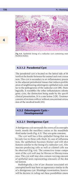
106 P.J. Slootweg4Fig. 4.4. Epi<strong>the</strong>lial lining <strong>of</strong> a radicular cyst containing manyRushton bodiesFig. 4.5. Cyst lining composed <strong>of</strong> squamous as well as mucous epi<strong>the</strong>lium.This can be found in radicular cysts as well as in dentigerousor residual cysts4.3.1.2 Paradental CystThe paradental cyst is located on <strong>the</strong> lateral side <strong>of</strong> <strong>the</strong>tooth at <strong>the</strong> border between <strong>the</strong> enamel <strong>and</strong> root cementum.This cyst is secondary to an inflammatory processin <strong>the</strong> adjacent periodontal tissues that induces proliferation<strong>of</strong> neighbouring odontogenic epi<strong>the</strong>lial rests, similarto <strong>the</strong> pathogenesis <strong>of</strong> <strong>the</strong> radicular cyst [88]. Histologically,it resembles <strong>the</strong> o<strong>the</strong>r inflammatory odontogeniccysts, <strong>the</strong> distinction being made by <strong>the</strong> specificclinical presentation. It is a rare lesion [128]. Treatmentconsists <strong>of</strong> excision with or without concomitant extraction<strong>of</strong> <strong>the</strong> involved tooth [45].4.3.2 Odontogenic Cysts –Developmental4.3.2.1 Dentigerous CystA dentigerous cyst surrounds <strong>the</strong> crown <strong>of</strong> an uneruptedtooth, mostly <strong>the</strong> maxillary canine or <strong>the</strong> m<strong>and</strong>ibularthird molar tooth (Fig. 4.3). They are quite common.The cyst wall has a thin epi<strong>the</strong>lial lining that maybe only two to three cells thick. In case <strong>of</strong> inflammation,<strong>the</strong> epi<strong>the</strong>lium becomes thicker <strong>and</strong> will showfeatures similar to <strong>the</strong> lining <strong>of</strong> a radicular cyst. Also,mucous-producing cells as well as ciliated cells maybe observed (Fig. 4.6). The connective tissue component<strong>of</strong> <strong>the</strong> cyst wall may be fibrous or fibromyxomatous.The cyst wall may also contain varying amounts<strong>of</strong> epi<strong>the</strong>lial nests representing remnants <strong>of</strong> <strong>the</strong> dentallamina.Radiologically, a lot <strong>of</strong> jaw diseases associated withunerupted teeth may have an appearance similar to that<strong>of</strong> a dentigerous cyst. Histologic examination, however,will be decisive in ruling out <strong>the</strong>se possibilities amongFig. 4.6. Lining <strong>of</strong> a dentigerous cyst mainly composed <strong>of</strong> mucouscellswhich keratocyst <strong>and</strong> unicystic ameloblastoma (seeSects. 4.3.2.4 <strong>and</strong> 4.4.1.1) are <strong>the</strong> most prevalent. Moreover,<strong>the</strong> radiologic picture <strong>of</strong> <strong>the</strong> dentigerous cyst maybe mimicked by hyperplasia <strong>of</strong> <strong>the</strong> dental follicle, <strong>the</strong>connective tissue capsule that surrounds <strong>the</strong> uneruptedtooth [33].Fibromyxomatous areas in <strong>the</strong> connective tissue wall<strong>of</strong> <strong>the</strong> dentigerous cyst may resemble <strong>the</strong> odontogenicmyxoma (see Sect. 4.4.2.1). The presence <strong>of</strong> odontogenicepi<strong>the</strong>lial rests may lead to <strong>the</strong> erroneous diagnosis<strong>of</strong> one or ano<strong>the</strong>r type <strong>of</strong> epi<strong>the</strong>lial odontogenic tumour[71]. However, identification <strong>of</strong> <strong>the</strong> epi<strong>the</strong>lial cyst liningwill rule out <strong>the</strong>se alternatives.In most instances, dentigerous cysts are a fortuitousfinding on oral radiographs. Only when excessively largemay <strong>the</strong>y cause swelling <strong>of</strong> <strong>the</strong> involved part <strong>of</strong> <strong>the</strong> jaw.If <strong>the</strong>re is inflammation, <strong>the</strong>y will cause pain <strong>and</strong> swelling.Removal <strong>of</strong> <strong>the</strong> cyst wall <strong>and</strong> <strong>the</strong> tooth involved willyield a permanent cure.The eruption cyst is a specific type <strong>of</strong> dentigerous cystlocated in <strong>the</strong> gingival s<strong>of</strong>t tissues overlying <strong>the</strong> crown


