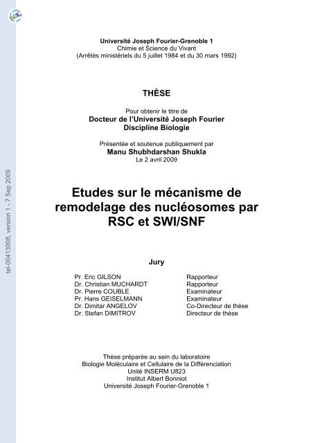
Etudes sur le mécanisme de remodelage des nucléosomes par ...
Etudes sur le mécanisme de remodelage des nucléosomes par ...
Etudes sur le mécanisme de remodelage des nucléosomes par ...

You also want an ePaper? Increase the reach of your titles
YUMPU automatically turns print PDFs into web optimized ePapers that Google loves.
tel-00413908, version 1 - 7 Sep 2009<br />
Université Joseph Fourier-Grenob<strong>le</strong> 1<br />
Chimie et Science du Vivant<br />
(Arrêtés ministériels du 5 juil<strong>le</strong>t 1984 et du 30 mars 1992)<br />
THÈSE<br />
Pour obtenir <strong>le</strong> titre <strong>de</strong><br />
Docteur <strong>de</strong> l’Université Joseph Fourier<br />
Discipline Biologie<br />
Présentée et soutenue publiquement <strong>par</strong><br />
Manu Shubhdarshan Shukla<br />
Le 2 avril 2009<br />
<strong>Etu<strong>de</strong>s</strong> <strong>sur</strong> <strong>le</strong> <strong>mécanisme</strong> <strong>de</strong><br />
remo<strong>de</strong>lage <strong>de</strong>s <strong>nucléosomes</strong> <strong>par</strong><br />
RSC et SWI/SNF<br />
Jury<br />
Pr. Eric GILSON Rapporteur<br />
Dr. Christian MUCHARDT Rapporteur<br />
Dr. Pierre COUBLE Examinateur<br />
Pr. Hans GEISELMANN Examinateur<br />
Dr. Dimitar ANGELOV Co-Directeur <strong>de</strong> thèse<br />
Dr. Stefan DIMITROV Directeur <strong>de</strong> thèse<br />
Thèse pré<strong>par</strong>ée au sein du laboratoire<br />
Biologie Moléculaire et Cellulaire <strong>de</strong> la Différenciation<br />
Unité INSERM U823<br />
Institut Albert Bonniot<br />
Université Joseph Fourier-Grenob<strong>le</strong> 1
tel-00413908, version 1 - 7 Sep 2009
tel-00413908, version 1 - 7 Sep 2009<br />
To,<br />
My Grand Pa<br />
Late Shri Uday Narain Shukla
tel-00413908, version 1 - 7 Sep 2009
tel-00413908, version 1 - 7 Sep 2009<br />
Acknow<strong>le</strong>dgements<br />
I have always believed that any personal achievement has many contributions from different<br />
quarters. As I ref<strong>le</strong>ct back, I see that without these it would not have been possib<strong>le</strong> to realize the<br />
goal of comp<strong>le</strong>ting my thesis. I wish to take this opportunity to thank those who have been<br />
instrumental in making this possib<strong>le</strong>.<br />
First and foremost, I wish to express my <strong>de</strong>epest gratitu<strong>de</strong> to my mentor Dr. Stefan Dimitrov<br />
for his unstinted support and encouragement. I sincerely thank him for his ab<strong>le</strong> guidance,<br />
being patient with me when I strayed, giving me the freedom to do things in my own ways, and<br />
most importantly keeping my focused at the same time. His sing<strong>le</strong> min<strong>de</strong>d <strong>de</strong>dication towards<br />
science consistently inspired my growth as a researcher.<br />
No words are enough to thank Dr. Dimitar Angelov, my thesis co-gui<strong>de</strong>, for teaching me the<br />
essentials of chromatin biology and his guidance in <strong>de</strong>signing and performing experiments. Not<br />
to mention his tire<strong>le</strong>ss work in optimizing most of the experiments in our laboratory which ma<strong>de</strong><br />
my job so much easier. Our long discussions before performing any experiment will be a thing to<br />
cherish in future.<br />
I wish to express my sincere thanks to Prof. Hans Geiselmann, Prof. Eric Gilson, Dr. Pierre<br />
Coub<strong>le</strong> and Dr. Christian Muchardt for their invaluab<strong>le</strong> time and effort spent on evaluation of<br />
thesis.<br />
I would like to thank Prof. Arthur Soucemarianadin for his support and help throughout the<br />
course of my thesis. Also, I wish to thank Joseph Fourier University for “La Bourse<br />
Presi<strong>de</strong>ntiel<strong>le</strong>” and the scientific franco-indian program ARCUS.<br />
I am grateful to Dr. Gyan Chandra Mishra, my first teacher during my early stage of research,<br />
for his crucial contribution in my <strong>de</strong>velopment as a stu<strong>de</strong>nt.
tel-00413908, version 1 - 7 Sep 2009<br />
I am in<strong>de</strong>bted to Dr. Sanjeev Galan<strong>de</strong> and Dr. R. R. Bhon<strong>de</strong> for their support and help. It<br />
was only for their support that I could continue in my pursuit of a scientific carrier. Thank<br />
you so much.<br />
I wish to thank Prof. Philippe Bouvet for his excel<strong>le</strong>nt support during my stay in the Jolliot<br />
Curie Laboratory.<br />
The scope of this thesis required multi-disciplinary approaches and I was fortunate to have very<br />
ab<strong>le</strong> collaborators whose contribution was indispensab<strong>le</strong> for successful conclusion of this work. I<br />
wish to thank Dr. Cendrine Moska<strong>le</strong>nko and Dr. Jan Bednar for their help in Atomic Force<br />
Microscopy and Cryo-E<strong>le</strong>ctron Microscopy experiments. I wish to especially thank Cendrine for<br />
explaining me the fundamentals of AFM, interpreting the results and also for her help in<br />
correcting the thesis.<br />
Special thanks for Fabien Montel, Sajad and Hervé for their help in my experiments, their<br />
critical inputs and most importantly for the great work environment we shared.<br />
I wish to thank Zofia, He<strong>le</strong>ne, Maggy and Bennoit for their willingness to help whenever I<br />
nee<strong>de</strong>d it. In fact, I would like to express my gratitu<strong>de</strong> to all the members of LJC for their<br />
constant support, encouragement, and making me feel as a <strong>par</strong>t of the big family. They all have<br />
been source of inspiration and the things I have <strong>le</strong>arned through my interaction with them will<br />
help me grow as a scientist in time to come.<br />
I would like to thank my <strong>par</strong>ents, family members and friends who stood by me at all the times.<br />
Thank you Sumit, Janesh and Farhan for being there.<br />
Finally I wish to thank one person without whom it was not possib<strong>le</strong> at all. Thank you Ruchi,<br />
for your support, standing by me with all my <strong>de</strong>cisions, un<strong>de</strong>rstanding me, taking many things<br />
off my shoul<strong>de</strong>rs and most of all for giving me the most precious gift “Chinmaya”.
tel-00413908, version 1 - 7 Sep 2009<br />
Tab<strong>le</strong> of Contents<br />
Tab<strong>le</strong> of Contents……….……………………………………………………………………. 7<br />
In<strong>de</strong>x of Figures and Tab<strong>le</strong>s……………………………………………………………12<br />
Thesis <strong>de</strong>tails………………………………………………………………………………… 14<br />
A. TITLE<br />
a. Titre en Français……………………………………………………………………14<br />
b. Tit<strong>le</strong> in English…………………………………………………………………….. 14<br />
B. ABSTRACT<br />
a. Résumé en Français………………………………………………………………...14<br />
b. Abstract in English…………………………………………………………………15<br />
C. KEYWORDS<br />
a. Mots clés en français……………………………………………………………….16<br />
b. Keywords in English……………………………………………………………….16<br />
D. DETAILS OF THE LABORATORIES OF THESIS PREPARATION<br />
a. Laboratory of affiliation ...........................................................................................17<br />
b. Other laboratory……………………………………………………………………17<br />
List of Abbreviations...............................................................................................................18<br />
CHAPTER I: INTRODUCTION<br />
Chromatin Structure, Organization and dynamics……………………………19<br />
I.1 The Nuc<strong>le</strong>osome.................................................................................................................20<br />
I.1.1 Histones…………………………………………………………………………20<br />
I.1.2 Crystal structure of the nuc<strong>le</strong>osome……………………………………………23<br />
I.2 Higher or<strong>de</strong>rs of Chromatin structure…………………………………………………26<br />
I.3 Chromatin Territories…………………………………………………………………..28<br />
I.4 Regulation of Chromatin Dynamics……………………………………………………29<br />
I.4.1 Incorporation of Histone variants……………………………………………....30<br />
I.4.1.1 H3 variants……………………………………………………………31<br />
I.4.1.2 H2A variants………………………………………………………….32<br />
I.4.1.2.1 H2A.Z……………………………………………………………….33<br />
I.4.1.2.2 H2A.X……………………………………………………………….35<br />
I.4.1.2.3 macroH2A…………………………………………………………...36<br />
I.4.1.2.4 H2A.Bbd…………………………………………………………….37<br />
7
tel-00413908, version 1 - 7 Sep 2009<br />
I.4.2 Posttranslational modifications of histones………………………………….39<br />
I.4.2.1 Histone acetylation……………………………………………………40<br />
I.4.2.2 Histone methylation…………………………………………………...41<br />
I.4.2.3 Other histone cova<strong>le</strong>nt modifications…………………………………42<br />
I.4.3 ATP <strong>de</strong>pen<strong>de</strong>nt Chromatin remo<strong>de</strong>ling.......................................................................43<br />
I.4.3.1 Different classes of Chromatin remo<strong>de</strong><strong>le</strong>rs…………………………………....44<br />
I.4.3.1.1 SWI/SNF family…………………………………………………….45<br />
I.4.3.1.1.1 SWI/SNF…………………………………………………………..46<br />
I.4.3.1.1.2 RSC………………………………………………………………..47<br />
I.4.3.1.1.3 SWI/SNF comp<strong>le</strong>xes in higher eukaryotes…………………........48<br />
I.4.3.1.1.4 Structural domains in SWI/SNF subfamily comp<strong>le</strong>xes…………..49<br />
I.4.3.1.2 ISWI family………………………………………………………….50<br />
I.4.3.1.3 INO80 family………………………………………………………..53<br />
I.4.3.1.4 CHD family………………………………………………………….54<br />
I.4.3.2 Targeting of chromatin remo<strong>de</strong><strong>le</strong>rs……………………………………………56<br />
I.4.3.3 Regulation of chromatin remo<strong>de</strong>ling………………………………………….58<br />
I.4.3.3.1 Posttranslational modification of active subunit…………………….58<br />
I.4.3.3.2 Subunit composition of the remo<strong>de</strong>ling comp<strong>le</strong>x…………………...59<br />
I.4.3.3.3 Interaction with secondary messenger mo<strong>le</strong>cu<strong>le</strong>s…………………...60<br />
I.4.3.3.4 Interaction with non-histone proteins………………………….........61<br />
I.4.3.4 Functions of ATP <strong>de</strong>pen<strong>de</strong>nt chromatin remo<strong>de</strong><strong>le</strong>rs………………………….61<br />
I.4.3.4.1 Regulation of transcription………………………………………….63<br />
I.4.3.4.2 Regulation of cell cyc<strong>le</strong>……………………………………………..65<br />
I.4.3.4.3 Effect on cell differentiation and <strong>de</strong>velopment……………………..67<br />
I.4.3.4.4 Regulation of DNA replication and repair………………………….67<br />
I.4.3.4.5 Ro<strong>le</strong> in tumor suppression ………………………………………….68<br />
I.4.3.4.6 Conclusions ………………………………………………………....69<br />
I.5 Mechanism of ATP <strong>de</strong>pen<strong>de</strong>nt chromatin remo<strong>de</strong>ling………………………………..69<br />
I.5.1 Biochemical properties of remo<strong>de</strong><strong>le</strong>rs…………………………………………..69<br />
I.5.1.1 Substrate binding………………………………………………………70<br />
I.5.1.2 ATP binding and hydrolysis…………………………………………...73<br />
I.5.1.3 Nuc<strong>le</strong>osome disruption activities……………………………………...73<br />
I.5.1.3.1 Generation of superhelical torsion…………………………………..74<br />
I. 5.1.3.2 Nuc<strong>le</strong>osome sliding…………………………………………………74<br />
8
tel-00413908, version 1 - 7 Sep 2009<br />
I.5.1.3.3 Changes in Nuc<strong>le</strong>osomal DNA conformation: ‘Remo<strong>de</strong>ling’…........76<br />
I.5.1.3.4 Histone H2A-H2B dimer expulsion or exchange…………………...77<br />
I.5.1.4 Conclusion…………………………………………………………….77<br />
I.5.2 ATP <strong>de</strong>pen<strong>de</strong>nt remo<strong>de</strong><strong>le</strong>rs as DNA translocases………………………………79<br />
I.5.3 Mo<strong>de</strong>ls for Nuc<strong>le</strong>osome remo<strong>de</strong>ling……………………………………………80<br />
I.6 Objectives………………………………………………………………………………...82<br />
CHAPTER II: Manuscript communicated- A Two-Step Mechanism for<br />
Nuc<strong>le</strong>osome Remo<strong>de</strong>ling by RSC: Initial Formation of a Remosome<br />
Containing ~180-190 bp DNA Followed by Sliding…………....……………85<br />
II.1 Summary...............................................................................................................86<br />
II.2 Introduction..........................................................................................................87<br />
II.3 Results<br />
II.3.1 RSC generates stab<strong>le</strong> non-mobilized nuc<strong>le</strong>osome-like <strong>par</strong>tic<strong>le</strong>s associated<br />
with 180-190 bp DNA………………………………………….......................89<br />
II.3.2 The remosomes are ensemb<strong>le</strong> of distinct structures with different DNA<br />
conformation………………………………………………………..................93<br />
II.3.3 The “in gel one pot assay” shows highly perturbed histone-DNA<br />
interactions within the remosome……………………………………………..96<br />
II.3.4 The remosomes are intermediate structures generated during the RSC<br />
nuc<strong>le</strong>osome mobilization process……………………………………………..99<br />
II.3.5 The remosomes are bona fi<strong>de</strong> substrates for mobilization by RSC…102<br />
II.4 Discussion………………………………………………………………………103<br />
II.5 Experimental Procedures<br />
II.5.1 Pre<strong>par</strong>ation of DNA fragments……………………………………….106<br />
II.5.2 Proteins and Nuc<strong>le</strong>osome reconstitutions……………………………..107<br />
II.5.3 Nuc<strong>le</strong>osome remo<strong>de</strong>ling reaction……………………………………..107<br />
II.5.4 Dnase I footprinting assay………………………………………….....107<br />
II.5.5 Sliding assay on gel eluted nuc<strong>le</strong>osome………………………………108<br />
II.5.6 In Gel One Pot assay…………………………………………………..109<br />
II.5.7 Gel elution of nuc<strong>le</strong>osomes for AFM analysis………………………..109<br />
II.5.8 Atomic Force Microscopy, Image Analysis and construction of the 2D<br />
maps Lc/∆L………………………………………………………………….110<br />
9
tel-00413908, version 1 - 7 Sep 2009<br />
II.5.9 E<strong>le</strong>ctron-Cryo microscopy…………………………………………….111<br />
II.6 Acknow<strong>le</strong>dgements……………………………………………………112<br />
Chapter III: Manuscript un<strong>de</strong>r pre<strong>par</strong>ation- I<strong>de</strong>ntification and<br />
Characterization of Novel Intermediates of SWI/SNF Induced Nuc<strong>le</strong>osome<br />
Sliding………………………………………………………………………....115<br />
III.1 Introduction…………………………………………………………………..116<br />
III.2 Results<br />
III.2.1 The initial step of SWI/SNF nuc<strong>le</strong>osome mobilization mechanism is the<br />
perturbation of the histone-DNA interactions and the generation of a non-<br />
mobilized nuc<strong>le</strong>osome-like <strong>par</strong>tic<strong>le</strong> associated with ∼180 bp of DNA….…..118<br />
III.2.2 Restriction enzyme c<strong>le</strong>avage of remosome DNA shows dramatic<br />
perturbations of the histone-DNA interactions………………………………123<br />
III.2.3 The remosomes represent a multitu<strong>de</strong> of remo<strong>de</strong><strong>le</strong>d nuc<strong>le</strong>osomes, in<br />
which each individual <strong>par</strong>tic<strong>le</strong> exhibits a distinctly perturbed path of<br />
nuc<strong>le</strong>osomal DNA…………………………………………………………...127<br />
III.3 Discussion……………………………………………………………………..129<br />
III.4 Experimental Procedures<br />
III.4.1 Nuc<strong>le</strong>osome remo<strong>de</strong>ling reactions…………………………………...131<br />
III.4.2 DNase I footprinting assay…………………………………………...132<br />
III.4.3 Restriction enzyme assay on gel eluted nuc<strong>le</strong>osome………………....132<br />
III.4.4 Atomic Force Microscopy…………………………………………....133<br />
Chapter IV: Manuscript un<strong>de</strong>r pre<strong>par</strong>ation - H2A Docking of H2A is<br />
essential for SWI/SNF and RSC induced nuc<strong>le</strong>osome sliding through<br />
generation of remosome intermediates……………………………………………135<br />
IV.1 Introduction……………………………………………………………..…….135<br />
IV.2 Results<br />
IV.2.1 Nuc<strong>le</strong>osome reconstitutions with H2A C-terminal <strong>de</strong><strong>le</strong>tion, chimeric<br />
and variant proteins………………………………………………………….137<br />
IV.2.2 Changes in C-terminal region of H2A results in structural perturbations<br />
in nuc<strong>le</strong>osomes…………………………………………………………...….138<br />
10
tel-00413908, version 1 - 7 Sep 2009<br />
IV.2.3 The base of H2A docking domain is essential for SWI/SNF mediated<br />
mobilization of nuc<strong>le</strong>osomes………………………………………...………141<br />
IV.2.4 RSC is more sensitive to perturbations in the docking domain of H2A<br />
……………………………………………………………………………….144<br />
IV.2.5 Generation of distinct remo<strong>de</strong><strong>le</strong>d forms by SWI/SNF on H2A.Bbd<br />
nuc<strong>le</strong>osomes…………………………………………………………………146<br />
IV.2.6 The docking domain of H2A.Bbd is responsib<strong>le</strong> for anomalous<br />
remo<strong>de</strong>ling by SWI/SNF…………………………………………………….149<br />
I V.2.7 R SC mediated r e mo<strong>de</strong>ling i s s i milar t o S WI/SNF o n<br />
H2A.dockingdomain.Bbd nuc<strong>le</strong>osomes……………………………………..151<br />
IV.3 Discussion………………………………………………………………….......152<br />
IV.4 Experimental procedures<br />
IV.4.1 Pre<strong>par</strong>ation of DNA probes…………………………………………..154<br />
IV.4.2 Proteins……………………………………………………………….154<br />
IV.4.3 Nuc<strong>le</strong>osome reconstitutions………………………………………….154<br />
IV.4.4 DNaseI and hydroxyl radical footprinting…………………………...155<br />
IV.4.5 Nuc<strong>le</strong>osome sliding assay……………………………………………155<br />
IV.4.6 AFM analysis…………………………...…………………………….156<br />
IV.4.7 One pot restriction assay……………………………………………..156<br />
IV.4.8 Supp<strong>le</strong>mentary information for Chapter II, III and IV……………….157<br />
V. General conclusions and perspective…………………………………………159<br />
BIBLIOGRAPHY..................................................................................................................163<br />
11
tel-00413908, version 1 - 7 Sep 2009<br />
In<strong>de</strong>x of Figures and Tab<strong>le</strong>s<br />
Figure I.1 Nuc<strong>le</strong>osomes: Beads-on-a-string mo<strong>de</strong>l…………………………………….20<br />
Figure I.2 Histone fold of the core histones…………………………………………….21<br />
Figure I.3 Nuc<strong>le</strong>osome assembly……………………………………………………….22<br />
Figure I.4 Structure and potential position of linker histone on nuc<strong>le</strong>osome…………..23<br />
Figure I.5. Structure of the 147 bp nuc<strong>le</strong>osome core <strong>par</strong>tic<strong>le</strong> at 1.9 Å resolution……...25<br />
Figure I.6 Different or<strong>de</strong>rs of chromatin architecture……………………………………….26<br />
Figure I.7 Mo<strong>de</strong>ls of 30 nm fiber organization…………....……………………………27<br />
Figure I.8 General Properties of euchromatin and heterochromatin……………………29<br />
Figure I.9 Canonical histones and their variants………………………………………..31<br />
Figure I.10 Phylogenetic tree of H2A variants………………………………………….33<br />
Figure I.11 Sequence and structure of H2A.Bbd………………………………………..38<br />
Figure I.12 Posttranslational Modifications of the Core Histones………………………40<br />
Figure I.13. Classification of SNF2 family ATPases……………………………….…...44<br />
Figure I.14 Classes of ATP <strong>de</strong>pen<strong>de</strong>nt Chromatin remo<strong>de</strong>ling enzymes………………..45<br />
Figure I.15 Subunit compositions of SWI2/SNF2 family comp<strong>le</strong>xes…….……………..45<br />
Figure I.16 Domain organizations of SWI/SNF subunits………….…………………….49<br />
Figure I.17 Subunit compositions of ISWI subfamily members…………………………50<br />
Figure I.18 Domain organization of ISWI subunits……………………………………...53<br />
Figure I.19 Subunit composition of INO80 subfamily members………………………...54<br />
Figure I.20 Subunit compositions of CHD subfamily members…………………………55<br />
Figure I.21 Mo<strong>de</strong>ls for SWI/SNF recruitment to target genes…………………………...57<br />
Tab<strong>le</strong> I.1 Biological functions of chromatin remo<strong>de</strong><strong>le</strong>rs…………………………………62<br />
Figure I.22 Structures of SWI/SNF and RSC comp<strong>le</strong>xes reconstructed from Cryo-E<strong>le</strong>ctron<br />
micrographs……………………………………………………………………………….72<br />
Figure I.23 Summary of various biochemical activities of ATP <strong>de</strong>pen<strong>de</strong>nt remo<strong>de</strong><strong>le</strong>rs.<br />
……………………………………………………………………………………….......78<br />
Figure I.24 Proposed mo<strong>de</strong>ls of nuc<strong>le</strong>osome sliding by ATP <strong>de</strong>pen<strong>de</strong>nt remo<strong>de</strong><strong>le</strong>rs......81<br />
Figure II.1. AFM visualization of RSC mobilized nuc<strong>le</strong>osomes…………..………….90<br />
Figure II.2. The initial step of the RSC nuc<strong>le</strong>osome mobilization reaction is the generation of<br />
a stab<strong>le</strong>, non-mobilized <strong>par</strong>tic<strong>le</strong> containing 180-190 bp of histone octamer associated DNA.<br />
…………………………………………………………………………………………91<br />
Figure II.3. E<strong>le</strong>ctron Cryo-Microscopy (EC-M) of the RSC treated mono- and trinuc<strong>le</strong>osomes<br />
shows that different species are present in the RSC remo<strong>de</strong>ling reaction.<br />
………….………………………………………………………………………………95<br />
12
tel-00413908, version 1 - 7 Sep 2009<br />
Figure II.4. In gel “one pot” restriction accessibility assay of the RSC generated remosomes<br />
………………………………………………………………………………………….98<br />
Figure II.5. The remosomes are intermediate structures generated during the RSC<br />
mobilization reaction…………………….………………………………………………101<br />
Figure II.6. The remosomes are bona fi<strong>de</strong> substrates for RSC…….…………………….103<br />
Figure II.7. Schematic representation of the two step RSC-induced nuc<strong>le</strong>osome mobilization<br />
………………………………………………………………………………….………..105<br />
Figure II.S1 The reconstituted nuc<strong>le</strong>osomes are efficiently mobilized by RSC………...113<br />
Figure II.S2 AFM experiments show that the remosomes are intermediary <strong>par</strong>tic<strong>le</strong>s generated<br />
during the RSC nuc<strong>le</strong>osome mobilization reaction……………………………………...113<br />
Figure III.1. Nuc<strong>le</strong>osome mobilization with SWI/SNF………………………………........119<br />
Figure III.2. DNase I footprinting analysis shows that nuc<strong>le</strong>osome treatment with SWI/SNF<br />
resulted in perturbation of the histone-DNA interactions prior to nuc<strong>le</strong>osome mobilization.<br />
……………………………………………………………………………………………120<br />
Figure III.3. SWI/SNF generates non-mobilized nuc<strong>le</strong>osomes <strong>par</strong>tic<strong>le</strong>s associated with ~180<br />
bp of DNA………………………………………………………………………………...122<br />
Figure III.4. Mea<strong>sur</strong>ements of the DNA accessibility to Hae III along the nuc<strong>le</strong>osomal DNA<br />
<strong>le</strong>ngth in control and SWI/SNF treated nuc<strong>le</strong>osomes by using the in gel one pot assay.....124<br />
Figure III.5. HaeIII digestion kinetics of control nuc<strong>le</strong>osomes and remosomes in solution.<br />
……………………………………………………………………………………………..126<br />
Figure III.6. Observation of different species in SWI/SNF treated mono- and trinuc<strong>le</strong>osomal<br />
substrates by E<strong>le</strong>ctron Cryo-Microscopy (EC-M)…………………………………………128<br />
Figure IV.1. Reconstitution of nuc<strong>le</strong>osomes with H2A C terminal <strong>de</strong><strong>le</strong>tion and chimeric<br />
proteins………………………..……………………………………………………………138<br />
Figure IV.2 Biochemical characterization of conventional, variant and mutant nuc<strong>le</strong>osomes by<br />
DNase I and OH° footprinting……………………………………………………………...140<br />
Figure IV.3. H2A docking domain is essential for SWI/SNF induced nuc<strong>le</strong>osome<br />
mobilization…………………………………………………………………………….…143<br />
Figure IV.4. AFM analysis of SWI/SNF induced remo<strong>de</strong>ling on H2A.ddBbd nuc<strong>le</strong>osomes.<br />
……………………………………………………………………………………………..144<br />
Figure IV.5. RSC is more sensitive to perturbations in the docking domain of H2A for<br />
nuc<strong>le</strong>osome mobilization…………………………………………………………….…….145<br />
Figure IV.6. Generation of distinct remo<strong>de</strong><strong>le</strong>d forms by SWI/SNF on H2A.Bbd nuc<strong>le</strong>osomes.<br />
……………………………………………………………………………………………...148<br />
Figure IV.7. The docking domain of H2A.Bbd is responsib<strong>le</strong> for anomalous remo<strong>de</strong>ling by<br />
SWI/SNF……………………………………………………………………………………150<br />
Figure IV.8. The docking domain of H2A.Bbd is responsib<strong>le</strong> for anomalous remo<strong>de</strong>ling by<br />
SWI/SNF…………………………………………………………………………………….152<br />
13
tel-00413908, version 1 - 7 Sep 2009<br />
Thesis Details<br />
A. Tit<strong>le</strong><br />
a. Titre en français<br />
<strong>Etu<strong>de</strong>s</strong> <strong>sur</strong> <strong>le</strong> <strong>mécanisme</strong> <strong>de</strong> remo<strong>de</strong>lage <strong>de</strong>s <strong>nucléosomes</strong> <strong>par</strong> RSC et SWI/SNF<br />
b. Tit<strong>le</strong> in english<br />
Studies on the mechanism of nuc<strong>le</strong>osome remo<strong>de</strong>ling by RSC and SWI/SNF<br />
B. Abstract<br />
a. Résumé en français<br />
Dans <strong>le</strong>s cellu<strong>le</strong>s eucaryotes l’ADN nucléaire est organisé sous la forme <strong>de</strong> chromatine, dont<br />
l’unité <strong>de</strong> répétition est <strong>le</strong> nuc<strong>le</strong>osome. En règ<strong>le</strong> généra<strong>le</strong>, la chromatine est considérée comme<br />
répressive pour <strong>le</strong>s processus nécessitant un accès à l’ADN tels que la transcription, la<br />
réplication ou la ré<strong>par</strong>ation. Le nucléosome représente une forte barrière pour <strong>de</strong>s protéines<br />
nécessitant l’accès à l’ADN. Pour <strong>sur</strong>monter cette barrière, la cellu<strong>le</strong> a développé <strong>de</strong>s<br />
métho<strong>de</strong>s variées, dont la plus importante semb<strong>le</strong> être <strong>le</strong> remo<strong>de</strong>lage <strong>de</strong>s <strong>nucléosomes</strong><br />
dépendant <strong>de</strong> l’ATP. Une propriété commune à tous ces facteurs <strong>de</strong> remo<strong>de</strong>lage est <strong>le</strong>ur<br />
capacité <strong>de</strong> repositionner <strong>le</strong>s <strong>nucléosomes</strong> <strong>le</strong> long <strong>de</strong> l’ADN.<br />
Dans ce travail, nous avons étudié <strong>le</strong> <strong>mécanisme</strong> <strong>de</strong> déplacement <strong>de</strong>s <strong>nucléosomes</strong> <strong>par</strong> RSC et<br />
SWI/SNF, <strong>de</strong>ux facteurs <strong>de</strong> remo<strong>de</strong>lage <strong>de</strong> <strong>le</strong>vure bien caractérisés. Nous avons combiné <strong>de</strong>s<br />
approches basées <strong>sur</strong> la visualisation à haute résolution, notamment la microscopie à force<br />
atomique (AFM) et la cryo-microscopie é<strong>le</strong>ctronique, avec <strong>de</strong>s approches nouvel<strong>le</strong>s à pointe<br />
<strong>de</strong> la biochimie et <strong>de</strong> la biologie moléculaire.<br />
Nous avons montré que la mobilisation <strong>de</strong>s <strong>nucléosomes</strong> <strong>par</strong> RSC ou SWI/SNF implique <strong>de</strong>s<br />
espèces réactionnel<strong>le</strong>s intermédiaires métastab<strong>le</strong>s dont l’existence et la structure étaient<br />
jusqu’alors inconnues. Ces <strong>par</strong>ticu<strong>le</strong>s nucléosoma<strong>le</strong>s, que nous avons nommé ‘remosomes’,<br />
possè<strong>de</strong>nt certaines propriétés structura<strong>le</strong>s distinctes <strong>de</strong>s <strong>nucléosomes</strong> canoniques. En<br />
<strong>par</strong>ticulier, <strong>le</strong>s ‘remosomes’ contiennent ~180 pb d’ADN associées à l’octamère d’histones au<br />
14
tel-00413908, version 1 - 7 Sep 2009<br />
lieu <strong>de</strong> 147 pb pour <strong>le</strong>s <strong>nucléosomes</strong> canoniques. En utilisant, l’empreinte à la DNase I nous<br />
avons montré que <strong>le</strong> ‘remosome’ représente un ensemb<strong>le</strong> <strong>de</strong> structures multip<strong>le</strong>s caractérisées<br />
<strong>par</strong> un enrou<strong>le</strong>ment fortement perturbé <strong>de</strong> l’ADN <strong>sur</strong> l’octamère d’histones. Pour caractériser<br />
ces ‘remosomes’ avec une gran<strong>de</strong> précision, nous avons mis au point une nouvel<strong>le</strong> technique<br />
« one pot in gel assay » qui consiste à cartographier toutes <strong>le</strong>s 10 pb l’accessibilité d’une<br />
enzyme <strong>de</strong> restriction au ‘remosome’ fractionné. L’application <strong>de</strong> cette technique a révélé que<br />
<strong>le</strong> profil <strong>de</strong> l’accessibilité du ‘remosome’ est très différent <strong>de</strong> celui du nucléosome. Alors que<br />
celui du nucléosome peut être extrapolé <strong>par</strong> une fonction <strong>de</strong> type hyperbolique, <strong>le</strong> profil du<br />
‘remosome’ est ajusté <strong>par</strong> une fonction <strong>par</strong>abolique.<br />
Nous avons voulu répondre à la question du <strong>mécanisme</strong> <strong>de</strong> l’inhibition <strong>de</strong> la mobilisation du<br />
nucléosome variant H2A.Bbd <strong>par</strong> SWI/SNF. En utilisant <strong>le</strong>s techniques décrites plus haut <strong>sur</strong><br />
<strong>de</strong>s <strong>nucléosomes</strong> variants ou chimériques (contenant <strong>de</strong>s délétions ou translocations <strong>de</strong><br />
domaines d’histones) nous avons montré que <strong>le</strong> domaine d’accrochage (‘docking domain’) <strong>de</strong><br />
l’histone H2A est essentiel pour la mobilisation <strong>de</strong>s <strong>nucléosomes</strong>. Nous avons aussi montré<br />
que l’incapacité du nucléosome à glisser est due à la génération d’états intermédiaires<br />
‘remosomes erronés’, distincts <strong>de</strong> ceux ap<strong>par</strong>aissant dans <strong>le</strong> cas du nucléosome<br />
conventionnel.<br />
b. Abstract in English<br />
In eukaryotic cell the DNA is organized in the nuc<strong>le</strong>us in the form of chromatin, the<br />
fundamental unit of which is cal<strong>le</strong>d as the nuc<strong>le</strong>osome. Organization of DNA into the<br />
nuc<strong>le</strong>osomes presents a strong barrier for various processes which require access to the DNA<br />
like transcription, replication and repair. To overcome this prob<strong>le</strong>m cells utilize a variety of<br />
methods, ATP <strong>de</strong>pen<strong>de</strong>nt chromatin remo<strong>de</strong>ling being one of the most important of them. A<br />
common feature of all the remo<strong>de</strong><strong>le</strong>rs is that they are ab<strong>le</strong> to reposition the nuc<strong>le</strong>osomes along<br />
the DNA at the expense of ATP.<br />
In the present work, we have studied the mechanism of nuc<strong>le</strong>osome mobilization by RSC and<br />
SWI/SNF, two well characterized remo<strong>de</strong><strong>le</strong>rs from yeast. A combinatorial approach was<br />
employed using high resolution microscopy namely E<strong>le</strong>ctron cryo-Microscopy (EC-M) and<br />
Atomic Force Microscopy (AFM) together with novel biochemical approaches. We have<br />
shown that the nuc<strong>le</strong>osome mobilization by RSC and SWI/SNF involves hitherto unknown<br />
15
tel-00413908, version 1 - 7 Sep 2009<br />
intermediate structures. These remo<strong>de</strong><strong>le</strong>d nuc<strong>le</strong>osome <strong>par</strong>tic<strong>le</strong>s ‘The Remosomes’ possess<br />
characteristic structural features. Our AFM studies show that ~180 bp of DNA is associated<br />
with the histone octamer as com<strong>par</strong>ed to ~147 bp in the canonical nuc<strong>le</strong>osomes. Using<br />
DNaseI footprinting and EC-M we have shown that the path of DNA around the histone<br />
octamer is highly perturbed. Moreover, these <strong>par</strong>tic<strong>le</strong>s represent an ensemb<strong>le</strong> many different<br />
structures rather than one <strong>de</strong>fined specie. The novel ‘in gel one pot assay’ showed that<br />
accessibility profi<strong>le</strong> of these <strong>par</strong>tic<strong>le</strong>s is comp<strong>le</strong>tely different from that of canonical<br />
nuc<strong>le</strong>osomes and they are accessib<strong>le</strong> all along the path of DNA.<br />
We have also addressed the question of inhibition of nuc<strong>le</strong>osome mobilization due to<br />
incorporation of histone variant H2A.Bbd in the nuc<strong>le</strong>osomes. We show that the docking<br />
domain of histone H2A is essential for SWI/SNF and RSC induced nuc<strong>le</strong>osome sliding.<br />
Furthermore, we <strong>de</strong>monstrate that the reason for inability of these nuc<strong>le</strong>osomes to sli<strong>de</strong> is due<br />
to a faulty generation of ‘Remosome’ intermediates.<br />
C. Keywords<br />
a. Mots clés en Français<br />
Variants d’histones, remo<strong>de</strong>lage <strong>de</strong> la chromatine, nucléosome, RSC, SWI SNF, chromatine,<br />
H2A.Bbd<br />
b. Keywords in English<br />
Histone variants, chromatin remo<strong>de</strong>ling, nuc<strong>le</strong>osome, RSC, SWI/SNF, chromatin, H2A.Bbd<br />
16
tel-00413908, version 1 - 7 Sep 2009<br />
D. Details of the laboratories of thesis pre<strong>par</strong>ation<br />
a. Laboratory of affiliation<br />
b. Other Laboratory<br />
Unité INSERM U823<br />
Institut Albert Bonniot<br />
Domain <strong>de</strong> la Merci<br />
38706 La Tronche ce<strong>de</strong>x<br />
FRANCE<br />
Laboratoire Joliot Curie<br />
Éco<strong>le</strong> Norma<strong>le</strong> Supérieure<br />
46 allée d’Italie<br />
69364 Lyon ce<strong>de</strong>x 07<br />
FRANCE<br />
17
tel-00413908, version 1 - 7 Sep 2009<br />
List of Abbreviations<br />
ACF ATP-<strong>de</strong>pen<strong>de</strong>nt Chromatin assembly and remo<strong>de</strong>ling Factor<br />
AFM Atomic Force Microscopy<br />
ATP A<strong>de</strong>nosine-5'-triphosphate<br />
Bbd Barr Body Deficient<br />
bp Base Pair<br />
CHD Chromodomain Helicase DNA-binding<br />
ChIP Chromatin Immuno Precipitation<br />
CHRAC CHRomatin Accessibility Comp<strong>le</strong>x<br />
Da Dalton (1g/mol)<br />
dd Docking Domain<br />
DNA Deoxyribo Nuc<strong>le</strong>ic Acid<br />
DTT Dithiothreitol<br />
ECM E<strong>le</strong>ctron Cryo Microscopy<br />
EDTA Ethy<strong>le</strong>ne Diamine Tetra Acetic acid<br />
EM E<strong>le</strong>ctron Microscopy<br />
EMSA E<strong>le</strong>ctrophoretic Mobility Shift Assay<br />
HA Hemagglutinin<br />
ISWI Imititation SWItch<br />
Lc Length of DNA comp<strong>le</strong>xed within the nuc<strong>le</strong>osome<br />
NaCl Sodium Chlori<strong>de</strong><br />
NCP Nuc<strong>le</strong>osome Core Partic<strong>le</strong><br />
NFR Nuc<strong>le</strong>osome Free Region<br />
NURF Nuc<strong>le</strong>osome Remo<strong>de</strong>ling Factor<br />
ORF Open Reading Frame<br />
PAGE Polyacrylami<strong>de</strong> Gel E<strong>le</strong>ctrophoresis<br />
PCR Polymerase Chain Reaction<br />
RSC Remo<strong>de</strong>ls Structure of Chromatin<br />
SDS Sodium Do<strong>de</strong>cyl Sulfate<br />
∆L Distance of nucelosome from the center of the fragment<br />
18
tel-00413908, version 1 - 7 Sep 2009<br />
Chapter I: Introduction<br />
Chromatin Structure, Organization and Dynamics<br />
The genetic instructions which are used for <strong>de</strong>velopment and functioning of all living<br />
organisms are contained in nuc<strong>le</strong>ic acid cal<strong>le</strong>d as <strong>de</strong>oxyribonuc<strong>le</strong>ic acid (DNA). DNA<br />
contains instructions for synthesis of other components of cells. Other DNA sequences<br />
between the genes have structural purposes and are known to be involved in regulation of<br />
gene expression. DNA was first isolated in 1869 by Friedrich Miescher as a microscopic<br />
substance in the pus of discar<strong>de</strong>d <strong>sur</strong>gical bandages. Since, it resi<strong>de</strong>d in the nuc<strong>le</strong>i of cells, he<br />
cal<strong>le</strong>d "nuc<strong>le</strong>in" (Dahm R, 2005). Later, in 1919 Phoebus Levene i<strong>de</strong>ntified the base, sugar<br />
and phosphate components of nuc<strong>le</strong>oti<strong>de</strong>s (Leven P, 1919) and suggested that DNA consisted<br />
of a string of nuc<strong>le</strong>oti<strong>de</strong>s linked together through the phosphate groups. Finally, DNA's ro<strong>le</strong> in<br />
heredity was confirmed in 1952 by the famous Hershey-Chase experiment (Hershey and<br />
Chase 1952) and based on X-ray diffraction data by Rosalind Franklin and the information<br />
that the bases were paired; James D. Watson and Francis Crick suggested the doub<strong>le</strong> helix<br />
structure of DNA what is now accepted as the first accurate mo<strong>de</strong>l of DNA structure (Watson<br />
and Crick, 1953).<br />
In the nuc<strong>le</strong>us DNA exists as a comp<strong>le</strong>x structure cal<strong>le</strong>d chromatin, a combination of DNA<br />
with proteins. The term ‘Chromatin’ was suggested for the first time by W. F<strong>le</strong>mming<br />
(~1880), owing to its affinity to stains, whi<strong>le</strong> studying the process of nuc<strong>le</strong>ar division. The<br />
purpose of chromatin organization is to package DNA into a smal<strong>le</strong>r volume to fit in the cell,<br />
to strengthen the DNA to allow mitosis and meiosis, and to serve as a mechanism to control<br />
vital processes like transcription, repair and DNA replication. Three basic <strong>le</strong>vels of chromatin<br />
organization occur in the cell:<br />
i. DNA wrapping around nuc<strong>le</strong>osomes - <strong>le</strong>ading to the primary structure of chromatin<br />
cal<strong>le</strong>d as the "beads on a string" structure.<br />
ii. A 30 nm con<strong>de</strong>nsed chromatin fiber resulting from specific interactions between<br />
nuc<strong>le</strong>osomes (Secondary structure of chromatin).<br />
iii. Highest <strong>le</strong>vel of DNA packaging resulting in the most compact form of chromatin: the<br />
metaphase chromosomes.<br />
19
tel-00413908, version 1 - 7 Sep 2009<br />
I.1 The Nuc<strong>le</strong>osome<br />
The basic repeat e<strong>le</strong>ments of chromatin are the nuc<strong>le</strong>osomes which are interconnected by<br />
stretchess of linker DNA. Kornberg (1974) first <strong>de</strong>fined nuc<strong>le</strong>osomes to be composed of<br />
about 200 bp DNA associated with two copies each of the core histones H2A, H2B, H3 and<br />
H4. Hence, the protein core of nuc<strong>le</strong>osomes is also cal<strong>le</strong>d as histone octamer. Further,<br />
nuc<strong>le</strong>osomes an also be associated with one unit of linker histones. Non-con<strong>de</strong>nsed<br />
nuc<strong>le</strong>osomes without the linker histones resemb<strong>le</strong> "beads on a string of DNA" un<strong>de</strong>r an<br />
e<strong>le</strong>ctron microscope (Figure I.1, Thoma et al 1979; Olins and Olins 1974). Linker histones<br />
such as H1 or H5 and their isoforms are involved in chromatin compaction and bind to the<br />
linker region of the DNA at the base of the nuc<strong>le</strong>osome near the DNA entry and exit site<br />
(Zhou et al., 1998). The structure of nuc<strong>le</strong>osome is highly preserved in all eukaryotes due to<br />
various antagonistic se<strong>le</strong>ctive pres<strong>sur</strong>es during evolution. The first is the need for compaction<br />
of DNA. In<strong>de</strong>ed, in a human cell the DNA, about 2 meter long in exten<strong>de</strong>d form, has to be<br />
compacted to fit within the nuc<strong>le</strong>us about 10 μm in diameter. On the other hand the cell must<br />
be ab<strong>le</strong> to access specific regions in its genome in or<strong>de</strong>r to produce certain RNA<br />
(transcription) or to duplicate its contents (replication) or to repair damage to its DNA. These<br />
vital needs for the cells <strong>le</strong>d to a structure compact and stab<strong>le</strong> but quickly modifiab<strong>le</strong> and very<br />
dynamic at the same time.<br />
Figure I.1 Nuc<strong>le</strong>osomes: Beads-on-a-string mo<strong>de</strong>l. E<strong>le</strong>ctron microscopic image of H1-<strong>de</strong>p<strong>le</strong>ted<br />
isolated chromatin. Adapted from Thoma et al., 1979<br />
I.1.1 Histones<br />
Histone were discovered as an acid extractab<strong>le</strong> material isolated from avian erythrocyte nuc<strong>le</strong>i<br />
and first <strong>de</strong>scribed by A. Kossel (1884) which he termed as ‘histon’ (Olins and Olins, 2003).<br />
Histones are small basic proteins of about 10-15 kDa (100 to 130 amino-acids) found in all<br />
20
tel-00413908, version 1 - 7 Sep 2009<br />
eukaryotes and are among proteins that are most conserved during evolution. Histone<br />
sequences have even been i<strong>de</strong>ntified in many archaeal genomes and they constitute a family<br />
of proteins that are structural homologs of the eukaryotic core histones and are cal<strong>le</strong>d as<br />
archaeal histones (Sandman and Reeve, 2006). There are 5 canonical forms of histones: ·<br />
H2A (14 kDa), H2B (14 kDa), H3 (15 kDa) and H4 (11 kDa) are cal<strong>le</strong>d as core histones and<br />
H1 (21 kDa) is cal<strong>le</strong>d as linker histone. The core histones have three functional domains:<br />
(i) Histone fold domain,<br />
(ii) N-terminal tail domain, and<br />
(iii) Various accessory helices and <strong>le</strong>ss structured regions.<br />
Figure I.2 Histone fold of the core histones (H2A, H2B, H3 and H4). All adopt the same<br />
secondary structure cal<strong>le</strong>d as "histone fold", which consists of a sequence of three propel<strong>le</strong>rs α<br />
represented by the cylin<strong>de</strong>rs. The histone fold is present at the base of histone dimerisation<br />
(H2A-H2B) and (H3-H4). Adapted from Son<strong>de</strong>rmann et al., 2003<br />
The "histone fold" is composed of symmetric duplication of helix-loop-helix motif with a<br />
long median helix and two shorter terminal helices joined by loops to the median helix<br />
(FigureI.2) allowing the histones to interact between them (H2A-H2B) and (H3-H4), via<br />
hydrophobic interactions (Son<strong>de</strong>rmann et al., 2003). These heterodimeric pairing is<br />
commonly cal<strong>le</strong>d as "handshake" pairing wherein median helices of the <strong>par</strong>tners align in<br />
opposite orientations (Arents et al., 1991). In the absence of DNA and un<strong>de</strong>r conditions of<br />
mo<strong>de</strong>rate salt concentration (150 mM NaCl), H3 and H4 join to form a tetramer (H3-H4)2<br />
whereas H2A and H2B remain associated in the form of dimer (Figure I.3). In high salt<br />
21
tel-00413908, version 1 - 7 Sep 2009<br />
concentration (2M NaCl), the octamer of histones forms spontaneously, in vitro (Eickbush<br />
and Moudrianakis, 1978).<br />
Besi<strong>de</strong>s histone fold, each histone has distinct N-terminal and C-terminal regions. N-terminal<br />
tails of histones are located outsi<strong>de</strong> of nuc<strong>le</strong>osomes and are subjected to cova<strong>le</strong>nt<br />
modifications which may <strong>le</strong>ad to modification of local chromatin structure either directly or<br />
through other interacting proteins. These accessib<strong>le</strong> regions serve as a platform for interaction<br />
between chromatin and regulatory proteins. The amino-terminal <strong>par</strong>ts of the histones do not<br />
take <strong>par</strong>t significantly in the structure of the nuc<strong>le</strong>osome; they seem to be rather committed in<br />
interactions with other proteins or others nuc<strong>le</strong>osomes. Tails of the histones H2B and H4 in<br />
<strong>par</strong>ticular are important for the formation of higher or<strong>de</strong>r structure of chromatin. The integrity<br />
of the tail of H4 is necessary for the formation of 30 nm fiber (Dorigo et al., 2003) and the<br />
amino-terminal <strong>par</strong>t of H2B is necessary for the chromosome assembly (<strong>de</strong> la Barre et al.,<br />
2000; <strong>de</strong> la Barre et al., 2001). This higher or<strong>de</strong>r architecture is facilitated and stabilized by<br />
linker histones.<br />
Figure I.3 Nuc<strong>le</strong>osome assembly. The four core histones are organized in a tetramer (H3-H4)2 and<br />
two dimers (H2A-H2B). Un<strong>de</strong>r ionic concentrations lower than 0.5 M and in the presence of DNA<br />
these species are assemb<strong>le</strong>d to form the nuc<strong>le</strong>osome or “nuc<strong>le</strong>osomal core <strong>par</strong>tic<strong>le</strong>” (NCP). Adapted<br />
from Richard Whee<strong>le</strong>r.<br />
22
tel-00413908, version 1 - 7 Sep 2009<br />
The linker histone H1 represents another family of histones as it does not have the same<br />
structure as the core histones. It adopts a tri<strong>par</strong>tite structure, ma<strong>de</strong> up of a conserved globular<br />
central domain of about 80 amino acids flanked by long, highly positive N- and C-terminal<br />
tails which diverge by their size and their sequence among different H1 variants (Wolffe et<br />
al., 1997). The globular domain of H1 interacts with the nuc<strong>le</strong>osome core <strong>par</strong>tic<strong>le</strong>s at the entry<br />
and exit site of DNA into the core <strong>par</strong>tic<strong>le</strong>. It has been shown to influence the ang<strong>le</strong> of<br />
entry/exit of linker DNA and many have suggested its ro<strong>le</strong> in organization of 30 nm fiber.<br />
However, knockout studies of H1 have posed question on its significance for nuc<strong>le</strong>ar<br />
assembly. Moreover for the location earlier it was thought to be present at the nuc<strong>le</strong>osomal<br />
dyad axis (Widom, 1989) but Zhou et al., (1998) argued it to be positioned asymmetrically,<br />
com<strong>par</strong>ed to the centre of symmetry of the nuc<strong>le</strong>osome. The <strong>de</strong>bate on its actual position and<br />
function is still on (Figure I.4, Brown et al., 2006).<br />
Figure I.4 Structure and potential position of linker histone on nuc<strong>le</strong>osome. Linker histone is<br />
represented in red and nuc<strong>le</strong>osomal dyad in the blue DNA is represented in yellow. Adapted from<br />
Brown et al., 2006.<br />
23
tel-00413908, version 1 - 7 Sep 2009<br />
I.1.2 Crystal structure of the nuc<strong>le</strong>osome<br />
The structure of histone octamer in absence of DNA was solved by X-ray crystallography at<br />
3.1 Å resolution (Arents et al., 1991) and later the crystal structure of comp<strong>le</strong>te nuc<strong>le</strong>osome<br />
core <strong>par</strong>tic<strong>le</strong> was solved at 2.8 Å resolution (Luger et al., 1997) providing <strong>de</strong>tails of protein<br />
and DNA interactions within the nuc<strong>le</strong>osome. In the histone core, H2A and H2B form two<br />
dimers (H2A-H2B) whereas H3 and H4 are present in the form of a tetramer (H3-H4)2.<br />
Structurally, the two dimers (H2A-H2B) enclose tetramer (H3-H4)2 and form a sandwich<br />
structure around which 147 bp DNA is wrapped in about 1 ¾ <strong>le</strong>ft superhelical turns (Figure<br />
I.5). The nuc<strong>le</strong>osome dimensions <strong>de</strong>rived from this structure are 11 nm in diameter and 6 nm<br />
in height (Luger et al., 1997). The nuc<strong>le</strong>osome displays an ap<strong>par</strong>ent two-fold symmetry with<br />
the axis passing through the octamer and intersects DNA perpendicularly at midpoint of the<br />
wrapped sequence. DNA interacts with the histone proteins through 14 hydrogen bonds at<br />
every 10 bp <strong>le</strong>ngth. This bonding makes nuc<strong>le</strong>osomes e<strong>le</strong>ctrostatically stab<strong>le</strong> between 20 and<br />
30 kT according to the ionic conditions, temperature and sequence (Richmond and Davey,<br />
2003).<br />
24
tel-00413908, version 1 - 7 Sep 2009<br />
11 nm<br />
Figure I.5. Structure of the 147 bp nuc<strong>le</strong>osome core <strong>par</strong>tic<strong>le</strong> at 1.9 Å resolution. (a) View down<br />
the axis of 2-fold pseudo-symmetry (dyad axis, black) with the DNA superhelix axis oriented<br />
vertically (broken line). The dyad axis bisects the central base-pair. The 147 bp palindromic DNA<br />
sequence shows nearly perfect 2-fold symmetry relating the two 73 bp halves of the DNA superhelix<br />
extending from the central base-pair. The DNA strands are cyan and brown. The histone-fold domains<br />
of the histone proteins are blue for H3, green for H4, yellow for H2A and red for H2B. The histonefold<br />
extensions and N-terminal tail regions shown are white. (b) View down the DNA superhelix axis<br />
showing one half of the structure to illustrate the organization of histone and DNA. Colors are as for<br />
(a). The superhelix locations are labe<strong>le</strong>d at the DNA-binding sites of the histone-fold pairs and the H3αN<br />
helix (SHL: 0.5, 1.5, 2.5, 3.5, 4.5, 5.5, 6.5). The central base-pair is indicated (0). The histone-fold<br />
substructure for histones H3 and H2B are labe<strong>le</strong>d (α1, L1, α2, L2, α3) as are histone-fold extensions<br />
(αN, αC) and segments of the N and C-terminal tails (N, C, N′, C′). Adapted from Davey et al., 2002.<br />
25<br />
5.6 nm
tel-00413908, version 1 - 7 Sep 2009<br />
I.2 Higher or<strong>de</strong>rs of Chromatin structure<br />
In the chromatin, 11nm fiber of nuc<strong>le</strong>osomal beads on DNA string compacts to form higher<br />
<strong>le</strong>vels of organization. Between the final structure of chromatin i.e. the mitotic chromosome<br />
and the nuc<strong>le</strong>osomal array, certain intermediate <strong>le</strong>vels of organization have been postulated<br />
(Figure I.6).<br />
Figure I.6 Different or<strong>de</strong>rs of chromatin architecture. The DNA is wrapped around the histone<br />
octamers to fom nuc<strong>le</strong>osomes which are connected by linker DNA. This represents the primary<br />
structure of chromatin. The compaction of this array of nuc<strong>le</strong>osomes constitutes the secondary<br />
structure of the chromatin, commonly cal<strong>le</strong>d as 30 nm fiber. The extreme compaction of chromatin is<br />
illustrated by the mitotic chromosome. The mitotic chromosome consists of four arms protected by a<br />
telomeric end. The point where anchoring of the mitotic spind<strong>le</strong> occurs is named as centromere.<br />
Adapted from Boulard 2007.<br />
Un<strong>de</strong>r physiological conditions the 11 nm fiber further compacts and forms 30 nm chromatin<br />
fibers which subsequently folds into higher or<strong>de</strong>r structures. In<strong>de</strong>ed, preliminary studies on<br />
chromatin, which were carried out by employing e<strong>le</strong>ctron microscopy and digestion with<br />
nuc<strong>le</strong>ases, revea<strong>le</strong>d the presence of a regular fiber which compacted in the presence of linker<br />
histones (H1 or H5) and by interactions between H2A with the N-terminal of histone H4.<br />
However, since then, the general information and the internal organization of this type of fiber<br />
are largely prone to <strong>de</strong>bate and several contradictory mo<strong>de</strong>ls have been proposed. Two<br />
principal architectures of 30 nm fiber arrangement proposed are the so<strong>le</strong>noid and zig-zag<br />
mo<strong>de</strong>ls (Figure I.7).<br />
DNA Nuc<strong>le</strong>osomes 30 nm fiber Chromosome<br />
26<br />
Telomere Centromere
tel-00413908, version 1 - 7 Sep 2009<br />
Figure I.7 Mo<strong>de</strong>ls of 30 nm fiber organization (A) So<strong>le</strong>noid Mo<strong>de</strong>l, (B) Zigzag mo<strong>de</strong>l.<br />
Adapted from Robinson et al., 2006.<br />
So<strong>le</strong>noid or sing<strong>le</strong>-start helices involve wrapping of a tightly packed array of nuc<strong>le</strong>osomes<br />
into a helix, which is stabilized by inter-nuc<strong>le</strong>osomal interactions. In or<strong>de</strong>r to overcome the<br />
constraint posed by linker DNA and to establish necessary contacts between successive<br />
nuc<strong>le</strong>osomes the linker DNA should be bent insi<strong>de</strong> the fiber or must continue the superhelical<br />
path of nuc<strong>le</strong>osomal DNA between nuc<strong>le</strong>osomes (Finch and Klug, 1976). ‘Zig-zag’ mo<strong>de</strong>l<br />
proposes existence of two helices connected by straight linker DNA (Bednar et al., 1998).<br />
Here, consecutive nuc<strong>le</strong>osomes are alternatively packed and dimensions of the helix <strong>de</strong>pend<br />
on linker <strong>le</strong>ngth which is not true for nuc<strong>le</strong>osome <strong>de</strong>pen<strong>de</strong>nt so<strong>le</strong>noid mo<strong>de</strong>l. Till date the<br />
issue of two-start versus one-start helices is not sett<strong>le</strong>d. Schalch et al (2005) <strong>de</strong>scribed X-ray<br />
crystallographic structure of reconstituted tetranuc<strong>le</strong>osome wherein very short linker DNA<br />
connects two stacks of non-consecutive nuc<strong>le</strong>osomes thus supporting ‘zig-zag’ mo<strong>de</strong>l. On the<br />
other hand, Robinson et al. (2006) combined the techniques of chromatin reconstitution and<br />
e<strong>le</strong>ctron microscopy and found that helix diameter and <strong>le</strong>ngth remains almost constant over<br />
consi<strong>de</strong>rab<strong>le</strong> linker <strong>le</strong>ngth variations as expected on the basis of so<strong>le</strong>noid mo<strong>de</strong>l. Recently,<br />
van Hol<strong>de</strong> and Zlatanova (2007) have reviewed the 30 nm fiber structures and discussed the<br />
controversy associated with it from last 30 years.<br />
Internal organization of this fiber <strong>de</strong>pends on many <strong>par</strong>ameters like concentration of diva<strong>le</strong>nt<br />
ions and spacing between consecutive nuc<strong>le</strong>osomes for examp<strong>le</strong>, the diameter of the fiber can<br />
27
tel-00413908, version 1 - 7 Sep 2009<br />
vary from 30 to 40 nm when space between nuc<strong>le</strong>osomes is changed from 50 to 70 bp<br />
(Robinson et al., 2006). Hence, the organization of nuc<strong>le</strong>osomes within chromatin is highly<br />
dynamic and changes according to the <strong>par</strong>ticular conditions in the microenvironment like the<br />
presence of transcription factor or the regularity of the positioning of the nuc<strong>le</strong>osomes.<br />
Chromatin organization is stabilized by multip<strong>le</strong> chromatin-associated proteins especially the<br />
linker histones. Linker histones are located between two nuc<strong>le</strong>osomes and stabilize both<br />
intramo<strong>le</strong>cular folding as well as fiber-fiber interactions (Carruthers et al., 1998).<br />
Furthermore, histone tails of core histones interact with other proteins to stabilize the<br />
nuc<strong>le</strong>osomal organization (Hansen, 2002; Zheng and Hayes, 2003). These fibers fold further<br />
to form compacted tertiary structures but its <strong>de</strong>tai<strong>le</strong>d structure is still not well un<strong>de</strong>rstood, it<br />
has been postulated that the fibers roll-up on itself to form thicker fibers, cal<strong>le</strong>d as<br />
chromoneme mo<strong>de</strong>l (Belmont and Bruce, 1994). These structures then interact with nuc<strong>le</strong>ar<br />
matrix to form more con<strong>de</strong>nsed and organized sections. Various experimental observations<br />
tend to show the presence of loops (from 200 to 300 nm diameter is several hundreds of kbp)<br />
or chromomers (Cook and Brazell, 1975; Old et al., 1977; Fraser and Bickmore, 2007). One<br />
functional mo<strong>de</strong>l proposed by Cook (1995) is that each one of these loops could constitute a<br />
unit of transcription. These fibers finally fold into an organized manner to form a<br />
chromosome (Fisher and Merkenschlanger, 2002; Hancock 2000). Compact organizations<br />
within chromosomes probably correspond to nonactive zones of chromatin that are not<br />
transcribed by the cell (Dubochet et al., 1988; Woodcock 1994; Gilbert and Allain, 2001) and<br />
are cal<strong>le</strong>d as ‘heterochromatin’. In the active and/or <strong>le</strong>ss <strong>de</strong>nse zones of genes, nuc<strong>le</strong>osomes<br />
are <strong>le</strong>ss regularly organized and chromatin might be present in a conformation of 11 nm fiber<br />
(McDowall et al., 1986; Horowitz et al., 1994; Gilbert et al., 2005). This poses a question on<br />
the existence of 30 nm fiber in vivo.<br />
I.3 Chromatin Territories<br />
In the nuc<strong>le</strong>us chromatin is organized as con<strong>de</strong>nsed regions with a <strong>de</strong>fined spatial<br />
arrangement so that distinct com<strong>par</strong>tments within the nuc<strong>le</strong>us can influence chromatin<br />
dynamics such as gene expression and si<strong>le</strong>ncing (Pe<strong>de</strong>rson, 2004; Baxter et al., 2002; Chubb<br />
and Bickmore, 2003). This positioning of chromatin within nuc<strong>le</strong>us is cal<strong>le</strong>d as ‘chromatin<br />
territories’ and was first <strong>de</strong>scribed by Heitz in 1928 as <strong>le</strong>ss stained, <strong>de</strong>con<strong>de</strong>nsed<br />
‘euchromatin’ and more compact, highly stained ‘heterochromatin’ (Passarge 1979).<br />
Euchromatin contains highly accessib<strong>le</strong> DNA and most protein coding genes are located<br />
28
tel-00413908, version 1 - 7 Sep 2009<br />
within this region. It <strong>de</strong>con<strong>de</strong>nses during interphase and is replicated early in S-phase. On the<br />
other hand, heterochromatin are transcriptionally inactive regions and replicated late in the<br />
cell cyc<strong>le</strong> (Fisher and Merkenschlager, 2002; Grewal and Elgin 2002; Grewal and Moazed,<br />
2003). These regions are mainly associated with centromeres and telomeres of chromosomes,<br />
however short stretches of interspersed heterochromatin are also found throughout the<br />
chromosome (Fahrner and Baylin, 2003). It is thought to stabilize the genome and regulate<br />
gene expression during <strong>de</strong>velopment and differentiation (Grewal and Moazed, 2003).<br />
Sometimes heterochromatin can spread to euchromatic regions <strong>le</strong>ading to changes in their<br />
chromatin structure thus resulting in gene inactivation (Reuter and Spierer, 1992; Schotta et<br />
al., 2003). Furthermore, histone modifications and special histone variants are known to be<br />
associated with heterochromatin. Establishment and maintenance of heterochromatic state of<br />
chromatin is mainly achieved by chromatin remo<strong>de</strong>ling by histone modification, DNA<br />
methylation and RNAi machinery (Vermaak et al 2003).<br />
Figure I.8 General properties of euchromatin and heterochromatin. Adapted from Grewal and<br />
Elgin 2002.<br />
I.4 Regulation of Chromatin Dynamics<br />
Nuc<strong>le</strong>osomes, as shown by the crystal structure, exhibit strong interaction between DNA and<br />
core histones and are highly stab<strong>le</strong> but f<strong>le</strong>xib<strong>le</strong> structures. Chromatin, at all <strong>le</strong>vels of<br />
organization, is very dynamic and this plasticity is crucial to en<strong>sur</strong>e proper functioning of the<br />
cell. Modification of chromatin structure is the prime step in regulation of all the processes for<br />
which genetic information is stored in the DNA. In<strong>de</strong>ed, chromatin provi<strong>de</strong>s the substrate<br />
29
tel-00413908, version 1 - 7 Sep 2009<br />
upon which most important biological processes like transcription, replication, repair and<br />
recombination takes place. These processes require quick changes in chromatin organization<br />
and structure. In or<strong>de</strong>r to make the DNA accessib<strong>le</strong> to enzymatic machinery, the compacted<br />
DNA fiber needs to be unrave<strong>le</strong>d (van Hol<strong>de</strong> and Zlatanova, 1996). Physical <strong>par</strong>ameters such<br />
as the affinity of the DNA sequence to the histone octamer or the intrinsic curvature of the<br />
DNA sequence can have strong effects on the structure of chromatin. In<strong>de</strong>ed, nuc<strong>le</strong>osomes on<br />
some DNA sequences are more prone to temperature induced octamer repositioning than<br />
sequences which have more affinity towards the octamer (Beard et al., 1978; Meersseman et<br />
al., 1992; Falus et al., 2004; Lowary and Widom, 1998). Moreover, nuc<strong>le</strong>osomes are ab<strong>le</strong> to<br />
adapt to strong distortions induced by binding of ligand on the DNA without losing the<br />
contact with the histone octamer (Edayathumangalam and Luger, 2005). Certain transcription<br />
factors such as NF-κB can bind to DNA without inhibition or major modification of the<br />
nuc<strong>le</strong>osome (Angelov et al., 2004). This intrinsic dynamics of the nuc<strong>le</strong>osome (or breathing)<br />
does not allow the comp<strong>le</strong>te DNA to be accessib<strong>le</strong> for all the cellular machineries. Moreover<br />
this “breathing” of the nuc<strong>le</strong>osomal DNA is limited to the ends of the nuc<strong>le</strong>osome (An<strong>de</strong>rson<br />
et al., 2002). Therefore, cells have <strong>de</strong>veloped certain mechanisms to en<strong>sur</strong>e modulation of<br />
DNA accessibility. Three principal methods to en<strong>sur</strong>e this plasticity are, as <strong>de</strong>scribed in the<br />
following sections, incorporation of histone variants, histone cova<strong>le</strong>nt modifications and ATP<br />
<strong>de</strong>pen<strong>de</strong>nt chromatin remo<strong>de</strong>ling.<br />
I.4.1 Incorporation of Histone variants<br />
The structure of chromatin can be adapted to perform specialized functions by variation in its<br />
core histone composition. Histones <strong>de</strong>posited at the time of DNA replication are cal<strong>le</strong>d as<br />
conventional histones (H2A, H2B, H3 and H4). They represent majority of histones (60-90<br />
%) in the cells and are synthesized only during S-phase of cell cyc<strong>le</strong>. However, synthesis of<br />
histones out of this phase of replication also takes place. These are non-al<strong>le</strong>lic counter<strong>par</strong>ts of<br />
conventional histones and are cal<strong>le</strong>d as ‘variants’. They can be <strong>de</strong>posited in the nuc<strong>le</strong>osome a<br />
manner in<strong>de</strong>pen<strong>de</strong>nt of the replication and have the capacity to substitute canonical histones<br />
within the nuc<strong>le</strong>osome. Hence, these are also cal<strong>le</strong>d as ‘replacement histones’. Except H4,<br />
multip<strong>le</strong> variant forms of all other core histones exist, however, alternative mRNA forms of<br />
H4 also seem to be present (Boulard et al., 2007; Poirier et al., 2006; Gendron et al., 1998).<br />
The percentage i<strong>de</strong>ntity of each histone with its conventional counter<strong>par</strong>t is highly variab<strong>le</strong><br />
(from 48 to 99.9%) (Figure I.9). Some are much conserved and are present throughout the<br />
30
tel-00413908, version 1 - 7 Sep 2009<br />
animal kingdom such as H2A.Z whereas some are present only in the mammals, like<br />
H2A.Bbd. The presence of histone variants within the nuc<strong>le</strong>osome modifies the structure and<br />
dynamics of the nuc<strong>le</strong>osomes <strong>le</strong>ading to significant impact on several cellular processes<br />
involving DNA, including transcription, repair, cell division and meiosis; and could have<br />
important epigenetic consequences as well. Contrary to their conventional counter<strong>par</strong>ts,<br />
mRNA of variants is <strong>de</strong>prived of stem loop structure at its 3' end, which is necessary for the<br />
<strong>de</strong>gradation control<strong>le</strong>d by the cellular cyc<strong>le</strong> (Pan<strong>de</strong>y et al., 1987). In place of this stem loop<br />
structure, these mRNA’s are polya<strong>de</strong>nylated, which increases their stability (Challoner et al.,<br />
1989). These specificities imply that histone variants are incorporated in the nuc<strong>le</strong>osome by<br />
various ways and are <strong>de</strong>alt with by specific protein chaperones.<br />
Figure I.9 Canonical histones and their variants. Adapted from Sarma and Reinberg, 2005.<br />
I.4.1.1 H3 variants<br />
In mammals there are five isoforms of H3: H3.1, H3.2, H3.3, H3t and CENP-A. There are<br />
minor differences between the variants, but a very strong positive se<strong>le</strong>ction is observed upon<br />
each histone (Marzluff et al., 2002). Each difference, even small, thus seems to be related to<br />
an important functional consequence.<br />
31
tel-00413908, version 1 - 7 Sep 2009<br />
Centromere specific H3 variant (CenH3 or CENP-A) is absolutely required for assembly of<br />
the proteinaceous kinetochore to which spind<strong>le</strong> microtub<strong>le</strong>s attach during cell division<br />
(Blower and Karpen, 2001). Inactivation of this variant of histone is <strong>le</strong>thal at the embryonic<br />
stage in the mouse, as it prevents correct mitosis. The assembly of CenH3-containing<br />
nuc<strong>le</strong>osome is in<strong>de</strong>pen<strong>de</strong>nt of replication (Ahmad and Henikoff, 2001; Shelby et al., 2000). In<br />
contrast to the nearly invariant N-terminal tail of canonical H3, the N-terminal tail of CenH3<br />
is highly diverse and significantly varies in <strong>le</strong>ngth and sequence among different species<br />
(Malik and Henikoff, 2003).<br />
Another H3 variant, H3.3 is very similar to H3 and differs only at 4 amino acid positions,<br />
three of which <strong>de</strong>termine nuc<strong>le</strong>osome assembly behavior. Changes from H3 to H3.3 form<br />
allow replication in<strong>de</strong>pen<strong>de</strong>nt assembly (Ahmad and Henikoff, 2002). It is enriched in the<br />
transcriptionally active zones of chromatin of insects, plants and humans (Ahmad and<br />
Henikoff, 2002; McKittrick et al., 2004; Chow et al 2005). H3.3 containing comp<strong>le</strong>xes are<br />
copurified with replication-in<strong>de</strong>pen<strong>de</strong>nt histone chaperone, HIRA, which differentiates it<br />
from other H3 which gets copurified with CAF-1 (replication competent assembly comp<strong>le</strong>x)<br />
(Tagami et al., 2004). Furthermore, this variant is post-translationally modified in a way that<br />
favors transcriptional activity, namely hyper-acetylation and dimethylation of H3K36 and<br />
H3K79 (Hake and Allis, 2006).<br />
The variants H3.1 and H3.2 were confused with each other for a long time, but the study of<br />
their post translational modifications suggest distinct ro<strong>le</strong>s (Hake and Allis, 2006). H3.2 is<br />
methylated on H3K27 and is implied in gene si<strong>le</strong>ncing, whereas H3.1 has marks associated<br />
with activation of genes (H3K14ac) as well as repression of transcription (H3K9me2).<br />
I.4.1.2 H2A variants<br />
Besi<strong>de</strong>s the conserved histone-fold domain, the histone H2A has very long N-terminal tail<br />
which intercalates between two turns of nuc<strong>le</strong>osomal DNA and its C-terminal tail has a<br />
docking domain through which it can interact with (H3-H4)2 tetramer via N-terminal tail of<br />
H4 (Luger et al., 1997; Suto et al., 2000). This interaction between H2A-H2B diamer with the<br />
(H3-H4)2 tetramer is essential for compaction of chromatin fiber (Horn et al., 2002; Zhou et<br />
al., 2007). Being an important player in regulation of chromatin dynamics a number of<br />
variants of H2A exists. Based on the sequence these variants can be <strong>de</strong>scribed by their<br />
evolutionary origin (Figure I.10, Malik and Henikoff, 2003). H2A.Z has originated very early<br />
32
tel-00413908, version 1 - 7 Sep 2009<br />
in eukaryotic evolution and is present in the mammals, birds (H2A.F), the Drosophila (H<br />
2Av), C. e<strong>le</strong>gans, sea urchins (H2AZ/F), various protozoans like Tetrahymena (H2Ahv1) and<br />
yeast (Htz1) (Raisner et al., 2005). Likewise, H2A.X is also present in all eukaryotes whereas<br />
the variant macroH2A exists only in vertebrates and H2A.Bbd is found exclusively in<br />
mammals (Malik and Henikoff, 2003; Eirin-Lopez et al., 2008).<br />
Figure I.10 Phylogenetic tree of H2A variants. Adapted from Gonza<strong>le</strong>z-Romero et al., 2008.<br />
I.4.1.2.1 H2A.Z The variant H2A.Z shares only around 60% homology with the canonical<br />
H2A and 90% homology between species. The resolution of crystal structure of nuc<strong>le</strong>osome<br />
containing H2A.Z by Suto et al., (2000) revea<strong>le</strong>d that DNA trajectory is not distorted by<br />
replacement with this variant however, protein-protein interactions are affected. Differences<br />
in affinity between H2A-H2B dimer and the core tetramer were observed. Three hydrogen<br />
bonds were found to be lost, thus <strong>de</strong>stabilizing the interaction between H3 and H2A.Z.<br />
However, the dimer H2A.Z-H2B forms a strong acidic patch and a diva<strong>le</strong>nt cation binding<br />
site on the <strong>sur</strong>face through which it could bind more strongly to H4 tail or other interacting<br />
non-histone proteins (Suto et al., 2000). Also, it could support the formation of 30 nm fiber.<br />
Several studies reporting contradictory physical properties of H2A.Z variant have been<br />
published; these differences in observations are might be due to the difference in techniques<br />
used in the studies. For examp<strong>le</strong>, certain mea<strong>sur</strong>ements conclu<strong>de</strong> that the nuc<strong>le</strong>osome<br />
containing H2A.Z would be <strong>le</strong>ss stab<strong>le</strong> (Abbott et al., 2001) whi<strong>le</strong> others found it to be highly<br />
stab<strong>le</strong> (Fan et al., 2002; Park et al., 2004; Jin and Felsenfeld, 2007).<br />
33
tel-00413908, version 1 - 7 Sep 2009<br />
H2A.Z has been observed to be located at yeast promoters and display a redundant ro<strong>le</strong> with<br />
ATP-<strong>de</strong>pen<strong>de</strong>nt nuc<strong>le</strong>osome remo<strong>de</strong>ling comp<strong>le</strong>xes (Santisteban et al., 2000) and found to<br />
interact directly with transcriptional machinery during gene expression (Adam et al., 2001).<br />
Flaus et al., (2004) observed that H2A.Z nuc<strong>le</strong>osomes can sli<strong>de</strong> thermodynamically more<br />
quickly than conventional nuc<strong>le</strong>osomes at around 30°C. This important observation<br />
strengthened their property similar to remo<strong>de</strong>ling comp<strong>le</strong>xes such as SWI/SNF. Various<br />
groups <strong>de</strong>termined genome wi<strong>de</strong> localization of H2A.Z nuc<strong>le</strong>osomes (Guil<strong>le</strong>mette et al.,<br />
2005; Li et al., 2005a; Raisner et al., 2005; Zhang et al., 2005; Millar et al., 2006; Barski et<br />
al., 2007). All these works point towards localization of a large fraction of H2A.Z<br />
nuc<strong>le</strong>osomes on promoter regions. However, the correlation between the presence of H2A.Z<br />
on the promoter and activity of the gene is not known for all and is still discussed. Recently,<br />
Baski et al., (2007) carried out high-resolution analysis of H2A.Z positioning in human<br />
genome, using SOLEXA© sequencing technique and found that, contrary to yeast, the<br />
presence of H2A.Z on the promoter is correlated with an active transcription in humans.<br />
These studies with high-resolution positioning of H2A.Z on the genome show that H2A.Z is<br />
strongly enriched in the nuc<strong>le</strong>osome free area (NFR) of promoters (Raisner et al., 2005;<br />
Barski et al., 2007). This work shows that two nuc<strong>le</strong>osomes containing H2A.Z flank the NFR<br />
of the promoter, in yeast and in human (Raisner et al., 2005; Barski et al., 2007). This <strong>le</strong>d to<br />
i<strong>de</strong>ntification of a 22 bp consensus sequence that could promote formation of NFR and<br />
incorporation of H2A.Z in both nuc<strong>le</strong>osomes flanking this area (Raisner et al., 2005). In yeast,<br />
acetylation of histones also directs incorporation of H2A.Z in euchromatic regions (Raisner et<br />
al., 2005; Zhang et al., 2005). Hence, H2A.Z targets specific nuc<strong>le</strong>osomes within promoters<br />
and can create promoter-specific chromatin structures, However, H2A.Z enrichment in active<br />
chromatin may even <strong>le</strong>ad to repression of gene expression (Dhillon and Kamakaka, 2000)<br />
hence; its ro<strong>le</strong> in functional chromatin dynamics is enigmatic.<br />
In vitro, the presence of H2A.Z facilitates nuc<strong>le</strong>osomal fiber compaction, but inhibits<br />
oligomerization (Fan et al., 2002). This suggests that chromatin containing H2A.Z could be<br />
present in the heterochromatic region. In <strong>par</strong>al<strong>le</strong>l, HP1α (a protein known to be related to<br />
constitution and compaction of heterochromatin) was found to bind preferentially to<br />
chromatins reconstituted with H2A.Z in vitro and absence of H2A.Z changes HP1α protein<br />
localization, in vivo (Fan et al., 2004). These results indicate involvement of H2A.Z in<br />
formation of pericentric heterochromatin.<br />
34
tel-00413908, version 1 - 7 Sep 2009<br />
H2A.Z is assemb<strong>le</strong>d by SWR1, a member of SWI/SNF family (Krogan et al., 2003; Kobor et<br />
al 2004; Krogan et al., 2004; Mizuguchi et al., 2004) but the exact mechanism is still not<br />
known. Another histone chaperone cal<strong>le</strong>d Chz1 preferentially <strong>de</strong>posits H2A.Z-H2B dimer<br />
(Luk et al., 2007). Taken together, the availab<strong>le</strong> data suggests that H2A.Z plays important ro<strong>le</strong><br />
in several cellular processes and can affect architecture of chromatin towards both increased<br />
gene expression as well as gene si<strong>le</strong>ncing. These distinct and even antagonistic functions are<br />
probably <strong>de</strong>pen<strong>de</strong>nt on the <strong>par</strong>ticular dynamics of these nuc<strong>le</strong>osomes and their distinct<br />
mechanisms of <strong>de</strong>position.<br />
I.4.1.2.2 H2A.X<br />
H2A.X represents about 10-15% of total H2A in most of the mammalian cells. Its sequence is<br />
very similar to the canonical H2A at amino terminal and core regions however, varies<br />
consi<strong>de</strong>rably at carboxy-terminal end (West and Bonner, 1983). In humans, the carboxy-<br />
terminal end of H2A.X differs in both <strong>le</strong>ngth as well as sequence from H2A. It contains 20<br />
amino acids more than H2A and exhibits homology with lower vertebrate species. In<br />
<strong>par</strong>ticular, it contains a very conserved tetrapepti<strong>de</strong> motif (Ser-Gln-acidic-aliphatic) whose<br />
serine (position 139) gets phosphorylated upon introduction of a doub<strong>le</strong> strand break<br />
(Marzluff and Pan<strong>de</strong>y, 1988; Rogakou et al., 1998; Li et al., 2005b). In mammals, a second<br />
(S,T)Q motif is present upstream of this region which also gets phosphorylated (position 136)<br />
but to a <strong>le</strong>sser extent (Rogakou et al., 2000). Furthermore, another upstream conserved region<br />
is formed by GKK cassette and posttranslational modifications of these three residues play<br />
important functional ro<strong>le</strong> (Li et al., 2005b).<br />
Redon et al., (2002) <strong>de</strong>monstrated phosphorylation of H2A.X (γH2A.X) as a general<br />
phenomenon correlated with DNA doub<strong>le</strong> strand breaks and suggested its ro<strong>le</strong> in DNA repair.<br />
This phosphorylation is carried out by three kinases of PIKK family namely, ATM, DNA-PK<br />
and ATR (Stiff et al., 2004). ATM (ataxia telangiectasia (A-T) mutated protein) is a crucial<br />
kinase for the signal transduction DSB pathway (Savitsky et al., 1995) and is known to play a<br />
dominant ro<strong>le</strong> in H2A.X phosphorylation than the other two kinases (Burma et al., 2001;<br />
Fernan<strong>de</strong>z-Capetillo et al., 2002; Redon et al., 2002). The phosphorylation of H2A.X could<br />
either directly open chromatin or can affect histone interactions and thus carryout opening of<br />
30 nm fiber (Li et al., 2005b). For DNA repair chromatin <strong>de</strong>con<strong>de</strong>nsation is a prerequisite.<br />
The phosphorylated serine of γH2A.X is present near the C-terminal end and is accessib<strong>le</strong> for<br />
35
tel-00413908, version 1 - 7 Sep 2009<br />
interaction with other proteins. This interacts with and plays essential ro<strong>le</strong> in accumulation of<br />
various DNA repair proteins and formation of DNA damage-induced repair foci. However,<br />
Ce<strong>le</strong>ste et al., (2003) <strong>de</strong>monstrated that it does not signal migration of repairing proteins to<br />
the damage site. γH2A.X is recognized by Arp4 (a common subunit of NuA4, Ino80 and<br />
Swr1 chromatin remo<strong>de</strong><strong>le</strong>rs) as a mark of DNA damage (Downs et al., 2004). These<br />
comp<strong>le</strong>xes would then make it possib<strong>le</strong> to modify structure of chromatin, so that the repair of<br />
the DNA can take place.<br />
I.4.1.2.3 MacroH2A<br />
MacroH2A (mH2A) is about three times more than the size of conventional H2A and is<br />
unique among the known histone variants with special tri<strong>par</strong>tite structure. The N-terminal<br />
third of its amino acid sequence (amino acids [aa] 1 through 122) is 64% i<strong>de</strong>ntical to major<br />
H2A. A C-terminal nonhistone region (aa 161 through 371) is linked to the histone homology<br />
domain via a linker region (aa 123 through 160) cal<strong>le</strong>d as L1 loop. The large C-terminal<br />
region is also cal<strong>le</strong>d as macro domain (Pehrson and Fried, 1992). In humans, two isoal<strong>le</strong>clic<br />
forms of mH2A, mH2A1 and mH2A2, are found and they exhibit about 80% homology<br />
(Chadwick and Willard, 2001; Costanzi and Pehrson, 2001). mH2A1 has two spliced variants,<br />
mH2A1.1 and mH2A1.2 (Chadwick et al., 2001).<br />
The variant mH2A is associated with strong repression of transcription and is found to be<br />
especially enriched in inactive X chromosome (Costanzi and Pehrson, 1998; Costanzi and<br />
Pehrson, 2001). Moreover, it is also suggested to be involved in assembly and maintenance of<br />
heterochromatin (Chadwick and Willard, 2002; Choo et al., 2006; Costanzi and Pehrson,<br />
1998; Grigoryev et al., 2004; Ma et al., 2005; Perche et al., 2000; Rasmussen et al., 2000). In<br />
senescent cells the si<strong>le</strong>nt senescence-associated heterochromatin foci (SAHF) were found to<br />
be enriched in mH2A (Zhang et al., 2005). mH2a can repress transcription at two <strong>le</strong>vels. It<br />
can block posttranslational modifications of histones by blocking HAT p300 (Doyen et al.,<br />
2006a) and can interact with HDAC1 and HDAC2 (Chakravarthy et al., 2005) thus affecting<br />
acetylation status of mH2A containing and neighboring histones. Moreover, mH2A can block<br />
the action of chromatin remo<strong>de</strong><strong>le</strong>rs through its L1 loop (Doyen et al., 2006a).<br />
The crystal structure of only the macro domain has been resolved by several groups (Al<strong>le</strong>n et<br />
al., 2003; Chakravarthy et al., 2005; Kustatscher et al., 2005) revealing its interesting<br />
structural and functional properties. It is characterized by a mixed alpha/beta fold and<br />
36
tel-00413908, version 1 - 7 Sep 2009<br />
exhibited similarity to the N-terminal binding domain of the E. Coli <strong>le</strong>ucine aminopeptidase<br />
PepA and to members of the P-loop family of nuc<strong>le</strong>oti<strong>de</strong> hydrolysases (Al<strong>le</strong>n et al., 2003).<br />
Recently, an ADP ribose binding motif has been found in the macro domain. Karras et al.,<br />
(2005) <strong>de</strong>monstrated that the macrodomain contains a conserved pocket, which binds to ADP-<br />
ribose with high affinity. However, macro domain of only mH2A1.1 but not of mH2A.2 can<br />
bind with O-acetyl-ADP-ribose (Kustatscher et al., 2005). Since the two proteins differ only<br />
by a short amino acid stretch embed<strong>de</strong>d within the macro domain, this points to a regulation<br />
of the binding of NAD (nicotinami<strong>de</strong> a<strong>de</strong>nine dinuc<strong>le</strong>oti<strong>de</strong>) metabolites through alternative<br />
splicing (Kustatscher et al., 2005). Experimental data supporting this suggestion are still<br />
missing.<br />
I.4.1.2.4 H2A.Bbd<br />
H2A.Bbd (Barr body <strong>de</strong>ficient) is the most recent discovered and <strong>le</strong>ast studied histone variant<br />
of the H2A family. It was found exclu<strong>de</strong>d from the inactive X chromosome, its name is thus<br />
<strong>de</strong>rived from this localization property (Chadwick and Willard, 2001). It exhibits only 48%<br />
i<strong>de</strong>ntity with the conventional H2A and is found to exhibit variations between species (Eirin-<br />
Lopez et al., 2008; Gonza<strong>le</strong>z-Romero et al., 2008). In contrast to mH2A, H2A.Bbd is shorter<br />
than H2A (115 amino-acids only) and lacks the f<strong>le</strong>xib<strong>le</strong> C-terminus and the histone H3<br />
docking domain (Figure I.11, Chadwick and Willard, 2001; Luger et al., 1997). However, it<br />
contains a row of six arginines at its N-terminal tail, indicating that it could interact more<br />
strongly than the N-tail of H2A with nuc<strong>le</strong>osomal DNA (Chadwick and Willard, 2001).<br />
Moreover, microccocal nuc<strong>le</strong>ase digestion of H2A.Bbd nuc<strong>le</strong>osome suggests that only 118 bp<br />
are protected from the enzyme insi<strong>de</strong> these nuc<strong>le</strong>osomes (Bao et al., 2004) whereas the <strong>le</strong>ngth<br />
of DNA protected by a conventional nuc<strong>le</strong>osome is 146 bp. Recently, Doyen et al., (2006b)<br />
carried out a more limited digestion of the variab<strong>le</strong> nuc<strong>le</strong>osomes H2A.Bbd and found 130 bp<br />
to be organized around the H2A.Bbd octamer (Doyen et al., 2006b). Hence, nuc<strong>le</strong>osomes<br />
containing H2A.Bbd instead of conventional H2A exhibit altered structure and chromatin<br />
remo<strong>de</strong><strong>le</strong>rs like SWI/SNF and ACF are unab<strong>le</strong> to remo<strong>de</strong>l them (Angelov et al., 2004).<br />
Ultracentrifugation of H2A.Bbd nuc<strong>le</strong>osomal arrays shows that compaction of these fibers is<br />
lower than that of conventional nuc<strong>le</strong>osomes (Zhou et al., 2007) and addition of Mg2+ ions<br />
does not results in compaction of these fibers. Very few data are availab<strong>le</strong> as for localization<br />
of H2A.Bbd on the genome. Its biological ro<strong>le</strong> is not known. In humans, H2A.Bbd is <strong>de</strong>tected<br />
by northern-blot in the testis and by RT-PCR in certain cellular lines (Chadwick et al., 2001).<br />
37
tel-00413908, version 1 - 7 Sep 2009<br />
Recently H2A.Bbd was also <strong>de</strong>tected in brain, liver, kidney and prostate of mouse (Eirin-<br />
Lopez et al., 2008). In vivo, H2A.Bbd colocalizes with hyperacetylated regions suggesting<br />
its positive ro<strong>le</strong> in gene transcription (Chadwick et al., 2001). This assumption is supported by<br />
in vitro experiments showing that a array of H2A.Bbd containing nuc<strong>le</strong>osomes is more easily<br />
transcribed and the histones are more effectively acetylated (Angelov et al., 2004).<br />
Mea<strong>sur</strong>ements of FRAP, FRET and of sedimentation, highlighted that H2A.Bbd nuc<strong>le</strong>osome<br />
is <strong>le</strong>ss stab<strong>le</strong> than canonical nuc<strong>le</strong>osome, which means that the H2A.Bbd-H2B dimer can be<br />
ejected and can be transferred more easily than H2A-H2B dimer (Angelov et al., 2004; Bao et<br />
al., 2004; Gautier et al., 2004). The instability of H2A.Bbd-H2B dimer implies that H2A.Bbd<br />
containing nuc<strong>le</strong>osome has a more open structure facilitating access to effector proteins like<br />
HATs, transcription factors and the polymerase; this could explain the positive ro<strong>le</strong> of<br />
H2A.Bbd on the transcription. Due to greater accessibility, the DNA of H2A.Bbd nuc<strong>le</strong>osome<br />
is more permissive for basic excision repair (BER) for which the canonical nuc<strong>le</strong>osome<br />
posses a strong barrier (Menoni et al., 2007).<br />
Figure I.11 Sequence and structure of H2A.Bbd Com<strong>par</strong>ison of H2A and H2A.Bbd sequences<br />
(on top). Position of anchorage domain of H2A in the nuc<strong>le</strong>osome structure is indicated by black<br />
arrow (bottom, <strong>le</strong>ft) and <strong>sur</strong>face of nuc<strong>le</strong>osome and position of ‘acidic patch’ is represented in red<br />
(bottom, right). Adapted from Bao et al., 2004 and Caterino and Hayes, 2007.<br />
38
tel-00413908, version 1 - 7 Sep 2009<br />
I.4.2 Posttranslational modifications of histones<br />
Conserved structure of a nuc<strong>le</strong>osome can attain a unique i<strong>de</strong>ntity by chromatin modifications.<br />
The variations in the DNA organization takes place either through histone variants or<br />
posttranslational modifications of the amino-terminal tails of core histones. Histone<br />
modifications were first <strong>de</strong>scribed in 1960’s (Allfrey et al., 1964). Since then, they have been<br />
an important focus of chromatin research since these cova<strong>le</strong>nt modifications of histones can<br />
regulate gene expression either directly or through recruitment of non-histone effector<br />
proteins. Several protein families of histone modifying enzymes and chromatin binding<br />
effector proteins have now been recognized. Since the amino-terminal tails of histones<br />
protru<strong>de</strong> out of the nuc<strong>le</strong>osome core, they are accessib<strong>le</strong> to modifying enzymes. These<br />
modifications inclu<strong>de</strong> lysine acetylation, lysine and arginine methylation, serine and threonine<br />
phosphorylation, ADP-ribosylation and ubiquitination (Figre I.12, Khorasaniza<strong>de</strong>h, 2004;<br />
Kouzari<strong>de</strong>s, 2007). However, these modifications do not affect integrity of the nuc<strong>le</strong>osome<br />
directly, as nuc<strong>le</strong>osome is stabilized by globular regions of the four core histones. “The<br />
histone fold” imposes strong accessibility constraint because of which very few modifications<br />
are found in the globular domain of core histones eg. methylation of lysin 79 of the histone<br />
H3 (H3K79me) ( Freitas et al., 2004).<br />
The cova<strong>le</strong>nt modifications <strong>le</strong>ads to alteration in e<strong>le</strong>ctrostatic charge of the histones further<br />
<strong>le</strong>ading to change in structural properties of histones and alteration in amino-terminal tail<br />
interactions. It is well established that these histone modifications are used as signals by<br />
chromatin modifying proteins however, the e<strong>le</strong>ctrostatic force produced by these<br />
modifications might not be sufficient to affect the accessibility of nuc<strong>le</strong>osomal DNA (Polach<br />
et al., 2000; Mutskov et al., 1998). Specific proteins are known to bind to the amino-terminal<br />
tail of histones and carryout or influence its modification. Two principal protein motifs that<br />
play major ro<strong>le</strong> in interaction between histone modifications and effector proteins are<br />
‘bromodomains’ and ‘chromodomains’, allowing the recognition of acetylated and methylated<br />
residues respectively. For examp<strong>le</strong>, protein HP1 (heterochromatin protein 1) binds to amino-<br />
terminal tail of H3 when methylated at lysine 9 residue via its chromodomain.<br />
39
tel-00413908, version 1 - 7 Sep 2009<br />
Figure I.12 Posttranslational Modifications of the Core Histones (A) The histone octamer portion<br />
of the nuc<strong>le</strong>osome core <strong>par</strong>tic<strong>le</strong> is shown. The sites of modifications are marked. For clarity, the<br />
modifications are shown on one copy of each protein. (B) The cova<strong>le</strong>nt modifications of the amino<br />
acids are shown. Adapted from Khorasaniza<strong>de</strong>h, 2004.<br />
Different modifications of histone amino-terminal tails constitute the so-cal<strong>le</strong>d ‘histone co<strong>de</strong>’.<br />
According to histone co<strong>de</strong> hypothesis a specific combination of histone modifications dictates<br />
recruitment of <strong>par</strong>ticular transacting factors to accomplish specific functions (Jenuwein and<br />
Allis, 2001; Strahl and Allis, 2000; Turner, 2002; Turner et al., 1992). These histone co<strong>de</strong>s<br />
can be read individually or as a combination.<br />
I.4.2.1 Histone acetylation<br />
Histone acetylation seems to play an important ro<strong>le</strong> in gene expression regulation through<br />
chromatin assembly as in general; increased acetylation positively correlates with increased<br />
transcriptional activity whi<strong>le</strong> <strong>de</strong>creased acetylation corresponds to transcriptionally repressed<br />
state (Fisch<strong>le</strong> et al., 2003; Grunstein 1997; Katan-Khaykovich and Struhl, 2002). Allfrey et<br />
al., (1964) first proposed the ro<strong>le</strong> of histone acetylation in gene expression regulation however<br />
40
tel-00413908, version 1 - 7 Sep 2009<br />
its c<strong>le</strong>ar evi<strong>de</strong>nce came with <strong>de</strong>velopment of antibodies against specific acetylated histones<br />
(Turner et al., 1992). Later, Brownell et al., (1996) and others i<strong>de</strong>ntified enzymes mediating<br />
histone acetylation modifications. Now, histone acetylation has been recognized as a dynamic<br />
modification of histone control<strong>le</strong>d by two antagonistic reactions mediated by histone<br />
acetyltransferases (HAT) and histone <strong>de</strong>actylases (HDAC).<br />
HATs form multiprotein comp<strong>le</strong>xes that display different histone tail specificities. Bromo-<br />
domain is present in many of these proteins through which they recognize acetylated histones<br />
(Dhalluin et al., 1999; Jacobson et al., 2000). Moreover, these proteins can physically<br />
associate with various transcription factors helping them to target the modified histones thus<br />
helps in targeting transcription machinery to specific genes. Likewise, many transcription<br />
repressors are known to be associated with HDAC’s and that comp<strong>le</strong>x plays ro<strong>le</strong> in gene<br />
si<strong>le</strong>ncing (Vaquero et al., 2003). Recently, HDACs are also <strong>de</strong>scribed to be involved in<br />
upregulation of gene expression (Kurdistani and Grunstein, 2003; Robyr et al 2002; Wang et<br />
al 2002). Besi<strong>de</strong>s gene regulation, histone acetylation plays an important ro<strong>le</strong> in many other<br />
nuc<strong>le</strong>ar processes like DNA repair and apoptosis, VDJ recombination and dosage<br />
compensation in Drosophila (Iizuka and Smith, 2003).<br />
I.4.2.2 Histone methylation<br />
Methylation of lysine or arginine by histone methyltransferases (HMTs) was supposed to be a<br />
stab<strong>le</strong> mark and was discovered more than 30 years ago but its functional significance has<br />
been recognized only recently (Rice and Allis, 2001). Moreover several <strong>de</strong>methylating<br />
enzymes have now been recognized such as JHDM1 (Schnei<strong>de</strong>r and Shilatifard, 2006). Thus<br />
like acetylation, even methylation is a reversib<strong>le</strong> posttranslational modification of histones<br />
and is associated with transcriptional regulation of genes and epigenetic si<strong>le</strong>ncing via<br />
heterochromatin assembly. This posttranslational modification has best been <strong>de</strong>scribed for<br />
H3 and H4 (Fisch<strong>le</strong> et al., 2003; Vaquero et al., 2003).<br />
HMTs catalyze transfer of up to three methyl groups from S-a<strong>de</strong>nosyl-methionine to a sing<strong>le</strong><br />
lysine residue and PRMTs (protein arginine methyltransferases) can make mono- or<br />
dimethylated arginines, either symmetrically or asymmetrically (Kouzari<strong>de</strong>s, 2002). Both, the<br />
site of the residue and number of methyl groups attached to it, <strong>de</strong>termine the functional ro<strong>le</strong> of<br />
the modification (Lachner and Jenuwein, 2002; Lachner et al., 2003). For examp<strong>le</strong>,<br />
methylation of lysine 4, 36 and 79 of H3 is associated with transcriptional activation (Beisel<br />
41
tel-00413908, version 1 - 7 Sep 2009<br />
et al., 2002; Ng et al., 2003; Santos-Rosa, et al., 2002) whi<strong>le</strong> di- and tri-methylation of lysine<br />
9 or 23 of H3 <strong>le</strong>ads to gene si<strong>le</strong>ncing (Bannister et al., 2001; Cao et al., 2002; Czermin et al.,<br />
2002; Lachner et al., 2001). Heterochromatic regions are especially enriched in methylated<br />
histones. HP1 binds to di- and tri-methylated form of lysine 9 of H3 (Bannister et al., 2001;<br />
Lachner et al., 2001) but this binding is inhibited at the beginning of phase S due to<br />
phosphorylation of its serine 10 residue by AuroraB kinase (Fisch<strong>le</strong> et al., 2005). Another<br />
protein polycomb, involved in si<strong>le</strong>ncing of homeotic genes during <strong>de</strong>velopment, recognizes<br />
methylation of lysine 27 of H3. These proteins bind to methylated histones through their<br />
chromo-domain (Brehm et al., 2004).<br />
I.4.2.3 Other histone cova<strong>le</strong>nt modifications<br />
Besi<strong>de</strong>s acetylation and methylation, histones can un<strong>de</strong>rgo phosphorylation at serine residues<br />
e.g. serine 10 and 28 of H3 (Fisch<strong>le</strong> et al., 2003). Several kinases and phosphatases are<br />
involved in regulation of histone phosphorylation such as aurora kinase and Glc7/PP1<br />
phosphatase (Hsu et al., 2000). This modification is associated with mitotic chromosome<br />
con<strong>de</strong>nsation. Besi<strong>de</strong>s core histones, the linker histone H1 has also been shown to un<strong>de</strong>rgo<br />
phosphorylation, methylation and ADP-ribosylation (God<strong>de</strong> and Ura, 2008; Villar-Garea and<br />
Imhof, 2008). Like H3, methylation of lysine 26 of H1.4 supports HP1 binding whereas<br />
phosphorylation of serine 27 blocks this (Daujat et al., 2005). HP1 binding can be blocked by<br />
phosphorylation of H1.5 (or H1b) suggesting a simp<strong>le</strong> redundancy between the five<br />
phosphorylation sites of this histone (Ha<strong>le</strong> et al., 2006). Phosphorylation of H2A variant,<br />
H2A.X has also been well <strong>de</strong>scribed in DNA repair (Doub<strong>le</strong> strand breaks) (Marzluff and<br />
Pan<strong>de</strong>y, 1988; Rogakou et al., 1998; Li et al., 2005b).<br />
Further, histones can get ubiquitinated by addition of a 76 aa pepti<strong>de</strong> to lysine residue e.g.<br />
lysine 123 of H2B. This modification is a prerequisite for methylation of lysine 4 and 79 of<br />
H3 (Briggs et al., 2002; Sun and Allis, 2002). Hence, there seems to be a crosstalk between<br />
these cova<strong>le</strong>nt modifications and together they make a signature on the chromatin. In<br />
addition, a variety of other histone modifications has been <strong>de</strong>scribed, such as ADP-<br />
ribosylation, biotinylation, glycosylation and sumoylation. Ro<strong>le</strong> of ADP-ribosylation has been<br />
implied in DNA repair (D'Amours et al., 1999; Lindahl et al., 1997). Further ro<strong>le</strong> of histone<br />
modifications has been implicated in cell ageing (Vaquero et al., 2003).<br />
42
tel-00413908, version 1 - 7 Sep 2009<br />
I.4.3 ATP <strong>de</strong>pen<strong>de</strong>nt Chromatin remo<strong>de</strong>ling<br />
As <strong>de</strong>scribed before, chromatin is the natural substrate for all the DNA related transactions in<br />
the nuc<strong>le</strong>us. Even the most fundamental unit of the chromatin, the nuc<strong>le</strong>osome, presents a<br />
great hindrance to factors involved in such processes. Besi<strong>de</strong>s histone modifying enzymes and<br />
incorporation of histone variants, cells use a set of mo<strong>le</strong>cular machines which use the energy<br />
of ATP to change chromatin structure to overcome this barrier. These enzymes range from<br />
sing<strong>le</strong> catalytic unit to multi-subunit comp<strong>le</strong>xes which may exceed ~1 MDa in mass.<br />
Yeast SWI/SNF comp<strong>le</strong>x is the founding member of chromatin remo<strong>de</strong>ling enzymes. Several<br />
components of this comp<strong>le</strong>x were originally i<strong>de</strong>ntified in genetic screens searching for genes<br />
affecting expression of HO endonuc<strong>le</strong>ase that is required for mating type Switching and<br />
SUC2, which enco<strong>de</strong>s an enzyme required for Sucrose utilization. The name SWItch genes<br />
was <strong>de</strong>rived from i<strong>de</strong>ntification of SWI1, SWI2 and SWI3 genes which act as positive<br />
regulator of HO transcription (Stern et al., 1984). On the other hand, genes SNF2, SNF5 and<br />
SNF6 were found to positively regulate the expression of SUC2, hence the name Sucrose Non<br />
Fermentors (Neigeborn and Carlson, 1984). Ensuing studies showed that all these 5 gene<br />
products function together as a comp<strong>le</strong>x involved in positive regulation of transcription<br />
(Peterson and Herskowitz, 1992; Peterson et al., 1994). Further investigations resulted in the<br />
purification of SWI/SNF comp<strong>le</strong>x of 11 subunits (1.15 Mda) (Côté et al., 1994). The<br />
importance of this comp<strong>le</strong>x in context of chromatin was established by studies on mutations<br />
which could al<strong>le</strong>viate the effects of swi mutations (SWI in<strong>de</strong>pen<strong>de</strong>nt or SIN). Two chromatin<br />
proteins were i<strong>de</strong>ntified, enco<strong>de</strong>d by genes namely SIN1 and SIN2 (Kruger and Herskowitz,<br />
1991). SIN1 was found to be a nonhistone protein similar to HMG 1/2 and SIN2 was shown<br />
to enco<strong>de</strong> histone H3. Moreover, an altered chromatin structure of SUC2 promoter was seen<br />
in snf5 mutant strains (Hirschhorn et al., 1992; Kruger et al. 1995). SNF5 is a core subunit of<br />
SWI/SNF comp<strong>le</strong>x and essential for its assembly and catalytic functions (Geng et al., 2001).<br />
In <strong>par</strong>al<strong>le</strong>l, in-vitro studies <strong>de</strong>monstrated that SWI/SNF is DNA <strong>de</strong>pen<strong>de</strong>nt ATPase (Laurent<br />
et al., 1993). Furthermore, the SWI/SNF comp<strong>le</strong>x was shown to be ab<strong>le</strong> to disrupt nuc<strong>le</strong>osome<br />
structure and enhance transcription factor binding to chromatin (Côté et al., 1994). The<br />
i<strong>de</strong>ntification of SWI/SNF paved way for subsequent i<strong>de</strong>ntification of numerous comp<strong>le</strong>xes<br />
involved in ATP <strong>de</strong>pen<strong>de</strong>nt chromatin remo<strong>de</strong>ling.<br />
43
tel-00413908, version 1 - 7 Sep 2009<br />
I.4.3.1 Different classes of Chromatin remo<strong>de</strong><strong>le</strong>rs<br />
A common feature of all the chromatin remo<strong>de</strong><strong>le</strong>rs is the presence of a motor subunit ATPase<br />
sharing sequence homology with the DEXX-box helicase superfamily 2 (SF2) (Eisen et al.,<br />
1995). The helicase related proteins are characterized by presence of 7 se<strong>par</strong>ated motifs<br />
label<strong>le</strong>d sequentially I, Ia, II, III, IV, V and VI. The helicases themselves are classified into<br />
three superfamilies viz. SF1, 2 and 3 based on their sequence and spacing of the motifs<br />
(Gorba<strong>le</strong>nya et al., 1989). Superfamiy SF2 inclu<strong>de</strong>s several families like the DEAD Box or<br />
DEAH box helicases and the so cal<strong>le</strong>d family ‘Snf2-like’ (Caruthers et al., 2002). However,<br />
Snf2- like family proteins differ with DEAD or DEAH box members with respect to helicase<br />
related motif III and IV where the spacing is significantly elongated. Also, their helicase-<br />
related motifs Ia, III, IV, V and VI have a rather conserved characteristic, and they contain a<br />
number of other conserved sequence blocks (Flaus and Owen-Hughes, 2001). It is noteworthy<br />
that, SNF2 family proteins do not posses a strand se<strong>par</strong>ation activity like other helicases, due<br />
to absence of a PIN motif which is required for this function (Dürr et al., 2005; Sing<strong>le</strong>ton et<br />
al., 2007).<br />
Helicases<br />
Figure I.13. Classification of SNF2 family ATPases. Adaped from Lusser and Kadonaga, 2003.<br />
The helicase-containing subunits of these enzymes are large multi-domain proteins which<br />
contain additional domains like bromodonains, PHD (p<strong>le</strong>ctron homology domain),<br />
chromodomains, SANT domains and AT hook regions. These additional domains play ro<strong>le</strong> in<br />
stabilizing interaction of the enzyme with chromatin and also helps in recognizing special<br />
histone co<strong>de</strong>s eg. Bromodomain interact with acetylated lysines, AT hook region interact with<br />
AT-rich minor groove of DNA and SANT domains interact with histone tails (Aravind and<br />
Landsman, 1998; Boyer et al., 2000; Goodwin and Nicolas, 2001). Based on characteristic<br />
44
tel-00413908, version 1 - 7 Sep 2009<br />
domain features and functional properties, chromatin remo<strong>de</strong><strong>le</strong>rs are subdivi<strong>de</strong>d into at <strong>le</strong>ast<br />
four major subfamilies: SWI2/SNF2, ISWI, INO80 and CHD (Figure I.14).<br />
Figure I.14 Classes of ATP <strong>de</strong>pen<strong>de</strong>nt Chromatin remo<strong>de</strong>ling enzymes. Adapted from<br />
Mohrmann and Varrijzer (2005)<br />
I.4.3.1.1 SWI/SNF family<br />
This sub-group constitutes five members, including the yeast SWI/SNF and RSC comp<strong>le</strong>x,<br />
the human hbrm and hBRGI comp<strong>le</strong>xes, and the Drosophila Brahma comp<strong>le</strong>x (Cairns et al.,<br />
1996; Dingwall et al., 1995; Wang et al., 1996) (See figure I.15 for their subunit composition,<br />
homologous and shared subunits).<br />
Figure I.15 Subunit compositions of SWI2/SNF2 family comp<strong>le</strong>xes. Adapted from Mohrmann<br />
and Verrijzer, 2005.<br />
45
tel-00413908, version 1 - 7 Sep 2009<br />
I.4.3.1.1.1 SWI/SNF<br />
The yeast SWI/SNF comp<strong>le</strong>x, consi<strong>de</strong>red as the foun<strong>de</strong>r for ATP-<strong>de</strong>pen<strong>de</strong>nt chromatin<br />
remo<strong>de</strong>ling enzymes, is required by many transcriptional activators to enhance transcription<br />
in yeast (Peterson and Tamkun, 1995; Winston and Carlson 1992). The yeast SWI/SNF<br />
comp<strong>le</strong>x consists of 11 subunits viz. SWI1, SWI2/SNF2, SWI3, SNF5, SNF6, SNF11,<br />
SWP82, SWP73, SWP29, ARP7 and ARP9 (Cairns et al., 1994; Cairns et al., 1996a; Côté et<br />
al., 1994; Peterson et al., 1994; Treich et al., 1995). Mutation in ATP binding domain of<br />
Arp7p or Arp9p has shown no phenotypic <strong>de</strong>fect but their <strong>de</strong><strong>le</strong>tion mutants are unviab<strong>le</strong> or<br />
show reduced growth (Cairns et al., 1998). Thus, the actin-related proteins Arp7 and Arp9 are<br />
suggested to share structural but not functional similarities with actin and their ro<strong>le</strong> has been<br />
implicated in interaction with nuc<strong>le</strong>ar matrix.<br />
Several of the yeast SWI/SNF components (Swi2p, Swi3p, Snf5p, Swp73p and the Arp<br />
subunits) have homologous counter<strong>par</strong>ts that are constituents of other SWI/SNF-like<br />
chromatin remo<strong>de</strong>ling comp<strong>le</strong>xes. This indicates a functional conservation among these<br />
comp<strong>le</strong>xes. However some subunits either show homology in a subset of comp<strong>le</strong>xes or are<br />
unique to their comp<strong>le</strong>x. For instance, yeast Swi1p shows homology to the OSA and Baf250p<br />
components of Drosophila Brahma and hSWI/SNF (comp<strong>le</strong>x A) respectively, whereas Snf6p,<br />
Swp82p, Swp29p and Snf11p appear to be unique to the yeast SWI/SNF comp<strong>le</strong>x (Figure<br />
I.15).<br />
Although litt<strong>le</strong> is known about the functional ro<strong>le</strong> of individual subunits of the SWI/SNF-like<br />
comp<strong>le</strong>xes, the size and comp<strong>le</strong>xity of these comp<strong>le</strong>xes suggest that they perform multip<strong>le</strong><br />
functions. SWI/SNF comp<strong>le</strong>x displays various ATP-<strong>de</strong>pen<strong>de</strong>nt biochemical activities. Despite<br />
the strong homology with the helicases, no strand se<strong>par</strong>ation activity is found during the<br />
remo<strong>de</strong>ling (Côté et al., 1994; Quinn et al., 1996; Côté et al., 1998). In an ATP-in<strong>de</strong>pen<strong>de</strong>nt<br />
manner SWI/SNF like comp<strong>le</strong>xes have the ability to bind naked and nuc<strong>le</strong>osomal DNA with<br />
high affinity (Côté et al., 1998; Moreira and Holmberg, 1999; Quinn et al., 1996). SWI/SNF<br />
binding properties are similar to high mobility group (HMG)-box containing proteins which<br />
recognize structured DNA without sequence specificity in a DNA <strong>le</strong>ngth <strong>de</strong>pen<strong>de</strong>nt manner<br />
(Côté et al., 1998; Grosschedl et al., 1994; Pil et al., 1993; Quinn et al., 1996).<br />
46
tel-00413908, version 1 - 7 Sep 2009<br />
I.4.3.1.1.2 RSC<br />
RSC (Remo<strong>de</strong>ls Structure of Chromatin) is a comp<strong>le</strong>x of about 1MDa isolated from yeast on<br />
the basis of similarities between its catalytic subunit protein Sth1 and SWI2/SNF2 (Cairns et<br />
al., 1996b). The RSC comp<strong>le</strong>x is composed of at <strong>le</strong>ast 15 subunits (Cairns et al., 1996b;<br />
San<strong>de</strong>rs et al., 2002). In addition to Sth1, several other sub-units of RSC are similar to sub-<br />
units of SWI/SNF comp<strong>le</strong>x. RSC subunits, Sfh1, Rsc8 and Rsc6 have respective counter<strong>par</strong>ts<br />
in SWI2/SNF2, SNF5, Swi3 and Swp73. The two comp<strong>le</strong>xes share actin related proteins<br />
namely Arp7 and Arp9 (also named Rsc11/Swp61 and Rsc12/Swp59). In addition, at <strong>le</strong>ast 6<br />
sub-units Rsc1, Rsc2, Rsc3, Rsc4, Rsc30 and Rsc58 are specific to this comp<strong>le</strong>x. Despite their<br />
resemblance, the SWI/SNF and RSC comp<strong>le</strong>x are not redundant. Unlike SWI/SNF, the RSC<br />
comp<strong>le</strong>x is essential for mitosis. On the other hand, estimating yields of purification suggests<br />
that RSC is at <strong>le</strong>ast ten times more abundant in the cell than SWI/SNF (Cairns et al., 1996b).<br />
This indicates that RSC could act on many promoters or be involved in several other<br />
processes like repair or replication of DNA.<br />
The RSC comp<strong>le</strong>x exists in multip<strong>le</strong> isoforms. Carins et al., (1996b) purified two distinct<br />
forms of RSC, using Rsc6 antibody, having a difference of a 90kDa component and cal<strong>le</strong>d<br />
them as RSC and RSCa. The form RSCa was <strong>de</strong>void of Rsc3 and Rsc30 (proteins having zinc<br />
cluster domain, which may help in targeting to genomic loci) and represented 10 to 20% of<br />
the purified comp<strong>le</strong>x (Cairns et al., 1996b; Angus-Hill et al., 2001). Also there are two other<br />
RSC isoforms, containing either protein Rsc1 or Rsc2. The isoform containing Rsc2 protein is<br />
most abundant. Proteins Rsc1 and Rsc2 are highly similar and are not essential. However, the<br />
<strong>de</strong><strong>le</strong>tion of RSC1 or RSC2 genes confers specific growth <strong>de</strong>fects. The simultaneous <strong>de</strong><strong>le</strong>tion<br />
of the two genes is <strong>le</strong>thal (Cairns et al., 1999). Rsc1 and Rsc2 proteins therefore share<br />
common functions but are not totally redundant and interchangeab<strong>le</strong>. Each contains two<br />
bromodomains, bromo-adjacent homology (BAH) domain and an AT hook (Cairns et al.,<br />
1999). BAH domain is found in all eukaryotes and is present in the DNA binding regions of a<br />
large number of proteins, which are involved in transcriptional regulation (Cal<strong>le</strong>baut et al.,<br />
1999). Besi<strong>de</strong>s BAH, the proteins Rsc1 and Rsc2 also contain an AT hook. Chromatin<br />
immunoprecipitation experiments were performed on both isoforms comp<strong>le</strong>xes but they do<br />
not reveal major differences in the location of the protein comp<strong>le</strong>x containing Rsc1 or Rsc2<br />
(Ng et al., 2002). The RSC comp<strong>le</strong>x was generally found to be recruited to Pol III promoters<br />
and it was specifically recruited to Pol II promoters by transcriptional activators and<br />
47
tel-00413908, version 1 - 7 Sep 2009<br />
repressors. Moreover RSC plays essential ro<strong>le</strong> in cell cyc<strong>le</strong> progression as Rsc3 mutants<br />
exhibit G2/M arrest (Angus-Hill et al., 2001).<br />
I.4.3.1.1.3 SWI/SNF comp<strong>le</strong>xes in higher eukaryotes<br />
Homology searches with the yeast Swi2/Snf2 ATPase sequence and biochemical studies have<br />
<strong>le</strong>d to the i<strong>de</strong>ntification of SWI/SNF counter<strong>par</strong>ts in higher eukaryotes. The comp<strong>le</strong>xes in<br />
Drosophila and in mammals contain subunits homologous to Swi2/Snf2 Swi3, Snf5, Swp73<br />
and actin-related proteins (Arp7 or 9) (Phelan et al., 1999).<br />
Drosophila has two SWI/SNF-like comp<strong>le</strong>xes BAP (Brahma associated proteins) and PBAP<br />
(Polybromo-associated BAP). These contain a common catalytic subunit, Brahma, but differ<br />
by additional subunits. BAP contains OSA whi<strong>le</strong> PBAP contains Polybromo and BAP170<br />
subunits (Mohrmann and Verrijzer, 2005). Similarly, at <strong>le</strong>ast two SWI/SNF-related<br />
comp<strong>le</strong>xes are found in humans as well namely, BRG1/BAF (hBRM-Associated Factors) or<br />
hSwi/Snf-A and hBRM/PBAF (Polybromo-associated BAF) or hSwi/Snf-B. The ATPase<br />
subunits of the comp<strong>le</strong>xes i.e. BRG1 and hBRM are highly homologous to each other and to<br />
yeast Snf2 but they appear to be functionally distinct. hBRM is not essential in mice but<br />
BRG1 null mutants die in early <strong>de</strong>velopment and BRG1 heterozygotes are predisposed to<br />
tumor <strong>de</strong>velopment (Bultman et al., 2000). It has been shown that the human Swi2p, Swi3p,<br />
and Snf5p homologues constitute the minimal core of subunits that are required for efficient<br />
remo<strong>de</strong>ling activity (Phelan et al., 1999). This suggests that the other conserved components<br />
(e.g. Swp73p and Arp proteins) are possibly nee<strong>de</strong>d to regulate the minimal core remo<strong>de</strong>ling<br />
activities. Recently BAF250, yeast Swi1 related, protein has been i<strong>de</strong>ntified and is found in<br />
hSwi/Snf-A but not in hSwi/Snf-B comp<strong>le</strong>x (Nie et al., 2000). Thus there can be a closer<br />
relationship between human comp<strong>le</strong>x A and yeast SWI/SNF and between comp<strong>le</strong>x B and<br />
RSC. Besi<strong>de</strong>s these two major isoforms, many forms of human SWI/SNF are found as<br />
hSwi/Snf can acquire tissue-specific subunits (Wang, 2003) or can associate with other<br />
factors such as BRCA1 (Bochar et al., 2000), components of histone <strong>de</strong>acetylase Sin3<br />
comp<strong>le</strong>x (Sif et al., 2001) and histone methylases (Pal et al., 2004).<br />
48
tel-00413908, version 1 - 7 Sep 2009<br />
I.4.3.1.1.4 Structural domains in SWI/SNF subfamily comp<strong>le</strong>xes<br />
The subunits of SWI/SNF comp<strong>le</strong>xes contain several structural domains with histone or DNA<br />
binding activity (Figure I.16). The ATPase domain consists of seven subdomains that forms<br />
two lobes cal<strong>le</strong>d as DEXD and helicase motifs which form a c<strong>le</strong>ft to which DNA binds<br />
(Thoma et al., 2005; Dürr et al., 2005). Swi2/Snf2 protein contains a bromodomain at its C-<br />
terminus. Swi1 contains an ARID domain (AT-rich interaction domain), which forms a helix-<br />
turn-helix structure and preferably binds to AT rich DNA. Swi1 belongs to ARID family but<br />
exhibits weaker binding affinity to DNA due to changes in key residues that are normally<br />
involved in the interaction (Wilsker et al., 2004). ARID domain is also found in Rsc9 subunit,<br />
OSA (in Drosophila) and BAF250 and BAP170 (in mammals). It is also cal<strong>le</strong>d as BRIGHT<br />
Figure I.16 Domain organizations of SWI/SNF subunits. Bromo means bromodomain, Q is Q<br />
rich region, CC stands for coi<strong>le</strong>d coil region, R/K is arginine lysine rich basic region and LZ is<br />
lucine zipper motif. Adapted from Gangaraju and Bartholomew, 2007.<br />
domain (B-cell-specific transactivator of IgH transcription) and exhibits both sequence<br />
specific as well as sequence in<strong>de</strong>pen<strong>de</strong>nt DNA binding activity (Patsialou et al., 2005;<br />
Wilsker et al, 2002; Herrscher et al., 1995; Gregory et al., 1996). Swi3 contains two domains<br />
SWIRM and SANT (SWI3, ADA2, N-CoR and TFIIIB”) which show affinity for<br />
nuc<strong>le</strong>osomes and DNA. SWIRM is a conserved domain of about 85 residues and is essential<br />
for proper assembly of Swi3 into SWI/SNF comp<strong>le</strong>x and activity of SWI/SNF in vivo. It is<br />
also found in Rsc8, Moira (in Drosophila), Ada2 (a component of HAT comp<strong>le</strong>x) and<br />
LSD1/BHC110 (histone <strong>de</strong>methylase) (Qian et al., 2005; Aravind and Iyer, 2002; Da et al.,<br />
2006). The SANT domain contains about 50 residues and is structurally related to c-Myb<br />
DNA binding domain (Mo et al., 2005). It has three alpha helices containing bulky aromatic<br />
residues in a helix-turn-helix arrangement and may bind to histones (Grüne et al., 2003). This<br />
domain is common among several ATP-<strong>de</strong>pen<strong>de</strong>nt chromatin remo<strong>de</strong>ling comp<strong>le</strong>xes such as<br />
RSC and ISWI and in histone modifying enzymes such as Ada2, Sin3, NCoR (interacts with<br />
HDAC) as well as in repressor comp<strong>le</strong>xes such as MLL, SMRT and some members of the<br />
49
tel-00413908, version 1 - 7 Sep 2009<br />
polycomb group of proteins (Shi et al., 2005; Boyer et al., 2002; Sterner et al., 2002; Guenther<br />
et al., 2001, Yu et al., 2003; Boyer et al., 2004).<br />
I.4.3.1.2 ISWI family<br />
An ATPase, highly similar to brahma, was discovered in Drosophila and was named as ISWI<br />
(Imitation SWItch) because of its similarity with SWI2 ATPase (Elfring et al., 1994).<br />
However, ATPase domains of the two factors can not replace each other hence the name is<br />
quite mis<strong>le</strong>ading. ISWI type ATPases are characterized by two SANT-like domains in the C-<br />
terminal end and absence of a bromodomain (Aasland et al., 1996; Grüne et al, 2003). ISWI<br />
comp<strong>le</strong>xes preferably bind to nuc<strong>le</strong>osomes containing extranuc<strong>le</strong>osomal DNA than to<br />
nuc<strong>le</strong>osome core <strong>par</strong>tic<strong>le</strong>s. This might take place via SANT domain (Langst et al., 1999). The<br />
ISWI family members appear to take <strong>par</strong>t in a variety of nuc<strong>le</strong>ar processes unlike SWI/SNF<br />
comp<strong>le</strong>xes that are <strong>de</strong>dicated to transcriptional control. Homozygous null mutation of ISWI is<br />
<strong>le</strong>thal to flies (Deuring et al., 2000). ISWI comp<strong>le</strong>xes play ro<strong>le</strong> both in transcription activation<br />
as well as repression eg. in Drosophila it is involved in activation of hsp70 transcription<br />
(Okada and Hirose, 1998) and represses specific genes during <strong>de</strong>velopment. Its<br />
<strong>de</strong>velopmental ro<strong>le</strong> has also been documented in mammals (Stopka and Skoultchi, 2003).<br />
The comp<strong>le</strong>xes contain 2 to 4 subunits and are about 200-800 kDa in size. The first members<br />
to be discovered of this group were dNURF and dCHRAC in Drosophila (Tsukiyama and<br />
Wu, 1995; Varga-Weisz et al., 1997). Later other members belonging to this family were<br />
i<strong>de</strong>ntified in different organisms like yeast, mouse and human. (See figure I.17 for their<br />
subunit composition, homologous and shared subunits).<br />
Figure I.17 Subunit compositions of ISWI subfamily members. The catalytic subunit is marked by<br />
an asterisk. Subunits which are shared by multip<strong>le</strong> comp<strong>le</strong>xes in the same organism are un<strong>de</strong>rlined.<br />
50
tel-00413908, version 1 - 7 Sep 2009<br />
Subunits which are homologous in different organisms by virtue of their sequence are shadowed in<br />
grey. Adapted from Gangaraju and Bartholomew, 2007.<br />
Drosophila contains three ISWI comp<strong>le</strong>xes namely, NURF (NUc<strong>le</strong>osome Remo<strong>de</strong>ling<br />
Factor), ACF (ATP-utilising Chromatin Factor) and CHRAC (CHRomatin Accessibility<br />
Comp<strong>le</strong>x). NURF is a four subunit comp<strong>le</strong>x where ISWI is found to be associated with<br />
BPTF/Nurf301, Nurf55 (pyrophosphatase) and Nurf55 (Tsukiyama and Wu, 1995). Nurf301<br />
forms organizing scaffold of the comp<strong>le</strong>x and shares many domains with Acf1 (largest<br />
subunit of ACF and CHRAC). Unlike SWI/SNF which gets equally stimulated by<br />
nuc<strong>le</strong>osomes and DNA, the ATPase activity of this comp<strong>le</strong>x is specifically activated by<br />
nuc<strong>le</strong>osomes and not by DNA. NURF interact with N-terminal tail of H4 and this interaction<br />
is essential for its ATPase and nuc<strong>le</strong>osome mobilising activity (Georgel et al., 1997). NURF<br />
has been shown to activate transcription in vitro as well as in vivo (Mizuguchi et al., 1997;<br />
Ba<strong>de</strong>nhorst et al., 2005). The ACF comp<strong>le</strong>x contains ISWI and Acf1, a bromodomain and<br />
PHD finger protein (Ito et al., 1999). This comp<strong>le</strong>x can <strong>de</strong>posit histone octamers along the<br />
DNA in presence of another histone chaperon NAP1 and facilitates regular spacing of<br />
nuc<strong>le</strong>osomes in an array (Ito et al., 1997; Fyodorov et al., 2004). However, it also possesses<br />
nuc<strong>le</strong>osome sliding activity and can activate transcription (Eberharter et al., 2001). CHRAC is<br />
very closely related to ACF. Besi<strong>de</strong>s ISWI and Acf1 it also contains two histone fold<br />
containing proteins CHRAC-14 and CHRAC-16 (Varga-Weisz et al., 1997). These additional<br />
subunits play ro<strong>le</strong> in early Drosophila <strong>de</strong>velopment (Corona et al., 2000). Like ACF, it can<br />
also make nuc<strong>le</strong>osomal DNA accessib<strong>le</strong> by sliding as well as it generates nuc<strong>le</strong>osome arrays<br />
with regular spacing. ISWI comp<strong>le</strong>xes are essential for viability and are associated with<br />
numerous nuc<strong>le</strong>ar processes in Drosophila (Corona and Tamkun, 2004).<br />
In yeast, S. cerevisiae, two ISWI genes- ISW1 and ISW2 have been i<strong>de</strong>ntified based on their<br />
homology with dISWI (Tsukiyama et al., 1999). Unlike Drosophila and mice, ISWI is not<br />
essential in yeast due to presence of these two redundant copies of ISWI. ISW1p forms two<br />
distinct comp<strong>le</strong>xes ISW1a and ISW1b (Figure I.17, Vary et al., 2003). ISW1a exhibits strong<br />
nuc<strong>le</strong>osome spacing activity whi<strong>le</strong> ISW1b does not. Isw2p associates with Itc 1p, a 140kDa<br />
protein having <strong>par</strong>tial similarity with Acf1. ISW2 also contains two additional smal<strong>le</strong>r<br />
subunits Dpb4 and Dls1 that have histone fold domain and are homologous to dCHRAC<br />
14/16 respectively (hCHRAC 15/17 in humans). ISW2 also exhibits nuc<strong>le</strong>osome spacing<br />
activity but is not as tightly regulated as in ISW1a. Moreover, ISW2 does not possess any<br />
51
tel-00413908, version 1 - 7 Sep 2009<br />
nuc<strong>le</strong>osome disruption activity (Tsukiyama et al., 1999; Gelbart et al., 2001). Thus, ISW2 can<br />
be consi<strong>de</strong>red as CHRAC homolog of yeast.<br />
Likewise, several ISWI-containing comp<strong>le</strong>xes such as RSF, hACF, WCRF and hCHRAC<br />
(reviewed by Längst and Becker, 2001) have been i<strong>de</strong>ntified in higher eukaryotes including<br />
Xenopus laevis (Guschin et al., 2000), mouse (Lazzaro and Picketts, 2001) and human<br />
(Strohner et al., 2001; Aalfs et al., 2001). These comp<strong>le</strong>xes contain homologous counter<strong>par</strong>ts<br />
of Drosophila proteins for examp<strong>le</strong> hCHRAC contains subunits that are conserved in<br />
Drosophila ISWI comp<strong>le</strong>xes: hACF1, the human homologue of Drosophila Acf1, a subunit of<br />
ACF, and the human counter<strong>par</strong>ts of two novel histone-fold proteins hCHRAC 15 and 17 that<br />
are <strong>par</strong>t of Drosophila CHRAC. Similar to yeast, two ISWI genes have been i<strong>de</strong>ntified in<br />
humans namely hSNF2L and hSNF2H (Okabe et al, 1992., Aihara et al., 1998). Both the<br />
genes enco<strong>de</strong> for proteins with about 70% homology to dISWI. hSNF2H is a member of at<br />
<strong>le</strong>ast two comp<strong>le</strong>xes: RSF (Remo<strong>de</strong>ling and Spacing Factor) and hACF/WCRF (Williams<br />
syndrome transcription related Chromatin Remo<strong>de</strong>ling Factor). RSF consists of hSNF2H and<br />
a 325kDa polypepti<strong>de</strong> and it exhibits promoter-specific remo<strong>de</strong>ling and nuc<strong>le</strong>osome spacing<br />
activities (LeRoy et al, 1998). On the other hand, in hACF comp<strong>le</strong>x hSNF2H is found to be<br />
associated with WCRF180/BAZ1A (Bochar et al., 2000). WCRF180 shares all conserved<br />
motifs of Acf1 thus hACF exhibits chromatin remo<strong>de</strong>ling activities similar to Drosophila<br />
ACF comp<strong>le</strong>x. Any comp<strong>le</strong>x containing hSNF2L has not yet been i<strong>de</strong>ntified.<br />
Besi<strong>de</strong>s the conserved Swi2/Snf2 ATPase domain, several structural domains have been<br />
i<strong>de</strong>ntified in the catalytic and accessory subunits of ISWI comp<strong>le</strong>xes such as SANT, SLIDE<br />
(SANT-like ISWI domain), HAND and AID (Acf1 Interaction Domain) domains (Figure I.18,<br />
Grüne et al., 2003). The SANT and SLIDE domains are connected by highly conserved<br />
spacer helix. SLIDE domain mediates binding of the comp<strong>le</strong>x to DNA. However, <strong>de</strong><strong>le</strong>tion of<br />
either SANT or SLIDE domains do not affect binding of the comp<strong>le</strong>x to nuc<strong>le</strong>osomes but<br />
<strong>de</strong><strong>le</strong>tion of SLIDE largely abolished its ATPase activity. Further, <strong>de</strong><strong>le</strong>tion of both domains<br />
adversely affected nuc<strong>le</strong>osome binding activity of the comp<strong>le</strong>x. Acf1 contains WAC (WSTF,<br />
Acf1, Cbp146p), WAKZ (WSTF Acf1, KIAA0314, ZK783.4), DDT (DNA binding<br />
homeobox and Different Transcription factors), BAZ, two PHD fingers and a bromodomain<br />
(Ito et al., 1999). Isw1p and Isw2p of yeast share the same domain organization as that of<br />
dISWI except that AID domain is absent in yeast counter<strong>par</strong>t. Ioc3 of ISW1a comp<strong>le</strong>x has no<br />
52
tel-00413908, version 1 - 7 Sep 2009<br />
<strong>de</strong>tectab<strong>le</strong> domain organization, whi<strong>le</strong> Ioc2 and Ioc4 of ISW1b comp<strong>le</strong>x have PHD and<br />
PWWP domains, respectively (Vary et al., 2003).<br />
Figure I.18 Domain organization of ISWI subunits. Adapted from Gangaraju and Bartholomew,<br />
2007.<br />
I.4.3.1.3 INO80 family<br />
INO80 is a large comp<strong>le</strong>x with 15 subunits and is known to be involved in transcription<br />
activation and DNA repair (See figure I.19 for its subunit composition, homologous and<br />
shared subunits amongst different species). Ino80p, the largest subunit of the comp<strong>le</strong>x<br />
contains a conserved but discontinuous ATPase/helicase domain which is split by a large<br />
spacer region, contrary to ATPase domain of Swi2/Snf2 and ISWI which are continuous.<br />
Also, it contains two conserved regions, TELY motif at the amino terminus and GTIE motif<br />
at carboxy terminus (Shen et al., 2000). In addition to Ino80, actin (Act1) and actin-related<br />
proteins (Arp 4, 5 and 8) are found to be associated with the comp<strong>le</strong>x. Rvb1 and Rvb2<br />
subunits are found to be present in multip<strong>le</strong> copies per Ino80 mo<strong>le</strong>cu<strong>le</strong> and are responsib<strong>le</strong> for<br />
3’-5’ helicase activity of the comp<strong>le</strong>x. Yeast mutants of INO80 exhibit mis-regulated<br />
transcription as well as hypersensitivity to DNA damaging agents implicating its ro<strong>le</strong> in both<br />
transcription regulation as well as DNA repair. Moreover INO80 comp<strong>le</strong>xes have been found<br />
to be recruited to doub<strong>le</strong> strand break sites through Nhp10 subunit (Morrison et al., 2004; van<br />
Attikum et al., 2004).<br />
Another large comp<strong>le</strong>x SWR1 (Sw12/Snf2 related) was discovered by three groups at the<br />
same time as a large comp<strong>le</strong>x that can interact with variant H2A.Z (Krogan et al., 2003;<br />
Kobor et al., 2004; Mizuguchi et al., 2004). It contains 14 subunits and shares 4 subunits with<br />
INO80 viz. Rvb1, Rvb2, Act1 and Arp4. Moreover like Ino80p, it also contains<br />
discontinuous ATPase domain. Like INO80, SWR1 comp<strong>le</strong>x has been shown to play<br />
53
tel-00413908, version 1 - 7 Sep 2009<br />
important ro<strong>le</strong> in DNA repair and it exhibits a new mo<strong>de</strong> of ATP <strong>de</strong>pen<strong>de</strong>nt chromatin<br />
remo<strong>de</strong>ling – histone variant exchange. In vivo, SWR1 is required for incorporation of H2A.Z<br />
variant in yeast genome (Meneghini et al., 2003). Further, Mizuguchi et al., 2004<br />
<strong>de</strong>monstrated that in vitro SWR1 can catalyze replacement of H2A/H2B diamers with<br />
H2A.Z/H2B diamers in an ATP-<strong>de</strong>pen<strong>de</strong>nt and replication in<strong>de</strong>pen<strong>de</strong>nt manner.<br />
Figure I.19 Subunit composition of INO80 subfamily members. The catalytic subunit is marked by<br />
an asterisk. Subunits which are shared by multip<strong>le</strong> comp<strong>le</strong>xes in the same organism are un<strong>de</strong>rlined.<br />
Sub units which are homologous in different organisms by virtue of their sequence are shadowed in<br />
grey. Adapted from Gangaraju and Bartholomew, 2007.<br />
I.4.3.1.4 CHD family<br />
Like other chromatin remo<strong>de</strong>ling comp<strong>le</strong>xes CHD or Mi-2 comp<strong>le</strong>xes play important ro<strong>le</strong>s in<br />
<strong>de</strong>velopment as mutations in Drosophila Mi-2 is embryonically <strong>le</strong>thal (Khattak et al., 2002).<br />
The CHD (Chromodomain Helicase DNA-binding) or Mi-2 comp<strong>le</strong>xes contain ATPases with<br />
one or more chromodomains. The comp<strong>le</strong>xes bind to nuc<strong>le</strong>osomal DNA in a histone tail<br />
in<strong>de</strong>pen<strong>de</strong>nt manner through the chromodomains (Bouazoune et al., 2002). In vertebrates,<br />
several members of CHD family are found. The first CHD protein (CHD-1) was isolated from<br />
mouse as a protein which exhibits features of both Swi2/Snf2 family of ATPases and the<br />
Polycomb/HP1 chromodomain family of proteins. But unlike HP1, it is not localised to<br />
con<strong>de</strong>nsed chromatin (Delmas et al., 1993). CHD1 homolog of Drosophila also localizes in<br />
transcriptionally active and exten<strong>de</strong>d chromatin regions (Stokes et al., 1996). CHD1 of yeast<br />
exhibit ATP <strong>de</strong>pen<strong>de</strong>nt nuc<strong>le</strong>osome remo<strong>de</strong>ling activity and can reposition nuc<strong>le</strong>osomes<br />
however unlike SWI/SNF it can not expose large regions of nuc<strong>le</strong>osomal DNA (Tran et al.,<br />
54
tel-00413908, version 1 - 7 Sep 2009<br />
2000). CHD2 is highly related to CHD1. Yoo et al., (2000) reported another CHD-type<br />
ATPase in fission yeast, cal<strong>le</strong>d as Hrp1, and found it to be involved in chromosome<br />
con<strong>de</strong>nsation during mitosis. CHD3 (Mi-2a) and CHD4 (Mi-2b) contains two PHD fingers.<br />
Mi-2 comp<strong>le</strong>xes are also cal<strong>le</strong>d as NURD (NUc<strong>le</strong>osome Remo<strong>de</strong>ling and Deacetylation) due<br />
to the subunit composition of the comp<strong>le</strong>xes. (See figure I.20 for their subunit composition,<br />
homologous and shared subunits among different species). These comp<strong>le</strong>xes contain<br />
HDAC1/2 as subunits (Keh<strong>le</strong> et al., 1998; Wa<strong>de</strong> et al., 1998). Besi<strong>de</strong>s ATPase and HDAC<br />
modu<strong>le</strong>s, two additional proteins are found in the human NURD comp<strong>le</strong>xes: MTA-1 and<br />
MTA-2 (Metastasis Associated Antigens). MTA-2 is a 70 kDa protein and is highly<br />
associated to MTA-1 and is essential for efficient <strong>de</strong>acetylase activity of the comp<strong>le</strong>x (Xue et<br />
al., 1998; Zhang et al., 1999). Since hypoacetylated histones are known to be associated with<br />
repression of transcription, these comp<strong>le</strong>xes are thought to be involved in gene si<strong>le</strong>ncing.<br />
Moreover, it contains another subunit MBD3 which is highly related to methyl cytosine<br />
binding protein, MBD2 (Wa<strong>de</strong> et al., 1999). Furthermore, MBD2 itself can associate with the<br />
comp<strong>le</strong>x and form a chromatin remo<strong>de</strong>ling comp<strong>le</strong>x (formally cal<strong>le</strong>d as MeCP1 comp<strong>le</strong>x)<br />
which preferentially binds to CpG islands of methylated DNA (Ng and Bird, 1999; Feng and<br />
Zhang, 2001). This indicates their ro<strong>le</strong> in coordinating histone <strong>de</strong>acetylation with DNA<br />
methylation during gene si<strong>le</strong>ncing. In addition, they are also involved in several other<br />
repression processes in cells such as repression of homeotic genes during <strong>de</strong>velopment (Keh<strong>le</strong><br />
et al., 1998), cell-type specific regulation of genes in lymphocytes (Kim et al., 1999; Cobb et<br />
al., 2000) and regulation of cell cyc<strong>le</strong> through human papillomaviruses (Brehm et al., 1999).<br />
Figure I.20 Subunit compositions of CHD subfamily members. The catalytic subunit is marked by<br />
an asterisk and subunits which are homologous in different organisms by virtue of their sequence are<br />
shadowed in grey. Adapted from Gangaraju and Bartholomew, 2007.<br />
55
tel-00413908, version 1 - 7 Sep 2009<br />
I.4.3.2 Targeting of chromatin remo<strong>de</strong><strong>le</strong>rs<br />
In general, chromatin remo<strong>de</strong><strong>le</strong>rs do not exhibit any intrinsic DNA sequence specificity hence<br />
their recruitment to specific genes must involve other factors which ultimately <strong>le</strong>ad to<br />
targeting of the remo<strong>de</strong>ling comp<strong>le</strong>xes to specific loci. Several theories and mo<strong>de</strong>ls have<br />
been proposed to explain the chromatin remo<strong>de</strong><strong>le</strong>rs targeting.<br />
Over the past few years three mo<strong>de</strong>ls have been proposed for SWI/SNF targeting (Figure<br />
I.21). The ‘Catalytic mo<strong>de</strong>l’ proposes that SWI/SNF catalyses transient changes in the<br />
chromatin structure randomly and persistent, targeted changes occur only in presence of a<br />
transcription factor (Owen-Hughes et al., 1996). This mo<strong>de</strong>l is insufficient to explain the<br />
specificity of SWI/SNF as it is a relatively rare enzyme (Côté et al., 1994) however it can be<br />
true for other abundant comp<strong>le</strong>xes like NURF and CHRAC, in Drosophila. Alternatively,<br />
‘Holoenzyme mo<strong>de</strong>l’ was proposed based on its association with RNA polymerase II<br />
holoenzyme. However, mutations in holoenzyme do not yield a characteristic Swi −<br />
phenotype. Moreover, works of Natarajan et al., (1999) and Yudkovsky et al., (1999) have<br />
raised questions against this mo<strong>de</strong>l as they do not found an obligatory association between Pol<br />
II holoenzyme and SWI/SNF. In contrast to the previous two, a relatively simp<strong>le</strong> ‘Activator<br />
mo<strong>de</strong>l’ was proposed according to which gene specific activators recruit the SWI/SNF<br />
comp<strong>le</strong>x directly to the target gene. The mo<strong>de</strong>l was initially supported by Yoshinaga et al.,<br />
(1992) study wherein they <strong>de</strong>monstrated SWI/SNF association with glucocorticoid receptor.<br />
Further, SWI/SNF has been shown to interact directly with a variety of transcription<br />
activators in yeast such as GCN4, SWI5, GAL4-VP16 and GAL4-AH through transcriptional<br />
activation domain (Natarajan et al., 1999; Yudkovsky et al., 1999; Neely et al., 1999).<br />
Besi<strong>de</strong>s yeast, human SWI/SNF also associates with glucocorticoid receptor, in vivo (Fryer<br />
and Archer, 1998). Moreover, SWI/SNF appears to be recruited by C/EBPβ and collaborates<br />
with c-myb to activate myeloid gene transcription (Kowenz-Leutz and Leutz, 1999). Lee et<br />
al., (1999) <strong>de</strong>tected hSWI/SNF near β-globin transcription initiation site and the recruitment<br />
required erythroid Krüppel-like factor binding site and TATA e<strong>le</strong>ment. On the other hand,<br />
factors binding to cytomegalovirus enhancer were unab<strong>le</strong> to recruit hSWI/SNF.<br />
56
tel-00413908, version 1 - 7 Sep 2009<br />
Figure I.21 Mo<strong>de</strong>ls for SWI/SNF recruitment to target genes. Adapted from Peterson and<br />
Workman, 2000.<br />
Hence, there is specificity between transcriptional activators and SWI/SNF comp<strong>le</strong>x which<br />
dictates targeting of the comp<strong>le</strong>x to specific loci <strong>de</strong>pending upon the binding site present in<br />
the promoter region of the gene. Xu et al., (2006) <strong>de</strong>monstrated that SWI/SNF protein Brg1 is<br />
recruited to the P4.2 promoter by E box–GATA-binding comp<strong>le</strong>x and is involved in<br />
transcriptional repression in murine erythroid progenitors. Hence the ‘Activator mo<strong>de</strong>l’ holds<br />
true even in higher eukaryotes.<br />
ISWI-comp<strong>le</strong>xes show two mo<strong>de</strong>s of binding to chromatin: a basal <strong>le</strong>vel of binding globally<br />
throughout the genome (catalytic mo<strong>de</strong>l), and a more target specific interaction (Fazzio et al.,<br />
2005). Like SWI/SNF, ISWI also requires presence of sequence specific DNA binding<br />
proteins for in vivo target specific binding (activator mo<strong>de</strong>l) (Bachman et al., 2005). Another<br />
mo<strong>de</strong> of targeting is seen with the Williams Syndrome Transcription Factor (WSTF) which<br />
interacts with PCNA directly to target chromatin remo<strong>de</strong>ling by SNF2H to replication foci<br />
(Poot et al., 2004). ISWI comp<strong>le</strong>xes can also ‘sense’ histone modification. They require the<br />
57
tel-00413908, version 1 - 7 Sep 2009<br />
H4 ‘basic’ patch of amino acids R17H18R19 to specifically associate with the target sites on<br />
chromatin (Clapier et al., 2002). ISWI is also targeted to nuc<strong>le</strong>osomes containing specific<br />
methylation marks, however, the mechanism of interaction with methylated histones is not<br />
well un<strong>de</strong>rstood (Mellor and Morillon, 2004; Santos-Rosa et al., 2003). Another examp<strong>le</strong> of<br />
specific recognition of histone modifications is the interaction of CHD1 with methylated<br />
Lysine 4 of histone H3 (Flanagan et al., 2005; Pray-Grant et al., 2005; Sims et al., 2005).<br />
In summary, chromatin remo<strong>de</strong><strong>le</strong>r targeting may occur primarily via interactions with other<br />
regulatory proteins or to epitopes on the histones marked by specific modifications.<br />
I.4.3.3 Regulation of chromatin remo<strong>de</strong>ling<br />
Besi<strong>de</strong>s targeting of remo<strong>de</strong>ling comp<strong>le</strong>xes to required loci, the activities of the comp<strong>le</strong>xes<br />
themselves must be tightly regulated, because aberrant activity could have <strong>de</strong><strong>le</strong>terious effects<br />
on the organization and expression of eukaryotic genomes. This regulation takes place<br />
through variety of ways including posttranslational modifications of subunits and changes in<br />
subunit composition of the comp<strong>le</strong>x or through interaction with secondary messenger<br />
mo<strong>le</strong>cu<strong>le</strong>s and non histone proteins.<br />
I.4.3.3.1 Posttranslational modification of active subunit<br />
Like cell signaling proteins, ATPase subunit of chromatin remo<strong>de</strong>ling comp<strong>le</strong>xes are directly<br />
regulated by posttranslational modifications especially phosphorylation and acetylation.<br />
These modifications may cause a conformational change that can alter mobility of the<br />
comp<strong>le</strong>x or there can be a monomer-dimer transition of the comp<strong>le</strong>x. For examp<strong>le</strong>, two<br />
subunits BRG1/BRM and SWI3 of hSWI/SNF gets phosphorylated during mitosis so as to<br />
inactivate the comp<strong>le</strong>x and exclu<strong>de</strong> from chromatin to facilitate chromosome compaction and<br />
this is reversed by <strong>de</strong>phosphorylation as cells exit mitosis (Muchardt et al., 1996; Sif et al.,<br />
1998). This reactivated comp<strong>le</strong>x then helps to maintain active and open chromatin structure.<br />
Similarly, Mi-2 is phosphorylated by Casein Kinase 2 (CK2) in Drosophila cell extracts<br />
(Bouazoune and Brehm, 2005). In contrast to BRG1 and BRM, Mi-2 is found to be<br />
phosphorylated through out the cell cyc<strong>le</strong>. Dephosphorylation increases its affinity for the<br />
nuc<strong>le</strong>osomal substrate, nuc<strong>le</strong>osome stimulated ATPase and ATP-<strong>de</strong>pen<strong>de</strong>nt nuc<strong>le</strong>osome<br />
mobilization activities. This suggests that constitutive phosphorylation serves to restrain<br />
58
tel-00413908, version 1 - 7 Sep 2009<br />
enzymatic activity and once recruited to chromatin it gets fully activated by an uni<strong>de</strong>ntified<br />
phosphatase. This presents a possib<strong>le</strong> mechanism to rapidly and reversibly control Mi-2<br />
activity, subsequent to chromatin association. Furthermore, Mi-2 associates with HDAC<br />
subunits in the final remo<strong>de</strong>ling comp<strong>le</strong>x. The HDAC component of the comp<strong>le</strong>x is also a<br />
target of CK2 but here phosphorylation upregulates <strong>de</strong>acetylase activity (Tsai and Seto,<br />
2002).<br />
Like phosphorylation, acetylation of BRM at its carboxy terminus also limits the activity of<br />
SWI/SNF comp<strong>le</strong>x (Bourachot et al., 2003). This could be because; the modification could<br />
alter the structure and thus affect interaction with other mo<strong>le</strong>cular <strong>par</strong>tners. The acetylation<br />
sites are not found in the highly homologous BRG1 protein. Moreover BRG1 can associate<br />
with HDACs (NCoR co-repressor comp<strong>le</strong>x) which can help to maintain <strong>de</strong>acetylated state of<br />
BRG1 and thus its catalytic activity (Un<strong>de</strong>rhill et al., 2000).<br />
I.4.3.3.2 Subunit composition of the remo<strong>de</strong>ling comp<strong>le</strong>x<br />
In general, ATP <strong>de</strong>pen<strong>de</strong>nt chromatin remo<strong>de</strong><strong>le</strong>rs are multi-subunit comp<strong>le</strong>xes, consisting of<br />
2 o 15 subunits. The non-ATPase subunits play an important ro<strong>le</strong> in regulation of the activity<br />
of ATPase subunit. For examp<strong>le</strong>, in SWI/SNF comp<strong>le</strong>xes, presence of BAF155, BAF170 and<br />
SNF5 stimulate the activity of BRG1 and hBRM (Phelan et al., 1999; Geng et al., 2001).<br />
Likewise, ACF1 subunit increases the ability of ISWI comp<strong>le</strong>xes to assemb<strong>le</strong> regular<br />
nuc<strong>le</strong>osomal arrays, it enhances its nuc<strong>le</strong>osome sliding efficiency and alters the direction in<br />
which it moves nuc<strong>le</strong>osome on DNA (Ito et al., 1999; Eberharter et al., 2001). Furthermore,<br />
interaction of ISWI with ACF1 alters nuc<strong>le</strong>osome structural requirement for the comp<strong>le</strong>x to<br />
target a locus. ISWI alone requires the histone tail domains of H4, H2A and H3 whi<strong>le</strong> in<br />
presence of ACF1 only the H4 tail is required (Clapier et al., 2001) and it targets ISWI<br />
comp<strong>le</strong>x to heterochromatin replication sites (Collins et al., 2002). Also, ACF1-ISWI<br />
comp<strong>le</strong>x associates with histone-fold proteins (CHARC-15 and CHARC-17 in humans) that<br />
facilitate nuc<strong>le</strong>osome sliding and possibly DNA bending (Kukimoto et al., 2004; Hart<strong>le</strong>pp et<br />
al., 2005). Similarly, NURF comp<strong>le</strong>x requires NURF301 for efficient nuc<strong>le</strong>osome sliding and<br />
targeting (Xiao et al., 2001). Like CHARC, may proteins having ability to bend or stabilize<br />
bent DNA are found to be associated with the chromatin remo<strong>de</strong>ling comp<strong>le</strong>xes as a subunits<br />
and are known to facilitate remo<strong>de</strong>ling activity of the comp<strong>le</strong>x e.g. BAP111 subunits of the<br />
Drosphila BRM comp<strong>le</strong>x, BAF57 of SWI/SNF-like comp<strong>le</strong>xes in mammals, Nhp10 of<br />
59
tel-00413908, version 1 - 7 Sep 2009<br />
INO80 and Nhp6 of RSC (Papoulas et al., 2001; Chi et al., 2002; Shen et al., 2000; Szerlong<br />
et al., 2003).<br />
Moreover, sometimes ATP <strong>de</strong>pen<strong>de</strong>nt chromatin remo<strong>de</strong>ling comp<strong>le</strong>xes can exist in different<br />
forms having distinct subunits composition e.g. RSC exists in two functionally distinct forms<br />
(Cairns et al., 1999). Also, BAF can have BRG1 or hBRM as the core motor subunit and<br />
accordingly its association with the class of transcription factors is <strong>de</strong>ci<strong>de</strong>d and hence<br />
targeting of the comp<strong>le</strong>x to a promoter <strong>de</strong>pends upon the subunit composition (Bultman et al.,<br />
2000; Kadam and Emerson, 2003). In mammals, SWI/SNF comp<strong>le</strong>xes are also known to<br />
contain tissue-specific subunits and can form additional subcomp<strong>le</strong>xes upon association with<br />
other factors like BRCA1 or components of histone <strong>de</strong>acetylating Sin3 comp<strong>le</strong>x. Actin and<br />
actin related proteins (ARPs) can dock together different remo<strong>de</strong>ling comp<strong>le</strong>xes (Szerlong et<br />
al., 2003) and can modulate binding of remo<strong>de</strong>ling comp<strong>le</strong>xes to chromatin or nuc<strong>le</strong>ar matrix.<br />
Moreover, they stimulate ATPase activity and promote comp<strong>le</strong>x assembly and stability e.g.<br />
Arp4 is essential for INO80, SWR1 and HAT comp<strong>le</strong>x, as it recognizes phosphorylated H2A<br />
(at ser 129) of damaged DNA and mediates binding of the comp<strong>le</strong>xes to the doub<strong>le</strong> stran<strong>de</strong>d<br />
break region (Downs et al., 2004). Hence, additional subunits regulate ATPase activity of the<br />
catalytic subunit along with overall stability of the comp<strong>le</strong>x and plays important ro<strong>le</strong> in<br />
targeting the comp<strong>le</strong>x.<br />
I.4.3.3.3 Interaction with secondary messenger mo<strong>le</strong>cu<strong>le</strong>s<br />
Chromatin remo<strong>de</strong><strong>le</strong>rs can directly respond to cell signaling pathways by interacting with<br />
secondary messenger mo<strong>le</strong>cu<strong>le</strong>s, most important of them all are lipid inositol 4,5 biphosphate<br />
(PIP2) and solub<strong>le</strong> inositol polyphosphates (IPs). The PHD finger domain of various<br />
remo<strong>de</strong>ling comp<strong>le</strong>x subunits like BAF and ACF1 have been implicated in interacting with<br />
phosphoinositi<strong>de</strong>s (Gozani et al., 2003). IPs have been found to regulate the activity of several<br />
yeast nuc<strong>le</strong>osome remo<strong>de</strong>ling comp<strong>le</strong>xes which have been implicated in regulating genes<br />
involved in inositol and phosphate metabolism (Rando et al., 2002; Shen et al., 2003; Steger<br />
et al., 2003).<br />
60
tel-00413908, version 1 - 7 Sep 2009<br />
I.4.3.3.4 Interaction with non-histone proteins<br />
Besi<strong>de</strong>s histones, HMG (High Mobility Group) proteins are found abundantly in chromatin<br />
and are known to play an important ro<strong>le</strong> in regulation of gene transcription in response to<br />
rapid environmental changes. They are divi<strong>de</strong>d into three groups: HMGB, HMGA and<br />
HMGN. Like linker histone H1, HMGBs can interact directly with nuc<strong>le</strong>osomes but both<br />
have contrary effects. HMGBs loosen up the DNA and make it more accessib<strong>le</strong> to remo<strong>de</strong>ling<br />
comp<strong>le</strong>xes and transcription factors (Wu and Travers, 2004) whereas, H1 limits spontaneous<br />
nuc<strong>le</strong>osome sliding and remo<strong>de</strong>ling by SWI/SNF comp<strong>le</strong>x (Ramachandran et al., 2003; Hill<br />
and Imbalzano, 2000). Transient interaction of HMGB1 with nuc<strong>le</strong>osomal linker DNA<br />
enhances the ability of ACF to bind nuc<strong>le</strong>osomal DNA and acce<strong>le</strong>rates its sliding activity<br />
(Bonaldi et al., 2002). Hence, HMGB1 acts as a DNA chaperone and facilitates the rate-<br />
limiting DNA distortion during nuc<strong>le</strong>osome remo<strong>de</strong>ling. Moreover, HMG-type proteins also<br />
play important ro<strong>le</strong> in chromatin remo<strong>de</strong><strong>le</strong>rs targeting by facilitating interactions between<br />
remo<strong>de</strong>ling comp<strong>le</strong>xes and site-specific targeting factors e.g. targeting of hSWI/SNF<br />
containing BRG-1 to HIV-1 promoter by ATF-3 transcription factor requires HMGA1<br />
(Hen<strong>de</strong>rson et al., 2004). Another <strong>le</strong>vel of regulation is ad<strong>de</strong>d to this system by<br />
posttranslational modifications of HMG proteins (Bergel et al., 2000; Munshi et al., 2001).<br />
Another group of non-histone proteins that facilitate ATP-<strong>de</strong>pen<strong>de</strong>nt nuc<strong>le</strong>osome remo<strong>de</strong>ling<br />
are ‘histone chaperones’ e.g. ASF-1, a histone chaperone, has been reported to cooperate with<br />
Brahma remo<strong>de</strong>ling comp<strong>le</strong>x in Drosophila (Moshkin et. al., 2002). Another interesting<br />
examp<strong>le</strong> of regulation of remo<strong>de</strong><strong>le</strong>r activity comes from the observation that Nuc<strong>le</strong>olin, a<br />
nuc<strong>le</strong>olar protein which also possesses histone chaperone activity, has been shown to greatly<br />
enhance SWI/SNF and ACF <strong>de</strong>pen<strong>de</strong>nt remo<strong>de</strong>ling (Angelov et. al., 2006).<br />
I.4.3.4 Functions of ATP <strong>de</strong>pen<strong>de</strong>nt chromatin remo<strong>de</strong><strong>le</strong>rs<br />
ATP-<strong>de</strong>pen<strong>de</strong>nt chromatin remo<strong>de</strong>ling comp<strong>le</strong>xes play an important ro<strong>le</strong> in the regulation of<br />
all the processes involving DNA such as transcription, replication, recombination and repair<br />
(Corona and Tamkun, 2004). Moreover, remo<strong>de</strong>ling factors may also play an important<br />
regulatory and architectural ro<strong>le</strong> in the maintenance of higher or<strong>de</strong>r structure of chromatin<br />
(Varga-Weisz and Becker, 2006; MacCallum et al., 2002). Hence they have an impact on the<br />
cell fate during cell division and differentiation. The ro<strong>le</strong>s of chromatin remo<strong>de</strong><strong>le</strong>rs have been<br />
61
tel-00413908, version 1 - 7 Sep 2009<br />
well documented in various cellular processes including <strong>de</strong>velopment, cell cycling and some<br />
disease mainly carcinogenesis. A snapshot of functions of ATP-<strong>de</strong>pen<strong>de</strong>nt chromatin<br />
remo<strong>de</strong>ling comp<strong>le</strong>xes is presented in Tab<strong>le</strong> I.1.<br />
Tab<strong>le</strong> I.1 Biological functions of chromatin remo<strong>de</strong><strong>le</strong>rs. Chromatin comp<strong>le</strong>xes carry out<br />
various functions in different organisms. The tab<strong>le</strong> summarizes their functions in different<br />
species. Sc, Saccharomyces cerevisiae; Dm, Drosophila melanogaster; Hs, Homo sapiens;<br />
*Based on data from many species; At, Arabidopsis thaliana. Adapted from Saha et al., 2006.<br />
Remo<strong>de</strong>ling<br />
Comp<strong>le</strong>x<br />
SWI/SNF family<br />
Sc SWI/SNF<br />
Sc RSC<br />
Dm Brahma<br />
Hs SWI/SNF<br />
ISWI-family<br />
ISWI*<br />
Dm ACF and<br />
CHRAC<br />
Biological functions References<br />
Pol II activation<br />
Elongation<br />
Doub<strong>le</strong> strand break (DSB) repair<br />
Targeting by activators<br />
Pol II regulation<br />
Pol III regulation<br />
Cell signalling<br />
Spind<strong>le</strong>-assembly checkpoint<br />
Chromosome/plasmid segregation<br />
Cohesion<br />
DSB repair<br />
Cell-cyc<strong>le</strong> progression<br />
Targeting by activators<br />
Octamer transfer/ejection<br />
Pol II regulation<br />
Development<br />
Elongation<br />
Tumor suppressor<br />
Differentiation<br />
Development<br />
Elongation<br />
Signaling<br />
Splicing<br />
Elongation<br />
Pol II repression<br />
Replication<br />
X-chromosome regulation<br />
Cohesion<br />
Embryonic <strong>de</strong>velopment<br />
Chromatin assembly<br />
Nuc<strong>le</strong>osome spacing<br />
62<br />
Hirschhorn et al., 1992; Sudarsanam et al.,<br />
2000.<br />
Davie and Kane, 2000.<br />
Chai et al., 2005.<br />
Neely et al., 2002; Yudkovsky et al., 1999.<br />
Ng et al., 2002; Angus-Hill et al., 2001;<br />
Moreira and Holmberg, 1999.<br />
Ng et al., 2002.<br />
Angus-Hill et al., 2001; Damelin et al., 2002.<br />
Angus-Hill et al., 2001.<br />
Huang and Laurent 2004; Wong et al., 2002.<br />
Huang et al., 2004.<br />
Chai et al., 2005<br />
Cao et al., 1997<br />
Neely et al., 2002<br />
Reinke and Horz, 2003; Boeger et al., 2004<br />
Armstrong et al., 2002<br />
Zraly et al., 2004; Marenda et al., 2003<br />
Srinivasan S. et al., 2005<br />
Roberts et al., 2000; Wong et al., 2000;<br />
Hendricks et al., 2004.<br />
Gresh L. et al., 2005; Vradii et al., 2006; <strong>de</strong><br />
la Serna et al., 2001a.<br />
Bultman, S. et al.2000; Wang et al., 2004;<br />
Lickert, et al., 2004.<br />
Corey et al., 2003.<br />
Zhao et al., 1998.<br />
Batsche et al., 2006.<br />
Morillon et al., 2003<br />
Goldmark et al., 2000; Vary et al., 2003.<br />
Bozhenok et al., 2002; Collins et al., 2002.<br />
Deuring et al., 2000.<br />
Hakimi et al., 2002.<br />
Stopka and Skoultchi, 2003.<br />
Ito et al., 1997; Varga-Weisz et al., 1997;<br />
Fyodorov and Kadonaga, 2002.<br />
Varga-Weisz et al., 1997.<br />
Dm NURF Transcriptional activation Ba<strong>de</strong>nhorst et al., 2002.
tel-00413908, version 1 - 7 Sep 2009<br />
INO80 family<br />
Sc INO80 DNA repair<br />
Shen et al., 2000; van Attikum et al., 2004;<br />
Morrison et al., 2004.<br />
Pol II activation<br />
Jonsson et al., 2004.<br />
At INO80<br />
SWR1 family<br />
Homologous recombination<br />
Gene transcription<br />
Fritsch et al., 2004.<br />
Fritsch et al., 2004.<br />
Sc SWR1 Htz1 <strong>de</strong>position Mizuguchi et al., 2004; Krogan et al., 2003;<br />
Kobor et al., 2004.<br />
Dm SWR1 DNA repair Kusch et al., 2004.<br />
CHD family<br />
Hs NURD<br />
Ce NURD<br />
Transcriptional repression and<br />
si<strong>le</strong>ncing<br />
Development<br />
I.4.3.4.1 Regulation of transcription<br />
63<br />
Wa<strong>de</strong> et al., 1999; Jones et al., 1998.<br />
Unhavaithaya et al., 2002; von Ze<strong>le</strong>wsky et<br />
al., 2000.<br />
SWI/SNF comp<strong>le</strong>xes in yeast and mammalian cells are involved in the regulation of<br />
transcription and are recruited to promoters by sequence specific transcription factors (Kadam<br />
and Emerson, 2003; Prochasson et al., 2003). The chromatin remo<strong>de</strong>ling activity then<br />
facilitates binding of both specific and general transcription factors, and it also facilitates the<br />
binding of factors involved in repression, such as HDACs. It is important to recognize that<br />
chromatin remo<strong>de</strong>ling per se does not <strong>de</strong>termine whether transcription will be activated or<br />
repressed, although SWI/SNF activity has so far mostly been associated with activation<br />
several examp<strong>le</strong>s of transcriptional repression have also been documented (Moeh<strong>le</strong> and Jones,<br />
1990; Trouche et al., 1997; Moreira and Holmberg, 1999; Murphy et al., 1999). A well<br />
studied aspect of SWI/SNF mediated transcriptional activation is the interaction between<br />
SWI/SNF and nuc<strong>le</strong>ar hormone receptors. The GR (glucocorticoid receptor) recruits<br />
SWI/SNF to the MMTV promoter, resulting in increased DNA accessibility that is essential<br />
for transcriptional activation (Fryer and Archer, 1998; Ostlund Farrants et al., 1997). The<br />
comp<strong>le</strong>x regulates transcription either directly or via various regulatory proteins e.g. proteins<br />
Pho2 and Pho4 activatetranscription of PHO5 gene (Sudarsanam et al., 2000).<br />
The loss of function mutation of SWI/SNF <strong>le</strong>ads to various different phenotypes including<br />
poor growth, inability to use <strong>par</strong>ticular carbon sources, and a <strong>de</strong>fect in sporulation, however, it
tel-00413908, version 1 - 7 Sep 2009<br />
is not required for viability (Cairns, 1998). Further, studies involving DNA chips revea<strong>le</strong>d that<br />
only a small fraction (3-6 %) of genes <strong>de</strong>pends on SWI/SNF for their transcription and hence<br />
it does not play a general ro<strong>le</strong> in transcription of the who<strong>le</strong> genome (Holstege et al., 1998;<br />
Sudarsanam et al., 2000). SWI/SNF regulated genes are distributed throughout the genome<br />
and are not concentrated to a <strong>par</strong>ticular chromatin region. In general, SWI/SNF appears to be<br />
involved in regulating pol II genes. The human SWI/SNF comp<strong>le</strong>x can facilitate binding of<br />
TBP (TATA binding protein) to a nuc<strong>le</strong>osomal TATA e<strong>le</strong>ment (Imbalzano et al., 1994). It is<br />
highly interesting that remo<strong>de</strong>ling by SWI/SNF is not only promoter specific, but also varies<br />
<strong>de</strong>pending on cell type. For examp<strong>le</strong>, BRG1 expression in SW13 cells strongly induces cd44,<br />
osteonectin and csf1, whi<strong>le</strong> BRG1 expression in ALAB cells induces only osteonectin<br />
(Hendricks et al., 2004). One explanation for this cell type specific <strong>de</strong>pen<strong>de</strong>nce is that<br />
additional transcription factors besi<strong>de</strong>s SWI/SNF are simply not expressed in the cells,<br />
preventing SWI/SNF mediated promoter activation/stimulation. Alternatively, epigenetic<br />
patterns established during <strong>de</strong>velopment could result in the same promoter having tissue<br />
specific chromatin topology and, consequently, to require different promoter activities for<br />
transcriptional activation or repression. Depending on tissue origin, the same gene could<br />
subsequently show variations in SWI/SNF <strong>de</strong>pen<strong>de</strong>ncy for its expression in different cell<br />
types.<br />
Like SWI/SNF, RSC comp<strong>le</strong>x is also involved in controlling the transcription. However in<br />
contrast with human and yeast SWI/SNF comp<strong>le</strong>xes, RSC has not been co-purified with RNA<br />
polymerase II of yeast (Cairns et al., 1996, Wilson et al., 1996; Neish et al., 1998). Moreover,<br />
it is much more abundant than SWI/SNF and genome-wi<strong>de</strong> location analysis indicates that the<br />
yeast nuc<strong>le</strong>osome-remo<strong>de</strong>ling comp<strong>le</strong>x RSC has about 700 physiological targets especially<br />
tRNA promoters, suggesting that the comp<strong>le</strong>x is recruited by the RNA polymerase III<br />
transcription machinery. At RNA polymerase II promoters, RSC specifically targets several<br />
gene classes, including histones, small nuc<strong>le</strong>olar RNAs, the nitrogen discrimination pathway,<br />
nonfermentative carbohydrate metabolism, and mitochondrial function. At the histone<br />
HTA1/HTB1 promoter, RSC recruitment requires the Hir1 and Hir2 corepressors, and it is<br />
associated with transcriptional inactivity. Furthermore, RSC binds to promoters involved in<br />
carbohydrate metabolism in response to transcriptional activation, but prior to association of<br />
the Pol II machinery. Hence, the RSC comp<strong>le</strong>x is generally recruited to Pol III promoters and<br />
it is specifically recruited to Pol II promoters by transcriptional activators and repressors (Ng<br />
et al., 2002).<br />
64
tel-00413908, version 1 - 7 Sep 2009<br />
Who<strong>le</strong>-genome analysis of gene expression in rsc3 and rsc30 mutants indicated that RSC<br />
affects the expression of ribosomal protein and cell wall genes (Angus-Hill et al., 2001).<br />
However, it is unc<strong>le</strong>ar whether these transcriptional effects are directly or indirectly mediated<br />
by RSC. Localization of Rsc9 on the genome indicated a relationship between genes targeted<br />
by Rsc9 and genes regulated by stress (Damelin et al., 2002). Rsc9 is involved in both<br />
repression and activation of mRNAs. Another interesting examp<strong>le</strong> of gene repression by RSC<br />
is yeast CHA1 (Moreira and Holmberg, 1999). This gene is strongly induced when the cells<br />
are grown in the presence of serine/threonine rich media. In the absence of Sth1p/Nps1p (a<br />
homolog of Swi2p/Snf2p) or of Swh3p (a homolog of Swi3p), expression of CHA1 in non-<br />
induced cells is increased to a <strong>le</strong>vel com<strong>par</strong>ab<strong>le</strong> with that of fully induced cells. These<br />
transcriptional changes are correlated with disturbances of the chromatin structure of the<br />
promoter. Hence, RSC comp<strong>le</strong>x represses CHA1 basal transcription by establishing and<br />
maintaining a repressive nuc<strong>le</strong>osome structure.<br />
Other examp<strong>le</strong>s of transcriptional repression by chromatin remo<strong>de</strong><strong>le</strong>rs come from NURD<br />
comp<strong>le</strong>xes (containing the CHD-type ATPase Mi-2), which have both nuc<strong>le</strong>osome<br />
remo<strong>de</strong>ling and histone <strong>de</strong>acetylation activities (reviewed in Bowen et al., 2004). Also, NoRC<br />
(for Nuc<strong>le</strong>olar chromatin Remo<strong>de</strong>ling Comp<strong>le</strong>x) containing the ISWI-homologue SNF2H is<br />
involved in the repression of PolI transcription through the recruitment of the SIN3/HDAC<br />
co-repressor to the ribosomal DNA promoter (Santoro and Grummt, 2005).<br />
I.4.3.4.2 Regulation of cell cyc<strong>le</strong><br />
RSC is the only remo<strong>de</strong>ling comp<strong>le</strong>x that is required for cell viability (Cairns et al., 1996).<br />
NPS1/STH1 gene (encoding RSC) of Saccharomyces cerevisiae is shown to be essential for<br />
mitotic growth, especially for the progression through the G2/M phase. The G2/M arrest<br />
conferred by four temperature-sensitive (ts) RSC mutations suggests a requirement for RSC<br />
function in cell cyc<strong>le</strong> progression (Tsuchiya et al,. 1992; Cao et al., 1997; Du et al., 1998;<br />
Angus-Hill et al., 2001).The homozygote of the temperature sensitive nps1 mutant, nps1-105,<br />
showed reduced and <strong>de</strong>layed <strong>le</strong>vels of sporulation, accompanied with a notab<strong>le</strong> <strong>de</strong>crease and<br />
<strong>de</strong>lay of the expression of several early meiotic genes (IME2, SPO11 and SPO13) (Yukawa et<br />
al., 1999). The basis for this G2/M arrest is unknown, but it <strong>de</strong>pends on the spind<strong>le</strong> body<br />
checkpoint. Mutants nps1-105 and sth1-3TS are sensitive to drugs <strong>de</strong>stabilizing microtubu<strong>le</strong>s<br />
65
tel-00413908, version 1 - 7 Sep 2009<br />
(Tsuchiya and al., 1998; Chai et al., 2002). In the mutant nps1-105, the chromatin structure<br />
around the centromere is disrupted. Tsuchiya et al., (1998) digested the centromeric regions<br />
by nuc<strong>le</strong>ases and restriction enzymes and found a change in the digestion profi<strong>le</strong>. This<br />
alteration is ap<strong>par</strong>ently not due to a loss of nuc<strong>le</strong>osomes in centromeric regions. In addition, a<br />
recent study showed the existence of genetic and physical interactions between RSC and<br />
components of the kinetochore (Hsu et al., 2003). It is localized at centromeres and plays a<br />
ro<strong>le</strong> in the se<strong>par</strong>ation of mitotic chromosomes (Hsu et al., 2003, Huang et al., 2004). On the<br />
other hand, the human counter<strong>par</strong>t of RSC, SWI/SNF-B is located at the kinetochores (Xue et<br />
al., 2000). These data indicate that the RSC comp<strong>le</strong>x is involved in cell cyc<strong>le</strong> progression.<br />
This function could be due to direct effect of RSC on segregation of chromosomes and the<br />
structure of the centromere and indirectly via the regulation of transcription of genes that<br />
control the cell cyc<strong>le</strong>.<br />
Besi<strong>de</strong>s RSC, other SWI/SNF comp<strong>le</strong>xes also interact with a number of regulatory<br />
components in the cell cyc<strong>le</strong> machinery thus affecting cell cycling. For examp<strong>le</strong>, BRG1 and<br />
BAF155 directly interact with cyclin E (Shanahan et al., 1999). Overexpression of BRG1 or<br />
BRM in human SW13 cells, which are <strong>de</strong>ficient in these proteins, causes cell cyc<strong>le</strong> arrest and<br />
cell senescence due to interaction between BRG1 and the cell cyc<strong>le</strong> repressor protein pRb<br />
(Dunaief et al., 1994; Shanahan et al., 1999).<br />
Moreover, <strong>le</strong>vels of various SWI/SNF comp<strong>le</strong>xes are also found to be regulated in a cell cyc<strong>le</strong><br />
<strong>de</strong>pen<strong>de</strong>nt manner. For examp<strong>le</strong>, in humans BRG1 and BRM proteins are both<br />
phosphorylated and exclu<strong>de</strong>d from con<strong>de</strong>nsed chromosomes during the M-phase, but the<br />
outcome of the phosphorylation is different. The <strong>le</strong>vel of BRG1 remains constant throughout<br />
the cell cyc<strong>le</strong>, whi<strong>le</strong> BRM <strong>le</strong>vel drops down during M phase due to <strong>de</strong>gradation in response to<br />
phosphorylation. BRG1 <strong>le</strong>vel increases again in late M/early G1 due to <strong>de</strong>phosphorylation of<br />
the remaining protein and, at the same time, <strong>de</strong> novo synthesis of BRM rapidly brings the<br />
protein back up to normal <strong>le</strong>vels (Muchardt et al., 1996; Stukenberg et al., 1997). The<br />
SWI/SNF subunit BAF155 is also phosphorylated in a cell cyc<strong>le</strong> <strong>de</strong>pen<strong>de</strong>nt pattern similar to<br />
BRG1 and BRM, and SWI/SNF comp<strong>le</strong>xes isolated from M phase cells are inactive in<br />
remo<strong>de</strong>ling assays (Sif et al., 1998). Data from yeast show that genes that must be activated in<br />
the boundary between M and G1 in the cell cyc<strong>le</strong>, where chromatin is still very con<strong>de</strong>nsed,<br />
<strong>de</strong>pend on SWI/SNF for transcriptional activation (Krebs et al., 2000).<br />
66
tel-00413908, version 1 - 7 Sep 2009<br />
I.4.3.4.3 Effect on cell differentiation and <strong>de</strong>velopment<br />
The expression patterns of BRG1 and BRM during embryo differentiation have spatial and<br />
temporal tissue specific distribution in mice, in which BRM is specifically expressed as soon<br />
as the blastula starts to differentiate (Dauvillier et al., 2001; LeGouy et al., 1998; Randazzo et<br />
al., 1994). Similar patterns are seen in <strong>de</strong>veloping chicken embryos, emphasizing the ro<strong>le</strong> of<br />
SWI/SNF comp<strong>le</strong>xes in <strong>de</strong>velopment (Schofield et al., 1999). Moreover, SWI/SNF activity<br />
has been associated with differentiation and <strong>de</strong>velopment of murine musc<strong>le</strong>, neural, and<br />
endo<strong>de</strong>rmal and meso<strong>de</strong>rmal cell types (Machida et al., 2001). Other reports have shown that<br />
differentiation of NIH3T3 fibroblasts into musc<strong>le</strong> cells <strong>de</strong>pends on both BRG1 and BRM in<br />
cooperation with the transcription factor MyoD. Expression of dominant negative ATPase-<br />
<strong>de</strong>ficient forms of BRG1 and BRM severely inhibits this process and specifically represses<br />
remo<strong>de</strong>ling of promoters of MyoD-activated genes in vivo (<strong>de</strong> la Serna et al., 2001a; <strong>de</strong> la<br />
Serna et al., 2001b). A new ro<strong>le</strong> of ATP-<strong>de</strong>pen<strong>de</strong>nt chromatin remo<strong>de</strong><strong>le</strong>rs ISWI and DOM in<br />
stem cell renewal was <strong>de</strong>monstrated by Xi and Xie (2005). They do this by regulating<br />
responses to pepti<strong>de</strong> factor signaling in the stem cell microenvironment (‘niche’). In<br />
Drosophila, ISWI was found to control Germline Stem Cell self-renewal and DOM was<br />
shown to be essential for Somatic Stem Cell self renewal. Likewise the remo<strong>de</strong><strong>le</strong>rs may play a<br />
ro<strong>le</strong> in stem cell self-renewal in other organisms, including humans, because of their<br />
conserved nature. Recently, Osipovich et al., (2007) <strong>de</strong>monstrated the importance of<br />
SWI/SNF comp<strong>le</strong>x in initiation of Tcrb gene assembly and T cell <strong>de</strong>velopment. Here they<br />
found that recruitment of SWI/SNF to promoters exposes the gene segments to variab<strong>le</strong>-<br />
(diversity)-joining (VDJ) recombinase in thymocytes. Together these studies c<strong>le</strong>arly show<br />
that chromatin remo<strong>de</strong><strong>le</strong>rs play an important ro<strong>le</strong> in <strong>de</strong>velopment and differentiation.<br />
I.4.3.4.4 Regulation of DNA replication and repair<br />
Eukaryotic DNA replication is efficiently regulated by chromatin remo<strong>de</strong>ling comp<strong>le</strong>xes at<br />
various <strong>le</strong>vels (reviewed in Falbo and Shen, 2006). It may help to open up the chromatin to<br />
make it accessib<strong>le</strong> to various effector mo<strong>le</strong>cu<strong>le</strong>s involved in making the origin of replication<br />
and also it can keep the chromatin in an open state after the replication fork passes, thereby<br />
creating an opportunity for the epigenetic marks to be copied and transmitted to the next<br />
generation (Poot et al., 2005). The mammalian ISWI isoform SNF2H has been shown to be<br />
required for efficient DNA replication from a viral origin of replication and through<br />
67
tel-00413908, version 1 - 7 Sep 2009<br />
heterochromatin (Collins et al., 2002; Zhou et al., 2005). Likewise, SNF2H may also have a<br />
ro<strong>le</strong> in chromatin maturation and the maintenance of epigenetic patterns through replication.<br />
SNF2H is found to be associated with WSTF, which directly binds to replication factor<br />
PCNA (Poot et al., 2004).<br />
Ro<strong>le</strong>s of various ATP-<strong>de</strong>pen<strong>de</strong>nt nuc<strong>le</strong>osome remo<strong>de</strong>ling factors in DNA repair and<br />
recombination have also been i<strong>de</strong>ntified (Huang et al., 2005; Shaked et al., 2006). In<br />
<strong>par</strong>ticular, chromatin-modifying comp<strong>le</strong>xes, such as the INO80, SWR1, RSC, and SWI/SNF<br />
are implicated in DNA repair. The activity of these chromatin-modifying comp<strong>le</strong>xes<br />
influences the efficiency of the DNA repair process, which ultimately affects genome<br />
integrity and carcinogenesis (Morrison and Shen, 2006). Morrison et al., (2004) illustrated the<br />
ro<strong>le</strong> of INO80 in DNA damage repair through interaction with phosphorylated histone H2A.<br />
Moreover, the Ies4 subunit of the remo<strong>de</strong>ling INO80 comp<strong>le</strong>x is phosphorylated by<br />
ATM/ATR, a necessary step for certain DNA checkpoints to work properly but it does not<br />
regulate DNA repair pathways. Detection of a DNA doub<strong>le</strong> strand break (DSB) is necessary<br />
to initiate DSB repair. Recently, Liang et al., (2007) illustrated an early ro<strong>le</strong> of RSC in<br />
sensing the cells’ DNA damage response. RSC is required for full <strong>le</strong>vels of H2A<br />
phosphorylation by facilitating the recruitment of Tel1/ATM and Mec1/ATR to the break site.<br />
I.4.3.4.5 Ro<strong>le</strong> in tumor suppression<br />
Several links have emerged between remo<strong>de</strong>ling comp<strong>le</strong>xes and oncogenesis however the<br />
mechanisms by which remo<strong>de</strong><strong>le</strong>rs contribute to tumor suppression are not fully un<strong>de</strong>rstood<br />
(Cairns, 2001). Subunits of the mammalian SWI/SNF comp<strong>le</strong>x possess intrinsic tumor<br />
suppressor function or are required for the activity of other tumor suppressor genes. Mutations<br />
in subunits of the remo<strong>de</strong>ling comp<strong>le</strong>xes have been associated with various tumors. Many<br />
human cancer cell lines show a down regulation of expression or lack expression altogether of<br />
several SWI/SNF components and a number of mutations in genes coding for SWI/SNF<br />
components have been i<strong>de</strong>ntified (Decristofaro et al., 2001; DeCristofaro et al., 1999;<br />
Reisman et al., 2003; Reisman et al., 2002; Wong et al., 2000). The SWI/SNF subunit Ini1 is<br />
strongly connected to cancer <strong>de</strong>velopment and is mutated or un<strong>de</strong>tectab<strong>le</strong> in several forms of<br />
cancer, in <strong>par</strong>ticular in pediatric rhabdoid tumors (Roberts and Orkin, 2004; Biegel et al.,<br />
1999; Versteege et al., 1998). Specific mutations in BRG1 have been i<strong>de</strong>ntified in pancreatic,<br />
breast, lung and prostrate cancer cell lines (Wong et al., 2000). Moreover, SWI/SNF can<br />
68
tel-00413908, version 1 - 7 Sep 2009<br />
directly interact with various tumor suppressors and proto-oncogenes such as RB, BRCA1, c-<br />
Myc and MLL (Bochar et al., 2000; Cheng et al., 1999; Dunaief et al., 1994). F9 murine<br />
embryonal carcinoma cells have an absolute requirement for BRG1 (Sumi-Ichinose et al.,<br />
1997) and mouse zygotes with a homozygous <strong>de</strong><strong>le</strong>tion of BRG1 cannot grow into viab<strong>le</strong><br />
embryos (Bultman et al., 2000). Heterozygous BRG1 +/-<br />
mice are viab<strong>le</strong>, but the number of<br />
offsprings is significantly lower than that for wild type animals. These mice also display an<br />
increased predisposition for exencephaly and tumors.<br />
I.4.3.4.6 Conclusions<br />
As <strong>de</strong>scribed above, the ATP <strong>de</strong>pen<strong>de</strong>nt chromatin remo<strong>de</strong>ling constitutes a very important<br />
component in regulation of chromatin dynamics. Owing to their ro<strong>le</strong> in the fundamental step<br />
of modulating DNA accessibility to factors, un<strong>sur</strong>prisingly, any <strong>de</strong>fect in their function <strong>le</strong>ads<br />
to a multitu<strong>de</strong> of effects including serious consequences on important functions like<br />
<strong>de</strong>velopment, DNA damage repair and carcinogenesis.<br />
I.5 Mechanism of ATP <strong>de</strong>pen<strong>de</strong>nt chromatin remo<strong>de</strong>ling<br />
Un<strong>de</strong>rstanding the process of nuc<strong>le</strong>osome remo<strong>de</strong>ling has been an area of intense studies for<br />
last 10 years. Numerous biochemical and sing<strong>le</strong> mo<strong>le</strong>cu<strong>le</strong> studies have provi<strong>de</strong>d insights<br />
about how this process occurs. However, some questions still remain about the action and<br />
finer <strong>de</strong>tails of the process (For review see Becker and Horz, 2002; Saha et al., 2006; Cairns,<br />
2007). In the following sections the advances ma<strong>de</strong> in the un<strong>de</strong>rstanding the mechanism of<br />
their mo<strong>de</strong> of action is summarized.<br />
I.5.1 Biochemical properties of remo<strong>de</strong><strong>le</strong>rs<br />
As <strong>de</strong>scribed before, a common feature of all the remo<strong>de</strong><strong>le</strong>rs is the presence of a highly<br />
conserved ATPase domain. On the expense of ATP, structural alterations are ma<strong>de</strong> in the<br />
substrate i.e. the nuc<strong>le</strong>osomes. However, different families of remo<strong>de</strong><strong>le</strong>rs display some<br />
common features as well as dissimilarities in their biochemical activities. In the present<br />
section, the properties of these remo<strong>de</strong><strong>le</strong>rs are summarized.<br />
69
tel-00413908, version 1 - 7 Sep 2009<br />
I.5.1.1 Substrate binding<br />
In or<strong>de</strong>r to remo<strong>de</strong>l the nuc<strong>le</strong>osomes the chromatin remo<strong>de</strong><strong>le</strong>rs must recognize their<br />
substrates. It is expected from an enzyme that brings about changes in chromatin to interact<br />
with DNA. Initial methods to isolate human SWI/SNF inclu<strong>de</strong>d DNA affinity columns<br />
indicating towards a nonspecific binding of these comp<strong>le</strong>xes to DNA (Kwon et al., 1994). The<br />
nuc<strong>le</strong>osome binding activity was evi<strong>de</strong>nt from initial gel shift experiments with yeast<br />
SWI/SNF comp<strong>le</strong>x (Côté et al., 1994). Later, the binding properties of both SWI/SNF and<br />
ISWI group of remo<strong>de</strong><strong>le</strong>rs have been established. SWI/SNF remo<strong>de</strong><strong>le</strong>rs display an affinity of<br />
~10 -8 M -1 for DNA substrates in an ATP <strong>de</strong>pen<strong>de</strong>nt manner (Quinn et al., 1996; Lorch et al.,<br />
1998). No difference in binding affinity between DNA and nuc<strong>le</strong>osomes was observed.<br />
However, the binding affinity to nuc<strong>le</strong>osomes increases more than three fold in presence of<br />
ATP (Lorch et al., 1998). Similarly, RSC remo<strong>de</strong>ling comp<strong>le</strong>x does not show any preference<br />
for the presence of linker DNA for binding. For ISWI group of remo<strong>de</strong><strong>le</strong>rs the binding<br />
preferences are slightly different. ISWI can bind DNA but with a lower affinity than<br />
SWI/SNF group of remo<strong>de</strong><strong>le</strong>rs (Whitehouse et al., 2003). Moreover, the presence of linker<br />
DNA increases the binding affinity towards the nuc<strong>le</strong>osomes (Brehm et al., 2000). It is known<br />
that SWI/SNF exhibits a high affinity for four way junction (4WJ) DNA. This property is<br />
similar to as shown by HMG-box domain proteins (Quinn et al., 1996). It is noteworthy that<br />
this structure is very similar to the entry exit site nuc<strong>le</strong>osomal DNA. Therefore, it was<br />
proposed that SWI/SNF and related comp<strong>le</strong>xes may bind the entry exit segment of<br />
nuc<strong>le</strong>osomal DNA.<br />
Further <strong>de</strong>tails about the nuc<strong>le</strong>osome binding of remo<strong>de</strong><strong>le</strong>rs have been obtained using<br />
structural studies using cross linking (Sengupta et al., 2001) and E<strong>le</strong>ctron microscopy<br />
methods. It has been shown that ISWI contacts three distinct regions within the nuc<strong>le</strong>osomal<br />
DNA (i)~10bp of nuc<strong>le</strong>osomal DNA at super helical location 2 (SHL2); (ii) 10 bp region near<br />
the entry exit site of DNA and (iii) linker DNA (Kagalwala et al., 2004). SWI/SNF makes<br />
contact with ~60 bp of nuc<strong>le</strong>osomal DNA from entry site of DNA to SHL2 (Dechassa et al.,<br />
2008). Similarly, RSC has been shown to interact with DNA near the SHL2, however, the<br />
interaction data was based on DNaseI footprinting experiment and needs to be confirmed by<br />
<strong>de</strong>finitive cross-linking studies (Saha et al., 2006).<br />
70
tel-00413908, version 1 - 7 Sep 2009<br />
On the other hand, structural analyses of yeast RSC and SWI/SNF have been performed<br />
three-dimensional micrographs from individual e<strong>le</strong>ctron micrographs (Smith et al., 2003;<br />
Leschziner et al., 2007; Asturias et al., 2002; Chaban et al., 2008; Dechassa et al., 2008).<br />
Using a Orthogonal Tilt Reconstruction method (OTR), Leschziner et al., have shown that<br />
RSC possesses a <strong>de</strong>ep central cavity, interestingly, of perfect size to fit one nuc<strong>le</strong>osome.<br />
Moreover, the authors have also shown the conformational variability in the RSC comp<strong>le</strong>x.<br />
Similar reconstructions for SWI/SNF also exhibited a cavity sufficient to accommodate at<br />
most one nuc<strong>le</strong>osome at a time (Figure I.22). Though the structures are different, the two<br />
structures share the ap<strong>par</strong>ent feature of the capacity to interact with a sing<strong>le</strong> nuc<strong>le</strong>osome in an<br />
environment largely <strong>sur</strong>roun<strong>de</strong>d by enzymatic subunits. Although these studies do not give<br />
information about the involvement of individual subunits of the comp<strong>le</strong>x, they c<strong>le</strong>arly<br />
<strong>de</strong>monstrate that the substrate recognition occurs via involvement of <strong>sur</strong>faces comprising<br />
multip<strong>le</strong> subunit proteins.<br />
71
tel-00413908, version 1 - 7 Sep 2009<br />
I.<br />
G. H.<br />
Figure I.22 Structures of SWI/SNF and RSC comp<strong>le</strong>xes reconstructed from Cryo-E<strong>le</strong>ctron<br />
micrographs.<br />
(A-F) Cryo-EM reconstruction of SWI/SNF and mo<strong>de</strong>l of the SWI/SNF-nuc<strong>le</strong>osome comp<strong>le</strong>x. Panels<br />
A, C, and E show three different views of the SWI/SNF structure obtained from cryoEM. Panels B, D,<br />
and F are the mo<strong>de</strong>ls of the SWI/SNF-nuc<strong>le</strong>osome comp<strong>le</strong>x obtained by fitting the crystal structure of<br />
the nuc<strong>le</strong>osome low pass filtered to 25 Å into the putative nuc<strong>le</strong>osome binding <strong>sur</strong>face of SWI/SNF.<br />
Features of the nuc<strong>le</strong>osome binding face of SWI/SNF are a trough whose base (TB) is met by a high<br />
wall (HW), a low wall (LW), and a back wall (BW). (G) Mo<strong>de</strong>l of path of DNA insi<strong>de</strong> the nuc<strong>le</strong>osome<br />
binding pocket, (H) SWI/SNF subunits which interact with histones and/or DNA as <strong>de</strong>rived from cross<br />
linking studies (Adapted from Dechassa et al., 2008)<br />
(I) Reconstructions of two conformers of RSC (J) Mo<strong>de</strong>l of nuc<strong>le</strong>osome binding by RSC. The x-ray<br />
crystal structure of the nuc<strong>le</strong>osome was manually fitted into the central cavity of RSC. The<br />
nuc<strong>le</strong>osome is shown as a ribbon diagram within a translucent <strong>sur</strong>face representation filtered to 10 Å.<br />
The DNA is represented in gold, and the protein is represented in orange. Back (Left) and front (Right)<br />
views of the comp<strong>le</strong>x are shown. The entry/exit points of the nuc<strong>le</strong>osomal DNA are indicated with<br />
green arrows, the dyad axis (blue cylin<strong>de</strong>r) is indicated with a blue arrow, the histone H3 tail visib<strong>le</strong> in<br />
the crystal structure is indicated with an orange arrow, and the binding site for the translocase domain<br />
is shown on the DNA with maroon arrows (Adapted from Leschziner et al., 2007 ).<br />
72<br />
J.
tel-00413908, version 1 - 7 Sep 2009<br />
I.5.1.2 ATP binding and hydrolysis<br />
As the name implies, remo<strong>de</strong><strong>le</strong>rs require ATP hydrolysis to carry out structural alterations in<br />
the nuc<strong>le</strong>osomes. For SWI/SNF remo<strong>de</strong><strong>le</strong>rs, the ATPase activity is stimulated by sing<strong>le</strong>-<br />
stran<strong>de</strong>d, doub<strong>le</strong>-stran<strong>de</strong>d, or nuc<strong>le</strong>osomal DNA to the same extent (Côté et al., 1994; Cairns<br />
et al., 1996). In contrast, ISWI group of remo<strong>de</strong><strong>le</strong>rs exhibit maximal ATPase activity with<br />
nuc<strong>le</strong>osomes whi<strong>le</strong> presence of free DNA does not stimulate it (Tsukiyama and Wu, 1995;<br />
Georgel et al., 1997). Moreover, ISWI group of remo<strong>de</strong><strong>le</strong>rs require the N-terminal tail of H4<br />
for full stimulation of their ATPase activity (Clapier et al., 2001; Corona et al., 2002).<br />
However, removal of H4 tail does not diminish binding of ISWI, suggesting that this tail may<br />
play a ro<strong>le</strong> in coupling ATP hydrolysis to conformational changes in the nuc<strong>le</strong>osomes. Un<strong>de</strong>r<br />
optimal conditions, SWI/SNF remo<strong>de</strong><strong>le</strong>rs exhibit 2-3 fold higher turnover for ATP as<br />
com<strong>par</strong>ed to ISWI remo<strong>de</strong><strong>le</strong>rs. For, both SWI/SNF and ISWI group of remo<strong>de</strong><strong>le</strong>rs, the<br />
stimulation of ATPase activity by DNA shows a <strong>le</strong>ngth <strong>de</strong>pen<strong>de</strong>nce over a limited range of<br />
20-70 bases (Saha et al., 2002; Whitehouse et al., 2003). As mentioned before, although the<br />
remo<strong>de</strong><strong>le</strong>rs belong to SF2 superfamily of helicases they lack doub<strong>le</strong> strand displacement<br />
activity (Côté et al., 1994). SWI/SNF action does not <strong>le</strong>ad to enhanced sensitivity of<br />
nuc<strong>le</strong>osomal DNA to potassium permanganate, indicating a lack of transient dup<strong>le</strong>x<br />
unwinding (Côté et al., 1998). However, the helicase regions present in the ATPase subunit<br />
are essential for SWI/SNF activity as mutations in these regions diminish the ATPase activity<br />
(Côté et al., 1994). Furthermore, the ATPase domains in isolation exhibit limited activity<br />
(Corona et al., 1999; Phelan et al., 1999). In summary, different remo<strong>de</strong><strong>le</strong>rs exhibit both<br />
similarity and differences in terms of substrate preference for ATPase activity.<br />
I.5.1.3 Nuc<strong>le</strong>osome disruption activities<br />
ATP <strong>de</strong>pen<strong>de</strong>nt remo<strong>de</strong>ling on nuc<strong>le</strong>osomes results in a variety of changes in the nuc<strong>le</strong>osome<br />
structure. A common feature of all chromatin remo<strong>de</strong><strong>le</strong>rs is the ability to enhance accessibility<br />
to nuc<strong>le</strong>ases or transcription factors. In the following sections, the different outcomes of<br />
nuc<strong>le</strong>osome remo<strong>de</strong>ling <strong>le</strong>ad to enhanced accessibility are summarized (See Figure I.23 for a<br />
general summary of various outcomes of nuc<strong>le</strong>osome remo<strong>de</strong>ling)<br />
73
tel-00413908, version 1 - 7 Sep 2009<br />
I.5.1.3.1 Generation of superhelical torsion<br />
Since the remo<strong>de</strong><strong>le</strong>rs belong to SF2 superfamily of helicases it was expected that some<br />
helicases like behaviour would be exhibited by these. The evi<strong>de</strong>nce was provi<strong>de</strong>d by Havas<br />
and col<strong>le</strong>agues (Havas et al., 2000) by testing various chromatin remo<strong>de</strong><strong>le</strong>rs for the ability to<br />
generate super helical torsion in DNA and chromatin substrates. The assay mea<strong>sur</strong>ed<br />
extrusion of cruciform from a DNA construct containing an inverted [AT]34 repeat. Any<br />
superhelical torsion created by the enzyme would result in formation of a cruciform, c<strong>le</strong>avab<strong>le</strong><br />
by the junction resolving enzyme, T4 Endonuc<strong>le</strong>ase VII. It was shown that, SWI/SNF,<br />
Xenopus Mi-2, ISWI, and recombinant BRG1 were all ab<strong>le</strong> to generate superhelical torsion in<br />
an ATP <strong>de</strong>pen<strong>de</strong>nt manner. However, only BRG1 and SWI/SNF were ab<strong>le</strong> to generate torsion<br />
on chromatin templates whi<strong>le</strong> Mi-2 and ISWI only functioned on nuc<strong>le</strong>osomal template. It<br />
must be noted that, however, the generation of superhelical torsion could either be<br />
consequence of remo<strong>de</strong>ling or may represent a way by which histone DNA contacts are<br />
disrupted.<br />
I.5.1.3.2 Nuc<strong>le</strong>osome sliding<br />
Passive movement of nuc<strong>le</strong>osomes along DNA i.e translational repositioning can occur in<br />
response to e<strong>le</strong>vated temperatures or ionic conditions (Meersseman et al., 1992; Pennings et<br />
al., 1991). Given the strong interaction between histone octamer and DNA, this process is<br />
energetically unfavourab<strong>le</strong>. To achieve this ATP <strong>de</strong>pen<strong>de</strong>nt chromatin remo<strong>de</strong><strong>le</strong>rs use the<br />
energy of ATP. In fact, it is a common feature of all the remo<strong>de</strong><strong>le</strong>rs to mobilize the histone<br />
octamer along the DNA (Längst and Becker, 2001). This was first <strong>de</strong>monstrated in initial<br />
studies testing un<strong>de</strong>fined ATP <strong>de</strong>pen<strong>de</strong>nt activities in Drosophila extracts (Tsukiyama et al.,<br />
1994; Varga-Weisz et al., 1995). Later on NURF, CHRAC and ISWI were shown to<br />
directionally reposition the mononuc<strong>le</strong>osomes reconstituted on DNA fragments longer than<br />
200 bp in <strong>le</strong>ngth (Hamiche et al., 1999; Längst et al, 1999). Similarly, yeast and human<br />
SWI/SNF comp<strong>le</strong>xes as well as the Mi-2 comp<strong>le</strong>xes were shown to reposition nuc<strong>le</strong>osomes<br />
on short linear as well as small circular plasmid DNAs (Brehm et al., 2000; Gavin et al.,<br />
2001; Goschin et al., 2000; Guyon et al., 2001; Jaskelioff et al., 2000; Whitehouse et al.,<br />
1999). Additionally, ISWI group of remo<strong>de</strong><strong>le</strong>rs exhibit the ability to generate regularly spaced<br />
arrays (Längst and Becker, 2001). This property was not shared by other families of<br />
remo<strong>de</strong><strong>le</strong>rs indicating that ISWI remo<strong>de</strong><strong>le</strong>rs may have a ro<strong>le</strong> in chromatin assembly.<br />
74
tel-00413908, version 1 - 7 Sep 2009<br />
Some of the ISWI family of remo<strong>de</strong><strong>le</strong>rs tend to move the nuc<strong>le</strong>osomes to central position on a<br />
DNA template, whi<strong>le</strong> others seem to randomise nuc<strong>le</strong>osome positioning (Fan et al., 2003;<br />
Hamiche et al., 2001). Ro<strong>le</strong> of additional subunits have been implicated in such observed<br />
behaviour of these remo<strong>de</strong><strong>le</strong>rs (Yang et al., 2006). On the other hand SWI/SNF group of<br />
remo<strong>de</strong><strong>le</strong>rs shift nuc<strong>le</strong>osomes to the end of the DNA template, away from the<br />
thermodynamically preferred position (Flaus and Owen-Hughes, 2003). An interesting feature<br />
of SWI/SNF induced nuc<strong>le</strong>osome shifting is that the nuc<strong>le</strong>osomes could be moved ~50 bp<br />
beyond the end of the DNA (Kassabov et al., 2003). The ability of SWI/SNF to move the<br />
nuc<strong>le</strong>osomes off the ends of DNA could explain some previously reported outcomes of<br />
SWI/SNF mediated remo<strong>de</strong>ling. SWI/SNF has been shown to generate di-nuc<strong>le</strong>osome like<br />
species or transfer of histone octamer by remo<strong>de</strong>ling mononuc<strong>le</strong>osomes (Lorch et al., 1998,<br />
2001; Schnitz<strong>le</strong>r et al., 1998; Phelan et al., 2000). One can imagine that as the nuc<strong>le</strong>osome is<br />
pushed off the DNA fragment, it can be transferred to another DNA or to other sli<strong>de</strong>d<br />
nuc<strong>le</strong>osome. It must be noted that, however, the abovementioned two outcomes are not the<br />
major products of remo<strong>de</strong>ling, at <strong>le</strong>ast in vitro, and could be generated in the specific reaction<br />
conditions used by the authors.<br />
There is some evi<strong>de</strong>nce that nuc<strong>le</strong>osome sliding happens in vivo. It has been shown that on<br />
the interferon beta promoter, which is activated by infection of cells with RNA viruses, the<br />
assembly of a comp<strong>le</strong>te enhancesome and preinitiation comp<strong>le</strong>x occurs lacking only in TBP<br />
on the promoter. However, the interaction of SWI/SNF to the promoter is essential for<br />
initiation of transcription. Examination of nuc<strong>le</strong>osome positioning before and after<br />
transcriptional activation revea<strong>le</strong>d that a nuc<strong>le</strong>osome obscuring TATA sequence was moved<br />
to position about 35 bp downstream, thereby permitting TBP to bind and allowing<br />
transcription to occur (Agalioti et al., 2000; Lomvardas and Thanos, 2002). Similarly, in yeast<br />
Isw2 has been shown to mobilize nuc<strong>le</strong>osomes. The authors used a galactose inducib<strong>le</strong> al<strong>le</strong><strong>le</strong><br />
of ISW2 to study changes in chromatin structure of promoters of test genes. The data<br />
suggested that changes were unidirectional and only involved a few nuc<strong>le</strong>osomes (Fazzio and<br />
Tsukiyama, 2003).<br />
In summary, ATP <strong>de</strong>pen<strong>de</strong>nt chromatin remo<strong>de</strong><strong>le</strong>rs are ab<strong>le</strong> to mobilize nuc<strong>le</strong>osomes in vitro<br />
as well as in vivo. The obvious consequence of nuc<strong>le</strong>osome sliding would be to expose or<br />
75
tel-00413908, version 1 - 7 Sep 2009<br />
shield regulatory regions, thereby permitting or restricting DNA binding factors involved in<br />
vital processes like transcription.<br />
I.5.1.3.3 Changes in Nuc<strong>le</strong>osomal DNA conformation: ‘Remo<strong>de</strong>ling’<br />
Although all the remo<strong>de</strong><strong>le</strong>rs have the ability to translationally reposition the nuc<strong>le</strong>osomes, in<br />
some cases this activity can not explain the how substantial tracts of DNA are ma<strong>de</strong><br />
accessib<strong>le</strong> e.g. in closely spaced nuc<strong>le</strong>osomal arrays. Therefore mechanisms, which could<br />
expose DNA sequence within the boundaries of histone octamer without the need for<br />
translational repositioning, would facilitate DNA expo<strong>sur</strong>e in <strong>de</strong>nsely spaced nuc<strong>le</strong>osomal<br />
regions. This property is exclusive for SWI/SNF group of remo<strong>de</strong><strong>le</strong>rs. SWI/SNF family<br />
members can increase the DNase and restriction enzyme sensitivity of DNA sites within the<br />
nuc<strong>le</strong>osomes (Kingston and Narlikar, 1999; Narlikar et al., 2002). This is achieved even in<br />
absence of a linker DNA where nuc<strong>le</strong>osomes could be repositioned. Restriction sites which<br />
are close to center of DNA are c<strong>le</strong>aved with similar rates as those situated at the end of the<br />
DNA (Narlikar et al., 2001). Further, site specific cross-linking of DNA to the octamer, which<br />
would prevent sliding of nuc<strong>le</strong>osome, does not prevent remo<strong>de</strong>ling by hSWI/SNF (Lee et al.,<br />
1999). Moreover, hSWI/SNF and ySWI/SNF can introduce stab<strong>le</strong> topological changes in<br />
closed circular arrays (Guyon et al., 2001; Jasekelioff et al., 2000; Kwon et al., 1994). These<br />
results can not be explained on the basis of translational repositioning of nuc<strong>le</strong>osomes. Any<br />
transient change caused by movement of DNA would be expected to resolve quickly on the<br />
unconstrained templates used in the studies.<br />
In summary, remo<strong>de</strong>ling events distinct from nuc<strong>le</strong>osome sliding can be induced by the action<br />
of SWI/SNF group of remo<strong>de</strong><strong>le</strong>rs. Such changes could occur via change in histone octamer<br />
conformation or perturbation in the path of DNA around the octamer. It must be noted that<br />
most of the aforementioned studies based on nuc<strong>le</strong>ase sensitivity assays did not fractionate the<br />
remo<strong>de</strong><strong>le</strong>d nuc<strong>le</strong>osome and repositioned nuc<strong>le</strong>osome. Moreover, it is known that SWI/SNF is<br />
ab<strong>le</strong> to translationally reposition nuc<strong>le</strong>osomes even in absence of linker DNA (Kassabov et<br />
al., 2003). Therefore, further validation of these events is required and we have, as we shall<br />
see in chapter II and III, tried to resolve this issue by fractionation of unmobilized remo<strong>de</strong><strong>le</strong>d<br />
species.<br />
76
tel-00413908, version 1 - 7 Sep 2009<br />
I.5.1.3.4 Histone H2A-H2B dimer expulsion or exchange<br />
A highly <strong>de</strong>bated question in the field of ATP <strong>de</strong>pen<strong>de</strong>nt chromatin remo<strong>de</strong>ling is whether<br />
histone octamer is disrupted during this process. Initially, it was suggested that remo<strong>de</strong>ling by<br />
SWI/SNF could involve dissociation of H2A-H2B dimer or alteration of the core histone<br />
folds (Côté et al., 1994; Peterson and Tamkun, 1995). Histone cross-linking studies have<br />
shown that octamer disruption is not a necessary requirement for allowing restriction enzyme<br />
access or nuc<strong>le</strong>osome sliding (Boyer et al., 2000). However, some studies suggest that<br />
expulsion of dimers can be catalysed by chromatin remo<strong>de</strong>ling enzyme. This was based the<br />
fact the remo<strong>de</strong><strong>le</strong>rs are ab<strong>le</strong> to move the nuc<strong>le</strong>osomes beyond the edge of DNA template. It<br />
was suggested that this phenomenon would loosen the dimer-tetramer interface and facilitate<br />
expulsion or exchange of dimers. Bruno et al., (2003) have shown that SWI/SNF, RSC and<br />
ISw1b were ab<strong>le</strong> to transfer H2A-H2B dimers from a mononuc<strong>le</strong>osomal substrate to H3-H4<br />
tetramers. Similar phenomenon was observed in an in<strong>de</strong>pen<strong>de</strong>nt study on SWI/SNF (Yang et<br />
al., 2007). It was shown that swi3p unit of the SWI/SNF comp<strong>le</strong>x was responsib<strong>le</strong> for this<br />
action. It must be noted that, however, that these results could occur from the <strong>par</strong>ticular DNA<br />
template used in the experiment. In both of these studies the DNA template used for<br />
nuc<strong>le</strong>osome reconstitution was mouse mammary tumor virus promoter (MMTV) sequence.<br />
This sequence is known to be more prone for dimer loss than 5S, another nuc<strong>le</strong>osome<br />
positioning sequence (Kelbauskas et al., 2008). There is in vivo evi<strong>de</strong>nce for this process but<br />
only for Ino80 family. An Ino80 family member, SWR1 comp<strong>le</strong>x, has been shown to swap<br />
H2A.Z-H2B dimers for H2A-H2B dimers (Kobor et al., 2004; Krogan et al; 2003; Mizuguchi<br />
et al., 2004).<br />
I.5.1.4 Conclusion<br />
Action of ATP <strong>de</strong>pen<strong>de</strong>nt remo<strong>de</strong><strong>le</strong>rs on nuc<strong>le</strong>osomes results in a multitu<strong>de</strong> of outcomes as<br />
enumerated above. These observations have <strong>le</strong>d to proposal of different mo<strong>de</strong>ls of remo<strong>de</strong>ling<br />
which could reconci<strong>le</strong> these outcomes. These mo<strong>de</strong>ls are discussed later in following sections.<br />
77
tel-00413908, version 1 - 7 Sep 2009<br />
Sliding<br />
Altered DNase I<br />
c<strong>le</strong>avage pattern<br />
78<br />
Remo<strong>de</strong><strong>le</strong>d<br />
dinuc<strong>le</strong>osome<br />
species<br />
Octamer transfer<br />
Assembly and spacing of nuc<strong>le</strong>osomes Generation of topological changes<br />
(H2A/H2B) 2<br />
(H3/H4) 2<br />
SNF2 subfamily specific<br />
activities<br />
ATPase stimulated by DNA and<br />
Nuc<strong>le</strong>osomes<br />
DNase I pattern of chromatin<br />
changed<br />
Histone octamer transfer in trans<br />
Dinuc<strong>le</strong>osome formation<br />
Dimer loss<br />
Dimer replacement<br />
Mediates translocation of nuc<strong>le</strong>osomes<br />
by ~80 bp<br />
Common<br />
activities<br />
Repositioning of<br />
nuc<strong>le</strong>osomes in<br />
cis<br />
Generation of<br />
superhelical<br />
torsion<br />
Trip<strong>le</strong> helix<br />
displacement<br />
ISWI subfamily<br />
specific activities<br />
ATPase stimulated by<br />
nuc<strong>le</strong>osomes<br />
ATPase stimulation H4 tail<br />
<strong>de</strong>pen<strong>de</strong>nt<br />
Chromatin assembly<br />
Mediates mo<strong>de</strong>st ~10 bp<br />
repositioning of<br />
nuc<strong>le</strong>osomes<br />
Figure I. 23 Summary of various biochemical activities of ATP <strong>de</strong>pen<strong>de</strong>nt remo<strong>de</strong><strong>le</strong>rs<br />
Adapted from Narlikar et al., 2002; Lusser and Kadonaga., 2003.
tel-00413908, version 1 - 7 Sep 2009<br />
I.5.2 ATP <strong>de</strong>pen<strong>de</strong>nt remo<strong>de</strong><strong>le</strong>rs as DNA translocases<br />
Over the past years, a number of studies on chromatin remo<strong>de</strong><strong>le</strong>rs have established that their<br />
ATPase subunits are ATP <strong>de</strong>pen<strong>de</strong>nt DNA translocases (Saha et al., 2002; Jaskelioff et al.,<br />
2003; Whitehouse et al., 2003). The evi<strong>de</strong>nce for DNA translocation activity was <strong>de</strong>rived<br />
from the observations that the remo<strong>de</strong><strong>le</strong>r ATPase activity is proportional to the <strong>le</strong>ngth of the<br />
DNA. DNA mini circ<strong>le</strong>s induce maximal ATPase activity as they represent a DNA of infinite<br />
<strong>le</strong>ngth. Moreover, the chromatin remo<strong>de</strong>ling enzymes are ab<strong>le</strong> to displace the third helix from<br />
a short trip<strong>le</strong> helix DNA in ATP <strong>de</strong>pen<strong>de</strong>nt manner. So far, SWI/SNF, ISWI and RSC; all of<br />
them have been shown to possess a directional 3’-5’ translocase activity. It was suggested<br />
that SWI/SNF, RSC and ISWI translocate DNA from an internal nuc<strong>le</strong>osomal site located ~2<br />
turns from the dyad. Nuc<strong>le</strong>oti<strong>de</strong> gaps created within this region interfered with nuc<strong>le</strong>osome<br />
mobilization (Zofall et al., 2006; Saha et al., 2005).<br />
Further insights about DNA translocase activity of remo<strong>de</strong><strong>le</strong>rs have come from a series of<br />
sing<strong>le</strong> mo<strong>le</strong>cu<strong>le</strong> experiments involving optical or magnetic tweezers. By combining atomic<br />
force microscopy with a magnetic trap, Lia et al., (2006), for the first time, have <strong>de</strong>monstrated<br />
that RSC is ab<strong>le</strong> to generate loops on naked DNA. RSC translocated DNA at high speeds (200<br />
bp per second) and for consi<strong>de</strong>rab<strong>le</strong> distances (averaging ~420 bp) un<strong>de</strong>r conditions of very<br />
low tension (0.3 pN). However, the processivity of RSC on free DNA in stopped-flow<br />
conditions (bulk mea<strong>sur</strong>ements) was ~20 bp (Fischer et al., 2007), and bulk <strong>le</strong>ngth <strong>de</strong>pen<strong>de</strong>nt<br />
ATPase assays estimated the average translocation distance at ~20–25 bp indicating the<br />
occurrence of <strong>par</strong>ticularly processive translocation events in the experimental conditions of<br />
abovementioned study. Another study by Zhang et al., (2006), using optical tweezer<br />
approach, has monitored RSC and SWI/SNF <strong>de</strong>pen<strong>de</strong>nt remo<strong>de</strong>ling in real time. Both RSC<br />
and SWI/SNF were shown to cause DNA shortening events which were interpreted as<br />
formation of loops on the nuc<strong>le</strong>osome <strong>sur</strong>face. DNA was translocated at ~13 bp per second<br />
and for distances averaging ~105 bp un<strong>de</strong>r a mo<strong>de</strong>rately high tension range (3–7 pN). It must<br />
be noted that, although these studies have provi<strong>de</strong>d direct observation of DNA translocase<br />
activity as well as mea<strong>sur</strong>ement of force applied by the remo<strong>de</strong><strong>le</strong>rs, a common shortcoming is<br />
a bias towards bigger translocation events due to instrument noise. Moreover, the possibility<br />
of many remo<strong>de</strong><strong>le</strong>r mo<strong>le</strong>cu<strong>le</strong>s working simultaneously or <strong>de</strong>stabilization of the nuc<strong>le</strong>osomes<br />
in typical sing<strong>le</strong> mo<strong>le</strong>cu<strong>le</strong> experimental conditions can not be ru<strong>le</strong>d out (Clau<strong>de</strong>t et al., 2005).<br />
79
tel-00413908, version 1 - 7 Sep 2009<br />
Further studies are <strong>de</strong>finitely required for elucidation of physical <strong>par</strong>ameters of DNA<br />
translocation as well as how the DNA translocation is applied on the nuc<strong>le</strong>osomes.<br />
I.5.3 Mo<strong>de</strong>ls for Nuc<strong>le</strong>osome remo<strong>de</strong>ling<br />
To reconci<strong>le</strong> the aforementioned outcomes of ATP <strong>de</strong>pen<strong>de</strong>nt nuc<strong>le</strong>osome remo<strong>de</strong>ling, two<br />
major mo<strong>de</strong>ls have been proposed. Both of these mo<strong>de</strong>ls assume that at a time only a subset<br />
of histone DNA interactions are disrupted at any given time and that the energy cost involved<br />
in disrupting the histone DNA interaction are compensated, in <strong>par</strong>t, by formation of new<br />
bonds. This hypothesis is supported by the observation that SWI/SNF or RSC motors stall at<br />
forces above 12 pN whi<strong>le</strong> the force required to comp<strong>le</strong>tely disrupt all DNA histone<br />
interactions in the nuc<strong>le</strong>osomes is ~20pN (Zhang et al., 2006). The first mo<strong>de</strong>l was “Twist<br />
Diffusion” mo<strong>de</strong>l discussed by van Hol<strong>de</strong> and Yager (1985) and readdressed later (van Hol<strong>de</strong><br />
and Yager, 2003). According to this mo<strong>de</strong>l (See Figure I.24), the migration of DNA around<br />
the histone octamer results due to propagation of small twist <strong>de</strong>fects that cause un<strong>de</strong>rwinding<br />
of the DNA helix which are then diffused around the nuc<strong>le</strong>osome. If the <strong>de</strong>fect collapses back<br />
upon itself no net movement of nuc<strong>le</strong>osome occurs. However, if the <strong>de</strong>fect is propagated<br />
forward, this results into small slipping steps to occur, resulting in net movement of histone<br />
octamer with respect to DNA (van Hol<strong>de</strong> and Yager, 2003). This view is supported by the<br />
observations that chromatin remo<strong>de</strong>ling enzymes generate superhelical torsion (Gavin et al.,<br />
2001: Havas et al., 2000).<br />
However, there are observations which chal<strong>le</strong>nge the universality of this mo<strong>de</strong>l in all cases.<br />
ISWI and SWI/SNF group of remo<strong>de</strong><strong>le</strong>rs are ab<strong>le</strong> to mobilize nuc<strong>le</strong>osomes even in presence<br />
of DNA containing nicks, hairpins or gaps (Aoyagi and Hayes, 2002; Längst and Becker,<br />
2001; Saha et al., 2002) which would be expected to interfere with the propagation of twist<br />
<strong>de</strong>fect. Nicks in the DNA might dissipate the torsional stress whi<strong>le</strong> hairpins might interfere<br />
with the rotation of the DNA relative to the nuc<strong>le</strong>osome.<br />
An alternate mo<strong>de</strong>l, “The bulge propagation or “Loop recapture” mo<strong>de</strong>l was proposed (Längst<br />
and Becker, 2004). In this mo<strong>de</strong>l it is suggested that that a wave of DNA is re<strong>le</strong>ased from the<br />
histone octamer and propagated along the <strong>sur</strong>face of the nuc<strong>le</strong>osome. The formation of this<br />
bulge is the rate limiting step of the remo<strong>de</strong>ling reaction (Strohner et al., 2005). Initial support<br />
for this mo<strong>de</strong>l came from experiments conducted by Aoyagi et al., (2002). H2B was<br />
80
tel-00413908, version 1 - 7 Sep 2009<br />
crosslinked to the DNA, and the remo<strong>de</strong>ling was assessed by sensitivity to nuc<strong>le</strong>ases.<br />
Interestingly, hSWI/SNF could still increase the sensitivity towards DNaseI even in the<br />
absence of nuc<strong>le</strong>osome movement. Using a photo affinity labelling and crosslinking<br />
approach Kassabov et al., (2003) have shown that SWI/SNF moves the nuc<strong>le</strong>osomes in<br />
increments of ~50 bp whi<strong>le</strong> for ISWI the step size was ~10 bp. These were interpreted as the<br />
size of the loop or the bulge created by these remo<strong>de</strong><strong>le</strong>rs. Using this information about the<br />
step size authors have tried to explain the observed differences in nuc<strong>le</strong>osome disruption<br />
properties of these two remo<strong>de</strong><strong>le</strong>rs. It must be noted that, however, that new histone DNA<br />
crosslinks generated due to remo<strong>de</strong>ling could represent final products of the remo<strong>de</strong>ling rather<br />
than reaction intermediates. Another support towards formation of bulge by remo<strong>de</strong><strong>le</strong>rs come<br />
from the fact that remo<strong>de</strong><strong>le</strong>rs are ATP <strong>de</strong>pen<strong>de</strong>nt DNA translocases and are ab<strong>le</strong> to pump<br />
DNA insi<strong>de</strong> the nuc<strong>le</strong>osome (Saha et al., 2005; Zofall et al., 2006).<br />
Figure I.24 Proposed mo<strong>de</strong>ls of nuc<strong>le</strong>osome sliding by ATP <strong>de</strong>pen<strong>de</strong>nt remo<strong>de</strong><strong>le</strong>rs.<br />
(A) Schematic drawing of the SHL locations that form DNA– histone interaction clusters. (B) and (C)<br />
The essential features of the nuc<strong>le</strong>osome remo<strong>de</strong>ling mo<strong>de</strong>ls. Adapted from Langst and Becker, 2004.<br />
Another mo<strong>de</strong>l for RSC mediated nuc<strong>le</strong>osome movement was proposed by Saha et at., (2005).<br />
Un<strong>de</strong>r this mo<strong>de</strong>l DNA is moved in form of a 1 bp wave from the internal translocation site to<br />
the end of the nuc<strong>le</strong>osome. However, if it was the case nuc<strong>le</strong>oti<strong>de</strong> gaps anywhere within this<br />
region would interfere with nuc<strong>le</strong>osome mobilization. It was seen that nuc<strong>le</strong>oti<strong>de</strong> gaps created<br />
only within or near the translocation site interfere with nuc<strong>le</strong>osome mobilization questioning<br />
the validity of this mo<strong>de</strong>l. Importantly, till date, no direct <strong>de</strong>monstration of a bulge formation<br />
on the nuc<strong>le</strong>osome <strong>sur</strong>face has been done and evi<strong>de</strong>nces provi<strong>de</strong>d are only indicative.<br />
81
tel-00413908, version 1 - 7 Sep 2009<br />
Moreover, the discrepancies observed in the step size (1-50 bp un<strong>de</strong>r different studies)<br />
probably resulted from different indirect mea<strong>sur</strong>ements.<br />
In summary, there is no <strong>de</strong>finite consensus about how the chromatin remo<strong>de</strong><strong>le</strong>rs work <strong>de</strong>spite<br />
of a lot effort put in this direction. In fact, it is only the beginning of our un<strong>de</strong>rstanding<br />
towards the mechanism of ATP <strong>de</strong>pen<strong>de</strong>nt chromatin remo<strong>de</strong><strong>le</strong>rs. Further studies are required<br />
to address the questions that these intriguing mo<strong>le</strong>cular machines have posed before us.<br />
I.6 Objectives<br />
As we can see, although a lot of effort has been put, a lot of grey areas exist in our<br />
un<strong>de</strong>rstanding of the mechanism of chromatin remo<strong>de</strong>ling. Many questions about the<br />
structural features of remo<strong>de</strong><strong>le</strong>d nuc<strong>le</strong>osome <strong>par</strong>tic<strong>le</strong>s, remo<strong>de</strong>ling intermediates and<br />
discrimination between nuc<strong>le</strong>osome remo<strong>de</strong>ling and sliding still remain. Moreover, all the<br />
proposed mo<strong>de</strong>ls assume the nuc<strong>le</strong>osome mobilization process to be a non-interrupted,<br />
continuing process. Although the bulge propagation mo<strong>de</strong>l is currently favoured mo<strong>de</strong>l in the<br />
literature, no direct evi<strong>de</strong>nce of the existence of a bulge has been provi<strong>de</strong>d.<br />
The present study aims to address these issues using yeast RSC and SWI/SNF, one of the best<br />
characterized remo<strong>de</strong><strong>le</strong>rs, as a mo<strong>de</strong>l system. In chapter II and III we have used a combination<br />
of high resolution microscopy and biochemical methods to elucidate the nuc<strong>le</strong>osome<br />
remo<strong>de</strong>ling mechanism of RSC and SWI/SNF respectively. Atomic force microscopy<br />
approach is employed to obtain precise information about the organisation of DNA on RSC<br />
and SWI/SNF remo<strong>de</strong><strong>le</strong>d nuc<strong>le</strong>osomes. Cryo-E<strong>le</strong>ctron microscopy is used to capture the<br />
remo<strong>de</strong>ling products in their native form, as well as to study the conformation of DNA on<br />
nuc<strong>le</strong>osomes. The biochemical method like “one pot restriction enzyme assay” allows to<br />
mea<strong>sur</strong>e the accessibility of remo<strong>de</strong><strong>le</strong>d nuc<strong>le</strong>osomes with 10 bp resolution. Moreover, special<br />
stress is given to discriminate between unmobilized remo<strong>de</strong><strong>le</strong>d <strong>par</strong>tic<strong>le</strong>s and mobilized<br />
nuc<strong>le</strong>osomes. By using these approaches we aim to circumvent the prob<strong>le</strong>m in analysis that<br />
could arise if an un<strong>de</strong>fined mixture of remo<strong>de</strong><strong>le</strong>d and sli<strong>de</strong>d nuc<strong>le</strong>osome <strong>par</strong>tic<strong>le</strong>s are analysed<br />
through classical biochemical methods like restriction enzyme accessibility assay.<br />
Incorporation of histone variants like H2A.Bbd confers the nuc<strong>le</strong>osomes special structural and<br />
biological properties. As summarized before, incorporation of H2A.Bbd in nuc<strong>le</strong>osomes<br />
results in an open structure of the nuc<strong>le</strong>osomes <strong>le</strong>ading to facilitated factor access to<br />
nuc<strong>le</strong>osomal DNA. On the other hand, <strong>de</strong>spite of their open structure, H2A.Bbd containing<br />
82
tel-00413908, version 1 - 7 Sep 2009<br />
nuc<strong>le</strong>osomes are resistant to remo<strong>de</strong>ling by ATP <strong>de</strong>pen<strong>de</strong>nt chromatin remo<strong>de</strong><strong>le</strong>rs like<br />
SWI/SNF and ACF. Since most of the structural features of H2A.Bbd nuc<strong>le</strong>osomes have been<br />
attributed to its <strong>de</strong>fective docking domain we hypothesised that this ap<strong>par</strong>ent inhibition of<br />
remo<strong>de</strong>ling could be due to this feature. Using a series of H2A mutant proteins, coup<strong>le</strong>d with<br />
biochemical and AFM methods, we have aimed to resolve this issue in chapter IV.<br />
83
tel-00413908, version 1 - 7 Sep 2009<br />
84
tel-00413908, version 1 - 7 Sep 2009<br />
Chapter II: Manuscript communicated<br />
Tit<strong>le</strong>: A Two-Step Mechanism for Nuc<strong>le</strong>osome Remo<strong>de</strong>ling by RSC: Initial<br />
Formation of a Remosome Containing ~180-190 bp DNA Followed by<br />
Sliding<br />
Manu Shubhdarshan Shukla 1 , Sajad Syed 1 , Fabien Montel 3,4 , Cendrine Faivre-Moska<strong>le</strong>nko 3,4 ,<br />
Jan Bednar 5,6 , Andrew Travers 7 , Dimitar Angelov 2* and Stefan Dimitrov 1*<br />
1 Institut Albert Bonniot, INSERM/UJF-U823, Site Santé-BP 170, 38042 Grenob<strong>le</strong> Ce<strong>de</strong>x 9,<br />
France<br />
2 Université <strong>de</strong> Lyon, Laboratoire <strong>de</strong> Biologie Moléculaire <strong>de</strong> la Cellu<strong>le</strong>, CNRS-UMR<br />
5239/INRA 1237/IFR128 Biosciences, Eco<strong>le</strong> Norma<strong>le</strong> Supérieure <strong>de</strong> Lyon, 46 Allée d'Italie,<br />
69007 Lyon, France<br />
3 Université <strong>de</strong> Lyon, Laboratoire Joliot-Curie (CNRS USR 3010), Eco<strong>le</strong> Norma<strong>le</strong> Supérieure<br />
<strong>de</strong> Lyon, 46 Allée d’Italie, 69007 Lyon, France<br />
4 Université <strong>de</strong> Lyon, Laboratoire <strong>de</strong> Physique (CNRS UMR 5672) Eco<strong>le</strong> Norma<strong>le</strong> Supérieure<br />
<strong>de</strong> Lyon, 46 Allée d’Italie, 69007 Lyon, France<br />
5 CNRS, Laboratoire <strong>de</strong> Spectrometrie Physique, UMR 5588, BP87, 140 Av. <strong>de</strong> la Physique ,<br />
38402 St. Martin d'Heres Ce<strong>de</strong>x, France<br />
6 Institute of Cellular Biology and Pathology, First Faculty of Medicine, Char<strong>le</strong>s University in<br />
Prague and De<strong>par</strong>tment of Cell Biology, Institute of Physiology,Aca<strong>de</strong>my of Sciences of the<br />
Czech Republic, Albertov 4, 128 01 Prague 2, Czech Republic<br />
7 MRC Laboratory of Mo<strong>le</strong>cular Biology, Hills Road, Cambridge CB2 2QH, UK<br />
*Corresponding authors :<br />
e-mail: stefan.dimitrov@ujf-grenob<strong>le</strong>.fr Phone:+33476549473; Fax: +33476549595<br />
e-mail: Dimitar.Anguelov@ens-lyon.fr Phone:+33472728898; Fax: +33472728016<br />
Running Tit<strong>le</strong>: A Two-Step Mechanism for Nuc<strong>le</strong>osome Remo<strong>de</strong>ling by RSC<br />
85
tel-00413908, version 1 - 7 Sep 2009<br />
II.1 Summary<br />
We have studied the mechanism of RSC nuc<strong>le</strong>osome mobilization by using high resolution<br />
microscopy and biochemical techniques. AFM analysis shows that two types of products are<br />
generated during the RSC remo<strong>de</strong>ling: (i) stab<strong>le</strong> non-mobilized <strong>par</strong>tic<strong>le</strong>s, termed remosomes,<br />
which contain 180-190 bp of DNA associated with the histone octamer and, (ii) mobilized<br />
<strong>par</strong>tic<strong>le</strong>s located at the end of DNA. E<strong>le</strong>ctron-cryo microscopy reveals that individual<br />
remosomes exhibit a distinct, variab<strong>le</strong> highly irregular DNA trajectory. The use of the novel<br />
“in gel one pot assay” for studying the accessibility of nuc<strong>le</strong>osomal DNA towards restriction<br />
enzymes all along its <strong>le</strong>ngth and DNase I footprinting <strong>de</strong>monstrate that the histone-DNA<br />
interactions within the remosomes are strongly perturbed, <strong>par</strong>ticularly in the vicinity of the<br />
nuc<strong>le</strong>osome dyad. The data suggest a two step mechanism of RSC nuc<strong>le</strong>osome remo<strong>de</strong>ling<br />
consisting of initial formation of a remosome followed by mobilization. In agreement with<br />
this mo<strong>de</strong>l, we experimentally show that the remosomes are intermediate products generated<br />
during the first step of the remo<strong>de</strong>ling reaction, which are further efficiently mobilized by<br />
RSC.<br />
86
tel-00413908, version 1 - 7 Sep 2009<br />
II.2 Introduction<br />
In all eukaryotes DNA is packaged into chromatin (van Hol<strong>de</strong> et al., 1980), which exhibits a<br />
repeating structure with a fundamental unit, the nuc<strong>le</strong>osome, consisting of an octamer of core<br />
histones (two each of H2A, H2B, H3 and H4) around which 147 bp of DNA is wrapped.<br />
Nuc<strong>le</strong>osomes constitute a barrier for several processes including transcription, repair and<br />
replication (reviewed in (Beato and Eisfeld, 1997)). Cells use three main strategies to<br />
overcome this barrier: post-translational histone modifications (Strahl and Allis, 2000),<br />
chromatin remo<strong>de</strong>ling comp<strong>le</strong>xes (Becker and Hörz, 2002) and histone variants (Boulard et<br />
al., 2007).<br />
Remo<strong>de</strong>ling comp<strong>le</strong>xes are large protein assemblies, consisting of an ATP-requiring DNA<br />
translocase of the SWI/SNF family associated with variab<strong>le</strong> numbers of subunits (Becker and<br />
Hörz, 2002). According to the type of ATPase, the remo<strong>de</strong>ling factors are classified in at <strong>le</strong>ast<br />
four distinct groups: the SWI2/SNF2, ISWI, CHD and INO80 families (Bao and Shen, 2007).<br />
These four main groups of remo<strong>de</strong><strong>le</strong>rs also exhibit distinct biochemical properties and<br />
specific remo<strong>de</strong>ling characterisitics. A general property of the remo<strong>de</strong><strong>le</strong>rs is their ability to<br />
mobilize the nuc<strong>le</strong>osome without disruption or trans-displacement of the histone octamer<br />
(Längst et al., 1999). In addition, the remo<strong>de</strong><strong>le</strong>rs belonging to the SWI/SNF group can<br />
efficiently alter histone-DNA interactions and even evict the histone octamer from DNA<br />
(Lorch et al., 1999). It has been also shown that the recently i<strong>de</strong>ntified Swr1 remo<strong>de</strong>ling<br />
comp<strong>le</strong>x, which belongs to the INO80 group, possesses novel properties and is implicated in<br />
the exchange of the histone variant H2A.Z (Mizuguchi et al., 2003). Interestingly, the<br />
presence of the histone variants mH2A and H2A.Bbd interferes with the ability of chromatin<br />
remo<strong>de</strong><strong>le</strong>rs to mobilize these variant nuc<strong>le</strong>osomes (Angelov et al., 2004; Doyen et al., 2006a;<br />
Doyen et al., 2006b).<br />
The yeast RSC (Remo<strong>de</strong>ls Structure of Chromatin) comp<strong>le</strong>x belongs to the SWI2/SNF2<br />
family (Cairns et al., 1996). It is abundant, essential for viability and comprises 15 subunits.<br />
RSC is involved in several processes including transcriptional activation, DNA repair and<br />
chromosome segregation (Cairns et al., 1999, Huang and Laurent, 2004; Chai et al., 2005).<br />
The structural analysis of RSC reveals the presence of a central cavity within the comp<strong>le</strong>x<br />
sufficient for binding a sing<strong>le</strong> nuc<strong>le</strong>osome (Leschziner et al., 2007; Asturias et al., 2002). This<br />
87
tel-00413908, version 1 - 7 Sep 2009<br />
mo<strong>de</strong>l was recently confirmed by the cryo-EM <strong>de</strong>termined structure of a RSC-nuc<strong>le</strong>osome<br />
comp<strong>le</strong>x (Chaban et al., 2008). The binding of the nuc<strong>le</strong>osome in the RSC cavity could allow<br />
a <strong>par</strong>tial se<strong>par</strong>ation of the DNA from histones whi<strong>le</strong> maintaining their mutual proximity<br />
(Asturias et al., 2002).<br />
It should be noted that <strong>de</strong>spite many efforts, neither the mechanism of the remo<strong>de</strong>ling<br />
assembly action nor the conformation of the remo<strong>de</strong><strong>le</strong>d nuc<strong>le</strong>osomes are yet established<br />
(reviewed in (Eberharter and Becker, 2004; Gangaraju and Bartholomew, 2007). It is,<br />
however, c<strong>le</strong>ar that the chromatin remo<strong>de</strong><strong>le</strong>rs exhibit a DNA translocase activity (Lia et al.,<br />
2006; Zhang et al., 2006). The reported biochemical data have <strong>le</strong>d to at <strong>le</strong>ast two mo<strong>de</strong>ls for<br />
chromatin remo<strong>de</strong>ling (Gangaraju and Bartholomew, 2007). According to the first mo<strong>de</strong>l,<br />
initially proposed for the remo<strong>de</strong><strong>le</strong>r RSC, DNA moves in 1 bp waves on the histone octamer<br />
<strong>sur</strong>face (Saha et al., 2005). According to the second mo<strong>de</strong>l, proposed for both SWI/SNF and<br />
ISW2 remo<strong>de</strong><strong>le</strong>rs, a DNA loop is formed on the nuc<strong>le</strong>osome <strong>sur</strong>face, which further allows the<br />
sliding of the histone octamer (Längst and Becker, 2001; Zofall et al., 2006). Recently it was<br />
inferred from data from experiments with optical tweezers that, in contrast to the biochemical<br />
reports, RSC is ab<strong>le</strong> to generate a loop with average size of about 110 bp at the dyad axis of<br />
the nuc<strong>le</strong>osome. This loop was proposed to be a prerequisite for the mobilization of the<br />
nuc<strong>le</strong>osome (Zhang et al., 2006). Note that each of these mo<strong>de</strong>ls implicitly assumes that the<br />
nuc<strong>le</strong>osome-induced mobilization is a non-interrupted, continuing process, not requiring the<br />
dissociation of the remo<strong>de</strong><strong>le</strong>r from the nuc<strong>le</strong>osome. Importantly, no direct experimental<br />
evi<strong>de</strong>nce for the existence of a remo<strong>de</strong><strong>le</strong>r-induced DNA loop on the nuc<strong>le</strong>osome <strong>sur</strong>face has<br />
been reported.<br />
In this work we show that RSC uses an intriguing two-step mechanism for nuc<strong>le</strong>osome<br />
mobilization. The first step consists of pumping of 15-20 bp of the DNA of both linkers<br />
towards the centre and the generation of stab<strong>le</strong> non-mobilized remo<strong>de</strong>ling intermediate<br />
containing ∼ 180-190 bp DNA associated loosely with the histone octamer. During the second<br />
step, the mobilization of the histone octamer is achieved. The physiological re<strong>le</strong>vance of such<br />
a RSC nuc<strong>le</strong>osome remo<strong>de</strong>ling mechanism is discussed.<br />
88
tel-00413908, version 1 - 7 Sep 2009<br />
II.3 Results<br />
II.3.1 RSC generates stab<strong>le</strong> non-mobilized nuc<strong>le</strong>osome-like <strong>par</strong>tic<strong>le</strong>s associated with 180-<br />
190 bp DNA<br />
To study the mechanism of nuc<strong>le</strong>osome mobilization by RSC we used reconstituted<br />
nuc<strong>le</strong>osomes. Briefly, recombinant core histones were purified to homogeneity and<br />
nuc<strong>le</strong>osomes were reconstituted on a 255 bp 601 positioning sequence (Supp<strong>le</strong>mentary Figure<br />
II.S1). The reconstitution, un<strong>de</strong>r the conditions used, was very efficient since no free DNA<br />
was <strong>de</strong>tected in the nuc<strong>le</strong>osome reconstituted samp<strong>le</strong>s (Supp<strong>le</strong>mentary Figure II.S1). Note<br />
that reconstitution on the 255 bp 601 fragment generates a precisely centrally positioned<br />
nuc<strong>le</strong>osome with 52 bp and 56 bp free DNA arms, respectively (results not shown). The gel-<br />
shift assay shows that RSC was ab<strong>le</strong> to efficiently mobilize the reconstituted <strong>par</strong>tic<strong>le</strong>s in the<br />
presence of ATP, <strong>de</strong>monstrating that the reconstituted <strong>par</strong>tic<strong>le</strong>s are bona fi<strong>de</strong> substrates for<br />
this remo<strong>de</strong><strong>le</strong>r (Supp<strong>le</strong>mentary Figure II.S1).<br />
Once the reconstituted <strong>par</strong>tic<strong>le</strong>s were characterized, we next used AFM to study the<br />
organization of the nuc<strong>le</strong>osomes upon incubation with RSC. AFM permits the simultaneous<br />
<strong>de</strong>termination of the nuc<strong>le</strong>osome position on the DNA and the <strong>le</strong>ngth of DNA wrapped<br />
around the histone octamer (Montel et al., 2007). This makes this technique extremely useful<br />
for characterizing the chromatin remo<strong>de</strong><strong>le</strong>r-induced nuc<strong>le</strong>osome mobilization through the<br />
evolution of nuc<strong>le</strong>osome position and wrapped DNA <strong>le</strong>ngth mapping (Montel et al., 2007). In<br />
our experiments the APTES-mica <strong>sur</strong>face was functionalized so as to trap the 3D<br />
conformation of the nuc<strong>le</strong>osomes (Val<strong>le</strong> et al., 2005) and the <strong>par</strong>ameters of interest were<br />
obtained by using a specially <strong>de</strong>signed algorithm, which allows the analysis of several<br />
hundred nuc<strong>le</strong>osomes in each AFM experiment and makes the results statistically significant<br />
(see Materials and Methods section and Montel et al., 2007).<br />
Figure II.1 shows a series of representative images for the nuc<strong>le</strong>osomes incubated for 30<br />
minutes in the absence of RSC (control samp<strong>le</strong>, first row) or in the presence of RSC (2 nd , 3 rd<br />
and 4 th rows). In the control samp<strong>le</strong>, the nuc<strong>le</strong>osome core <strong>par</strong>tic<strong>le</strong> (pink <strong>par</strong>t of the structure)<br />
is c<strong>le</strong>arly distinguishab<strong>le</strong> from the free DNA “arms” (labe<strong>le</strong>d in yellow) and the histone<br />
octamer is centrally positioned. Upon incubation with RSC (in the presence of ATP) three<br />
89
tel-00413908, version 1 - 7 Sep 2009<br />
different groups of structures were observed. The organization of the first group (2 nd row) is<br />
indistinguishab<strong>le</strong> from the control samp<strong>le</strong> (Figure II.1, com<strong>par</strong>e the images of the 1 st row with<br />
that of the 2 nd row). The second group (3 rd row) exhibited shorter DNA arms than the control<br />
and the third group consisted of comp<strong>le</strong>tely sli<strong>de</strong>d nuc<strong>le</strong>osomes with the histone octamer<br />
located at one end of the DNA (4 th row).<br />
100 nm<br />
− RSC<br />
+ RSC<br />
Figure II.1. AFM visualization of RSC mobilized nuc<strong>le</strong>osomes. AFM topography images of<br />
centrally positioned nuc<strong>le</strong>osomes reconstituted on 255 bp 601 positioning sequence and incubated for<br />
30 minutes with ATP at 29°C in the absence of RSC (first row) or in the presence of RSC (2 nd , 3 rd and<br />
4 th rows). In the absence of RSC only centrally positioned “standard” nuc<strong>le</strong>osomes are observed,<br />
whi<strong>le</strong> in the presence of RSC three types of nuc<strong>le</strong>osomes were <strong>de</strong>tected, “standard” centrally<br />
positioned nuc<strong>le</strong>osomes (second row), nuc<strong>le</strong>osomes with shorter “arms” (third row) and sli<strong>de</strong>d endpositioned<br />
nuc<strong>le</strong>osomes (fourth row).<br />
To further study how the different groups of <strong>par</strong>tic<strong>le</strong>s were generated we have carried out<br />
remo<strong>de</strong>ling reactions with two different amounts of RSC (30 and 60 fmol) and se<strong>par</strong>ated the<br />
reaction mixtures on PAGE un<strong>de</strong>r native conditions (see schematics in Figure II.2A) (Note<br />
that even at the higher amount of RSC used in the remo<strong>de</strong>ling reaction it was at subsaturating<br />
concentration relative to the nuc<strong>le</strong>osomes, i.e. roughly 10 times <strong>le</strong>ss RSC per nuc<strong>le</strong>osome).<br />
Then the upper and the lower nuc<strong>le</strong>osome bands were excised from the gel, the nuc<strong>le</strong>osomes<br />
were eluted from the gel slices and visualized by AFM (Figure II.2B-E). The control samp<strong>le</strong><br />
(incubated with ATP in the absence of RSC and consisting of a sing<strong>le</strong> upper band) contained,<br />
as expected, only centrally positioned nuc<strong>le</strong>osomes (see inset of Figure II.2B).<br />
90<br />
2.5<br />
nm<br />
1.25<br />
1.25<br />
nm<br />
0<br />
0<br />
nm
tel-00413908, version 1 - 7 Sep 2009<br />
In contrast, the <strong>par</strong>tic<strong>le</strong>s isolated from the upper band of the samp<strong>le</strong>s incubated with RSC<br />
were either i<strong>de</strong>ntical to the controls or exhibited short free DNA arms (see insets in Figure<br />
II.2C). The frequency of nuc<strong>le</strong>osomes with short arms dramatically increased when a higher<br />
amount of RSC was used in the remo<strong>de</strong>ling reaction (Figure II.2D, inset). The lower band<br />
contained mainly comp<strong>le</strong>tely mobilized nuc<strong>le</strong>osomes (inset in Figure II.2E).<br />
A<br />
RSC − + + + PAGE purification<br />
I II III<br />
IV<br />
Native PAGE<br />
L -<br />
L +<br />
L c = L tot – (L + + L - )<br />
ΔL = 1/2 (L + – L - )<br />
AFM analysis<br />
Figure II.2. The initial step of the RSC nuc<strong>le</strong>osome mobilization reaction is the generation of a<br />
stab<strong>le</strong>, non-mobilized <strong>par</strong>tic<strong>le</strong> containing 180-190 bp of histone octamer associated DNA. (A)<br />
Schematics of the experiment. Centrally positioned nuc<strong>le</strong>osomes, reconstituted on 255 bp 601<br />
positioning sequence, were incubated in the presence of ATP for 30 minutes at 30°C in either the<br />
absence (-) or the presence of 30 fmol (+) or 60 fmol (++) of RSC. After arresting the reaction, the<br />
mixtures were run on a 5% PAGE un<strong>de</strong>r native conditions. Then both the upper and the lower<br />
nuc<strong>le</strong>osomal bands were excised from the gel, the nuc<strong>le</strong>osomes were eluted and visualized with AFM.<br />
The different gel eluted <strong>par</strong>tic<strong>le</strong>s (fractions I, II, III and IV) were indicated by arrows. The lower right<br />
<strong>par</strong>t of the figure illustrates the schematics of the mea<strong>sur</strong>ement of histone octamer DNA comp<strong>le</strong>xed<br />
<strong>le</strong>ngth Lc and the position ΔL of the nuc<strong>le</strong>osome relative to the center of the DNA sequence. Dark blue<br />
line: contour of the nuc<strong>le</strong>osome. Light blue point: centroïd of the histone octamer. Blue dot circ<strong>le</strong>:<br />
exclu<strong>de</strong>d area of the histone octamer. Light blue line: ske<strong>le</strong>tons of the free DNA arms. Color sca<strong>le</strong>:<br />
from 0 to 1.5 nm. The color indicates the probability to find a nuc<strong>le</strong>osome with the DNA comp<strong>le</strong>xed<br />
<strong>le</strong>ngth Lc and the position ΔL. Blue corresponds to a low probability and red to a high probability. (B)<br />
2D histogram Lc/ΔL representing the DNA comp<strong>le</strong>xed <strong>le</strong>ngth Lc along with the nuc<strong>le</strong>osome position<br />
ΔL (N = 1254 nuc<strong>le</strong>osomes) for nuc<strong>le</strong>osomes incubated in absence of RSC (control) un<strong>de</strong>r the<br />
91<br />
position ΔL (bp)<br />
B 0<br />
C<br />
D<br />
E<br />
20<br />
40<br />
60<br />
80<br />
0<br />
20<br />
40<br />
60<br />
80 0<br />
20<br />
40<br />
60<br />
80<br />
0<br />
20<br />
40<br />
60<br />
I<br />
II<br />
III<br />
IV<br />
DNA comp<strong>le</strong>xed <strong>le</strong>ngth L c (bp)<br />
80<br />
0 50 100 150 200 250 300<br />
DNA comp<strong>le</strong>xed <strong>le</strong>ngth L c (bp)
tel-00413908, version 1 - 7 Sep 2009<br />
conditions <strong>de</strong>scribed in (A) and gel eluted (fraction I, see (A). (C) and (D), 2D histograms for the<br />
upper gel band eluted nuc<strong>le</strong>osomes incubated with 30 fmol (fraction II, see (A), N=635 nuc<strong>le</strong>osomes<br />
and 60 fmol, (fraction III, see (A) N=255 nuc<strong>le</strong>osomes, of RSC. (E) 2D histogram for the nuc<strong>le</strong>osomes<br />
eluted from the excised lower gel band after incubation for 30 minutes with RSC (N= 538<br />
nuc<strong>le</strong>osomes). The inserts show the distinct nuc<strong>le</strong>osome species corresponding to the different regions<br />
of the 2D histograms.<br />
The c<strong>le</strong>ar visualization of the free DNA arms allows the precise mea<strong>sur</strong>ement of the DNA<br />
<strong>le</strong>ngth of each arm (indicated as L+ and L- for the longer and the shorter arm, respectively). To<br />
mea<strong>sur</strong>e the <strong>le</strong>ngth of each arm, we have exclu<strong>de</strong>d the octamer <strong>par</strong>t and the trajectory of the<br />
free DNA was <strong>de</strong>termined by using morphological tools avoiding false ske<strong>le</strong>tonization by<br />
heuristic algorithm (Figure II.2A and Materials and Methods). The precise mea<strong>sur</strong>ements of<br />
the <strong>le</strong>ngth of the arms allowed the calculation of both the <strong>le</strong>ngth of the DNA comp<strong>le</strong>xed with<br />
the histone octamer Lc (Lc = Ltot - L+ - L-, where Ltot= 255 bp is the <strong>le</strong>ngth of the 601 fragment<br />
used for reconstitution) and the position of the nuc<strong>le</strong>osome relative DNA template center ∆L=<br />
(L+ - L-)/2. The 2D histogram Lc/∆L for the control nuc<strong>le</strong>osomes (treated with ATP in the<br />
absence of RSC and eluted from the gel <strong>par</strong>tic<strong>le</strong>s) is presented in Figure II.2B. The maximum<br />
of the distribution peaked at ∼ 145 bp and ∆L is ∼5-8 bp, which is in a good qualitative<br />
agreement with the <strong>de</strong>termination of the nuc<strong>le</strong>osome position by biochemical approaches.<br />
Importantly, in the absence of ATP, RSC has no effect on the Lc/∆L map (data not shown)<br />
The 2D histograms Lc/∆L for the nuc<strong>le</strong>osomes incubated with RSC (in the presence of ATP)<br />
and eluted from the gel slice nuc<strong>le</strong>osomes were, however, quite different (Figure II.2 C-E).<br />
The data show that both variab<strong>le</strong>s, Lc and ∆L, are significantly different in the distinct RSC<br />
generated nuc<strong>le</strong>osome populations. In<strong>de</strong>ed, at the lower amount (30 fmol) of RSC present in<br />
the remo<strong>de</strong>ling reaction the Lc/∆L map for the nuc<strong>le</strong>osomes isolated from the upper<br />
e<strong>le</strong>ctrophoretic band was getting wi<strong>de</strong>r indicating that <strong>par</strong>tic<strong>le</strong>s with overcomp<strong>le</strong>xed DNA<br />
(more than 150 bp in <strong>le</strong>ngth) were generated (Figure II.2C). The presence of the higher<br />
amount (60 fmol) of RSC resulted in the generation of mainly <strong>par</strong>tic<strong>le</strong>s with short free DNA<br />
arms (isolated from the upper band) and containing about 180-190 bp DNA in comp<strong>le</strong>x with<br />
the histone octamer (Figure II.2D). Importantly, the nuc<strong>le</strong>osome position ∆L relative to the<br />
DNA ends in these <strong>par</strong>tic<strong>le</strong>s remained essentially the same as in the control <strong>par</strong>tic<strong>le</strong>s,<br />
suggesting that the increased amount of DNA associated with the octamer is achieved through<br />
pumping of about 15-20 bp of DNA from each free DNA arm without nuc<strong>le</strong>osome<br />
repositioning. For simplicity, further in the text we will call these <strong>par</strong>tic<strong>le</strong>s remosomes<br />
(remo<strong>de</strong><strong>le</strong>d nuc<strong>le</strong>osomes).<br />
92
tel-00413908, version 1 - 7 Sep 2009<br />
The Lc/∆L map for the <strong>par</strong>tic<strong>le</strong>s, eluted from the lower e<strong>le</strong>ctrophoretic band of the RSC<br />
incubated samp<strong>le</strong>s in the presence of ATP, showed that both the comp<strong>le</strong>xed DNA <strong>le</strong>ngth Lc<br />
and their position ∆L have altered and had average values of Lc ∼150 bp and ∆L∼50 bp. Thus,<br />
they represented a population of nuc<strong>le</strong>osomes relocalized to the DNA end.<br />
II.3.2 The remosomes are ensemb<strong>le</strong> of distinct structures with different DNA<br />
conformation<br />
The AFM visualization of the RSC remo<strong>de</strong>ling reaction products gave an intriguing insight<br />
into their organization. The AFM experiments could be, however, affected by the <strong>de</strong>position<br />
of the samp<strong>le</strong>s on the functionalized mica <strong>sur</strong>face. To overcome this potential prob<strong>le</strong>m the<br />
RSC remo<strong>de</strong>ling reaction products were also visualized by E<strong>le</strong>ctron Cryo-Microscopy (EC-<br />
M). In<strong>de</strong>ed, EC-M experiments, carried out in vitrified solution without any fixation and use<br />
of contrasting reagents, provi<strong>de</strong> high resolution images of the “native” 3D structure of the<br />
studied material. EC-M has very successfully been used to investigate the structure of<br />
different chromatin samp<strong>le</strong>s, including isolated nuc<strong>le</strong>osomes and 30 nm chromatin fibers<br />
(Bednar et al., 1995; Bednar and Woodcock, 1999). The EC-M pictures of the RSC reaction<br />
products c<strong>le</strong>arly show, as in the case of AFM images, the presence of three different types of<br />
structures, namely unperturbed centrally positioned nuc<strong>le</strong>osomes, end-positioned<br />
nuc<strong>le</strong>osomes and remosome-like structures (Figure II.3A). Typically, the remosomes<br />
exhibited shorter free DNA arms. Importantly, the DNA conformation of each individual<br />
remosome was distinct and irregular and differed from the round shaped DNA conformation<br />
of the centrally positioned or sli<strong>de</strong>d end-positioned <strong>par</strong>tic<strong>le</strong>s (Figure II.3A). These results are<br />
in comp<strong>le</strong>te agreement with the AFM data (com<strong>par</strong>e Figure II.3A with Figure II.1) and<br />
<strong>de</strong>monstrate that the remosomes do not exhibit a sing<strong>le</strong>, well <strong>de</strong>fined organization but instead<br />
represent an ensemb<strong>le</strong> of different nuc<strong>le</strong>osome-like <strong>par</strong>tic<strong>le</strong>s with distinct trajectories of an<br />
exten<strong>de</strong>d associated DNA.<br />
The <strong>de</strong>scribed above results were obtained by using nuc<strong>le</strong>osomes reconstituted on 601 DNA<br />
sequences. The 601 sequence is, however, an “artificial” sequence, which was not so far<br />
i<strong>de</strong>ntified in the studied genomes. Then the question arises whether the <strong>de</strong>scribed remosome<br />
structures could be generated when using natural DNA sequences for nuc<strong>le</strong>osome<br />
reconstitution. To test this we have studied the remo<strong>de</strong>ling of nuc<strong>le</strong>osomes reconstituted on a<br />
255 bp DNA fragment, containing the 5S RNA gene of Xenopus borealis (Figure II.3B).<br />
Un<strong>de</strong>r our conditions of reconstitution the majority of the nuc<strong>le</strong>osomes were centrally located<br />
93
tel-00413908, version 1 - 7 Sep 2009<br />
(Figure II.3B, the two first pictures of the 1 st row). Some amount of end-positioned<br />
nuc<strong>le</strong>osomes was also observed, which ref<strong>le</strong>cts the weaker positioning signal of the 5S DNA.<br />
In both cases the nuc<strong>le</strong>osomes exhibit well <strong>de</strong>fined round shape and relatively long free DNA<br />
arms (Figure II.3B, 1 st and 2 nd rows). Upon incubation with RSC, as expected, the amount of<br />
the centrally positioned nuc<strong>le</strong>osomes strongly <strong>de</strong>creases whi<strong>le</strong> that of end-positioned<br />
nuc<strong>le</strong>osomes increases (results not shown). Importantly, remosome-like structures with larger<br />
dimensions, irregular shape and shorter free DNA arms were observed (Figure II.3C, 3 rd and<br />
4 th rows). We conclu<strong>de</strong> that RSC has the capacity to generate remosomes on natural DNA<br />
sequences.<br />
We have also studied the RSC remo<strong>de</strong>ling of trinuc<strong>le</strong>osomes, reconstituted on a DNA<br />
fragment, containing three 601 sequences. The individual nuc<strong>le</strong>osomes within the<br />
trinuc<strong>le</strong>osomes showed a well <strong>de</strong>fined round shape and are equally spaced (Figure II.3C, 1 st<br />
row). Incubation of these templates with RSC (in the presence of ATP) resulted either in<br />
nuc<strong>le</strong>osome sliding and consequently in closely spaced nuc<strong>le</strong>osomes within the<br />
trinuc<strong>le</strong>osomes (Figure II.3C, 4 th row) or in the generation of remo<strong>de</strong><strong>le</strong>d templates (Figure<br />
II.3C, 2 nd and 3 rd rows), where one of the nuc<strong>le</strong>osomes exhibits remosome-like conformation<br />
with larger and irregular shape. No such remo<strong>de</strong><strong>le</strong>d trinuc<strong>le</strong>osomes were observed upon<br />
incubation with RSC, but in the absence of ATP. These data illustrate the capacity of RSC to<br />
generate remosomes within nuc<strong>le</strong>osomal arrays.<br />
Since a sing<strong>le</strong> nuc<strong>le</strong>osome can be converted into a remosome within the trinuc<strong>le</strong>osomal array,<br />
this suggests that RSC is associated with a sing<strong>le</strong> nuc<strong>le</strong>osome within the array and that it<br />
remo<strong>de</strong>ls only one nuc<strong>le</strong>osome at a time. To study the association of RSC with the<br />
trinuc<strong>le</strong>osomes, H1-<strong>de</strong>p<strong>le</strong>ted trinuc<strong>le</strong>osomes were isolated from chicken erythrocyte nuc<strong>le</strong>i<br />
and comp<strong>le</strong>xed with RSC. Then they were fixed with formal<strong>de</strong>hy<strong>de</strong>, negatively stained and<br />
used for the EM experiments. Note that un<strong>de</strong>r the conditions used in the AFM and EC-M<br />
experiments, we were ab<strong>le</strong> to observe only very few RSC-nuc<strong>le</strong>osome comp<strong>le</strong>xes, suggesting<br />
that once the remosomes are formed or the nuc<strong>le</strong>osomes are mobilized, RSC dissociates from<br />
its substrate. Fixation was, thus, required to visualize the RSC-nuc<strong>le</strong>osome comp<strong>le</strong>x un<strong>de</strong>r our<br />
experimental conditions.<br />
The RSC alone showed the typical “crescent” shape conformation with a central cavity<br />
(Figure II.3D, 1 st row), a result in agreement with the previous reports (Leschziner et al.,<br />
2007; Asturias et al., 2002). However, when RSC was allowed to associate with the<br />
94
tel-00413908, version 1 - 7 Sep 2009<br />
trinuc<strong>le</strong>osome, a much larger structure than a sing<strong>le</strong> nuc<strong>le</strong>osome was observed (Figure II.3D,<br />
com<strong>par</strong>e the structures of the trinuc<strong>le</strong>osomes of the 2 nd row with those of the 3 rd row). The<br />
linker DNA connecting this large structure with the adjacent nuc<strong>le</strong>osomes was c<strong>le</strong>arly visib<strong>le</strong><br />
(Figure II.3D, 3 rd row). We attributed this structure to the RSC-sing<strong>le</strong> nuc<strong>le</strong>osome comp<strong>le</strong>x.<br />
Interestingly, this large structure exhibited a uniform staining, <strong>de</strong>monstrating that the<br />
nuc<strong>le</strong>osome in<strong>de</strong>ed fil<strong>le</strong>d the RSC cavity (Figure II.3D, 3 rd row). This result is in agreement<br />
with the recent cryo-EM data showing that RSC forms a comp<strong>le</strong>x with a sing<strong>le</strong> isolated<br />
nuc<strong>le</strong>osome (Chaban et al., 2008) and further illustrates that this is also the case when<br />
nuc<strong>le</strong>osomal arrays are used as substrate for the remo<strong>de</strong><strong>le</strong>r.<br />
A C<br />
B D<br />
Figure II.3. E<strong>le</strong>ctron Cryo-Microscopy (EC-M) of the RSC treated mono- and trinuc<strong>le</strong>osomes<br />
shows that different species are present in the RSC remo<strong>de</strong>ling reaction. (A) Centrally positioned<br />
nuc<strong>le</strong>osomes reconstituted on a 255 bp 601 DNA were treated with RSC for 30 minutes at 29°C in the<br />
presence of ATP (un<strong>de</strong>r these conditions ∼ 30% of the nuc<strong>le</strong>osomes were comp<strong>le</strong>tely mobilized) and<br />
then immediately vitrified. The first two rows show the nuc<strong>le</strong>osomes exhibiting ‘standard’ structure,<br />
i.e. non-mobilized nuc<strong>le</strong>osomes (the first row) and comp<strong>le</strong>tely mobilized nuc<strong>le</strong>osomes (the second<br />
row). The remaining four rows show the EC-M micrographs of the nuc<strong>le</strong>osomes with altered structure.<br />
Each micrograph is accompanied by schematic drawing illustrating the shape of the DNA observed in<br />
the micrographs. (B) Incubation of 5S nuc<strong>le</strong>osomes with RSC results in the generation of remosomes.<br />
Centrally positioned nuc<strong>le</strong>osomes were reconstituted on a 255 bp DNA fragment containing the 5S<br />
somatic gene of Xenopus borealis. The 5S nuc<strong>le</strong>osomes were treated with RSC as <strong>de</strong>scribed in (A),<br />
vitrified and visualized by cryo-EM. Non-affected (first row) and end-mobilized (second row) by<br />
95
tel-00413908, version 1 - 7 Sep 2009<br />
RSC <strong>par</strong>tic<strong>le</strong>s as well as RSC-generated remosomes (third and fourth rows) are shown. (C) RSC has<br />
the capacity to generate remosomes in nuc<strong>le</strong>osomal arrays. Trinuc<strong>le</strong>osomes were reconstituted on a<br />
DNA fragment consisting of three 601 repeats. The <strong>le</strong>ngth of each repeat was 197 bp. The<br />
trinuc<strong>le</strong>osomes (containing a centrally positioned nuc<strong>le</strong>osome within each 601 repeat) were treated<br />
with RSC as <strong>de</strong>scribed in (A) and then immediately vitrified. The first row illustrates the structure of a<br />
trinuc<strong>le</strong>osome un-affected by RSC. The second and the third rows show a typical structure of<br />
trinuc<strong>le</strong>osome, containing a remosome (the black arrow indicates the centrally located remosome<br />
within the trinuc<strong>le</strong>osome). Note the altered structure of the remosome com<strong>par</strong>ed to the end-positioned<br />
nuc<strong>le</strong>osome in the trinuc<strong>le</strong>osome. The fourth row shows a trinuc<strong>le</strong>osme in which the centrally<br />
positioned nuc<strong>le</strong>osome has been mobilized. Each micrograph is accompanied by schematic drawing<br />
illustrating the shape of the DNA observed in the micrographs. (D) The RSC comp<strong>le</strong>x is associated<br />
with a sing<strong>le</strong> nuc<strong>le</strong>osome within a trinuc<strong>le</strong>osome. Native H1-<strong>de</strong>p<strong>le</strong>ted trinuc<strong>le</strong>osomes were incubated<br />
with RSC and the RSC-trinuc<strong>le</strong>osome comp<strong>le</strong>xes were fixed by 0.1 % formal<strong>de</strong>hy<strong>de</strong>. The material<br />
was then negatively stained and visualized by conventional EM. The first and the second row show<br />
representative e<strong>le</strong>ctron micrographs of RSC and trinuc<strong>le</strong>osomes alone, respectively. On the third row<br />
are shown the RSC-trinuc<strong>le</strong>osome comp<strong>le</strong>xes. Note that RSC is associated with a sing<strong>le</strong> nuc<strong>le</strong>osome<br />
(see arrows). (Sca<strong>le</strong> bar 50nm)<br />
II.3.3 The “in gel one pot assay” shows highly perturbed histone-DNA interactions<br />
within the remosome<br />
To biochemically characterize the DNA path within the remosome at higher resolution we<br />
have <strong>de</strong>veloped a novel method based on the recently reported “one-pot” assay for the<br />
accessibility of DNA towards restriction enzymes in the nuc<strong>le</strong>osome core <strong>par</strong>tic<strong>le</strong> (Wu and<br />
Travers, 2004). We cal<strong>le</strong>d this method “in gel one pot assay” (see the schematics of the<br />
method in Figure II.4A). Briefly, we have used eight different mutated 255 bp 601.2 DNA<br />
sequences. Each one of the sequences bears a HaeIII restriction site (<strong>de</strong>signated dyad-0 (d0) to<br />
dyad-7 (d7), where the number indicates the number of helical turns from the dyad). Each<br />
restriction site has the same rotational position with an outward-facing minor groove (Wu and<br />
Travers, 2004). With this system it is possib<strong>le</strong> to mea<strong>sur</strong>e the accessibility of the nuc<strong>le</strong>osomal<br />
DNA at many different sites in a sing<strong>le</strong> reaction and any change in the rotational position or<br />
protection of the site (s) could be readily <strong>de</strong>tected.<br />
We have produced the above <strong>de</strong>scribed eight 601.2 sequences by PCR amplification by using<br />
32 P-end labe<strong>le</strong>d primers and then we used them for reconstitution of centrally positioned<br />
nuc<strong>le</strong>osomes. An equimolar mixture of the eight centrally positioned nuc<strong>le</strong>osomes was<br />
incubated with RSC in the presence of ATP in a way to produce about 50% (relative to the<br />
total initial amount of nuc<strong>le</strong>osomes) of sli<strong>de</strong>d end-positioned nuc<strong>le</strong>osomes and the reaction<br />
mixture was run on a 5% PAGE un<strong>de</strong>r native conditions (Figure II.4A). Then the upper band<br />
(containing the non-sli<strong>de</strong>d <strong>par</strong>tic<strong>le</strong>s) was excised and digested in gel with increasing amount<br />
96
tel-00413908, version 1 - 7 Sep 2009<br />
of HaeIII un<strong>de</strong>r appropriate conditions. DNA fragments were isolated from the in gel HaeIII<br />
digested nuc<strong>le</strong>osome <strong>par</strong>tic<strong>le</strong>s and se<strong>par</strong>ated on 8% sequencing PAGE. The same experiment<br />
was carried out with control (incubated with RSC but in the absence of ATP) nuc<strong>le</strong>osomes.<br />
After expo<strong>sur</strong>e of the dried gel, product bands from the experiment were quantified and<br />
expressed as percentage of cut fraction.<br />
A typical experiment is presented in Figure II.4B and C. In the absence of ATP, the<br />
accessibility of dyad-7 to HaeIII differed from that of the other dyads. In<strong>de</strong>ed, even at low<br />
concentration (0.125 u /μl) of HaeIII used ∼30% of dyad-7 was c<strong>le</strong>aved (Figure II.4B and C).<br />
Increasing the concentration of the restriction enzyme resulted in an increased dyad-7<br />
c<strong>le</strong>avage, which reaches ∼70-75 % at 8 u/μl HaeIII. An ap<strong>par</strong>ent increase of the accessibility<br />
was also observed for dyad-6, which reached 20-25% c<strong>le</strong>avage at the highest concentration (8<br />
u/μl) of HaeIII. The c<strong>le</strong>avage at all the other sites was very low and remained largely<br />
unchanged at all concentrations of HaeIII, suggesting a weak accessibility of these sites.<br />
These results are in comp<strong>le</strong>te agreement with the previously reported data and are consistent<br />
with a transient unwrapping of DNA between dyads-7 and -5 (Wu and Travers, 2004). The<br />
picture was, however, comp<strong>le</strong>tely different for the remosome fraction. In this latter case, the<br />
accessibility of dyad-7 sharply <strong>de</strong>creased upon increasing the concentration of the enzyme<br />
(down to ∼ threefold <strong>de</strong>crease at the highest concentration 8 u/μl HaeIII). The accessibility of<br />
all the other sites (from dyad-6 to dyad-0) dramatically increased, the most pronounced<br />
increase (up to 10-15 fold in the different experiments) being observed at dyad-0. These data<br />
<strong>de</strong>monstrate that within the RSC generated remosome the DNA organization differed<br />
substantially from that of the unremo<strong>de</strong><strong>le</strong>d <strong>par</strong>tic<strong>le</strong>. The <strong>de</strong>crease accessibility at dyad-7<br />
would ref<strong>le</strong>ct the RSC “pumping” of 15-20 bp free linker DNA and the association of the sites<br />
around dyad-7 with the histone octamer and respectively protection of these sites against<br />
HaeIII digestion. The increased accessibility in the remosome of all the remaining dyads<br />
could be viewed as an evi<strong>de</strong>nce for strong perturbations in the histone-DNA interactions at<br />
these internally located sites within the remosome. Note that the efficiency of HaeIII c<strong>le</strong>avage<br />
along the nuc<strong>le</strong>osomal DNA was not comp<strong>le</strong>tely uniform, but instead displayed a <strong>par</strong>abolic-<br />
like shape (see Figure II.4C) with highest values at d0 and d7. Since within the native<br />
nuc<strong>le</strong>osome the strongest histone-DNA interactions are found around d0, (Luger et al., 1997)<br />
this shows that RSC has specifically altered these interactions and suggests that this alteration<br />
97
tel-00413908, version 1 - 7 Sep 2009<br />
of the histone DNA-interactions around d0 is important for further mobilization of the<br />
remosomes.<br />
A<br />
32 P<br />
% c<strong>le</strong>avage<br />
C<br />
+RSC<br />
ATP<br />
+<br />
−<br />
gel fractionation<br />
d7 HaeIII<br />
in-gel HaeIII<br />
restriction analysis by<br />
sequencing<br />
PAGE<br />
50 HaeIII 0.125 u/μl<br />
40<br />
30<br />
20<br />
10<br />
0<br />
80<br />
60<br />
40<br />
20<br />
0<br />
HaeIII 2 u/μl<br />
d 0<br />
d 1<br />
d 2<br />
d 3<br />
d 0<br />
d 4<br />
B<br />
−ATP<br />
+ATP<br />
−ATP<br />
+ATP<br />
d 5<br />
HaeIII<br />
(u/μl)<br />
d 6<br />
#<br />
d7 d6 d5 d 4<br />
d 3<br />
d 2<br />
d 1<br />
d 0<br />
d 7<br />
.03 .12 .5 2 8 .03 .12 .5 2 8<br />
Figure II.4. In gel “one pot” restriction accessibility assay of the RSC generated remosomes. (A)<br />
Schematics of the in gel “one pot” assay. (B) Hae III DNA digestion pattern of the non-sli<strong>de</strong>d<br />
nuc<strong>le</strong>osomes incubated with RSC in the absence (<strong>le</strong>ft panel) or presence (right panel) of ATP. The<br />
excised gel slice containing the control (incubated in the absence of ATP) or the non-mobilized (but<br />
treated with RSC in the presence of ATP) nuc<strong>le</strong>osomes were incubated with the indicated units of<br />
HaeIII for 5 minutes at 29°C. DNA was then isolated and run on an 8% sequencing PAGE. Lane 11,<br />
naked DNA digested 0.5 U/μl Hae III. The # indicates a fragment which corresponds to a Hae III site<br />
present only in “dyad 7” 601.2 fragment and located at 4 bp from the dyad 7 (d7) site (C)<br />
Quantification of the data presented in (B).<br />
98<br />
- ATP + ATP<br />
DNA<br />
1 2 3 4 5 6 7 8 9 10 11<br />
HaeIII 0.5 u/μl<br />
HaeIII 8 u/μl<br />
d 0<br />
d 1<br />
d 2<br />
d 3<br />
d 4<br />
−ATP<br />
+ATP<br />
−ATP<br />
+ATP<br />
d 5<br />
d 6<br />
.5<br />
d 7
tel-00413908, version 1 - 7 Sep 2009<br />
II.3.4 The remosomes are intermediate structures generated during the RSC nuc<strong>le</strong>osome<br />
mobilization process.<br />
All the above data strongly suggest that the remosomes are intermediate structures generated<br />
by RSC that are further mobilized and converted comp<strong>le</strong>tely into end-positioned<br />
nuc<strong>le</strong>osomes. To further confirm this we have <strong>de</strong>signed an experiment, which allowed the<br />
mea<strong>sur</strong>ement of the amount of the various nuc<strong>le</strong>osome species present at different stages of<br />
the remo<strong>de</strong>ling reaction (Figure II.5). The protocol for these experiments is presented in<br />
Figure II.5A. Centrally positioned nuc<strong>le</strong>osomes were incubated with RSC either in the<br />
presence or absence of ATP for time points ranging from 0 to 64 minutes. After arresting the<br />
reaction they were submitted to <strong>par</strong>tial DNase I digestion and run on PAGE un<strong>de</strong>r native<br />
conditions. Then the fractions containing the remosomes (the e<strong>le</strong>ctrophoretic band with lower<br />
mobility) and two of the sli<strong>de</strong>d fractions (obtained after 48 and 64 minutes of incubation with<br />
RSC, respectively) were excised from the gel, the DNA was eluted and run on a sequencing<br />
gel (Figure II.5B). Upon increasing the time of incubation with RSC the accessibility of<br />
DNA within the remosome fractions was strongly altered (lanes 2-8) and in contrast to the<br />
digestion pattern of the control nuc<strong>le</strong>osomes (incubated with RSC in the absence of ATP, lane<br />
1) becomes very similar to naked DNA (lane DNA) and that of the sli<strong>de</strong>d nuc<strong>le</strong>osomes (lanes<br />
9, 10). Since no mobilization of the histone octamer was observed in the remosome fraction<br />
(see Figure II.2), we attributed the altered DNase I digestion pattern to ref<strong>le</strong>ct strong<br />
perturbations of the histone-DNA interactions within the remosome, a result in comp<strong>le</strong>te<br />
agreement with the data of “one pot in gel assay “ (Figure II.4).<br />
As the RSC remo<strong>de</strong>ling reaction proceeds, the alterations in the DNase I patterns of the<br />
fractions containing the remosomes are characterized by the disappearance or <strong>de</strong>crease of<br />
intensity of some specific for the nuc<strong>le</strong>osome bands and the appearance (or increase of<br />
intensity) of some bands specific for naked DNA (Figure II.5B, see bands indicated by<br />
asterisks). We have used this effect to mea<strong>sur</strong>e the <strong>par</strong>t of intact nuc<strong>le</strong>osomes in the<br />
remosome fraction (see Materials & Methods section for <strong>de</strong>tail). The <strong>par</strong>t of the sli<strong>de</strong>d<br />
nuc<strong>le</strong>osomes was directly mea<strong>sur</strong>ed from the native PAGE (Figure II.5A). Since the total<br />
amount of all type of nuc<strong>le</strong>osomes in the RSC reaction was known, this has allowed the<br />
calculation of the <strong>par</strong>t of remosomes present in the reaction mixture (Figure II.5C).<br />
99
tel-00413908, version 1 - 7 Sep 2009<br />
As seen, during the remo<strong>de</strong>ling reaction, the amount of intact nuc<strong>le</strong>osomes rapidly <strong>de</strong>creases,<br />
whi<strong>le</strong> that of the sli<strong>de</strong>d nuc<strong>le</strong>osomes increases, but with lower rate (Figure II.5C, com<strong>par</strong>e the<br />
initial slope of the “intact” nuc<strong>le</strong>osome curve with that of the “sli<strong>de</strong>d” nuc<strong>le</strong>osome curve).<br />
Consequently, at the initial times of the remo<strong>de</strong>ling reaction the amount of remosomes<br />
increases, reaches a plateau, which is followed by its gradual <strong>de</strong>crease as the remo<strong>de</strong>ling<br />
reaction proceeds (Figure II.5C). Note that the initial rate of remosome formation is higher<br />
than that of sli<strong>de</strong>d nuc<strong>le</strong>osomes (Figure II.6C, com<strong>par</strong>e the initial slope of the “remosome”<br />
curve with that of the “sli<strong>de</strong>d” nuc<strong>le</strong>osome curve). Importantly, when using our AFM data to<br />
mea<strong>sur</strong>e the proportion of each individual <strong>par</strong>tic<strong>le</strong> species in the RSC reaction mixture very<br />
similar curves were obtained (See supp<strong>le</strong>mentary Figure II.S2). Therefore, the use of two<br />
comp<strong>le</strong>tely in<strong>de</strong>pen<strong>de</strong>nt techniques has <strong>le</strong>d to the same results. This <strong>de</strong>monstrates that in<strong>de</strong>ed<br />
the remosomes are intermediate products generated by RSC in ATP-<strong>de</strong>pen<strong>de</strong>nt manner, which<br />
are further converted into sli<strong>de</strong>d, end-positioned <strong>par</strong>tic<strong>le</strong>s.<br />
100
tel-00413908, version 1 - 7 Sep 2009<br />
Relative yield (%)<br />
A<br />
Time<br />
(min)<br />
C<br />
100<br />
75<br />
50<br />
25<br />
0<br />
Nuc+RSC+ATP<br />
DNase I<br />
0’ 2’ 4’ 8’ 16’ 32’ 48’ 64<br />
1 2 3 4 5 6 7 8<br />
native PAGE purification<br />
Sequencing<br />
gel analysis<br />
intact<br />
remosome<br />
sli<strong>de</strong>d<br />
9 10<br />
0 10 20 30 40 50 60 70<br />
Time (min)<br />
Time<br />
(min)<br />
Figure II.5. The remosomes are intermediate structures generated during the RSC mobilization<br />
reaction. (A) Schematics of the experiment. Centrally positioned nuc<strong>le</strong>osomes were treated with RSC<br />
(in the presence of ATP) for the times indicated (ranging from 0 to 64 minutes) and after arresting the<br />
reaction they were incubated with DNase I and they were se<strong>par</strong>ated on a native PAGE. Then the upper<br />
bands (from 1 to 8) and the lower bands 9 and 10 (obtained upon incubation with RSC for 48 and 64<br />
minutes, respectively) were cut from the gel and the nuc<strong>le</strong>osomal <strong>par</strong>tic<strong>le</strong>s were eluted. DNA was<br />
isolated from the different eluted samp<strong>le</strong>s and run on DNA sequencing gel. The changes in the<br />
intensity of the DNA bands (in the DNase I digestion pattern) specific either for the nuc<strong>le</strong>osome or<br />
free DNA (marked with asterisk in panel B) were used to quantify the fraction of intact nuc<strong>le</strong>osomes<br />
present at a given time in the remo<strong>de</strong>ling reaction mixture (see Material and Method section for<br />
<strong>de</strong>tail). The amount of mobilized nuc<strong>le</strong>osome was directly mea<strong>sur</strong>ed from the native PAGE (see panel<br />
A). The fraction of remosomes present in the remo<strong>de</strong>ling reaction mixture at a given incubation time<br />
was calculated as: %(remosome) = 1- %(intact nuc<strong>le</strong>osomes) – %(sli<strong>de</strong>d nuc<strong>le</strong>osomes). (B) 8%<br />
sequencing PAGE of the isolated DNA from the RSC remo<strong>de</strong><strong>le</strong>d and DNase I digested <strong>par</strong>tic<strong>le</strong>s. At<br />
the bottom of the gel are indicated the numbers of the different fractions presented in panel (A). At<br />
the top of the gel are indicated the times of incubation with RSC. The last two lanes (48 and 64<br />
minutes) show the DNase I digestion pattern of the gel purified mobilized <strong>par</strong>tic<strong>le</strong>s (see panel A).<br />
DNA, DNase I digestion profi<strong>le</strong> of free 601 DNA. At the right <strong>par</strong>t of the figure is presented<br />
schematically the position of the nuc<strong>le</strong>osome; the arrow shows the location of the nuc<strong>le</strong>osome dyad.<br />
Bands, which change in intensity (indicated with asterisk), were used for calculation of the fraction of<br />
intact nuc<strong>le</strong>osomes remaining in each remo<strong>de</strong>ling reaction. (C) Normalized fractions of intact<br />
nuc<strong>le</strong>osomes, remosomes and sli<strong>de</strong>d nuc<strong>le</strong>osomes (relative yields) <strong>de</strong>termined from A and B versus<br />
the reaction time. Note that upon incubation with RSC an initial rapid increase of the amount of<br />
remosomes is observed, then it reaches a plateau, which is next followed by its gradual <strong>de</strong>crease as the<br />
remo<strong>de</strong>ling reaction proceeds.<br />
B<br />
101<br />
DNA<br />
*<br />
*<br />
*<br />
*<br />
*<br />
0 2 4 8 16 32 48 64 48 64<br />
1 2 3 4 5 6 7 8 9 10<br />
dyad
tel-00413908, version 1 - 7 Sep 2009<br />
II.3.5 The remosomes are bona fi<strong>de</strong> substrates for mobilization by RSC<br />
If the remosomes are intermediates of the RSC nuc<strong>le</strong>osome mobilization reaction, they should<br />
be efficiently mobilized by RSC. We have addressed this question by using gel purified<br />
remosome fractions. Briefly, we have incubated with RSC (in the presence of ATP and un<strong>de</strong>r<br />
the same conditions as in Figure II.5) centrally positioned 601 nuc<strong>le</strong>osomes for 16 and 48<br />
minutes and after arresting the reaction we have se<strong>par</strong>ated the different species on native<br />
e<strong>le</strong>ctrophoresis (Figure II.6A). Then we have cut the gel slices containing the remosome<br />
fractions (R and R+, obtained after 16 and 48 minutes of incubation, respectively), the sli<strong>de</strong>d<br />
nuc<strong>le</strong>osomes (S), as well as the control fraction (N) (Figure II.6A). Note that un<strong>de</strong>r these<br />
conditions of incubation with RSC, both fractions (R and R+) contained mainly remosomes<br />
(see Figure II.5C). The <strong>par</strong>tic<strong>le</strong>s from R, R+, S and N fractions were eluted from the gel and a<br />
RSC mobilization assay was carried out in the presence of ATP (Figure II.6B). As seen, the<br />
remosome fractions (R and R+) as well as the control nuc<strong>le</strong>osomes (N) were efficiently<br />
mobilized by RSC, whi<strong>le</strong> the sli<strong>de</strong>d fraction, as expected, was not affected. In the absence of<br />
ATP, no one of the different nuc<strong>le</strong>osome species was mobilized (results not shown). We<br />
conclu<strong>de</strong> that the remosomes are good substrates for RSC, which can be mobilized by the<br />
remo<strong>de</strong><strong>le</strong>r in a ATP-<strong>de</strong>pen<strong>de</strong>nt manner.<br />
102
tel-00413908, version 1 - 7 Sep 2009<br />
Figure II.6. The remosomes are bona fi<strong>de</strong> substrates for RSC. (A) Schematics of the remosome<br />
mobilization experiment. Centrally positioned 601 nuc<strong>le</strong>osomes were treated (un<strong>de</strong>r the same<br />
conditions as in Figure II.5) with RSC in the presence of ATP for the times indicated. The reaction<br />
was arrested and the reaction mixtures were loa<strong>de</strong>d on native PAGE. After se<strong>par</strong>ation of the different<br />
nuc<strong>le</strong>osome species, the bands containing the remosomes (R and R+), the sli<strong>de</strong>d nuc<strong>le</strong>osomes (S) and<br />
the control nuc<strong>le</strong>osomes (N) were excised from the gel and the <strong>par</strong>tic<strong>le</strong>s were eluted. Then they were<br />
incubated again with increasing amount of RSC and the RSC-induced <strong>par</strong>tic<strong>le</strong> mobilization was<br />
visualized by using native PAGE. The different nuc<strong>le</strong>osome species are indicated on the right <strong>par</strong>t of<br />
the panel. (B) Mobilization of the remosome fractions R and R+, the control nuc<strong>le</strong>osomes (N) and the<br />
sli<strong>de</strong>d end-positioned nuc<strong>le</strong>osomes (S). Note that both remosome fractions R and R+, in contrast to<br />
the end-positioned nuc<strong>le</strong>osomes (S), are mobilized by RSC.<br />
II.4 Discussion<br />
A B<br />
Time 0’ 16’ 48’<br />
N R R+<br />
S<br />
PAGE purification<br />
RSC<br />
RSC sliding<br />
PAGE<br />
− −<br />
R R+<br />
N S<br />
In this work we have studied the mechanism of nuc<strong>le</strong>osome mobilization by the remo<strong>de</strong>ling<br />
assembly RSC. Reconstituted centrally positioned nuc<strong>le</strong>osomes flanked by two free DNA<br />
arms were incubated with RSC and the products of the reaction were visualized by AFM, EM<br />
and EC-M. EM was also used to analyze the comp<strong>le</strong>x of RSC with tri-nuc<strong>le</strong>osomal templates.<br />
Our results, in agreement with the recently reported data (Chaban et al., 2008), <strong>de</strong>monstrate<br />
that RSC is associated with a sing<strong>le</strong> nuc<strong>le</strong>osome suggesting that it remo<strong>de</strong>ls only one<br />
nuc<strong>le</strong>osome at a time. We show that as a result of the remo<strong>de</strong>ling reaction two types of<br />
103
tel-00413908, version 1 - 7 Sep 2009<br />
products were generated: nuc<strong>le</strong>osome-like <strong>par</strong>tic<strong>le</strong>s (remosomes) containing 180-190 bp DNA<br />
and mobilized <strong>par</strong>tic<strong>le</strong>s with the histone octamer located at either one of the DNA ends. RSC<br />
has also the capacity to generate remosomes in short nuc<strong>le</strong>osomal arrays. Remosomes are<br />
stab<strong>le</strong> <strong>par</strong>tic<strong>le</strong>s that can be se<strong>par</strong>ated from the sli<strong>de</strong>d end-positioned nuc<strong>le</strong>osomes by PAGE<br />
un<strong>de</strong>r native conditions and eluted from the gel. Both free DNA arms of the remosomes are<br />
shorter com<strong>par</strong>ed to those of the non-remo<strong>de</strong><strong>le</strong>d <strong>par</strong>tic<strong>le</strong>s and the position of the histone<br />
octamer relative to the center of the DNA fragment remains i<strong>de</strong>ntical to that of the non-<br />
remo<strong>de</strong><strong>le</strong>d structures. EC-M visualization <strong>de</strong>monstrated that the DNA wrapping around the<br />
histone octamer of the individual remosomes was distinct but quite irregular, and importantly<br />
strongly differed from the helical projection of the DNA path of both the non-remo<strong>de</strong><strong>le</strong>d or<br />
sli<strong>de</strong>d end-positioned <strong>par</strong>tic<strong>le</strong>s. The histone-DNA interactions within the remosome were<br />
strongly perturbed as shown by both the novel “in gel one pot assay” method and DNase I<br />
footprinting. These data, taken together, allow the conclusion that the remosomes do not<br />
exhibit a sing<strong>le</strong>, well <strong>de</strong>fined organization, but instead represent a multitu<strong>de</strong> of structures,<br />
each structure exhibiting a distinct DNA trajectory around the associated histone octamer.<br />
The AFM visualization of the products of the remo<strong>de</strong>ling reaction carried out at different<br />
concentrations of RSC strongly suggests that the remosomes are intermediate structures in the<br />
mobilization process, which are subsequently converted into normal, but end-positioned<br />
nuc<strong>le</strong>osomes. This claim was supported by the experiments <strong>de</strong>monstrating the evolution of<br />
the different nuc<strong>le</strong>osome species during the mobilization process and the capacity of RSC to<br />
efficiently mobilize the remosomes.<br />
Based on our and previous data we propose the following mo<strong>de</strong>l for the mechanism of RSC<br />
nuc<strong>le</strong>osome remo<strong>de</strong>ling (see Figure II.7). A sing<strong>le</strong> RSC comp<strong>le</strong>x associates with a sing<strong>le</strong><br />
<strong>par</strong>tic<strong>le</strong> when using mononuc<strong>le</strong>osomal (Leschziner et al., 2007; Chaban et al., 2008) or<br />
polynuc<strong>le</strong>osomal template (Figure II.3). This nuc<strong>le</strong>osome “fills” the cavity of RSC with its<br />
dyad axis accessib<strong>le</strong> from the solution as suggested (Leschziner et al., 2007; Chaban et al.,<br />
2008). It utilizes a two-step mechanism to mobilize the nuc<strong>le</strong>some (Figure II.7). By using the<br />
energy of ATP hydrolysis, RSC pumps 15-20 bp DNA from the each one of the free DNA<br />
linkers without repositioning of the histone octamer (the AFM data). This has two major<br />
consequences: (i) creation of a 30-40 bp loop (or bulge) in the vicinity of the dyad and thus,<br />
disruption of the strongest histone-DNA interaction within the nuc<strong>le</strong>osome and, (ii) changes<br />
in the DNA path within the nuc<strong>le</strong>osome. The <strong>par</strong>tic<strong>le</strong> created in this way no longer fits in the<br />
RSC cavity and the remo<strong>de</strong><strong>le</strong>r dissociates from the nuc<strong>le</strong>osome. The loop is, however,<br />
104
tel-00413908, version 1 - 7 Sep 2009<br />
unstab<strong>le</strong>, it propagates and stops at different sites along the nuc<strong>le</strong>osomal DNA, where it<br />
<strong>par</strong>tially spreads. Since the pumped additional 15-20 bp DNA of each linker is found<br />
associated with the histone octamer (the “in gel one pot assay” results), the spread loop cannot<br />
dissipate. As a result, a multitu<strong>de</strong> of stab<strong>le</strong> structures with distinct, irregular DNA path is<br />
generated, i.e. the remosome is formed. During the second step of the reaction, RSC functions<br />
as a true translocase, by pumping and re<strong>le</strong>asing DNA as it has been suggested by sing<strong>le</strong><br />
mo<strong>le</strong>cu<strong>le</strong> experiments (Lia et al., 2006; Zhang et al., 2006). To fulfill its translocase activity,<br />
RSC has, however, to change its conformation in or<strong>de</strong>r to properly interact with the<br />
remosomes and to translocate DNA.<br />
Figure II.7. Schematic representation of the two step RSC-induced nuc<strong>le</strong>osome mobilization. In a<br />
first step (I) ATP hydrolysis is used by RSC to remo<strong>de</strong>l a midd<strong>le</strong> positioned nuc<strong>le</strong>osome by pumping<br />
~15-20 bp from both si<strong>de</strong>s. The resulting remosomes can exhibit various configuration of their overcomp<strong>le</strong>xed<br />
DNA. In a second step (II), ATP hydrolysis by RSC results into the translocation of the<br />
DNA to produce an end-positioned nuc<strong>le</strong>osome.<br />
The proposed mo<strong>de</strong>l indirectly implies that the translocation of DNA is performed through<br />
the remosome, a claim which is in comp<strong>le</strong>te agreement with the experimental data showing<br />
that the remosomes are efficiently mobilized by RSC.<br />
Earlier reports have suggested that chromatin remo<strong>de</strong>ling machines from the SWI2/SNF2<br />
family are ab<strong>le</strong> to generate stab<strong>le</strong> remo<strong>de</strong><strong>le</strong>d nuc<strong>le</strong>osomes in which the DNA-histone<br />
interactions are altered (Fan et al., 2003; Fan et al., 2004; Narlikar et al., 2002). In this study<br />
we have for the first time directly <strong>de</strong>monstrated the existence of such <strong>par</strong>tic<strong>le</strong>s (remosomes)<br />
and have both visualized the path of the DNA in remosomes and also importantly have<br />
105
tel-00413908, version 1 - 7 Sep 2009<br />
distinguished these <strong>par</strong>tic<strong>le</strong>s from mobilized nuc<strong>le</strong>osomes. We conclu<strong>de</strong> that the remosome<br />
contains up to ~40 bp more DNA than the initial unremo<strong>de</strong><strong>le</strong>d core <strong>par</strong>tic<strong>le</strong>. This observation<br />
shows that remo<strong>de</strong>ling by RSC proceeds initially by the formation of a bulge or loop rather<br />
than by a twist propagation mechanism. In formal terms the rotational tracking of the RSC<br />
comp<strong>le</strong>x around the sugar-phosphate backbone is manifested principally as a change in writhe<br />
of the octamer-associated DNA. Further the location of disrupted contacts in the vicinity of<br />
the dyad indicates that ‘loop’ propagation does not proceed from one of the outer extremities<br />
of the wrapped DNA. Rather this central position is consistent with the facilitation of<br />
remo<strong>de</strong>ling by HMGB proteins (Bonaldi et al., 2002) which also can increase the accessibility<br />
of octamer-bound DNA at the dyad (Ragab and Travers, 2003). We speculate that the major<br />
in vivo function of RSC is the generation of remosomes. Since this process would minimize<br />
nuc<strong>le</strong>osome collision, it would in princip<strong>le</strong> facilitate several vital nuc<strong>le</strong>ar processes including<br />
both DNA repair and transcription factor binding.<br />
II.5 Experimental Procedures<br />
II.5.1 Pre<strong>par</strong>ation of DNA fragments<br />
The 255 bp 601 DNA probe used for reconstitution of centrally positioned nuc<strong>le</strong>osomes was<br />
PCR amplified from pGEM-3Z-601.1 plasmid (kindly provi<strong>de</strong>d by J. Widom). 5’ end labeling<br />
was performed by using 32 P-labe<strong>le</strong>d primer in PCR. For ‘One Pot Restriction enzyme Assay’<br />
a set of eight pGEM-3Z-601.2 mutants were utilized, each containing HaeIII site at a different<br />
superhelical location, as <strong>de</strong>scribed before (Wu and Travers, 2004; note that the “dyad 7”<br />
fragment contains an additional HaeIII site located at 4 bp away from the d7 site). Briefly, a<br />
281 bp fragment was amplified using primers targeting the vector specific sequence flanking<br />
the 601.2 sequence. Labeling of the fragment was done as <strong>de</strong>scribed above. The fragments<br />
were subsequently digested with SphI to get a fragment of 255 bp with 57 and 51 bp linker<br />
DNA on <strong>le</strong>ft and right si<strong>de</strong> respectively. All the fragments were purified on 6% native<br />
acrylami<strong>de</strong> gel prior to use for nuc<strong>le</strong>osome reconstitutions. Additionally, A 255 bp 5S DNA<br />
was PCR amplified from pXP-10 plasmid for E<strong>le</strong>ctron Cryo-Microscopy experiments to<br />
visualize nuc<strong>le</strong>osome remo<strong>de</strong>ling reaction products.<br />
106
tel-00413908, version 1 - 7 Sep 2009<br />
II.5.2 Proteins and Nuc<strong>le</strong>osome reconstitutions<br />
Recombinant Xenopus laevis full-<strong>le</strong>ngth histone proteins were expressed in form of inclusion<br />
bodies in E. coli Strain BL21(DE3) and purified as <strong>de</strong>scribed (Luger et al., 1999). Yeast RSC<br />
comp<strong>le</strong>x was purified essentially as <strong>de</strong>scribed (Cairns et al., 1996). Nuc<strong>le</strong>osome reconstitution<br />
was performed by the salt dialysis procedure (Mutskov et al., 1998). For biochemical<br />
experiments requiring 32 P-end label<strong>le</strong>d DNA, 100 ng of 32 P- label<strong>le</strong>d 255 bp 601.1 or an<br />
equimolar mixture of the eight different 32 P-label<strong>le</strong>d 255 bp 601.2 mutated DNA fragments<br />
(100 ng) were ad<strong>de</strong>d to the reconstitution mixture.<br />
II.5.3 Nuc<strong>le</strong>osome remo<strong>de</strong>ling reaction<br />
Typical remo<strong>de</strong>ling reactions were performed with 150 fmol of nuc<strong>le</strong>osomes and ~15 fmol of<br />
RSC in remo<strong>de</strong>ling buffer (RB) 10 mM Tris pH 7.4, 5% glycerol, 1 mM rATP, 2.5 mM<br />
MgCl2, 1 mM DTT, 100 μg/ml BSA, 50 mM NaCl, 0.01% NP40) in a volume of 7.5 μl at 29°<br />
C. In sca<strong>le</strong>d up remo<strong>de</strong>ling experiments nuc<strong>le</strong>osome to RSC concentration ratio (~10:1) was<br />
maintained if not mentioned otherwise. It is to note that un<strong>de</strong>r our experimental conditions<br />
this nuc<strong>le</strong>osome to remo<strong>de</strong><strong>le</strong>r ratio was sufficient to mobilize nuc<strong>le</strong>osomes to saturation in 45<br />
minutes.<br />
II.5.4 DNase I footprinting assay<br />
300 fmol of nuc<strong>le</strong>osomes, reconstituted on 32 P- end-labe<strong>le</strong>d 255 bp 601.1 DNA, were<br />
incubated with 30 fmol RSC in 15 μl RB for indicated time intervals. Reactions were stopped<br />
by addition of 0.02 units of apyrase and 2 μg of plasmid DNA. In the ‘0 time’ control<br />
reaction, apyrase was ad<strong>de</strong>d before addition of RSC. All the reactions were divi<strong>de</strong>d into two<br />
equal <strong>par</strong>ts. In the first <strong>par</strong>t, DNase I digestion was performed by addition of 0.5 units of<br />
Dnase I. EDTA was ad<strong>de</strong>d to 25 mM to stop the DNase I c<strong>le</strong>avage. Both the undigested and<br />
DNase I digested samp<strong>le</strong>s were resolved in <strong>par</strong>al<strong>le</strong>l on se<strong>par</strong>ate native polyacrylami<strong>de</strong> gels<br />
(29:1) in 0.25X TBE at room temperature. The native gel corresponding to undigested samp<strong>le</strong><br />
was used for quantitation of nuc<strong>le</strong>osome sliding. From the second gel, done for resolving<br />
Dnase I digested samp<strong>le</strong>s, bands corresponding unmobilized and mobilized nuc<strong>le</strong>osomes were<br />
107
tel-00413908, version 1 - 7 Sep 2009<br />
excised. DNA was eluted, filtered, <strong>de</strong>proteinized through phenol:chloroform treatment,<br />
precipitated and run on 8% <strong>de</strong>naturing PAGE.<br />
The gel bands (Figure II.5) were quantified by integration of rectang<strong>le</strong>s using the Multi Gauge<br />
v3.0 (Fuji) software. In the case of figure 5A, the fraction of mobilized nuc<strong>le</strong>osomes (S) was<br />
found by dividing the signal of the fast migrating band to the total radioactivity, i.e. to the<br />
sum of the signals of the slow and fast migrating bands. The quantification of the fraction of<br />
native nuc<strong>le</strong>osomes (N) present at each studied time point of the remo<strong>de</strong>ling reaction (Figure<br />
II.5C) was based on the observation that upon generation of remosomes some typical<br />
nuc<strong>le</strong>osomal bands disappear, whi<strong>le</strong> other typical naked DNA bands in the DNase I digestion<br />
profi<strong>le</strong> appear concomitantly (Figure II.5B, see bands marked with asterisks). Therefore, the<br />
relative intensity of these bands is a mea<strong>sur</strong>e of the amount of intact nuc<strong>le</strong>osomes in the<br />
remo<strong>de</strong>ling reaction at the respective time point. The signals of these bands for each time<br />
point of RSC remo<strong>de</strong>ling, normalized to the sum of the signals of all bands (the total<br />
radioactivity in the lane) were <strong>de</strong>termined by integration. These values were further<br />
normalized assuming 100% and 0% intact nuc<strong>le</strong>osomes at the time points t=0 and t=64<br />
minutes respectively. This assumption is based on the observation of a full saturation at 48<br />
and 64 minutes of the <strong>de</strong>pen<strong>de</strong>ncies of the intensity of each band versus time of RSC<br />
remo<strong>de</strong>ling (data not shown, but see Fig II.5C, “intact nuc<strong>le</strong>osomes”). Finally, values for<br />
different bands in each line were averaged and then multiplied to the corresponding fractions<br />
N+R=1-S (<strong>de</strong>termined from Fig II.5A, see above). This allows the <strong>de</strong>termination of the<br />
fraction of intact nuc<strong>le</strong>osomes (N) present at the given time point of the remo<strong>de</strong>ling reaction.<br />
The fractions of remosomes R at each time point were calculated as R=1-N-S.<br />
II.5.5 Sliding assay on gel eluted nuc<strong>le</strong>osome<br />
Centrally positioned 150 fmo<strong>le</strong> 601.1 nuc<strong>le</strong>osomes were incubated with RSC in the<br />
remo<strong>de</strong>ling reaction as <strong>de</strong>scribed above. Reaction was stopped 16 and 48 minutes by addition<br />
of 0.01 units of Apyrase and 1 μg of plasmid DNA, as un<strong>de</strong>r these conditions the non-<br />
mobilized fraction contains essentially remo<strong>de</strong><strong>le</strong>d nuc<strong>le</strong>osome <strong>par</strong>tic<strong>le</strong>s. Reaction products<br />
were resolved on 5% native polyacrylami<strong>de</strong> gel. Bands, corresponding to unmobilized<br />
fractions from 0, 16 and 48 minute, and mobilized fraction from 48 minute reaction time<br />
points were excised. Excised bands were then cut in small pieces and soaked in 80 μl Elution<br />
Buffer (EB) containing Tris 10 mM pH7.4, 0.25 mM EDTA and 10 mM NaCl, at 4 ° C for 3<br />
108
tel-00413908, version 1 - 7 Sep 2009<br />
hours with gent<strong>le</strong> shaking. 0.75 nmol of cold 601 255 bp nuc<strong>le</strong>osomes were ad<strong>de</strong>d in the<br />
elution buffer to maintain the stability of eluted nuc<strong>le</strong>osomes. Eluted nuc<strong>le</strong>osomes were<br />
filtered through glass fibre filter un<strong>de</strong>r low speed centrifugation (200g) to remove acrylami<strong>de</strong><br />
<strong>par</strong>tic<strong>le</strong>s, washed and concentrated using 100 kDa cutoff spin filters. Eluted nuc<strong>le</strong>osomes,<br />
divi<strong>de</strong>d into equal aliquots, were further subjected to next round of sliding reaction in the<br />
standard remo<strong>de</strong>ling conditions, as <strong>de</strong>scribed above, for 45 mintutes with increasing amount<br />
of RSC (in two fold increments) with the maximum being 15 fmol.<br />
II.5.6 In Gel One Pot assay<br />
The remo<strong>de</strong>ling reaction was performed in a five times sca<strong>le</strong>d up reaction with nuc<strong>le</strong>osomes<br />
reconstituted on equimolar mixture of the eight 601.2 mutants. 0.75 pmol (Control reactions<br />
with no ATP) or 1.50 pmol (Remo<strong>de</strong>ling reactions) of nuc<strong>le</strong>osomes were incubated with the<br />
amount of RSC (35 fmol for control and 70 fmol for remo<strong>de</strong>ling reaction respectively)<br />
sufficient to mobilize 45-60% of the nuc<strong>le</strong>osomes. Reactions were stopped by adding 0.05<br />
units of apyrase. Prior to loading on 5% native polyacrylami<strong>de</strong> gel, 6.25 pmol of cold 255 bp<br />
601.1 midd<strong>le</strong> positioned nuc<strong>le</strong>osomes were ad<strong>de</strong>d to each reaction as a carrier in or<strong>de</strong>r to<br />
maintain stability during subsequent procedures. Both control and remo<strong>de</strong>ling reaction were<br />
equally divi<strong>de</strong>d in five aliquots and resolved on 5% native polyacrylami<strong>de</strong> gel. Bands<br />
corresponding to control unremo<strong>de</strong><strong>le</strong>d and unmobilized remo<strong>de</strong><strong>le</strong>d nuc<strong>le</strong>osomes were<br />
excised, col<strong>le</strong>cted in siliconized eppendorf tubes, crushed very gently and immersed with 50<br />
μl restriction buffer (10 mM Tris pH7.6, 10 mM MgCl2, 50 mM NaCl, 1 mM DTT and 100<br />
μg/ml BSA) containing increasing amount of HaeIII (0.03, 0.12, 0.50, 2.0, 8 units/μl) for 5<br />
minutes at 29 ° C. The reaction was stopped by addition of an equal volume (50 μl) of stop<br />
buffer containing 0.2% SDS and 40 mM EDTA. DNA was eluted from the gel slices, purified<br />
as <strong>de</strong>scribed above, and run on 8% <strong>de</strong>naturing gel. The quantification of extent of<br />
accessibility at different superhelical locations in the nuc<strong>le</strong>osome was performed using Multi-<br />
Gauge Software (Fuji).<br />
II.5.7 Gel elution of nuc<strong>le</strong>osomes for AFM analysis<br />
600 fmol of the 255 bp 601.1 nuc<strong>le</strong>osomes were incubated with increasing amount of RSC<br />
(30 and 60 fmol respectively) in the remo<strong>de</strong>ling reaction as <strong>de</strong>scribed above for 30 minutes.<br />
109
tel-00413908, version 1 - 7 Sep 2009<br />
However, the final reaction volumes in this experiment were adjusted to 10 μl to be<br />
convenient for loading the samp<strong>le</strong>s on gel. After stopping the reaction with apyrase, reaction<br />
products were resolved on 5% native polyacrylami<strong>de</strong> gel. To ascertain the migration of<br />
unmobilized and mobilized species, a replicate of the experimental set containing 32 P- labe<strong>le</strong>d<br />
601-255 bp nuc<strong>le</strong>osomes was done and run on the same gel. Nuc<strong>le</strong>somes were eluted from<br />
excised bands, corresponding to control, remo<strong>de</strong><strong>le</strong>d and sli<strong>de</strong>d species, as <strong>de</strong>scribed before.<br />
Eluted nuc<strong>le</strong>osomes were filtered through glass fibre filter, prior to samp<strong>le</strong> pre<strong>par</strong>ation for<br />
AFM analysis.<br />
II.5.8 Atomic Force Microscopy, Image Analysis and construction of the 2D maps Lc/�L<br />
For the AFM imaging, the nuc<strong>le</strong>osomes were immobilized onto APTES-mica <strong>sur</strong>faces as<br />
<strong>de</strong>scribed previously (Montel et al., 2007). To automatically analyze AFM images, we have<br />
written a Matlab © (The Mathworks, Natick, MA) script based on morphological tools. Using<br />
this script we are ab<strong>le</strong> to isolate sing<strong>le</strong> mono-nuc<strong>le</strong>osomes from other objects present on the<br />
image (<strong>sur</strong>face roughness, naked DNA, two connected nuc<strong>le</strong>osomes).<br />
In or<strong>de</strong>r to remove the piezoe<strong>le</strong>ctric scanner thermal drift, flatten of the image is performed.<br />
The use of a height criteria (h>0.5nm where h is the height of the object) allows to avoid the<br />
shadow artifact induced by high objects on the image. Then we se<strong>le</strong>ct nuc<strong>le</strong>osomes based on<br />
area criteria and height thresholding. Using a hysteresis height thresholding, we verify the<br />
presence of an NCP on each se<strong>le</strong>cted objects. For each mono-nuc<strong>le</strong>osome, the NCP center of<br />
mass is localized and an Euclidian distance map can be calculated from this origin. After<br />
exclusion of the NCP <strong>par</strong>t, the ske<strong>le</strong>tons of the free arm regions are obtained by thinning. By<br />
applying the previous distance map, the <strong>le</strong>ngth of each arm is mea<strong>sur</strong>ed from the NCP<br />
centroid. The longest arm is named L + and the shortest L -. DNA comp<strong>le</strong>xed <strong>le</strong>ngth is <strong>de</strong>duced<br />
by Lc = Ltot - L- - L+ where L tot is 255 bp in this case. The position of the nuc<strong>le</strong>osome<br />
relatively to the DNA template center is calculated as ∆L = (L+ - L-)/2. It is important to<br />
notice that the position <strong>de</strong>fined in this way corresponds to the location of the most <strong>de</strong>eply<br />
buried base pair, which might differ from dyad axis position (strictly <strong>de</strong>fined for symmetric<br />
nuc<strong>le</strong>osomes).<br />
110
tel-00413908, version 1 - 7 Sep 2009<br />
As the <strong>le</strong>ngth of each nuc<strong>le</strong>osome arm (L + and L -) is mea<strong>sur</strong>ed from the centroid of the NCP,<br />
it is necessary to subtract the crystallographic radius (5.5 nm) of the NCP to get the actual arm<br />
<strong>le</strong>ngth.<br />
To construct the 2D-histogram a 10 bp-sliding box is used. For each coordinates (∆L0, Lc0) in<br />
(0, 150 bp)×(0, 300 bp), nuc<strong>le</strong>osomes with a DNA comp<strong>le</strong>xed <strong>le</strong>ngth inclu<strong>de</strong>d in the range<br />
(Lc0 – 5 bp, Lc0 + 5 bp) and a position inclu<strong>de</strong>d in the range (∆L0 – 5bp, ∆L0 + 5 bp) are<br />
counted. After normalization a smooth distribution is obtained that represents mathematically<br />
the convolution of the real experimental 2D-distribution with an 8 bp square rectangular<br />
pulse.<br />
During the AFM mobilization assays, we have observed nuc<strong>le</strong>osomes where only one DNA<br />
arm is visib<strong>le</strong>. The sing<strong>le</strong> DNA arm exhibits the same <strong>le</strong>ngth as one arm of the over-<br />
comp<strong>le</strong>xed two-arm nuc<strong>le</strong>osome, and is also c<strong>le</strong>arly different from the sli<strong>de</strong>d end-positioned<br />
one arm nuc<strong>le</strong>osome. Cryo-EM experiments do not show any of such over-comp<strong>le</strong>xed one-<br />
arm nuc<strong>le</strong>osome. This type of objects most probably results from the interaction with the<br />
functionalized mica <strong>sur</strong>face during the <strong>de</strong>position process that might perturb the more labi<strong>le</strong><br />
structure of the ‘remosomes’. This type of ‘false one arm’ nuc<strong>le</strong>osome is very rarely observed<br />
on control nuc<strong>le</strong>osomes (-RSC). Accordingly, those objects were discar<strong>de</strong>d during the<br />
analysis.<br />
II.5.9 E<strong>le</strong>ctron-Cryo microscopy<br />
Samp<strong>le</strong>s for e<strong>le</strong>ctron cryo-microscopy were pre<strong>par</strong>ed as <strong>de</strong>scribed previously (Dubochet et al.,<br />
1988). The e<strong>le</strong>ctron microscopy grids covered with perforated support film were used. The<br />
film <strong>sur</strong>face was treated by subsequent evaporation of carbon and carbon-platinum layers and<br />
the plastic support was dissolved prior to use. 3 µl of solution was <strong>de</strong>posited on the grid held<br />
in the tweezers mounted in the plunger. The majority of the liquid was blotted away with<br />
Whatman No 4 blotting paper and the grid immediately plunged into liquid ethane held at -<br />
183°C. The specimen was transferred without re-warming into the e<strong>le</strong>ctron microscope using<br />
Gatan 626 cryo-transfer hol<strong>de</strong>r. Images were acquired at 80 kV acce<strong>le</strong>rating voltage either on<br />
Philips CM200 using Kodak SO 163 negative films, 66000x direct magnification and 1.5 µm<br />
un<strong>de</strong>rfocus or Philips Tecnai G2 Sphera microscope equipped with Ultrascan 1000 CCD<br />
camera (Gatan) using 14500x direct microscope magnification (0.7 nm final pixel size) and<br />
111
tel-00413908, version 1 - 7 Sep 2009<br />
2.5 µm un<strong>de</strong>rfocus. Negatives were <strong>de</strong>veloped for 12 minutes in full strength Kodak D19<br />
<strong>de</strong>veloper.<br />
II.6 Acknow<strong>le</strong>dgements<br />
This work was supported by grants from INSERM and CNRS. S.D. acknow<strong>le</strong>dges La Ligue<br />
Nationa<strong>le</strong> contre <strong>le</strong> Cancer (Equipe labellisée La Ligue). J.B. acknow<strong>le</strong>dges the support of the<br />
Grant Agency of the Czech Republic (Grant #304/05/2168), the Ministry of Education, Youth<br />
and Sports (MSM0021620806 and LC535) and the Aca<strong>de</strong>my of Sciences of the Czech<br />
Republic (Grant #AV0Z50110509). We thank Dr. J. Workman for kindly providing us with<br />
the yeast strain expressing tagged RSC.<br />
112
tel-00413908, version 1 - 7 Sep 2009<br />
II.7 Supp<strong>le</strong>mentary Figures<br />
A B C<br />
M H2A<br />
RSC −<br />
H2B<br />
H3<br />
H4<br />
oct<br />
Figure II.S1. The reconstituted nuc<strong>le</strong>osomes are efficiently mobilized by RSC. (A) 18% SDS-<br />
PAGE of the recombinant histones used for reconstitution and the histone composition (oct) of the<br />
reconstituted <strong>par</strong>tic<strong>le</strong>s. (B) Band shift assay of the reconstituted nuc<strong>le</strong>osomes. Nuc<strong>le</strong>osomes were<br />
reconstituted on a 255 bp 601 positioning sequence. Note that un<strong>de</strong>r the conditions of reconstitution no<br />
free DNA was observed. (C) RSC mobilization assay. Reconstituted nuc<strong>le</strong>osomes were incubated<br />
with increasing amounts of RSC in the presence of ATP.<br />
Relative yield (%)<br />
100<br />
75<br />
50<br />
25<br />
0<br />
intact<br />
remosome<br />
sli<strong>de</strong>d<br />
0 10 20 30 40 50 60 70<br />
Time (min)<br />
Figure II.S2. AFM experiments show that the remosomes are intermediary <strong>par</strong>tic<strong>le</strong>s generated<br />
during the RSC nuc<strong>le</strong>osome mobilization reaction. Centrally positioned 601 nuc<strong>le</strong>osomes were<br />
incubated with RSC in the presence of ATP and the reaction was stopped at the times indicated. Then<br />
the different species present in the reaction mixture were visualized by AFM. The amount of each<br />
individual type of <strong>par</strong>tic<strong>le</strong>s was mea<strong>sur</strong>ed and after normalization, the percentage of intact<br />
nuc<strong>le</strong>osomes, remosomes and sli<strong>de</strong>d nuc<strong>le</strong>osomes was presented as a function of the time of the<br />
remo<strong>de</strong>ling reaction. Note that the initial increase of remosome amount is followed by a gradual<br />
<strong>de</strong>crease of the amount of this type of <strong>par</strong>tic<strong>le</strong>s as the remo<strong>de</strong>ling reaction proceeds.<br />
113
tel-00413908, version 1 - 7 Sep 2009<br />
114
tel-00413908, version 1 - 7 Sep 2009<br />
Chapter III: Manuscript un<strong>de</strong>r pre<strong>par</strong>ation.<br />
Tit<strong>le</strong>: I<strong>de</strong>ntification and Characterization of Novel Intermediates of<br />
SWI/SNF Induced Nuc<strong>le</strong>osome Sliding.<br />
Manu Shubhdarshan Shukla 1 , Sajad Syed 1 , Fabien Montel 3,4 , Cendrine Faivre-Moska<strong>le</strong>nko 3,4 ,<br />
Jan Bednar 5,6 , Andrew Travers 7 , Dimitar Angelov 2* and Stefan Dimitrov 1*<br />
1 Institut Albert Bonniot, INSERM/UJF-U823, Site Santé-BP 170, 38042 Grenob<strong>le</strong> Ce<strong>de</strong>x 9,<br />
France<br />
2 Université <strong>de</strong> Lyon, Laboratoire <strong>de</strong> Biologie Moléculaire <strong>de</strong> la Cellu<strong>le</strong>, CNRS-UMR<br />
5239/INRA 1237/IFR128 Biosciences, Eco<strong>le</strong> Norma<strong>le</strong> Supérieure <strong>de</strong> Lyon, 46 Allée d'Italie,<br />
69007 Lyon, France<br />
3 Université <strong>de</strong> Lyon, Laboratoire Joliot-Curie (CNRS USR 3010), Eco<strong>le</strong> Norma<strong>le</strong> Supérieure<br />
<strong>de</strong> Lyon, 46 Allée d’Italie, 69007 Lyon, France<br />
4 Université <strong>de</strong> Lyon, Laboratoire <strong>de</strong> Physique (CNRS UMR 5672) Eco<strong>le</strong> Norma<strong>le</strong> Supérieure<br />
<strong>de</strong> Lyon, 46 Allée d’Italie, 69007 Lyon, France<br />
5 CNRS, Laboratoire <strong>de</strong> Spectrometrie Physique, UMR 5588, BP87, 140 Av. <strong>de</strong> la Physique ,<br />
38402 St. Martin d'Heres Ce<strong>de</strong>x, France<br />
6 Institute of Cellular Biology and Pathology, First Faculty of Medicine, Char<strong>le</strong>s University in<br />
Prague and De<strong>par</strong>tment of Cell Biology, Institute of Physiology,Aca<strong>de</strong>my of Sciences of the<br />
Czech Republic, Albertov 4, 128 01 Prague 2, Czech Republic<br />
7 MRC Laboratory of Mo<strong>le</strong>cular Biology, Hills Road, Cambridge CB2 2QH, UK<br />
*Corresponding authors :<br />
e-mail: stefan.dimitrov@ujf-grenob<strong>le</strong>.fr Phone:+33476549473; Fax: +33476549595<br />
e-mail: Dimitar.Anguelov@ens-lyon.fr Phone:+33472728898; Fax: +33472728016<br />
115
tel-00413908, version 1 - 7 Sep 2009<br />
III.1 Introduction<br />
Chromatin exhibits a repeating structure and its repeating unit, the nuc<strong>le</strong>osome, is a comp<strong>le</strong>x<br />
of an octamer of the core histones (two each of H2A, H2B, H3 and H4) and ∼150 bp of DNA,<br />
which is wrapped around the histone octamer in ∼1,65 <strong>le</strong>ft-han<strong>de</strong>d turns (van Hol<strong>de</strong>, 1988 ).<br />
The structure of both the histone octamer (Arents et al., 1991) and the nuc<strong>le</strong>osome (Luger et<br />
al., 1997) was solved by X-ray crystallography. The individual histones consist of a “histone-<br />
fold” structured domain and non-structural, highly f<strong>le</strong>xib<strong>le</strong> NH2-termini, which are protruding<br />
from the nuc<strong>le</strong>osome. The nuc<strong>le</strong>osomes are connected by the linker DNA and a fifth histone,<br />
the linker histone, is associated with this DNA (van Hol<strong>de</strong>, 1988). The nuc<strong>le</strong>osomal arrays are<br />
further fol<strong>de</strong>d into the thick 30 nm chromatin fiber and this folding is assisted by the linker<br />
histones and the NH2-core histone termini (Thoma et al., 1979; Wolffe et al., 1997; Hayes and<br />
Haysen, 2001). The NH2-core histone termini are also involved in the assembly of the mitotic<br />
chromosomes (<strong>de</strong> la Barre et al., 2001).<br />
The nuc<strong>le</strong>osomes are stab<strong>le</strong> <strong>par</strong>tic<strong>le</strong>s and they interfere with the cellular processes requiring<br />
access to genomic DNA (reviewed in Beato and Eisfeld, 1997). The cell uses three main<br />
strategies to overcome the nuc<strong>le</strong>osomal barrier and to get access to nuc<strong>le</strong>osomal DNA,<br />
namely histone modifications (reviewed in Strahl and Allis, 2000), histone variants (reviewed<br />
in Boulard et al., 2007) and chromatin remo<strong>de</strong>ling comp<strong>le</strong>xes (reviewed in Becker and Horz,<br />
2002).<br />
Chromatin remo<strong>de</strong>ling comp<strong>le</strong>xes are multiprotein assemblies comprising variab<strong>le</strong> number of<br />
subunits [Becker and Horz, 2002; Peterson, 2000; Langst and Becker, 2001; Havas et al.,<br />
2001). Each remo<strong>de</strong>ling comp<strong>le</strong>x contains an ATPase, which possesses a DNA translocase<br />
property and is essential for the function of the comp<strong>le</strong>x. According to the type of ATPase,<br />
the chromatin remo<strong>de</strong>ling comp<strong>le</strong>xes are divi<strong>de</strong>d in al <strong>le</strong>ast four distinct families:<br />
SWI2/SNF2, ISWI, CHD and INO80 families (Bao and Shen, 2007; Gangaraju and<br />
Bartholomew, 2007). The comp<strong>le</strong>xes from the different groups exhibit a common property,<br />
they are ab<strong>le</strong> to mobilize the histone octamer at the expense of the energy freed by the<br />
hydrolysis of ATP. In addition, the comp<strong>le</strong>xes from the SWI2/SNF family (SWI/SNF and<br />
RSC) induce strong perturbation in the histone-DNA interactions and can evict the histone<br />
octamer from nuc<strong>le</strong>osomal DNA (Côté et al., 1998; Lorch et al., 1999). On the other hand,<br />
116
tel-00413908, version 1 - 7 Sep 2009<br />
alterations in the nuc<strong>le</strong>osome structure, induced by the incorporation of some histone variants,<br />
affect the capacity of chromatin remo<strong>de</strong><strong>le</strong>rs to mobilize the histone variant nuc<strong>le</strong>osomes<br />
(Angelov et al., 2004; Doyen et al., 2006a; Gautier et al., 2004).<br />
SWI/SNF was the first discovered chromatin remo<strong>de</strong>ling comp<strong>le</strong>x (Peterson and Herskowitz,<br />
1992). SWI/SNF in involved several processes, including transcription (Peterson and<br />
Herskowitz, 1992), DNA repair (Chai et al., 2005), splicing (Batsche et al., 2006) and<br />
telomeric and ribosomal DNA si<strong>le</strong>ncing (Dror and Winston, 2004). It consists of ∼11 subunits<br />
and exhibits a central cavity. The dimensions of the cavity (∼15 nm in diameter and ∼5 nm in<br />
<strong>de</strong>pth) fit well with these of the nuc<strong>le</strong>osome, suggesting that the cavity would be viewed as a<br />
nuc<strong>le</strong>osome-binding pocket (Smith et al., 2003). This indicates that SWI/SNF would interact<br />
and remo<strong>de</strong>l only one nuc<strong>le</strong>osome at the time.<br />
Despite numerous studies, the mechanism of action of the remo<strong>de</strong>ling comp<strong>le</strong>xes is far from<br />
being c<strong>le</strong>ar. Two different general classes of mo<strong>de</strong>ls were proposed (recently reviewed in<br />
(Gangaraju and Bartholomew, 2007). According to the first class of mo<strong>de</strong>ls, DNA moves on<br />
the <strong>sur</strong>face of the histone octamer in 1 bp waves. This mo<strong>de</strong>l is, however, inconsistent with<br />
several recent reports (see for review Gangaraju and Bartholomew, 2007). According to the<br />
second class of mo<strong>de</strong>ls, favored in the literature, the remo<strong>de</strong><strong>le</strong>r creates a bulge on the<br />
nuc<strong>le</strong>osomal <strong>sur</strong>face, which is further directionally propagated (Gangaraju and Bartholomew,<br />
2007). Since the dimensions of SWI/SNF are quite large and its contacts with DNA are<br />
extensive (the nuc<strong>le</strong>osome is supposed to “fill” the SWI/SNF cavity), a large fragment of<br />
DNA could be involved in the SWI/SNF induced bulge formation and in<strong>de</strong>ed, according to<br />
the sing<strong>le</strong>-mo<strong>le</strong>cu<strong>le</strong> experiments the average size of the bulge was found to be about 110 bp<br />
(Zhang et al., 2006). Note that each one of the mo<strong>de</strong>ls <strong>de</strong>scribed the mobilization of the<br />
nuc<strong>le</strong>osome as a continuing, non-interrupted process, which is achieved without dissociation<br />
of the remo<strong>de</strong><strong>le</strong>r from the nuc<strong>le</strong>osome.<br />
In this manuscript we have studied the SWI/SNF nuc<strong>le</strong>osome mobilization mechanism by<br />
using a combination of high resolution microscopy techniques (Atomic Force Microscopy<br />
(AFM) and E<strong>le</strong>ctron Cryo-Microscopy (EC-M)) and novel biochemistry approaches, which<br />
allowed mea<strong>sur</strong>ements with high precision of the DNA accessibility towards restriction<br />
enzymes at 10 bp resolution all along the nuc<strong>le</strong>osomal DNA <strong>le</strong>ngth. We showed that<br />
SWI/SNF uses a two-step mechanism to mobilize the nuc<strong>le</strong>osome. The first step involves<br />
117
tel-00413908, version 1 - 7 Sep 2009<br />
pumping towards the center of 15-20 bp DNA from each individual linker, which is<br />
accompanied with extensive perturbation in the histone-DNA interactions. This results in the<br />
formation of a multitu<strong>de</strong> of nuc<strong>le</strong>osome-like <strong>par</strong>tic<strong>le</strong>s, termed remosomes, which contain 175-<br />
180 bp DNA associated with the histones. During the second step, the SWI/SNF acts as a true<br />
translocase by pumping and re<strong>le</strong>asing DNA in one direction<br />
III.2 Results<br />
III.2.1 The initial step of SWI/SNF nuc<strong>le</strong>osome mobilization mechanism is the<br />
perturbation of the histone-DNA interactions and the generation of a non-mobilized<br />
nuc<strong>le</strong>osome-like <strong>par</strong>tic<strong>le</strong> associated with ∼180 bp of DNA.<br />
By using AFM it was recently shown that during the SWI/SNF nuc<strong>le</strong>osome remo<strong>de</strong>ling<br />
reaction, in addition to both the initial non-sli<strong>de</strong>d nuc<strong>le</strong>osomes (associated with ∼150 bp of<br />
DNA) and the comp<strong>le</strong>tely sli<strong>de</strong>d nuc<strong>le</strong>osomes, a third group of <strong>par</strong>tic<strong>le</strong>s was observed, which<br />
consisted of non-mobilized nuc<strong>le</strong>osome-like <strong>par</strong>tic<strong>le</strong>s, but associated with ∼175-180 bp of<br />
DNA (Montel et al., 2007). The presence of the additional 30-35 bp associated with the<br />
histone octamer suggests that the histone-DNA interactions within these non-mobilized<br />
nuc<strong>le</strong>osome-like <strong>par</strong>tic<strong>le</strong>s might be perturbed. To test this, we used DNase I footprinting.<br />
Briefly, we reconstituted centrally positioned nuc<strong>le</strong>osomes by using highly purified<br />
recombinant histones and 255 bp 601.1 DNA. Un<strong>de</strong>r the conditions used the efficiency of<br />
reconstitution was very high (essentially no free DNA was observed in the reconstituted<br />
samp<strong>le</strong>s) and the reconstituted <strong>par</strong>tic<strong>le</strong>s exhibited the typical nuc<strong>le</strong>osomal organization. The<br />
centrally positioned 32 P-end labe<strong>le</strong>d nuc<strong>le</strong>osomes were incubated with different amounts of<br />
SWI/SNF at 29°C with in the presence of ATP, the reaction was arrested with apyrase and<br />
run on a 5% native PAGE (Figure III.1).<br />
118
tel-00413908, version 1 - 7 Sep 2009<br />
SWI/SNF<br />
units<br />
0 .07 .15 .3 .6 1.2 2.4<br />
Figure III.1. Nuc<strong>le</strong>osome mobilization with SWI/SNF.<br />
Centrally positioned nuc<strong>le</strong>osomes on 601.1 DNA were<br />
incubated in presence of increasing amount of SWI/SNF<br />
(as indicated) for 45 minutes at 29° C. Reactions were<br />
arrested by addition of 0.01 units of apyrase and the<br />
reaction products were resolved on 5% native PAGE.<br />
Positions of unremo<strong>de</strong><strong>le</strong>d and sli<strong>de</strong>d nuc<strong>le</strong>osomes as well<br />
as free DNA are indicated.<br />
Conditions were found where ∼50% of the nuc<strong>le</strong>osomes were sli<strong>de</strong>d. Then the nuc<strong>le</strong>osomes<br />
were incubated with SWI/SNF un<strong>de</strong>r these conditions and after arresting the reaction they<br />
were treated with increasing amount of DNase I (Figure III.2A). The digested <strong>par</strong>tic<strong>le</strong>s were<br />
se<strong>par</strong>ated on the gel and the upper band (containing the non-sli<strong>de</strong>d <strong>par</strong>tic<strong>le</strong>s) and the lower<br />
band (consisting of sli<strong>de</strong>d <strong>par</strong>tic<strong>le</strong>s) were cut, the DNA was extracted from the gel slices and<br />
run on a 8% <strong>de</strong>naturing PAGE (Figure III.2B). The digestion pattern of both the sli<strong>de</strong>d<br />
<strong>par</strong>tic<strong>le</strong>s and the non-sli<strong>de</strong>d ones, in contrast to that of the control <strong>par</strong>tic<strong>le</strong>s (incubated with<br />
SWI/SNF in the absence of ATP and gel-eluted after native PAGE), were similar and close to<br />
that of naked DNA (Figure III.2B, com<strong>par</strong>e lanes 4-6 and lanes 7-9 with lane 10). Note that<br />
this effect was stronger for the sli<strong>de</strong>d <strong>par</strong>tic<strong>le</strong>s (com<strong>par</strong>e lanes 4 and 5 with lanes 7 and 8).<br />
This suggests that the histone-DNA interactions in the non-sli<strong>de</strong>d <strong>par</strong>tic<strong>le</strong>s are perturbed,<br />
which in turn suggests, that the non-sli<strong>de</strong>d nuc<strong>le</strong>osome band might consist either of only<br />
SWI/SNF remo<strong>de</strong><strong>le</strong>d nuc<strong>le</strong>osomes or represent of a mixed population of structurally non-<br />
modified <strong>par</strong>tic<strong>le</strong>s and SWI/SNF remo<strong>de</strong><strong>le</strong>d <strong>par</strong>tic<strong>le</strong>s.<br />
119
tel-00413908, version 1 - 7 Sep 2009<br />
A<br />
DNase I<br />
Nuc<br />
SWI/SNF<br />
±<br />
ATP<br />
DNase I<br />
− ATP + ATP<br />
native PAGE<br />
purification<br />
B<br />
Sequencing<br />
PAGE<br />
Figure III.2. DNase I footprinting analysis shows that nuc<strong>le</strong>osome treatment with SWI/SNF<br />
resulted in perturbation of the histone-DNA interactions prior to nuc<strong>le</strong>osome mobilization.<br />
Centrally positioned nuc<strong>le</strong>osomes were reconstituted on 255 bp 601.2 DNA sequence and incubated<br />
with SWI/SNF at 29°C in the presence of ATP. Then the reaction was arrested with apyrase and<br />
aliquots were incubated with increasing amounts of DNase I for 2.5 minutes at room temperature.<br />
After arresting the DNase I digestion reaction, the samp<strong>le</strong>s were se<strong>par</strong>ated on a 5% PAGE un<strong>de</strong>r<br />
native conditions. The bands corresponding to either the non-sli<strong>de</strong>d <strong>par</strong>tic<strong>le</strong>s (upper band) or sli<strong>de</strong>d<br />
<strong>par</strong>tic<strong>le</strong>s (lower band) were excised from the gel, the DNase I digested DNA was eluted from the gel<br />
slices and run on a 8% sequencing gel. (A) Schematics of the experiment and 5% native PAGE<br />
fractionation of SWI/SNF treated and DNase I digested nuc<strong>le</strong>osomes. (B) DNase I digestion pattern<br />
of control nuc<strong>le</strong>osomes (lanes 1-3) and SWI/SNF treated nuc<strong>le</strong>osomes isolated from the upper band<br />
(non-sli<strong>de</strong>d <strong>par</strong>tic<strong>le</strong>s, lanes 4-6) and the lower band (sli<strong>de</strong>d <strong>par</strong>tic<strong>le</strong>s, lanes 7-9). On the <strong>le</strong>ft si<strong>de</strong> both<br />
the position of the histone octamer relative to the ends of the 601 DNA sequence and the nuc<strong>le</strong>osome<br />
dyad are indicated. Lane 10, DNase I digestion pattern of naked DNA.<br />
According to previously reported AFM data the remo<strong>de</strong><strong>le</strong>d <strong>par</strong>tic<strong>le</strong>s would be associated with<br />
175-180 bp of DNA (Montel et al., 2007). We tested this hypothesis by AFM visualization of<br />
the SWI/SNF treated nuc<strong>le</strong>osomes isolated from upper and lower e<strong>le</strong>ctrophoretic bands<br />
(Figure III.3). We found that the upper bands contained in<strong>de</strong>ed two types of <strong>par</strong>tic<strong>le</strong>s (Figure<br />
III.3B row 2). The first types were <strong>par</strong>tic<strong>le</strong>s undistinguishab<strong>le</strong> from the control <strong>par</strong>tic<strong>le</strong>s (α)<br />
120<br />
DNase I<br />
7−<br />
6−<br />
5−<br />
4−<br />
3−<br />
2−<br />
1−<br />
dyad 0−<br />
-1−<br />
-2−<br />
-3−<br />
-4−<br />
-5−<br />
-6−<br />
-7−<br />
*<br />
DNA<br />
/20<br />
1 2 3 4 5 6 7 8 9 10
tel-00413908, version 1 - 7 Sep 2009<br />
with the same free DNA arms in <strong>le</strong>ngth. The second type (β) exhibited, however, shorter arms<br />
but appeared to be still localized close to the center of the DNA fragment (Figure III.3B,<br />
com<strong>par</strong>e rows 1 and 2). The nuc<strong>le</strong>osome fraction isolated from the lower e<strong>le</strong>ctrophoretic band<br />
contained only sli<strong>de</strong>d (γ) nuc<strong>le</strong>osomes (Figure III.3B row 3).<br />
To precisely mea<strong>sur</strong>e both the <strong>le</strong>ngth of DNA associated with the histone octamer and the<br />
position of the histone octamer relative to the center of the DNA of one and the same<br />
nuc<strong>le</strong>osome, we analyzed several thousands of AFM visualized gel-isolated <strong>par</strong>tic<strong>le</strong>s by a<br />
specially <strong>de</strong>veloped image analysis program which allows us to precisely mea<strong>sur</strong>e the <strong>le</strong>ngth<br />
of both DNA arms (see chapter II experimental procedure section for <strong>de</strong>tail). The data were<br />
presented as LC and ΔL distributions respectively, where Lc is the <strong>le</strong>ngth of DNA comp<strong>le</strong>xed<br />
with the histone octamer and ΔL is the position of the nuc<strong>le</strong>osome relative to the center of the<br />
of DNA fragment (Figure III.3 C and D). In these AFM studies, the <strong>le</strong>ngth of the 601 used for<br />
reconstitution was 255 bp and the histone octamer was centrally positioned relative to the<br />
ends of DNA, <strong>le</strong>aving (according to the biochemical characterization) a longer free DNA arm<br />
(L+=56 bp) and a shorter one L-= 52 bp. For the LC distribution, Lc was calculated as Lc=Lt –<br />
L+ - L-, where Lt is the total <strong>le</strong>ngth of the 601 DNA used for reconstitution whereas; ΔL was<br />
calculated as, ΔL= (L+ - L-)/2. As seen, the control nuc<strong>le</strong>osomes (α) in the Lc distribution<br />
exhibited peak value of Lc= ∼150 bp, a result in good agreement with the previous<br />
biochemical and AFM data (Montel et al., 2007, Doyen et al., 2006b) as well as with the<br />
crystallographic value (Luger et al., 1997). The sli<strong>de</strong>d nuc<strong>le</strong>osomes isolated from lower<br />
e<strong>le</strong>ctrophoretic band (γ) also exhibit average DNA comp<strong>le</strong>xed <strong>le</strong>ngth similar to unremo<strong>de</strong><strong>le</strong>d<br />
control nuc<strong>le</strong>osomes only with a narrower distribution along the Lc axis. This is probably<br />
indicative of <strong>le</strong>ss fluctuation of one linker DNA arm as com<strong>par</strong>ed to nuc<strong>le</strong>osomes with two<br />
DNA arms. However, the nuc<strong>le</strong>osomes isolated from SWI/SNF remo<strong>de</strong><strong>le</strong>d upper<br />
e<strong>le</strong>ctrophoretic band show an increase in DNA comp<strong>le</strong>xed <strong>le</strong>ngth with the mean value ~165<br />
bp (Figure III.3 C). Consi<strong>de</strong>ring that the nuc<strong>le</strong>osomes isolated from this band contain an<br />
approximately equal mixture of unremo<strong>de</strong><strong>le</strong>d as well as remo<strong>de</strong><strong>le</strong>d nuc<strong>le</strong>osomes, the mean<br />
values of DNA comp<strong>le</strong>x <strong>le</strong>ngth should fall between 150 and 180 bp, hence are in agreement<br />
with previously reported values (~180 bp) for SWI/SNF remo<strong>de</strong><strong>le</strong>d nuc<strong>le</strong>osomes (Montel et<br />
al., 2007). Importantly, the nuc<strong>le</strong>osomes isolated from upper bands (unremo<strong>de</strong><strong>le</strong>d as well as<br />
remo<strong>de</strong><strong>le</strong>d) exhibited the same ΔL distribution profi<strong>le</strong>, confirming that both of these <strong>par</strong>tic<strong>le</strong>s<br />
were not mobilized (Figure III.3 D). As expected the nuc<strong>le</strong>osome eluted from the lower band<br />
121
tel-00413908, version 1 - 7 Sep 2009<br />
showed a shift in ΔL distribution with the peak value at ~ 50 bp indicative of octamer<br />
movement to the end of the DNA (Figure III.3 D).<br />
We conclu<strong>de</strong> that prior to mobilization, SWI/SNF generates <strong>par</strong>tic<strong>le</strong>s associated with<br />
additional ~30 bp DNA and this results in strong perturbations of the histone DNA-<br />
interactions. For simplicity we will refer to these <strong>par</strong>tic<strong>le</strong>s, further in the text, as remosomes<br />
(remo<strong>de</strong><strong>le</strong>d nuc<strong>le</strong>osomes). The remosomes were stab<strong>le</strong> since we have observed them after gel<br />
elution and gel eluted remosomes exhibited the same morphology as the remosomes observed<br />
directly in the reaction mixture without gel purification (Montel et al., 2007).<br />
A B<br />
Probability <strong>de</strong>nsity<br />
Nuc<strong>le</strong>osome Remo<strong>de</strong>ling<br />
SWI - + ++<br />
1 2<br />
3<br />
PAGE fractionation<br />
0.014<br />
0.012<br />
0.01<br />
0.008<br />
0.006<br />
0.004<br />
0.002<br />
Elution of nuc<strong>le</strong>osome<br />
<strong>par</strong>tic<strong>le</strong>s from gel<br />
AFM<br />
Band 1 (α)<br />
Band 2 (α+β)<br />
Band 3 (γ)<br />
0<br />
0 50 100 150 200 250 300<br />
DNA comp<strong>le</strong>xed <strong>le</strong>ngth (bp)<br />
Band 1 (α)<br />
Band 2 (α+β)<br />
Band 3 (γ)<br />
C D<br />
Probability <strong>de</strong>nsity<br />
Figure III.3. SWI/SNF generates non-mobilized nuc<strong>le</strong>osomes <strong>par</strong>tic<strong>le</strong>s associated with ~180 bp of<br />
DNA. (A) Schematics of the experiment. Centrally positioned nuc<strong>le</strong>osomes were reconstituted on 255<br />
bp 601 DNA sequence. The histone octamer is localized close to the center of the fragment, <strong>le</strong>aving<br />
two free DNA arms with <strong>le</strong>gths of 56 bp (L+) and 52 bp (L-), respectively. The nuc<strong>le</strong>osomes were<br />
incubated with increasing amounts of SWI/SNF for 45 minutes at 29°C and after arresting the reaction<br />
with apyrase, they were run on a 5% PAGE. The bands corresponding to either the non-sli<strong>de</strong>d (upper<br />
band) or sli<strong>de</strong>d (lower band) nuc<strong>le</strong>osomes were cut, the nuc<strong>le</strong>osome <strong>par</strong>tic<strong>le</strong>s were eluted from the gel<br />
slices and analyzed with AFM. (B) AFM visualization of the gel-eluted nuc<strong>le</strong>osomes. First row, gel<br />
eluted control nuc<strong>le</strong>osomes (incubated in the absence of SWI/SNF); 2 nd row, nuc<strong>le</strong>osomes from upper<br />
e<strong>le</strong>ctrophoretic band incubated with SWI/SNF in the presence of ATP; 3 rd row, nuc<strong>le</strong>osomes eluted<br />
from the lower gel band. (C) Lc distribution of the gel eluted nuc<strong>le</strong>osomes from the non-sli<strong>de</strong>d and<br />
sli<strong>de</strong>d nuc<strong>le</strong>osome fractions, Lc is the <strong>le</strong>ngth of the DNA associated with the histone octamer [Lc= Lt-<br />
(L+-L-)]. (D) ΔL distribution of gel eluted nuc<strong>le</strong>osomes to mea<strong>sur</strong>e the position of octamer with respect<br />
to DNA arms. For unremo<strong>de</strong><strong>le</strong>d (α) (n=5806), remo<strong>de</strong><strong>le</strong>d (α+β) (n=4448) and sli<strong>de</strong>d (γ) (n= 6410)<br />
nuc<strong>le</strong>osome Lc and ΔL distributions (C and D) are represented in blue, red and green color<br />
respectively.<br />
122<br />
0.01<br />
0.008<br />
0.006<br />
0.004<br />
0.002<br />
100 nm<br />
Band 1 (α)<br />
Band 2 (α+β)<br />
Band 3 (γ)<br />
0<br />
0 50 100<br />
Position ΔL (bp)<br />
150
tel-00413908, version 1 - 7 Sep 2009<br />
III.2.2 Restriction enzyme c<strong>le</strong>avage of remosome DNA shows dramatic perturbations of<br />
the histone-DNA interactions<br />
As mentioned above, the perturbed DNase I digestion pattern of the remosome pointed to a<br />
strong perturbation of the histone-DNA interactions. However, if the generation of the<br />
remosomes is associated with some very weak (few bases) oscillation of the histone octamer<br />
around its initial precise position (resulting in the formation of a multitu<strong>de</strong> of nuc<strong>le</strong>osomes<br />
with very slightly changed translational positions, which cannot be <strong>de</strong>tected by AFM), this<br />
would also <strong>le</strong>ad to changes in the DNase I digestion pattern. In other words, the alteration in<br />
the DNase I footprinting of the remosome could not be unambiguously attributed only to<br />
alterations in the histone-DNA interactions. To <strong>de</strong>monstrate that the remosomes really<br />
exhibited strongly perturbed histone-DNA interactions we have <strong>de</strong>veloped an approach,<br />
termed “In gel one pot assay” (see Figure III.4A). This approach allows the unambiguous<br />
<strong>de</strong>tection of the alterations in the histone-DNA interactions at 10 bp resolution all along the<br />
nuc<strong>le</strong>osomal DNA and it is based on the restriction enzyme assay <strong>de</strong>veloped originally by Wu<br />
and Travers (Wu and Travers, 2004). Briefly, eight mutated 32 P-end labe<strong>le</strong>d 255 bp 601.2<br />
sequences were used to reconstitute centrally positioned nuc<strong>le</strong>osomes (Figure III.4). Within<br />
each one of these sequences a sing<strong>le</strong> Hae III restriction site was introduced (<strong>de</strong>signated as<br />
dyad 0 (d0) to dyad 7 (d7), where the number refers to the number of helical turns from the<br />
dyad). Note that each restriction site exhibits i<strong>de</strong>ntical rotational position with an outward-<br />
facing minor groove (Wu and Travers, 2004). Then the nuc<strong>le</strong>osomes were incubated with an<br />
appropriate amount of SWI/SNF (in the presence of ATP) to produce 50-60% of mobilized<br />
<strong>par</strong>tic<strong>le</strong>s (as judged by gel-shift, see Figure III.4A) and the upper e<strong>le</strong>ctrophoretic band,<br />
containing the remosome fraction was excised and in gel digested with increasing amount of<br />
Hae III. The digested DNA was purified from the gel and run on an 8% PAGE un<strong>de</strong>r<br />
<strong>de</strong>naturing conditions. Similar experiment was performed but with control (incubated with<br />
SWI/SNF in the absence of ATP) nuc<strong>le</strong>osomes. The gel was dried, the products bands were<br />
visualized by expo<strong>sur</strong>e on a PhosphorImager and quantified.<br />
123
tel-00413908, version 1 - 7 Sep 2009<br />
B<br />
HaeIII<br />
(u/μl)<br />
d 7<br />
d6 d5 d4 d 3<br />
d 2<br />
d 1<br />
d 0<br />
*<br />
A<br />
Sequencing<br />
PAGE<br />
In-gel HaeIII<br />
restriction<br />
− ATP + ATP<br />
+ ATP − ATP<br />
DNA<br />
.03 .12 .5 2 8 .03 .12 .5 2 8 .5<br />
1 2 3 4 5 6 7 8 9 10 11<br />
PAGE fractionation<br />
% c<strong>le</strong>avage<br />
C<br />
60<br />
40<br />
20<br />
0<br />
80<br />
60<br />
40<br />
20<br />
0<br />
124<br />
HaeIII 0.125 u/μl HaeIII 0.5 u/μl<br />
HaeIII 2 u/μl<br />
SWI/SNF<br />
remo<strong>de</strong>ling<br />
− ATP<br />
+ ATP<br />
− ATP<br />
+ ATP<br />
d 0 d 1 d 2 d 3 d 4 d 5 d 6 d 7<br />
HaeIII 8 u/μl<br />
HaeIII<br />
− ATP<br />
+ ATP<br />
− ATP<br />
+ ATP<br />
d 0 d 1 d 2 d 3 d 4 d 5 d 6 d 7<br />
Figure III.4. Mea<strong>sur</strong>ements of the DNA accessibility to Hae III along the nuc<strong>le</strong>osomal DNA<br />
<strong>le</strong>ngth in control and SWI/SNF treated nuc<strong>le</strong>osomes by using the in gel one pot assay. (A)<br />
Schematics of the “in gel one pot assay”. (B) Left panel, Hae III restriction nuc<strong>le</strong>ase digestion pattern<br />
of control nuc<strong>le</strong>osomes (incubated with SWI/SNF in the absence of ATP): right panel, same as (A),<br />
but for the treated with SWI/SNF (in the presence of ATP) non-mobilized nuc<strong>le</strong>osomes. After<br />
incubation with 2 units of SWI/SNF at 29°C for 45 minutes and se<strong>par</strong>ation on a 5% native PAGE , the<br />
control and the non-mobilized by SWI/SNF nuc<strong>le</strong>osome fraction were in gel digested with the<br />
indicated amount of HaeIII for 5 minutes at 29°C. Then the samp<strong>le</strong>s were eluted from the gel slices,<br />
DNA was isolated and run on 8% PAGE un<strong>de</strong>r <strong>de</strong>naturing conditions. Lane 11, in gel digested naked<br />
DNA with 0.5U/μl of Hae III. * indicates a fragment which corresponds to an additional HaeIII site<br />
present only in D7 fragment 4 bp away from the d7 (C) Quantification of the data presented in (B).<br />
As seen (Figure III.4 B and C), in the control <strong>par</strong>tic<strong>le</strong>s the accessibility to the restriction<br />
enzyme strongly <strong>de</strong>creases from d7 to d0. In fact, d7 and d6 behaved differently com<strong>par</strong>ed to<br />
the other dyads since even at the lowest concentration (0.125 U/μl) of HaeIII, about 50% of d7<br />
were accessib<strong>le</strong> to the enzyme and this accessibility increases up to 80% at the highest<br />
enzyme concentration (8U/μl). The internally located dyads (from d4 to d0) are poorly c<strong>le</strong>aved<br />
at any concentration of HaeIII used. These results are in compete agreement with the<br />
reported data of Wu and Travers (Wu and Travers, 2004). Upon nuc<strong>le</strong>osome remo<strong>de</strong>ling the<br />
HaeIII accessibility changed dramatically all along the nuc<strong>le</strong>osome <strong>le</strong>ngth (Figure III.4 B and<br />
C). The accessibility of d7 is <strong>de</strong>creased relative to that f the control <strong>par</strong>tic<strong>le</strong>s, whi<strong>le</strong> that of the
tel-00413908, version 1 - 7 Sep 2009<br />
other dyads is strongly increased with highest increase (up to 10-12 folds in the different<br />
experiments) observed at d0. Intriguingly, the HaeIII c<strong>le</strong>avage efficiency distribution showed<br />
a <strong>par</strong>abolic-like shape (Figure III.4C). These data allowed us to conclu<strong>de</strong> that within the<br />
remosomes the histone-DNA interactions are markedly perturbed with the strongest SWI/SNF<br />
induced perturbations in the vicinity of d0 close to the center of the <strong>par</strong>tic<strong>le</strong>.<br />
The data from “in gel one pot assay” provi<strong>de</strong>d us an average distribution of accessibility<br />
across the octamer <strong>sur</strong>face. However, it does not give us kinetics of accessibility at individual<br />
superhelical locations. To further elucidate the accessibility profi<strong>le</strong> of remosomes, we gel<br />
purified the remosomes and carried out HaeIII digestion kinetics experiments in solution with<br />
the unremo<strong>de</strong><strong>le</strong>d nuc<strong>le</strong>osomes and remosomes (Figure III.5). Note that un<strong>de</strong>r our conditions<br />
of elution from the gel the remosomes did not disassemb<strong>le</strong>, i.e. ~5% and ~10% of free DNA<br />
was observed in the eluted nuc<strong>le</strong>osome and remosome <strong>par</strong>tic<strong>le</strong>s solution as judged by both<br />
band shift and AFM (data not shown). Un<strong>de</strong>r the HaeIII digestion condition used (2 units/μl),<br />
free DNA was comp<strong>le</strong>tely digested within 1 minute of digestion (Figure III.5A lane 13).<br />
Therefore, these free DNA values, as mentioned above, were subtracted from the calculated<br />
% c<strong>le</strong>avage values for unremo<strong>de</strong><strong>le</strong>d nuc<strong>le</strong>osomes and remosomes. The experimental data<br />
(Figure III.4B) show that the kinetics curves of the HaeIII accessibility of dyads 0-4 for the<br />
control nuc<strong>le</strong>osomes are smooth, at the first time point a very small c<strong>le</strong>avage is <strong>de</strong>tected,<br />
which further increases with time with a slow digestion kinetics. The characteristic<br />
accessibility profi<strong>le</strong> of unremo<strong>de</strong><strong>le</strong>d nuc<strong>le</strong>osomes is preserved, i.e very high accessibility at<br />
d7, a successive drop in accessibility at d6 and d5 and very low accessibility observed at d4-d0.<br />
This indicates that the gel purification does not alter the native nuc<strong>le</strong>osome state after gel<br />
purification which is also consistent with our AFM data.<br />
125
tel-00413908, version 1 - 7 Sep 2009<br />
HaeIII<br />
time<br />
D7<br />
D6<br />
D5<br />
D4<br />
D3<br />
D2<br />
D1<br />
D0<br />
SWI + +<br />
ATP − +<br />
A B<br />
Elution from gel<br />
Nuc<strong>le</strong>osome Remosome<br />
DNA<br />
1’ 2’ 4’ 8’ 16’ 32’ 1’ 2’ 4’ 8’ 16’ 32’ 1’<br />
1 2 3 4 5 6 7 8 9 10 11 12 13<br />
Figure III.5. Hae III digestion kinetics of control nuc<strong>le</strong>osomes and remosomes in solution. (A)<br />
Nuc<strong>le</strong>osomes were reconstituted by using the eight 32 P-labe<strong>le</strong>d 255 bp 601.2 sequences, each<br />
containing a unique Hae III site (see figure III.4A) and incubated with 2 units of SWI/SNF for 45<br />
minutes at 29°C. After running of the samp<strong>le</strong>s on a 5% PAGE, the control nuc<strong>le</strong>osomes (incubated<br />
with SWI/SNF in the absence of ATP) and the SWI/SNF non-mobilized fraction were eluted from the<br />
gel in presence of unlabel<strong>le</strong>d 601 nuc<strong>le</strong>osomes. One and the same amount of both types of<br />
nuc<strong>le</strong>osomes were digested with 2 U/μl of Hae III for different times, DNA from control nuc<strong>le</strong>osomes<br />
(<strong>le</strong>ft panel lanes 1-6) and remosomes (right panel lanes 7-12) was isolated, purified and run on 8%<br />
PAGE un<strong>de</strong>r <strong>de</strong>naturing conditions. The times of digestion and the positions of the different dyads are<br />
indicated. Free DNA eluted in presence of same amount of unlabe<strong>le</strong>d 601 nuc<strong>le</strong>osomes was digested<br />
for 1 minute (Lane 13). (B) Quantification of the data presented in (A). Kinetic curves for HaeIII<br />
accessibility are shown for unremo<strong>de</strong><strong>le</strong>d nuc<strong>le</strong>osomes (in blue) and remosomes (in red).<br />
The picture is, however, quite different for the time-<strong>de</strong>pen<strong>de</strong>nt HaeIII c<strong>le</strong>avage for the<br />
remosome DNA. The kinetics of HaeIII digestion of each individual dyad consist of three<br />
well <strong>de</strong>fined <strong>par</strong>ts: (i) an immediate c<strong>le</strong>avage (time point 1 min) indicative of bulge or <strong>de</strong>fect<br />
present at that specific location, (ii) a kinetic <strong>par</strong>t (time points 1-8 minutes) indicative of a<br />
SWI/SNF induced <strong>de</strong>fect/bulge created in vicinity, <strong>le</strong>ading to transient changes in interaction<br />
between the octamer and DNA at this location and (iii) a later <strong>par</strong>t exhibiting relatively slow<br />
126<br />
Percentage c<strong>le</strong>avage<br />
100<br />
80<br />
60<br />
40<br />
20<br />
0<br />
30<br />
20<br />
10<br />
0<br />
30<br />
20<br />
10<br />
0<br />
30<br />
20<br />
10<br />
D6<br />
D4 D5<br />
D2 D3<br />
D0 D1<br />
Nuc<strong>le</strong>osome<br />
Remosome<br />
D7<br />
Nuc<strong>le</strong>osome<br />
Remosome<br />
0<br />
0 10 20 30 0 10 20 30 40<br />
Time in minute
tel-00413908, version 1 - 7 Sep 2009<br />
c<strong>le</strong>avge com<strong>par</strong>ab<strong>le</strong> of that of control nuc<strong>le</strong>osomes. The percentage of the immediate c<strong>le</strong>avage<br />
varies for different dyads. For all dyads, with the exception of d7 and d6, the c<strong>le</strong>avage is<br />
higher than respective one for the control nuc<strong>le</strong>osomes (com<strong>par</strong>e the c<strong>le</strong>avage of the 1 min<br />
time points for control nuc<strong>le</strong>osomes and remosomes for the respective dyads, Figure III.5B).<br />
The highest increase in the c<strong>le</strong>avage is observed in the case of d0, where the c<strong>le</strong>avage is up to<br />
10-12 folds higher com<strong>par</strong>ed to control nuc<strong>le</strong>osomes consistent with the “in gel one pot<br />
assay” results.<br />
III.2.3 The remosomes represent a multitu<strong>de</strong> of remo<strong>de</strong><strong>le</strong>d nuc<strong>le</strong>osomes, in which each<br />
individual <strong>par</strong>tic<strong>le</strong> exhibits a distinctly perturbed path of nuc<strong>le</strong>osomal DNA<br />
The HaeIII digestion pattern of the remosomes is strongly suggestive of a multitu<strong>de</strong> of<br />
structures, each one exhibiting a distinct altered organization of DNA. To further test this<br />
hypothesis, we used E<strong>le</strong>ctron Cryo-Microscopy (EC-M) which allowed the visualization of<br />
the path of DNA within in an individual nuc<strong>le</strong>osome in unfixed and unstained samp<strong>le</strong>s with<br />
high resolution (Angelov et al, 2004; Doyen et al, 2006b). Briefly, we incubated centrally<br />
positioned nuc<strong>le</strong>osomes with SWI/SNF in the presence of ATP (un<strong>de</strong>r conditions where<br />
∼30% of nuc<strong>le</strong>osome mobilization is achieved) and then an aliquot of the reaction mixture<br />
was vitrified and used for EC-M visualization. The cryo-e<strong>le</strong>ctron micrographs c<strong>le</strong>arly show<br />
three types of structures: (i) centrally positioned nuc<strong>le</strong>osomes, which are undistinguishab<strong>le</strong><br />
from the control ones (Figure III.6A, <strong>le</strong>ft panel rows 1, 2 and 3); (ii) comp<strong>le</strong>tely sli<strong>de</strong>d<br />
nuc<strong>le</strong>osomes (Figure III.6A <strong>le</strong>ft panel, row 4 and 5); note that these nuc<strong>le</strong>osomes are round-<br />
shaped and thus, their DNA path appeared to be very similar to this of the control non-sli<strong>de</strong>d<br />
<strong>par</strong>tic<strong>le</strong>s and, (iii) “non-standard” multitu<strong>de</strong> of different structures that we attributed to<br />
remosomes. Typically, each such individual structure is larger, shows both shorter free DNA<br />
arms and distinct, irregular path of DNA com<strong>par</strong>ed to the unremo<strong>de</strong><strong>le</strong>d nuc<strong>le</strong>osomes (Figure<br />
III.6A right panel, rows 1-5). We conclu<strong>de</strong> that the remosomes are not a sing<strong>le</strong>, well <strong>de</strong>fined<br />
<strong>par</strong>tic<strong>le</strong> (as the conventional nuc<strong>le</strong>osomes), but instead represent a multitu<strong>de</strong> of structures<br />
with distinct and highly perturbed path of DNA. We also studied SWI/SNF mediated<br />
remo<strong>de</strong>ling products on trinuc<strong>le</strong>osomes reconstituted on a DNA fragment containing three<br />
601 repeats in tan<strong>de</strong>m (Figure III.6B). Consistent with the data from mononuc<strong>le</strong>osomes,<br />
SWI/SNF action on trinuc<strong>le</strong>osomes resulted in generation of typical remosomes like<br />
structures characterized by shorter linker DNA and concomitant increase in the diameter of<br />
remo<strong>de</strong><strong>le</strong>d nuc<strong>le</strong>osomes (Figure III.6B, com<strong>par</strong>e the <strong>le</strong>ft panel representing unremo<strong>de</strong><strong>le</strong>d<br />
127
tel-00413908, version 1 - 7 Sep 2009<br />
nuc<strong>le</strong>osomes with right panel representing remo<strong>de</strong><strong>le</strong>d nuc<strong>le</strong>osomes). Interestingly, within one<br />
trinuc<strong>le</strong>osomal template both remo<strong>de</strong><strong>le</strong>d as well as unremo<strong>de</strong><strong>le</strong>d nuc<strong>le</strong>osomes could be seen.<br />
Within the same reaction, a small fraction of trinuc<strong>le</strong>osomes could also be seen in comp<strong>le</strong>x<br />
with SWI/SNF. Consistent with the dimensions reported in a previous study about SWI/SNF<br />
structure (Smith et al., 2003) only one SWI/SNF comp<strong>le</strong>x was seen bound to one nuc<strong>le</strong>osome<br />
(Figure III.6C). Taken together, this can be taken as evi<strong>de</strong>nce that SWI/SNF remo<strong>de</strong>ls one<br />
nuc<strong>le</strong>osome at a time.<br />
A<br />
B<br />
Figure III.6. Observation of different species in SWI/SNF treated mono- and trinuc<strong>le</strong>osomal<br />
substrates by E<strong>le</strong>ctron Cryo-Microscopy (EC-M). (A) Centrally positioned mononuc<strong>le</strong>osomes were<br />
incubated in presence of SWI/SNF and ATP for 30 minutes at 29° C (un<strong>de</strong>r these conditions ~40% of<br />
nuc<strong>le</strong>osomes were mobilized to the end of the 601 DNA fragment). Left panel shows nuc<strong>le</strong>osomes<br />
which are either unperturbed or sli<strong>de</strong>d to the end of the DNA fragment by SWI/SNF action. In the<br />
right panel nuc<strong>le</strong>osomes with altered structure are represented. (B) SWI/SNF is ab<strong>le</strong> to alter<br />
nuc<strong>le</strong>osomes in a trinuc<strong>le</strong>osomal array. Trinuc<strong>le</strong>osomal template was reconstituted on DNA fragment<br />
containing three tan<strong>de</strong>m repeats of 601 sequence. The trinuc<strong>le</strong>osomal array was remo<strong>de</strong><strong>le</strong>d in presence<br />
of SWI/SNF as in (A). Left panel represents unaltered trinuc<strong>le</strong>osomes whi<strong>le</strong> the right panel represents<br />
trinuc<strong>le</strong>osomes altered by SWI/SNF. Note that all the nuc<strong>le</strong>osomes are altered by SWI/SNF (right<br />
panel row one), only one nuc<strong>le</strong>osome remains unaltered (midd<strong>le</strong> row, indicated by black arrow) or<br />
only one nuc<strong>le</strong>osome is altered (bottom row, indicated by white arrow). All the EC-M micrographs are<br />
accompanied with line drawing illustrative of the shape of DNA observed in micrographs. (C)<br />
SWI/SNF comp<strong>le</strong>x associates with a sing<strong>le</strong> nuc<strong>le</strong>osome in a trinuc<strong>le</strong>osome array. SWI/SNF bound<br />
nuc<strong>le</strong>osomes are indicated by red arrows. Unaltered nuc<strong>le</strong>osomes are indicated by black arrows. An<br />
altered but unbound nuc<strong>le</strong>osome is indicated by a white arrow. (Sca<strong>le</strong> bar 50nm)<br />
128<br />
C<br />
50nm
tel-00413908, version 1 - 7 Sep 2009<br />
III.3 Discussion<br />
In this work we have studied the type and structure of the products of the SWI/SNF<br />
nuc<strong>le</strong>osome remo<strong>de</strong>ling reaction by using highly resolution microscopy methods combined<br />
with novel biochemistry approaches. This has allowed a <strong>de</strong>tai<strong>le</strong>d structural characterization of<br />
the SWI/SNF reaction products. In the microscopy study we have used centrally positioned<br />
nuc<strong>le</strong>osomes, reconstituted on a 255 bp 601 DNA sequence. These nuc<strong>le</strong>osomes exhibited<br />
two free ∼50 bp DNA arms, which permitted the visualization of the structural alterations in<br />
nuc<strong>le</strong>osomal DNA upon remo<strong>de</strong>ling. We found that, in addition to the mobilized<br />
nuc<strong>le</strong>osomes, SWI/SNF generates a multitu<strong>de</strong> of nuc<strong>le</strong>osome-like <strong>par</strong>tic<strong>le</strong>s that we cal<strong>le</strong>d<br />
remosomes and which are associated with ∼180 bp of DNA, instead of 147 bp of DNA as in<br />
the non-remo<strong>de</strong><strong>le</strong>d control nuc<strong>le</strong>osomes. Importantly, the AFM data <strong>de</strong>monstrated that the<br />
position of the histone octamer relative to the center of the DNA remained unchanged,<br />
indicating that the remosome is generated by SWI/SNF “pumping” of 15-20 bp of DNA from<br />
each individual free DNA arm. The “in gel one pot assay” illustrates that the histone-DNA<br />
interactions within the remosomes are markedly perturbed all along the remosome DNA.<br />
Importantly, the accessibility of dyad 6 and 7 (located at the very end of the nuc<strong>le</strong>osomal<br />
DNA) to HaeIII, in contrast to those of all the remaining dyads, is <strong>de</strong>creased in the<br />
remosomes, which could be viewed as an evi<strong>de</strong>nce for generation of a stronger histone-DNA<br />
interactions in the vicinity of this location, i.e. the “pumped” DNA interacting with the<br />
histone octamer.<br />
The DNase I footprinting pattern of the remosomes is c<strong>le</strong>arly different from that of the<br />
nuc<strong>le</strong>osomes and is similar to free DNA. Since the remosomes appeared to be generated<br />
without mobilization of the histone octamer, this points that the remosomes are not a set of<br />
well <strong>de</strong>fined <strong>par</strong>tic<strong>le</strong>s as the <strong>par</strong>ental nuc<strong>le</strong>osomes are, but instead represent an ensemb<strong>le</strong> of<br />
heterogenous structures. The EC-M visualization of the remosomes confirms that this is really<br />
the case. A common feature of the remosomes is their larger size than that of nuc<strong>le</strong>osomes.<br />
Importantly, each remosome shows an irregular and distinct DNA path, the strongest<br />
irregulatities being observed at different locations relative to the center of the <strong>par</strong>tic<strong>le</strong>s. This<br />
indicates that within each individual remosome, a distinctly localized region with very<br />
strongly perturbed histone-DNA interaction should exist. The presence of HaeIII immediate<br />
c<strong>le</strong>avage regions all along the remosomal DNA is in perfect agreement with this statement.<br />
129
tel-00413908, version 1 - 7 Sep 2009<br />
Interestingly, un<strong>de</strong>r our experimental conditions very few SWI/SNF-nuc<strong>le</strong>osome comp<strong>le</strong>xes<br />
were <strong>de</strong>tected by both gel shift assays and EC-M, indicating that once the remosome is<br />
generated, SWI/SNF dissociates from it.<br />
Taken together, all the above data suggest a two-step mechanism of SWI/SNF nuc<strong>le</strong>osome<br />
mobilization. SWI/SNF, as RSC (a comp<strong>le</strong>x belonging to the same remo<strong>de</strong><strong>le</strong>r’s family)<br />
exhibits a central cavity (Smith et al., 2003; Leschziner et al., 2007; Asturias et al., 2002). The<br />
dimensions of the cavity fit well to these of the nuc<strong>le</strong>osome, suggesting that the nuc<strong>le</strong>osome is<br />
localized in the cavity (Smith et al., 2003). We hypothesize that the entry/exit nuc<strong>le</strong>osomal<br />
DNA ends and thus, the center of the nuc<strong>le</strong>osome, are oriented towards the solution. Upon<br />
hydrolysis of ATP, SWI/SNF generates a bulge in vicinity to the nuc<strong>le</strong>osome center and in<br />
this way it perturbs the strongest histone-DNA interactions within the nuc<strong>le</strong>osome. This bulge<br />
is generated through “pumping” of DNA from both free DNA arms without repositioning of<br />
the histone octamer. The pumped DNA interacts with the histone octamer and a topologically<br />
“closed” structure is formed. Once the remosome is generated, SWI/SNF dissociates from it.<br />
The bulge is, however, unstab<strong>le</strong> and it can “travel” along the remosomal DNA, but it cannot<br />
dissipate since the ends (the “pumped” DNA) of the remosome are “stuck” to the histone<br />
octamer <strong>sur</strong>face. In this way a multitu<strong>de</strong> of structures, containing bulges at different sites<br />
along the <strong>par</strong>tic<strong>le</strong> are created, and this <strong>de</strong>termines the irregular and distinct DNA shape of<br />
each individual remosome.<br />
During the second step of the reaction, SWI/SNF binds again to the remosome, but this time it<br />
acts as a true translocase by pulling and re<strong>le</strong>asing DNA around the <strong>sur</strong>face of the histone<br />
octamer. Once the histone octamer is moved to the end of the DNA fragment, the excess of<br />
remosomal DNA is pul<strong>le</strong>d out and a regular round shaped structure associated with ∼150 bp<br />
DNA is then formed.<br />
According to the mo<strong>de</strong>l, the remosome, and not the nuc<strong>le</strong>osome, is used to translocate DNA.<br />
This is a crucial feature of the mo<strong>de</strong>l, since within the remosome the histone-DNA<br />
interactions are highly perturbed and thus, the translocation of DNA could be achieved at the<br />
expense of <strong>le</strong>ss energy. The mo<strong>de</strong>l also suggests that SWI/SNF would be highly processive<br />
and it would not dissociate from the remosome until the histone octamer is moved to the end<br />
of the DNA. In addition, the suggested DNA translocation mechanism requires a high<br />
f<strong>le</strong>xibility of SWI/SNF in or<strong>de</strong>r to be ab<strong>le</strong> to bind to the multitu<strong>de</strong> of different remosomes and<br />
130
tel-00413908, version 1 - 7 Sep 2009<br />
translocate DNA. Since the remosomes are larger and very distinct in shape, the translocation<br />
step might not be realized through the binding of the remosome to the SWI/SNF central<br />
cavity, i.e. the binding of the nuc<strong>le</strong>osome to the central cavity would be required only for the<br />
generation of the remosomes.<br />
Earlier reports have suggested that SWI2/SNF2 family of remo<strong>de</strong><strong>le</strong>rs may generate<br />
structurally altered nuc<strong>le</strong>osomes (Fan et al., 2003; Fan et al., 2004; Narlikar et al., 2002). The<br />
evi<strong>de</strong>nces presented in these reports, could be viewed, however, only as indicative since the<br />
data did not allow the differentiation between mobilized and remo<strong>de</strong><strong>le</strong>d non-sli<strong>de</strong>d<br />
nuc<strong>le</strong>osomes. In the present work we have firmly i<strong>de</strong>ntified, isolated and characterized the<br />
remosomes, a population of non-mobilized remo<strong>de</strong><strong>le</strong>d nuc<strong>le</strong>osomes with unexpected<br />
properties. We predict that the generation of remosomes could be the main in vivo function of<br />
SWI/SNF as well as of other remo<strong>de</strong><strong>le</strong>rs of this family. The remosome is relatively stab<strong>le</strong> and<br />
could be isolated after se<strong>par</strong>ation of the SWI/SNF reaction products on a native gel. In<br />
addition, its generation requires only some “pumping” of 15-20 bp of both linkers DNA. This<br />
would be low cost and would avoid nuc<strong>le</strong>osome collision, a typical prob<strong>le</strong>m encountered in<br />
the nuc<strong>le</strong>osome mobilization. Moreover, since the histone-DNA interactions within the<br />
remosomes are highly altered, the histones could be easier evicted from the remosomes<br />
com<strong>par</strong>ed to the conventional nuc<strong>le</strong>osomes and the creation of histone-free regions would be<br />
facilitated. We predict that the generation of such relatively long-lived nuc<strong>le</strong>osome-like<br />
<strong>par</strong>tic<strong>le</strong>s within the cell may significantly assist several processes, including DNA repair and<br />
transcription.<br />
III.4 Experimental Procedures<br />
III.4.1 Nuc<strong>le</strong>osome remo<strong>de</strong>ling reactions<br />
Typical remo<strong>de</strong>ling reactions were performed with 150 fmol of nuc<strong>le</strong>osomes in remo<strong>de</strong>ling<br />
buffer (RB) 10 mM Tris pH 7.4, 5% glycerol, 1 mM rATP, 2.5 mM MgCl2, 1 mM DTT, 100<br />
μg/ml BSA, 50 mM NaCl, 0.01% NP40) in a volume of 7.5 μl at 29° C. For sake of<br />
convenience SWI/SNF amounts are expressed in units. The SWI/SNF units were <strong>de</strong>fined as<br />
<strong>de</strong>scribed before (Angelov et al., 2006). However, un<strong>de</strong>r the experimental conditions<br />
131
tel-00413908, version 1 - 7 Sep 2009<br />
<strong>de</strong>scribed nuc<strong>le</strong>osomes were always in 10-15 molar excess with respect to SWI/SNF<br />
concentration even un<strong>de</strong>r the highest concentration of SWI/SNF used.<br />
III.4.2 Dnase I footprinting assay<br />
The remo<strong>de</strong>ling reaction was performed in Remo<strong>de</strong>ling buffer (RB) 10 mM Tris pH 7.4, 5%<br />
glycerol, 1 mM rATP, 2.5 mM MgCl2, 1 mM DTT, 100 μg/ml BSA, 50 mM NaCl, 0.01%<br />
NP40) in a volume of 7.5 μl at 29 ° C for 50 min. The control reactions did not receive ATP.<br />
450 fmol (Control reactions) or 900 fmol (Remo<strong>de</strong>ling reactions) of Nuc<strong>le</strong>osomes<br />
reconstituted on 32 P- end label<strong>le</strong>d 255 bp 601.2 DNA were incubated with the amount of<br />
SWI/SNF sufficient to mobilize ~50% of the nuc<strong>le</strong>osomes. Reactions were stopped by<br />
addition of 0.03 Units of Apyrase and 3μg of plasmid DNA. Reaction products were divi<strong>de</strong>d<br />
into three equal aliquots and increasing amount of DNaseI (0.6, 0.12, 0.25 for control<br />
nuc<strong>le</strong>osomes; 0.12, 0.25 and 0.5 units for remo<strong>de</strong><strong>le</strong>d nuc<strong>le</strong>osomes respectively) was ad<strong>de</strong>d to<br />
remo<strong>de</strong><strong>le</strong><strong>le</strong>d or control nuc<strong>le</strong>osomes. EDTA was ad<strong>de</strong>d to 20mM to stop the DNaseI<br />
c<strong>le</strong>avage. Unmobilized and mobilized fractions were resolved on Native PAGE (29:1) in<br />
0.25X TBE. Bands, corresponding to Unremo<strong>de</strong><strong>le</strong>d, Remo<strong>de</strong><strong>le</strong>d-unmobilized and Sli<strong>de</strong>d<br />
nuc<strong>le</strong>osomes were excised from the gel, DNA was eluted, filtered, <strong>de</strong>proteinized through<br />
phenol:chloroform treatment, precipitated and run on 8% Denaturing PAGE.<br />
.<br />
III.4.3 Restriction enzyme assay on gel eluted nuc<strong>le</strong>osome<br />
Centrally positioned 150 fmol of 601.2 nuc<strong>le</strong>osomes were incubated with SWI/SNF in the<br />
remo<strong>de</strong>ling reaction as <strong>de</strong>scribed above. Reaction was stopped 45 minutes by addition of 0.01<br />
units of Apyrase and 1 μg of plasmid DNA, as un<strong>de</strong>r these conditions the non-mobilized<br />
fraction contains essentially remo<strong>de</strong><strong>le</strong>d nuc<strong>le</strong>osome <strong>par</strong>tic<strong>le</strong>s. Reaction products were<br />
resolved on 5% native polyacrylami<strong>de</strong> gel. Bands, corresponding to unmobilized fractions<br />
(Unremo<strong>de</strong><strong>le</strong>d as well as remo<strong>de</strong><strong>le</strong>d) were excised. Excised bands were then cut in small<br />
pieces and soaked in 80 μl Elution Buffer (EB) containing Tris 10 mM pH7.4, 0.25 mM<br />
EDTA and 10 mM NaCl, at 4 ° C for 3 hours with gent<strong>le</strong> shaking. 0.75 pmol of cold 601 255<br />
bp nuc<strong>le</strong>osomes were ad<strong>de</strong>d in the elution buffer to maintain the stability of eluted<br />
nuc<strong>le</strong>osomes. Eluted nuc<strong>le</strong>osomes were filtered through glass fibre filter un<strong>de</strong>r low speed<br />
centrifugation (200g) to remove acrylami<strong>de</strong> <strong>par</strong>tic<strong>le</strong>s, washed and concentrated using 100 kDa<br />
132
tel-00413908, version 1 - 7 Sep 2009<br />
cutoff spin filters. Eluted nuc<strong>le</strong>osomes were adjusted to buffer conditions of the restriction<br />
digestion conditions (10 mM Tris pH7.6, 10 mM MgCl2, 50 mM NaCl, 1 mM DTT and 100<br />
μg/ml BSA). HaeIII was ad<strong>de</strong>d to 2 units/μl and the reaction was allowed to proceed at 29° C.<br />
At indicated time points aliquots were taken and the reaction was stopped by addition of 0.1%<br />
SDS and 20 mM EDTA. DNA was extracted through phenol:chloroform, precipitated and run<br />
on 8% <strong>de</strong>naturing PAGE. Gels were dried, autoradiographed, scanned on phosphorimager and<br />
quantified using Multigauge software (Fuji).<br />
III.4.4 Atomic Force Microscopy<br />
For the AFM imaging, the nuc<strong>le</strong>osomes were immobilized onto APTES-mica <strong>sur</strong>faces as<br />
<strong>de</strong>scribed previously. Image acquisition and analysis were done as <strong>de</strong>scribed in chapter II.<br />
DNA comp<strong>le</strong>xed <strong>le</strong>ngth (Lc) and position (ΔL) distributions were constructed as <strong>de</strong>scribed<br />
(Montel et al., 2007).<br />
Other experimental procedures were essentially similar to and as <strong>de</strong>scribed in chapter II.<br />
133
tel-00413908, version 1 - 7 Sep 2009<br />
134
tel-00413908, version 1 - 7 Sep 2009<br />
Chapter IV: Manuscript un<strong>de</strong>r pre<strong>par</strong>ation<br />
Tit<strong>le</strong>: H2A Docking of H2A is essential for SWI/SNF and RSC induced<br />
nuc<strong>le</strong>osome sliding through generation of remosome intermediates.<br />
IV.1 Introduction<br />
Nuc<strong>le</strong>osomes, the fundamental repeating unit of chromatin, consist of an octamer of histones<br />
containing two copies of each of H2A, H2B, H3 and H4. Around this histone core about 146<br />
bp of DNA is wound in 1.65 superhelical turns (Luger et al., 1997). The organization of<br />
DNA into chromatin is generally repressive for various DNA related transactions like<br />
replication, transcription, repair and recombination. Two well un<strong>de</strong>rstood mo<strong>de</strong>s to overcome<br />
this nuc<strong>le</strong>osomal barrier are cova<strong>le</strong>nt modifications of histones and ATP <strong>de</strong>pen<strong>de</strong>nt chromatin<br />
remo<strong>de</strong>ling (Strahl and Allis, 2000; Becker and Horz, 2002). An emerging concept in<br />
regulation of chromatin dynamics is incorporation of histone variants within the nuc<strong>le</strong>osome<br />
(Boulard et al., 2007)<br />
Histone variants are nonal<strong>le</strong>lic isoforms of conventional histones (van Hol<strong>de</strong>, 1988 and<br />
Russanova et al., 1989). The primary structure of histone variants shows various <strong>de</strong>grees of<br />
homology with the corresponding conventional histone (Malik and Henikoff, 2003).<br />
Incorporation of histone variants within the nuc<strong>le</strong>osome im<strong>par</strong>ts new structural and functional<br />
properties influencing vital cellular processes like transcription, repair, cell division and<br />
meiosis etc (Suto et al., 2000; Abbott et al., 2001; Angelov et al., 2003; Gautier et al., 2004;<br />
Ahmad and Henikoff, 2002; Ausio and Abbott, 2002; Kamakaka and Biggins, 2005; Boulard<br />
et al., 2007 ).<br />
The histone H2A family encompasses the greatest diversity of variants among core histones<br />
(Redon et al., 2002; Sarma and Reinberg 2005; Boulard et al., 2007). The members of histone<br />
H2A family (H2A.1, H2A.X, H2A.Z, mH2A and H2A.Bbd) exhibit significant sequence<br />
variability at both N and C terminal ends (Ausio and Abbott, 2002; Ausio, 2006). Whi<strong>le</strong> the<br />
implications of N terminal heterogeneity still remains unc<strong>le</strong>ar, most of the recent work has<br />
been focussed on C terminal domain variations. Initially, Eickbush et al., (1988) <strong>de</strong>monstrated<br />
that the carboxy terminal tail of H2A is essential for the stability of nuc<strong>le</strong>osomal <strong>par</strong>tic<strong>le</strong>s and<br />
that the H2A-H2B dimer displays a significant <strong>de</strong>crease in the affinity for the (H3-H4)2<br />
tetramer when the terminal 15 amino acids are removed by an endogenous protease.<br />
135
tel-00413908, version 1 - 7 Sep 2009<br />
Interestingly, one of the latest <strong>de</strong>scribed H2A variant, H2A.Bbd exhibits a similar C-terminal<br />
truncation (Chadwick and Willard, 2001).<br />
H2A.Bbd (Barr body <strong>de</strong>ficient) <strong>de</strong>rives its name from its property to be exclu<strong>de</strong>d from the<br />
fema<strong>le</strong> inactive X chromosome. It is found to be localized to histone H4 acetylated regions in<br />
the nuc<strong>le</strong>us thus suggesting its association with transcriptionally active euchromatin. It is<br />
quite divergent as the primary sequence exhibits only 48% homology to the conventional<br />
H2A counter<strong>par</strong>t (Chadwick and Willard, 2001). Major structural hallmarks of H2A.Bbd as<br />
com<strong>par</strong>ed to conventional H2A are presence of a stretch of 6 arginine residues at the N<br />
terminal, presence of only one lysine residue as com<strong>par</strong>ed to 14 lysine residues in H2A, and<br />
absence of C terminal tail and the very last segment of the docking domain. Moreover, most<br />
of the amino acid variations are concentrated in the docking domain (Chadwick and Willard,<br />
2001; Bao et al., 2004; Doyen et al., 2006b). In the conventional H2A, amino acids 82-119<br />
form a distinct lad<strong>le</strong> shaped structure (the docking domain) which is involved in organizing<br />
last turn of DNA through guiding the H3αN helix. The short α-C helix (amino acids 92-96) of<br />
H2A forms a short β sheet interaction with C-terminal region of H4 (amino acids 95-102).<br />
The who<strong>le</strong> docking domain of H2A constitutes about 2000 Å 2 of interaction area with (H3-<br />
H4)2 tetramer (Luger et al., 1997).<br />
Not <strong>sur</strong>prisingly, incorporation of H2A.Bbd results in profound changes in structural and<br />
functional properties of nuc<strong>le</strong>osomes. These changes inclu<strong>de</strong>, a more relaxed structure and<br />
organisation of only ~130 bp of DNA in contrast to ~147 bp on canonical NCPs suggesting<br />
re<strong>le</strong>ase of ~10 bp nuc<strong>le</strong>osomal DNA from each end of the octamer (Doyen et al., 2006b).<br />
Moreover, the H2A.Bbd-H2B dimer is <strong>le</strong>ss strongly associated with the tetramer resulting in<br />
lower stability of the nuc<strong>le</strong>osomes containing H2A.Bbd (Bao et al., 2004; Doyen et al.,<br />
2006b; Gautier et al., 2004).<br />
These structural changes result in increased transcription factor access and a <strong>le</strong>ss prohibitive<br />
chromatin to transcription (Angelov et al., 2004). Note that these changes are usually<br />
associated with action of ATP <strong>de</strong>pen<strong>de</strong>nt chromatin remo<strong>de</strong>ling machines. It is intriguing,<br />
however, <strong>de</strong>spite of having a relaxed structure the H2A.Bbd containing nuc<strong>le</strong>osomes or<br />
chromatin are refractory to action of ATP <strong>de</strong>pen<strong>de</strong>nt remo<strong>de</strong><strong>le</strong>rs like SWI/SNF and ACF.<br />
This property was largely attributed to the presence of a <strong>de</strong>fective docking domain in<br />
H2A.Bbd (Angelov et al., 2004; Doyen et al., 2006b). However, SWI/SNF action on<br />
136
tel-00413908, version 1 - 7 Sep 2009<br />
H2A.Bbd nuc<strong>le</strong>osomes resulted in a <strong>par</strong>tial increase in restriction enzyme accessibility and<br />
base excision repair (Angelov et al., 2004; Menoni et al., 2007).<br />
The aforementioned studies strongly indicate the importance of H2A docking domain and C-<br />
terminal region in the process of nuc<strong>le</strong>osome remo<strong>de</strong>ling. In this work, we have tried to<br />
further elucidate the ro<strong>le</strong> and the mechanistic aspects of involvement of these domains in<br />
nuc<strong>le</strong>osome remo<strong>de</strong>ling by SWI/SNF and RSC, two of the best characterized ATP <strong>de</strong>pen<strong>de</strong>nt<br />
chromatin remo<strong>de</strong>ling comp<strong>le</strong>xes from yeast.<br />
IV.2 Results<br />
IV.2.1 Nuc<strong>le</strong>osome reconstitutions with H2A C-terminal <strong>de</strong><strong>le</strong>tion, chimeric and variant<br />
proteins<br />
In or<strong>de</strong>r to un<strong>de</strong>rstand the ro<strong>le</strong> of H2A docking domain and C-terminal <strong>par</strong>t in nuc<strong>le</strong>osome<br />
remo<strong>de</strong>ling we first ma<strong>de</strong> serial <strong>de</strong><strong>le</strong>tion mutants using the X. laevis N-terminal HA-tagged<br />
H2A protein as the <strong>par</strong>ent clone. A chimeric protein H2A.ddBbd was constructed in which the<br />
docking domain of H2A was replaced with the docking domain of H2A.Bbd (H2A.ddBbd).<br />
As a control full <strong>le</strong>ngth H2A and H2A.Bbd were also used. Alignment of human H2A.1 and<br />
H2a.Bbd are shown in figure IV.1A. Truncation points in <strong>de</strong><strong>le</strong>tion mutants are indicated by<br />
arrowheads (in red) above the H2A.1 sequence. All the proteins were bacterially expressed<br />
and purified in <strong>de</strong>naturing conditions as <strong>de</strong>scribed in materials and methods section. The<br />
purity of the recombinant proteins was checked by 18% SDS-PAGE (Figure IV.1B). We next<br />
checked if the mutant proteins could be reconstituted in nuc<strong>le</strong>osomes. For this, nuc<strong>le</strong>osome<br />
reconstitutions were performed using salt dialysis method and replacing conventional H2A by<br />
mutant proteins in the reconstitution mixtures containing all the four histones and a NotI<br />
digested 601 DNA. This DNA fragment strongly positions nuc<strong>le</strong>osomes at one end and is an<br />
i<strong>de</strong>al substrate for DNase I based footprinting assays. All the mutants and variant H2A<br />
proteins were efficiently reconstituted in the nuc<strong>le</strong>osomes as shown in figure IV.1C. Un<strong>de</strong>r<br />
the reconstitution conditions very litt<strong>le</strong> free DNA was observed (with the exception of Δ79<br />
nuc<strong>le</strong>osomes where the amount of free DNA was slightly higher). This evi<strong>de</strong>nces for good<br />
incorporation of mutant histones and reconstitution of bona fi<strong>de</strong> nuc<strong>le</strong>osomes. Note that the<br />
nuc<strong>le</strong>osomes containing <strong>de</strong><strong>le</strong>tion mutants of H2A exhibit a slower migration in the gel and<br />
137
tel-00413908, version 1 - 7 Sep 2009<br />
this ten<strong>de</strong>ncy increases with successive <strong>de</strong><strong>le</strong>tion in the C-terminal region. We attribute this to<br />
change in conformation of linker DNA which affects the migration in the gel.<br />
B<br />
H4<br />
Figure IV.1. Reconstitution of nuc<strong>le</strong>osomes with H2A C terminal <strong>de</strong><strong>le</strong>tion and chimeric proteins.<br />
(A) Alignment of human H2A.1 and H2A.Bbd proteins. Domain structure of histone H2A is<br />
represented in the form of cartoon drawing below the sequence. H2A docking domain is represented<br />
as dotted line below the sequence. Inverted arrowheads above the H2A sequence (in red) represent the<br />
last amino acid in truncated proteins. In H2A.ddBbd chimeric protein the docking domain and the last<br />
C-terminal <strong>par</strong>t was replaced with docking domain of H2A.Bbd. (B) 18% SDS PAGE of different<br />
histones and H2A mutant proteins. All the proteins were bacterially expressed in <strong>de</strong>naturing condition<br />
and purified from inclusion bodies using SP-sepharose medium. Note that the proteins in lane 7-11 are<br />
N terminal HA tagged. However, it does not change the properties of nuc<strong>le</strong>osomes (Discussed in text)<br />
(C) EMSA of the end-positioned conventional (lane 1 and 6), variant (lane 8) and mutant (lanes 2, 3,<br />
4, 5 and 7) nuc<strong>le</strong>osomes, reconstituted on NotI-restricted and 3'-labe<strong>le</strong>d 601 DNA (upper strand) to be<br />
used in DNase I footprinting experiments. The 3'- 32 P label position is indicated by an asterisk.<br />
Positions of nuc<strong>le</strong>osomes and free DNA are indicated.<br />
IV.2.2 Changes in C-terminal region of H2A results in structural perturbations in<br />
nuc<strong>le</strong>osomes<br />
H3<br />
A<br />
N<br />
H2B<br />
H2A<br />
Bbd<br />
dd.Bbd<br />
10 20 30 40 50 60<br />
N-terminal HA tag<br />
H2A<br />
Δ109<br />
Δ97<br />
Δ90<br />
Δ79<br />
1 2 3 4 5 6 7 8 9 10 11<br />
αN α1 α2<br />
70 80 90 100 110 120<br />
α2 α3 αC<br />
Docking domain<br />
The migration profi<strong>le</strong> of nuc<strong>le</strong>osomes containing mutant and chimeric H2A (Figure IV.1C) is<br />
indicative of structural changes in the nuc<strong>le</strong>osomes they are incorporated in. To test this<br />
possibility we performed DNase I footprinting assay (Figure IV.2A). This assay is very useful<br />
138<br />
C N-terminal HA tag<br />
H2A Δ109 Δ97 Δ90 Δ79 H2A dd.Bbd<br />
1 2 3 4 5 6 7<br />
C
tel-00413908, version 1 - 7 Sep 2009<br />
in <strong>de</strong>ciphering site specific changes in the conformation of nuc<strong>le</strong>osomal DNA. DNase I<br />
digestion of canonical nuc<strong>le</strong>osomes gives a 10 bp repeat, typical for 601 nuc<strong>le</strong>osomes,<br />
indicative of minor groove of nuc<strong>le</strong>osomal DNA facing towards the solution (lanes 2-4).<br />
Incorporation of H2A Δ109 in the nuc<strong>le</strong>osome showed no major structural perturbations.<br />
However, subt<strong>le</strong> changes were observed in the vicinity of nuc<strong>le</strong>osomal dyad (lanes 5-7).<br />
Further <strong>de</strong><strong>le</strong>tion of C teminal residues, i.e H2A Δ97 which lacks α-C helix and H2A Δ90<br />
which lacks all of the C-terminal tail as well as the last two α helices, results in c<strong>le</strong>ar<br />
perturbation in the conformation of nuc<strong>le</strong>osomal DNA (lanes 8-10, 11-13). Prominent<br />
changes are indicated by the asterisk. Similar perturbations are also seen when all of H2A C-<br />
terminal as well the docking domain is comp<strong>le</strong>tely <strong>de</strong><strong>le</strong>ted (H2A Δ79) as seen in lanes 14-16<br />
or replaced with docking domain of H2A.Bbd (lanes 17-19) <strong>le</strong>ading to a DNase I digestion<br />
profi<strong>le</strong> quasi-i<strong>de</strong>ntical to H2A.Bbd nuc<strong>le</strong>osomes (lanes 20-22). Note that the N-terminal HA<br />
tag on <strong>de</strong><strong>le</strong>tion proteins does not contribute to these changes as HA tagged conventional H2A<br />
and untagged H2A containing nuc<strong>le</strong>osomes exhibit i<strong>de</strong>ntical DNase I digestion profi<strong>le</strong><br />
(Com<strong>par</strong>e lane 4 to 23).<br />
In <strong>par</strong>al<strong>le</strong>l, we performed OH° footprinting (Figure IV.2B) on the nuc<strong>le</strong>osomes containing<br />
H2A.Bbd (lane 2), H2A.ddBbd (lane 3), and H2A Δ79 (lane 4). A 10 base periodic repeat was<br />
found similar to nuc<strong>le</strong>osomes containing canonical H2A (lane 1) confirming the wrapping of<br />
DNA around the histone octamer. This is not <strong>sur</strong>prising as either type of nuc<strong>le</strong>osomes may<br />
not pose a steric hindrance towards OH° as seen with DNase I (Hayes and Lee, 1997).<br />
An interesting phenomenon observed here is the progressive appearance of specific bands in<br />
DNase I profi<strong>le</strong> with progressive <strong>de</strong><strong>le</strong>tion of C-terminal region of H2A. As <strong>de</strong>scribed before<br />
the C terminal domain H2A perform two major functions (i) organisation of last turn of DNA<br />
through interaction with H3 αN helix and (ii) formation of a β-sheet interaction with C-<br />
terminal of H4, thus contributes to the strength of dimer-tetramer interaction (Luger et al.,<br />
1997). Note that histone octamer is not stab<strong>le</strong> at physiological salt conditions (Eickbush et al.,<br />
1978). This is due to weak nature of interactions between H2A-H2B dimer and (H3-H4)2<br />
tetramer and wrapping of DNA contributes significantly in maintaining the interaction<br />
between the two (Luger et al., 1997; Bao et al., 2004). Therefore, the perturbations observed<br />
<strong>de</strong>ep insi<strong>de</strong> the nuc<strong>le</strong>osome by DNase I footprinting could be largely attributed to weakened<br />
dimer-tetramer interactions. This weakening could be caused indirectly by (i) loss of<br />
139
tel-00413908, version 1 - 7 Sep 2009<br />
organization of last turns of DNA in mutants lacking the last <strong>par</strong>t of C terminal domain and<br />
(ii) by directly affecting the strength of dimer-tetramer interface in mutants lacking the base<br />
of the docking domain.<br />
A B<br />
DNase I<br />
OH°<br />
DNA<br />
DNase I 1/20<br />
Units<br />
H2A Δ109 Δ97 Δ90 Δ79 ddBbd Bbd<br />
Figure IV.2 Biochemical characterization of conventional, variant and mutant nuc<strong>le</strong>osomes by<br />
DNase I and OH° footprinting. Nuc<strong>le</strong>osomes, <strong>de</strong>scribed in figure IV.1C, were subjected to DNase I<br />
or OH° footprinting. After stopping the reaction DNA was <strong>de</strong>proteinized, ethanol precipitated and run<br />
on 8% <strong>de</strong>naturing PAGE. (A) Nuc<strong>le</strong>osomes were digested with increasing amount of DNase I (0.2, 0.3<br />
and 0.45 units) for 2.5 minute at room temperature (lane 2-23). Free DNA (lane1) was digested with<br />
0.01 units of DNase I in the same conditions. As a control of nuc<strong>le</strong>osomes containing HA tagged H2A<br />
(lane 2-16), DNase I digested untagged H2A nuc<strong>le</strong>osomes (Lane 23) were also run. Major structural<br />
perturbations are indicated by asterisk (*). Position of nuc<strong>le</strong>osomal dyad is indicated by Φ. (B) In<br />
<strong>par</strong>al<strong>le</strong>l, conventional H2A (lane 1), H2A.Bbd (lane 2), H2A-dd.Bbd (Lane 3), and H2A-Δ79 (lane 4)<br />
nuc<strong>le</strong>osomes were subjected to OH° footprinting.<br />
140<br />
H2A<br />
1 2 3 4 5 6 7 8 9 10 11 12 13 14 15 16 17 18 19 20 21 22 23<br />
*<br />
*<br />
Φ Dyad<br />
*<br />
*<br />
H2A<br />
Bbd<br />
ddBbd<br />
Δ79<br />
1 2 3 4
tel-00413908, version 1 - 7 Sep 2009<br />
IV.2.3 The base of H2A docking domain is essential for SWI/SNF mediated mobilization<br />
of nuc<strong>le</strong>osomes<br />
It is well documented that H2A.Bbd containing nuc<strong>le</strong>osomes are refractory to SWI/SNF and<br />
ACF mediated (Angelov et al., 2004) as well as heat induced mobilization (Bao et al, 2004).<br />
Moreover, the observation that truncations in H2A C-terminal domain and swapping of H2A<br />
docking domain with that of H2A.Bbd result in perturbations similar to H2A.Bbd containing<br />
nuc<strong>le</strong>osomes <strong>le</strong>d us to test if these structural changes result in affecting SWI/SNF catalyzed<br />
mobilization of nuc<strong>le</strong>osomes. To this end, we performed a sliding assay with nuc<strong>le</strong>osomes<br />
containing truncated H2A proteins or chimeric H2A protein with swapped docking domain.<br />
Nuc<strong>le</strong>osomes were reconstituted on a 255 bp 601 DNA. This DNA fragment strongly<br />
positions nuc<strong>le</strong>osome in the centre and sliding of nuc<strong>le</strong>osomes to the end of the DNA could<br />
be ascertained by faster migration of the sli<strong>de</strong>d species in native PAGE. All the nuc<strong>le</strong>osomes<br />
were incubated with increasing amount of SWI/SNF for 45 minutes at 29° C. Reactions were<br />
stopped by addition of apyrase and the reaction products were resolved on 5% native PAGE<br />
(Figure IV.3A). Conventional H2A containing nuc<strong>le</strong>osomes are sli<strong>de</strong>d efficiently by<br />
SWI/SNF as seen in figure IV.3A. However, nuc<strong>le</strong>osomes containing H2A truncated till the<br />
Δ90 are also sli<strong>de</strong>d with similar efficiency by SWI/SNF and very litt<strong>le</strong> <strong>de</strong>crease in sliding<br />
efficiency was observed. The results were further confirmed by quantitation of the gel<br />
pictures in figure IV.3A and the percentage of sli<strong>de</strong>d species was plotted against SWI/SNF<br />
units (Figure IV.3B). The situation, however, changes drastically when the very last <strong>par</strong>t of<br />
the docking domain is <strong>de</strong><strong>le</strong>ted (Δ79) or when the H2A docking domain is swapped with<br />
H2A.Bbd. No sliding was observed even with highest concentration of SWI/SNF.<br />
We also validated the results of H2A.ddBbd nuc<strong>le</strong>osomes using AFM analysis. Briefly,<br />
centrally positioned 601 255bp H2A.ddBbd nuc<strong>le</strong>osomes were remo<strong>de</strong><strong>le</strong>d in presence of 2<br />
units of SWI/SNF in conditions similar to the sliding assays and the reaction mixtures were<br />
<strong>de</strong>posited on treated mica <strong>sur</strong>face for AFM imaging in air. Using specially <strong>de</strong>signed software<br />
(see chapter II experimental procedure section for <strong>de</strong>tail) we were ab<strong>le</strong> to precisely mea<strong>sur</strong>e<br />
both the <strong>le</strong>ngth of DNA associated with the histone octamer (Lc) and the position of the<br />
histone octamer relative to the center of the DNA (ΔL) of one and the same nuc<strong>le</strong>osome. To<br />
get statistically significant results we analyzed several hundreds of AFM visualized <strong>par</strong>tic<strong>le</strong>s.<br />
The data were presented as ΔL and LC distributions respectively (Figure IV.4 A and B).<br />
141
tel-00413908, version 1 - 7 Sep 2009<br />
Similar to the sliding assay conditions, in this AFM experiment, the <strong>le</strong>ngth of the 601 DNA<br />
used for reconstitution was 255 bp and the histone octamer was centrally positioned relative<br />
to the ends of DNA, having a longer free DNA arm L+=56 bp and a shorter one L-= 52 bp.<br />
For ΔL distribution, ΔL was calculated as ΔL= (L+ - L-)/2. For the LC distribution, Lc was<br />
calculated as Lc=Lt –L+ - L-, where Lt is the total <strong>le</strong>ngth of the 601 DNA used for<br />
reconstitution.<br />
As seen, the control (incubated in absence of ATP) as well as remo<strong>de</strong><strong>le</strong>d H2A.ddBbd<br />
nuc<strong>le</strong>osomes exhibited a wi<strong>de</strong> ΔL peak distribution (in contrast to conventional nuc<strong>le</strong>osomes,<br />
see figure III.3C). Importantly, it does not change significantly by action of SWI/SNF,<br />
confirming SWI/SNF is unab<strong>le</strong> to mobilize these <strong>par</strong>tic<strong>le</strong>s to the end of the DNA (ΔL=60bp)<br />
(Figure IV.4 A), consistent with the results obtained from nuc<strong>le</strong>osome sliding assays. The Lc<br />
distribution profi<strong>le</strong> of control nuc<strong>le</strong>osomes shows a peak value at ~130 bp meaning that only<br />
130 bp of DNA is attached to the histone octamer. Similar values were obtained in an AFM<br />
study on H2A.Bbd nuc<strong>le</strong>osomes (Montel et al., 2007) further emphasizing the ro<strong>le</strong> of docking<br />
domain of H2A.Bbd in open structure of these nuc<strong>le</strong>osomes. The remo<strong>de</strong><strong>le</strong>d H2A.ddBbd<br />
nuc<strong>le</strong>osomes, however, show an increase in DNA comp<strong>le</strong>xed <strong>le</strong>ngth (mean value ~145 bp)<br />
indicating towards structural perturbations im<strong>par</strong>ted by SWI/SNF (Figure IV.4B). Note that<br />
the Lc distribution of remo<strong>de</strong><strong>le</strong>d nuc<strong>le</strong>osomes is very wi<strong>de</strong>. This strongly indicates<br />
fluctuations of linker DNA arms and suggests that the action of SWI/SNF on H2A.ddBbd<br />
nuc<strong>le</strong>osomes results in pumping of linker DNA insi<strong>de</strong> the nuc<strong>le</strong>osome and that the interaction<br />
of the DNA to the octamer remains dynamic.<br />
The data from nuc<strong>le</strong>osome sliding assays and AFM analysis, taken together, proves that the<br />
docking domain of H2A is required for nuc<strong>le</strong>osome mobilization mediated through SWI/SNF.<br />
142
tel-00413908, version 1 - 7 Sep 2009<br />
A<br />
SWI units<br />
SWI units<br />
H2A Nuc<strong>le</strong>osomes Δ109 Nuc<strong>le</strong>osomes Δ97 Nuc<strong>le</strong>osomes<br />
− .12 .25 .5 1 2 4 − .12 .25 .5 1 2 4 − .12 .25 .5 1 2 4<br />
1 2 3 4 5 6 7 8 9 10 11 12 13 14 15 16 17 18 19 20 21<br />
Δ90 Nuc<strong>le</strong>osomes Δ79 Nuc<strong>le</strong>osomes H2AddBbd Nuc<strong>le</strong>osomes<br />
− .12 .25 .5 1 2 4 − .12 .25 .5 1 2 4 − .12 .25 .5 1 2 4<br />
22 23 24 25 26 27 28 29 30 31 32 33 34 35 36 37 38 39 40 41 42<br />
B<br />
% Sli<strong>de</strong>d<br />
100<br />
80<br />
60<br />
40<br />
20<br />
0<br />
0 1 2 3 4<br />
SWI/SNF Units<br />
Figure IV.3. H2A docking domain is essential for SWI/SNF induced nuc<strong>le</strong>osome mobilization.<br />
(A) Centrally positioned conventional H2A (upper row <strong>le</strong>ft panel), Δ109 (upper row midd<strong>le</strong> panel),<br />
Δ97 (upper row right panel), Δ90 (lower row <strong>le</strong>ft panel), Δ79 (lower row midd<strong>le</strong> panel) and<br />
H2A.ddBbd (lower row right panel) containing nuc<strong>le</strong>osomes on a 255 bp 601 DNA were incubated<br />
with increasing amounts of SWI/SNF in presence of 1mM ATP for 45 minutes at 29° C. Lanes 1, 8,<br />
15, 22, 28 and 36 represent control reactions for respective nuc<strong>le</strong>osomes without ad<strong>de</strong>d SWI/SNF.<br />
Reactions were stopped by addition of 0.01 units of apyrase. Samp<strong>le</strong>s were resolved on 5% native<br />
PAGE. Gels were dried and visualized by expo<strong>sur</strong>e on a PhosphorImager. Positions of unmobilized<br />
and sli<strong>de</strong>d nuc<strong>le</strong>osomes in the gel are shown by cartoon drawing. (B) Quantitation of gel data for<br />
conventional H2A, Δ109, Δ97 and Δ90 nuc<strong>le</strong>osomes presented in A.<br />
143<br />
H2A<br />
Del 109<br />
Del 97<br />
Del 90
tel-00413908, version 1 - 7 Sep 2009<br />
Probability <strong>de</strong>nsity (X10 -3 )<br />
A B<br />
6<br />
5<br />
4<br />
3<br />
2<br />
1<br />
−ATP<br />
+ATP<br />
0 0 50 100 150<br />
Position ΔL (bp)<br />
Figure IV.4. AFM analysis of SWI/SNF induced remo<strong>de</strong>ling on H2A.ddBbd nuc<strong>le</strong>osomes<br />
Centrally positioned nuc<strong>le</strong>osomes were reconstituted on 255 bp 601 DNA sequence. The histone<br />
octamer is localized close to the center of the fragment, <strong>le</strong>aving two free DNA arms with <strong>le</strong>gths of 56<br />
bp (L+) and 52 bp (L-), respectively. The nuc<strong>le</strong>osomes were incubated with 2 units of SWI/SNF for 45<br />
minutes at 29°C and after reaction products were analyzed by AFM. (A) ΔL distribution of control<br />
(incubated in absence of ATP) and SWI/SNF remo<strong>de</strong><strong>le</strong>d nuc<strong>le</strong>osomes to mea<strong>sur</strong>e the position of<br />
octamer with respect to DNA arms, ΔL= (L+ - L-)/2 (B) Lc distribution of the unremo<strong>de</strong><strong>le</strong>d and<br />
remo<strong>de</strong><strong>le</strong>d nuc<strong>le</strong>osomes, Lc is the <strong>le</strong>ngth of the DNA associated with the histone octamer [Lc= Lt-(L+-<br />
L-)]. For unremo<strong>de</strong><strong>le</strong>d nuc<strong>le</strong>osomes N=1510, for remo<strong>de</strong><strong>le</strong>d N=585.<br />
IV.2.4 RSC is more sensitive to perturbations in the docking domain of H2A<br />
The yeast RSC (Remo<strong>de</strong>ls Structure of Chromatin) comp<strong>le</strong>x is another comp<strong>le</strong>x from yeast<br />
belonging to the SWI2/SNF2 family (Cairns et al., 1996). It is shown to be similar in all the<br />
biochemical activities associated with SWI/SNF comp<strong>le</strong>x. However, they are not redundant<br />
and exhibit different functional properties in vivo (Becker and Hörz, 2002). This prompted us<br />
to test the effect of H2A C-terminal <strong>de</strong>fect on RSC mediated nuc<strong>le</strong>osome sliding as well. As<br />
in previous experiment, different nuc<strong>le</strong>osomes were incubated with RSC in presence of ATP<br />
and sliding efficiency was checked by standard gel shift assay (Figure IV.5A). As with<br />
SWI/SNF, the nuc<strong>le</strong>osomes containing full <strong>le</strong>ngth H2A were mobilized efficiently (lanes 2-7).<br />
2.4 units of RSC were sufficient to sli<strong>de</strong> nuc<strong>le</strong>osomes to saturation in 45 minutes. However,<br />
contrary to SWI/SNF, C-terminal truncations of H2A exhibit a profound effect on RSC<br />
mediated sliding (Figure IV.5A, lanes 16-21 and 23-28) which is c<strong>le</strong>arly represented in<br />
conditions when nuc<strong>le</strong>osomes were incubated with 1.2 units of RSC. In this condition, whi<strong>le</strong><br />
RSC is ab<strong>le</strong> to sli<strong>de</strong> ~60% of canonical nuc<strong>le</strong>osomes, only ~30% and ~15% of Δ97 and Δ90<br />
nuc<strong>le</strong>osomes were sli<strong>de</strong>d respectively (Figure IV.5B). Even with highest amount of RSC the<br />
reduction in sliding efficiency is c<strong>le</strong>arly seen. Δ79 and H2A.ddBbd nuc<strong>le</strong>osomes were not<br />
144<br />
Probability <strong>de</strong>nsity (X10 -3 )<br />
6<br />
5<br />
4<br />
3<br />
2<br />
1<br />
−ATP<br />
+ATP<br />
0<br />
0 50 100 150 200 250 300<br />
DNA comp<strong>le</strong>xed <strong>le</strong>ngth (Lc ) (bp)
tel-00413908, version 1 - 7 Sep 2009<br />
sli<strong>de</strong>d even un<strong>de</strong>r the highest concentration of RSC (Figure IV.5B and C). As previously, the<br />
results on H2A.ddBbd nuc<strong>le</strong>osomes were verified by AFM analysis and no RSC induced<br />
nuc<strong>le</strong>osome mobilization was seen on these nuc<strong>le</strong>osomes (data not shown). We conclu<strong>de</strong> that<br />
RSC is more sensitive than SWI/SNF to perturbations in the nuc<strong>le</strong>osome structure resulting<br />
from <strong>de</strong>fects in the docking domain of H2A.<br />
A<br />
RSC units − .15 .3 .6 1.2 2.4 4.8<br />
RSC units<br />
H2A Nuc<strong>le</strong>osomes Δ109 Nuc<strong>le</strong>osomes Δ97 Nuc<strong>le</strong>osomes<br />
1 2 3 4 5 6 7 8 9 10 11 12 13 14 15 16 17 18 19 20 21<br />
Δ90 Nuc<strong>le</strong>osomes Δ79 Nuc<strong>le</strong>osomes H2AddBbd Nuc<strong>le</strong>osomes<br />
− .15 .3 .6 1.2 2.4 4.8 − .0.6 1.2 2.4 4.8<br />
− .0.6 1.2 2.4 4.8<br />
22 23 24 25 26 27 28 29 30 31 32 33 34 35 36 37 38<br />
B<br />
% Sli<strong>de</strong>d<br />
100<br />
80<br />
60<br />
− .15 .3 .6 1.2 2.4 4.8 − .15 .3 .6 1.2 2.4 4.8<br />
40<br />
20<br />
H2A<br />
Del 109<br />
Del 97<br />
0<br />
Del 90<br />
0 1.2 2.4 3.6 4.8<br />
RSC Units<br />
Figure IV.5. RSC is more sensitive to perturbations in the docking domain of H2A for<br />
nuc<strong>le</strong>osome mobilization. (A) Centrally positioned conventional H2A (upper row <strong>le</strong>ft panel), Δ109<br />
(upper row midd<strong>le</strong> panel), Δ97 (upper row right panel), Δ90 (lower row <strong>le</strong>ft panel), Δ79 (lower row<br />
midd<strong>le</strong> panel) and H2A.ddBbd (lower row right panel) containing nuc<strong>le</strong>osomes on a 255 bp 601 DNA<br />
were incubated with increasing amounts of RSC (as indicated) in presence of 1mM ATP for 45<br />
minutes at 29° C. Lanes 1, 8, 15, 22, 29 and 34 represent control reactions for respective nuc<strong>le</strong>osomes<br />
without ad<strong>de</strong>d RSC. Reactions were stopped by addition of 0.01 units of apyrase. Samp<strong>le</strong>s were<br />
resolved on 5% native PAGE. Gels were dried and visualized by expo<strong>sur</strong>e on a PhosphorImager.<br />
Positions of unmobilized and sli<strong>de</strong>d nuc<strong>le</strong>osomes in the gel are shown by cartoon drawing. (B)<br />
Quantitation of gel data for conventional H2A, Δ109, Δ97 and Δ90 nuc<strong>le</strong>osomes presented in A.<br />
145
tel-00413908, version 1 - 7 Sep 2009<br />
IV.2.5 Generation of distinct remo<strong>de</strong><strong>le</strong>d forms by SWI/SNF on H2A.Bbd nuc<strong>le</strong>osomes<br />
Recently, we have <strong>de</strong>scribed a two step mechanism for SWI/SNF and RSC mediated sliding<br />
of nuc<strong>le</strong>osomes where the first step is generation of remosomes characterized by having ~30-<br />
40 bp of DNA pumped in and distinct restriction enzyme accessibility profi<strong>le</strong> of the<br />
nuc<strong>le</strong>osomal DNA without translational repositioning (See Results chapter II & III). This step<br />
is followed by second binding of the remo<strong>de</strong><strong>le</strong>r comp<strong>le</strong>x and ATPase activity <strong>le</strong>ading to<br />
mobilization of nuc<strong>le</strong>osomes to the end of the DNA fragment. Therefore, inhibition of<br />
nuc<strong>le</strong>osome sliding by incorporation of H2A.Bbd into nuc<strong>le</strong>osomes can happen at either at the<br />
remosomes formation or the subsequent step of sliding. To dissect this issue we took<br />
advantage of the ‘One pot restriction enzyme assay’ (Wu and Travers, 2004). Note that, no<br />
translational repositioning was observed due to remo<strong>de</strong><strong>le</strong>r action on H2A.Bbd and<br />
H2A.ddBbd nuc<strong>le</strong>osomes (Angelov et al., 2004; Figure IV.3, 4 and 5A). This allowed us to<br />
probe the true DNA accessibility (without the contribution of nuc<strong>le</strong>osome repositioning) of<br />
remo<strong>de</strong><strong>le</strong>d nuc<strong>le</strong>osomes in solution without the need for gel fractionation.<br />
Briefly, we reconstituted H2A.Bbd containing nuc<strong>le</strong>osomes on an equimolar mixture of 8<br />
different 601.2 mutants containing HaeIII restriction site at different super helical locations<br />
(<strong>de</strong>scribed in materials and methods). This allowed us to look at the accessibility of<br />
nuc<strong>le</strong>osomal DNA with 10 bp resolution. 223 bp DNA fragments containing these sequences<br />
were PCR amplified and used for reconstitutions which position the nuc<strong>le</strong>osome at one end of<br />
the DNA. Reconstituted nuc<strong>le</strong>osomes were verified by gel shift assay and DNase I foot<br />
printing (data not shown).<br />
Nuc<strong>le</strong>osomes were remo<strong>de</strong><strong>le</strong>d in presence of SWI/SNF and ATP and the reaction was stopped<br />
by addition of apyrase. As a control, nuc<strong>le</strong>osomes were incubated in presence of SWI/SNF<br />
but in absence of ATP. After stopping the reaction HaeIII was introduced in the reaction<br />
mixture to 5 units/μl and the restriction digestion was allowed to proceed. At indicated time<br />
points aliquots were taken and the reaction was stopped with addition of SDS and EDTA.<br />
DNA was extracted from the samp<strong>le</strong>s and resolved on <strong>de</strong>naturing PAGE. Figure IV.6A shows<br />
a representative experiment. Lanes 2-8 show the restriction enzyme accessibility profi<strong>le</strong> of<br />
unremo<strong>de</strong><strong>le</strong>d H2A.Bbd nuc<strong>le</strong>osomes. H2A.Bbd nuc<strong>le</strong>osomes exhibit a characteristic<br />
accessibility profi<strong>le</strong>. Last 3 superhelical locations d7, d6 and d5 (SHLs or dyads) are readily<br />
accessib<strong>le</strong> to the restriction enzyme which is consistent with the previous observations where<br />
146
tel-00413908, version 1 - 7 Sep 2009<br />
the DNA ends in H2A.Bbd nuc<strong>le</strong>osomes were shown to be <strong>le</strong>ss constrained (Doyen et al.,<br />
2006b; Bao et al., 2004). The accessibility drops sud<strong>de</strong>nly from d4 to d1 and displays slow<br />
reaction kinetics (see Figure IV.6B for quantitation of the gel data). A characteristic feature<br />
of H2A.Bbd nuc<strong>le</strong>osomes was the unusual accessibility profi<strong>le</strong> at the dyad (d0). At this<br />
location the nuc<strong>le</strong>osomal DNA seems to be highly dynamic and is accessib<strong>le</strong> to restriction<br />
enzyme in a distinct manner. This is in agreement with the DNase I foot printing data where<br />
the most prominent perturbations were observed in the vicinity of the dyad (Figure IV.2).<br />
Note that, the restriction enzyme accessibility profi<strong>le</strong> of these nuc<strong>le</strong>osomes is comp<strong>le</strong>tely<br />
different from canonical nuc<strong>le</strong>osomes. In canonical nuc<strong>le</strong>osomes maximum accessibility is<br />
seen at the end of the nuc<strong>le</strong>osomes (i.e. d7) whi<strong>le</strong> d4-d0 are essentially inaccessib<strong>le</strong> to the<br />
restriction enzyme (Chapter 3, Figure III.5). The picture, however, is changed when SWI/SNF<br />
remo<strong>de</strong><strong>le</strong>d nuc<strong>le</strong>osomes are analyzed (Figure IV.6A lanes 9-15). The remo<strong>de</strong><strong>le</strong>d nuc<strong>le</strong>osomes<br />
exhibit a peculiar accessibility profi<strong>le</strong> where d7-d5 are largely unaffected. At dyads 2, 3 and 4<br />
an initial jump in accessibility (<strong>le</strong>ading to about 10-15 fold increase in accessibility) is<br />
observed after which it follows kinetics similar to that of unremo<strong>de</strong><strong>le</strong>d nuc<strong>le</strong>osomes. At dyads<br />
0 and 1 neither the accessibility nor the shape of the curve changes indicating that there is no<br />
effect of remo<strong>de</strong>ling at this location.<br />
The typical remosomes, as <strong>de</strong>scribed previously, exhibit two characteristic features (i) A<br />
sharp <strong>de</strong>crease in accessibility at d7, indicating a strong attachment of pumped linker DNA<br />
insi<strong>de</strong> the nuc<strong>le</strong>osome and (ii) An overall increase in accessibility with the maxima at d0.<br />
Interestingly, none of these features were observed in SWI/SNF remo<strong>de</strong><strong>le</strong>d H2A.Bbd<br />
nuc<strong>le</strong>osomes. Taken together, these results strongly suggest that the lack of nuc<strong>le</strong>osome<br />
sliding observed in H2A.Bbd nuc<strong>le</strong>osomes is due to a <strong>de</strong>fective remosome formation.<br />
147
tel-00413908, version 1 - 7 Sep 2009<br />
HaeIII<br />
time<br />
A B<br />
DNA<br />
- ATP + ATP<br />
.5’ 1’ 2’ 4’ 8’ 16’ 32’ .5’ 1’ 2’ 4’ 8’ 16’ 32’<br />
1 2 3 4 5 6 7 8 9 10 11 12 13 14 15<br />
− D7<br />
− D6<br />
-D5<br />
-D4<br />
-D3<br />
-D2<br />
-D1<br />
-D0<br />
Percent c<strong>le</strong>avage<br />
Figure IV.6. Generation of distinct remo<strong>de</strong><strong>le</strong>d forms by SWI/SNF on H2A.Bbd nuc<strong>le</strong>osomes.<br />
100<br />
(A) H2A.Bbd nuc<strong>le</strong>osomes were reconstituted by using the eight 32 P-labe<strong>le</strong>d 223 bp 601.2 sequences,<br />
each containing a unique Hae III site (see material and methods for <strong>de</strong>tail) and incubated with 2 units<br />
of SWI/SNF for 45 minutes at 29°C. After stopping the remo<strong>de</strong>ling reaction by addition of apyrase,<br />
both control nuc<strong>le</strong>osomes (incubated with SWI/SNF in the absence of ATP) and the SWI/SNF<br />
remo<strong>de</strong><strong>le</strong>d nuc<strong>le</strong>osomes (incubated with SWI/SNF in the presence of ATP) were restriction digested<br />
with HaeIII (5units/μl). Aliquots were taken at indicated time points and reactions were stopped by<br />
adding SDS (0.1%) and EDTA (20mM). DNA was isolated, purified and run on 8% PAGE un<strong>de</strong>r<br />
<strong>de</strong>naturing conditions. Unremo<strong>de</strong><strong>le</strong>d (lanes 2-8) and remo<strong>de</strong><strong>le</strong>d (right panel lanes 9-15) nuc<strong>le</strong>osomes,<br />
times of digestion with HaeIII and the positions of the different dyads are indicated. Free DNA, in the<br />
same condition, was digested for 1 minute (Lane 1). (B) Quantification of the data presented in (A).<br />
Kinetic curves for HaeIII accessibility are shown for unremo<strong>de</strong><strong>le</strong>d (in blue) and remo<strong>de</strong><strong>le</strong>d (in red)<br />
nuc<strong>le</strong>osomes.<br />
80<br />
60<br />
40<br />
20<br />
0<br />
80<br />
60<br />
40<br />
20<br />
148<br />
0<br />
80<br />
D2 D3<br />
60<br />
40<br />
20<br />
0<br />
80<br />
D0 D1<br />
60<br />
40<br />
20<br />
D6 D7<br />
D4<br />
− ATP<br />
+ ATP<br />
D5<br />
0<br />
0 10 20 30 0 10 20 30<br />
Time (min)<br />
− ATP<br />
+ ATP
tel-00413908, version 1 - 7 Sep 2009<br />
IV.2.6 The docking domain of H2A.Bbd is responsib<strong>le</strong> for anomalous remo<strong>de</strong>ling by<br />
SWI/SNF<br />
The previous result of lack of characteristic remosomes formation on H2A.Bbd nuc<strong>le</strong>osomes,<br />
also seen in lieu of availab<strong>le</strong> literature (Doyen et al., 2006b., Bao et al., 2004), strongly<br />
suggestive of the ro<strong>le</strong> of a <strong>de</strong>fective docking domain. However, from previous experiment it<br />
can not be ru<strong>le</strong>d out that whether the who<strong>le</strong> histone fold domain of H2A.Bbd is responsib<strong>le</strong><br />
for this behaviour. To test this, we performed similar assay with nuc<strong>le</strong>osomes containing<br />
chimeric H2A where the docking domain was swapped with that of H2A.Bbd (H2A.ddBbd).<br />
A representative experiment is shown in Figure IV.7. Incorporation of this protein into<br />
nuc<strong>le</strong>osome, expectedly so, ameliorates highly perturbed structure as seen with H2A.Bbd<br />
nuc<strong>le</strong>osomes (Fig IV.7A lanes 1-7). At d7 (the end of the nuc<strong>le</strong>osomes) the accessibility is<br />
very high as ~40 % of DNA at this dyad is c<strong>le</strong>aved within the first 30 seconds of HaeIII<br />
digestion and further goes to about 70% at 32 minutes (Figure IV.7B). However, unlike<br />
H2A.Bbd nuc<strong>le</strong>osomes the accessibility of d6 and d5 is <strong>de</strong>creased and somewhat close to<br />
canonical nuc<strong>le</strong>osomes (see Figure III.5). Another major difference is seen at d0 where<br />
histone DNA contacts seem to be greatly stabilized (com<strong>par</strong>e d0 c<strong>le</strong>avage kinetics in figure<br />
IV.6B to that of 7B). The restriction enzyme accessibility profi<strong>le</strong> of remo<strong>de</strong><strong>le</strong>d nuc<strong>le</strong>osomes<br />
(figure IV.7A lanes 8-14) is, however, qualitatively very similar to those of remo<strong>de</strong><strong>le</strong>d<br />
H2A.Bbd nuc<strong>le</strong>osomes. As with H2A.Bbd nuc<strong>le</strong>osomes, SWI/SNF mediated remo<strong>de</strong>ling<br />
resulted in increase in accessibility at d2, d3, and d4 with maximum at d3 (about 10-12 fold<br />
increase at d3 as seen in 30 seconds digestion with HaeIII). Additionally ~5 fold increase in<br />
accessibility at d5 was also seen at this time point. Note that this location DNA was highly<br />
accessib<strong>le</strong> in H2A.Bbd nuc<strong>le</strong>osomes and remained essentially unchanged after remo<strong>de</strong>ling by<br />
SWI/SNF (Figure IV.6B). Importantly, no reduction in accessibility was observed at d7 and<br />
d6 as seen and rather a small increase was observed, indicative of lack of firm attachment of<br />
pumped DNA tightly associated with the octamer. Similar to H2A.Bbd nuc<strong>le</strong>osomes, at d0<br />
and d1 very litt<strong>le</strong> change in accessibility is seen (~ 2-3 fold as com<strong>par</strong>ed to ~10 folds with d3)<br />
at 30 second digestion of HaeIII (Figure IV.7B).<br />
Taken together, these results c<strong>le</strong>arly show that docking domain of H2A is essential for correct<br />
remosomes formation thereby affecting nuc<strong>le</strong>osome mobilization by SWI/SNF.<br />
149
tel-00413908, version 1 - 7 Sep 2009<br />
HaeIII<br />
time<br />
A B<br />
- ATP/+ SWI + ATP/+ SWI<br />
.5’ 1’ 2’ 4’ 8’ 16’ 32’ .5’ 1’ 2’ 4’ 8’ 16’ 32’ DNA<br />
1 2 3 4 5 6 7 8 9 10 11 12 13 14 15<br />
-D7<br />
-D6<br />
-D5<br />
-D4<br />
-D3<br />
-D2<br />
-D1<br />
-D0<br />
Percent c<strong>le</strong>avage<br />
80<br />
60<br />
40<br />
20<br />
0<br />
40<br />
30<br />
20<br />
10<br />
30<br />
20<br />
10<br />
0<br />
30<br />
20<br />
10<br />
0<br />
150<br />
D6 D7<br />
+SWI/−ATP<br />
+SWI/+ATP<br />
D4 D5<br />
0<br />
40 D2 D3<br />
D0 D1<br />
0 10 20 30<br />
Time (min)<br />
+SWI/−ATP<br />
+SWI/+ATP<br />
0 10 20 30<br />
Figure IV.7. The docking domain of H2A.Bbd is responsib<strong>le</strong> for anomalous remo<strong>de</strong>ling by<br />
SWI/SNF (A) SWI/SNF remo<strong>de</strong>ling reaction was performed on H2A.ddBbd nuc<strong>le</strong>osomes as<br />
<strong>de</strong>scribed in Figure IV.6. Lanes 1-7 represent HaeIII digestion of control H2A.ddBbd nuc<strong>le</strong>osomes<br />
(incubated in absence of ATP) at different time points. Similarly, lanes 8-14 represent HaeIII digestion<br />
of SWI/SNF remo<strong>de</strong><strong>le</strong>d nuc<strong>le</strong>osomes. HaeIII concentration is kept similar (5 units/μl) as in Figure<br />
IV.6. Times of digestion with HaeIII and the positions of the different dyads are indicated. Free DNA,<br />
in the same condition, was digested for 1 minute (Lane 15). (B) Quantification of the data presented in<br />
(A). Kinetic curves for HaeIII accessibility are shown for unremo<strong>de</strong><strong>le</strong>d (in blue) and remo<strong>de</strong><strong>le</strong>d (in<br />
red) nuc<strong>le</strong>osomes.
tel-00413908, version 1 - 7 Sep 2009<br />
IV.2.7 RSC mediated remo<strong>de</strong>ling is similar to SWI/SNF on H2A.dockingdomain.Bbd<br />
nuc<strong>le</strong>osomes<br />
Although SWI/SNF was not ab<strong>le</strong> to mobilize H2A.ddBbd nuc<strong>le</strong>osomes, however, it was ab<strong>le</strong><br />
to induce structural perturbations in the nuc<strong>le</strong>osomes seen c<strong>le</strong>arly in the restriction enzyme<br />
assay. The results of nuc<strong>le</strong>osome sliding assays show that RSC is more sensitive to <strong>de</strong>fects in<br />
the docking domain of H2A (Figure IV.5). This raised the question whether the initial<br />
remo<strong>de</strong>ling process by RSC is also affected by these <strong>de</strong>fects. To test this, we performed a<br />
similar one pot restriction enzyme accessibility assay. H2A.ddBbd nuc<strong>le</strong>osomes were<br />
remo<strong>de</strong><strong>le</strong>d in presence of RSC and the accessibility of remo<strong>de</strong><strong>le</strong>d nuc<strong>le</strong>osomes was assayed<br />
as <strong>de</strong>scribed previously. Note that the activity of RSC was normalized with SWI/SNF by<br />
com<strong>par</strong>ing its sliding activity on nuc<strong>le</strong>osomes containing conventional H2A. A representative<br />
experiment is shown in figure IV.8. It is c<strong>le</strong>arly seen that the RSC action on H2A.ddBbd<br />
nuc<strong>le</strong>osomes gives rise to accessibility changes essentially similar to that of SWI/SNF (Figure<br />
IV.8A, com<strong>par</strong>e lanes 1-7 to 8-14). The results were further confirmed when a quantitation of<br />
the accessibility of unremo<strong>de</strong><strong>le</strong>d and RSC remo<strong>de</strong><strong>le</strong>d nuc<strong>le</strong>osomes was performed (Figure.<br />
IV.8B). Accessibility at different super helical locations of unremo<strong>de</strong><strong>le</strong>d H2A.ddBbd<br />
nuc<strong>le</strong>osomes was com<strong>par</strong>ed to RSC remo<strong>de</strong><strong>le</strong>d nuc<strong>le</strong>osomes at 16 minute time point of<br />
HaeIII digestion. A 2-3 fold increase in accessibility at d2-d4 was seen, consistent with the<br />
previous result of SWI/SNF mediated remo<strong>de</strong>ling on these nuc<strong>le</strong>osomes. As expected,<br />
accessibility at d0 does not change significantly. Moreover, no <strong>de</strong>crease in accessibility at d7<br />
and d6 was observed, rather remo<strong>de</strong>ling by RSC results in a small increase of accessibility at<br />
these super helical locations. We conclu<strong>de</strong> that the first step of nuc<strong>le</strong>osome remo<strong>de</strong>ling by<br />
RSC is affected by a <strong>de</strong>fective docking domain of H2A.<br />
151
tel-00413908, version 1 - 7 Sep 2009<br />
HaeIII<br />
time<br />
A B<br />
Figure IV.8. The docking domain of H2A.Bbd is responsib<strong>le</strong> for anomalous remo<strong>de</strong>ling by<br />
SWI/SNF (A) RSC remo<strong>de</strong>ling reaction was performed on H2A.ddBbd nuc<strong>le</strong>osomes as <strong>de</strong>scribed in<br />
Figure IV.6. RSC activity was normalized to SWI/SNF as <strong>de</strong>scribed in the text. Lanes 1-7 represent<br />
HaeIII digestion of control H2A.ddBbd nuc<strong>le</strong>osomes (incubated in absence of ATP) at different time<br />
points. Similarly, lanes 8-14 represent HaeIII digestion of RSC remo<strong>de</strong><strong>le</strong>d nuc<strong>le</strong>osomes. HaeIII<br />
concentration was kept at 5 units/μl. Times of digestion with HaeIII and the positions of the different<br />
dyads are indicated. Free DNA, in the same condition, was digested for 1 minute (Lane 15). (B)<br />
Quantification of HaeIII accessibility of unremo<strong>de</strong><strong>le</strong>d and RSC remo<strong>de</strong><strong>le</strong>d nuc<strong>le</strong>osomes at 16 minute<br />
time point from (A). Light grey bars indicate unremo<strong>de</strong><strong>le</strong>d nuc<strong>le</strong>osomes whi<strong>le</strong> dark grey bars represent<br />
remo<strong>de</strong><strong>le</strong>d H2A.ddBbd nuc<strong>le</strong>osomes. Positions of respective dyads are <strong>de</strong>noted on x-axis. (C) Figure<br />
II.4C, lower right panel, reproduced here for com<strong>par</strong>ison of accessibility profi<strong>le</strong> of remosomes (from<br />
conventional nuc<strong>le</strong>osomes) to that of remo<strong>de</strong><strong>le</strong>d H2A.dd.Bbd nuc<strong>le</strong>osomes.<br />
IV.3 Discussion<br />
- ATP/+ RSC + ATP/+ RSC<br />
.5’ 1’ 2’ 4’ 8’ 16’ 32’ .5’ 1’ 2’ 4’ 8’ 16’ 32’ DNA<br />
1 2 3 4 5 6 7 8 9 10 1112 13 1415<br />
-D7<br />
-D6<br />
-D5<br />
-D4<br />
-D3<br />
-D2<br />
-D1<br />
-D0<br />
% c<strong>le</strong>avage<br />
% c<strong>le</strong>avage<br />
In the present work we have studied the ro<strong>le</strong> of H2A docking domain in nuc<strong>le</strong>osome<br />
mobilization mediated by SWI/SNF and RSC. Nuc<strong>le</strong>osome sliding assays using H2A C-<br />
terminal <strong>de</strong><strong>le</strong>tion as well the H2A.ddBbd chimeric proteins c<strong>le</strong>arly <strong>de</strong>monstrated the<br />
importance of H2A docking domain in this process (Figure IV.3, 4 and 5). It is important to<br />
note that neither SWI/SNF binding nor ATPase activity is affected on H2A.Bbd nuc<strong>le</strong>osomes<br />
(Angelov et al., 2004). The results presented here rather indicate towards an active structural<br />
ro<strong>le</strong> of histone octamer in chromatin remo<strong>de</strong>ling process. SWI/SNF and RSC <strong>de</strong>pen<strong>de</strong>nt<br />
remo<strong>de</strong>ling of conventional nuc<strong>le</strong>osomes starts with unwrapping and/or pumping DNA from<br />
152<br />
80<br />
60<br />
40<br />
20<br />
0<br />
C<br />
80<br />
60<br />
40<br />
20<br />
0<br />
-ATP/+RSC<br />
+ATP/+RSC<br />
D 0 D 1 D 2 D 3 D 4 D 5 D 6 D 7<br />
D 0<br />
D 1<br />
D 2<br />
D 3<br />
D 4<br />
−ATP<br />
+ATP<br />
D 5<br />
D 6<br />
D 7
tel-00413908, version 1 - 7 Sep 2009<br />
the linkers which is attached to the octamer forming a typical remosome structure. This, in<br />
turn, <strong>le</strong>ads to <strong>de</strong>crease in the accessibility of dyads 7 and 6 and a concomitant increase in the<br />
accessibility at nuc<strong>le</strong>osomal dyad (see results Chapter II&III). The situation is, however,<br />
comp<strong>le</strong>tely different when the docking domain is <strong>de</strong>fective, since no <strong>de</strong>crease is observed in<br />
the SWI/SNF or RSC remo<strong>de</strong><strong>le</strong>d H2A.Bbd and H2A.ddBbd nuc<strong>le</strong>osomes (in the case of<br />
H2A.ddBbd nuc<strong>le</strong>osome even a small increase in the accessibility of these dyads was<br />
<strong>de</strong>tected) as shown by our ‘one pot restriction enzyme assays’ (Figure IV.6, 7 and 8). This<br />
suggests that the presence of a <strong>de</strong>fective docking domain resulted in an inability to firmly<br />
attach the pumped extranuc<strong>le</strong>osomal DNA on the octamer. The overall accessibility increase<br />
at all the dyads in the SWI/SNF H2A.Bbd and H2A.ddBbd remo<strong>de</strong><strong>le</strong>d nuc<strong>le</strong>osomes<br />
evi<strong>de</strong>nces, however, that the remo<strong>de</strong><strong>le</strong>r is ab<strong>le</strong> to strongly perturb the histone-DNA<br />
interactions in these <strong>par</strong>tic<strong>le</strong>s. Our AFM results are also in agreement to that (Figure IV.4B).<br />
These results are consistent with previous observation that SWI/SNF is ab<strong>le</strong> to increase<br />
accessibility of DNA on reconstituted (H3-H4)2 tetramers arrays (Boyer et al., 2000)<br />
indicating that the (H3-H4)2 tetramer is the minimal structural substrate for the first step<br />
remo<strong>de</strong>ling i.e unwrapping and pumping of extranuc<strong>le</strong>osomal DNA.<br />
In our previous work (results chapter II and III), we have shown that nuc<strong>le</strong>osome remo<strong>de</strong>ling<br />
on canonical nuc<strong>le</strong>osomes is a two step process where remosomes have been shown to be<br />
essential intermediates in the nuc<strong>le</strong>osome mobilization process by SWI/SNF and RSC. Our<br />
data, presented in this study, c<strong>le</strong>arly <strong>de</strong>monstrates that the formation is remosomes in<br />
nuc<strong>le</strong>osomes lacking a correct docking domain is faulty and does not conform to typical<br />
remosomes structure. We believe that due to this the second round of binding and ATPase<br />
activity by SWI/SNF and RSC is non-productive and does not <strong>le</strong>ad to nuc<strong>le</strong>osome<br />
moblization.<br />
We speculate that the inability of nuc<strong>le</strong>osomes with a <strong>de</strong>fective domain to firmly attach<br />
pumped DNA is due to a weakened H2A-H2B dimer and (H3-H4)2 tetramer interface. In<strong>de</strong>ed,<br />
structural perturbations as seen by DNase I footprinting (Figure IV.2) and <strong>de</strong>crease in<br />
nuc<strong>le</strong>osome sliding efficiency with RSC (Figure IV.5) were additive in nature and increased<br />
with progressive <strong>de</strong><strong>le</strong>tion of C-terminal H2A. These, together with the observations that H2A<br />
C-terminal truncations or incorporation of H2A.Bbd in nuc<strong>le</strong>osomes weakens the H2A-H2B<br />
dimer and (H3-H4)2 tetramer interaction (Eickbush et al., 1988; Doyen et al., 2006b) strongly<br />
suggest the ro<strong>le</strong> of H2A-H2B dimer in characteristic remosome formation. On the other hand,<br />
153
tel-00413908, version 1 - 7 Sep 2009<br />
the data presented here reinforces our proposed mo<strong>de</strong>l where remosomes are essential<br />
intermediates during the nuc<strong>le</strong>osome mobilization process by SWI/SNF and RSC.<br />
IV.4 Experimental procedures<br />
IV.4.1 Pre<strong>par</strong>ation of DNA probes<br />
The 255 bp DNA probe was PCR amplified from pGEM-3Z-601 plasmid containing 601<br />
positioning sequence in the midd<strong>le</strong> (Kindly provi<strong>de</strong>d by J. Widom and B. Bartholomew). 5’<br />
end labeling was performed by using 32 P-labe<strong>le</strong>d primer in the PCR. For ‘One Pot Restriction<br />
enzyme Assay’ a set of eight pGEM-3Z-601.2 mutants were used as a template, each<br />
containing HaeIII site at a different superhelical location, as <strong>de</strong>scribed before (Wu et al.,<br />
2004). Briefly, a 223 bp fragment was amplified by PCR and 5’ end labeling was performed.<br />
Labeling of the fragment was done as <strong>de</strong>scribed above. For DNaseI and OH° footprinting a<br />
NotI restricted 601.1 fragment was 3’ labe<strong>le</strong>d using K<strong>le</strong>now enzyme with [α-32P]CTP in the<br />
presence of 50 µM dGTP. All the DNA fragments were purified on 6% Native acrylami<strong>de</strong> gel<br />
prior to use for nuc<strong>le</strong>osome reconstitutions. 255 bp cold 601.1 DNA was amplified using PCR<br />
for reconstitution of nuc<strong>le</strong>osomes used in AFM experiments.<br />
IV.4.2 Proteins<br />
pET3a, containing HA tagged Xenopus laevis H2A between N<strong>de</strong>I and BamHI sites was used<br />
as the <strong>par</strong>ent clone for contsruction of H2A C-terminal <strong>de</strong><strong>le</strong>tion mutants. ORFs corresponding<br />
to HA-H2A Δ109, Δ97, Δ90 and Δ79 were PCR amplified and cloned into N<strong>de</strong>I and BamHI<br />
digested pET3a vector (see supp<strong>le</strong>mentary methods for primer <strong>de</strong>tails). H2A.ddBbd chimeric<br />
protein was generated by primer overlap extension method (Constructed by Ceci<strong>le</strong> Doyen).<br />
All the recombinant proteins including full <strong>le</strong>ngth Xenopus laevis H2A, H2B, H3 and H4<br />
were expressed in form of inclusion bodies in E. coli Strain BL21(DE3) and purified as<br />
<strong>de</strong>scribed (Luger et al., 1999). Yeast SWI/SNF and RSC comp<strong>le</strong>xes were purified as<br />
<strong>de</strong>scribed (Cairns et al., 1996; Côté et al., 1994).<br />
IV.4.3 Nuc<strong>le</strong>osome reconstitutions<br />
Nuc<strong>le</strong>osome reconstitution was performed by the salt dialysis procedure (Mutskov et al.,<br />
1998). Briefly, 2.4 μg Chicken erythrocyte Carrier DNA (200 bp average size) and 100ng of<br />
154
tel-00413908, version 1 - 7 Sep 2009<br />
either 32 P- label<strong>le</strong>d 255 bp 601, NotI restricted 601.1 fragment, or an equimolar mixture of 8<br />
different 223 bp 601.2 mutant DNA fragments (100ng) were mixed with equimolar amount of<br />
histone octamer in Nuc<strong>le</strong>osome Reconstitution Buffer (NRB) 2M NaCl, 10 mM Tris (pH 7.4),<br />
1 mM EDTA, 5mM β MeEtOH. Reconstitutions with 255 bp unlabe<strong>le</strong>d 601 DNA were also<br />
performed in the same way. In case of nuc<strong>le</strong>osome reconstitutions with H2A <strong>de</strong><strong>le</strong>tion mutant<br />
or H2A.ddBbd proteins, H2A was replace by an equimolar amount of corresponding protein<br />
in the histone octamer. All the nuc<strong>le</strong>osome reconstitutions were verified on 5% native PAGE<br />
run with 0.25X TBE.<br />
IV.4.4 DNaseI and hydroxyl radical footprinting<br />
150 fmol of nuc<strong>le</strong>osomes, reconstituted on NotI digested 601 fragment, were digested with<br />
DNaseI in a volume of 7.5 μl buffer (10 mM Tris pH 7.4, 2.5 mM MgCl2, 1 mM DTT, 100<br />
μg/ml BSA, 50 mM NaCl, 0.01% NP40) for 2.5 minute at room temperature. Additionally 1<br />
μg of plasmid DNA was ad<strong>de</strong>d to the reaction mixture. DNaseI conditons for H2A and Δ109<br />
were 0.14, 0.2 and 0.3 units. For other nuc<strong>le</strong>osomes 0.9, 0.14 and 0.2 units of DNaseI were<br />
used. Reactions were stopped by adding 100 μl of 0.1% SDS and 20 mM EDTA. Hydroxyl<br />
radical footprinting was performed as <strong>de</strong>scribed (Hayes and Lee, 1997). DNA was<br />
phenol:choloroform extracted, precipitated and run on 8% <strong>de</strong>naturing PAGE. Gels were dried,<br />
exposed and imaged on phosphorimager (Fuji-FLA5100).<br />
IV.4.5 Nuc<strong>le</strong>osome sliding assay<br />
Nuc<strong>le</strong>osome sliding reactions were performed with 150 fmol of nuc<strong>le</strong>osomes in remo<strong>de</strong>ling<br />
buffer (RB) 10 mM Tris pH 7.4, 5% glycerol, 1 mM rATP, 2.5 mM MgCl2, 1 mM DTT, 100<br />
μg/ml BSA, 50 mM NaCl, 0.01% NP40) in a volume of 7.5 μl at 29° C. The SWI/SNF and<br />
RSC units were <strong>de</strong>fined as <strong>de</strong>scribed before (Angelov et al., 2006). Nuc<strong>le</strong>osomes were<br />
incubated with increasing amount of RSC or SWI/SNF for 45 minutes. Reactions were<br />
arrested by addition of 0.01 units of apyarse. Reaction products were resolved on 5% native<br />
PAGE. Gels were run in 0.25X TBE at room temperature and processed as <strong>de</strong>scribed above.<br />
Sliding efficiency of indicated nuc<strong>le</strong>osomes were calculated from quantitaion of gel scans.<br />
155
tel-00413908, version 1 - 7 Sep 2009<br />
IV.4.6 AFM analysis<br />
For the AFM imaging, the SWI/SNF or RSC remo<strong>de</strong><strong>le</strong>d H2A.ddBbd nuc<strong>le</strong>osomes were<br />
immobilized onto APTES-mica <strong>sur</strong>faces as <strong>de</strong>scribed previously. Image acquisition and<br />
analysis were done as <strong>de</strong>scribed in chapter II. DNA comp<strong>le</strong>xed <strong>le</strong>ngth (Lc) and position (ΔL)<br />
distributions were constructed as <strong>de</strong>scribed (Montel et al., 2007).<br />
IV.4.7 One pot restriction assay<br />
1 pmol of the H2A.Bbd or H2A.ddBbd nuc<strong>le</strong>osomes on a mixture of 8 different 601.2<br />
sequences (223 bp) were remo<strong>de</strong><strong>le</strong>d in presence of SWI/SNF or RSC in a volume of 42 μl in<br />
0.4X restriction buffer (4mM Tris pH7.4, 0.4 mM DTT, 50 mM NaCl, 100μg/ml BSA) at 29°<br />
for 45 minutes . Only the remo<strong>de</strong>ling reaction was supp<strong>le</strong>mented with 1mM rATP whi<strong>le</strong> in<br />
control reaction no ATP was ad<strong>de</strong>d. Amounts of SWI/SNF and RSC were sca<strong>le</strong>d up<br />
proportionally (14 units). Reactions were arrested by adding 0.07 units of apyrase and HaeIII<br />
was ad<strong>de</strong>d to 5 units/μl. Restriction digestion was allowed to proceed at 29° for indicated time<br />
points. Aliquots were taken and the reaction was stopped by addition of 0.1% SDS and 20<br />
mM EDTA. DNA was extracted as <strong>de</strong>scribed before and resolved on 8% <strong>de</strong>naturing gel. Gel<br />
scans were quantified using Multi-Gauge (Fuji). Data were normalized to the amount of<br />
radioactivity in each lane and % c<strong>le</strong>avage for each SHL (or dyads) were calculated and plotted<br />
against time of HaeIII digestion.<br />
156
tel-00413908, version 1 - 7 Sep 2009<br />
IV.4.8 Supp<strong>le</strong>mentary information for Chapter II, III and IV<br />
Wild type 601.2 sequence in pGEM-3Z<br />
CAGTGAATTGTAATACGACTCACTATAGGGCGAATTCGAGCTCGGTACCCTACATGCACA<br />
GGATGTATATATCTGACACGTGCCTGGAGACTAGGGAGTAATCCCCTTGGCGGTTAAAAC<br />
GCGGGGGACAGCGCGTACGTTCGATCAAGCGGATCCAGAGCTTGCTACGACCAATTGAG<br />
CGGCCCCGGGACCAAGCTTCTGCAGGGCGCCCGCGTATAGGGTCCGGGGATCCTCTAGAG<br />
TCGACCTGCAGGCATGCAAGCTTGAGTATTCTATAGTGTCACC<br />
Representation of HaeIII sites in the 601.2 sequences used for ‘one pot assay’<br />
CAGTGAATTGTAATACGACTCACTATAGGGCGAATTCGAGCTCGGTACCCTACATGCACA<br />
GGATGTATATATCTGACACGTGCCTGGAGACTAGGGAGTAATCCCCTTGGCGGTTAAAAC<br />
Dyad Dyad1 Dyad2 Dyad3 Dyad4 Dyad5<br />
GGCCGGGACAGGCCGTACGTGGCCTCAAGCGGCCCCAGAGGGCCCTACGAGGCCTTGAG<br />
Dyad6 Dyad7<br />
CGGCCCCGGGAGGCCGCTTCTGGCCGGCGCCGGCCTATAGGGTCCGGGGATCCTCTAGAG<br />
TCGACCTGCAGGCATGCAAGCTTGAGTATTCTATAGTGTCACC<br />
Oligos for 282 bp fragment :<br />
New_Trav_link_2 nd : 5' CAGTGAATTGTAATACGACTC AC 3'<br />
AT_Rev223: 5' GGTGACACTATAGAATACTCAAGC 3'<br />
Oligos for 223 bp fragment :<br />
AT_For: CAGGATGTATATATCTGACAC<br />
AT_Rev223: GGTGACACTATAGAATACTCAAGC<br />
601.1WT (pGEM3Z-601):<br />
CTATCCGACTGGCACCGGCAAGGTCGCTGTTCAATACATGCACAGGATGTATATATCTGA<br />
CACGTGCCTGGAGACTAGGGAGTAATCCCCTTGGCGGTTAAAACGCGGGGGACAGCGCG<br />
TACGTGCGTTTAAGCGGTGCTAGAGCTTGCTACGACCAATTGAGCGGCCTCGGCACCGGG<br />
ATTCTCCAGGGCGGCCGCGTATAGGGTCCATCACATAAGGGATGAACTCGGTGTGAAGA<br />
ATCATGC<br />
Oligoes for 255 bp fragment:<br />
601-Eco: GCTCGGAATTCTATCCGACTGGCACCGGCAAG<br />
601-Bst: GCATGATTCTTAAGACCGAGTTCATCCCTTATGTG<br />
157
tel-00413908, version 1 - 7 Sep 2009<br />
158
tel-00413908, version 1 - 7 Sep 2009<br />
Chapter V: General conclusions and perspectives<br />
The mechanism of ATP <strong>de</strong>pen<strong>de</strong>nt nuc<strong>le</strong>osome remo<strong>de</strong>ling has been a subject of numerous<br />
studies over the last <strong>de</strong>ca<strong>de</strong>. The basic outcome of chromatin remo<strong>de</strong>ling is structural<br />
alterations in the nuc<strong>le</strong>osome which facilitate access to factors involved in vital cell processes<br />
like replication, transcription, recombination and repair. The act of remo<strong>de</strong>ling on<br />
nuc<strong>le</strong>osomes results in at <strong>le</strong>ast 4 major outcomes (i) nuc<strong>le</strong>osome sliding or movement of<br />
hostone octamer along the DNA in cis, (ii) removal of H2A-H2B dimers, (iii) nuc<strong>le</strong>osome<br />
ejection i.e comp<strong>le</strong>te displacement of the histone octamer and (iv) replacement of H2A-H2B<br />
dimers by a variant histone like H2A.Z containing dimer (Cairns, 2007). Moreover,<br />
accessibility to factors can be generated through structural alteration in the DNA (nuc<strong>le</strong>osome<br />
remo<strong>de</strong>ling) around the histone octamer (Fan et al., 2004; Narlikar et al., 2004). Various<br />
mo<strong>de</strong>ls have been proposed for the nuc<strong>le</strong>osome sliding, bulge propagation mo<strong>de</strong>l being the<br />
currently favored mo<strong>de</strong>l (Gangaraju and Bartholomew, 2007). However, no direct evi<strong>de</strong>nce of<br />
a bulge is presented. Moreover, the long standing question of generation of accessibility via<br />
nuc<strong>le</strong>osome sliding or remo<strong>de</strong>ling still remained unanswered as the experimental approaches<br />
used did not discriminate between a translational repositioning of the histone octamer or<br />
structural alterations.<br />
The present study aimed at dissecting these issues by using a combinatorial approach of high<br />
resolution microscopy techniques and biochemical methods. We have i<strong>de</strong>ntified, isolated and<br />
characterized novel intermediates of nuc<strong>le</strong>osomes remo<strong>de</strong>ling by RSC and SWI/SNF, two<br />
well characterized chromatin remo<strong>de</strong><strong>le</strong>rs from yeast. These intermediates, termed remosomes,<br />
are peculiar structures which have distinct properties i.e. ~180-190 bp of DNA as com<strong>par</strong>ed to<br />
147 bp in the canonical nuc<strong>le</strong>osomes. An important feature of these <strong>par</strong>tic<strong>le</strong>s was that <strong>de</strong>spite<br />
of extra DNA pumped in si<strong>de</strong> the nuc<strong>le</strong>osomes no translational repositioning was observed<br />
through our AFM experiments. Moreover, the EC-M approach <strong>de</strong>monstrated that these<br />
<strong>par</strong>tic<strong>le</strong>s do not represent a sing<strong>le</strong> well <strong>de</strong>fined specie, but rather an ensemb<strong>le</strong> of differently<br />
altered structures. Using biochemical techniques we were ab<strong>le</strong> to fractionate the remosomes<br />
as well as to visualize them by AFM. A very important feature of the remosomes was a<br />
distinct accessibility profi<strong>le</strong> where the nuc<strong>le</strong>osomal DNA was ren<strong>de</strong>red accessib<strong>le</strong> to a<br />
restriction enzyme all along the <strong>sur</strong>face of the octamer. Further, we <strong>de</strong>monstrated that these<br />
remosomes are bona-fi<strong>de</strong> intermediates of nuc<strong>le</strong>osomes sliding process. The i<strong>de</strong>ntification<br />
159
tel-00413908, version 1 - 7 Sep 2009<br />
of remosomes has allowed us, for the first time, to <strong>de</strong>monstrate the process of nuc<strong>le</strong>osomes<br />
remo<strong>de</strong>ling. Further, another major outcome of the study is the <strong>de</strong>monstration of the fact that<br />
nuc<strong>le</strong>osome sliding is not a non-interrupted one step process but rather an iterative process<br />
going through the intermediary remosome generation.<br />
We also addressed the issue of inference of nuc<strong>le</strong>osome sliding by incorporation of histone<br />
variant H2A.Bbd in the nuc<strong>le</strong>osomes. We <strong>de</strong>monstrated that a <strong>de</strong>fective remosomes<br />
generation is the reason for this interference. Further we <strong>de</strong>monstrated that H2A docking<br />
domain is essential for nuc<strong>le</strong>osome sliding by RSC and SWI/SNF through generation of<br />
characteristic remosomes. This observation also un<strong>de</strong>rscored our view that remosomes are<br />
essential intermediates in the nuc<strong>le</strong>osome sliding process.<br />
The i<strong>de</strong>ntification of remosomes has raised many important questions. Are remosomes the<br />
structures responsib<strong>le</strong> for the observed outcomes of ATP <strong>de</strong>pen<strong>de</strong>nt nuc<strong>le</strong>osomes remo<strong>de</strong>ling<br />
like H2A-H2B dimer loss, exchange or who<strong>le</strong> octamer ejection? The biochemical evi<strong>de</strong>nce<br />
provi<strong>de</strong>d in our study strongly suggests that the interaction between the octamer and the DNA<br />
are highly perturbed. It is known that the tight wrapping of DNA is responsib<strong>le</strong> for stabilizing<br />
the octamer and DNA interaction ((Luger et al., 1997; Bao et al., 2004)). Further analysis of<br />
stability of these <strong>par</strong>tic<strong>le</strong>s would allow us to <strong>de</strong>cipher this issue. Moreover, in vivo, these<br />
outcomes could be mediated through involvement of histone chaperones which could either<br />
<strong>de</strong>stabilize or replace the H2A-H2B dimer with a variant histone containing dimer (Heo et al.,<br />
2008; Belotserkovskaya et al., 2003; Mizuguchi et al., 2004). In<strong>de</strong>ed, there is evi<strong>de</strong>nce that<br />
histone chaperones like Asf-1 can <strong>de</strong>stabilize nuc<strong>le</strong>osomes though interaction with H3-H4<br />
tetramers in vivo and in vitro (English et al., 2006; Natsume et al., 2007; Schwabish and<br />
Struhl, 2006; and Korber et al., 2006). Further, <strong>de</strong>pending upon the temporal availability of<br />
specific histones variants, histone exchange could be facilitated by generation of remosomes<br />
owing to their highly perturbed structure. We would like to test these hypotheses using<br />
purified remosomes and testing them as the source of histone related transactions in<br />
nuc<strong>le</strong>osomes.<br />
Generation of accessibility to factors without translational repositioning raises an attractive<br />
possibility in vivo scenario where generation of remosomes could help in overcoming<br />
nuc<strong>le</strong>osome collision which is expected if nuc<strong>le</strong>osome sliding is consi<strong>de</strong>red as the major<br />
outcome of ATP <strong>de</strong>pen<strong>de</strong>nt remo<strong>de</strong>ling by RSC and SWI/SNF. Furthermore, since no<br />
160
tel-00413908, version 1 - 7 Sep 2009<br />
translational repositioning is required for generation of remosomes, it could help in<br />
maintaining the positional memory for the nuc<strong>le</strong>osomes whi<strong>le</strong> still allowing factor access to<br />
nuc<strong>le</strong>osomal DNA.<br />
Probably, the most interesting property of remosomes observed here is the random<br />
distribution of accessibility. This feature can be especially important for repair of DNA<br />
<strong>le</strong>sions encountered due to ionizing radiations or reactive oxygen species generated through<br />
the cell metabolism itself which are random in nature. It is established that organization of<br />
DNA into nuc<strong>le</strong>osomes poses a strong barrier to these processes (Menoni et al., 2007). The<br />
inherent random distribution of factor accessibility of remosomes could help in overcoming<br />
this prob<strong>le</strong>m and possibly represent a major way of DNA repair in vivo. One may imagine<br />
that stochastic generation of remosomes is a necessary step for initiating global genome repair<br />
(GBR) by facilitating the initial recognition and binding of DNA glycosylases, the first<br />
enzymes in base excision repair. We plan, at <strong>le</strong>ast for the moment, to study the ro<strong>le</strong><br />
remosomes in repair by a series of in vitro experiments.<br />
In addition, we will also study how transcription factors can inva<strong>de</strong> the nuc<strong>le</strong>osome. The<br />
expectation is that within the remosomes, in contrast to conventional nuc<strong>le</strong>osomes, the histone<br />
octamer would become “invisib<strong>le</strong>” for transcription factors, i.e. the transcription factors<br />
would be ab<strong>le</strong> to inva<strong>de</strong> the remosome with affinity very similar to that of naked DNA. If this<br />
is the case, the generation of remosomes would be a key factor in transcriptional regulation.<br />
Are remosomes also formed upon nuc<strong>le</strong>osome remo<strong>de</strong>ling by other remo<strong>de</strong><strong>le</strong>rs, belonging to<br />
the three other families, different from that of SWI/SNF family? If yes, what are their<br />
structures? Are they different or very close to those of the SWI/SNF and RSC generated<br />
remosomes? Do histone chaperones or other proteins with co-remo<strong>de</strong>ling activity affect<br />
remosome formation? If yes, how do they do this?<br />
The discovery of remosomes has presented a multitu<strong>de</strong> of question of which only a <strong>par</strong>t were<br />
enumerated above. Addressing these questions remains a chal<strong>le</strong>nge for future studies.<br />
161
tel-00413908, version 1 - 7 Sep 2009<br />
162
tel-00413908, version 1 - 7 Sep 2009<br />
Bibliography<br />
Aalfs,J.D., Narlikar,G.J., and Kingston,R.E. (2001). Functional differences between the human ATP-<strong>de</strong>pen<strong>de</strong>nt<br />
nuc<strong>le</strong>osome remo<strong>de</strong>ling proteins BRG1 and SNF2H. J. Biol. Chem. 276, 34270-34278.<br />
Abbott,D.W., Ivanova,V.S., Wang,X., Bonner,W.M., and Ausio,J. (2001). Characterization of the stability and<br />
folding of H2A.Z chromatin <strong>par</strong>tic<strong>le</strong>s: implications for transcriptional activation. J. Biol. Chem. 276, 41945-<br />
41949.<br />
Adam,M., Robert,F., Larochel<strong>le</strong>,M., and Gaudreau,L. (2001). H2A.Z is required for global chromatin integrity<br />
and for recruitment of RNA polymerase II un<strong>de</strong>r specific conditions. Mol. Cell Biol. 21, 6270-6279.<br />
Agalioti,T., Lomvardas,S., Parekh,B., Yie,J., Maniatis,T., and Thanos,D. (2000). Or<strong>de</strong>red recruitment of<br />
chromatin modifying and general transcription factors to the IFN-beta promoter. Cell 103, 667-678.<br />
Ahmad,K. and Henikoff,S. (2001). Centromeres are specialized replication domains in heterochromatin. J. Cell<br />
Biol. 153, 101-110.<br />
Ahmad,K. and Henikoff,S. (2002). Histone H3 variants specify mo<strong>de</strong>s of chromatin assembly. Proc. Natl. Acad.<br />
Sci. U. S. A 99 Suppl 4, 16477-16484.<br />
Ahmad,K. and Henikoff,S. (2002). The histone variant H3.3 marks active chromatin by replication-in<strong>de</strong>pen<strong>de</strong>nt<br />
nuc<strong>le</strong>osome assembly. Mol. Cell 9, 1191-1200.<br />
Aihara,T., Miyoshi,Y., Koyama,K., Suzuki,M., Takahashi,E., Mon<strong>de</strong>n,M., and Nakamura,Y. (1998). Cloning<br />
and mapping of SMARCA5 encoding hSNF2H, a novel human homologue of Drosophila ISWI. Cytogenet. Cell<br />
Genet. 81, 191-193.<br />
Al<strong>le</strong>n,M.D., Buck<strong>le</strong>,A.M., Cor<strong>de</strong>ll,S.C., Lowe,J., and Bycroft,M. (2003). The crystal structure of AF1521 a<br />
protein from Archaeoglobus fulgidus with homology to the non-histone domain of macroH2A. J. Mol. Biol. 330,<br />
503-511.<br />
Allfrey,V.G., Faulkner,R., and Mirsky,A.E. (1964). Acetylation and methylation of histones and their possib<strong>le</strong><br />
ro<strong>le</strong> in the regulation of RNA synthesis. Proc. Natl. Acad. Sci. U. S. A 51, 786-794.<br />
An<strong>de</strong>rson,J.D., Thastrom,A., and Widom,J. (2002). Spontaneous access of proteins to buried nuc<strong>le</strong>osomal DNA<br />
target sites occurs via a mechanism that is distinct from nuc<strong>le</strong>osome translocation. Mol. Cell Biol. 22, 7147-<br />
7157.<br />
Angelov,D., Molla,A., Perche,P.Y., Hans,F., Cote,J., Khochbin,S., Bouvet,P., and Dimitrov,S. (2003). The<br />
histone variant macroH2A interferes with transcription factor binding and SWI/SNF nuc<strong>le</strong>osome remo<strong>de</strong>ling.<br />
Mol. Cell 11, 1033-1041.<br />
Angelov,D., Ver<strong>de</strong>l,A., An,W., Bondarenko,V., Hans,F., Doyen,C.M., Studitsky,V.M., Hamiche,A.,<br />
Roe<strong>de</strong>r,R.G., Bouvet,P., and Dimitrov,S. (2004). SWI/SNF remo<strong>de</strong>ling and p300-<strong>de</strong>pen<strong>de</strong>nt transcription of<br />
histone variant H2ABbd nuc<strong>le</strong>osomal arrays. EMBO J. 23, 3815-3824.<br />
Angelov,D., Bondarenko,V.A., Almagro,S., Menoni,H., Mongelard,F., Hans,F., Mietton,F., Studitsky,V.M.,<br />
Hamiche,A., Dimitrov,S., and Bouvet,P. (2006). Nuc<strong>le</strong>olin is a histone chaperone with FACT-like activity and<br />
assists remo<strong>de</strong>ling of nuc<strong>le</strong>osomes. EMBO J. 25, 1669-1679.<br />
Aoyagi,S. and Hayes,J.J. (2002). hSWI/SNF-catalyzed nuc<strong>le</strong>osome sliding does not occur so<strong>le</strong>ly via a twistdiffusion<br />
mechanism. Mol. Cell Biol. 22, 7484-7490.<br />
Aoyagi,S., Narlikar,G., Zheng,C., Sif,S., Kingston,R.E., and Hayes,J.J. (2002). Nuc<strong>le</strong>osome remo<strong>de</strong>ling by the<br />
human SWI/SNF comp<strong>le</strong>x requires transient global disruption of histone-DNA interactions. Mol. Cell Biol. 22,<br />
3653-3662.<br />
Aravind,L. and Landsman,D. (1998). AT-hook motifs i<strong>de</strong>ntified in a wi<strong>de</strong> variety of DNA-binding proteins.<br />
Nuc<strong>le</strong>ic Acids Res. 26, 4413-4421.<br />
Aravind,L. and Iyer,L.M. (2002). The SWIRM domain: a conserved modu<strong>le</strong> found in chromosomal proteins<br />
points to novel chromatin-modifying activities. Genome Biol. 3, RESEARCH0039.<br />
Arents,G., Burlingame,R.W., Wang,B.C., Love,W.E., and Moudrianakis,E.N. (1991). The nuc<strong>le</strong>osomal core<br />
histone octamer at 3.1 A resolution: a tri<strong>par</strong>tite protein assembly and a <strong>le</strong>ft-han<strong>de</strong>d superhelix. Proc. Natl. Acad.<br />
Sci. U. S. A 88, 10148-10152.<br />
163
tel-00413908, version 1 - 7 Sep 2009<br />
Armstrong,J.A., Papoulas,O., Daubresse,G., Sperling,A.S., Lis,J.T., Scott,M.P., and Tamkun,J.W. (2002). The<br />
Drosophila BRM comp<strong>le</strong>x facilitates global transcription by RNA polymerase II. EMBO J. 21, 5245-5254.<br />
Asturias,F.J., Chung,W.H., Kornberg,R.D., and Lorch,Y. (2002). Structural analysis of the RSC chromatinremo<strong>de</strong>ling<br />
comp<strong>le</strong>x. Proc. Natl. Acad. Sci. U. S. A 99, 13477-13480.<br />
Ausio,J. and Abbott,D.W. (2002). The many ta<strong>le</strong>s of a tail: carboxyl-terminal tail heterogeneity specializes<br />
histone H2A variants for <strong>de</strong>fined chromatin function. Biochemistry 41, 5945-5949.<br />
Ausio,J. (2006). Histone variants--the structure behind the function. Brief. Funct. Genomic. Proteomic. 5, 228-<br />
243.<br />
Bachman,N., Gelbart,M.E., Tsukiyama,T., and Boeke,J.D. (2005). TFIIIB subunit Bdp1p is required for periodic<br />
integration of the Ty1 retrotransposon and targeting of Isw2p to S. cerevisiae tDNAs. Genes Dev. 19, 955-964.<br />
Ba<strong>de</strong>nhorst,P., Voas,M., Rebay,I., and Wu,C. (2002). Biological functions of the ISWI chromatin remo<strong>de</strong>ling<br />
comp<strong>le</strong>x NURF. Genes Dev. 16, 3186-3198.<br />
Bannister,A.J., Zegerman,P., Partridge,J.F., Miska,E.A., Thomas,J.O., Allshire,R.C., and Kouzari<strong>de</strong>s,T. (2001).<br />
Se<strong>le</strong>ctive recognition of methylated lysine 9 on histone H3 by the HP1 chromo domain. Nature 410, 120-124.<br />
Bao,Y., Konesky,K., Park,Y.J., Rosu,S., Dyer,P.N., Rangasamy,D., Tremethick,D.J., Laybourn,P.J., and<br />
Luger,K. (2004). Nuc<strong>le</strong>osomes containing the histone variant H2A.Bbd organize only 118 base pairs of DNA.<br />
EMBO J. 23, 3314-3324.<br />
Bao,Y. and Shen,X. (2007). SnapShot: chromatin remo<strong>de</strong>ling comp<strong>le</strong>xes. Cell 129, 632.<br />
Barski,A., Cuddapah,S., Cui,K., Roh,T.Y., Schones,D.E., Wang,Z., Wei,G., Chepe<strong>le</strong>v,I., and Zhao,K. (2007).<br />
High-resolution profiling of histone methylations in the human genome. Cell 129, 823-837.<br />
Batsche,E., Yaniv,M., and Muchardt,C. (2006). The human SWI/SNF subunit Brm is a regulator of alternative<br />
splicing. Nat. Struct. Mol. Biol. 13, 22-29.<br />
Baxter,J., Merkenschlager,M., and Fisher,A.G. (2002). Nuc<strong>le</strong>ar organisation and gene expression. Curr. Opin.<br />
Cell Biol. 14, 372-376.<br />
Beard,P. (1978). Mobility of histones on the chromosome of simian virus 40. Cell 15, 955-967.<br />
Beato,M. and Eisfeld,K. (1997). Transcription factor access to chromatin. Nuc<strong>le</strong>ic Acids Res. 25, 3559-3563.<br />
Becker,P.B. and Horz,W. (2002). ATP-<strong>de</strong>pen<strong>de</strong>nt nuc<strong>le</strong>osome remo<strong>de</strong>ling. Annu. Rev. Biochem. 71, 247-273.<br />
Bednar,J., Horowitz,R.A., Dubochet,J., and Woodcock,C.L. (1995). Chromatin conformation and salt-induced<br />
compaction: three-dimensional structural information from cryoe<strong>le</strong>ctron microscopy. J. Cell Biol. 131, 1365-<br />
1376.<br />
Bednar,J., Horowitz,R.A., Grigoryev,S.A., Carruthers,L.M., Hansen,J.C., Koster,A.J., and Woodcock,C.L.<br />
(1998). Nuc<strong>le</strong>osomes, linker DNA, and linker histone form a unique structural motif that directs the higher-or<strong>de</strong>r<br />
folding and compaction of chromatin. Proc. Natl. Acad. Sci. U. S. A 95, 14173-14178.<br />
Beisel,C., Imhof,A., Greene,J., Kremmer,E., and Sauer,F. (2002). Histone methylation by the Drosophila<br />
epigenetic transcriptional regulator Ash1. Nature 419, 857-862.<br />
Belmont,A.S. and Bruce,K. (1994). Visualization of G1 chromosomes: a fol<strong>de</strong>d, twisted, supercoi<strong>le</strong>d<br />
chromonema mo<strong>de</strong>l of interphase chromatid structure. J. Cell Biol. 127, 287-302.<br />
Belotserkovskaya,R., Oh,S., Bondarenko,V.A., Orphani<strong>de</strong>s,G., Studitsky,V.M., and Reinberg,D. (2003). FACT<br />
facilitates transcription-<strong>de</strong>pen<strong>de</strong>nt nuc<strong>le</strong>osome alteration. Science 301, 1090-1093.<br />
Bergel,M., Herrera,J.E., Thatcher,B.J., Prymakowska-Bosak,M., Vassi<strong>le</strong>v,A., Nakatani,Y., Martin,B., and<br />
Bustin,M. (2000). Acetylation of novel sites in the nuc<strong>le</strong>osomal binding domain of chromosomal protein HMG-<br />
14 by p300 alters its interaction with nuc<strong>le</strong>osomes. J. Biol. Chem. 275, 11514-11520.<br />
Biegel,J.A., Zhou,J.Y., Rorke,L.B., Stenstrom,C., Wainwright,L.M., and Fogelgren,B. (1999). Germ-line and<br />
acquired mutations of INI1 in atypical teratoid and rhabdoid tumors. Cancer Res. 59, 74-79.<br />
Blower,M.D. and Karpen,G.H. (2001). The ro<strong>le</strong> of Drosophila CID in kinetochore formation, cell-cyc<strong>le</strong><br />
progression and heterochromatin interactions. Nat. Cell Biol. 3, 730-739.<br />
164
tel-00413908, version 1 - 7 Sep 2009<br />
Bochar,D.A., Wang,L., Beniya,H., Kinev,A., Xue,Y., Lane,W.S., Wang,W., Kashanchi,F., and Shiekhattar,R.<br />
(2000). BRCA1 is associated with a human SWI/SNF-related comp<strong>le</strong>x: linking chromatin remo<strong>de</strong>ling to breast<br />
cancer. Cell 102, 257-265.<br />
Bochar,D.A., Savard,J., Wang,W., Laf<strong>le</strong>ur,D.W., Moore,P., Cote,J., and Shiekhattar,R. (2000). A family of<br />
chromatin remo<strong>de</strong>ling factors related to Williams syndrome transcription factor. Proc. Natl. Acad. Sci. U. S. A<br />
97, 1038-1043.<br />
Boeger,H., Griesenbeck,J., Strattan,J.S., and Kornberg,R.D. (2004). Removal of promoter nuc<strong>le</strong>osomes by<br />
disassembly rather than sliding in vivo. Mol. Cell 14, 667-673.<br />
Bonaldi,T., Langst,G., Strohner,R., Becker,P.B., and Bianchi,M.E. (2002). The DNA chaperone HMGB1<br />
facilitates ACF/CHRAC-<strong>de</strong>pen<strong>de</strong>nt nuc<strong>le</strong>osome sliding. EMBO J. 21, 6865-6873.<br />
Bouazoune,K., Mitterweger,A., Langst,G., Imhof,A., Akhtar,A., Becker,P.B., and Brehm,A. (2002). The dMi-2<br />
chromodomains are DNA binding modu<strong>le</strong>s important for ATP-<strong>de</strong>pen<strong>de</strong>nt nuc<strong>le</strong>osome mobilization. EMBO J.<br />
21, 2430-2440.<br />
Bouazoune,K. and Brehm,A. (2005). dMi-2 chromatin binding and remo<strong>de</strong>ling activities are regulated by dCK2<br />
phosphorylation. J. Biol. Chem. 280, 41912-41920.<br />
Boulard,M., Bouvet,P., Kundu,T.K., and Dimitrov,S. (2007). Histone variant nuc<strong>le</strong>osomes: structure, function<br />
and implication in disease. Subcell. Biochem. 41, 71-89.<br />
Boulard, M. Variants d'histones H2BFWT et macroH2A1: <strong>de</strong> la structure à la fonction épigénétique, Université<br />
JosephFourier. 2007.<br />
Boulard,M., Bouvet,P., Kundu,T.K., and Dimitrov,S. (2007). Histone variant nuc<strong>le</strong>osomes: structure, function<br />
and implication in disease. Subcell. Biochem. 41, 71-89.<br />
Bourachot,B., Yaniv,M., and Muchardt,C. (2003). Growth inhibition by the mammalian SWI-SNF subunit Brm<br />
is regulated by acetylation. EMBO J. 22, 6505-6515.<br />
Bowen,N.J., Fujita,N., Kajita,M., and Wa<strong>de</strong>,P.A. (2004). Mi-2/NuRD: multip<strong>le</strong> comp<strong>le</strong>xes for many purposes.<br />
Biochim. Biophys. Acta 1677, 52-57.<br />
Boyer,L.A., Shao,X., Ebright,R.H., and Peterson,C.L. (2000). Ro<strong>le</strong>s of the histone H2A-H2B dimers and the<br />
(H3-H4)(2) tetramer in nuc<strong>le</strong>osome remo<strong>de</strong>ling by the SWI-SNF comp<strong>le</strong>x. J. Biol. Chem. 275, 11545-11552.<br />
Boyer,L.A., Logie,C., Bonte,E., Becker,P.B., Wa<strong>de</strong>,P.A., Wolffe,A.P., Wu,C., Imbalzano,A.N., and<br />
Peterson,C.L. (2000). Functional <strong>de</strong>lineation of three groups of the ATP-<strong>de</strong>pen<strong>de</strong>nt family of chromatin<br />
remo<strong>de</strong>ling enzymes. J. Biol. Chem. 275, 18864-18870.<br />
Boyer,L.A., Langer,M.R., Crow<strong>le</strong>y,K.A., Tan,S., Denu,J.M., and Peterson,C.L. (2002). Essential ro<strong>le</strong> for the<br />
SANT domain in the functioning of multip<strong>le</strong> chromatin remo<strong>de</strong>ling enzymes. Mol. Cell 10, 935-942.<br />
Boyer,L.A., Latek,R.R., and Peterson,C.L. (2004). The SANT domain: a unique histone-tail-binding modu<strong>le</strong>?<br />
Nat. Rev. Mol. Cell Biol. 5, 158-163.<br />
Bozhenok,L., Wa<strong>de</strong>,P.A., and Varga-Weisz,P. (2002). WSTF-ISWI chromatin remo<strong>de</strong>ling comp<strong>le</strong>x targets<br />
heterochromatic replication foci. EMBO J. 21, 2231-2241.<br />
Brehm,A., Nielsen,S.J., Miska,E.A., McCance,D.J., Reid,J.L., Bannister,A.J., and Kouzari<strong>de</strong>s,T. (1999). The E7<br />
oncoprotein associates with Mi2 and histone <strong>de</strong>acetylase activity to promote cell growth. EMBO J. 18, 2449-<br />
2458.<br />
Brehm,A., Langst,G., Keh<strong>le</strong>,J., Clapier,C.R., Imhof,A., Eberharter,A., Mul<strong>le</strong>r,J., and Becker,P.B. (2000). dMi-2<br />
and ISWI chromatin remo<strong>de</strong>lling factors have distinct nuc<strong>le</strong>osome binding and mobilization properties. EMBO<br />
J. 19, 4332-4341.<br />
Brehm,A., Tufteland,K.R., Aasland,R., and Becker,P.B. (2004). The many colours of chromodomains. Bioessays<br />
26, 133-140.<br />
Briggs,S.D., Bryk,M., Strahl,B.D., Cheung,W.L., Davie,J.K., Dent,S.Y., Winston,F., and Allis,C.D. (2001).<br />
Histone H3 lysine 4 methylation is mediated by Set1 and required for cell growth and rDNA si<strong>le</strong>ncing in<br />
Saccharomyces cerevisiae. Genes Dev. 15, 3286-3295.<br />
Brown,D.T., Izard,T., and Misteli,T. (2006). Mapping the interaction <strong>sur</strong>face of linker histone H1(0) with the<br />
nuc<strong>le</strong>osome of native chromatin in vivo. Nat. Struct. Mol. Biol. 13, 250-255.<br />
165
tel-00413908, version 1 - 7 Sep 2009<br />
Brownell,J.E., Zhou,J., Ranalli,T., Kobayashi,R., Edmondson,D.G., Roth,S.Y., and Allis,C.D. (1996).<br />
Tetrahymena histone acetyltransferase A: a homolog to yeast Gcn5p linking histone acetylation to gene<br />
activation. Cell 84, 843-851.<br />
Bruno,M., Flaus,A., Stockda<strong>le</strong>,C., Rencurel,C., Ferreira,H., and Owen-Hughes,T. (2003). Histone H2A/H2B<br />
dimer exchange by ATP-<strong>de</strong>pen<strong>de</strong>nt chromatin remo<strong>de</strong>ling activities. Mol. Cell 12, 1599-1606.<br />
Bultman,S., Gebuhr,T., Yee,D., La,M.C., Nicholson,J., Gilliam,A., Randazzo,F., Metzger,D., Chambon,P.,<br />
Crabtree,G., and Magnuson,T. (2000). A Brg1 null mutation in the mouse reveals functional differences among<br />
mammalian SWI/SNF comp<strong>le</strong>xes. Mol. Cell 6, 1287-1295.<br />
Burma,S., Chen,B.P., Murphy,M., Kurimasa,A., and Chen,D.J. (2001). ATM phosphorylates histone H2AX in<br />
response to DNA doub<strong>le</strong>-strand breaks. J. Biol. Chem. 276, 42462-42467.<br />
Cairns,B.R., Kim,Y.J., Sayre,M.H., Laurent,B.C., and Kornberg,R.D. (1994). A multisubunit comp<strong>le</strong>x<br />
containing the SWI1/ADR6, SWI2/SNF2, SWI3, SNF5, and SNF6 gene products isolated from yeast. Proc. Natl.<br />
Acad. Sci. U. S. A 91, 1950-1954.<br />
Cairns,B.R., Lorch,Y., Li,Y., Zhang,M., Lacomis,L., Erdjument-Bromage,H., Tempst,P., Du,J., Laurent,B., and<br />
Kornberg,R.D. (1996). RSC, an essential, abundant chromatin-remo<strong>de</strong>ling comp<strong>le</strong>x. Cell 87, 1249-1260.<br />
Cairns,B.R., Levinson,R.S., Yamamoto,K.R., and Kornberg,R.D. (1996). Essential ro<strong>le</strong> of Swp73p in the<br />
function of yeast Swi/Snf comp<strong>le</strong>x. Genes Dev. 10, 2131-2144.<br />
Cairns,B.R., Erdjument-Bromage,H., Tempst,P., Winston,F., and Kornberg,R.D. (1998). Two actin-related<br />
proteins are shared functional components of the chromatin-remo<strong>de</strong>ling comp<strong>le</strong>xes RSC and SWI/SNF. Mol.<br />
Cell 2, 639-651.<br />
Cairns,B.R. (1998). Chromatin remo<strong>de</strong>ling machines: similar motors, ulterior motives. Trends Biochem. Sci. 23,<br />
20-25.<br />
Cairns,B.R., Schlichter,A., Erdjument-Bromage,H., Tempst,P., Kornberg,R.D., and Winston,F. (1999). Two<br />
functionally distinct forms of the RSC nuc<strong>le</strong>osome-remo<strong>de</strong>ling comp<strong>le</strong>x, containing essential AT hook, BAH,<br />
and bromodomains. Mol. Cell 4, 715-723.<br />
Cairns,B.R. (2001). Emerging ro<strong>le</strong>s for chromatin remo<strong>de</strong>ling in cancer biology. Trends Cell Biol. 11, S15-S21.<br />
Cairns,B.R. (2007). Chromatin remo<strong>de</strong>ling: insights and intrigue from sing<strong>le</strong>-mo<strong>le</strong>cu<strong>le</strong> studies. Nat. Struct. Mol.<br />
Biol. 14, 989-996.<br />
Cal<strong>le</strong>baut,I., Courvalin,J.C., and Mornon,J.P. (1999). The BAH (bromo-adjacent homology) domain: a link<br />
between DNA methylation, replication and transcriptional regulation. FEBS Lett. 446, 189-193.<br />
Cao,R., Wang,L., Wang,H., Xia,L., Erdjument-Bromage,H., Tempst,P., Jones,R.S., and Zhang,Y. (2002). Ro<strong>le</strong><br />
of histone H3 lysine 27 methylation in Polycomb-group si<strong>le</strong>ncing. Science 298, 1039-1043.<br />
Cao,Y., Cairns,B.R., Kornberg,R.D., and Laurent,B.C. (1997). Sfh1p, a component of a novel chromatinremo<strong>de</strong>ling<br />
comp<strong>le</strong>x, is required for cell cyc<strong>le</strong> progression. Mol. Cell Biol. 17, 3323-3334.<br />
Carruthers,L.M., Bednar,J., Woodcock,C.L., and Hansen,J.C. (1998). Linker histones stabilize the intrinsic salt<strong>de</strong>pen<strong>de</strong>nt<br />
folding of nuc<strong>le</strong>osomal arrays: mechanistic ramifications for higher-or<strong>de</strong>r chromatin folding.<br />
Biochemistry 37, 14776-14787.<br />
Caruthers,J.M. and McKay,D.B. (2002). Helicase structure and mechanism. Curr. Opin. Struct. Biol. 12, 123-<br />
133.<br />
Caterino,T.L. and Hayes,J.J. (2007). Chromatin structure <strong>de</strong>pends on what's in the nuc<strong>le</strong>osome's pocket. Nat.<br />
Struct. Mol. Biol. 14, 1056-1058.<br />
Ce<strong>le</strong>ste,A., Fernan<strong>de</strong>z-Capetillo,O., Kruhlak,M.J., Pilch,D.R., Staudt,D.W., Lee,A., Bonner,R.F., Bonner,W.M.,<br />
and Nussenzweig,A. (2003). Histone H2AX phosphorylation is dispensab<strong>le</strong> for the initial recognition of DNA<br />
breaks. Nat. Cell Biol. 5, 675-679.<br />
Chaban,Y., Ezeokonkwo,C., Chung,W.H., Zhang,F., Kornberg,R.D., Maier-Davis,B., Lorch,Y., and<br />
Asturias,F.J. (2008). Structure of a RSC-nuc<strong>le</strong>osome comp<strong>le</strong>x and insights into chromatin remo<strong>de</strong>ling. Nat.<br />
Struct. Mol. Biol. 15, 1272-1277.<br />
Chadwick,B.P. and Willard,H.F. (2001). Histone H2A variants and the inactive X chromosome: i<strong>de</strong>ntification of<br />
a second macroH2A variant. Hum. Mol. Genet. 10, 1101-1113.<br />
166
tel-00413908, version 1 - 7 Sep 2009<br />
Chadwick,B.P., Val<strong>le</strong>y,C.M., and Willard,H.F. (2001). Histone variant macroH2A contains two distinct<br />
macrochromatin domains capab<strong>le</strong> of directing macroH2A to the inactive X chromosome. Nuc<strong>le</strong>ic Acids Res. 29 ,<br />
2699-2705.<br />
Chadwick,B.P. and Willard,H.F. (2002). Cell cyc<strong>le</strong>-<strong>de</strong>pen<strong>de</strong>nt localization of macroH2A in chromatin of the<br />
inactive X chromosome. J. Cell Biol. 157, 1113-1123.<br />
Chai,B., Huang,J., Cairns,B.R., and Laurent,B.C. (2005). Distinct ro<strong>le</strong>s for the RSC and Swi/Snf ATP-<strong>de</strong>pen<strong>de</strong>nt<br />
chromatin remo<strong>de</strong><strong>le</strong>rs in DNA doub<strong>le</strong>-strand break repair. Genes Dev. 19, 1656-1661.<br />
Chakravarthy,S., Gundimella,S.K., Caron,C., Perche,P.Y., Pehrson,J.R., Khochbin,S., and Luger,K. (2005).<br />
Structural characterization of the histone variant macroH2A. Mol. Cell Biol. 25, 7616-7624.<br />
Challoner,P.B., Moss,S.B., and Groudine,M. (1989). Expression of replication-<strong>de</strong>pen<strong>de</strong>nt histone genes in avian<br />
spermatids involves an alternate pathway of mRNA 3'-end formation. Mol. Cell Biol. 9, 902-913.<br />
Cheng,S.W., Davies,K.P., Yung,E., Beltran,R.J., Yu,J., and Kalpana,G.V. (1999). c-MYC interacts with<br />
INI1/hSNF5 and requires the SWI/SNF comp<strong>le</strong>x for transactivation function. Nat. Genet. 22, 102-105.<br />
Chi,T.H., Wan,M., Zhao,K., Taniuchi,I., Chen,L., Littman,D.R., and Crabtree,G.R. (2002). Reciprocal regulation<br />
of CD4/CD8 expression by SWI/SNF-like BAF comp<strong>le</strong>xes. Nature 418, 195-199.<br />
Choo,J.H., Kim,J.D., Chung,J.H., Stubbs,L., and Kim,J. (2006). Al<strong>le</strong><strong>le</strong>-specific <strong>de</strong>position of macroH2A1 in<br />
imprinting control regions. Hum. Mol. Genet. 15, 717-724.<br />
Chow,C.M., Georgiou,A., Szutorisz,H., Maia e Silva, Pombo,A., Barahona,I., Dargelos,E., Canzonetta,C., and<br />
Dillon,N. (2005). Variant histone H3.3 marks promoters of transcriptionally active genes during mammalian cell<br />
division. EMBO Rep. 6, 354-360.<br />
Chubb,J.R. and Bickmore,W.A. (2003). Consi<strong>de</strong>ring nuc<strong>le</strong>ar com<strong>par</strong>tmentalization in the light of nuc<strong>le</strong>ar<br />
dynamics. Cell 112, 403-406.<br />
Clapier,C.R., Langst,G., Corona,D.F., Becker,P.B., and Nightinga<strong>le</strong>,K.P. (2001). Critical ro<strong>le</strong> for the histone H4<br />
N terminus in nuc<strong>le</strong>osome remo<strong>de</strong>ling by ISWI. Mol. Cell Biol. 21, 875-883.<br />
Clapier,C.R., Nightinga<strong>le</strong>,K.P., and Becker,P.B. (2002). A critical epitope for substrate recognition by the<br />
nuc<strong>le</strong>osome remo<strong>de</strong>ling ATPase ISWI. Nuc<strong>le</strong>ic Acids Res. 30, 649-655.<br />
Clau<strong>de</strong>t,C., Angelov,D., Bouvet,P., Dimitrov,S., and Bednar,J. (2005). Histone octamer instability un<strong>de</strong>r sing<strong>le</strong><br />
mo<strong>le</strong>cu<strong>le</strong> experiment conditions. J. Biol. Chem. 280, 19958-19965.<br />
Cobb,B.S., Mora<strong>le</strong>s-Alcelay,S., K<strong>le</strong>iger,G., Brown,K.E., Fisher,A.G., and Sma<strong>le</strong>,S.T. (2000). Targeting of<br />
Ikaros to pericentromeric heterochromatin by direct DNA binding. Genes Dev. 14, 2146-2160.<br />
Collins,N., Poot,R.A., Kukimoto,I., Garcia-Jimenez,C., Dellaire,G., and Varga-Weisz,P.D. (2002). An ACF1-<br />
ISWI chromatin-remo<strong>de</strong>ling comp<strong>le</strong>x is required for DNA replication through heterochromatin. Nat. Genet. 32,<br />
627-632.<br />
Cook,P.R. and Brazell,I.A. (1975). Supercoils in human DNA. J. Cell Sci. 19, 261-279.<br />
Cook,P.R. (1995). A chromomeric mo<strong>de</strong>l for nuc<strong>le</strong>ar and chromosome structure. J. Cell Sci. 108 ( Pt 9), 2927-<br />
2935.<br />
Corey,L.L., Weirich,C.S., Benjamin,I.J., and Kingston,R.E. (2003). Localized recruitment of a chromatinremo<strong>de</strong>ling<br />
activity by an activator in vivo drives transcriptional elongation. Genes Dev. 17, 1392-1401.<br />
Corona,D.F., Langst,G., Clapier,C.R., Bonte,E.J., Ferrari,S., Tamkun,J.W., and Becker,P.B. (1999). ISWI is an<br />
ATP-<strong>de</strong>pen<strong>de</strong>nt nuc<strong>le</strong>osome remo<strong>de</strong>ling factor. Mol. Cell 3, 239-245.<br />
Corona,D.F., Eberharter,A., Bud<strong>de</strong>,A., Deuring,R., Ferrari,S., Varga-Weisz,P., Wilm,M., Tamkun,J., and<br />
Becker,P.B. (2000). Two histone fold proteins, CHRAC-14 and CHRAC-16, are <strong>de</strong>velopmentally regulated<br />
subunits of chromatin accessibility comp<strong>le</strong>x (CHRAC). EMBO J. 19, 3049-3059.<br />
Corona,D.F., Clapier,C.R., Becker,P.B., and Tamkun,J.W. (2002). Modulation of ISWI function by site-specific<br />
histone acetylation. EMBO Rep. 3, 242-247.<br />
Corona,D.F. and Tamkun,J.W. (2004). Multip<strong>le</strong> ro<strong>le</strong>s for ISWI in transcription, chromosome organization and<br />
DNA replication. Biochim. Biophys. Acta 1677, 113-119.<br />
Costanzi,C. and Pehrson,J.R. (1998). Histone macroH2A1 is concentrated in the inactive X chromosome of<br />
fema<strong>le</strong> mammals. Nature 393, 599-601.<br />
167
tel-00413908, version 1 - 7 Sep 2009<br />
Costanzi,C. and Pehrson,J.R. (2001). MACROH2A2, a new member of the MARCOH2A core histone family. J.<br />
Biol. Chem. 276, 21776-21784.<br />
Cote,J., Quinn,J., Workman,J.L., and Peterson,C.L. (1994). Stimulation of GAL4 <strong>de</strong>rivative binding to<br />
nuc<strong>le</strong>osomal DNA by the yeast SWI/SNF comp<strong>le</strong>x. Science 265, 53-60.<br />
Cote,J., Peterson,C.L., and Workman,J.L. (1998). Perturbation of nuc<strong>le</strong>osome core structure by the SWI/SNF<br />
comp<strong>le</strong>x persists after its <strong>de</strong>tachment, enhancing subsequent transcription factor binding. Proc. Natl. Acad. Sci.<br />
U. S. A 95, 4947-4952.<br />
Czermin,B., Melfi,R., McCabe,D., Seitz,V., Imhof,A., and Pirrotta,V. (2002). Drosophila enhancer of Zeste/ESC<br />
comp<strong>le</strong>xes have a histone H3 methyltransferase activity that marks chromosomal Polycomb sites. Cell 111, 185-<br />
196.<br />
D'Amours,D., Desnoyers,S., D'Silva,I., and Poirier,G.G. (1999). Poly(ADP-ribosyl)ation reactions in the<br />
regulation of nuc<strong>le</strong>ar functions. Biochem. J. 342 ( Pt 2), 249-268.<br />
Da,G., Lenkart,J., Zhao,K., Shiekhattar,R., Cairns,B.R., and Marmorstein,R. (2006). Structure and function of<br />
the SWIRM domain, a conserved protein modu<strong>le</strong> found in chromatin regulatory comp<strong>le</strong>xes. Proc. Natl. Acad.<br />
Sci. U. S. A 103, 2057-2062.<br />
Dahm,R. (2005). Friedrich Miescher and the discovery of DNA. Dev. Biol. 278, 274-288.<br />
Damelin,M., Simon,I., Moy,T.I., Wilson,B., Komili,S., Tempst,P., Roth,F.P., Young,R.A., Cairns,B.R., and<br />
Silver,P.A. (2002). The genome-wi<strong>de</strong> localization of Rsc9, a component of the RSC chromatin-remo<strong>de</strong>ling<br />
comp<strong>le</strong>x, changes in response to stress. Mol. Cell 9, 563-573.<br />
Daujat,S., Zeiss<strong>le</strong>r,U., Waldmann,T., Happel,N., and Schnei<strong>de</strong>r,R. (2005). HP1 binds specifically to Lys26methylated<br />
histone H1.4, whereas simultaneous Ser27 phosphorylation blocks HP1 binding. J. Biol. Chem. 280,<br />
38090-38095.<br />
Dauvillier,S., Ott,M.O., Renard,J.P., and Legouy,E. (2001). BRM (SNF2alpha) expression is concomitant to the<br />
onset of vasculogenesis in early mouse postimplantation <strong>de</strong>velopment. Mech. Dev. 101, 221-225.<br />
Davey,C.A., Sargent,D.F., Luger,K., Mae<strong>de</strong>r,A.W., and Richmond,T.J. (2002). Solvent mediated interactions in<br />
the structure of the nuc<strong>le</strong>osome core <strong>par</strong>tic<strong>le</strong> at 1.9 a resolution. J. Mol. Biol. 319, 1097-1113.<br />
Davie,J.K. and Kane,C.M. (2000). Genetic interactions between TFIIS and the Swi-Snf chromatin-remo<strong>de</strong>ling<br />
comp<strong>le</strong>x. Mol. Cell Biol. 20, 5960-5973.<br />
<strong>de</strong> la Barre,A.E., Angelov,D., Molla,A., and Dimitrov,S. (2001). The N-terminus of histone H2B, but not that of<br />
histone H3 or its phosphorylation, is essential for chromosome con<strong>de</strong>nsation. EMBO J. 20, 6383-6393.<br />
<strong>de</strong>,l.S., I, Carlson,K.A., and Imbalzano,A.N. (2001). Mammalian SWI/SNF comp<strong>le</strong>xes promote MyoD-mediated<br />
musc<strong>le</strong> differentiation. Nat. Genet. 27, 187-190.<br />
<strong>de</strong>,l.S., I, Roy,K., Carlson,K.A., and Imbalzano,A.N. (2001). MyoD can induce cell cyc<strong>le</strong> arrest but not musc<strong>le</strong><br />
differentiation in the presence of dominant negative SWI/SNF chromatin remo<strong>de</strong>ling enzymes. J. Biol. Chem.<br />
276, 41486-41491.<br />
Dechassa,M.L., Zhang,B., Horowitz-Scherer,R., Persinger,J., Woodcock,C.L., Peterson,C.L., and<br />
Bartholomew,B. (2008). Architecture of the SWI/SNF-nuc<strong>le</strong>osome comp<strong>le</strong>x. Mol. Cell Biol. 28, 6010-6021.<br />
Decristofaro,M.F., Betz,B.L., Wang,W., and Weissman,B.E. (1999). Alteration of hSNF5/INI1/BAF47 <strong>de</strong>tected<br />
in rhabdoid cell lines and primary rhabdomyosarcomas but not Wilms' tumors. Oncogene 18, 7559-7565.<br />
Decristofaro,M.F., Betz,B.L., Rorie,C.J., Reisman,D.N., Wang,W., and Weissman,B.E. (2001). Characterization<br />
of SWI/SNF protein expression in human breast cancer cell lines and other malignancies. J. Cell Physiol 186,<br />
136-145.<br />
Delmas,V., Stokes,D.G., and Perry,R.P. (1993). A mammalian DNA-binding protein that contains a<br />
chromodomain and an SNF2/SWI2-like helicase domain. Proc. Natl. Acad. Sci. U. S. A 90, 2414-2418.<br />
Deuring,R., Fanti,L., Armstrong,J.A., Sarte,M., Papoulas,O., Prestel,M., Daubresse,G., Verardo,M.,<br />
Mose<strong>le</strong>y,S.L., Berloco,M., Tsukiyama,T., Wu,C., Pimpinelli,S., and Tamkun,J.W. (2000). The ISWI chromatinremo<strong>de</strong>ling<br />
protein is required for gene expression and the maintenance of higher or<strong>de</strong>r chromatin structure in<br />
vivo. Mol. Cell 5, 355-365.<br />
Dhalluin,C., Carlson,J.E., Zeng,L., He,C., Aggarwal,A.K., and Zhou,M.M. (1999). Structure and ligand of a<br />
histone acetyltransferase bromodomain. Nature 399, 491-496.<br />
168
tel-00413908, version 1 - 7 Sep 2009<br />
Dhillon,N. and Kamakaka,R.T. (2000). A histone variant, Htz1p, and a Sir1p-like protein, Esc2p, mediate<br />
si<strong>le</strong>ncing at HMR. Mol. Cell 6, 769-780.<br />
Dingwall,A.K., Beek,S.J., McCallum,C.M., Tamkun,J.W., Kalpana,G.V., Goff,S.P., and Scott,M.P. (1995). The<br />
Drosophila snr1 and brm proteins are related to yeast SWI/SNF proteins and are components of a large protein<br />
comp<strong>le</strong>x. Mol. Biol. Cell 6, 777-791.<br />
Dorigo,B., Schalch,T., Bystricky,K., and Richmond,T.J. (2003). Chromatin fiber folding: requirement for the<br />
histone H4 N-terminal tail. J. Mol. Biol. 327, 85-96.<br />
Doyen,C.M., An,W., Angelov,D., Bondarenko,V., Mietton,F., Studitsky,V.M., Hamiche,A., Roe<strong>de</strong>r,R.G.,<br />
Bouvet,P., and Dimitrov,S. (2006). Mechanism of polymerase II transcription repression by the histone variant<br />
macroH2A. Mol. Cell Biol. 26, 1156-1164.<br />
Doyen,C.M., Montel,F., Gautier,T., Menoni,H., Clau<strong>de</strong>t,C., acour-Larose,M., Angelov,D., Hamiche,A.,<br />
Bednar,J., Faivre-Moska<strong>le</strong>nko,C., Bouvet,P., and Dimitrov,S. (2006). Dissection of the unusual structural and<br />
functional properties of the variant H2A.Bbd nuc<strong>le</strong>osome. EMBO J. 25, 4234-4244.<br />
Dror,V. and Winston,F. (2004). The Swi/Snf chromatin remo<strong>de</strong>ling comp<strong>le</strong>x is required for ribosomal DNA and<br />
telomeric si<strong>le</strong>ncing in Saccharomyces cerevisiae. Mol. Cell Biol. 24, 8227-8235.<br />
Du,J., Nasir,I., Benton,B.K., Klad<strong>de</strong>,M.P., and Laurent,B.C. (1998). Sth1p, a Saccharomyces cerevisiae<br />
Snf2p/Swi2p homolog, is an essential ATPase in RSC and differs from Snf/Swi in its interactions with histones<br />
and chromatin-associated proteins. Genetics 150, 987-1005.<br />
Dubochet,J., Adrian,M., Chang,J.J., Homo,J.C., Lepault,J., McDowall,A.W., and Schultz,P. (1988). Cryoe<strong>le</strong>ctron<br />
microscopy of vitrified specimens. Q. Rev. Biophys. 21, 129-228.<br />
Dunaief,J.L., Strober,B.E., Guha,S., Khavari,P.A., Alin,K., Luban,J., Begemann,M., Crabtree,G.R., and<br />
Goff,S.P. (1994). The retinoblastoma protein and BRG1 form a comp<strong>le</strong>x and cooperate to induce cell cyc<strong>le</strong><br />
arrest. Cell 79, 119-130.<br />
Durr,H., Korner,C., Mul<strong>le</strong>r,M., Hickmann,V., and Hopfner,K.P. (2005). X-ray structures of the Sulfolobus<br />
solfataricus SWI2/SNF2 ATPase core and its comp<strong>le</strong>x with DNA. Cell 121, 363-373.<br />
Eberharter,A., Ferrari,S., Langst,G., Straub,T., Imhof,A., Varga-Weisz,P., Wilm,M., and Becker,P.B. (2001).<br />
Acf1, the largest subunit of CHRAC, regulates ISWI-induced nuc<strong>le</strong>osome remo<strong>de</strong>lling. EMBO J. 20, 3781-<br />
3788.<br />
Eberharter,A. and Becker,P.B. (2004). ATP-<strong>de</strong>pen<strong>de</strong>nt nuc<strong>le</strong>osome remo<strong>de</strong>lling: factors and functions. J. Cell<br />
Sci. 117, 3707-3711.<br />
Edayathumangalam,R.S. and Luger,K. (2005). The temperature of flash-cooling has dramatic effects on the<br />
diffraction quality of nuc<strong>le</strong>osome crystals. Acta Crystallogr. D. Biol. Crystallogr. 61, 891-898.<br />
Eickbush,T.H. and Moudrianakis,E.N. (1978). The histone core comp<strong>le</strong>x: an octamer assemb<strong>le</strong>d by two sets of<br />
protein-protein interactions. Biochemistry 17, 4955-4964.<br />
Eickbush,T.H. and Moudrianakis,E.N. (1978). The histone core comp<strong>le</strong>x: an octamer assemb<strong>le</strong>d by two sets of<br />
protein-protein interactions. Biochemistry 17, 4955-4964.<br />
Eickbush,T.H., Godfrey,J.E., Elia,M.C., and Moudrianakis,E.N. (1988). H2a-specific proteolysis as a unique<br />
probe in the analysis of the histone octamer. J. Biol. Chem. 263, 18972-18978.<br />
Eirin-Lopez,J.M., Ishibashi,T., and Ausio,J. (2008). H2A.Bbd: a quickly evolving hypervariab<strong>le</strong> mammalian<br />
histone that <strong>de</strong>stabilizes nuc<strong>le</strong>osomes in an acetylation-in<strong>de</strong>pen<strong>de</strong>nt way. FASEB J. 22, 316-326.<br />
Eisen,J.A., Swe<strong>de</strong>r,K.S., and Hanawalt,P.C. (1995). Evolution of the SNF2 family of proteins: subfamilies with<br />
distinct sequences and functions. Nuc<strong>le</strong>ic Acids Res. 23, 2715-2723.<br />
English,C.M., Adkins,M.W., Carson,J.J., Churchill,M.E., and Ty<strong>le</strong>r,J.K. (2006). Structural basis for the histone<br />
chaperone activity of Asf1. Cell 127, 495-508.<br />
Fahrner,J.A. and Baylin,S.B. (2003). Heterochromatin: stab<strong>le</strong> and unstab<strong>le</strong> invasions at home and abroad. Genes<br />
Dev. 17, 1805-1812.<br />
Falbo,K.B. and Shen,X. (2006). Chromatin remo<strong>de</strong>ling in DNA replication. J. Cell Biochem. 97, 684-689.<br />
Fan,H.Y., He,X., Kingston,R.E., and Narlikar,G.J. (2003). Distinct strategies to make nuc<strong>le</strong>osomal DNA<br />
accessib<strong>le</strong>. Mol. Cell 11, 1311-1322.<br />
169
tel-00413908, version 1 - 7 Sep 2009<br />
Fan,H.Y., Narlikar,G.J., and Kingston,R.E. (2004). Noncova<strong>le</strong>nt modification of chromatin: different remo<strong>de</strong><strong>le</strong>d<br />
products with different ATPase domains. Cold Spring Harb. Symp. Quant. Biol. 69, 183-192.<br />
Fan,J.Y., Gordon,F., Luger,K., Hansen,J.C., and Tremethick,D.J. (2002). The essential histone variant H2A.Z<br />
regulates the equilibrium between different chromatin conformational states. Nat. Struct. Biol. 9, 172-176.<br />
Fan,J.Y., Rangasamy,D., Luger,K., and Tremethick,D.J. (2004). H2A.Z alters the nuc<strong>le</strong>osome <strong>sur</strong>face to<br />
promote HP1alpha-mediated chromatin fiber folding. Mol. Cell 16, 655-661.<br />
Fazzio,T.G. and Tsukiyama,T. (2003). Chromatin remo<strong>de</strong>ling in vivo: evi<strong>de</strong>nce for a nuc<strong>le</strong>osome sliding<br />
mechanism. Mol. Cell 12 , 1333-1340.<br />
Fazzio,T.G., Gelbart,M.E., and Tsukiyama,T. (2005). Two distinct mechanisms of chromatin interaction by the<br />
Isw2 chromatin remo<strong>de</strong>ling comp<strong>le</strong>x in vivo. Mol. Cell Biol. 25, 9165-9174.<br />
Feng,Q. and Zhang,Y. (2001). The MeCP1 comp<strong>le</strong>x represses transcription through preferential binding,<br />
remo<strong>de</strong>ling, and <strong>de</strong>acetylating methylated nuc<strong>le</strong>osomes. Genes Dev. 15, 827-832.<br />
Fernan<strong>de</strong>z-Capetillo,O., Chen,H.T., Ce<strong>le</strong>ste,A., Ward,I., Romanienko,P.J., Mora<strong>le</strong>s,J.C., Naka,K., Xia,Z.,<br />
Camerini-Otero,R.D., Motoyama,N., Carpenter,P.B., Bonner,W.M., Chen,J., and Nussenzweig,A. (2002). DNA<br />
damage-induced G2-M checkpoint activation by histone H2AX and 53BP1. Nat. Cell Biol. 4, 993-997.<br />
Finch,J.T. and Klug,A. (1976). So<strong>le</strong>noidal mo<strong>de</strong>l for superstructure in chromatin. Proc. Natl. Acad. Sci. U. S. A<br />
73, 1897-1901.<br />
Fischer,C.J., Saha,A., and Cairns,B.R. (2007). Kinetic mo<strong>de</strong>l for the ATP-<strong>de</strong>pen<strong>de</strong>nt translocation of<br />
Saccharomyces cerevisiae RSC along doub<strong>le</strong>-stran<strong>de</strong>d DNA. Biochemistry 46, 12416-12426.<br />
Fisch<strong>le</strong>,W., Wang,Y., and Allis,C.D. (2003). Binary switches and modification cassettes in histone biology and<br />
beyond. Nature 425, 475-479.<br />
Fisch<strong>le</strong>,W., Tseng,B.S., Dormann,H.L., Ueberhei<strong>de</strong>,B.M., Garcia,B.A., Shabanowitz,J., Hunt,D.F., Funabiki,H.,<br />
and Allis,C.D. (2005). Regulation of HP1-chromatin binding by histone H3 methylation and phosphorylation.<br />
Nature 438, 1116-1122.<br />
Fisher,A.G. and Merkenschlager,M. (2002). Gene si<strong>le</strong>ncing, cell fate and nuc<strong>le</strong>ar organisation. Curr. Opin.<br />
Genet. Dev. 12, 193-197.<br />
Flanagan,J.F., Mi,L.Z., Chruszcz,M., Cymborowski,M., Clines,K.L., Kim,Y., Minor,W., Rastinejad,F., and<br />
Khorasaniza<strong>de</strong>h,S. (2005). Doub<strong>le</strong> chromodomains cooperate to recognize the methylated histone H3 tail.<br />
Nature 438, 1181-1185.<br />
Flaus,A. and Owen-Hughes,T. (2001). Mechanisms for ATP-<strong>de</strong>pen<strong>de</strong>nt chromatin remo<strong>de</strong>lling. Curr. Opin.<br />
Genet. Dev. 11, 148-154.<br />
Flaus,A. and Owen-Hughes,T. (2003). Dynamic properties of nuc<strong>le</strong>osomes during thermal and ATP-driven<br />
mobilization. Mol. Cell Biol. 23, 7767-7779.<br />
Flaus,A., Rencurel,C., Ferreira,H., Wiechens,N., and Owen-Hughes,T. (2004). Sin mutations alter inherent<br />
nuc<strong>le</strong>osome mobility. EMBO J. 23, 343-353.<br />
Fraser,P. and Bickmore,W. (2007). Nuc<strong>le</strong>ar organization of the genome and the potential for gene regulation.<br />
Nature 447, 413-417.<br />
Freitas,M.A., Sk<strong>le</strong>nar,A.R., and Parthun,M.R. (2004). Application of mass spectrometry to the i<strong>de</strong>ntification and<br />
quantification of histone post-translational modifications. J. Cell Biochem. 92, 691-700.<br />
Fritsch,O., Benvenuto,G., Bow<strong>le</strong>r,C., Molinier,J., and Hohn,B. (2004). The INO80 protein controls homologous<br />
recombination in Arabidopsis thaliana. Mol. Cell 16, 479-485.<br />
Fryer,C.J. and Archer,T.K. (1998). Chromatin remo<strong>de</strong>lling by the glucocorticoid receptor requires the BRG1<br />
comp<strong>le</strong>x. Nature 393, 88-91.<br />
Fyodorov,D.V. and Kadonaga,J.T. (2002). Dynamics of ATP-<strong>de</strong>pen<strong>de</strong>nt chromatin assembly by ACF. Nature<br />
418, 897-900.<br />
Fyodorov,D.V., Blower,M.D., Karpen,G.H., and Kadonaga,J.T. (2004). Acf1 confers unique activities to<br />
ACF/CHRAC and promotes the formation rather than disruption of chromatin in vivo. Genes Dev. 18, 170-183.<br />
Gangaraju,V.K. and Bartholomew,B. (2007). Mechanisms of ATP <strong>de</strong>pen<strong>de</strong>nt chromatin remo<strong>de</strong>ling. Mutat. Res.<br />
618, 3-17.<br />
170
tel-00413908, version 1 - 7 Sep 2009<br />
Gautier,T., Abbott,D.W., Molla,A., Ver<strong>de</strong>l,A., Ausio,J., and Dimitrov,S. (2004). Histone variant H2ABbd<br />
confers lower stability to the nuc<strong>le</strong>osome. EMBO Rep. 5, 715-720.<br />
Gavin,I., Horn,P.J., and Peterson,C.L. (2001). SWI/SNF chromatin remo<strong>de</strong>ling requires changes in DNA<br />
topology. Mol. Cell 7, 97-104.<br />
Gelbart,M.E., Rechsteiner,T., Richmond,T.J., and Tsukiyama,T. (2001). Interactions of Isw2 chromatin<br />
remo<strong>de</strong>ling comp<strong>le</strong>x with nuc<strong>le</strong>osomal arrays: analyses using recombinant yeast histones and immobilized<br />
templates. Mol. Cell Biol. 21, 2098-2106.<br />
Gendron,N., Dumont,M., Gagne,M.F., and Lemaire,S. (1998). Poly A-containing histone H4 mRNA variant<br />
(H4-v. 1): isolation and sequence <strong>de</strong>termination from bovine adrenal medulla. Biochim. Biophys. Acta 1396 ,<br />
32-38.<br />
Geng,F., Cao,Y., and Laurent,B.C. (2001). Essential ro<strong>le</strong>s of Snf5p in Snf-Swi chromatin remo<strong>de</strong>ling in vivo.<br />
Mol. Cell Biol. 21, 4311-4320.<br />
Georgel,P.T., Tsukiyama,T., and Wu,C. (1997). Ro<strong>le</strong> of histone tails in nuc<strong>le</strong>osome remo<strong>de</strong>ling by Drosophila<br />
NURF. EMBO J. 16, 4717-4726.<br />
Gilbert,N. and Allan,J. (2001). Distinctive higher-or<strong>de</strong>r chromatin structure at mammalian centromeres. Proc.<br />
Natl. Acad. Sci. U. S. A 98, 11949-11954.<br />
Gilbert,N., Gilchrist,S., and Bickmore,W.A. (2005). Chromatin organization in the mammalian nuc<strong>le</strong>us. Int.<br />
Rev. Cytol. 242, 283-336.<br />
God<strong>de</strong>,J.S. and Ura,K. (2008). Cracking the enigmatic linker histone co<strong>de</strong>. J. Biochem. 143, 287-293.<br />
Goldmark,J.P., Fazzio,T.G., Estep,P.W., Church,G.M., and Tsukiyama,T. (2000). The Isw2 chromatin<br />
remo<strong>de</strong>ling comp<strong>le</strong>x represses early meiotic genes upon recruitment by Ume6p. Cell 103, 423-433.<br />
Gonza<strong>le</strong>z-Romero,R., Men<strong>de</strong>z,J., Ausio,J., and Eirin-Lopez,J.M. (2008). Quickly evolving histones, nuc<strong>le</strong>osome<br />
stability and chromatin folding: all about histone H2A.Bbd. Gene 413, 1-7.<br />
Goodwin,G.H. and Nicolas,R.H. (2001). The BAH domain, polybromo and the RSC chromatin remo<strong>de</strong>lling<br />
comp<strong>le</strong>x. Gene 268, 1-7.<br />
Gorba<strong>le</strong>nya,A.E., Koonin,E.V., Donchenko,A.P., and Blinov,V.M. (1989). Two related superfamilies of putative<br />
helicases involved in replication, recombination, repair and expression of DNA and RNA genomes. Nuc<strong>le</strong>ic<br />
Acids Res. 17, 4713-4730.<br />
Gozani,O., Karuman,P., Jones,D.R., Ivanov,D., Cha,J., Lugovskoy,A.A., Baird,C.L., Zhu,H., Field,S.J.,<br />
Lessnick,S.L., Villasenor,J., Mehrotra,B., Chen,J., Rao,V.R., Brugge,J.S., Ferguson,C.G., Payrastre,B.,<br />
Myszka,D.G., Cant<strong>le</strong>y,L.C., Wagner,G., Divecha,N., Prestwich,G.D., and Yuan,J. (2003). The PHD finger of the<br />
chromatin-associated protein ING2 functions as a nuc<strong>le</strong>ar phosphoinositi<strong>de</strong> receptor. Cell 114, 99-111.<br />
Gregory,S.L., Kortschak,R.D., Kalionis,B., and Saint,R. (1996). Characterization of the <strong>de</strong>ad ringer gene<br />
i<strong>de</strong>ntifies a novel, highly conserved family of sequence-specific DNA-binding proteins. Mol. Cell Biol. 16, 792-<br />
799.<br />
Gresh,L., Bourachot,B., Reimann,A., Guigas,B., Fiette,L., Garbay,S., Muchardt,C., Hue,L., Pontoglio,M.,<br />
Yaniv,M., and Klochend<strong>le</strong>r-Yeivin,A. (2005). The SWI/SNF chromatin-remo<strong>de</strong>ling comp<strong>le</strong>x subunit SNF5 is<br />
essential for hepatocyte differentiation. EMBO J. 24, 3313-3324.<br />
Grewal,S.I. and Elgin,S.C. (2002). Heterochromatin: new possibilities for the inheritance of structure. Curr.<br />
Opin. Genet. Dev. 12, 178-187.<br />
Grewal,S.I. and Moazed,D. (2003). Heterochromatin and epigenetic control of gene expression. Science 301,<br />
798-802.<br />
Grigoryev,S.A., Nikitina,T., Pehrson,J.R., Singh,P.B., and Woodcock,C.L. (2004). Dynamic relocation of<br />
epigenetic chromatin markers reveals an active ro<strong>le</strong> of constitutive heterochromatin in the transition from<br />
proliferation to quiescence. J. Cell Sci. 117, 6153-6162.<br />
Grosschedl,R., Giese,K., and Pagel,J. (1994). HMG domain proteins: architectural e<strong>le</strong>ments in the assembly of<br />
nuc<strong>le</strong>oprotein structures. Trends Genet. 10, 94-100.<br />
Grune,T., Brzeski,J., Eberharter,A., Clapier,C.R., Corona,D.F., Becker,P.B., and Mul<strong>le</strong>r,C.W. (2003). Crystal<br />
structure and functional analysis of a nuc<strong>le</strong>osome recognition modu<strong>le</strong> of the remo<strong>de</strong>ling factor ISWI. Mol. Cell<br />
12, 449-460.<br />
171
tel-00413908, version 1 - 7 Sep 2009<br />
Grunstein,M. (1997). Histone acetylation in chromatin structure and transcription. Nature 389, 349-352.<br />
Guenther,M.G., Barak,O., and Lazar,M.A. (2001). The SMRT and N-CoR corepressors are activating cofactors<br />
for histone <strong>de</strong>acetylase 3. Mol. Cell Biol. 21, 6091-6101.<br />
Guil<strong>le</strong>mette,B., Batail<strong>le</strong>,A.R., Gevry,N., Adam,M., Blanchette,M., Robert,F., and Gaudreau,L. (2005). Variant<br />
histone H2A.Z is globally localized to the promoters of inactive yeast genes and regulates nuc<strong>le</strong>osome<br />
positioning. PLoS. Biol. 3, e384.<br />
Guschin,D., Geiman,T.M., Kikyo,N., Tremethick,D.J., Wolffe,A.P., and Wa<strong>de</strong>,P.A. (2000). Multip<strong>le</strong> ISWI<br />
ATPase comp<strong>le</strong>xes from xenopus laevis. Functional conservation of an ACF/CHRAC homolog. J. Biol. Chem.<br />
275, 35248-35255.<br />
Guyon,J.R., Narlikar,G.J., Sullivan,E.K., and Kingston,R.E. (2001). Stability of a human SWI-SNF remo<strong>de</strong><strong>le</strong>d<br />
nuc<strong>le</strong>osomal array. Mol. Cell Biol. 21, 1132-1144.<br />
Hake,S.B. and Allis,C.D. (2006). Histone H3 variants and their potential ro<strong>le</strong> in in<strong>de</strong>xing mammalian genomes:<br />
the "H3 barco<strong>de</strong> hypothesis". Proc. Natl. Acad. Sci. U. S. A 103, 6428-6435.<br />
Hakimi,M.A., Bochar,D.A., Schmiesing,J.A., Dong,Y., Barak,O.G., Speicher,D.W., Yokomori,K., and<br />
Shiekhattar,R. (2002). A chromatin remo<strong>de</strong>lling comp<strong>le</strong>x that loads cohesin onto human chromosomes. Nature<br />
418 , 994-998.<br />
Ha<strong>le</strong>,T.K., Contreras,A., Morrison,A.J., and Herrera,R.E. (2006). Phosphorylation of the linker histone H1 by<br />
CDK regulates its binding to HP1alpha. Mol. Cell 22, 693-699.<br />
Hamiche,A., Sandaltzopoulos,R., Gdula,D.A., and Wu,C. (1999). ATP-<strong>de</strong>pen<strong>de</strong>nt histone octamer sliding<br />
mediated by the chromatin remo<strong>de</strong>ling comp<strong>le</strong>x NURF. Cell 97, 833-842.<br />
Hamiche,A., Kang,J.G., Dennis,C., Xiao,H., and Wu,C. (2001). Histone tails modulate nuc<strong>le</strong>osome mobility and<br />
regulate ATP-<strong>de</strong>pen<strong>de</strong>nt nuc<strong>le</strong>osome sliding by NURF. Proc. Natl. Acad. Sci. U. S. A 98, 14316-14321.<br />
Hancock,R. (2000). A new look at the nuc<strong>le</strong>ar matrix. Chromosoma 109, 219-225.<br />
Hansen,J.C. (2002). Conformational dynamics of the chromatin fiber in solution: <strong>de</strong>terminants, mechanisms, and<br />
functions. Annu. Rev. Biophys. Biomol. Struct. 31, 361-392.<br />
Hart<strong>le</strong>pp,K.F., Fernan<strong>de</strong>z-Tornero,C., Eberharter,A., Grune,T., Mul<strong>le</strong>r,C.W., and Becker,P.B. (2005). The<br />
histone fold subunits of Drosophila CHRAC facilitate nuc<strong>le</strong>osome sliding through dynamic DNA interactions.<br />
Mol. Cell Biol. 25, 9886-9896.<br />
Havas,K., Flaus,A., Phelan,M., Kingston,R., Wa<strong>de</strong>,P.A., Lil<strong>le</strong>y,D.M., and Owen-Hughes,T. (2000). Generation<br />
of superhelical torsion by ATP-<strong>de</strong>pen<strong>de</strong>nt chromatin remo<strong>de</strong>ling activities. Cell 103, 1133-1142.<br />
Havas,K., Whitehouse,I., and Owen-Hughes,T. (2001). ATP-<strong>de</strong>pen<strong>de</strong>nt chromatin remo<strong>de</strong>ling activities. Cell<br />
Mol. Life Sci. 58, 673-682.<br />
Hayes,J.J. and Lee,K.M. (1997). In vitro reconstitution and analysis of mononuc<strong>le</strong>osomes containing <strong>de</strong>fined<br />
DNAs and proteins. Methods 12, 2-9.<br />
Hayes,J.J. and Hansen,J.C. (2001). Nuc<strong>le</strong>osomes and the chromatin fiber. Curr. Opin. Genet. Dev. 11, 124-129.<br />
Hen<strong>de</strong>rson,A., Holloway,A., Reeves,R., and Tremethick,D.J. (2004). Recruitment of SWI/SNF to the human<br />
immuno<strong>de</strong>ficiency virus type 1 promoter. Mol. Cell Biol. 24, 389-397.<br />
Hendricks,K.B., Shanahan,F., and Lees,E. (2004). Ro<strong>le</strong> for BRG1 in cell cyc<strong>le</strong> control and tumor suppression.<br />
Mol. Cell Biol. 24, 362-376.<br />
Heo,K., Kim,H., Choi,S.H., Choi,J., Kim,K., Gu,J., Lieber,M.R., Yang,A.S., and An,W. (2008). FACT-mediated<br />
exchange of histone variant H2AX regulated by phosphorylation of H2AX and ADP-ribosylation of Spt16. Mol.<br />
Cell 30, 86-97.<br />
Herrscher,R.F., Kaplan,M.H., Lelsz,D.L., Das,C., Scheuermann,R., and Tucker,P.W. (1995). The<br />
immunoglobulin heavy-chain matrix-associating regions are bound by Bright: a B cell-specific trans-activator<br />
that <strong>de</strong>scribes a new DNA-binding protein family. Genes Dev. 9, 3067-3082.<br />
HERSHEY,A.D. and CHASE,M. (1952). In<strong>de</strong>pen<strong>de</strong>nt functions of viral protein and nuc<strong>le</strong>ic acid in growth of<br />
bacteriophage. J. Gen. Physiol 36, 39-56.<br />
Hill,D.A. and Imbalzano,A.N. (2000). Human SWI/SNF nuc<strong>le</strong>osome remo<strong>de</strong>ling activity is <strong>par</strong>tially inhibited by<br />
linker histone H1. Biochemistry 39, 11649-11656.<br />
172
tel-00413908, version 1 - 7 Sep 2009<br />
Hirschhorn,J.N., Brown,S.A., Clark,C.D., and Winston,F. (1992). Evi<strong>de</strong>nce that SNF2/SWI2 and SNF5 activate<br />
transcription in yeast by altering chromatin structure. Genes Dev. 6, 2288-2298.<br />
Hobel,M. and Raithelhuber,A. (1976). [Studies on the metabolism and distribution of 14C-sodium nitroprussi<strong>de</strong><br />
in rats (author's transl)]. Arzneimittelforschung. 26, 2015-2019.<br />
Holstege,F.C., Jennings,E.G., Wyrick,J.J., Lee,T.I., Hengartner,C.J., Green,M.R., Golub,T.R., Lan<strong>de</strong>r,E.S., and<br />
Young,R.A. (1998). Dissecting the regulatory circuitry of a eukaryotic genome. Cell 95, 717-728.<br />
Horn,P.J., Crow<strong>le</strong>y,K.A., Carruthers,L.M., Hansen,J.C., and Peterson,C.L. (2002). The SIN domain of the<br />
histone octamer is essential for intramo<strong>le</strong>cular folding of nuc<strong>le</strong>osomal arrays. Nat. Struct. Biol. 9, 167-171.<br />
Horowitz,R.A., Agard,D.A., Sedat,J.W., and Woodcock,C.L. (1994). The three-dimensional architecture of<br />
chromatin in situ: e<strong>le</strong>ctron tomography reveals fibers composed of a continuously variab<strong>le</strong> zig-zag nuc<strong>le</strong>osomal<br />
ribbon. J. Cell Biol. 125, 1-10.<br />
Hsu,J.M., Huang,J., Meluh,P.B., and Laurent,B.C. (2003). The yeast RSC chromatin-remo<strong>de</strong>ling comp<strong>le</strong>x is<br />
required for kinetochore function in chromosome segregation. Mol. Cell Biol. 23, 3202-3215.<br />
Hsu,J.Y., Sun,Z.W., Li,X., Reuben,M., Tatchell,K., Bishop,D.K., Grushcow,J.M., Brame,C.J., Caldwell,J.A.,<br />
Hunt,D.F., Lin,R., Smith,M.M., and Allis,C.D. (2000). Mitotic phosphorylation of histone H3 is governed by<br />
Ipl1/aurora kinase and Glc7/PP1 phosphatase in budding yeast and nemato<strong>de</strong>s. Cell 102, 279-291.<br />
Huang,J. and Laurent,B.C. (2004). A Ro<strong>le</strong> for the RSC chromatin remo<strong>de</strong><strong>le</strong>r in regulating cohesion of sister<br />
chromatid arms. Cell Cyc<strong>le</strong> 3, 973-975.<br />
Huang,J., Hsu,J.M., and Laurent,B.C. (2004). The RSC nuc<strong>le</strong>osome-remo<strong>de</strong>ling comp<strong>le</strong>x is required for<br />
Cohesin's association with chromosome arms. Mol. Cell 13, 739-750.<br />
Huang,J., Liang,B., Qiu,J., and Laurent,B.C. (2005). ATP-<strong>de</strong>pen<strong>de</strong>nt chromatin-remo<strong>de</strong>ling comp<strong>le</strong>xes in DNA<br />
doub<strong>le</strong>-strand break repair: remo<strong>de</strong>ling, pairing and (re)pairing. Cell Cyc<strong>le</strong> 4, 1713-1715.<br />
Iizuka,M. and Smith,M.M. (2003). Functional consequences of histone modifications. Curr. Opin. Genet. Dev.<br />
13, 154-160.<br />
Imbalzano,A.N., Kwon,H., Green,M.R., and Kingston,R.E. (1994). Facilitated binding of TATA-binding protein<br />
to nuc<strong>le</strong>osomal DNA. Nature 370, 481-485.<br />
Ito,T., Bulger,M., Pazin,M.J., Kobayashi,R., and Kadonaga,J.T. (1997). ACF, an ISWI-containing and ATPutilizing<br />
chromatin assembly and remo<strong>de</strong>ling factor. Cell 90, 145-155.<br />
Ito,T., Levenstein,M.E., Fyodorov,D.V., Kutach,A.K., Kobayashi,R., and Kadonaga,J.T. (1999). ACF consists of<br />
two subunits, Acf1 and ISWI, that function cooperatively in the ATP-<strong>de</strong>pen<strong>de</strong>nt catalysis of chromatin<br />
assembly. Genes Dev. 13, 1529-1539.<br />
Jacobson,R.H., Ladurner,A.G., King,D.S., and Tjian,R. (2000). Structure and function of a human TAFII250<br />
doub<strong>le</strong> bromodomain modu<strong>le</strong>. Science 288, 1422-1425.<br />
Jaskelioff,M., Gavin,I.M., Peterson,C.L., and Logie,C. (2000). SWI-SNF-mediated nuc<strong>le</strong>osome remo<strong>de</strong>ling: ro<strong>le</strong><br />
of histone octamer mobility in the persistence of the remo<strong>de</strong><strong>le</strong>d state. Mol. Cell Biol. 20, 3058-3068.<br />
Jaskelioff,M., Van,K.S., Krebs,J.E., Sung,P., and Peterson,C.L. (2003). Rad54p is a chromatin remo<strong>de</strong>ling<br />
enzyme required for heterodup<strong>le</strong>x DNA joint formation with chromatin. J. Biol. Chem. 278, 9212-9218.<br />
Jenuwein,T. and Allis,C.D. (2001). Translating the histone co<strong>de</strong>. Science 293, 1074-1080.<br />
Jin,C. and Felsenfeld,G. (2007). Nuc<strong>le</strong>osome stability mediated by histone variants H3.3 and H2A.Z. Genes<br />
Dev. 21, 1519-1529.<br />
Jones,P.L., Veenstra,G.J., Wa<strong>de</strong>,P.A., Vermaak,D., Kass,S.U., Landsberger,N., Strouboulis,J., and Wolffe,A.P.<br />
(1998). Methylated DNA and MeCP2 recruit histone <strong>de</strong>acetylase to repress transcription. Nat. Genet. 19, 187-<br />
191.<br />
Jonsson,Z.O., Jha,S., Wohlsch<strong>le</strong>gel,J.A., and Dutta,A. (2004). Rvb1p/Rvb2p recruit Arp5p and assemb<strong>le</strong> a<br />
functional Ino80 chromatin remo<strong>de</strong>ling comp<strong>le</strong>x. Mol. Cell 16, 465-477.<br />
Kadam,S. and Emerson,B.M. (2003). Transcriptional specificity of human SWI/SNF BRG1 and BRM chromatin<br />
remo<strong>de</strong>ling comp<strong>le</strong>xes. Mol. Cell 11, 377-389.<br />
Kagalwala,M.N., Glaus,B.J., Dang,W., Zofall,M., and Bartholomew,B. (2004). Topography of the ISW2nuc<strong>le</strong>osome<br />
comp<strong>le</strong>x: insights into nuc<strong>le</strong>osome spacing and chromatin remo<strong>de</strong>ling. EMBO J. 23, 2092-2104.<br />
173
tel-00413908, version 1 - 7 Sep 2009<br />
Kamakaka,R.T. and Biggins,S. (2005). Histone variants: <strong>de</strong>viants? Genes Dev. 19, 295-310.<br />
Karras,G.I., Kustatscher,G., Buhecha,H.R., Al<strong>le</strong>n,M.D., Pugieux,C., Sait,F., Bycroft,M., and Ladurner,A.G.<br />
(2005). The macro domain is an ADP-ribose binding modu<strong>le</strong>. EMBO J. 24, 1911-1920.<br />
Kassabov,S.R., Zhang,B., Persinger,J., and Bartholomew,B. (2003). SWI/SNF unwraps, sli<strong>de</strong>s, and rewraps the<br />
nuc<strong>le</strong>osome. Mol. Cell 11, 391-403.<br />
Katan-Khaykovich,Y. and Struhl,K. (2002). Dynamics of global histone acetylation and <strong>de</strong>acetylation in vivo:<br />
rapid restoration of normal histone acetylation status upon removal of activators and repressors. Genes Dev. 16,<br />
743-752.<br />
Keh<strong>le</strong>,J., Beuch<strong>le</strong>,D., Treuheit,S., Christen,B., Kennison,J.A., Bienz,M., and Mul<strong>le</strong>r,J. (1998). dMi-2, a<br />
hunchback-interacting protein that functions in polycomb repression. Science 282, 1897-1900.<br />
Kelbauskas,L., Chan,N., Bash,R., DeBartolo,P., Sun,J., Woodbury,N., and Lohr,D. (2008). Sequence-<strong>de</strong>pen<strong>de</strong>nt<br />
variations associated with H2A/H2B <strong>de</strong>p<strong>le</strong>tion of nuc<strong>le</strong>osomes. Biophys. J. 94, 147-158.<br />
Khattak,S., Lee,B.R., Cho,S.H., Ahnn,J., and Spoerel,N.A. (2002). Genetic characterization of Drosophila Mi-2<br />
ATPase. Gene 293, 107-114.<br />
Khorasaniza<strong>de</strong>h,S. (2004). The nuc<strong>le</strong>osome: from genomic organization to genomic regulation. Cell 116, 259-<br />
272.<br />
Kim,J., Sif,S., Jones,B., Jackson,A., Koipally,J., Hel<strong>le</strong>r,E., Winandy,S., Viel,A., Sawyer,A., Ikeda,T.,<br />
Kingston,R., and Georgopoulos,K. (1999). Ikaros DNA-binding proteins direct formation of chromatin<br />
remo<strong>de</strong>ling comp<strong>le</strong>xes in lymphocytes. Immunity. 10, 345-355.<br />
Kingston,R.E. and Narlikar,G.J. (1999). ATP-<strong>de</strong>pen<strong>de</strong>nt remo<strong>de</strong>ling and acetylation as regulators of chromatin<br />
fluidity. Genes Dev. 13, 2339-2352.<br />
Kobor,M.S., Venkatasubrahmanyam,S., Meneghini,M.D., Gin,J.W., Jennings,J.L., Link,A.J., Madhani,H.D., and<br />
Rine,J. (2004). A protein comp<strong>le</strong>x containing the conserved Swi2/Snf2-related ATPase Swr1p <strong>de</strong>posits histone<br />
variant H2A.Z into euchromatin. PLoS. Biol. 2, E131.<br />
Korber,P., Barbaric,S., Luckenbach,T., Schmid,A., Schermer,U.J., Blaschke,D., and Horz,W. (2006). The<br />
histone chaperone Asf1 increases the rate of histone eviction at the yeast PHO5 and PHO8 promoters. J. Biol.<br />
Chem. 281, 5539-5545.<br />
Kornberg,R.D. (1974). Chromatin structure: a repeating unit of histones and DNA. Science 184, 868-871.<br />
Kouzari<strong>de</strong>s,T. (2002). Histone methylation in transcriptional control. Curr. Opin. Genet. Dev. 12, 198-209.<br />
Kouzari<strong>de</strong>s,T. (2007). Chromatin modifications and their function. Cell 128, 693-705.<br />
Kowenz-Leutz,E. and Leutz,A. (1999). A C/EBP beta isoform recruits the SWI/SNF comp<strong>le</strong>x to activate<br />
myeloid genes. Mol. Cell 4, 735-743.<br />
Krebs,J.E., Fry,C.J., Samuels,M.L., and Peterson,C.L. (2000). Global ro<strong>le</strong> for chromatin remo<strong>de</strong>ling enzymes in<br />
mitotic gene expression. Cell 102, 587-598.<br />
Krogan,N.J., Keogh,M.C., Datta,N., Sawa,C., Ryan,O.W., Ding,H., Haw,R.A., Pootoolal,J., Tong,A.,<br />
Canadien,V., Richards,D.P., Wu,X., Emili,A., Hughes,T.R., Buratowski,S., and Greenblatt,J.F. (2003). A Snf2<br />
family ATPase comp<strong>le</strong>x required for recruitment of the histone H2A variant Htz1. Mol. Cell 12, 1565-1576.<br />
Krogan,N.J., Baetz,K., Keogh,M.C., Datta,N., Sawa,C., Kwok,T.C., Thompson,N.J., Davey,M.G., Pootoolal,J.,<br />
Hughes,T.R., Emili,A., Buratowski,S., Hieter,P., and Greenblatt,J.F. (2004). Regulation of chromosome stability<br />
by the histone H2A variant Htz1, the Swr1 chromatin remo<strong>de</strong>ling comp<strong>le</strong>x, and the histone acetyltransferase<br />
NuA4. Proc. Natl. Acad. Sci. U. S. A 101 , 13513-13518.<br />
Kruger,W. and Herskowitz,I. (1991). A negative regulator of HO transcription, SIN1 (SPT2), is a nonspecific<br />
DNA-binding protein related to HMG1. Mol. Cell Biol. 11, 4135-4146.<br />
Kruger,W., Peterson,C.L., Sil,A., Coburn,C., Arents,G., Moudrianakis,E.N., and Herskowitz,I. (1995). Amino<br />
acid substitutions in the structured domains of histones H3 and H4 <strong>par</strong>tially relieve the requirement of the yeast<br />
SWI/SNF comp<strong>le</strong>x for transcription. Genes Dev. 9, 2770-2779.<br />
Kukimoto,I., El<strong>de</strong>rkin,S., Grimaldi,M., Oelgeschlager,T., and Varga-Weisz,P.D. (2004). The histone-fold<br />
protein comp<strong>le</strong>x CHRAC-15/17 enhances nuc<strong>le</strong>osome sliding and assembly mediated by ACF. Mol. Cell 13,<br />
265-277.<br />
174
tel-00413908, version 1 - 7 Sep 2009<br />
Kurdistani,S.K. and Grunstein,M. (2003). Histone acetylation and <strong>de</strong>acetylation in yeast. Nat. Rev. Mol. Cell<br />
Biol. 4, 276-284.<br />
Kusch,T., Florens,L., Macdonald,W.H., Swanson,S.K., Glaser,R.L., Yates,J.R., III, Abmayr,S.M.,<br />
Washburn,M.P., and Workman,J.L. (2004). Acetylation by Tip60 is required for se<strong>le</strong>ctive histone variant<br />
exchange at DNA <strong>le</strong>sions. Science 306, 2084-2087.<br />
Kustatscher,G., Hothorn,M., Pugieux,C., Scheffzek,K., and Ladurner,A.G. (2005). Splicing regulates NAD<br />
metabolite binding to histone macroH2A. Nat. Struct. Mol. Biol. 12, 624-625.<br />
Kwon,H., Imbalzano,A.N., Khavari,P.A., Kingston,R.E., and Green,M.R. (1994). Nuc<strong>le</strong>osome disruption and<br />
enhancement of activator binding by a human SW1/SNF comp<strong>le</strong>x. Nature 370, 477-481.<br />
Lachner,M., O'Carroll,D., Rea,S., Mecht<strong>le</strong>r,K., and Jenuwein,T. (2001). Methylation of histone H3 lysine 9<br />
creates a binding site for HP1 proteins. Nature 410, 116-120.<br />
Lachner,M. and Jenuwein,T. (2002). The many faces of histone lysine methylation. Curr. Opin. Cell Biol. 14,<br />
286-298.<br />
Lachner,M., O'Sullivan,R.J., and Jenuwein,T. (2003). An epigenetic road map for histone lysine methylation. J.<br />
Cell Sci. 116 , 2117-2124.<br />
Langst,G., Bonte,E.J., Corona,D.F., and Becker,P.B. (1999). Nuc<strong>le</strong>osome movement by CHRAC and ISWI<br />
without disruption or trans-displacement of the histone octamer. Cell 97, 843-852.<br />
Langst,G. and Becker,P.B. (2001). Nuc<strong>le</strong>osome mobilization and positioning by ISWI-containing chromatinremo<strong>de</strong>ling<br />
factors. J. Cell Sci. 114, 2561-2568.<br />
Langst,G. and Becker,P.B. (2001). ISWI induces nuc<strong>le</strong>osome sliding on nicked DNA. Mol. Cell 8, 1085-1092.<br />
Langst,G. and Becker,P.B. (2004). Nuc<strong>le</strong>osome remo<strong>de</strong>ling: one mechanism, many phenomena? Biochim.<br />
Biophys. Acta 1677, 58-63.<br />
Laurent,B.C., Treich,I., and Carlson,M. (1993). Ro<strong>le</strong> of yeast SNF and SWI proteins in transcriptional<br />
activation. Cold Spring Harb. Symp. Quant. Biol. 58, 257-263.<br />
Lazzaro,M.A. and Picketts,D.J. (2001). Cloning and characterization of the murine Imitation Switch (ISWI)<br />
genes: differential expression patterns suggest distinct <strong>de</strong>velopmental ro<strong>le</strong>s for Snf2h and Snf2l. J. Neurochem.<br />
77, 1145-1156.<br />
Lee,C.H., Murphy,M.R., Lee,J.S., and Chung,J.H. (1999). Targeting a SWI/SNF-related chromatin remo<strong>de</strong>ling<br />
comp<strong>le</strong>x to the beta-globin promoter in erythroid cells. Proc. Natl. Acad. Sci. U. S. A 96, 12311-12315.<br />
Lee,K.M., Sif,S., Kingston,R.E., and Hayes,J.J. (1999). hSWI/SNF disrupts interactions between the H2A Nterminal<br />
tail and nuc<strong>le</strong>osomal DNA. Biochemistry 38, 8423-8429.<br />
Legouy,E., Thompson,E.M., Muchardt,C., and Renard,J.P. (1998). Differential preimplantation regulation of two<br />
mouse homologues of the yeast SWI2 protein. Dev. Dyn. 212, 38-48.<br />
LeRoy,G., Orphani<strong>de</strong>s,G., Lane,W.S., and Reinberg,D. (1998). Requirement of RSF and FACT for transcription<br />
of chromatin templates in vitro. Science 282, 1900-1904.<br />
Leschziner,A.E., Saha,A., Wittmeyer,J., Zhang,Y., Bustamante,C., Cairns,B.R., and Noga<strong>le</strong>s,E. (2007).<br />
Conformational f<strong>le</strong>xibility in the chromatin remo<strong>de</strong><strong>le</strong>r RSC observed by e<strong>le</strong>ctron microscopy and the orthogonal<br />
tilt reconstruction method. Proc. Natl. Acad. Sci. U. S. A 104, 4913-4918.<br />
Levene P (1919). The structure of yeast nuc<strong>le</strong>ic acid. J. Biol. Chem. 40, 415-424.<br />
Li,A., Eirin-Lopez,J.M., and Ausio,J. (2005). H2AX: tailoring histone H2A for chromatin-<strong>de</strong>pen<strong>de</strong>nt genomic<br />
integrity. Biochem. Cell Biol. 83, 505-515.<br />
Li,B., Patten<strong>de</strong>n,S.G., Lee,D., Gutierrez,J., Chen,J., Sei<strong>de</strong>l,C., Gerton,J., and Workman,J.L. (2005). Preferential<br />
occupancy of histone variant H2AZ at inactive promoters influences local histone modifications and chromatin<br />
remo<strong>de</strong>ling. Proc. Natl. Acad. Sci. U. S. A 102, 18385-18390.<br />
Lia,G., Praly,E., Ferreira,H., Stockda<strong>le</strong>,C., Tse-Dinh,Y.C., Dunlap,D., Croquette,V., Bensimon,D., and Owen-<br />
Hughes,T. (2006). Direct observation of DNA distortion by the RSC comp<strong>le</strong>x. Mol. Cell 21, 417-425.<br />
Liang,B., Qiu,J., Ratnakumar,K., and Laurent,B.C. (2007). RSC functions as an early doub<strong>le</strong>-strand-break sensor<br />
in the cell's response to DNA damage. Curr. Biol. 17, 1432-1437.<br />
175
tel-00413908, version 1 - 7 Sep 2009<br />
Lickert,H., Takeuchi,J.K., Von,B., I, Walls,J.R., McAuliffe,F., Adamson,S.L., Henkelman,R.M., Wrana,J.L.,<br />
Rossant,J., and Bruneau,B.G. (2004). Baf60c is essential for function of BAF chromatin remo<strong>de</strong>lling comp<strong>le</strong>xes<br />
in heart <strong>de</strong>velopment. Nature 432, 107-112.<br />
Lindahl,T., Karran,P., and Wood,R.D. (1997). DNA excision repair pathways. Curr. Opin. Genet. Dev. 7, 158-<br />
169.<br />
Lomvardas,S. and Thanos,D. (2002). Modifying gene expression programs by altering core promoter chromatin<br />
architecture. Cell 110, 261-271.<br />
Lorch,Y., Cairns,B.R., Zhang,M., and Kornberg,R.D. (1998). Activated RSC-nuc<strong>le</strong>osome comp<strong>le</strong>x and<br />
persistently altered form of the nuc<strong>le</strong>osome. Cell 94, 29-34.<br />
Lorch,Y., Zhang,M., and Kornberg,R.D. (1999). Histone octamer transfer by a chromatin-remo<strong>de</strong>ling comp<strong>le</strong>x.<br />
Cell 96, 389-392.<br />
Lorch,Y., Zhang,M., and Kornberg,R.D. (2001). RSC unravels the nuc<strong>le</strong>osome. Mol. Cell 7, 89-95.<br />
Lowary,P.T. and Widom,J. (1998). New DNA sequence ru<strong>le</strong>s for high affinity binding to histone octamer and<br />
sequence-directed nuc<strong>le</strong>osome positioning. J. Mol. Biol. 276, 19-42.<br />
Luger,K., Ma<strong>de</strong>r,A.W., Richmond,R.K., Sargent,D.F., and Richmond,T.J. (1997). Crystal structure of the<br />
nuc<strong>le</strong>osome core <strong>par</strong>tic<strong>le</strong> at 2.8 A resolution. Nature 389, 251-260.<br />
Luger,K., Rechsteiner,T.J., and Richmond,T.J. (1999). Expression and purification of recombinant histones and<br />
nuc<strong>le</strong>osome reconstitution. Methods Mol. Biol. 119, 1-16.<br />
Luk,E., Vu,N.D., Patteson,K., Mizuguchi,G., Wu,W.H., Ranjan,A., Backus,J., Sen,S., Lewis,M., Bai,Y., and<br />
Wu,C. (2007). Chz1, a nuc<strong>le</strong>ar chaperone for histone H2AZ. Mol. Cell 25, 357-368.<br />
Lusser,A. and Kadonaga,J.T. (2003). Chromatin remo<strong>de</strong>ling by ATP-<strong>de</strong>pen<strong>de</strong>nt mo<strong>le</strong>cular machines. Bioessays<br />
25, 1192-1200.<br />
Ma,Y., Jacobs,S.B., Jackson-Grusby,L., Mastrangelo,M.A., Torres-Betancourt,J.A., Jaenisch,R., and<br />
Rasmussen,T.P. (2005). DNA CpG hypomethylation induces heterochromatin reorganization involving the<br />
histone variant macroH2A. J. Cell Sci. 118, 1607-1616.<br />
MacCallum,D.E., Losada,A., Kobayashi,R., and Hirano,T. (2002). ISWI remo<strong>de</strong>ling comp<strong>le</strong>xes in Xenopus egg<br />
extracts: i<strong>de</strong>ntification as major chromosomal components that are regulated by INCENP-aurora B. Mol. Biol.<br />
Cell 13, 25-39.<br />
Machida,Y., Murai,K., Miyake,K., and Iijima,S. (2001). Expression of chromatin remo<strong>de</strong>ling factors during<br />
neural differentiation. J. Biochem. 129, 43-49.<br />
Malik,H.S. and Henikoff,S. (2003). Phylogenomics of the nuc<strong>le</strong>osome. Nat. Struct. Biol. 10, 882-891.<br />
Malik,H.S. and Henikoff,S. (2003). Phylogenomics of the nuc<strong>le</strong>osome. Nat. Struct. Biol. 10, 882-891.<br />
Marenda,D.R., Zraly,C.B., Feng,Y., Egan,S., and Dingwall,A.K. (2003). The Drosophila SNR1 (SNF5/INI1)<br />
subunit directs essential <strong>de</strong>velopmental functions of the Brahma chromatin remo<strong>de</strong>ling comp<strong>le</strong>x. Mol. Cell Biol.<br />
23, 289-305.<br />
Marzluff,W.F. and Pan<strong>de</strong>y,N.B. (1988). Multip<strong>le</strong> regulatory steps control histone mRNA concentrations. Trends<br />
Biochem. Sci. 13 , 49-52.<br />
Marzluff,W.F., Gongidi,P., Woods,K.R., Jin,J., and Maltais,L.J. (2002). The human and mouse replication<strong>de</strong>pen<strong>de</strong>nt<br />
histone genes. Genomics 80, 487-498.<br />
McDowall,A.W., Smith,J.M., and Dubochet,J. (1986). Cryo-e<strong>le</strong>ctron microscopy of vitrified chromosomes in<br />
situ. EMBO J. 5, 1395-1402.<br />
McKittrick,E., Gafken,P.R., Ahmad,K., and Henikoff,S. (2004). Histone H3.3 is enriched in cova<strong>le</strong>nt<br />
modifications associated with active chromatin. Proc. Natl. Acad. Sci. U. S. A 101, 1525-1530.<br />
Meersseman,G., Pennings,S., and Bradbury,E.M. (1992). Mobi<strong>le</strong> nuc<strong>le</strong>osomes--a general behavior. EMBO J. 11,<br />
2951-2959.<br />
Mellor,J. and Morillon,A. (2004). ISWI comp<strong>le</strong>xes in Saccharomyces cerevisiae. Biochim. Biophys. Acta 1677,<br />
100-112.<br />
176
tel-00413908, version 1 - 7 Sep 2009<br />
Meneghini,M.D., Wu,M., and Madhani,H.D. (2003). Conserved histone variant H2A.Z protects euchromatin<br />
from the ectopic spread of si<strong>le</strong>nt heterochromatin. Cell 112, 725-736.<br />
Menoni,H., Gas<strong>par</strong>utto,D., Hamiche,A., Ca<strong>de</strong>t,J., Dimitrov,S., Bouvet,P., and Angelov,D. (2007). ATP<strong>de</strong>pen<strong>de</strong>nt<br />
chromatin remo<strong>de</strong>ling is required for base excision repair in conventional but not in variant H2A.Bbd<br />
nuc<strong>le</strong>osomes. Mol. Cell Biol. 27, 5949-5956.<br />
Millar,C.B., Xu,F., Zhang,K., and Grunstein,M. (2006). Acetylation of H2AZ Lys 14 is associated with genomewi<strong>de</strong><br />
gene activity in yeast. Genes Dev. 20, 711-722.<br />
Mizuguchi,G., Shen,X., Landry,J., Wu,W.H., Sen,S., and Wu,C. (2004). ATP-driven exchange of histone H2AZ<br />
variant catalyzed by SWR1 chromatin remo<strong>de</strong>ling comp<strong>le</strong>x. Science 303, 343-348.<br />
Mo,X., Kowenz-Leutz,E., Laumonnier,Y., Xu,H., and Leutz,A. (2005). Histone H3 tail positioning and<br />
acetylation by the c-Myb but not the v-Myb DNA-binding SANT domain. Genes Dev. 19, 2447-2457.<br />
Moeh<strong>le</strong>,C.M. and Jones,E.W. (1990). Consequences of growth media, gene copy number, and regulatory<br />
mutations on the expression of the PRB1 gene of Saccharomyces cerevisiae. Genetics 124, 39-55.<br />
Mohrmann,L. and Verrijzer,C.P. (2005). Composition and functional specificity of SWI2/SNF2 class chromatin<br />
remo<strong>de</strong>ling comp<strong>le</strong>xes. Biochim. Biophys. Acta 1681, 59-73.<br />
Montel,F., Fontaine,E., St-Jean,P., Castelnovo,M., and Faivre-Moska<strong>le</strong>nko,C. (2007). Atomic force microscopy<br />
imaging of SWI/SNF action: mapping the nuc<strong>le</strong>osome remo<strong>de</strong>ling and sliding. Biophys. J. 93, 566-578.<br />
Moreira,J.M. and Holmberg,S. (1999). Transcriptional repression of the yeast CHA1 gene requires the<br />
chromatin-remo<strong>de</strong>ling comp<strong>le</strong>x RSC. EMBO J. 18, 2836-2844.<br />
Morillon,A., Karabetsou,N., O'Sullivan,J., Kent,N., Proudfoot,N., and Mellor,J. (2003). Isw1 chromatin<br />
remo<strong>de</strong>ling ATPase coordinates transcription elongation and termination by RNA polymerase II. Cell 115 , 425-<br />
435.<br />
Morrison,A.J., Highland,J., Krogan,N.J., rbel-E<strong>de</strong>n,A., Greenblatt,J.F., Haber,J.E., and Shen,X. (2004). INO80<br />
and gamma-H2AX interaction links ATP-<strong>de</strong>pen<strong>de</strong>nt chromatin remo<strong>de</strong>ling to DNA damage repair. Cell 119,<br />
767-775.<br />
Morrison,A.J. and Shen,X. (2006). Chromatin modifications in DNA repair. Results Probl. Cell Differ. 41, 109-<br />
125.<br />
Moshkin,Y.M., Armstrong,J.A., Maeda,R.K., Tamkun,J.W., Verrijzer,P., Kennison,J.A., and Karch,F. (2002).<br />
Histone chaperone ASF1 cooperates with the Brahma chromatin-remo<strong>de</strong>lling machinery. Genes Dev. 16, 2621-<br />
2626.<br />
Muchardt,C., Reyes,J.C., Bourachot,B., Leguoy,E., and Yaniv,M. (1996). The hbrm and BRG-1 proteins,<br />
components of the human SNF/SWI comp<strong>le</strong>x, are phosphorylated and exclu<strong>de</strong>d from the con<strong>de</strong>nsed<br />
chromosomes during mitosis. EMBO J. 15, 3394-3402.<br />
Munshi,N., Agalioti,T., Lomvardas,S., Merika,M., Chen,G., and Thanos,D. (2001). Coordination of a<br />
transcriptional switch by HMGI(Y) acetylation. Science 293, 1133-1136.<br />
Murphy,D.J., Hardy,S., and Engel,D.A. (1999). Human SWI-SNF component BRG1 represses transcription of<br />
the c-fos gene. Mol. Cell Biol. 19, 2724-2733.<br />
Mutskov,V., Gerber,D., Angelov,D., Ausio,J., Workman,J., and Dimitrov,S. (1998). Persistent interactions of<br />
core histone tails with nuc<strong>le</strong>osomal DNA following acetylation and transcription factor binding. Mol. Cell Biol.<br />
18, 6293-6304.<br />
Narlikar,G.J., Phelan,M.L., and Kingston,R.E. (2001). Generation and interconversion of multip<strong>le</strong> distinct<br />
nuc<strong>le</strong>osomal states as a mechanism for catalyzing chromatin fluidity. Mol. Cell 8, 1219-1230.<br />
Narlikar,G.J., Fan,H.Y., and Kingston,R.E. (2002). Cooperation between comp<strong>le</strong>xes that regulate chromatin<br />
structure and transcription. Cell 108, 475-487.<br />
Natarajan,K., Jackson,B.M., Zhou,H., Winston,F., and Hinnebusch,A.G. (1999). Transcriptional activation by<br />
Gcn4p involves in<strong>de</strong>pen<strong>de</strong>nt interactions with the SWI/SNF comp<strong>le</strong>x and the SRB/mediator. Mol. Cell 4, 657-<br />
664.<br />
Natsume,R., Eitoku,M., Akai,Y., Sano,N., Horikoshi,M., and Senda,T. (2007). Structure and function of the<br />
histone chaperone CIA/ASF1 comp<strong>le</strong>xed with histones H3 and H4. Nature 446, 338-341.<br />
177
tel-00413908, version 1 - 7 Sep 2009<br />
Neely,K.E., Hassan,A.H., Wallberg,A.E., Steger,D.J., Cairns,B.R., Wright,A.P., and Workman,J.L. (1999).<br />
Activation domain-mediated targeting of the SWI/SNF comp<strong>le</strong>x to promoters stimulates transcription from<br />
nuc<strong>le</strong>osome arrays. Mol. Cell 4, 649-655.<br />
Neely,K.E., Hassan,A.H., Brown,C.E., Howe,L., and Workman,J.L. (2002). Transcription activator interactions<br />
with multip<strong>le</strong> SWI/SNF subunits. Mol. Cell Biol. 22, 1615-1625.<br />
Neigeborn,L. and Carlson,M. (1984). Genes affecting the regulation of SUC2 gene expression by glucose<br />
repression in Saccharomyces cerevisiae. Genetics 108, 845-858.<br />
Neish,A.S., An<strong>de</strong>rson,S.F., Sch<strong>le</strong>gel,B.P., Wei,W., and Parvin,J.D. (1998). Factors associated with the<br />
mammalian RNA polymerase II holoenzyme. Nuc<strong>le</strong>ic Acids Res. 26, 847-853.<br />
Ng,H.H. and Bird,A. (1999). DNA methylation and chromatin modification. Curr. Opin. Genet. Dev. 9, 158-163.<br />
Ng,H.H., Robert,F., Young,R.A., and Struhl,K. (2002). Genome-wi<strong>de</strong> location and regulated recruitment of the<br />
RSC nuc<strong>le</strong>osome-remo<strong>de</strong>ling comp<strong>le</strong>x. Genes Dev. 16, 806-819.<br />
Ng,H.H., Robert,F., Young,R.A., and Struhl,K. (2003). Targeted recruitment of Set1 histone methylase by<br />
elongating Pol II provi<strong>de</strong>s a localized mark and memory of recent transcriptional activity. Mol. Cell 11, 709-719.<br />
ngus-Hill,M.L., Schlichter,A., Roberts,D., Erdjument-Bromage,H., Tempst,P., and Cairns,B.R. (2001). A<br />
Rsc3/Rsc30 zinc cluster dimer reveals novel ro<strong>le</strong>s for the chromatin remo<strong>de</strong><strong>le</strong>r RSC in gene expression and cell<br />
cyc<strong>le</strong> control. Mol. Cell 7, 741-751.<br />
Nie,Z., Xue,Y., Yang,D., Zhou,S., Deroo,B.J., Archer,T.K., and Wang,W. (2000). A specificity and targeting<br />
subunit of a human SWI/SNF family-related chromatin-remo<strong>de</strong>ling comp<strong>le</strong>x. Mol. Cell Biol. 20, 8879-8888.<br />
Okabe,I., Bai<strong>le</strong>y,L.C., Attree,O., Srinivasan,S., Perkel,J.M., Laurent,B.C., Carlson,M., Nelson,D.L., and<br />
Nussbaum,R.L. (1992). Cloning of human and bovine homologs of SNF2/SWI2: a global activator of<br />
transcription in yeast S. cerevisiae. Nuc<strong>le</strong>ic Acids Res. 20, 4649-4655.<br />
Old,R.W., Callan,G.H., and Gross,K.W. (1977). Localization of histone gene transcripts in newt lampbrush<br />
chromosomes by in situ hybridization. J. Cell Sci. 27, 57-79.<br />
Olins,A.L. and Olins,D.E. (1974). Spheroid chromatin units (v bodies). Science 183, 330-332.<br />
Olins,D.E. and Olins,A.L. (2003). Chromatin history: our view from the bridge. Nat. Rev. Mol. Cell Biol. 4,<br />
809-814.<br />
Osipovich,O., Cobb,R.M., Oestreich,K.J., Pierce,S., Ferrier,P., and Oltz,E.M. (2007). Essential function for<br />
SWI-SNF chromatin-remo<strong>de</strong>ling comp<strong>le</strong>xes in the promoter-directed assembly of Tcrb genes. Nat. Immunol. 8,<br />
809-816.<br />
Ostlund Farrants,A.K., Blomquist,P., Kwon,H., and Wrange,O. (1997). Glucocorticoid receptor-glucocorticoid<br />
response e<strong>le</strong>ment binding stimulates nuc<strong>le</strong>osome disruption by the SWI/SNF comp<strong>le</strong>x. Mol. Cell Biol. 17, 895-<br />
905.<br />
Owen-Hughes,T., Ut<strong>le</strong>y,R.T., Cote,J., Peterson,C.L., and Workman,J.L. (1996). Persistent site-specific<br />
remo<strong>de</strong>ling of a nuc<strong>le</strong>osome array by transient action of the SWI/SNF comp<strong>le</strong>x. Science 273, 513-516.<br />
Pal,S., Vishwanath,S.N., Erdjument-Bromage,H., Tempst,P., and Sif,S. (2004). Human SWI/SNF-associated<br />
PRMT5 methylates histone H3 arginine 8 and negatively regulates expression of ST7 and NM23 tumor<br />
suppressor genes. Mol. Cell Biol. 24, 9630-9645.<br />
Pan<strong>de</strong>y,N.B. and Marzluff,W.F. (1987). The stem-loop structure at the 3' end of histone mRNA is necessary and<br />
sufficient for regulation of histone mRNA stability. Mol. Cell Biol. 7, 4557-4559.<br />
Papoulas,O., Daubresse,G., Armstrong,J.A., Jin,J., Scott,M.P., and Tamkun,J.W. (2001). The HMG-domain<br />
protein BAP111 is important for the function of the BRM chromatin-remo<strong>de</strong>ling comp<strong>le</strong>x in vivo. Proc. Natl.<br />
Acad. Sci. U. S. A 98, 5728-5733.<br />
Park,Y.J., Dyer,P.N., Tremethick,D.J., and Luger,K. (2004). A new fluorescence resonance energy transfer<br />
approach <strong>de</strong>monstrates that the histone variant H2AZ stabilizes the histone octamer within the nuc<strong>le</strong>osome. J.<br />
Biol. Chem. 279, 24274-24282.<br />
Passarge,E. (1979). Emil Heitz and the concept of heterochromatin: longitudinal chromosome differentiation was<br />
recognized fifty years ago. Am. J. Hum. Genet. 31, 106-115.<br />
178
tel-00413908, version 1 - 7 Sep 2009<br />
Patsialou,A., Wilsker,D., and Moran,E. (2005). DNA-binding properties of ARID family proteins. Nuc<strong>le</strong>ic Acids<br />
Res. 33, 66-80.<br />
Pe<strong>de</strong>rson,T. (2004). The spatial organization of the genome in mammalian cells. Curr. Opin. Genet. Dev. 14,<br />
203-209.<br />
Pehrson,J.R. and Fried,V.A. (1992). MacroH2A, a core histone containing a large nonhistone region. Science<br />
257, 1398-1400.<br />
Pennings,S., Meersseman,G., and Bradbury,E.M. (1991). Mobility of positioned nuc<strong>le</strong>osomes on 5 S rDNA. J.<br />
Mol. Biol. 220, 101-110.<br />
Perche,P.Y., Vourc'h,C., Konecny,L., Souchier,C., Robert-Nicoud,M., Dimitrov,S., and Khochbin,S. (2000).<br />
Higher concentrations of histone macroH2A in the Barr body are correlated with higher nuc<strong>le</strong>osome <strong>de</strong>nsity.<br />
Curr. Biol. 10, 1531-1534.<br />
Peterson,C.L. and Herskowitz,I. (1992). Characterization of the yeast SWI1, SWI2, and SWI3 genes, which<br />
enco<strong>de</strong> a global activator of transcription. Cell 68, 573-583.<br />
Peterson,C.L., Dingwall,A., and Scott,M.P. (1994). Five SWI/SNF gene products are components of a large<br />
multisubunit comp<strong>le</strong>x required for transcriptional enhancement. Proc. Natl. Acad. Sci. U. S. A 91, 2905-2908.<br />
Peterson,C.L. and Tamkun,J.W. (1995). The SWI-SNF comp<strong>le</strong>x: a chromatin remo<strong>de</strong>ling machine? Trends<br />
Biochem. Sci. 20, 143-146.<br />
Peterson,C.L. (2000). ATP-<strong>de</strong>pen<strong>de</strong>nt chromatin remo<strong>de</strong>ling: going mobi<strong>le</strong>. FEBS Lett. 476, 68-72.<br />
Peterson,C.L. and Workman,J.L. (2000). Promoter targeting and chromatin remo<strong>de</strong>ling by the SWI/SNF<br />
comp<strong>le</strong>x. Curr. Opin. Genet. Dev. 10, 187-192.<br />
Phelan,M.L., Sif,S., Narlikar,G.J., and Kingston,R.E. (1999). Reconstitution of a core chromatin remo<strong>de</strong>ling<br />
comp<strong>le</strong>x from SWI/SNF subunits. Mol. Cell 3, 247-253.<br />
Phelan,M.L., Schnitz<strong>le</strong>r,G.R., and Kingston,R.E. (2000). Octamer transfer and creation of stably remo<strong>de</strong><strong>le</strong>d<br />
nuc<strong>le</strong>osomes by human SWI-SNF and its isolated ATPases. Mol. Cell Biol. 20, 6380-6389.<br />
Pil,P.M., Chow,C.S., and Lip<strong>par</strong>d,S.J. (1993). High-mobility-group 1 protein mediates DNA bending as<br />
<strong>de</strong>termined by ring clo<strong>sur</strong>es. Proc. Natl. Acad. Sci. U. S. A 90, 9465-9469.<br />
Poirier,R., Lemaire,I., and Lemaire,S. (2006). Characterization, localization and possib<strong>le</strong> anti-inflammatory<br />
function of rat histone H4 mRNA variants. FEBS J. 273, 4360-4373.<br />
Polach,K.J., Lowary,P.T., and Widom,J. (2000). Effects of core histone tail domains on the equilibrium<br />
constants for dynamic DNA site accessibility in nuc<strong>le</strong>osomes. J. Mol. Biol. 298, 211-223.<br />
Poot,R.A., Bozhenok,L., van <strong>de</strong>n Berg,D.L., Steffensen,S., Ferreira,F., Grimaldi,M., Gilbert,N., Ferreira,J., and<br />
Varga-Weisz,P.D. (2004). The Williams syndrome transcription factor interacts with PCNA to target chromatin<br />
remo<strong>de</strong>lling by ISWI to replication foci. Nat. Cell Biol. 6, 1236-1244.<br />
Poot,R.A., Bozhenok,L., van <strong>de</strong>n Berg,D.L., Hawkes,N., and Varga-Weisz,P.D. (2005). Chromatin remo<strong>de</strong>ling<br />
by WSTF-ISWI at the replication site: opening a window of opportunity for epigenetic inheritance? Cell Cyc<strong>le</strong> 4,<br />
543-546.<br />
Pray-Grant,M.G., Daniel,J.A., Schieltz,D., Yates,J.R., III, and Grant,P.A. (2005). Chd1 chromodomain links<br />
histone H3 methylation with. Nature 433, 434-438.<br />
Prochasson,P., Neely,K.E., Hassan,A.H., Li,B., and Workman,J.L. (2003). Targeting activity is required for<br />
SWI/SNF function in vivo and is accomplished through two <strong>par</strong>tially redundant activator-interaction domains.<br />
Mol. Cell 12, 983-990.<br />
Qian,C., Zhang,Q., Li,S., Zeng,L., Walsh,M.J., and Zhou,M.M. (2005). Structure and chromosomal DNA<br />
binding of the SWIRM domain. Nat. Struct. Mol. Biol. 12, 1078-1085.<br />
Quinn,J., Fyrberg,A.M., Ganster,R.W., Schmidt,M.C., and Peterson,C.L. (1996). DNA-binding properties of the<br />
yeast SWI/SNF comp<strong>le</strong>x. Nature 379, 844-847.<br />
Ragab,A. and Travers,A. (2003). HMG-D and histone H1 alter the local accessibility of nuc<strong>le</strong>osomal DNA.<br />
Nuc<strong>le</strong>ic Acids Res. 31 , 7083-7089.<br />
179
tel-00413908, version 1 - 7 Sep 2009<br />
Raisner,R.M., Hart<strong>le</strong>y,P.D., Meneghini,M.D., Bao,M.Z., Liu,C.L., Schreiber,S.L., Rando,O.J., and<br />
Madhani,H.D. (2005). Histone variant H2A.Z marks the 5' ends of both active and inactive genes in<br />
euchromatin. Cell 123, 233-248.<br />
Ramachandran,A., Omar,M., Cheslock,P., and Schnitz<strong>le</strong>r,G.R. (2003). Linker histone H1 modulates nuc<strong>le</strong>osome<br />
remo<strong>de</strong>ling by human SWI/SNF. J. Biol. Chem. 278, 48590-48601.<br />
Randazzo,F.M., Khavari,P., Crabtree,G., Tamkun,J., and Rossant,J. (1994). brg1: a putative murine homologue<br />
of the Drosophila brahma gene, a homeotic gene regulator. Dev. Biol. 161, 229-242.<br />
Rando,O.J., Zhao,K., Janmey,P., and Crabtree,G.R. (2002). Phosphatidylinositol-<strong>de</strong>pen<strong>de</strong>nt actin filament<br />
binding by the SWI/SNF-like BAF chromatin remo<strong>de</strong>ling comp<strong>le</strong>x. Proc. Natl. Acad. Sci. U. S. A 99, 2824-<br />
2829.<br />
Rasmussen,T.P., Mastrangelo,M.A., E<strong>de</strong>n,A., Pehrson,J.R., and Jaenisch,R. (2000). Dynamic relocalization of<br />
histone MacroH2A1 from centrosomes to inactive X chromosomes during X inactivation. J. Cell Biol. 150,<br />
1189-1198.<br />
Redon,C., Pilch,D., Rogakou,E., Se<strong>de</strong>lnikova,O., Newrock,K., and Bonner,W. (2002). Histone H2A variants<br />
H2AX and H2AZ. Curr. Opin. Genet. Dev. 12, 162-169.<br />
Reinke,H. and Horz,W. (2003). Histones are first hyperacetylated and then lose contact with the activated PHO5<br />
promoter. Mol. Cell 11, 1599-1607.<br />
Reisman,D.N., Strobeck,M.W., Betz,B.L., Sciariotta,J., Funkhouser,W., Jr., Murchardt,C., Yaniv,M.,<br />
Sherman,L.S., Knudsen,E.S., and Weissman,B.E. (2002). Concomitant down-regulation of BRM and BRG1 in<br />
human tumor cell lines: differential effects on RB-mediated growth arrest vs CD44 expression. Oncogene 21,<br />
1196-1207.<br />
Reisman,D.N., Sciarrotta,J., Wang,W., Funkhouser,W.K., and Weissman,B.E. (2003). Loss of BRG1/BRM in<br />
human lung cancer cell lines and primary lung cancers: correlation with poor prognosis. Cancer Res. 63, 560-<br />
566.<br />
Reuter,G. and Spierer,P. (1992). Position effect variegation and chromatin proteins. Bioessays 14, 605-612.<br />
Rice,J.C. and Allis,C.D. (2001). Histone methylation versus histone acetylation: new insights into epigenetic<br />
regulation. Curr. Opin. Cell Biol. 13, 263-273.<br />
Richmond,T.J. and Davey,C.A. (2003). The structure of DNA in the nuc<strong>le</strong>osome core. Nature 423, 145-150.<br />
Roberts,C.W., Galusha,S.A., McMenamin,M.E., F<strong>le</strong>tcher,C.D., and Orkin,S.H. (2000). Haploinsufficiency of<br />
Snf5 (integrase interactor 1) predisposes to malignant rhabdoid tumors in mice. Proc. Natl. Acad. Sci. U. S. A<br />
97, 13796-13800.<br />
Roberts,C.W. and Orkin,S.H. (2004). The SWI/SNF comp<strong>le</strong>x--chromatin and cancer. Nat. Rev. Cancer 4, 133-<br />
142.<br />
Robinson,P.J., Fairall,L., Huynh,V.A., and Rho<strong>de</strong>s,D. (2006). EM mea<strong>sur</strong>ements <strong>de</strong>fine the dimensions of the<br />
"30-nm" chromatin fiber: evi<strong>de</strong>nce for a compact, interdigitated structure. Proc. Natl. Acad. Sci. U. S. A 103,<br />
6506-6511.<br />
Robyr,D., Suka,Y., Xenarios,I., Kurdistani,S.K., Wang,A., Suka,N., and Grunstein,M. (2002). Microarray<br />
<strong>de</strong>acetylation maps <strong>de</strong>termine genome-wi<strong>de</strong> functions for yeast histone <strong>de</strong>acetylases. Cell 109, 437-446.<br />
Rogakou,E.P., Pilch,D.R., Orr,A.H., Ivanova,V.S., and Bonner,W.M. (1998). DNA doub<strong>le</strong>-stran<strong>de</strong>d breaks<br />
induce histone H2AX phosphorylation on serine 139. J. Biol. Chem. 273, 5858-5868.<br />
Rogakou,E.P., Nieves-Neira,W., Boon,C., Pommier,Y., and Bonner,W.M. (2000). Initiation of DNA<br />
fragmentation during apoptosis induces phosphorylation of H2AX histone at serine 139. J. Biol. Chem. 275,<br />
9390-9395.<br />
Russanova,V., Stephanova,E., Pashev,I., and Tsanev,R. (1989). Histone variants in mouse centromeric<br />
chromatin. Mol. Cell Biochem. 90, 1-7.<br />
Saha,A., Wittmeyer,J., and Cairns,B.R. (2002). Chromatin remo<strong>de</strong>ling by RSC involves ATP-<strong>de</strong>pen<strong>de</strong>nt DNA<br />
translocation. Genes Dev. 16, 2120-2134.<br />
Saha,A., Wittmeyer,J., and Cairns,B.R. (2005). Chromatin remo<strong>de</strong>ling through directional DNA translocation<br />
from an internal nuc<strong>le</strong>osomal site. Nat. Struct. Mol. Biol. 12, 747-755.<br />
180
tel-00413908, version 1 - 7 Sep 2009<br />
Saha,A., Wittmeyer,J., and Cairns,B.R. (2006). Chromatin remo<strong>de</strong>lling: the industrial revolution of DNA around<br />
histones. Nat. Rev. Mol. Cell Biol. 7, 437-447.<br />
San<strong>de</strong>rs,S.L., Jennings,J., Canutescu,A., Link,A.J., and Weil,P.A. (2002). Proteomics of the eukaryotic<br />
transcription machinery: i<strong>de</strong>ntification of proteins associated with components of yeast TFIID by<br />
multidimensional mass spectrometry. Mol. Cell Biol. 22, 4723-4738.<br />
Sandman,K. and Reeve,J.N. (2006). Archaeal histones and the origin of the histone fold. Curr. Opin. Microbiol.<br />
9, 520-525.<br />
Santisteban,M.S., Kalashnikova,T., and Smith,M.M. (2000). Histone H2A.Z regulats transcription and is<br />
<strong>par</strong>tially redundant with nuc<strong>le</strong>osome remo<strong>de</strong>ling comp<strong>le</strong>xes. Cell 103, 411-422.<br />
Santoro,R. and Grummt,I. (2005). Epigenetic mechanism of rRNA gene si<strong>le</strong>ncing: temporal or<strong>de</strong>r of NoRCmediated<br />
histone modification, chromatin remo<strong>de</strong>ling, and DNA methylation. Mol. Cell Biol. 25, 2539-2546.<br />
Santos-Rosa,H., Schnei<strong>de</strong>r,R., Bannister,A.J., Sherriff,J., Bernstein,B.E., Emre,N.C., Schreiber,S.L., Mellor,J.,<br />
and Kouzari<strong>de</strong>s,T. (2002). Active genes are tri-methylated at K4 of histone H3. Nature 419, 407-411.<br />
Santos-Rosa,H., Schnei<strong>de</strong>r,R., Bernstein,B.E., Karabetsou,N., Morillon,A., Weise,C., Schreiber,S.L., Mellor,J.,<br />
and Kouzari<strong>de</strong>s,T. (2003). Methylation of histone H3 K4 mediates association of the Isw1p ATPase with<br />
chromatin. Mol. Cell 12, 1325-1332.<br />
Sarma,K. and Reinberg,D. (2005). Histone variants meet their match. Nat. Rev. Mol. Cell Biol. 6, 139-149.<br />
Savitsky,K., Sfez,S., Tag<strong>le</strong>,D.A., Ziv,Y., Sartiel,A., Collins,F.S., Shiloh,Y., and Rotman,G. (1995). The<br />
comp<strong>le</strong>te sequence of the coding region of the ATM gene reveals similarity to cell cyc<strong>le</strong> regulators in different<br />
species. Hum. Mol. Genet. 4, 2025-2032.<br />
Schalch,T., Duda,S., Sargent,D.F., and Richmond,T.J. (2005). X-ray structure of a tetranuc<strong>le</strong>osome and its<br />
implications for the chromatin fibre. Nature 436, 138-141.<br />
Schiessel,H. (2006). The nuc<strong>le</strong>osome: a trans<strong>par</strong>ent, slippery, sticky and yet stab<strong>le</strong> DNA-protein comp<strong>le</strong>x. Eur.<br />
Phys. J. E. Soft. Matter 19, 251-262.<br />
Schnei<strong>de</strong>r,J. and Shilatifard,A. (2006). Histone <strong>de</strong>methylation by hydroxylation: chemistry in action. ACS<br />
Chem. Biol. 1, 75-81.<br />
Schnitz<strong>le</strong>r,G., Sif,S., and Kingston,R.E. (1998). Human SWI/SNF interconverts a nuc<strong>le</strong>osome between its base<br />
state and a stab<strong>le</strong> remo<strong>de</strong><strong>le</strong>d state. Cell 94, 17-27.<br />
Schofield,J., Isaac,A., Golov<strong>le</strong>va,I., Craw<strong>le</strong>y,A., Goodwin,G., Tick<strong>le</strong>,C., and Brickell,P. (1999). Expression of<br />
Drosophila trithorax-group homologues in chick embryos. Mech. Dev. 80, 115-118.<br />
Schotta,G., Ebert,A., Dorn,R., and Reuter,G. (2003). Position-effect variegation and the genetic dissection of<br />
chromatin regulation in Drosophila. Semin. Cell Dev. Biol. 14, 67-75.<br />
Schwabish,M.A. and Struhl,K. (2006). Asf1 mediates histone eviction and <strong>de</strong>position during elongation by RNA<br />
polymerase II. Mol. Cell 22, 415-422.<br />
Sengupta,S.M., VanKanegan,M., Persinger,J., Logie,C., Cairns,B.R., Peterson,C.L., and Bartholomew,B. (2001).<br />
The interactions of yeast SWI/SNF and RSC with the nuc<strong>le</strong>osome before and after chromatin remo<strong>de</strong>ling. J.<br />
Biol. Chem. 276, 12636-12644.<br />
Shaked,H., vivi-Ragolsky,N., and Levy,A.A. (2006). Involvement of the Arabidopsis SWI2/SNF2 chromatin<br />
remo<strong>de</strong>ling gene family in DNA damage response and recombination. Genetics 173, 985-994.<br />
Shanahan,F., Seghezzi,W., Parry,D., Mahony,D., and Lees,E. (1999). Cyclin E associates with BAF155 and<br />
BRG1, components of the mammalian SWI-SNF comp<strong>le</strong>x, and alters the ability of BRG1 to induce growth<br />
arrest. Mol. Cell Biol. 19, 1460-1469.<br />
Shelby,R.D., Monier,K., and Sullivan,K.F. (2000). Chromatin assembly at kinetochores is uncoup<strong>le</strong>d from DNA<br />
replication. J. Cell Biol. 151, 1113-1118.<br />
Shen,X., Mizuguchi,G., Hamiche,A., and Wu,C. (2000). A chromatin remo<strong>de</strong>lling comp<strong>le</strong>x involved in<br />
transcription and DNA processing. Nature 406, 541-544.<br />
Shen,X., Xiao,H., Ranallo,R., Wu,W.H., and Wu,C. (2003). Modulation of ATP-<strong>de</strong>pen<strong>de</strong>nt chromatinremo<strong>de</strong>ling<br />
comp<strong>le</strong>xes by inositol polyphosphates. Science 299, 112-114.<br />
181
tel-00413908, version 1 - 7 Sep 2009<br />
Shi,Y.J., Matson,C., Lan,F., Iwase,S., Baba,T., and Shi,Y. (2005). Regulation of LSD1 histone <strong>de</strong>methylase<br />
activity by its associated factors. Mol. Cell 19, 857-864.<br />
Sif,S., Stukenberg,P.T., Kirschner,M.W., and Kingston,R.E. (1998). Mitotic inactivation of a human SWI/SNF<br />
chromatin remo<strong>de</strong>ling comp<strong>le</strong>x. Genes Dev. 12, 2842-2851.<br />
Sif,S., Saurin,A.J., Imbalzano,A.N., and Kingston,R.E. (2001). Purification and characterization of mSin3Acontaining<br />
Brg1 and hBrm chromatin remo<strong>de</strong>ling comp<strong>le</strong>xes. Genes Dev. 15, 603-618.<br />
Sims,R.J., III, Chen,C.F., Santos-Rosa,H., Kouzari<strong>de</strong>s,T., Patel,S.S., and Reinberg,D. (2005). Human but not<br />
yeast CHD1 binds directly and se<strong>le</strong>ctively to histone H3 methylated at lysine 4 via its tan<strong>de</strong>m chromodomains. J.<br />
Biol. Chem. 280, 41789-41792.<br />
Sing<strong>le</strong>ton,M.R., Dillingham,M.S., and Wig<strong>le</strong>y,D.B. (2007). Structure and mechanism of helicases and nuc<strong>le</strong>ic<br />
acid translocases. Annu. Rev. Biochem. 76, 23-50.<br />
Smith,C.L., Horowitz-Scherer,R., Flanagan,J.F., Woodcock,C.L., and Peterson,C.L. (2003). Structural analysis<br />
of the yeast SWI/SNF chromatin remo<strong>de</strong>ling comp<strong>le</strong>x. Nat. Struct. Biol. 10, 141-145.<br />
Son<strong>de</strong>rmann,H., Soisson,S.M., Bar-Sagi,D., and Kuriyan,J. (2003). Tan<strong>de</strong>m histone folds in the structure of the<br />
N-terminal segment of the ras activator Son of Seven<strong>le</strong>ss. Structure. 11, 1583-1593.<br />
Srinivasan,S., Armstrong,J.A., Deuring,R., Dahlsveen,I.K., McNeill,H., and Tamkun,J.W. (2005). The<br />
Drosophila trithorax group protein Kismet facilitates an early step in transcriptional elongation by RNA<br />
Polymerase II. Development 132, 1623-1635.<br />
Steger,D.J., Haswell,E.S., Mil<strong>le</strong>r,A.L., Wente,S.R., and O'Shea,E.K. (2003). Regulation of chromatin<br />
remo<strong>de</strong>ling by inositol polyphosphates. Science 299, 114-116.<br />
Stern,M., Jensen,R., and Herskowitz,I. (1984). Five SWI genes are required for expression of the HO gene in<br />
yeast. J. Mol. Biol. 178, 853-868.<br />
Sterner,D.E., Wang,X., Bloom,M.H., Simon,G.M., and Berger,S.L. (2002). The SANT domain of Ada2 is<br />
required for normal acetylation of histones by the yeast SAGA comp<strong>le</strong>x. J. Biol. Chem. 277, 8178-8186.<br />
Stiff,T., O'Driscoll,M., Rief,N., Iwabuchi,K., Lobrich,M., and Jeggo,P.A. (2004). ATM and DNA-PK function<br />
redundantly to phosphorylate H2AX after expo<strong>sur</strong>e to ionizing radiation. Cancer Res. 64, 2390-2396.<br />
Stokes,D.G., Tartof,K.D., and Perry,R.P. (1996). CHD1 is concentrated in interbands and puffed regions of<br />
Drosophila polytene chromosomes. Proc. Natl. Acad. Sci. U. S. A 93, 7137-7142.<br />
Stopka,T. and Skoultchi,A.I. (2003). The ISWI ATPase Snf2h is required for early mouse <strong>de</strong>velopment. Proc.<br />
Natl. Acad. Sci. U. S. A 100, 14097-14102.<br />
Strahl,B.D. and Allis,C.D. (2000). The language of cova<strong>le</strong>nt histone modifications. Nature 403, 41-45.<br />
Strohner,R., Nemeth,A., Jansa,P., Hofmann-Rohrer,U., Santoro,R., Langst,G., and Grummt,I. (2001). NoRC--a<br />
novel member of mammalian ISWI-containing chromatin remo<strong>de</strong>ling machines. EMBO J. 20, 4892-4900.<br />
Strohner,R., Wachsmuth,M., Dachauer,K., Mazurkiewicz,J., Hochstatter,J., Rippe,K., and Langst,G. (2005). A<br />
'loop recapture' mechanism for ACF-<strong>de</strong>pen<strong>de</strong>nt nuc<strong>le</strong>osome remo<strong>de</strong>ling. Nat. Struct. Mol. Biol. 12, 683-690.<br />
Stukenberg,P.T., Lustig,K.D., McGarry,T.J., King,R.W., Kuang,J., and Kirschner,M.W. (1997). Systematic<br />
i<strong>de</strong>ntification of mitotic phosphoproteins. Curr. Biol. 7, 338-348.<br />
Sudarsanam,P., Iyer,V.R., Brown,P.O., and Winston,F. (2000). Who<strong>le</strong>-genome expression analysis of snf/swi<br />
mutants of Saccharomyces cerevisiae. Proc. Natl. Acad. Sci. U. S. A 97, 3364-3369.<br />
Sumi-Ichinose,C., Ichinose,H., Metzger,D., and Chambon,P. (1997). SNF2beta-BRG1 is essential for the<br />
viability of F9 murine embryonal carcinoma cells. Mol. Cell Biol. 17, 5976-5986.<br />
Sun,Z.W. and Allis,C.D. (2002). Ubiquitination of histone H2B regulates H3 methylation and gene si<strong>le</strong>ncing in<br />
yeast. Nature 418, 104-108.<br />
Suto,R.K., Clarkson,M.J., Tremethick,D.J., and Luger,K. (2000). Crystal structure of a nuc<strong>le</strong>osome core <strong>par</strong>tic<strong>le</strong><br />
containing the variant histone H2A.Z. Nat. Struct. Biol. 7, 1121-1124.<br />
Szerlong,H., Saha,A., and Cairns,B.R. (2003). The nuc<strong>le</strong>ar actin-related proteins Arp7 and Arp9: a dimeric<br />
modu<strong>le</strong> that cooperates with architectural proteins for chromatin remo<strong>de</strong>ling. EMBO J. 22, 3175-3187.<br />
182
tel-00413908, version 1 - 7 Sep 2009<br />
Tagami,H., Ray-Gal<strong>le</strong>t,D., Almouzni,G., and Nakatani,Y. (2004). Histone H3.1 and H3.3 comp<strong>le</strong>xes mediate<br />
nuc<strong>le</strong>osome assembly pathways <strong>de</strong>pen<strong>de</strong>nt or in<strong>de</strong>pen<strong>de</strong>nt of DNA synthesis. Cell 116, 51-61.<br />
Thoma,F., Kol<strong>le</strong>r,T., and Klug,A. (1979). Involvement of histone H1 in the organization of the nuc<strong>le</strong>osome and<br />
of the salt-<strong>de</strong>pen<strong>de</strong>nt superstructures of chromatin. J. Cell Biol. 83, 403-427.<br />
Thoma,N.H., Czyzewski,B.K., A<strong>le</strong>xeev,A.A., Mazin,A.V., Kowalczykowski,S.C., and Pav<strong>le</strong>tich,N.P. (2005).<br />
Structure of the SWI2/SNF2 chromatin-remo<strong>de</strong>ling domain of eukaryotic Rad54. Nat. Struct. Mol. Biol. 12 ,<br />
350-356.<br />
Tran,H.G., Steger,D.J., Iyer,V.R., and Johnson,A.D. (2000). The chromo domain protein chd1p from budding<br />
yeast is an ATP-<strong>de</strong>pen<strong>de</strong>nt chromatin-modifying factor. EMBO J. 19, 2323-2331.<br />
Treich,I., Cairns,B.R., <strong>de</strong> los,S.T., Brewster,E., and Carlson,M. (1995). SNF11, a new component of the yeast<br />
SNF-SWI comp<strong>le</strong>x that interacts with a conserved region of SNF2. Mol. Cell Biol. 15, 4240-4248.<br />
Trouche,D., Le,C.C., Muchardt,C., Yaniv,M., and Kouzari<strong>de</strong>s,T. (1997). RB and hbrm cooperate to repress the<br />
activation functions of E2F1. Proc. Natl. Acad. Sci. U. S. A 94, 11268-11273.<br />
Tsai,S.C. and Seto,E. (2002). Regulation of histone <strong>de</strong>acetylase 2 by protein kinase CK2. J. Biol. Chem. 277,<br />
31826-31833.<br />
Tsuchiya,E., Uno,M., Kiguchi,A., Masuoka,K., Kanemori,Y., Okabe,S., and Mikayawa,T. (1992). The<br />
Saccharomyces cerevisiae NPS1 gene, a novel CDC gene which enco<strong>de</strong>s a 160 kDa nuc<strong>le</strong>ar protein involved in<br />
G2 phase control. EMBO J. 11, 4017-4026.<br />
Tsuchiya,E., Hosotani,T., and Miyakawa,T. (1998). A mutation in NPS1/STH1, an essential gene encoding a<br />
component of a novel chromatin-remo<strong>de</strong>ling comp<strong>le</strong>x RSC, alters the chromatin structure of Saccharomyces<br />
cerevisiae centromeres. Nuc<strong>le</strong>ic Acids Res. 26, 3286-3292.<br />
Tsukiyama,T., Becker,P.B., and Wu,C. (1994). ATP-<strong>de</strong>pen<strong>de</strong>nt nuc<strong>le</strong>osome disruption at a heat-shock promoter<br />
mediated by binding of GAGA transcription factor. Nature 367, 525-532.<br />
Tsukiyama,T. and Wu,C. (1995). Purification and properties of an ATP-<strong>de</strong>pen<strong>de</strong>nt nuc<strong>le</strong>osome remo<strong>de</strong>ling<br />
factor. Cell 83, 1011-1020.<br />
Tsukiyama,T., Palmer,J., Lan<strong>de</strong>l,C.C., Shiloach,J., and Wu,C. (1999). Characterization of the imitation switch<br />
subfamily of ATP-<strong>de</strong>pen<strong>de</strong>nt chromatin-remo<strong>de</strong>ling factors in Saccharomyces cerevisiae. Genes Dev. 13, 686-<br />
697.<br />
Turner,B.M., Bir<strong>le</strong>y,A.J., and Laven<strong>de</strong>r,J. (1992). Histone H4 isoforms acetylated at specific lysine residues<br />
<strong>de</strong>fine individual chromosomes and chromatin domains in Drosophila polytene nuc<strong>le</strong>i. Cell 69, 375-384.<br />
Turner,B.M. (2002). Cellular memory and the histone co<strong>de</strong>. Cell 111, 285-291.<br />
Un<strong>de</strong>rhill,C., Qutob,M.S., Yee,S.P., and Torchia,J. (2000). A novel nuc<strong>le</strong>ar receptor corepressor comp<strong>le</strong>x, N-<br />
CoR, contains components of the mammalian SWI/SNF comp<strong>le</strong>x and the corepressor KAP-1. J. Biol. Chem.<br />
275, 40463-40470.<br />
Unhavaithaya,Y., Shin,T.H., Miliaras,N., Lee,J., Oyama,T., and Mello,C.C. (2002). MEP-1 and a homolog of<br />
the NURD comp<strong>le</strong>x component Mi-2 act together to maintain germline-soma distinctions in C. e<strong>le</strong>gans. Cell<br />
111, 991-1002.<br />
Val<strong>le</strong>,F., Favre,M., De Los,R.P., Rosa,A., and Diet<strong>le</strong>r,G. (2005). Scaling exponents and probability distributions<br />
of DNA end-to-end distance. Phys. Rev. Lett. 95, 158105.<br />
Van Hol<strong>de</strong>,K.E., Al<strong>le</strong>n,J.R., Tatchell,K., Weischet,W.O., and Lohr,D. (1980). DNA-histone interactions in<br />
nuc<strong>le</strong>osomes. Biophys. J. 32, 271-282.<br />
Van Hol<strong>de</strong>, K. E. Chromatin. Springer-Verlag KG, Berlin, Germany. 1988.<br />
Ref Type: Generic<br />
van,A.H., Fritsch,O., Hohn,B., and Gasser,S.M. (2004). Recruitment of the INO80 comp<strong>le</strong>x by H2A<br />
phosphorylation links ATP-<strong>de</strong>pen<strong>de</strong>nt chromatin remo<strong>de</strong>ling with DNA doub<strong>le</strong>-strand break repair. Cell 119,<br />
777-788.<br />
183
tel-00413908, version 1 - 7 Sep 2009<br />
van,H.K. and Zlatanova,J. (1996). What <strong>de</strong>termines the folding of the chromatin fiber? Proc. Natl. Acad. Sci. U.<br />
S. A 93, 10548-10555.<br />
van,H.K. and Yager,T. (2003). Mo<strong>de</strong>ls for chromatin remo<strong>de</strong>ling: a critical com<strong>par</strong>ison. Biochem. Cell Biol. 81,<br />
169-172.<br />
van,H.K. and Zlatanova,J. (2007). Chromatin fiber structure: Where is the prob<strong>le</strong>m now? Semin. Cell Dev. Biol.<br />
18, 651-658.<br />
Vaquero,A., Loyola,A., and Reinberg,D. (2003). The constantly changing face of chromatin. Sci. Aging<br />
Know<strong>le</strong>dge. Environ. 2003, RE4.<br />
Varga-Weisz,P.D., Blank,T.A., and Becker,P.B. (1995). Energy-<strong>de</strong>pen<strong>de</strong>nt chromatin accessibility and<br />
nuc<strong>le</strong>osome mobility in a cell-free system. EMBO J. 14, 2209-2216.<br />
Varga-Weisz,P.D., Wilm,M., Bonte,E., Dumas,K., Mann,M., and Becker,P.B. (1997). Chromatin-remo<strong>de</strong>lling<br />
factor CHRAC contains the ATPases ISWI and topoisomerase II. Nature 388, 598-602.<br />
Varga-Weisz,P.D. and Becker,P.B. (2006). Regulation of higher-or<strong>de</strong>r chromatin structures by nuc<strong>le</strong>osomeremo<strong>de</strong>lling<br />
factors. Curr. Opin. Genet. Dev. 16, 151-156.<br />
Vary,J.C., Jr., Gangaraju,V.K., Qin,J., Lan<strong>de</strong>l,C.C., Kooperberg,C., Bartholomew,B., and Tsukiyama,T. (2003).<br />
Yeast Isw1p forms two se<strong>par</strong>ab<strong>le</strong> comp<strong>le</strong>xes in vivo. Mol. Cell Biol. 23, 80-91.<br />
Vary,J.C., Jr., Gangaraju,V.K., Qin,J., Lan<strong>de</strong>l,C.C., Kooperberg,C., Bartholomew,B., and Tsukiyama,T. (2003).<br />
Yeast Isw1p forms two se<strong>par</strong>ab<strong>le</strong> comp<strong>le</strong>xes in vivo. Mol. Cell Biol. 23, 80-91.<br />
Vermaak,D., Ahmad,K., and Henikoff,S. (2003). Maintenance of chromatin states: an open-and-shut case. Curr.<br />
Opin. Cell Biol. 15, 266-274.<br />
Versteege,I., Sevenet,N., Lange,J., Rousseau-Merck,M.F., Ambros,P., Handgretinger,R., Aurias,A., and<br />
Delattre,O. (1998). Truncating mutations of hSNF5/INI1 in aggressive paediatric cancer. Nature 394, 203-206.<br />
Villar-Garea,A. and Imhof,A. (2008). Fine mapping of posttranslational modifications of the linker histone H1<br />
from Drosophila melanogaster. PLoS. ONE. 3, e1553.<br />
von,Z.T., Palladino,F., Brunschwig,K., Tob<strong>le</strong>r,H., Hajnal,A., and Mul<strong>le</strong>r,F. (2000). The C. e<strong>le</strong>gans Mi-2<br />
chromatin-remo<strong>de</strong>lling proteins function in vulval cell fate <strong>de</strong>termination. Development 127, 5277-5284.<br />
Vradii,D., Wagner,S., Doan,D.N., Nickerson,J.A., Montecino,M., Lian,J.B., Stein,J.L., van Wijnen,A.J.,<br />
Imbalzano,A.N., and Stein,G.S. (2006). Brg1, the ATPase subunit of the SWI/SNF chromatin remo<strong>de</strong>ling<br />
comp<strong>le</strong>x, is required for myeloid differentiation to granulocytes. J. Cell Physiol 206, 112-118.<br />
Wa<strong>de</strong>,P.A., Jones,P.L., Vermaak,D., and Wolffe,A.P. (1998). A multip<strong>le</strong> subunit Mi-2 histone <strong>de</strong>acetylase from<br />
Xenopus laevis cofractionates with an associated Snf2 superfamily ATPase. Curr. Biol. 8, 843-846.<br />
Wa<strong>de</strong>,P.A., Gegonne,A., Jones,P.L., Bal<strong>le</strong>star,E., Aubry,F., and Wolffe,A.P. (1999). Mi-2 comp<strong>le</strong>x coup<strong>le</strong>s<br />
DNA methylation to chromatin remo<strong>de</strong>lling and histone <strong>de</strong>acetylation. Nat. Genet. 23, 62-66.<br />
Wang,W., Xue,Y., Zhou,S., Kuo,A., Cairns,B.R., and Crabtree,G.R. (1996). Diversity and specialization of<br />
mammalian SWI/SNF comp<strong>le</strong>xes. Genes Dev. 10, 2117-2130.<br />
Wang,W. (2003). The SWI/SNF family of ATP-<strong>de</strong>pen<strong>de</strong>nt chromatin remo<strong>de</strong><strong>le</strong>rs: similar mechanisms for<br />
diverse functions. Curr. Top. Microbiol. Immunol. 274, 143-169.<br />
Wang,X.Q., Alfaro,M.L., Evans,G.F., and Zuckerman,S.H. (2002). Histone <strong>de</strong>acetylase inhibition results in<br />
<strong>de</strong>creased macrophage CD9 expression. Biochem. Biophys. Res. Commun. 294, 660-666.<br />
Wang,Z., Zhai,W., Richardson,J.A., Olson,E.N., Meneses,J.J., Firpo,M.T., Kang,C., Skarnes,W.C., and Tjian,R.<br />
(2004). Polybromo protein BAF180 functions in mammalian cardiac chamber maturation. Genes Dev. 18, 3106-<br />
3116.<br />
WATSON,J.D. and CRICK,F.H. (1953). Mo<strong>le</strong>cular structure of nuc<strong>le</strong>ic acids; a structure for <strong>de</strong>oxyribose<br />
nuc<strong>le</strong>ic acid. Nature 171, 737-738.<br />
West,M.H. and Bonner,W.M. (1983). Structural com<strong>par</strong>isons of mouse histones 2A.X and 2A.Z with 2A.1 and<br />
2A.2. Comp Biochem. Physiol B 76, 455-464.<br />
Whitehouse,I., Flaus,A., Cairns,B.R., White,M.F., Workman,J.L., and Owen-Hughes,T. (1999). Nuc<strong>le</strong>osome<br />
mobilization catalysed by the yeast SWI/SNF comp<strong>le</strong>x. Nature 400, 784-787.<br />
184
tel-00413908, version 1 - 7 Sep 2009<br />
Whitehouse,I., Stockda<strong>le</strong>,C., Flaus,A., Szczelkun,M.D., and Owen-Hughes,T. (2003). Evi<strong>de</strong>nce for DNA<br />
translocation by the ISWI chromatin-remo<strong>de</strong>ling enzyme. Mol. Cell Biol. 23, 1935-1945.<br />
Widom,J. (1989). Toward a unified mo<strong>de</strong>l of chromatin folding. Annu. Rev. Biophys. Biophys. Chem. 18, 365-<br />
395.<br />
Wilsker,D., Patsialou,A., Dallas,P.B., and Moran,E. (2002). ARID proteins: a diverse family of DNA binding<br />
proteins implicated in the control of cell growth, differentiation, and <strong>de</strong>velopment. Cell Growth Differ. 13, 95-<br />
106.<br />
Wilsker,D., Patsialou,A., Zumbrun,S.D., Kim,S., Chen,Y., Dallas,P.B., and Moran,E. (2004). The DNA-binding<br />
properties of the ARID-containing subunits of yeast and mammalian SWI/SNF comp<strong>le</strong>xes. Nuc<strong>le</strong>ic Acids Res.<br />
32, 1345-1353.<br />
Wilson,C.J., Chao,D.M., Imbalzano,A.N., Schnitz<strong>le</strong>r,G.R., Kingston,R.E., and Young,R.A. (1996). RNA<br />
polymerase II holoenzyme contains SWI/SNF regulators involved in chromatin remo<strong>de</strong>ling. Cell 84, 235-244.<br />
Winston,F. and Carlson,M. (1992). Yeast SNF/SWI transcriptional activators and the SPT/SIN chromatin<br />
connection. Trends Genet. 8, 387-391.<br />
Wolffe,A.P., Khochbin,S., and Dimitrov,S. (1997). What do linker histones do in chromatin? Bioessays 19, 249-<br />
255.<br />
Wolffe,A.P. (1997). Histone H1. Int. J. Biochem. Cell Biol. 29, 1463-1466.<br />
Wong,A.K., Shanahan,F., Chen,Y., Lian,L., Ha,P., Hendricks,K., Ghaffari,S., Iliev,D., Penn,B.,<br />
Woodland,A.M., Smith,R., Salada,G., Carillo,A., Laity,K., Gupte,J., Swedlund,B., Tavtigian,S.V., Teng,D.H.,<br />
and Lees,E. (2000). BRG1, a component of the SWI-SNF comp<strong>le</strong>x, is mutated in multip<strong>le</strong> human tumor cell<br />
lines. Cancer Res. 60, 6171-6177.<br />
Wong,M.C., Scott-Drew,S.R., Hayes,M.J., Howard,P.J., and Murray,J.A. (2002). RSC2, encoding a component<br />
of the RSC nuc<strong>le</strong>osome remo<strong>de</strong>ling comp<strong>le</strong>x, is essential for 2 microm plasmid maintenance in Saccharomyces<br />
cerevisiae. Mol. Cell Biol. 22, 4218-4229.<br />
Woodcock,C.L. (1994). Chromatin fibers observed in situ in frozen hydrated sections. Native fiber diameter is<br />
not correlated with nuc<strong>le</strong>osome repeat <strong>le</strong>ngth. J. Cell Biol. 125, 11-19.<br />
Wu,C. and Travers,A. (2004). A 'one-pot' assay for the accessibility of DNA in a nuc<strong>le</strong>osome core <strong>par</strong>tic<strong>le</strong>.<br />
Nuc<strong>le</strong>ic Acids Res. 32, e122.<br />
Xi,R. and Xie,T. (2005). Stem cell self-renewal control<strong>le</strong>d by chromatin remo<strong>de</strong>ling factors. Science 310, 1487-<br />
1489.<br />
Xiao,H., Sandaltzopoulos,R., Wang,H.M., Hamiche,A., Ranallo,R., Lee,K.M., Fu,D., and Wu,C. (2001). Dual<br />
functions of largest NURF subunit NURF301 in nuc<strong>le</strong>osome sliding and transcription factor interactions. Mol.<br />
Cell 8, 531-543.<br />
Xu,Z., Meng,X., Cai,Y., Koury,M.J., and Brandt,S.J. (2006). Recruitment of the SWI/SNF protein Brg1 by a<br />
multiprotein comp<strong>le</strong>x effects transcriptional repression in murine erythroid progenitors. Biochem. J. 399, 297-<br />
304.<br />
Xue,Y., Wong,J., Moreno,G.T., Young,M.K., Cote,J., and Wang,W. (1998). NURD, a novel comp<strong>le</strong>x with both<br />
ATP-<strong>de</strong>pen<strong>de</strong>nt chromatin-remo<strong>de</strong>ling and histone <strong>de</strong>acetylase activities. Mol. Cell 2, 851-861.<br />
Xue,Y., Canman,J.C., Lee,C.S., Nie,Z., Yang,D., Moreno,G.T., Young,M.K., Salmon,E.D., and Wang,W.<br />
(2000). The human SWI/SNF-B chromatin-remo<strong>de</strong>ling comp<strong>le</strong>x is related to yeast rsc and localizes at<br />
kinetochores of mitotic chromosomes. Proc. Natl. Acad. Sci. U. S. A 97, 13015-13020.<br />
Yang,J.G., Madrid,T.S., Sevastopoulos,E., and Narlikar,G.J. (2006). The chromatin-remo<strong>de</strong>ling enzyme ACF is<br />
an ATP-<strong>de</strong>pen<strong>de</strong>nt DNA <strong>le</strong>ngth sensor that regulates nuc<strong>le</strong>osome spacing. Nat. Struct. Mol. Biol. 13, 1078-1083.<br />
Yang,X., Zaurin,R., Beato,M., and Peterson,C.L. (2007). Swi3p controls SWI/SNF assembly and ATP<strong>de</strong>pen<strong>de</strong>nt<br />
H2A-H2B displacement. Nat. Struct. Mol. Biol. 14, 540-547.<br />
Yoo,E.J., Jin,Y.H., Jang,Y.K., Bjerling,P., Tabish,M., Hong,S.H., Ekwall,K., and Park,S.D. (2000). Fission yeast<br />
hrp1, a chromodomain ATPase, is required for proper chromosome segregation and its overexpression interferes<br />
with chromatin con<strong>de</strong>nsation. Nuc<strong>le</strong>ic Acids Res. 28, 2004-2011.<br />
Yoshinaga,S.K., Peterson,C.L., Herskowitz,I., and Yamamoto,K.R. (1992). Ro<strong>le</strong>s of SWI1, SWI2, and SWI3<br />
proteins for transcriptional enhancement by steroid receptors. Science 258, 1598-1604.<br />
185
tel-00413908, version 1 - 7 Sep 2009<br />
Yu,J., Li,Y., Ishizuka,T., Guenther,M.G., and Lazar,M.A. (2003). A SANT motif in the SMRT corepressor<br />
interprets the histone co<strong>de</strong> and promotes histone <strong>de</strong>acetylation. EMBO J. 22, 3403-3410.<br />
Yudkovsky,N., Logie,C., Hahn,S., and Peterson,C.L. (1999). Recruitment of the SWI/SNF chromatin<br />
remo<strong>de</strong>ling comp<strong>le</strong>x by transcriptional activators. Genes Dev. 13, 2369-2374.<br />
Yukawa,M., Katoh,S., Miyakawa,T., and Tsuchiya,E. (1999). Nps1/Sth1p, a component of an essential<br />
chromatin-remo<strong>de</strong>ling comp<strong>le</strong>x of Saccharomyces cerevisiae, is required for the maximal expression of early<br />
meiotic genes. Genes Cells 4, 99-110.<br />
Zhang,H., Roberts,D.N., and Cairns,B.R. (2005). Genome-wi<strong>de</strong> dynamics of Htz1, a histone H2A variant that<br />
poises repressed/basal promoters for activation through histone loss. Cell 123, 219-231.<br />
Zhang,R., Poustovoitov,M.V., Ye,X., Santos,H.A., Chen,W., Daganzo,S.M., Erzberger,J.P., Serebriiskii,I.G.,<br />
Canutescu,A.A., Dunbrack,R.L., Pehrson,J.R., Berger,J.M., Kaufman,P.D., and Adams,P.D. (2005). Formation<br />
of MacroH2A-containing senescence-associated heterochromatin foci and senescence driven by ASF1a and<br />
HIRA. Dev. Cell 8, 19-30.<br />
Zhang,Y., Ng,H.H., Erdjument-Bromage,H., Tempst,P., Bird,A., and Reinberg,D. (1999). Analysis of the NuRD<br />
subunits reveals a histone <strong>de</strong>acetylase core comp<strong>le</strong>x and a connection with DNA methylation. Genes Dev. 13,<br />
1924-1935.<br />
Zhang,Y., Smith,C.L., Saha,A., Grill,S.W., Mihardja,S., Smith,S.B., Cairns,B.R., Peterson,C.L., and<br />
Bustamante,C. (2006). DNA translocation and loop formation mechanism of chromatin remo<strong>de</strong>ling by SWI/SNF<br />
and RSC. Mol. Cell 24, 559-568.<br />
Zhang,Y., Smith,C.L., Saha,A., Grill,S.W., Mihardja,S., Smith,S.B., Cairns,B.R., Peterson,C.L., and<br />
Bustamante,C. (2006). DNA translocation and loop formation mechanism of chromatin remo<strong>de</strong>ling by SWI/SNF<br />
and RSC. Mol. Cell 24, 559-568.<br />
Zhao,K., Wang,W., Rando,O.J., Xue,Y., Swi<strong>de</strong>rek,K., Kuo,A., and Crabtree,G.R. (1998). Rapid and<br />
phosphoinositol-<strong>de</strong>pen<strong>de</strong>nt binding of the SWI/SNF-like BAF comp<strong>le</strong>x to chromatin after T lymphocyte<br />
receptor signaling. Cell 95, 625-636.<br />
Zheng,C. and Hayes,J.J. (2003). Intra- and inter-nuc<strong>le</strong>osomal protein-DNA interactions of the core histone tail<br />
domains in a mo<strong>de</strong>l system. J. Biol. Chem. 278, 24217-24224.<br />
Zhou,J., Chau,C.M., Deng,Z., Shiekhattar,R., Spind<strong>le</strong>r,M.P., Schepers,A., and Lieberman,P.M. (2005). Cell<br />
cyc<strong>le</strong> regulation of chromatin at an origin of DNA replication. EMBO J. 24, 1406-1417.<br />
Zhou,J., Fan,J.Y., Rangasamy,D., and Tremethick,D.J. (2007). The nuc<strong>le</strong>osome <strong>sur</strong>face regulates chromatin<br />
compaction and coup<strong>le</strong>s it with transcriptional repression. Nat. Struct. Mol. Biol. 14, 1070-1076.<br />
Zhou,Y.B., Gerchman,S.E., Ramakrishnan,V., Travers,A., and Muyl<strong>de</strong>rmans,S. (1998). Position and orientation<br />
of the globular domain of linker histone H5 on the nuc<strong>le</strong>osome. Nature 395, 402-405.<br />
Zofall,M., Persinger,J., Kassabov,S.R., and Bartholomew,B. (2006). Chromatin remo<strong>de</strong>ling by ISW2 and<br />
SWI/SNF requires DNA translocation insi<strong>de</strong> the nuc<strong>le</strong>osome. Nat. Struct. Mol. Biol. 13, 339-346.<br />
Zraly,C.B., Marenda,D.R., and Dingwall,A.K. (2004). SNR1 (INI1/SNF5) mediates important cell growth<br />
functions of the Drosophila Brahma (SWI/SNF) chromatin remo<strong>de</strong>ling comp<strong>le</strong>x. Genetics 168, 199-214.<br />
186