Bioinformatics for DNA Sequence Analysis.pdf - Index of
Bioinformatics for DNA Sequence Analysis.pdf - Index of
Bioinformatics for DNA Sequence Analysis.pdf - Index of
- No tags were found...

Create successful ePaper yourself
Turn your PDF publications into a flip-book with our unique Google optimized e-Paper software.
M ETHODS IN M OLECULAR B IOLOGY TM<strong>Bioin<strong>for</strong>matics</strong> <strong>for</strong> <strong>DNA</strong> <strong>Sequence</strong><strong>Analysis</strong>Edited byDavid PosadaDepartamento de Genética, Bioquímica e Inmunología,Facultad de Biología, Universidad de Vigo,Vigo, Spain
To Mónica and Lucas
PrefaceThe recent accumulation <strong>of</strong> in<strong>for</strong>mation from genomes, including their sequences, hasresulted not only in new attempts to answer old questions and solve longstanding issuesin biology, but also in the <strong>for</strong>mulation <strong>of</strong> novel hypotheses that arise precisely from thiswealth <strong>of</strong> data. The storage, processing, description, transmission, connection, andanalysis <strong>of</strong> these data has prompted bioin<strong>for</strong>matics to become one the most relevantapplied sciences <strong>for</strong> this new century, walking hand-in-hand with modern molecularbiology and clearly impacting areas like biotechnology and biomedicine.<strong>Bioin<strong>for</strong>matics</strong> skills have now become essential <strong>for</strong> many scientists working with<strong>DNA</strong> sequences. With this idea in mind, this book aims to provide practical guidanceand troubleshooting advice <strong>for</strong> the computational analysis <strong>of</strong> <strong>DNA</strong> sequences, coveringa range <strong>of</strong> issues and methods that unveil the multitude <strong>of</strong> applications and relevancethat <strong>Bioin<strong>for</strong>matics</strong> has today. The analysis <strong>of</strong> protein sequences has been purposelyexcluded to gain focus. Individual book chapters are oriented toward the description <strong>of</strong>the use <strong>of</strong> specific bioin<strong>for</strong>matics tools, accompanied by practical examples, a discussionon the interpretation <strong>of</strong> results, and specific comments on strengths and limitations <strong>of</strong>the methods and tools. In a sense, chapters could be seen as enriched task-orientedmanuals that will direct the reader in completing specific bioin<strong>for</strong>matics analyses.The target audience <strong>for</strong> this book is biochemists, and molecular and evolutionarybiologists that want to learn how to analyze <strong>DNA</strong> sequences in a simple but meaningfulfashion. Readers do not need a special background in statistics, mathematics, orcomputer science, just a basic knowledge <strong>of</strong> molecular biology and genetics. All thetools described in the book are free and all <strong>of</strong> them can be downloaded or accessedthrough the web. Most chapters could be used <strong>for</strong> practical advanced undergraduate orgraduate-level courses in bioin<strong>for</strong>matics and molecular evolution.The book could not start in another place than describing one <strong>of</strong> the most widespreadbioin<strong>for</strong>matics tool: BLAST (Basic Local Alignment Search Tool). Indeed, one<strong>of</strong> the first steps in the analysis <strong>of</strong> <strong>DNA</strong> sequences is their collection. There<strong>for</strong>e,Chapter 1 guides the reader through the recognition <strong>of</strong> similar sequences usingBLAST. Next, the use <strong>of</strong> OrthologID <strong>for</strong> understanding the nature <strong>of</strong> this similarityis described in Chapter 2, followed by a Chapter 3 about one <strong>of</strong> the most importantstages in most bioin<strong>for</strong>matics pipelines, the alignment, which shows the basis andthe application <strong>of</strong> the program MAFFT. The next set <strong>of</strong> chapters is intimately relatedto the study <strong>of</strong> molecular evolution. Indeed, the <strong>DNA</strong> sequences that we see today arethe result <strong>of</strong> this process. In Chapter 4, SeqVis is used to detect compositional changesin <strong>DNA</strong> sequences through time, while Chapter 5 is focused on the selection <strong>of</strong> models<strong>of</strong> nucleotide substitution using jModelTest. Precisely the use <strong>of</strong> these models <strong>for</strong>phylogenetic reconstruction is described in Chapter 6, which capitalizes upon theestimation <strong>of</strong> maximum likelihood phylogenetic trees with Phyml. Indeed, the estimation<strong>of</strong> phylogenies is <strong>of</strong>ten the first step in many evolutionary analyses. How tocombine multiple trees in a single supertree is the basis <strong>of</strong> Chapter 7, which explainsvii
ContentsPreface ............................................................Contributors. .......................................................viixi1. Similarity Searching Using BLAST . . . . . . . . . . . . . . . . . . . . . . . . . . . . . . . . . . . . . 1Kit J. Menlove, Mark Clement, and Keith A. Crandall2. Gene Orthology Assessment with OrthologID ............................ 23Mary Egan, Ernest K. Lee, Joanna C. Chiu, Gloria Coruzzi,and Rob DeSalle3. Multiple Alignment <strong>of</strong> <strong>DNA</strong> <strong>Sequence</strong>s with MAFFT . . . . . . . . . . . . . . . . . . . . . . 39Kazutaka Katoh, George Asimenos, and Hiroyuki Toh4. SeqVis: A Tool <strong>for</strong> Detecting Compositional Heterogeneity Among AlignedNucleotide <strong>Sequence</strong>s . . . . . . . . . . . . . . . . . . . . . . . . . . . . . . . . . . . . . . . . . . . . . . . 65Lars Sommer Jermiin, Joshua Wing Kei Ho, Kwok Wai Lau,and Vivek Jayaswal5. Selection <strong>of</strong> Models <strong>of</strong> <strong>DNA</strong> Evolution with jMODELTEST ................... 93David Posada6. Estimating Maximum Likelihood Phylogenies with PhyML. . . . . . . . . . . . . . . . . . 113Stéphane Guindon, Frédéric Delsuc, Jean-François Dufayard,and Olivier Gascuel7. Trees from Trees: Construction <strong>of</strong> Phylogenetic Supertrees Using Clann . . . . . . . 139Christopher J. Creevey and James O. McInerney8. Detecting Signatures <strong>of</strong> Selection from <strong>DNA</strong> <strong>Sequence</strong>s Using Datamonkey. . . . . 163Art F.Y. Poon, Simon D.W. Frost, and Sergei L. Kosakovsky Pond9. Recombination Detection and <strong>Analysis</strong> Using RDP3 . . . . . . . . . . . . . . . . . . . . . . . 185Darren P. Martin10. CodonExplorer: An Interactive Online Database <strong>for</strong> the <strong>Analysis</strong><strong>of</strong> Codon Usage and <strong>Sequence</strong> Composition . . . . . . . . . . . . . . . . . . . . . . . . . . . . . 207Jesse Zaneveld, Micah Hamady, Noboru Sueoka, and Rob Knight11. Genetic Code Prediction <strong>for</strong> Metazoan Mitochondria with GenDecoder. . . . . . . . 233Federico Abascal, Rafael Zardoya, and David Posada12. Computational Gene Annotation in New Genome Assemblies Using GeneID . . . 243Enrique Blanco and Josep F. Abril13. Promoter <strong>Analysis</strong>: Gene Regulatory Motif Identification with A-GLAM . . . . . . . 263Leonardo Mariño-Ramírez, Kannan Tharakaraman, John L. Spouge,and David Landsman14. <strong>Analysis</strong> <strong>of</strong> Genomic <strong>DNA</strong> with the UCSC Genome Browser . . . . . . . . . . . . . . . . 277Jonathan Pevsner15. Mining <strong>for</strong> SNPs and SSRs Using SNPServer, dbSNPand SSR Taxonomy Tree . . . . . . . . . . . . . . . . . . . . . . . . . . . . . . . . . . . . . . . . . . . . 303Jacqueline Batley and David Edwardsix
xContents16. <strong>Analysis</strong> <strong>of</strong> Transposable Element <strong>Sequence</strong>s Using CENSORand RepeatMasker. . . . . . . . . . . . . . . . . . . . . . . . . . . . . . . . . . . . . . . . . . . . . . . . . . 323Ahsan Huda and I. King Jordan17. <strong>DNA</strong> <strong>Sequence</strong> Polymorphism <strong>Analysis</strong> Using DnaSP . . . . . . . . . . . . . . . . . . . . . . 337Julio Rozas<strong>Index</strong> ............................................................. 351
ContributorsFEDERICO ABASCAL • Departamento de Genética, Bioquímica e Inmunología, Facultadde Biología, Universidad de Vigo, Vigo, SpainJOSEP F. ABRIL • Departament de Genètica, Facultat de Biologia, Universitat deBarcelona, SpainGEORGE ASIMENOS • Department <strong>of</strong> Computer Science, Stan<strong>for</strong>d University, Stan<strong>for</strong>d,CA, USAJACQUELINE BATLEY • Australian Centre <strong>for</strong> Plant Functional Genomics, School<strong>of</strong> Land, Crop and Food Sciences, University <strong>of</strong> Queensland, Brisbane, AustraliaENRIQUE BLANCO • Departament de Genètica, Facultat de Biologia, Universitat deBarcelona, SpainJOANNA C. CHIU • Department <strong>of</strong> Molecular Biology and Biochemistry, RutgersUniversity, Piscataway, NJ, USAMARK CLEMENT • Department <strong>of</strong> Computer Science, Brigham Young University,Provo, UT, USAGLORIA CORUZZI • Department <strong>of</strong> Biology, New York University, New York, NY, USAKEITH A. CRANDALL • Department <strong>of</strong> Biology, Brigham Young University, Provo, UT,USACHRISTOPHER J. CREEVEY • EMBL Heidelberg, Heidelberg, GermanyFREDERIC DELSUC • Institut des Sciences de l’Evolution de Montpellier (ISEM), UMR5554-CNRS, Université Montpellier I I, Montpellier, FranceROB DESALLE • Sackler Institute <strong>of</strong> Comparative Genomics, American Museum<strong>of</strong> Natural History New York, NY, USAJEAN-FRANÇOIS DUFAYARD • Laboratoire d’In<strong>for</strong>matique, de Robotique et de Microélectroniquede Montpellier (LIRMM). UMR 5506-CNRS, Université Montpellier I I,Montpellier, FranceDAVID EDWARDS • Australian Centre <strong>for</strong> Plant Functional Genomics, School <strong>of</strong> Land,Crop and Food Sciences, University <strong>of</strong> Queensland, Brisbane, AustraliaMARY EGAN • Department <strong>of</strong> Biology, Montclair State University, Montclair, NJ, USASIMON D.W. FROST • Antiviral Research Center, Department <strong>of</strong> Pathology, University<strong>of</strong> Cali<strong>for</strong>nia San Diego, La Jolla, CA, USAOLIVIER GASCUEL • Laboratoire d’In<strong>for</strong>matique, de Robotique et de Microélectroniquede Montpellier (LIRMM). UMR 5506-CNRS, Université Montpellier I I, Montpellier,FranceSTÉPHANE GUINDON • Laboratoire d’In<strong>for</strong>matique, de Robotique et de Microélectroniquede Montpellier (LIRMM). UMR 5506-CNRS, Université Montpellier II,Montpellier, France; Department <strong>of</strong> Statistics, University <strong>of</strong> Auckland. Auckland,New ZealandMICAH HAMADY • Department <strong>of</strong> Computer Science, University <strong>of</strong> Colorado, Boulder,CO, USAxi
xiiContributorsJOSHUA WING KEI HO • School <strong>of</strong> In<strong>for</strong>mation Technologies, University <strong>of</strong> Sydney,Sydney, Australia; NICTA, Australian Technology Park, Eveleigh, AustraliaAHSAN HUDA • School <strong>of</strong> Biology, Georgia Institute <strong>of</strong> Technology, Atlanta, GA, USAVIVEK JAYASWAL • Centre <strong>for</strong> Mathematical Biology, Sydney <strong>Bioin<strong>for</strong>matics</strong>, School<strong>of</strong> Mathematics and Statistics, University <strong>of</strong> Sydney, Sydney, AustraliaLARS SOMER JERMIIN • School <strong>of</strong> Biological Sciences, Centre <strong>for</strong> Mathematical Biologyand Sydney <strong>Bioin<strong>for</strong>matics</strong>, University <strong>of</strong> Sydney, Sydney, AustraliaI. KING JORDAN • School <strong>of</strong> Biology, Georgia Institute <strong>of</strong> Technology, Atlanta, GA, USAKAZUTAKA KATOH • Digital Medicine Initiative, Kyushu University, Fukuoka812-8582, JapanROB KNIGHT • Department <strong>of</strong> Chemistry and Biochemistry, University <strong>of</strong> Colorado,Boulder, CO, USADAVID LANDSMAN • Computational Biology Branch, National Center <strong>for</strong> BiotechnologyIn<strong>for</strong>mation, National Library <strong>of</strong> Medicine, National Institutes <strong>of</strong> Health, Bethesda,MD, USAKWOK WAI LAU • School <strong>of</strong> Biological Sciences, University <strong>of</strong> Sydney, Sydney, AustraliaERNEST K. LEE • Department <strong>of</strong> Biology , New York University, New York, NY, USALEONARDO MARIÑO-RAMÍREZ • Computational Biology Branch, National Center <strong>for</strong>Biotechnology In<strong>for</strong>mation, National Library <strong>of</strong> Medicine, National Institutes<strong>of</strong> Health, Bethesda, MD, USADARREN P. MARTIN • Institute <strong>of</strong> Infectious Disease and Molecular Medicine,University <strong>of</strong> Cape Town, Observatory, Cape Town, South AfricaJAMES O. MCINERNEY • Department <strong>of</strong> Biology, National University <strong>of</strong> IrelandMaynooth, Co. Kildare, IrelandKIT J. MENLOVE • Department <strong>of</strong> Biology, Brigham Young University, Provo, UT, USAJONATHAN PEVSNER • Department <strong>of</strong> Neurology, Kennedy Krieger Institute, Baltimore,MD, USASERGEI L. KOSAKOVSKY POND • Antiviral Research Center, Department <strong>of</strong> Pathology,University <strong>of</strong> Cali<strong>for</strong>nia San Diego, La Jolla, CA, USAART F.Y. POON • Antiviral Research Center, Department <strong>of</strong> Pathology, University<strong>of</strong> Cali<strong>for</strong>nia San Diego, La Jolla, CA, USADAVID POSADA • Departamento de Genética, Bioquímica e Inmunología, Facultad deBiología, Universidad de Vigo, Vigo, SpainJULIO ROZAS • Departament de Genètica, Facultat de Biologia, Universitat de Barcelona,Barcelona, SpainJOHN L. SPOUGE • Computational Biology Branch, National Center <strong>for</strong> BiotechnologyIn<strong>for</strong>mation, National Library <strong>of</strong> Medicine, National Institutes <strong>of</strong> Health, Bethesda,MD, USANOBORU SUEOKA • Department <strong>of</strong> Molecular, Cellular, and Developmental Biology,University <strong>of</strong> Colorado, Boulder, CO, USAKANNAN THARAKARAMAN • Computational Biology Branch, National Center <strong>for</strong>Biotechnology In<strong>for</strong>mation, National Library <strong>of</strong> Medicine, National Institutes<strong>of</strong> Health, Bethesda, MD, USAHIROYUKI TOH • Medical Institute <strong>of</strong> Bioregulation, Kyushu University, Fukuoka,Japan
ContributorsxiiiJESSE ZANEVELD • Department <strong>of</strong> Molecular, Cellular, and Developmental Biology,University <strong>of</strong> Colorado, Boulder, CO, USARAFAEL ZARDOYA • Departamento de Biodiversidad y Biología Evolutiva, MuseoNacional de Ciencias Naturales, Madrid, Spain
Chapter 1Similarity Searching Using BLASTKit J. Menlove, Mark Clement, and Keith A. CrandallAbstractSimilarity searches are an essential component <strong>of</strong> most bioin<strong>for</strong>matic applications. They <strong>for</strong>m the bases <strong>of</strong>structural motif identification, gene identification, and insights into functional associations. With the rapidincrease in the available genetic data through a wide variety <strong>of</strong> databases, similarity searches are an essentialtool <strong>for</strong> accessing these data in an in<strong>for</strong>mative and productive way. In this chapter, we provide an overview<strong>of</strong> similarity searching approaches, related databases, and parameter options to achieve the best results <strong>for</strong> avariety <strong>of</strong> applications. We then provide a worked example and some notes <strong>for</strong> consideration.Key words: BLAST, sequence alignment, similarity search.1. Introduction1.1. An Introduction toNucleotide DatabasesPerhaps the central goal <strong>of</strong> genetics is to articulate the associations<strong>of</strong> phenotypes <strong>of</strong> interest with their underlying genetic componentsand then to understand the relationship between geneticvariation and variation in the phenotype. This goal has beenbuoyed by the tremendous increase in our ability to obtain moleculargenetic data, across both populations and species. As methods<strong>of</strong> gathering in<strong>for</strong>mation about various aspects <strong>of</strong> biologicalmacromolecules arose, biological in<strong>for</strong>mation became abundantand the need to consolidate and make this in<strong>for</strong>mation accessiblebecame increasingly apparent. In the early 1960s, Margaret Dayh<strong>of</strong>fand colleagues at the National Biomedical Research Foundation(NBRF) began collecting in<strong>for</strong>mation on protein sequencesand structure into a volume entitled Atlas <strong>of</strong> Protein <strong>Sequence</strong> andStructure (1). Since that beginning, databases have been an importantand essential part <strong>of</strong> biological and biochemical research.David Posada (ed.), <strong>Bioin<strong>for</strong>matics</strong> <strong>for</strong> <strong>DNA</strong> <strong>Sequence</strong> <strong>Analysis</strong>, Methods in Molecular Biology 537ª Humana Press, a part <strong>of</strong> Springer ScienceþBusiness Media, LLC 2009DOI 10.1007/978-1-59745-251-9_11
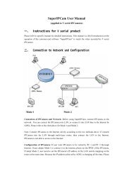
2 Menlove, Clement, and CrandallBy 1972, the size <strong>of</strong> the Atlas had become unwieldy, so Dr. Dayh<strong>of</strong>f,a pioneer <strong>of</strong> bioin<strong>for</strong>matics, developed a database infrastructureinto which this in<strong>for</strong>mation could be funneled. Though nucleotidein<strong>for</strong>mation was included in the Atlas as early as 1966 (2), itsbulkwas comprised <strong>of</strong> amino acid sequences with structural annotation.1.2. InternationalNucleotide <strong>Sequence</strong>Database Collaboration:DDBJ, EMBL, andGenBank1.3. Other Nucleotide<strong>Sequence</strong> DatabasesIt was not until 1982 that databases were developed with the expresspurpose <strong>of</strong> storing nucleotide sequences by the European MolecularBiology Laboratory (EMBL: http://www.embl.org/) in Europeand the National Institutes <strong>of</strong> Health (NIH – NCBI: http://www.ncbi.nlm.nih.gov/) inNorthAmerica.Japanfollowedsuitwith the creation <strong>of</strong> their <strong>DNA</strong> Databank (DDBJ: http://www.ddbj.nig.ac.jp/) in 1986. A sizeable amount <strong>of</strong> sharing naturallyoccurred between these three databases and the Genome<strong>Sequence</strong> Database, also in North America, a condition that led totheir coalition in 1988 under the title International Nucleotide<strong>Sequence</strong> Database Collaboration (INSDC). They still remain verydistinct entities, but in the 1988 meeting, they established policiesto govern the <strong>for</strong>matting <strong>of</strong> and stewardship over the sequenceseach receives. Their current policies include unrestricted access anduse <strong>of</strong> all data records, proper citation <strong>of</strong> data originators, and theresponsibilities <strong>of</strong> submitters to verify the validity <strong>of</strong> the data andtheir right to submit it. The INSDC currently contains approximately80 billion base pairs (bp) (not including whole-genomeshotgun sequences) and nearly 80 million sequence entries. Includingshotgun sequences (HTGS), it passed the 100-gigabase mark onAugust 22, 2005, and contains approximately 200 billion bp as <strong>of</strong>September 2007. For more than 10 years, the amount <strong>of</strong> data inthese databases doubled approximately every 18 months. Thisexpansion has begun to level <strong>of</strong>f as our capacity <strong>for</strong> high-throughputsequencing is gradually reaching a maximum. The next redoubling<strong>of</strong> the data is expected to occur in approximately 4 years (Fig. 1.1).Since the first nucleotide databases were initiated by EMBL andNIH (now held by NCBI), many <strong>DNA</strong> databases have been <strong>for</strong>medto cater to the needs <strong>of</strong> specialized research groups. The 2007Database issue <strong>of</strong> Nucleic Acid Research contained 109 nucleotidesequence databases that met the standards required to be includedin its listing (3). These databases are typically developed to includeancillary data associated with the genetic data, such as patient orspecimen in<strong>for</strong>mation, including clinical in<strong>for</strong>mation, images,downstream analyses. Many do not meet the standards <strong>of</strong> ‘‘quality,quantity and originality <strong>of</strong> data as well as the quality <strong>of</strong> the webinterface’’ that are required to be considered <strong>for</strong> the issue (4). Evenmore are privately held to permit access <strong>of</strong> costly data to a select few.All in all, the number <strong>of</strong> <strong>DNA</strong> databases is astounding and steadilyincreasing as we find new, powerful ways to gather, store, and utilizethe pieces that comprise the puzzle <strong>of</strong> life.
Similarity Searching Using BLAST 3Fig. 1.1. Growth <strong>of</strong> GenBank and DDBJ genetic databases over the past 10 years. TheINSDC databases have grown, over the past 10 years, approximately 168-fold in totalnumber <strong>of</strong> base pairs. While in the past the number <strong>of</strong> entries in INSDC databasesdoubled approximately every 2 years, a simple second-order polynomial regression(R 2 ¼0.9995) <strong>of</strong> the data over the past 10 years indicates that the next redoubling willtake a little over 4 years. This graph does not include HTG data.2. Program Usage2.1. Database FileFormatsOne <strong>of</strong> the largest sources <strong>of</strong> diversity among <strong>DNA</strong> databases liesin their file <strong>for</strong>mats. While great ef<strong>for</strong>ts have been made to standardizefile <strong>for</strong>mats, the various types and purposes <strong>of</strong> sequencein<strong>for</strong>mation and annotation entreat customized file types.2.1.1. FASTA Format First used with Pearson and Lipman’s FASTA program <strong>for</strong>sequence comparison (5), the FASTA file <strong>for</strong>mat is the simplest<strong>of</strong> the widely used <strong>for</strong>mats available through the INSDC. It iscomposed <strong>of</strong> a definition or description line followed by thesequence. The definition line begins with a greater-than symbol(>) and marks the beginning <strong>of</strong> each new entry. The in<strong>for</strong>mationfollowing the greater-than symbol varies according to its source.Generally, an identifier follows (Table 1.1), after which optionaldescription words may be included. If the sequence is retrievedthrough NCBI’s databases, a GI number precedes the identifier.Though it is recommended that the definition line be no greaterthan 80 characters, various types and levels <strong>of</strong> in<strong>for</strong>mation are<strong>of</strong>ten included. The definition line is followed by the <strong>DNA</strong>sequence itself, in single or multi-line <strong>for</strong>mat. Nucleotides arerepresented by their standard IUB/IUPAC codes, includingambiguity codes (Table 1.2).
4 Menlove, Clement, and CrandallTable 1.1FASTA File sequence identifiers. In<strong>for</strong>mation from the NCBIHandbook (25)Database nameGenBankEMBLDDBJNCBI RefSeqPDBPatentsNBRF PIRSWISS-PROTProtein Research FoundationGenInfo Backbone IdGeneral database identifierLocal <strong>Sequence</strong> identifierIdentifier syntaxgb|accession.versionemb|accession.versiondbj|accession.versionref|accession.versionpdb|entry|chainpat|country|numberpir||entrysp|accession|entryprf|namebbs|numbergnl|database|identifierlcl|identifierTable 1.2IUB/IUPAC nucleotide and ambiguity codesA adenosine M A or C (amino) V A, C, or GC cytidine K G or T (keto) H A, C, or TG guanine R A or G (purine) D A, G, or TT thymidine Y C or T (pyrimidine) B C, G, or TU uridine S A or T (strong) – Gap <strong>of</strong> indeterminate lengthW C or G (weak) N A, C, G, or T (any or unknown)2.1.2. Flat File Format GenBank, EMBL, and DDBJ each have their own flat file <strong>for</strong>mat,but contain basically the same in<strong>for</strong>mation. They are all basedupon the Feature Table, which can be found at http://www.ncbi.nlm.nih.gov/collab/FT. For references to these filetypes, see (6–9).2.1.3. Accession Numbers,Version Numbers, LocusNames, DatabaseIdentifiers, etc.The standard <strong>for</strong> identifying a nucleotide sequence record is by anaccession.version system where the accession number is an identifier<strong>of</strong> two letters followed by six digits and the version is an incrementalnumber indicating the number <strong>of</strong> changes that have been
Similarity Searching Using BLAST 5made to the sequence since it was first submitted. Locus names(see Note 1) are older, less standardized identifiers whose originalpurpose was to group entries with similar sequences (10). Theoriginal locus <strong>for</strong>mat was intended to hold in<strong>for</strong>mation about theorganism and other common group characteristics (such as geneproduct). That ten-character <strong>for</strong>mat is no longer able to hold suchin<strong>for</strong>mation <strong>for</strong> the large number and variety <strong>of</strong> sequences nowavailable, so the locus has become yet another unique identifier<strong>of</strong>ten set to be the same value as the accession number. Databaseidentifiers are simply two- or three-character strings that serve toindicate which database originally received and stored the in<strong>for</strong>mation.The database identifier is the first value listed in theFASTA identifier syntax (Table 1.1).When a sequence is first submitted to GenBank, it is submittedwith several defined features associated with the sequence. Someinclude CDS (coding sequence), RBS (ribosome binding site),rep_origin (origin <strong>of</strong> replication), and tRNA (mature transferRNA) in<strong>for</strong>mation. A translation <strong>of</strong> protein coding nucleotidesequences into amino acids is provided as part <strong>of</strong> the featuressection. Likewise, labeling <strong>of</strong> different open reading frames,introns, etc., are all part <strong>of</strong> the table <strong>of</strong> features. A list <strong>of</strong> featuresand their descriptions, <strong>for</strong>mats, and conventions that were agreedupon by INSDC can be found in the Feature Table (seeSection 2.1.2).2.2. Smith–Watermanand DynamicProgrammingIn 1970, Needleman and Wunsch adapted the idea <strong>of</strong> dynamicprogramming to the difficult problem <strong>of</strong> global sequence alignment(11). In 1981, Smith and Waterman adapted this algorithmto local alignments (12). A global alignment attempts to align twosequences throughout their entire length, whereas a local alignmentaligns regions <strong>of</strong> two sequences where high similarity isobserved. Both methods involve initializing, scoring, and tracinga matrix where the rows and columns correspond to the bases orresidues <strong>of</strong> the two sequences being aligned (Fig. 1.2). In the localalignment case, the first row and the first column are filled withzeroes. The remaining cells are filled with a metric value recursivelyderived from neighboring values:80>< left neighbor þ gap penaltymaxtop neighbor þ gap penatly>:top-left neighbor þ match/mismatch scoreIf the current cell corresponds to a match (identical bases), thematch score is added to the value from the diagonal neighbor,otherwise the mismatch score is used. The gap penalty and mismatchscores are generally zero or a small, negative number whilethe match score is a positive number, larger in magnitude. This
6 Menlove, Clement, and CrandallFig. 1.2. Smith-Waterman local alignment example. (A) shows an empty matrix, initialized <strong>for</strong> a Smith-Watermanalignment. (B) and (C) are alignments calculated using the specified scoring parameters. The alignment produced in(B) is drastically different from that in (C), though they only differ slightly in their scoring parameters, one using a matchscore <strong>of</strong> 1 and the other 2.method is used recursively, starting from the upper left corner <strong>of</strong> thematrix and proceeding to the lower right corner. Figure 1.2b and cshows matrices from two different sets <strong>of</strong> gap and match scores.To find a local alignment, one simply finds the largest numberin the matrix and traces a path back until a zero is reached, eachstep moving to a cell that was responsible <strong>for</strong> the current cell’svalue. While this method is robust and is guaranteed to give thebest alignment(s) <strong>for</strong> a given set <strong>of</strong> scores and penalties, it isimportant to note that <strong>of</strong>ten multiple paths and there<strong>for</strong>e multiplealignments are possible <strong>for</strong> any given matrix when these parametersare used. As an example, b and c <strong>of</strong> Fig. 1.2 only differslightly in their gap and match scores, but produce very differentalignments. In addition, the set <strong>of</strong> scores and penalties useddramatically affect the alignment, and finding the optimal set isneither trivial nor deterministic. Weight matrices <strong>for</strong> protein-codingsequences were developed in the late 1970s in an attempt to overcomethese challenges.2.3. Weighting/Models2.3.1. PAM MatricesTo increase the specificity <strong>of</strong> alignment algorithms and provide ameans to evaluate their statistical significance, it was necessary toimplement a meaningful scoring scheme <strong>for</strong> nucleotide and aminoacid substitutions. This was especially true when dealing with protein(or protein-coding) sequences. In 1978, Dayh<strong>of</strong>f et al. developedthe first scoring or weighting matrices created from substitutionsthat have been observed during evolutionary history (13). Thesesubstitutions, since they have been allowed or accepted by natural
Similarity Searching Using BLAST 7Fig. 1.3. PAM250 and BLOSUM45 substitution matrices.selection, are called accepted point mutations (PAM). ForDayh<strong>of</strong>f’sPAMmatrices,groups<strong>of</strong>proteinswith85%ormoresequence similarity were analyzed and their 1,571 substitutionswere cataloged. Each cell <strong>of</strong> a PAM matrix corresponds to thefrequency in substitutions per 100 residues between two givenamino acids. This frequency is referred to as one PAM unit. Backin the 1970s, when they were created, however, there was alimited number and variety <strong>of</strong> protein sequences available, sotheyarebiasedtowardsmall,globular proteins. It is also importantto note that each PAM matrix corresponds to a specificevolutionary distance and that each is simply an extrapolation<strong>of</strong> the original. For example, a PAM250 (Fig. 1.3) matrixisconstructed by multiplying the PAM1 matrix by itself 250times and is viewed as a typical scoring matrix <strong>for</strong> proteins thathave been separated by 250 million years <strong>of</strong> evolution.2.3.2. BLOSUM Matrices To overcome some <strong>of</strong> the drawbacks <strong>of</strong> PAM matrices, Henik<strong>of</strong>fand Henik<strong>of</strong>f developed the BLOSUM matrices in 1992 (14).These matrices were based on the BLOCKS database, which organizesproteins into blocks, where each block, defined by an alignment<strong>of</strong> motifs, corresponds to a family. Whereas the original PAMmatrix was calculated with proteins with at least 85% identity,BLOSUM matrices are each calculated separately using conservedmotifs at or below a specific evolutionary distance. This diversity <strong>of</strong>matrices coupled with being based on larger datasets makes theBLOSUM matrices more robust at detecting similarity at greaterevolutionary distances and more accurate, in many cases, at per<strong>for</strong>minglocal similarity searches (15).2.3.3. Choosing a Matrix When choosing a matrix, it is important to consider the alternatives.Do not simply choose the default setting without some initialconsideration. In general, finding similarity at increasing divergencecorresponds to increasing PAM matrices (PAM1, PAM40,PAM120, etc.) and decreasing BLOSUM matrices (BLOSUM90,
8 Menlove, Clement, and CrandallTable 1.3Suggested uses <strong>for</strong> common substitution matrices. The matrices highlighted in boldare available through NCBI’s BLAST web interface. BLOSUM62 has been shown toprovide the best results in BLAST searches overall due to its ability to detect largeranges <strong>of</strong> similarity. Nevertheless, the other matrices have their strengths. Forexample, if your goal is to only detect sequences <strong>of</strong> high similarity to infer homologywithin a species, the PAM30, BLOSUM90, and PAM70 matrices would provide thebest results. This table was adapted from results obtained by David Wheeler (16)Alignment size Best at detecting Similarity (%) PAM BLOSUMShort Similarity within a species 75–90 PAM30 BLOSUM95" Similarity within a genus 60–75 PAM70 BLOSUM85Medium Similarity within a family 50–60 PAM120 BLOSUM80" The largest range <strong>of</strong> similarity 40–50 PAM160 BLOSUM62Long Similarity within a class 30–40 PAM250 BLOSUM45" Similarity within the twilight zone 20–30 BLOSUM30BLOSUM80, BLOSUM62, etc.) (16). PAM matrices are strongat detecting high similarity due to their use <strong>of</strong> evolutionary in<strong>for</strong>mation.However, as evolutionary distance increases, BLOSUMmatrices are more sensitive and accurate than their PAM counterparts.Table 1.3 includes a list <strong>of</strong> suggested uses.2.4. BLAST Programs Nucleotide–nucleotide searches are beneficial because no in<strong>for</strong>mationis lost in the alignment. When a codon is translated fromnucleotides to amino acid, approximately 69% <strong>of</strong> the complexityis lost (64 possible nucleotide combinations mapped to 20 aminoacids). In contrast, however, the true physical relationshipbetweentwocodingsequencesisbestcapturedinthetranslatedview. Matrices that take into account physical properties, such asPAM and BLOSUM, can be used to add power to the search.Additionally, in a nucleotide search, there are only four possiblecharacterstatescomparedto20inanaminoacidsearch.Thustheprobability <strong>of</strong> a match due to chance versus a match due tocommon ancestry (identify in state versus identical by descent)is high.The Basic Local Alignment and Search Tools (BLAST) are themost widely used and among the most accurate in detectingsequence similarity (17)(see Note 2). The standard BLAST programsare Nucleotide BLAST (blastn), Protein BLAST (blastp),blastx, tblastn, and tblastx. Others have also been developed tomeet specific needs. When choosing a BLAST program, it is
Similarity Searching Using BLAST 9important to choose the correct one <strong>for</strong> your question <strong>of</strong> interest.Some <strong>of</strong> the most common mistakes in similarity searching comefrom misunderstandings <strong>of</strong> these different applications.l Nucleotide blast: compares a nucleotide query against anucleotide sequence databasel Protein blast: compares a protein query against a proteinsequence databasel blastx: compares a nucleotide query translated in all six readingframes against a protein databasel tblastn: compares a protein query against a nucleotide sequencedatabase dynamically translated in all six reading framesl tblastx: compares a nucleotide query in all six reading framesagainst a nucleotide sequence database in all six reading framesThe BLAST algorithm is an heuristic program, one that is notguaranteed to return the best result. It is, however, quite accurate.BLAST works by first making a look-up table <strong>of</strong> all the ‘‘words’’and ‘‘neighboring words’’ <strong>of</strong> the query sequence. Words are shortsubsequences <strong>of</strong> length W and neighboring words are words thatare highly accepted in the scoring matrix sense, determined by athreshold T. The database is then scanned <strong>for</strong> the words andneighboring words. Once a match is found, extensions with andwithout gaps are initiated there both upstream and downstream.The extension continues, adding gap existence (initiation) andextension penalties, and match and mismatch scores as appropriateas in the Smith-Waterman algorithm until a score threshold S isreached. Reaching this mark flags the sequence <strong>for</strong> output. Theextension then continues until the score drops by a value X fromthe maximum, at which point the extension stops and the alignmentis trimmed back to the point where the maximum score washit. Understanding this algorithm is important <strong>for</strong> users if they areto select optimal parameters <strong>for</strong> BLAST. The interaction betweenthe parameters T, W, S, X, and the scoring matrix allows the user t<strong>of</strong>ind a balance between sensitivity and specificity, alter the runningtime, and tweak the accuracy <strong>of</strong> the algorithm. The interactionsamong these variables will be discussed in Section 2.8.2.5. Query <strong>Sequence</strong> Query sequences may be entered by uploading a file or enteringone manually in the text box provided (Fig. 1.4). The uploadoption accepts files containing a single sequence, multiplesequences in FASTA <strong>for</strong>mat, or a list <strong>of</strong> valid sequence identifiers(accession numbers, GI numbers, etc.). In contrast to previousversions <strong>of</strong> BLAST on the NCBI website, the current versionallows the user to specify a descriptive job title. This allows theuser to track any adjustments or versions <strong>of</strong> a search as well as itspurpose and query in<strong>for</strong>mation. This is especially important whensequence identifiers are not included in the uploaded file.
10 Menlove, Clement, and CrandallFig. 1.4. NCBI nucleotide BLAST interface.2.6. Search Set2.6.1. DatabasesWhen choosing a database, it is important to understand theirpurpose, content, and limitations. The list <strong>of</strong> nucleotide databasesis divided into Genomic plus Transcript and Other Databases sections.Some <strong>of</strong> the databases, composed <strong>of</strong> reference sequences,come from the RefSeq database, a highly curated, all-inclusive,non-redundant set <strong>of</strong> INSDC (EMBL + GenBank + DDBJ) <strong>DNA</strong>,mRNA, and protein entries. RefSeq sequences have accessionnumbers <strong>of</strong> the <strong>for</strong>m AA_######, where AA is one <strong>of</strong> the followingcombination <strong>of</strong> letters (Table 1.4) and ###### is a uniquenumber representing the sequence.A description <strong>of</strong> the nucleotide databases is included below.A list <strong>of</strong> protein databases accessible through BLAST’s web interfacecan be found at http://www.ncbi.nlm.nih.gov/BLAST/blastcgihelp.shtml.l Human genomic plus transcript: contains all human genomicand RNA sequences.l Mouse genomic plus transcript: contains all mouse genomicand RNA sequences.
Similarity Searching Using BLAST 11Table 1.4RefSeq categoriesExperimentally determinedand curatedGenome annotation (computationalpredictions from <strong>DNA</strong>)NCNGComplete genomic moleculesIncomplete genomic regionNM mRNA XM Model mRNANRRNA (non-coding)NP Protein XP Model proteinlllllllllNucleotide collection (nr/nt): contains INSDC + RefSeqnucleotides + PDB sequences, not including EST, STS, GSS,or unfinished HGT sequences. The nucleotide collection is themost comprehensive set <strong>of</strong> nucleotide sequences availablethrough BLAST.Reference mRNA sequences (refseq_rna): contains the nonredundantRefSeq mRNA sequences.Reference genomic sequences (refseq_genomic): containsthe non-redundant RefSeq genomic sequences.Expressed sequence tags (est): contains short, single readsfrom mRNA sequencing (via c<strong>DNA</strong>). These c<strong>DNA</strong> sequencesrepresent the mRNA in a cell at a particular moment in aparticular tissue.Non-human, non-mouse ESTs (est_others): the previousdatabase with human and mouse sequences removed.Genomic survey sequences (gss): contains random genomicsequences obtained from single-pass genome surveys, cosmids,BACs, YACs, and other survey methods. Their quality varies.High-throughput genomic sequences (HTGS): containssequences obtained from high-throughput genome centers.<strong>Sequence</strong>s in this database contain a phase number, 0 beingthe initial phase and 3 being the finished phase. Once finished,the sequences move to the appropriate division in their respectivedatabase.Patent sequences (pat): contains sequences from the patent<strong>of</strong>fices at each <strong>of</strong> the INSDC organizations.Protein data bank (pdb): the nucleotide sequences from theBrookhaven Protein Data Bank managed by the Research Collaboratory<strong>for</strong> Structural <strong>Bioin<strong>for</strong>matics</strong> (http://www.rcsb.org/pdb).
12 Menlove, Clement, and CrandalllHuman ALU repeat elements (alu_repeats): contains a set <strong>of</strong>ALU repeat elements that can be used to mask repeat elementsfrom query sequences. ALU sequences are regions subject tocleavage by Alu restriction endonucleases, around 300 bp long,and estimated to constitute about 10% <strong>of</strong> the human genome(18).l <strong>Sequence</strong> tagged sites (dbsts): a collection <strong>of</strong> uniquesequences used in PCR and genome mapping that identify aparticular region <strong>of</strong> a genome.l Whole-genome shotgun reads (wgs): contains large-scaleshotgun sequences, mostly unassembled and non-annotated.l Environmental samples (env_nt): contains sets <strong>of</strong> whole-genomeshotgun reads from many sampled organisms, each setfrom a particular location <strong>of</strong> interest. These sets allow researchersto look into the genetic diversity existing at a particularlocation and environment.2.6.2. Organism The organism box allows the user to specify a particular organismto search. It automatically suggests organisms when you begintyping. This option is not available when Genomic plus Transcriptdatabases are selected (Fig. 1.5).2.6.3. Entrez Queries Entrez queries provide a way to limit your search to a specific type<strong>of</strong> organism or molecule. It is an efficient way to filter unwantedresults by excluding organisms or defining sequence lengthcriteria. In addition, Entrez queries allow the user to findsequences submitted by a particular author, from a particularjournal, with a particular property or feature key, or submitted ormodified within a specific date range. For help with Entrez queries,see the Entrez Help document at http://www.ncbi.nlm.nih.gov/entrez/query/static/help/helpdoc.html.2.7. BLAST SearchParametersIn addition to entering a query sequence, choosing a searchset, and selecting a program, several additional parameters areavailable, which allow you to fine-tune your search to yourneeds. These parameters are available by clicking the ‘‘Algorithmparameters’’ link at the bottom <strong>of</strong> the BLAST page (Fig. 1.6)(see Notes 3 and 4).Fig. 1.5. NCBI nucleotide BLAST algorithm parameters.
Similarity Searching Using BLAST 13Fig. 1.6. Organism selection when searching a multi-organism database.2.7.1. Max Target<strong>Sequence</strong>sThe maximum target sequences parameter allows you to select thenumber <strong>of</strong> sequences you would like displayed in your results.Lower numbers do not reduce the search time, but do reducethe time to send the results back. This is generally only an issueover a slow connection.2.7.2. Short Queries When using short queries (<strong>of</strong> length 30 or less), the parametersmust be adjusted or you will not receive statistically significantresults. Checking the ‘‘short queries’’ box automatically adjusts theparameters to return valid responses <strong>for</strong> a short query sequence.2.7.3. Expect Threshold The expect threshold limits the results displayed to those with anE-value lower than it. This value corresponds to the number <strong>of</strong>sequence matches that are expected to be found merely by chance.2.7.4. Word Size The word size, W, as discussed earlier determines the length <strong>of</strong> thewords and neighboring words used as initial search queries.Increasing the word size generally results in fewer extension initializations,increasing the speed <strong>of</strong> the BLAST search but decreasingits sensitivity.2.7.5. Scoring Parameters The scoring parameters <strong>of</strong> a nucleotide search are the matchand mismatch scores and gap costs. In protein searches, thematch and mismatch scores are indicated by a scoring matrix(see Section 2.3). A limited set <strong>of</strong> suggested match and mismatchscores are available from the dropdown menu on NCBI’s BLASTsearch <strong>for</strong>m. Increasing the ratio in the following fashion (match,mismatch): (1,–1) ! (4,–5) ! (2,–3) ! (1,–2) ! (1,–3) ! (1,–4)prevents mismatched nucleotides from aligning, increasing the
Similarity Searching Using BLAST 15seeding stage, where words and neighboring words are scanned,but unmask them during the extension phases. This prevents theE-values from being affected in biologically interesting resultswhile preventing regions <strong>of</strong> low complexity from slowing thesearch down and introducing uninteresting results.The ‘‘Mask lower case letters’’ option gives the user the optionto annotate his or her sequence by using lower case letters wheremasking is desired.2.8. Interpreting theResults2.9. Future <strong>of</strong> SimilaritySearchingBy default, BLAST results contain five basic sections: a summary <strong>of</strong>your input (query and parameters), a graphical overview <strong>of</strong> the topresults, a table <strong>of</strong> sequences producing significant alignments, thebest 100 alignments, and result statistics. The number <strong>of</strong> hitsshown in the graphical overview as well as the number <strong>of</strong> alignments,among other options, may be changed by clicking ‘‘Re<strong>for</strong>matthese results’’ at the top <strong>of</strong> the results page or by clicking‘‘Formatting options’’ on the Formatting Results page (the pagethat appears after you click BLAST and be<strong>for</strong>e the results appear).In the third section, the results table contains eight columns:accession, description, max score, total score, query coverage,E-value, max ident, and links. The Accession number provides a linkto detailed in<strong>for</strong>mation about the sequence. The description providesin<strong>for</strong>mation about the species and the kind <strong>of</strong> sample the hit wasgenerated from. The max score provides a metric <strong>for</strong> how good thebest local alignment is. The total score indicates how similar thesequence is to the query, accounting <strong>for</strong> all local alignments betweenthe two sequences. If the max score is greater than the total score,then more than one local alignment was found between the twosequences. Higher scores are correlated with more similar sequences.Both <strong>of</strong> these scores, reported in bits, are calculated from a <strong>for</strong>mulathat takes into account matches (or similar residues, if doing a proteinsearch) and mismatch penalties along with gap insertion penalties.Bit scores are normalized so that they can be directly compared eventhough the alignments between different sequences may be <strong>of</strong> differentlengths. The expectation value or E-value provides an estimate<strong>of</strong> how likely it is that this alignment occurred by random chance. AnE-value <strong>of</strong> 2e–02 indicates that similarity found in the alignment hasa 2 in 100 chance <strong>of</strong> occurring by chance. The lower the E-value, themore significant the score. An appropriate cut<strong>of</strong>f E-value depends onthe users’ goals. The max identity field shows the percentage <strong>of</strong> thequery sequence that was identical to the database hit. The links fieldprovides links to UniGene, the Gene Expression Omnibus, EntrezGene, Entrez’s Related Structures (<strong>for</strong> protein sequences), and theMap Viewer (<strong>for</strong> genomic sequences).Since both PAM and BLOSUM matrices are experimentallyderived from a limited set <strong>of</strong> sequences in a database that wasavailable at the time they were created, they will almost certainlynot provide optimal values <strong>for</strong> searches with new sequence
16 Menlove, Clement, and Crandallfamilies. Current research is being per<strong>for</strong>med to determine whichchemical properties are changing in a sequence in order to providea magnitude <strong>of</strong> change that is independent <strong>of</strong> scoring matrices.Current techniques to find promoter regions are severely lackingin accuracy (19). Techniques will arise in the future that mayimprove current methods by using BLAST-like algorithms toassess the similarity <strong>of</strong> a sequence to known promoter elements,thus helping to identify it as a promoter.3. ExamplesThis section will provide three examples <strong>of</strong> common BLAST uses:a nucleotide–nucleotide BLAST, a position-specific iteratedBLAST, and a blastx.3.1. Nucleotide–Nucleotide BLAST <strong>for</strong>Allele FindingHere we present an example <strong>of</strong> using BLAST to search <strong>for</strong> theknown alleles <strong>of</strong> a given nucleotide sequence. This approach can beused to answer the question: what are the known variants <strong>of</strong> mygene <strong>of</strong> interest (within its species)? Our example will be to find allknown variants <strong>of</strong> a Tp53 nucleotide sequence (accession numberAF151353) from a mouse. While this sequence does code <strong>for</strong> aprotein, non-coding sequences would work just as well using thisapproach.We will start by going to the BLAST homepage at http://www.ncbi.nlm.nih.gov/BLAST/ and selecting nucleotide blast.Inthe ‘‘Enter Query <strong>Sequence</strong>’’ box, we type the accession number:AF151353. You will notice that the ‘‘Job Title’’ box automaticallyfills in a title <strong>for</strong> you ‘‘AF151353:Mus musculus tumor suppressorp53...’’. If we were to paste a sequence instead <strong>of</strong> an accessionnumber or GI, we would want to enter a job title to help us keeptrack <strong>of</strong> our results. Under ‘‘Choose Search Set,’’ we select the‘‘Nucleotide collection (nr/nt)’’ database, since it is the mostcomprehensive database (remember that nr is no longer nonredundant).For a complete search, we should also per<strong>for</strong>m asearch on the ‘‘Expressed sequence tags (est)’’ database. In theOrganism box, we choose type ‘‘mouse’’ and select ‘‘mouse(taxid:10090),’’ which corresponds to Mus musculus, the housemouse. Since we are searching <strong>for</strong> alleles, we select ‘‘Highly similarsequences (megablast)’’ in the ‘‘Program Selection’’ box.Next, let us change the algorithm parameters. Click ‘‘Algorithmparameters’’ to display them. Since the sequence is 1,409 bpin length, we deselect the ‘‘Automatically adjust parameters <strong>for</strong>short input sequences’’ box. Since we expect that the p53 protein isa well-conserved protein (due to its critical function), we set theexpect threshold to a low value. Let us choose 1e-8. For a word
Similarity Searching Using BLAST 17size, we are not concerned about speed in this case, so the number<strong>of</strong> extensions per<strong>for</strong>med is not a concern. Let us select a word size<strong>of</strong> 20 to make sure we do not miss any matches (although in thiscase a larger word size should not make much difference). As <strong>for</strong>the scoring parameters, we choose the largest ratio, correspondingto the greatest identity: ‘‘1,–4.’’ Since this is a protein-codingsequence, we do not expect repeats to be a factor, so we leave theFilters and Masking section at the default settings.The results indicate that 108 hits were found on the querysequence. Looking at the graphical alignment (Fig. 1.7), wenotice that only about 2/3 <strong>of</strong> them span a good portion <strong>of</strong> thequery. When we scroll down to the gene descriptions, most <strong>of</strong> thelast fourth are pseudogenes (partial sequence) (Fig. 1.8), whichmay <strong>of</strong>fer insight into different alleles and their correspondingphenotypes, but which were not sequenced experimentally. Per<strong>for</strong>minga search on the EST database with the same parametersresults in 101 additional hits.3.2. PSI-BLAST <strong>for</strong>Distant HomologySearchingWhen searching <strong>for</strong> distantly related sequences, two BLASToptions are available. One is the standard nucleotide–nucleotideBLAST with discontiguous BLAST, a method very similar to MaFig. 1.7. Graphical distribution <strong>of</strong> top 100 BLAST hits.
18 Menlove, Clement, and CrandallFig. 1.8. Last 16 sequences producing significant alignments from a mouse p53 gene Nucleotide BLAST search. Nineteen<strong>of</strong> the last 26 reported sequences are pseudogenes.et al.’s work (20), selected as the program. The other is to use amore sensitive approach, PSI-BLAST, which per<strong>for</strong>ms an iterativesearch on a protein sequence query. Though the second approachwill only work if you are dealing with protein-coding sequences, it ismore sensitive and accurate than the first.In this example, we will search <strong>for</strong> relatives <strong>of</strong> the cytochrome bgene <strong>of</strong> the Durango night lizard (Xantusia extorris). We start byselecting protein blast from the BLAST home page and enteringthe accession number, ABY48155, into the query box. If yoursequence is not available as a protein sequence, you will need totranslate it. This can easily be done using a program such as MEGA(21), available at http://www.megas<strong>of</strong>tware.net, or an online toolsuch as the JustBio Translator (http://www.justbio.com/translator/)or the ExPASy Translate Tool (http://www.expasy.org/tools/dna.html).Once again, the ‘‘Job Title’’ box is filled with ‘‘ABY48155:cytochrome b [Xantusia extorris].’’ We will choose the ‘‘Referenceproteins (refseq_protein)’’ database, which is more highly curatedand non-redundant (per gene) than the default nr database. We donot specify an organism because we want results from any and allrelated organisms. For the algorithm, we select PSI-BLAST due toits ability to detect more distantly related sequences. We hope toinclude as many sequences as possible in our iterations, so wechoose 1,000 as the max target sequences. We can, once again,remove the ‘‘Automatically adjust parameters <strong>for</strong> short inputsequences’’ check, since our sequence is sufficiently long (380amino acids). Since we wish to detect all related sequences, wekeep the expect threshold at its default <strong>of</strong> 10. While decreasing itmay remove false-positives, it may also prevent some significantresults from being returned. Since we do not have a particularscope in mind (within the genus or family, <strong>for</strong> example), we willuse the BLOSUM62 matrix due to its ability to detect homologyover large ranges <strong>of</strong> similarity.
Similarity Searching Using BLAST 19The first iteration results in 1,000 hits on the query sequence, all<strong>of</strong> which cover at least 93% <strong>of</strong> the query sequence and have anE-value <strong>of</strong> 10 –126 or less. We leave all <strong>of</strong> the sequences selected andpress the ‘‘Run PSI-Blast iteration 2’’ button. The second iterationlikewise returns 1,000 hits, but this time they have E-values less than10 –99 and cover at least 65% <strong>of</strong> the query sequence (all but six cover90% or more). We uncheck the last hit, Bi4p [Saccharomyces cerevisiae],since we are unsure <strong>of</strong> its homology, and iterate one last time.At this point, it would be helpful to view the taxonomy report<strong>of</strong> the results. You can do so by clicking ‘‘Taxonomy Reports’’ nearthe bottom <strong>of</strong> the first section <strong>of</strong> the BLAST report. You willnotice that we have a good selection <strong>of</strong> organisms, ranging frombony fishes to Proteobacteria. While this list would need to benarrowed to produce a good taxonomy, it would be a good startingpoint if you wish to per<strong>for</strong>m a broad phylogenetic reconstruction.To per<strong>for</strong>m a search <strong>of</strong> more closely related sequences, youwould likely per<strong>for</strong>m a standard blastp (protein–protein BLAST)instead <strong>of</strong> a PSI-BLAST and use the PAM 70 or PAM 30 matrix.3.3. Blastx <strong>for</strong> ESTIdentificationWhat if you have a nucleotide sequence such as an expressed sequencetag and wish to know if it codes <strong>for</strong> a known protein? You can searchthe nucleotide database or take the more direct approach <strong>of</strong> blastx.Blastx allows you to search the protein database using a nucleotidequery, which it first translates into all six reading frames. In thisexample, we will per<strong>for</strong>m a blastx on the following sequence:TCTCTATAGTTATGGTGTTCTGAATCAGCCTTCCCTCATASince the sequence is only 40 bp long, we need to be carefulwith our parameters. We start by selecting blastx from the BLASThomepage. We then enter the sequence into the query box andenter a relevant job title, such as ‘‘EST blastx Search 1.’’ We willsearch the ‘‘Non-redundant protein sequences (nr)’’ database,since it has the largest number <strong>of</strong> annotated nucleotide sequences.Under ‘‘Algorithm parameters,’’ we need to choose an appropriateexpect threshold and matrix. If we choose too low an expectthreshold, we might not find anything. Likewise, if we choosethe wrong matrix, we may not obtain significant results due tothe short length <strong>of</strong> our sequence. We will choose 10 (the default)as our expect threshold and PAM70 as our matrix, since it correspondsto finding similarity at or below the family/genus level.Since we do not know what our sequence is, we want to filterregions <strong>of</strong> low complexity to ensure that if our sequence containssuch regions, they will not return deceptively significant results.Our search produces a large number (more than 1,000) <strong>of</strong>results with an E-value <strong>of</strong> 0.079 (Fig. 1.9). If we were to use thePAM70 matrix, essentially the same results would be obtained, buteach with an E-value <strong>of</strong> 3.0. Since all <strong>of</strong> the 2,117 results aredifferent entries <strong>of</strong> the nucleocapsid protein <strong>of</strong> the Influenza A
20 Menlove, Clement, and CrandallFig. 1.9. Blastx results showing E-values <strong>of</strong> 0.079 <strong>for</strong> the top ten hits, all <strong>of</strong> which are nucleocapsid proteins ornucleoproteins.virus, we can be somewhat confident that our protein is related,especially if we had any prior knowledge that would support ourfindings.4. Notes1. One <strong>of</strong> the options NCBI provides from their homepage is tosearch across their databases using an identifier (accessionnumber, sequence identification number, Locus ID, etc.).This option can be rather straight<strong>for</strong>ward if you are using anidentifier unique to a particular sequence; however, if you aresearching <strong>for</strong> a locus across organisms or individuals, you mayneed to pay close attention to the search terms you are using.For example, since the Cytochrome b/b6 subunit is known bythe terms ‘‘Cytochrome b,’’ ‘‘Cytochrome b6,’’ ‘‘cyt-b,’’‘‘cytb,’’ ‘‘cyb,’’ ‘‘COB,’’ ‘‘COB1,’’ ‘‘cyb6,’’ ‘‘petB,’’ ‘‘mtcyb,’’and ‘‘mt-cyb’’ in a search <strong>for</strong> all possible homologs <strong>of</strong> thissubunit, it is necessary to search <strong>for</strong> all <strong>of</strong> its names andabbreviations used in the organisms <strong>of</strong> interest. Since researchgroups studying different organisms create their own uniquelocus names <strong>for</strong> the same gene, it is important to use all <strong>of</strong>them in your search. IHOP (www.ihop-net.org) is an excellentresource <strong>for</strong> protein names (22). In addition, you will want toper<strong>for</strong>m a BLAST search to make sure you have everything!
Similarity Searching Using BLAST 21Fig. 1.10. Save search strategies.2. In addition to the BLAST program provided by NCBI, otherBLAST programs exist, which have improved the BLASTalgorithm in various ways. Dr. Warren Gish at WashingtonUniversity in St. Louis has developed WU-BLAST, the firstBLAST algorithm that allowed gaped alignments with statistics(23). It boasts speed, accuracy, and flexibility, taking oneven the largest jobs. Another program, FSA-BLAST (FasterSearch Algorithm), was developed to implement recently publishedimprovements to the original BLAST algorithm (24).Itpromises to be twice as fast as NCBI’s and just as accurate.WU-BLAST is free <strong>for</strong> academic and non-pr<strong>of</strong>it use and FSA-BLAST is an open source under the BSD license agreement.3. My NCBI is a tool that allows you to customize your preferences,save searches, and set up automatic searches that sendresults via e-mail. If you find yourself per<strong>for</strong>ming the samesearches (or even similar searches) repeatedly, you may wantto take advantage <strong>of</strong> this option! To register, go to the NCBIhome page and click the ‘‘My NCBI’’ link under ‘‘Hot Spots.’’Once you have registered and signed in, a new option will beavailable to you on all BLAST and Entrez searches (Fig. 1.10).4. To save a BLAST search strategy, simply click the ‘‘SaveSearch Strategies’’ link on the results page. This will add thesearch to your ‘‘Saved Strategies’’ page, which is availablethrough a tab on the top <strong>of</strong> each page in the BLAST websitewhen you are logged in to My NCBI. Doing so will not saveyour results, but it will save your query and all parameters youspecified <strong>for</strong> your search so you can run it later to retrieveupdated results.References1. Dayh<strong>of</strong>f, M. O., Eck, R. V., Chang, M. A.,and Sochard, M. R. (1965) Atlas <strong>of</strong> Protein<strong>Sequence</strong> and Structure, National BiomedicalResearch Foundation, Silver Spring, MD.2. Hersh, R. T. (1967) Reviews. Syst Zool 16,262–63.3. Galperin, M. Y. (2007) The molecular biologydatabase collection: 2007 update.Nucleic Acids Res 35, D3–D4.4. Batemen, A. (2007) Editorial. Nucleic AcidsRes 35, D1–2.5. Pearson, W. R., and Lipman, D. J. (1988)Improved tools <strong>for</strong> biological sequencecomparison. Proc Natl Acad Sci USA 85,2444–48.6. León, D., and Markel, S. (2003) <strong>Sequence</strong><strong>Analysis</strong> in a Nutshell, O’Reilly & Associates,Inc., Sebastopol, CA.
Chapter 2Gene Orthology Assessment with OrthologIDMary Egan, Ernest K. Lee, Joanna C. Chiu, Gloria Coruzzi, and Rob DeSalleAbstractOrthologID (http://nypg.bio.nyu.edu/orthologid/) allows <strong>for</strong> the rapid and accurate identification <strong>of</strong>gene orthology within a character-based phylogenetic framework. The Web application has two functions –an orthologous group search and a query orthology classification. The <strong>for</strong>mer determines orthologous genesets <strong>for</strong> complete genomes and identifies diagnostic characters that define each orthologous gene set; and thelatter allows <strong>for</strong> the classification <strong>of</strong> unknown query sequences to orthology groups. The first module <strong>of</strong> theWeb application, the gene family generator, uses an E-value based approach to sort genes into gene families.An alignment constructor then aligns members <strong>of</strong> gene families and the resulting gene family alignments aresubmitted to the tree builder to obtain gene family guide trees. Finally, the diagnostics generator extractsdiagnostic characters from guide trees and these diagnostics are used to determine gene orthology <strong>for</strong> querysequences.Key words: Single linkage cluster, orthology, phylogeny, alignment, diagnosis genomics.Homology is either the cornerstone <strong>of</strong> biology or a term ripe <strong>for</strong> burning.John Maynard Smith1. IntroductionSub-genomic studies (that analyze hundreds to thousands <strong>of</strong> generegions <strong>for</strong> perhaps hundreds <strong>of</strong> terminal taxa) face new computationalchallenges. Responding to these challenges is resulting inthe development <strong>of</strong> new methodologies and tools <strong>for</strong> tree buildingand analysis. Sub-genomic studies, however, follow the sameground plan as most molecular systematic studies, albeit on agrand scale. It may be that the next stimulus to intellectual debatein the field <strong>of</strong> systematics will come as a result <strong>of</strong> responding toDavid Posada (ed.), <strong>Bioin<strong>for</strong>matics</strong> <strong>for</strong> <strong>DNA</strong> <strong>Sequence</strong> <strong>Analysis</strong>, Methods in Molecular Biology 537ª Humana Press, a part <strong>of</strong> Springer ScienceþBusiness Media, LLC 2009DOI 10.1007/978-1-59745-251-9_223
24 Egan et al.challenges faced by true phylogenomic studies in which entiregenomes are used as the basis <strong>for</strong> comparison. The presence <strong>of</strong>gene families and horizontal gene transfer pose challenges <strong>for</strong> bothcharacter coding as well as <strong>for</strong> identifying which are the appropriateterminals in the analysis. These challenges to be faced in phylogenomicstudies hearken back to those faced in morphological studies.Thereislikelytobeashiftinthedirection<strong>of</strong>intellectualdebateinthe phylogenetic analysis equation, the direction <strong>of</strong> this shift beingtoward data matrix assembly and homology assessment. Severaltypes <strong>of</strong> genomic studies are already implicitly or explicitly addressingthe problem <strong>of</strong> homology.Homology is <strong>of</strong>ten considered one <strong>of</strong> Darwin’s most impressivecontributions to evolutionary thinking. Darwin was one <strong>of</strong> the firstto discern the differences between homology and analogy. Whereashomology refers to traits that are the same due to common ancestry,analogy refers to traits that are similar due to evolutionary convergence.Homology then becomes a term <strong>of</strong> absoluteness and must bediscovered via hypothesis testing, and similarity becomes a term <strong>of</strong>measurement and is calculated from observation <strong>of</strong> two entities. Theliterature on homology is rich with debate, hence the quote thatstarts <strong>of</strong>f this chapter. While phylogenomic studies may stimulateintellectual debate and growth in systematic theory, the conversemay also be true – that systematics may provide an aspect <strong>of</strong> theintellectual underpinning necessary <strong>for</strong> the continued development<strong>of</strong> genomic studies.In the 1980s Fitch and several colleagues published an importantclarification <strong>of</strong> terms then being used in the earliest <strong>of</strong> comparisons<strong>of</strong> molecular sequences. Their note suggested that scientists take carein using the word homology. Essentially, Fitch et al. pointed outthat most molecular biologists at the time were misusing and henceoverusing the term homology. For instance, a typical sequence comparisonpaper from that period would claim that two sequences <strong>of</strong>100 amino acids long had 70% homology if 70 out <strong>of</strong> 100 residues inthe two proteins were the same. Fitch and colleagues pointed out thatthis was a misuse <strong>of</strong> the term homology, which indicates commonancestry. When two sequences are compared there is necessarily a lack<strong>of</strong> investigation <strong>of</strong> common ancestry because it takes at least threetarget sequences and an outgroup sequence to discover commonancestry. Those readers who have heard Fitch speak are probablyfamiliar with his famous punch line about homology – ‘‘Homologyis like pregnancy. Someone is either pregnant or not. A personcannot be 70% pregnant.’’ In this context, two sequences can behomologous, but cannot be 70% homologous. Rather two sequencesare 70% similar (<strong>for</strong> further discussion <strong>of</strong> similarity versus commonancestry in orthology assessment, see Note 1).In addition to establishing this important distinction concerninghomology, Fitch and colleagues also established a framework <strong>for</strong>how we should examine genes in multigene families, by proposing
Gene Orthology Assessment with OrthologID 25that the term orthology refer to genes that are identical by descent asa result <strong>of</strong> speciation <strong>of</strong> two entities. The term paralogy then refersto a pair or set <strong>of</strong> genes related to each other but not through aspeciation event. In this case, paralogy refers to members <strong>of</strong> a genefamily that have arisen through duplication and not followed by aspeciation event. While both terms refer to kinds <strong>of</strong> homologousrelationships, orthology is what an anatomist would refer to ashomology and paralogy would be akin to serial homology. A thirdterm coined xenology refers to the similarity <strong>of</strong> entities as a result<strong>of</strong> horizontal transfer. It should be obvious from the terminologythat any departure from orthology <strong>for</strong> members <strong>of</strong> gene familiescomplicates and even negates sound evolutionary or biologicalanalysis. The old adage <strong>of</strong> comparing apples to oranges also appliesto genes in gene families.To automate the rapid assessment <strong>of</strong> orthology <strong>of</strong> genes in genefamilies we have developed a Web based program called OrthologID(1). This program uses the concept <strong>of</strong> common ancestry to establishhomologous relationships <strong>of</strong> genes obtained from whole genomesequencing, EST studies, or other genome analyses. There are othermethods that exist that have been used to establish orthology, suchas simple BLAST, BLAT, and COG approaches. Because BLASTbest hits have been shown not to identify the closest phylogeneticneighbor (2), a problem exists with relying solely on this approach(and indeed others that rely on BLAST or are similar to BLAST).However, BLAST, BLAT, and other techniques such as COGs andother distance measures (see Note 1) can be in<strong>for</strong>mative first stepsin topographical assessment. We consider these approaches to bevalid generators <strong>of</strong> hypotheses when determining orthology and aswe point out below they are incorporated into the algorithm wehave developed. Our description <strong>of</strong> the program will first describethe rationale <strong>for</strong> the approach we have devised, then describe indetail the algorithm and its component parts, and finally someworked examples are presented.An automated approximation <strong>of</strong> the phylogenetic gene treeapproach to orthology determination would need to be developedin order to be able to use this approach on a genomic scale. Forsmall gene families or <strong>for</strong> limited numbers <strong>of</strong> taxa, it is possible touse this approach with currently available analytical tools. Thesewould involve initial similarity searches to identify putative genefamilies (<strong>for</strong> multiple taxa simultaneously), alignment, tree building,and screening trees <strong>for</strong> diagnostic characters to identify orthogousgene family members <strong>of</strong> each taxon, These analyses would berepeated <strong>for</strong> each new sequence to be placed among its orthologousgroup. To use this approach on a genomic scale, new toolshave been developed. The main difference between the manualphylogenetic gene tree approach and the automated approachimplemented in OrthologID is the exclusive use <strong>of</strong> completelysequenced genomes <strong>for</strong> constructing gene family trees. These
26 Egan et al.trees (termed guide trees) are screened <strong>for</strong> the presence <strong>of</strong> charactersdiagnostic <strong>of</strong> orthologs using the CAOS algorithm (3) andthe identification <strong>of</strong> unknown queries is made through comparison<strong>of</strong> the guide tree diagnostics and the query sequence. In this waytrees are not required to be constructed each time a new query’sorthology is identified.Table 2.1Description <strong>of</strong> homology approaches showing the methods used to establishhomology and focus <strong>of</strong> applicationApproach and steps Description <strong>of</strong> step Purpose or method Applied todePinna (dP)1. Primary homology Establish charactercodingInterpret anatomy (similarto characterassignment)Anatomy2. SecondaryhomologyPhylogenetic analysisDiscover shared andderivedness to establishhomology (identicalto step 3 in BS)Brower Schawaroch (BS)1. Topographicalsimilarity<strong>Sequence</strong> alignment New aspect not in dP Molecularsequences2. Characterassignment3. PhylogeneticanalysisAssess charactertrans<strong>for</strong>mationsDiscover sharedand derivednessSimple <strong>for</strong> <strong>DNA</strong> andproteins to establishhomology(Identical to step 2 in dP)Gene family homology (GFH)1. TopographicalsimilarityBLAST/BLATEstablishes hypothesis <strong>of</strong>gene family inclusionGene presenceabsence studies;gene familystudies2. CharacterassignmentAlignment <strong>of</strong> geneEstablishes characterassignment <strong>for</strong>sequences3. PhylogeneticanalysisBecause target nowis organismalphylogenetic analysisaccomplishes genehomologyassessmentEstablishes gene familyhomologies bydemonstrating sharedderived origin <strong>for</strong> familymembers
Gene Orthology Assessment with OrthologID 29(3) The gene family alignments are then analyzed in the treebuilder (TB; Fig. 2.1) using parsimony in PAUP* (9).(4) The trees are then processed by a diagnostics generator (DG;Fig. 2.1) where they are used as guide trees. Diagnostics areextracted from the guide trees using the CAOS algorithm(3, 10).In this way <strong>for</strong> each gene family a set <strong>of</strong> diagnostic rules isgenerated that can then be used in the Web interface to facilitatethe identification <strong>of</strong> unknown query sequences supplied by the user.Because the diagnostic rules are used to generate the identification,the identifications can also include the diagnostics <strong>for</strong> inclusion <strong>of</strong>a query into an ortholog group.The OrthologID program is structured so that other phylogenetic,alignment and DGs can be interchanged. For instance, thebasic program can accommodate other alignment programs thanMAFFT (8), or other phylogenetic tree building programs thanPAUP* (9). In addition, while we prefer parsimony as the method<strong>for</strong> generating trees, the general program is built to also accommodatelikelihood, distance, or Bayesian methods.To date OrthologID has been constructed to facilitate identification<strong>of</strong> plant orthologs. We are in the process <strong>of</strong> extending theapproach to accommodate other databases, and these new databaseswill operate the same as the Plant OrthologID database. The OrthologIDwebsite(Fig. 2.2) can be accessed at http://nypg.bio.nyu.edu/orthologID/. There are three ‘‘hot’’ buttons on this page: an ‘‘orthologousgroup search’’ button, a ‘‘query orthology classification’’ button,and an ‘‘about OrthologID’’ button. These hot buttons gain theuser access to the two main ways to interact with OrthologID.Thefirst,the ‘‘orthologous group search’’ button, allows the user to accessFig. 2.2. Screenshot <strong>of</strong> OrthologID homepage (http://nypg.bio.nyu.edu/orthologid/).
Gene Orthology Assessment with OrthologID 31Fig. 2.4. Screenshot <strong>of</strong> results <strong>of</strong> querying the Group Search page with AT3G47520. The arrow indicates where thediagnostic query <strong>for</strong> Fig. 2.5 is clicked.This screen has two further interactive tools. First, any <strong>of</strong> thenodes can be clicked on to show the diagnostic amino acid sites andtheir states <strong>for</strong> the various ortholog groups displayed (by scrollingover the phylogenetic tree’s nodes and clicking on any node). Inthis example we have clicked on the node near the arrow. Whenthis node is clicked, all <strong>of</strong> the diagnostic sites in the guide tree arehighlighted in red in the accompanying alignment and the groupunder examination is boxed <strong>of</strong>f in light blue (Fig. 2.5).For any gene in the tree to the right, by simply clicking on thegene number, full in<strong>for</strong>mation on that gene can be obtained. Inthis case we have clicked on the Populus gene family member686130 and this links out to the JGI Populus trichocarpa websitewith all <strong>of</strong> the annotation in<strong>for</strong>mation <strong>for</strong> this specific protein.3.2. WorkedExample # 2.Query OrthologyClassificationPress the ‘‘query orthology classification’’ button on the OrthologIDhomepage.Paste a query FASTA sequence from an unknown proteinsequence. In this case we have pasted a Solanum malate dehydrogenasegene family member into the query box (Fig. 2.6). The searchbutton starts the search procedure. If the gene family that the querysequence belongs to is not in the Plant OrthologID database, then a‘‘No Hits’’ response will be returned.
32 Egan et al.Fig. 2.5. Screenshot <strong>of</strong> querying the tree at the position marked by an arrow in Fig. 2.4.Astar marks the node <strong>of</strong> interestand the characters that are involved in diagnosis <strong>of</strong> the group <strong>of</strong> genes in the box are highlighted.Fig. 2.6. Screenshot <strong>of</strong> the query ortholog classification in OrthologID (http://nypg.bio.nyu.edu/orthologid/classify.html).
Gene Orthology Assessment with OrthologID 33Fig. 2.7. Screenshot <strong>of</strong> the results <strong>of</strong> a query sequence placed in the query ortholog classification in OrthologID. The ovalindicates the gene family the query sequence is orthologous to. The query sequence is shown attached to a node in thetree.If the query sequence home gene family is in the Plant OrthologIDdatabase, the OrthologID web server will return the identification<strong>of</strong> the gene family at the top <strong>of</strong> the screen (circled in red),a phylogenetic tree on the left with the query sequence attached tothe tree (in red), and an alignment <strong>of</strong> sequences from gene familymembers on the right (Fig. 2.7).As with the results from the orthologous group search function,the tree can also return annotated gene in<strong>for</strong>mation <strong>for</strong> eachgene in the tree, by clicking on the gene name or number, and thediagnostic sites <strong>for</strong> each <strong>of</strong> the potential groups in the tree byclicking on the nodes in the tree (Fig. 2.8).Another interactive aspect <strong>of</strong> the ‘‘query orthology classification’’page is also shown in Fig. 2.8. When the node thatcontains the original query sequence is clicked, two numbersappear. The first gives the number <strong>of</strong> characters that are part <strong>of</strong>the diagnostics and the second number gives a score <strong>for</strong> theplacement <strong>of</strong> the query sequence into the group it is placed (seeNote 6).
34 Egan et al.Fig. 2.8. Screenshot <strong>of</strong> an examination <strong>of</strong> the placement <strong>of</strong> the query sequence from Fig. 2.7. The box shows the statistics<strong>for</strong> diagnosing the query sequence to its position in the tree. The diagnostics used to place the query sequence areindicated in the box and highlighted.4. Notes1. Distances and orthology. Some authors have argued that distancemeasures are adequate indicators <strong>of</strong> homology or <strong>of</strong> significantevolutionary or biological relationships. On the surfacethis assumption makes some sense. When comparing severalprotein sequences it is assumed that the two with the greatestsimilarity are each other’s closest relatives and hence, if theyoriginate in different species, they are assumed to be orthologs.Eisen (11) and Thornton and DeSalle (12) have discussed the insand outs <strong>of</strong> distance-based homology studies. They cite fourassumptions and problems <strong>of</strong> this approach: (1) divergencerates are identical in all lineages. This assumption is <strong>of</strong>ten notthecaseingenefamilyevolution;(2)thepairwisesimilarityscoresinclude not only phylogenetically in<strong>for</strong>mative synapomorphiesbut also shared ancestral characters (symplesiomorphies) andunique derived ones (autapomorphies); (3) multiple hits problems<strong>of</strong>ten violate the additivity <strong>of</strong> distances; and (4) accurateestimates <strong>of</strong> divergence are needed.
Gene Orthology Assessment with OrthologID 352. Phylogenetics and orthology assessment via sequence alignmentand OrthologID. DePinna (13) suggested that homology assessmentconsists <strong>of</strong> two steps. The first requires that a hypothesis <strong>of</strong>homology be posed based on some measure <strong>of</strong> sameness <strong>of</strong>entities he termed primary homology. This concept shouldsound familiar and <strong>of</strong>ten times this primary homology is establishedby similarity measures or assessing similarity <strong>of</strong> two entities,be they genes or organisms. The hypothesis <strong>of</strong> homology orprimary homology statement is then tested using phylogeneticanalysis. If the primary homology statement is not rejected, itbecomes a secondary homology statement. In this case, thesecondary homology statement is a shared derived character ora synapomorphy. Brower and Schawaroch (4) refined thishomology scheme by adding a step. They begin the assessment<strong>of</strong> homology with an initial step they call topographic similarityassessment. Once topographic similarity is established, a stepinvolving establishing character state identity follows. The finalstep is the testing <strong>of</strong> the hypotheses established in the first twosteps. Brower and Schawaroch (4) introduced their schemebecause they realized a basic difference between molecular andmorphological approaches to systematics.3. The importance <strong>of</strong> E-values. In addition to in<strong>for</strong>mation aboutfunctionally annotated genes and structure, one <strong>of</strong> the mostcommonly used tools <strong>for</strong> identifying genes is the use <strong>of</strong> anE-value cut<strong>of</strong>f. The choice <strong>of</strong> E-value is somewhat arbitrarythough and some studies have noticed a ‘‘big genome attraction’’effect. This phenomenon results from the idea that organismswith large genomes will include remnants <strong>of</strong> a large number<strong>of</strong> gene families. Some researchers have resorted to a processcalled ‘‘conditioning’’ in order to overcome this problem. Inrecent attempts to explore ‘‘E-value space,’’ researchers examinephylogenies constructed using matrices assembled using arange <strong>of</strong> E-value cut<strong>of</strong>fs. Lienau et al. (14) examined the impact<strong>of</strong> E-value choice on the gene presence absence tree <strong>of</strong> life.When very stringent E-values (very small E-values) are used asa cut<strong>of</strong>f, the trees become less resolved, but what resolutionexists agrees well with what we know about organismal history.The reason <strong>for</strong> the lack <strong>of</strong> resolution is that when very smallE-values (such as e-300) are used, most <strong>of</strong> the in<strong>for</strong>mation aboutpresence/absence <strong>of</strong> genes is eliminated from a matrix (14).4. Comparison <strong>of</strong> OrthologID with PhiG. The PhiG approach isa straight<strong>for</strong>ward application <strong>of</strong> Fitch’s (15) original solutionto the orthology paralogy problem. The approach involvesfive steps: ‘‘(1) an all against all BLASTP <strong>of</strong> the completeproteomes; (2) global alignment and distance calculation <strong>of</strong>the gene pairs identified by BLAST; (3) iterative, hierarchicalclustering; (4) multiple sequence alignment (MSA) creation
36 Egan et al.and editing; and (5) gene tree reconstruction’’ (7). Notethat these steps are also very similar to the steps outlined inTable 2.1, with steps 1, 2, and 3 approximating the topographicalsimilarity step in the Gfh approach, step 4 coincidingwith character state assessment, and step 5 coincidingwith the last step <strong>of</strong> all three approaches discussed inTable 2.1. The PhiG website adds an interesting aspect togene family identification by putting the orthology statementsderived from their five-step procedure into a chromosomallocation context. In addition to this aspect <strong>of</strong> PhiG thatrelates to fully sequenced genomes, the package also uses aHidden Markov Model (HMM) approach to facilitate theclassification <strong>of</strong> putative proteins from less densely sampledgenomes such as those generated by the EST approach.5. The CAOS algorithm (3, 10). The CAOS approach is basedon population aggregation analysis as articulated by Davis andNixon (16) and is used to discover the diagnostics in theOrthologID approach. In essence the guide tree allows <strong>for</strong> aphylogenetic grouping <strong>of</strong> protein sequences that can be usedby the CAOS algorithm to find diagnostics. Diagnostics can be‘‘single pure,’’ ‘‘compound private pure,’’ and even ‘‘compoundnonprivate (polymorphic) pure’’ (see OrthologID website –http://nypg.bio.nyu.edu/orthologid/diagnostics.html). Thecompound classes <strong>of</strong> diagnostics take advantage <strong>of</strong> combiningcolumns that in and <strong>of</strong> themselves are not diagnostic.The compound private pure diagnostics take advantage <strong>of</strong>private character states in particular ortholog groups andthe compound polymorphic pure diagnostics utilize complexcombinations <strong>of</strong> character states in positions to discoverdiagnostics in in<strong>for</strong>mation that at first seems simplypolymorphic.6. Interpreting OrthologID scores. Figure 2.9 shows an example<strong>of</strong> how to interpret the two numbers that are given whenscrolling over the query sequence node on the ‘‘query orthologyclassification’’ page. In Fig. 2.9, there are the two exampleswe used to explain these numbers. In both, the firstnumber indicates the number <strong>of</strong> diagnostic characters sharedbetween the query and the gene(s) belonging to the clade it isplaced into. In both the examples shown, this number is 50.Note that the absolute number <strong>of</strong> diagnostic characters maynot be a reliable indication <strong>of</strong> how decisively the query is beingplaced into a clade. The second number, represented as apercentage score, is a better indication <strong>of</strong> the strength <strong>of</strong>query classification. In example 1, query sequence Q1 shares50 characters (a ¼ 50) with the gene(s) in clade A, and zerocharacters (b ¼ 0) with the gene(s) in clade B. As a result,OrthologIDplaces Q1 into clade A with a percentage score <strong>of</strong>
Gene Orthology Assessment with OrthologID 37Fig. 2.9. An example <strong>of</strong> how to interpret the two numbers that are given when scrollingover the query sequence node on the ‘‘query orthology classification’’ page. The firstnumber in both examples indicates the number <strong>of</strong> diagnostic characters shared betweenthe query and the gene(s) belonging to the clade it is placed into. The second number,represented as a percentage score, is the better indication <strong>of</strong> the strength <strong>of</strong> queryclassification. In example 1, query sequence Q1 shares 50 characters (a ¼ 50) with thegene(s) in clade A, and zero characters (b ¼ 0) with the gene(s) in clade B. As a result,OrthologID places Q1 into clade A with a percentage score <strong>of</strong> 100%. The percentagescore is calculated using the <strong>for</strong>mula (a/(a+b) ) 100%. In example 2, a ¼ 50 andb ¼ 40; as a result, the percentage score is lower (55.6%), indicating that the strength <strong>of</strong>query placement <strong>for</strong> Q2 is weaker than that <strong>of</strong> Q1 in example 1.100%. The percentage score is calculated using the <strong>for</strong>mula (a/(a+b)) 100%. In example 2, a ¼ 50 and b ¼ 40; as a result,the percentage score is lower (55.6%), indicating that thestrength <strong>of</strong> query placement <strong>for</strong> Q2 is weaker than that <strong>of</strong> Q1in example 1.References1. Chiu, J. C., Lee, E. K., Egan, M. G., Sarkar,I. N., Coruzzi, G. M., and DeSalle,R. (2006) OrthologID: automation <strong>of</strong>genome-scale ortholog identification withina parsimony framework. <strong>Bioin<strong>for</strong>matics</strong> 22,699–707.2. Koski, L. B., and Golding, G. B. (2001) Theclosest BLAST hit is <strong>of</strong>ten not the nearestneighbor. J Mol Evol 52, 540–42.3. Sarkar, I. N., Thornton, J. W., Planet, P. J.,Figurski, D. H., Schierwater, B., andDeSalle, R. (2002) An automated phylogenetickey <strong>for</strong> classifying homeoboxes. MolPhylogenet Evol 24, 388–99.4. Brower, A. V. Z., and Schawaroch, V.(1996) Three steps <strong>of</strong> homology assessment.Cladistics 12, 265–72.5. Altschul, S. F., Gish, W., Miller, W., Meyers,E. W., and Lipman, D. J. (1990) Basic localalignment search tool. J Mol Biol 215,403–10.6. Tatusov, R. L., Galperin, M. Y., Natale, D.A., and Koonin, E. V. (2000) The COGdatabase: a tool <strong>for</strong> genome-scale analysis<strong>of</strong> protein functions and evolution. NucleicAcids Res 28, 33–6.7. Dehal, P. S., and Boore, J. L. (2006) Aphylogenomic gene cluster resource: thePhylogenetically Inferred Groups (PhIGs)Database. BMC <strong>Bioin<strong>for</strong>matics</strong> 7, 201.8. Katoh, K., Kuma, K., Toh, H., and Miyata,T. (2005) MAFFT version 5: improvementin accuracy <strong>of</strong> multiple sequence alignment.Nucleic Acids Res 33, 511–18.
38 Egan et al.9. Sw<strong>of</strong><strong>for</strong>d, D. L. (2003) PAUP* Phylogenetic<strong>Analysis</strong> Using Parsimony (*and OtherMethods). Version 4. Sinauer Associates,Sunderland, Massachusetts.10. Sarkar, I. N., Planet, P. J., Bael, T. E.,Stanley, S. E., Siddall, M., DeSalle, R.,and Figurski, D. H. (2002) Characteristicattributes in cancer microarrays. J BiomedIn<strong>for</strong>m Apr/May; 35(2), 111–2211. Eisen, J. A. (1998) Phylogenomics: improvingfunctional predictions <strong>for</strong> uncharacterizedgenes by evolutionary analysis. Genome Res 8,163–67.12. Thornton, J. W., and DeSalle, R. (2000)Phylogenetics meets genomics: homologyand evolution in gene families. Annu RevGenomics Hum Gene 1, 43–72.13. DePinna, M. C. C. (1991) Concepts andtests <strong>of</strong> homology in the cladistic paradigm.Cladistics 7, 367–94.14. Lienau,E.K.,DeSalle,R.,Rosenfeld,J.A.,andPlanet, P. J. (2006) Reciprocal illuminationin the gene content tree <strong>of</strong> life. Syst Biol55, 441–53.15. Fitch, W. M. (1970) Distinguishing homologousfrom analogous proteins. Syst Zool19, 99–113.16. Davis, J. J., and Nixon, K.C. (1992) Populations,genetic variation and the delimitation<strong>of</strong> phylogenetic species. Syst Biol 41, 121–35.
Chapter 3Multiple Alignment <strong>of</strong> <strong>DNA</strong> <strong>Sequence</strong>s with MAFFTKazutaka Katoh, George Asimenos, and Hiroyuki TohAbstractMultiple alignment <strong>of</strong> <strong>DNA</strong> sequences is an important step in various molecular biological analyses. As alarge amount <strong>of</strong> sequence data is becoming available through genome and other large-scale sequencingprojects, scalability, as well as accuracy, is currently required <strong>for</strong> a multiple sequence alignment (MSA)program. In this chapter, we outline the algorithms <strong>of</strong> an MSA program MAFFT and provide practicaladvice, focusing on several typical situations a biologist sometimes faces. For genome alignment, which isbeyond the scope <strong>of</strong> MAFFT, we introduce two tools: TBA and MAUVE.Key words: Multiple sequence alignment, progressive method, iterative refinement method,consistency objective function, genome comparison.1. IntroductionMultiple sequence alignment (MSA) is used in various phases <strong>of</strong>biological studies, such as in SSU rRNA-based phylogeny inference<strong>for</strong> organismal classification (1), or in in<strong>for</strong>mant sequence analysis<strong>for</strong> comparative gene finding (2). Most current MSA programssuccessfully create an accurate alignment in a reasonable amount <strong>of</strong>time <strong>for</strong> easy cases, in which the number <strong>of</strong> input sequences is small(several dozen), the length <strong>of</strong> each sequence is short (up to 500nucleotides), and the sequences are highly similar (percent identity<strong>of</strong> 70) to each other. Such cases require no special consideration. Ifall <strong>of</strong> these conditions are not met, however, then choosing appropriatetools and configurations, while considering the trade<strong>of</strong>fbetween accuracy and computational cost, can be difficult.Here we explain the basic algorithms implemented in an MSAprogram, MAFFT (3, 4), and then explain how to select an appropriateoption. Recent benchmark studies (5, 6) on RNA and <strong>DNA</strong>consistently identified MAFFT as one <strong>of</strong> the most accurate MSADavid Posada (ed.), <strong>Bioin<strong>for</strong>matics</strong> <strong>for</strong> <strong>DNA</strong> <strong>Sequence</strong> <strong>Analysis</strong>, Methods in Molecular Biology 537ª Humana Press, a part <strong>of</strong> Springer ScienceþBusiness Media, LLC 2009DOI 10.1007/978-1-59745-251-9_339
40 Katoh, Asimenos, and Tohmethods in a wide range <strong>of</strong> situations. However, MAFFT cannotprocess all <strong>of</strong> the <strong>DNA</strong> alignment problems we may encounter. Forexample, MAFFT assumes that the order <strong>of</strong> aligned sites or blocksis completely conserved among all <strong>of</strong> the sequences. This assumptioncan be violated by genome inversions, translocations, orduplications. That is, genome alignment is beyond the scope <strong>of</strong>MAFFT. Thus we also explain two tools specialized to genomealignment: TBA (7, 8) and MAUVE (9, 10). In addition, it isimportant to try out multiple different methods in order to obtaina high-quality alignment (6, 11).1.1. Basic Concepts A sequence alignment is a set <strong>of</strong> corresponding residues among acollection <strong>of</strong> nucleotide or amino acid sequences. Aligned residuesare usually interpreted to share an evolutionary origin and/or to befunctionally equivalent. We mention here the alignment <strong>of</strong> multiple(>2) <strong>DNA</strong> sequences. A pairwise sequence alignment (composed <strong>of</strong>two sequences) can be calculated by the dynamic programming (DP)algorithm (12–14). An MSA is usually displayed as a table, in whichcorresponding residues occupy the same column, as in Fig. 3.1c.When a sequence has no corresponding residue because <strong>of</strong> an insertionor deletion event, the position is displayed as ‘‘-’’ which is called a‘‘gap.’’ The sequences can be protein- or RNA-coding sequences ornon-coding nucleotide sequences, such as introns or spacers.The sequences involved in an MSA are usually assumed to behomologous, that is, derived from a single common ancestralsequence. Some MSA methods assume global homology while someother methods assume only a local homology (see Note 2). Most MSAprograms do not attempt to filter non-homologous sequences, leavingthe decision <strong>of</strong> what sequences to include in the MSA as an externaldecision <strong>for</strong> the user. However, this problem is sometimes importantin comparative analysis, as discussed in Section 3.3.1.2. MSA Strategiesin MAFFT1.2.1. Progressive MethodThe progressive method (15) is currently the most widely usedmultiple alignment algorithm. ClustalW (16) and most <strong>of</strong> theother packages use this strategy with various modifications. Inthis strategy, a guide tree is created based on all-to-all pairwisecomparisons, and an MSA is constructed using a group-to-groupalignment algorithm at each node <strong>of</strong> the guide tree. In principle,the DP algorithm <strong>for</strong> pairwise alignment can be extended to that<strong>for</strong> group-to-group alignment. To achieve a reasonable balancebetween speed and accuracy, MAFFT uses a two-cycle progressivemethod (FFT-NS-2) shown in Fig. 3.2. In this method, lowqualityall-pairwise distances are rapidly calculated, a tentativeMSA is constructed, refined distances are calculated from theMSA, and then the second progressive alignment is per<strong>for</strong>med(see Note 1). FFT-NS-1, a one-cycle progressive method, is fasterand less accurate than FFT-NS-2. There is a more scalable option,PartTree (17), applicable to 50,000 sequences.
Multiple Alignment <strong>of</strong> <strong>DNA</strong> <strong>Sequence</strong>s with MAFFT 41ac><strong>Sequence</strong>1nnnnnnattccggttgatcctgccggaggccactgctatcggggtccgactaagccatgcgagtcaaggggctcccttcggggagcaccggcgcacggctcagtaacacgtggctaacctaccctcgggtgggggataacctcgggaaactgaggctaatcccccataggggaggaggtctggaatgatccctccccgaaaggcgtaagctgcccgaggatggggctgcggcggattaggagtgaggccctgtccgcaagggcagggtcgaacttgggttcagcgaggggggcgaagtcgtaacaaggtagccgtaggggaactgcggctggatcacctccnnnnn><strong>Sequence</strong>2nnnnnnactccggttgatcctgccggaggccaccgctatcggggtccgactaagccatgcaagtcgagggccgcccggcaatgggggcggcccggcggacggctcagtaacacgtgggtaacctaccctcgggacggggataaccccggcgaaagtggggctaatccccgataggcggggcggcctggaacggtcctccgccgaaagggcccgggcccatgccgcccgggtccgcccgaggatgggcctgcggccgattaggtagttggcggggtaacggcccgccaagccgataatcggtacgggcggtgagagccggagcccggagacggggactga><strong>Sequence</strong>3agtggggaatcttggacaatgggggaaaccctgatccagcgacgccgcgtgcgggacgaagcccttcggggtgtaaaccgctgtggcgggggaagaataaggtagggaggaaatgccctatgtcggatgtgaaatcccacggctcaaccgtggggctgcatccgaaactaccaggcttggtctCLUSTAL W (1.83) multiple sequence alignment<strong>Sequence</strong>1 --NNNNNNATTCCGGTTGATCCTGCCGGAGGCCACTGCTATCGGGGTCCGACTAAGCCAT<strong>Sequence</strong>2 --NNNNNNACTCCGGTTGATCCTGCCGGAGGCCACCGCTATCGGGGTCCGACTAAGCCAT<strong>Sequence</strong>3 AGTGGGGAATCTTGGACAAT------GGGGGAAACCCTGATCCAG---CGACGCCGC-GT* ** ** ** ** ** *** * **** ** *b><strong>Sequence</strong>1nnnnnnattccggttgatcctgccggaggccactgctatcggggtccgactaagccatgcgagtcaaggggctccc----ttcggggagcaccggcgcacggctcagtaacacgtggctaacctaccctcgggtgggggataacctcgg-gaaactgaggctaatcccccataggggaggaggtctggaatgatccctccccgaaagg-----------------cgtaagctgcccgaggatggggctgcggcggattaggagtgaggccctgtccgcaagggcagggtcgaacttgggttcagcgaggggggcgaagtcgtaacaaggtagccgtag---gggaactgcggctggatcacctccnnnnn><strong>Sequence</strong>2nnnnnnactccggttgatcctgccggaggccaccgctatcggggtccgactaagccatgcaagtcgagggccgcccggcaatgggggcggcccggcggacggctcagtaacacgtgggtaacctaccctcgggacggggataaccccggcgaaagtggggctaatccccgataggcggggcggcctggaacggtcctccgccgaaagggcccgggcccatgccgcccgggtccgcccgaggatgggcctgcggccgattaggtagttggcggggt-----aacggcccgccaagccgataatcggtacgggcggtgag-------------agccggagcccggagacggggactga--------------><strong>Sequence</strong>3ag--------tggggaatcttg--gacaatgggggaaaccctgatccagcgacgccgcgtgcgggacgaagccctt----cggggtgtaaaccgctgtg-----------------------------gcgggggaagaataaggtagg--------gaggaaatgccctatgtcggatgtgaaatcccacg-----------------------------------------gctcaaccgtggggctgcatccga--aactaccaggcttggtct----------------------------------------------------------------------------------------------<strong>Sequence</strong>1 GCGAGTCAAGGGGCTCCCTTC----GGGGAGCACCGGCGCACGGCTCAGTAACACGTGGC<strong>Sequence</strong>2 GCAAGTCGAGGGCCGCCCGGCAATGGGGGCGGCCCGGCGGACGGCTCAGTAACACGTGGG<strong>Sequence</strong>3 GCGGGACGAAGCCCTTCGGGGTGT-AAACCGCTGTGGCGGG-GGAAGAATAAGGTAGGGA** * * * * * * * **** ** * *** **<strong>Sequence</strong>1 TAACCTACCCT---CGGGTGGGGGATAACCTCGG-GAAACTGAGGCTAATCCCCCATAGG<strong>Sequence</strong>2 TAACCTACCCT---CGGGACGGGGATAACCCCGGCGAAAGTGGGGCTAATCCCCGATAGG<strong>Sequence</strong>3 GGAAATGCCCTATGTCGGATGTGAAATCCCACGGCTCAACCGTGGGG------CTGCATC* * **** ** * * * ** *** ** * ** * *<strong>Sequence</strong>1 GGAGGAGGTCTGGAATGATCCCTCCCCGAAAGGCGTAAGCTGCCCGAGGATGGGGCTGCG<strong>Sequence</strong>2 CGGGGCGGCCTGGAACGGTCCTCCGCCGAAAGGGCCCGG--GCCCATG---CCGCCCGGG<strong>Sequence</strong>3 CGAAACTACCAGGCTTGGTCT---------------------------------------* * ** * **<strong>Sequence</strong>1 GCGGATTAGGAGTGAGGCCCTGTCCGCAAGGGCAGGGTCGAACTTGGGTTCAGCGAGGGG<strong>Sequence</strong>2 TCCGCCCGAGGATGGGCCTGCGGCCGATTAGGTA--GTTGG---CGGGGTAA---CGGCC<strong>Sequence</strong>3 ------------------------------------------------------------<strong>Sequence</strong>1 GGCGAAGTCGTAACAAGGTAGCCGTAG-GGGAACTGCGGCTGGATCACCTCCNNNNN<strong>Sequence</strong>2 CGCCAAGCCGATAATCGGTACGGGCGGTGAGAGCCGGAGCCCGGAGACGGGGACTGA<strong>Sequence</strong>3 ---------------------------------------------------------Fig. 3.1. (a) Multiple unaligned sequences in the FASTA <strong>for</strong>mat. (b) Multiple alignment in the FASTA <strong>for</strong>mat. (c) Multiple alignment in the CLUSTAL <strong>for</strong>mat.
42 Katoh, Asimenos, and TohabcdeAll pairwisecomparisonb c d eab cdeGroup-to-groupalignmentacd eabbca b c dcddea b c d eUnaligned sequences Distance matrix 1 Tree 1 Alignment 1abcdb c d eFig. 3.2. Calculation procedure <strong>of</strong> the progressive method.aa babcdeDistance matrix 2 Tree 2 Alignment 2cded eb c d eGroup-to-groupalignmentabcdeThe progressive method has a drawback in that once a gap isincorrectly introduced, especially at an early step (near a leaf <strong>of</strong> theguide tree), the gap is never removed in later steps. To overcomethis drawback, there are two types <strong>of</strong> solutions: the iterative refinementmethod and the consistency-based method. These two proceduresare quite different; the <strong>for</strong>mer tries to correct mistakes inthe initial alignment, whereas the latter tries to avoid mistakes inadvance. However, both work well to improve alignment accuracy.1.2.2. Iterative RefinementMethodIn the iterative refinement method, an objective function thatrepresents the ‘‘goodness’’ <strong>of</strong> the MSA is explicitly defined. Aninitial MSA, calculated by the progressive or another method, issubjected to an iterative process and is gradually modified so thatthe objective function is maximized, as shown in Fig. 3.3. VariousabcdeGroup-to-groupalignmentabcdeab c d eTree-dependent partitioningDivide intosubalignmentsBetter score?Yes NoInitial alignmentabcdeReplaceFig. 3.3. Calculation procedure <strong>of</strong> the iterative refinement method.
Multiple Alignment <strong>of</strong> <strong>DNA</strong> <strong>Sequence</strong>s with MAFFT 43combinations <strong>of</strong> objective functions and optimization strategieshave been proposed to date (18–22). Among them, Gotoh’s iterativerefinement method, PRRN with the weighted sum-<strong>of</strong>-pairs(WSP) objective function (23–25), is the most successful one, andit <strong>for</strong>ms the basis <strong>of</strong> recent iterative refinement methods includingthe FFT-NS-i option <strong>of</strong> MAFFT. In the iterative refinementmethod, an MSA is partitioned into two groups, and the twogroups are re-aligned using a group-to-group alignment algorithm.This process is repeated until no more improvements aremade. To save computation time, the partitions <strong>of</strong> the MSA arerestricted to those corresponding to the branches <strong>of</strong> the guidetree (26) (see Note 1).1.2.3. Consistency-BasedIterative RefinementMethodConsistency-based method is an entirely different approach toovercome the drawback <strong>of</strong> the progressive method. In consistencycriteria, the accuracy <strong>of</strong> an MSA is judged with regard to how it isconsistent with pairwise alignments. Several types <strong>of</strong> consistencycriteria were described previously (27–29), but TC<strong>of</strong>fee(30) achieved a great improvement in accuracy (see Note 3).MAFFT adopted a consistency criterion similar to COFFEE(29) and combined it with the WSP objective function. Thecombined objective score is iteratively maximized in theG-INS-i, L-INS-i, and E-INS-i options (4). The threeoptions should be separately used according to whether thehomology is expected to be global or local (see Note 2 andSection 2.3).1.3. Tools <strong>for</strong> GenomeAlignmentAlong with the availability <strong>of</strong> genome sequences has come theneed to construct genomic alignments. Since genomes undergovarious evolutionary events such as inversions, translocations, andduplications, the original order and orientation <strong>of</strong> the nucleotidesis no longer maintained. This makes the alignment problemharder, because genome alignment tools need to detect thehomologous pieces as well as per<strong>for</strong>m a nucleotide alignment <strong>of</strong>them, instead <strong>of</strong> per<strong>for</strong>ming one global alignment <strong>of</strong> the wholeinput. Since some homologous regions might not appear in allinput genomes, output alignments may relate a subset <strong>of</strong> the inputsequences.Determining the homologous pieces is complicated by thepresence <strong>of</strong> paralogs and segmental duplications, which leadto an increase in the number <strong>of</strong> possible combinations whenmany genomes are involved. There are different paradigmsfollowed by the alignment tools in order to handle duplicatedbases. Figure 3.4 (i) illustrates a toy example with three genomes(A, B, and C, depicted horizontally) and two genes (1 and 2).Genome A contains two copies <strong>of</strong> gene 1 (A1 and A1 0 , bothorthologous to B1 and C1), and genome B contains two copies
44 Katoh, Asimenos, and TohA1 A1’A1A1A1’B1 B2 B2’B1B2B1B1C1C2C1C2C1C1(i ) (ii ) (iii )Fig. 3.4. An example <strong>of</strong> three genomic sequences with tamdemly duplicated genes.<strong>of</strong> gene 2 (B2 and B2 0 , both orthologous to C2). The ideal outputwould include an alignment containing A1, A1 0 , B1, and C1, andanother alignment containing B2, B2 0 , and C2. Traditional multiplealignment output does not allow a given species to appear morethan once in the same alignment; <strong>for</strong> example, B2 and B2 0cannot appear in the same alignment. The relationship betweenB2, B2 0 , and C2 could be represented by two separate alignments(one between B2 and C2, and another between B2 0 andC2), in which case the sequence <strong>of</strong> C2 would appear twice inthe output, in two different alignments. What happens in practiceis that most multiple alignment tools allow each base fromeach genome to participate in at most one alignment, andthere<strong>for</strong>e provide a one-to-one mapping between the bases.An example <strong>of</strong> such an output is shown in Fig. 3.4 (ii), whereeach gene can only appear once, and two genes from the samegenome cannot appear in the same alignment; thus, one <strong>of</strong> thetwocopies<strong>of</strong>eachduplicatedgene(inthiscaseA1 0 and B2 0 )remains unaligned.Other alignment tools per<strong>for</strong>m a multiple alignment withrespect to a reference genome: the bases <strong>of</strong> the reference genomemust appear exactly once, but the bases <strong>of</strong> the other genomes areallowed to appear in more than one alignment. Alignments that donot contain the reference genome are not constructed. Figure 3.4(iii) shows an example <strong>of</strong> a multiple alignment which uses A as areference genome; there is exactly one alignment <strong>for</strong> each <strong>of</strong> theA1 and A1 0 genes and they both contain B1 and C1. In that case,gene 2 is not aligned since it is not present in A. An alignment withrespect to a reference genome G is suitable <strong>for</strong> studies that need toaddress G-centric questions, such as ‘‘How conserved is a givenbase <strong>of</strong> genome G ’’ or ‘‘How much <strong>of</strong> genome G is covered byeach species,’’ etc.Per<strong>for</strong>ming a nucleotide alignment at the whole-genomelevel can be a resource-intensive process; the analyses that canbe per<strong>for</strong>med on a single desktop computer are limited by thesize <strong>of</strong> the sequences involved. The alignment problem is easier<strong>for</strong> prokaryotes, which have relatively short genomes and are
Multiple Alignment <strong>of</strong> <strong>DNA</strong> <strong>Sequence</strong>s with MAFFT 45less repetitive, and is more complex <strong>for</strong> eukaryotes as the inputis longer and more repetitive. Most modern programs canhandle (on a single computer) up to 100 genomes that haveup to 10 M bases each, though the actual complexity dependson the similarity <strong>of</strong> the genomes and is hard to estimate. Verylarge alignment problems require either parallelization on acomputer cluster, or some other non-trivial setup; however,<strong>for</strong> any available large genome, an alignment between thatgenome and related genomes may already exist in some Internetresource.In Section 2.6, we explain how to use two genome alignmenttools: TBA and MAUVE. Other alignment tools that are simply tooslow <strong>for</strong> modern alignment problems, that are able to provide genomicalignments <strong>for</strong> only two genomes (such as BLAST and all itsvariants), or that require the sequences to be free from rearrangementsare not covered here.2. Program UsageMAFFT accepts a set <strong>of</strong> unaligned sequences in FASTA <strong>for</strong>mat(Fig. 3.1a) and returns an alignment <strong>of</strong> the sequences in FASTA(Fig. 3.1b) or CLUSTAL (Fig. 3.1c) <strong>for</strong>mat. The following methodsare implemented: progressive method (FFT-NS-1 and FFT-NS-2),iterative refinement method (FFT-NS-i), and consistency-based iterativerefinement method (G-INS-i, L-INS-i,andE-INS-i).MAFFTisacommand-line program with no graphical user interface. It runs on aUNIX-like environment, such as Linux, Terminal on Mac OS X, andCygwin on Windows. For users who prefer a graphical interface, webbasedinterfaces are available at several MAFFT servers (Table 3.1),but we do not explain here how to use them.Table 3.1MAFFT serversKyushu UniversityEBIGenomeNetMax PlanckInstituteMyHits, SiBhttp://align.bmr.kyushu-u.ac.jp/mafft/online/server/http://www.ebi.ac.uk/mafft/http://align.genome.jp/mafft/http://toolkit.tuebingen.mpg.de/maffthttp://myhits.isb- sib.ch/cgi-bin/mafft
46 Katoh, Asimenos, and TohMAFFT accepts the following command line arguments thatcontrol MSA strategy (See the MAFFT page <strong>for</strong> the full list <strong>of</strong>arguments).l --retree n Progressive alignment is repeated n times renewingthe guide tree. When n ¼ 0, only a guide tree is calculated andno alignment is computed. n ¼ 2 by default.llll--maxiterate n The number <strong>of</strong> cycles <strong>of</strong> iterative refinement.n ¼ 0 by default.--6merpair Distance between every pair <strong>of</strong> sequences iscalculated based on the number <strong>of</strong> shared 6mers (3, 31, 32).Enabled by default.--globalpair All pairwise alignments are computed with theNeedleman–Wunsch (12) algorithm. Disabled by default.--localpair All pairwise alignments are computed with theSmith–Waterman (13) algorithm. Disabled by default.l --genafpair All pairwise alignments are computed with a localalignment algorithm with the generalized affine gap cost (33).Disabled by default.Various MSA strategies can be executed by combining theabove arguments. Users have to select appropriate strategiesaccording to the nature <strong>of</strong> input sequences, considering the trade<strong>of</strong>fbetween accuracy and computational cost.As the easiest way, there is a fully automated option that selectsan appropriate strategy from L-INS-i, FFT-NS-i, and FFT-NS-2,depending on the size <strong>of</strong> the data.% mafft --auto input_file > output_fileHowever, we strongly recommend seeing how each strategyworks and trying some different ones, particularly when a set <strong>of</strong>distantly related sequences is being aligned. We list several typicalcases with possible difficulties and show what option <strong>of</strong> MAFFTcan be helpful. We also show command-line arguments.2.1. A Large Number (>1000) <strong>of</strong> <strong>Sequence</strong>s –FFT-NS-2, FFT-NS-1,or PartTreeOnly the progressive method can align many (> 1000) sequencesin a reasonable amount <strong>of</strong> time. As the distance calculation process isthe time-limiting factor in this case, progressive methods with fastdistance calculation are suitable. An approximate distance between apair <strong>of</strong> sequences can be roughly estimated by the number <strong>of</strong> sharedk-mers between them (3, 31, 32).% mafft input_file > output_fileThis command per<strong>for</strong>ms the two-cycle progressive method(FFT-NS-2) shown in Fig. 3.2. For faster computation, try theone-cycle progressive method (FFT-NS-1)% mafft --retree 1 input_file > output_filewhich omits the re-estimation <strong>of</strong> tree, and thus is approximatelytwo times faster than the two-cycle progressive method.
Multiple Alignment <strong>of</strong> <strong>DNA</strong> <strong>Sequence</strong>s with MAFFT 47When handling larger numbers <strong>of</strong> sequences (N>10,000),FFT-NS-1 can be still too slow. For that case, MAFFT has a fasterand more scalable option, PartTree (17), which relies on anapproximate guide tree construction method with a time complexity<strong>of</strong> O(N log N). For two-cycle progressive alignment,% mafft --parttree input_file > output_fileand <strong>for</strong> one-cycle progressive alignment% mafft --parttree --retree 1 input_file > output_fileNote that these methods are less accurate by 2 or 3% than thecorresponding methods with a full guide tree.2.2. Long <strong>Sequence</strong>s(> 10,000 nt.) – FFT-NS-2 or FFT-NS-i2.3. Low Similarity –E-INS-i, L-INS-i, orG-INS-iWhen handling long sequences, space complexity should be considered.FFT-NS-2 and FFT-NS-i automatically switch the DPalgorithm to a memory saving one (34) requiring only an O(L)RAM space when the length <strong>of</strong> the alignment exceeds a threshold,where L is sequence length. The FFT-based alignment algorithm(3) plays an important role to reduce the calculation time in cases<strong>of</strong> highly conserved and long sequences. This algorithm is alsoenabled by default. Thus no special argument is needed. For theprogressive method,% mafft input_file > output_fileand <strong>for</strong> the iterative refinement method,% mafft –-maxiterate 10 input_file > output_fileThe maximum number <strong>of</strong> the iterative refinement cycle islimited to 10 in this command.First <strong>of</strong> all, check whether the set <strong>of</strong> sequences is globally alignableor locally alignable: will the resulting alignment be like that inFig. 3.5a (needs long internal and terminal gaps), b (needs longterminal gaps), or c (globally alignable)? In general, we recommendaoooooooooXXX------XXXX---------------------------------XXXXXXXXXXX-XXXXXXXXXXXXXXXoooooooooooo---------XXXXXXXXXXXXXooo------------------------------XXXXXXXXXXXXXXXXXX-XXXXXXXX-----------------ooooXXXXXX---XXXXooooooooooo----------------------XXXXX----XXXXXXXXXXXXXXXXXXoooooooooooo---------XXXXX----XXXXoooooooooooooooooooooooooooooooooXXXXX-XXXXXXXXXXXX--XXXXXXX---------------------XXXXX----XXXX---------------------------------XXXXX---XXXXXXXXXX--XXXXXXXooooo-------booooooooooooooooooooooooooooooooXXXXXXXXXXX-XXXXXXXXXXXXXXX--------------------------------------------------XX-XXXXXXXXXXXXXXX-XXXXXXXXooooooooooo-------------------------ooooooooooooooXXXXX----XXXXXXXX---XXXXXXXooooooooooo---------------ooooooooooooooooooooooooXXXXX-XXXXXXXXXX----XXXXXXXoooooooooooooooooo--------------------------------XXXXXXXXXXXXXXXX----XXXXXXX------------------cXXXXXXXXXXX-XXXXXXXXXXXXXXXXX-XXXXXXXXXXXXXXX-XXXXXXXXXXXXX----XXXXXXXX---XXXXX---XXXX-XXXXXXXXXX----XXXXXXXXXXXXXXXXXXXXXXX----XXXXXXXFig. 3.5. Globally alignable, locally alignable, or long internal gaps? ‘‘-’’ represents a gap, ‘‘X’’ represents an alignednucleotide and ‘‘o’’ is an un-alignable nucleotide.
48 Katoh, Asimenos, and Tohassuming case a if the type <strong>of</strong> alignment is unclear, becausethe assumption <strong>of</strong> case a is the safest <strong>of</strong> the three. Once an initialalignment is obtained, then trimming the alignment to include onlythe relevant homologous parts can be done manually, and then themethods designed <strong>for</strong> case c can be applied. In addition, considerthe trade<strong>of</strong>f between computational costs and accuracy: highlyaccurate methods tend to be time- and space-consuming. Thethree methods (G-INS-i, L-INS-i, and E-INS-i) explained here canprocess only < 500 sequences on a standard desktop computer.Case a – E-INS-i When long internal gaps are expected. Thiscase corresponds to an SSU rRNA alignment composed <strong>of</strong> distantlyrelated organisms, such as from the three different domains (Eukarya,Bacteria, and Archaea), or a c<strong>DNA</strong> alignment with splicing variants,both <strong>of</strong> which need long gaps to be inserted. E-INS-i is suitable <strong>for</strong>such data. It employs a generalized affine gap cost (33) in the pairwisealignment stage, in which the un-alignable regions are left unaligned.% mafft --genafpair --maxiterate 1000 input_file >output_fileAn alias is available:% mafft-einsi input_file > output_fileCase b – L-INS-i Locally alignable. When only one alignableblock surrounded by long terminal gaps is expected, L-INS-i isrecommended.% mafft --localpair --maxiterate 1000 input_file >output_fileor% mafft-linsi input_file> output_fileCase c – G-INS-i Globally alignable. If the entire region <strong>of</strong>input sequences is expected to be aligned, we recommend G-INS-ithat assumes global homology.% mafft --globalpair --maxiterate 1000 input_file>output_fileor% mafft-ginsi input_file> output_fileTo obtain a high-quality MSA from the biological point <strong>of</strong>view, we recommend trying multiple independent methods (seeNote 5), different parameter sets (see Note 4), and comparingvarious alignments by eye (see Note 6).2.4. Adding New<strong>Sequence</strong>(s) to anExisting AlignmentIn this section, we explain how to align a group <strong>of</strong> alignedsequences with another group <strong>of</strong> aligned sequences or with unalignedsequences. The tools explained here can be useful <strong>for</strong> asemi-automatic alignment or <strong>for</strong> combining ‘‘eye-ball’’ alignmentsand automated alignments, although they are not needed <strong>for</strong> fullyautomated sequence analyses.
Multiple Alignment <strong>of</strong> <strong>DNA</strong> <strong>Sequence</strong>s with MAFFT 492.4.1. Pr<strong>of</strong>ile–Pr<strong>of</strong>ileAlignment2.4.2. ConstrainedAlignmentAn alignment between two alignments or between an alignmentand a single sequence is basically per<strong>for</strong>med by converting eachalignment/sequence to a pr<strong>of</strong>ile (35) and then applying a pairwisepr<strong>of</strong>ile–pr<strong>of</strong>ile alignment algorithm. Many packages includingMAFFT <strong>of</strong>fer this utility.% mafft-pr<strong>of</strong>ile aligned_group1 aligned_group2>output_filewhere aligned_group1 and aligned_group2 are alignment files inthe FASTA <strong>for</strong>mat. Each <strong>of</strong> them is converted to a pr<strong>of</strong>ile, and thenan alignment between aligned_group1 and aligned_group2 iscalculated. Note that this method assumes that the phylogeneticrelationship is like that in Fig. 3.6a or 3.6b.We sometimes encounter a situation like that in Fig. 3.6c, inwhich an in-group sequence is to be added. A simple pr<strong>of</strong>ile–pr<strong>of</strong>ile alignment should not be applied to such a case, becauseone may overlook a close relatedness between the new sequenceand a sequence (marked with an asterisk in Fig. 3.6c) in theexisting alignment. Instead, we recommend another option:% mafft-linsi --seed aligned_sequences new_sequence>output_fileThe alignment within aligned_sequences is fixed, and an MSAconsisting <strong>of</strong> both the aligned_sequences and new_sequence iscreated. This can be used even if many new sequences are to beadded into an existing ‘‘seed’’ alignment, as shown in Fig. 3.6d.abAligned group1Aligned group1Aligned group1Aligned group2Aligned group2Aligned group2Aligned group2Aligned group2Aligned group2Aligned group2Aligned group2New sequenceAligned sequenceAligned sequenceAligned sequenceAligned sequenceAligned sequenceAligned sequenceAligned sequenceAligned sequencecdAligned sequenceAligned sequenceAligned sequenceNew sequenceAligned sequenceAligned sequence*Aligned sequenceAligned sequenceAligned sequenceUnaligned sequenceAligned sequenceUnaligned sequenceUnaligned sequenceUnaligned sequenceAligned sequenceUnaligned sequenceUnaligned sequenceUnaligned sequenceAligned sequenceUnaligned sequenceUnaligned sequenceFig. 3.6. Possible relationships between a group <strong>of</strong> aligned sequences and new sequence(s). (a) and (b) can be subjectedto a pr<strong>of</strong>ile–pr<strong>of</strong>ile alignment, but (c) and (d) should not.
50 Katoh, Asimenos, and Toh2.5. Outputting a GuideTree% mafft-linsi --seed aligned_sequences unaligned_sequences \ > output_fileFurthermore, two or more fixed groups are acceptable.% mafft-linsi --seed group 1 --seed group 2 --seedgroup3 \ unaligned_sequences> output_fileUsers can select an appropriate algorithm, such as L-INS-i,E-INS-i, and G-INS-i, according to the characteristics <strong>of</strong> thesequences (global similarity or local similarity; see Section 2.3).The guide tree is output by the --treeout argument.% mafft --retree 0 --treeout --globalpair input_file>output_fileThis command creates the input_file.tree file, which showsthe guide tree based on pairwise alignments. The --retree 0option suppresses the progressive alignment calculation. Either<strong>of</strong> --localpair, --globalpair or --genafpair argument is recommendedwhen the number <strong>of</strong> sequences is less than 500.For larger dataset (number <strong>of</strong> sequences > 500), an approximatemethod based on 6mer counting is recommended, keepingin mind that the resulting tree is more approximate than thatgenerated by the above methods.% mafft --retree 0 --treeout --6 merpair input_file>output_fileIf the --retree 0 option is omitted, the second guide tree (Tree2inFig. 3.2) is output.% mafft --treeout input_file> output_fileThis tree is generally expected to be more accurate than theinitial guide tree (Tree 1) as explained in Section 1.2. However,this is not the case when evolutionary unrelated sequencesare included. This is because the second guide tree is builtbased on an MSA <strong>of</strong> unrelated sequences and such MSA maycontain erroneous in<strong>for</strong>mation, since the basic assumption thatall <strong>of</strong> the sequences in the alignment are homologous is violated(see Section 1.1). In such a case, the use <strong>of</strong> the --retree 0argument is recommended. See Section 3.3 <strong>for</strong> detaileddiscussion.2.6. Genome Alignment If the input sequences have translocations, inversions, or duplications,MAFFT is not suitable; MSA tools specially designed <strong>for</strong>such data should be used instead. In this section we focus on twogenome alignment tools: TBA and MAUVE.TBA (7, 8) is an MSA tool that can also be used to align wholegenomes, though this is tractable on a single computer only <strong>for</strong>genomes that are at most a few million bases long. The latestrevision (available on the web) can detect all types <strong>of</strong> genomicrearrangements. It outputs alignment blocks, where each blockcan contain any subset <strong>of</strong> the input species. An alignment block
Multiple Alignment <strong>of</strong> <strong>DNA</strong> <strong>Sequence</strong>s with MAFFT 51cannot contain a species more than once and, by default, anygenomic base <strong>of</strong> the input can appear in at most one alignmentblock in the output.The first step in utilizing TBA is to align all pairs <strong>of</strong> genomeswith BLASTZ (36), a BLAST-like pairwise alignment tool. TheTBA s<strong>of</strong>tware package provides a wrapper program that callsBLASTZ <strong>for</strong> all pairs and post-processes the results to resolve overlappingalignments. The resulting pairwise alignments are used asinput to the TBA program, along with a user-supplied binary tree <strong>of</strong>the genomes that dictates the order in which progressive alignmentshall be per<strong>for</strong>med. The algorithm works recursively: <strong>for</strong> each node<strong>of</strong> the tree, a set <strong>of</strong> alignment blocks corresponding to the multiplealignment <strong>of</strong> the genomes <strong>of</strong> the left subtree, and a set <strong>of</strong> alignmentblocks corresponding to the multiple alignment <strong>of</strong> the genomes <strong>of</strong>the right subtree are recursively obtained. The two sets are thencombined using pairwise alignments between genomes <strong>of</strong> the leftsubtree and genomes <strong>of</strong> the right subtree as guides (this step iscalled MULTIZ). The result is a set <strong>of</strong> alignment blocks correspondingto the multiple alignment <strong>of</strong> all the genomes under thatnode. Once the algorithm reaches the root, the final output isreturned in a single file using the MAF <strong>for</strong>mat (37) (Fig. 3.7).TBA can also be used to per<strong>for</strong>m a multiple alignment withrespect to a reference genome. The UCSC Genome Browser (38)utilizes MULTIZ to create and display reference-based wholegenome alignments, such as a human-centric alignment <strong>of</strong> variousvertebrates and a D. melanogaster-centric alignment <strong>of</strong> variousinsects. All such whole-genome MULTIZ alignments can beviewed in graphic and text <strong>for</strong>m and downloaded directly fromthe UCSC Genome Browser website.Both the TBA and BLASTZ packages can be downloadedfrom the CCGB website. To make an alignment with TBA, theinput sequences should be in the FASTA <strong>for</strong>mat, and each speciesshould be placed in a file <strong>of</strong> its own, named after the species (withno filename extension). The FASTA header <strong>of</strong> each sequenceneeds to have the following <strong>for</strong>mat:>species_name:chromosome_name:start_ position:orientation: chr_sizea score=4622.0s crescentus 277993 61 + 4016947 GCAAAACGTCGCTATAAGGCGCCATGTCAAACCAAGAAAAAGCAATCGCCGTCGTCGAACGs k31365323 61 + 5876911 GCGGAACGCTGATATAGGGCGCCATGTCAAACCAAGAAAAAGCAATCGCCGCTGTCGAGCGa score=7121.0s crescentus 278054 53 + 4016947 GCTCAATGACGAATACGAACGCGCCGTCGGCGCTCTGCGCGACGCGCTCCGCGs maris 1082062 53 - 3364850 GCTCAATGACCGCTTTGCCCGCGCCATCGACACGCTGCTGGCCCCGCTGCACGs k31365384 53 + 5876911 GCTAAACCAGGAATACGAACGCGCCGTCGACGCCCTGCGGACCGCGCTTCGCGa score=2608.0s crescentus 278107 47 + 4016947 CCTATCTCGAAGATGGAACCCGCCCGGATCCGGCCGCACGCTTTGACs k31365437 47 + 5876911 CCTATCTCGAACATGGAACCCGGCCCGATCCGCAGACGCGGTTCGACFig. 3.7. An example <strong>of</strong> alignment blocks generated by TBA from three bacterial genomes.
52 Katoh, Asimenos, and TohIn this <strong>for</strong>mat, species_name and chromosome_name are thenames <strong>of</strong> the species and the chromosome to which the sequencecorresponds, and chr_size is the original size <strong>of</strong> that chromosome. Incasethesequenceisapiece<strong>of</strong>achromosome (<strong>for</strong> example, if a smallerregion has been extracted from a larger chromosome in order tomake an alignment <strong>of</strong> a specific locus), start_position and orientationrefer to the beginning position (1-indexed, inclusive) and theorientation (the ASCII character þ or –) <strong>of</strong> the piece (otherwise theyshould be given as 1 and þ, respectively). If a species contains onlyone sequence in its respective FASTA file, then its header can besimplified to just the species name; otherwise, if a species consists <strong>of</strong>many sequences, the special header <strong>for</strong>mat must be used.To speed up BLASTZ, and also to remove spurious pairwisealignments from the BLASTZ output, low-complexity regions aswell as repeats should be s<strong>of</strong>tmasked (that is, converted to lowercasecharacters, with the rest <strong>of</strong> the genome in uppercase) in theinput sequences. This can be achieved by running RepeatMasker(39) and TRF (40) on the genomes. Repeats cover almost 50% <strong>of</strong>the human genome, so this step is essential <strong>for</strong> repeat-richsequences. Bacterial genomes are less prone to this problem asthey are not so repetitive, but it is still a good practice to mask anylow complexity regions they might contain.With the input sequences s<strong>of</strong>tmasked and in their proper <strong>for</strong>m,the following two commands should be used to make an alignmentwith default parameters:% all_bz "(guide tree)"% tba "(guide tree)" *.maf output_fileThe "(guide tree)"is a parenthesized expression that describesa binary tree <strong>of</strong> the species, such as "(((human chimp) (mouserat)) chicken)"; it also serves as a list <strong>of</strong> filenames, since eachgenome is expected to reside in a file named after its name. Thegenerated output_file will contain the resulting alignments inMAF <strong>for</strong>mat (Fig. 3.7). These can be visualized with Gmaj (41),an interactive viewer (written in Java) <strong>for</strong> MAF files.To make an alignment with respect to a reference genome,F¼genome should be added to the all bz call, and E¼genomeP¼multic to the tba call (where genome is the name <strong>of</strong> the genomeused as reference):% all_bz F¼genome"(guide tree)"%tbaE¼genome P¼multic "(guide tree)" *.maf output_fileThe all_bz call can also take an extra argument: the name <strong>of</strong> afile that describes the parameters <strong>of</strong> the underlying BLASTZ calls<strong>for</strong> each pair <strong>of</strong> genomes. By specifying such a file, different parameterscan be used <strong>for</strong> each genomic pair, allowing, <strong>for</strong> example,more sensitive parameters <strong>for</strong> distant pairs. More documentationon this feature is available on the web (42).
Multiple Alignment <strong>of</strong> <strong>DNA</strong> <strong>Sequence</strong>s with MAFFT 53MAUVE (9, 10) is a multiple genome alignment and visualizationtool available <strong>for</strong> various plat<strong>for</strong>ms. It does not require anexternal BLAST-like program, as it employs its own method <strong>for</strong>finding matching words (seeds) between the genomes, andextending them into larger anchors. Collinear anchors betweengenomes are combined into locally collinear blocks (LCBs).A multiple global alignment is then per<strong>for</strong>med <strong>for</strong> each significantLCB, and the results are output to a file using the XMFA <strong>for</strong>mat(43) and visualized in-place with a bundled Java application. Followingthe traditional paradigm, MAUVE aligns each genomicbase at most once in the output. Although the initially publishedversion (9) detected genomic rearrangements, it also required all<strong>of</strong> the species to be present in an alignment block; a newer versioncalled Progressive MAUVE (available on the web) allows <strong>for</strong> thegeneration <strong>of</strong> alignment blocks <strong>of</strong> any subset <strong>of</strong> species.MAUVE can be easily run through its graphical front-end.Unless the sequences compared are very closely related, the progressiveversion <strong>of</strong> the algorithm should be used in order to achievegreater sensitivity and relax the constraint requiring the presence <strong>of</strong>all species in a block. The progressive alignment dialog box can beused to select the FASTA sequences to be aligned, and also to tweakthe alignment parameters. The Match Seed Weight refers to the wordsize (more precisely, the number <strong>of</strong> matching positions in the seed)used to look <strong>for</strong> anchors. If the program cannot find any anchors withthe default parameters due to low sequence identity, and assumingFig. 3.8. A screenshot <strong>of</strong> MAUVE.
54 Katoh, Asimenos, and Tohthe genomes are small enough and not repetitive, the seed weight canbe gradually lowered to permit a more sensitive anchoring. TheMinimum LCB Weight refers to the minimum required pairwisescore <strong>of</strong> an LCB. The default value (which is calculated on the flyand reported in the console output) may need to be manuallyincreased <strong>for</strong> very closely related (nearly identical) species, ordecreased <strong>for</strong> distant genomes with a lot <strong>of</strong> rearrangements andfragmented assemblies. Once the alignment is done, it will be automaticallyvisualized (Fig. 3.8). The Java application can be also usedto visualize previous alignments by loading the respective XMFA file.3. Examples3.1. Constructing anMSA <strong>of</strong> 17 EukaryoticLSU rRNA <strong>Sequence</strong>s3.2. Adding a <strong>Sequence</strong>to an Existing MSAHere we show how to use MAFFT to construct an MSA <strong>of</strong> LSUrRNA sequences as an example. The dataset is downloadable fromhttp://align.bmr.kyushu-u.ac.jp/mafft/examples/lsu_euk. Thelsu_euk file contains 17 LSU rRNA sequences from various Eukaryotes,including animals, fungi, plants, and protists, with sequencelengths <strong>of</strong> 3500 (Tetrahymena)to5000 (human) residues. Thesequence file was subjected to the FFT-NS-2 option <strong>of</strong> MAFFT. Apart <strong>of</strong> the resulting alignment is shown in Fig. 3.9.Most<strong>of</strong>otheroptions, such as FFT-NS-i and L-INS-i, are applicable to this dataset,but the difference in alignment quality is small in such a highlyconserved dataset. The CPU times <strong>of</strong> FFT-NS-2 and L-INS-i were9 s and 2 min, respectively, on a 3 GHz Intel Xeon processorwith Mac OS X. A rough phylogenetic tree <strong>of</strong> the LSU rRNAsequences by the NJ method is shown in Fig. 3.10.Assume that a partial LSU rRNA sequence (1439 residues) <strong>of</strong>chimpanzee has been newly determined. The chimpanzee sequenceis at http://align.bmr.kyushu-u.ac.jp/mafft/examples/lsu_chimp.We explain here how to add the chimpanzee sequence into theexisting alignment <strong>of</strong> 17 Eukaryotes. Some consideration is neededif the new sequence is expected to be closely related to one <strong>of</strong>existing sequences, like human sequence in this case (Fig. 3.10).The mafft-pr<strong>of</strong>ile program, which per<strong>for</strong>ms a group-tosequencealignment or a group-to-group alignment, is very fast(requires less than 1 s) but not appropriate <strong>for</strong> this case. This isbecause mafft-pr<strong>of</strong>ile converts the alignment consisting <strong>of</strong> 17sequences into a pr<strong>of</strong>ile ignoring the close relationship betweenchimpanzee and human sequences. Figure 3.9b shows an example<strong>of</strong> misalignment introduced by an incorrect use <strong>of</strong> mafft-pr<strong>of</strong>ile.To avoid such misalignment, either <strong>of</strong> the following two proceduresis recommended.
Multiple Alignment <strong>of</strong> <strong>DNA</strong> <strong>Sequence</strong>s with MAFFT 55abEuglenactgatcgtgttgggattgatcccggcttgag---gcgttgcccagagcagatcttggt-tCrithidia gacgaagcttatggcgtgagcctaagatgga---ccggcctctagtgcagatcttggt-tTrypanosoma gacgaagccgatggcgcgagcctcggatgga---cccgcctctagtgcagatcttggt-tPhysarum gttgaagcgtgctgt-tgacagc-acgtgga---ccggccggggcagcagatcttggt-aTetrahymena --tagaagtattggcgtgagcct-atatgga---gcagcgattagtg-agatcttggt-gTetrahymena --tagaagtattgggcgtgagct-atatgga---gcagcgattagtgcagatcttggt-gProrocentrum gatgaagcttttggcgtaagccc-tggtgaa---acggctcctagtgcagatcttggt-gSaccharomyces gacgaagcctagaccgtaaggtc-gggtcga---acggcctctagtgcagatcttggt-gOryza-gcaaaacccggggcgcgagccc-gggcgga---gcggccgtcggtgcagatcttggt-gArabidopsis tgcaaaacctagggcgcgag----gcgcgga---gcggccgtcggtgcagatcttggt-gFragaria tccaaaacct-gggcgtgagccc-gggcgga---gcggccgtcggtgcagatcttggt-gCaenorhabditis ttgaaggctatgagcgtaggctc-ggctgga---gcttccgtcagtgcagatcgtaat-gHerdmania gtcgaagcgc-gagcgtgagctc-gcgtggaggagcagccgtcggtgcagatcttggtggDrosophila gtagaagtgtttggcgtaagcct-gcatgga---gctgccattggtacagatcttggtggXenopusgcggaagcgc-gcgcgagggccc-gggtgga---gccgccgcgggtgcagatcttggt-gMuscttgaagcctagggcgcgggccc-gggtgga---gccgccgcaggtgcagatcttggt-gRattuscttgaagcctagggcgcgggccc-gggtgga---gccgccgcaggtgcagatcttggt-gHomocttgaagcctagggcgcgggccc-gggtggag--gccgccgcaggtgcagatcttggt-g* ***** * *Euglenactgatcgtgttgggattgatcccggcttgag---gcgttgcccagagcagatcttggt-tCrithidia gacgaagcttatggcgtgagcctaagatgga---ccggcctctagtgcagatcttggt-tTrypanosoma gacgaagccgatggcgcgagcctcggatgga---cccgcctctagtgcagatcttggt-tPhysarum gttgaagcgtgctgt-tgacagc-acgtgga---ccggccggggcagcagatcttggt-aTetrahymena --tagaagtattggcgtgagcct-atatgga---gcagcgattagtg-agatcttggt-gTetrahymena --tagaagtattgggcgtgagct-atatgga---gcagcgattagtgcagatcttggt-gProrocentrum gatgaagcttttggcgtaagccc-tggtgaa---acggctcctagtgcagatcttggt-gSaccharomyces gacgaagcctagaccgtaaggtc-gggtcga---acggcctctagtgcagatcttggt-gOryza-gcaaaacccggggcgcgagccc-gggcgga---gcggccgtcggtgcagatcttggt-gArabidopsis tgcaaaacctagggcgcgag----gcgcgga---gcggccgtcggtgcagatcttggt-gFragaria tccaaaacct-gggcgtgagccc-gggcgga---gcggccgtcggtgcagatcttggt-gCaenorhabditis ttgaaggctatgagcgtaggctc-ggctgga---gcttccgtcagtgcagatcgtaat-gHerdmania gtcgaagcgc-gagcgtgagctc-gcgtggaggagcagccgtcggtgcagatcttggtggDrosophila gtagaagtgtttggcgtaagcct-gcatgga---gctgccattggtacagatcttggtggXenopusgcggaagcgc-gcgcgagggccc-gggtgga---gccgccgcgggtgcagatcttggt-gMuscttgaagcctagggcgcgggccc-gggtgga---gccgccgcaggtgcagatcttggt-gRattuscttgaagcctagggcgcgggccc-gggtgga---gccgccgcaggtgcagatcttggt-gHomocttgaagcctagggcgcgggccc-gggtggag--gccgccgcaggtgcagatcttggt-gchimpanzee cttgaagcctagggcgcgggcccgggtggag---gccgccgcaggtgcagatcttggt-g* ***** * *c_seed_Euglena ctgatcgtgttgggattgatcccggcttgag---gcgttgcccagagcagatcttggt-t_seed_Crithidia gacgaagcttatggcgtgagcctaagatgga---ccggcctctagtgcagatcttggt-t_seed_Trypanoso gacgaagccgatggcgcgagcctcggatgga---cccgcctctagtgcagatcttggt-t_seed_Physarum gttgaagcgtgctgt-tgacagc-acgtgga---ccggccggggcagcagatcttggt-a_seed_Tetrahyme --tagaagtattggcgtgagcct-atatgga---gcagcgattagtg-agatcttggt-g_seed_Tetrahyme --tagaagtattgggcgtgagct-atatgga---gcagcgattagtgcagatcttggt-g_seed_Prorocent gatgaagcttttggcgtaagccc-tggtgaa---acggctcctagtgcagatcttggt-g_seed_Saccharom gacgaagcctagaccgtaaggtc-gggtcga---acggcctctagtgcagatcttggt-g_seed_Oryza -gcaaaacccggggcgcgagccc-gggcgga---gcggccgtcggtgcagatcttggt-g_seed_Arabidops tgcaaaacctagggcgcgag----gcgcgga---gcggccgtcggtgcagatcttggt-g_seed_Fragaria tccaaaacct-gggcgtgagccc-gggcgga---gcggccgtcggtgcagatcttggt-g_seed_Caenorhab ttgaaggctatgagcgtaggctc-ggctgga---gcttccgtcagtgcagatcgtaat-g_seed_Herdmania gtcgaagcgc-gagcgtgagctc-gcgtggaggagcagccgtcggtgcagatcttggtgg_seed_Drosophil gtagaagtgtttggcgtaagcct-gcatgga---gctgccattggtacagatcttggtgg_seed_Xenopus gcggaagcgc-gcgcgagggccc-gggtgga---gccgccgcgggtgcagatcttggt-g_seed_Mus cttgaagcctagggcgcgggccc-gggtgga---gccgccgcaggtgcagatcttggt-g_seed_Rattus cttgaagcctagggcgcgggccc-gggtgga---gccgccgcaggtgcagatcttggt-g_seed_Homo cttgaagcctagggcgcgggccc-gggtggag--gccgccgcaggtgcagatcttggt-gchimpanzee cttgaagcctagggcgcgggccc-gggtggag--gccgccgcaggtgcagatcttggt-g* ***** * *Fig. 3.9. Alignments <strong>of</strong> eukaryotic LSU rRNA sequences. (a), a part <strong>of</strong> the FFT-NS-2 alignment consisting <strong>of</strong> 17sequences;(b), an example <strong>of</strong> misalignment (highlighted) when adding the chimpanzee sequence (in bold letters) with an incorrectuse <strong>of</strong> mafft-pr<strong>of</strong>ile; (c), the chimpanzee sequence was added by the --seed option.llIf the original alignment was fully automatically constructed,remove all <strong>of</strong> the gaps from the 17 sequences and then applythe same program to the 17 (original) þ 1 (chimpanzee)dataset to entirely re-construct an alignment consisting <strong>of</strong> the18 sequences. The CPU times <strong>of</strong> FFT-NS-2 and L-INS-i are10 s and 2 min, respectively, <strong>for</strong> this example.If the original alignment has already been manually inspected,the entire re-construction is not practical. Instead, the constrainedalignment, explained in Section 2.4, can be useful.Figure 3.9c shows a resulting alignment in which a
56 Katoh, Asimenos, and TohFragaria ananassa X58118Arabidopsis thaliana X52320Oryza sativa M11585Saccharomyces cerevisiae J01355Prorocentrum micans X16108Tetrahymena pyri<strong>for</strong>mis X54004Tetrahymena thermophila X54512rat X01069fruit fly M21017Caenorhabditis elegans X03680Herdmania momus X53538human M11167mouse X00525Xenopus laevis X00136chimpanzee K03429Physarum polycephalum V01159Trypanosoma brucei X14553Crithidia fasciculata Y00055Euglena gracilis X53361Fig. 3.10. Eukaryotic LSU rRNA sequences used in the example. Genbank accessionnumbers are shown after each species name. Note that this is a rough NJ tree just toshow what organisms are included and is expected to reflect the phylogenetic relationshipto some extent, but not completely.chimpanzee sequence was added into the 17-species alignmentby applying the constrained alignment option (mafft --seed).The FFT-NS-2 option was applied using the original alignment<strong>of</strong> the 17 sequences as a ‘‘seed.’’ CPU time was 17 s.The L-INS-i option can also be applied and it took 1 min.3.3. Determining Which<strong>Sequence</strong>s Should beIncluded in an MSAThe third example is related to a problem as to which sequencesshould be included in an MSA and which sequences should not. Weuse here an artifactual example <strong>of</strong> heterogeneous data consisting <strong>of</strong>32 sequences, including unrelated ones, SSU rRNA, tRNA, andrandomly selected c<strong>DNA</strong> sequences. The dataset is available athttp://align.bmr.kyushu-u.ac.jp/mafft/examples/unrelated. TheSSU rRNA sequences used here (1500 to 2000 residues inlength) were taken from all three domains, Eukarya, Bacteria, andArchaea, and they have sequence similarity. The tRNA sequences(70 residues in length) have very weak sequence similarity to each
Multiple Alignment <strong>of</strong> <strong>DNA</strong> <strong>Sequence</strong>s with MAFFT 57other and no homology to the SSU rRNA sequences. The rest wererandomly taken from mouse c<strong>DNA</strong> sequences with sequencelengths <strong>of</strong> 100 to 700 residues. How can we graphically displaythe relatedness, or correctly extract the two homologous groups(SSU rRNA and tRNA)?The most general criterion to determine whether a sequenceshould be included into an MSA is E-value calculated by BLAST(44) or other search tools. The sequences showing lower E-valuesto a query than a given threshold (<strong>for</strong> example, E 10 5 ) can bepresumed as homologs and then be included into an MSA. However,such criterion is not relevant when we need an MSA <strong>of</strong>distantly related homologs, such as tRNA sequences. In this example,the E-values between tRNA sequences are 1 or larger and itcannot discriminate tRNA sequences from non-homologoussequences as far as standard similarity search tools are used.In such a case, a guide tree calculated at the initial step <strong>of</strong> MSA issometimes useful. A guide tree can show the relationship among manysequences at a glance, regardless <strong>of</strong> whether the sequences are evolutionarilyrelated. It can be used to roughly classify (unrelated andrelated) sequences or to identify closely related subsets. Figure 3.11ashows a guide tree used in the initial step <strong>of</strong> the G-INS-i method. It wascalculated with the --treeout --globalpair --retree 0 arguments. CPUtime was 11 s. Two clusters, SSU rRNA and tRNA, are successfullydiscriminated. More approximate guide tree based on the 6mer countingmethod (--treeout --6merpair --retree 0) is shown in Fig. 3.11b.This method is very fast (requires CPU time <strong>of</strong> 0.4 s <strong>for</strong> this example)and thus has a merit in processing larger number (1000 to 10, 000)<strong>of</strong> sequences at the cost <strong>of</strong> accuracy.Note that these two procedures do not include the calculation<strong>of</strong> MSA <strong>of</strong> unrelated sequences, and thus are applicable to adataset containing evolutionarily unrelated sequences. On theother hand, the second guide tree in the progressive method(Tree 2 in Fig. 3.2) is not suitable <strong>for</strong> the present case withunrelated sequences because the resulting tree is built based onan MSA <strong>of</strong> unrelated sequences. There<strong>for</strong>e, when the inputsequences include evolutionary unrelated sequences, the --treeout option should be used always with the --retree 0 option.Figure 3.11c shows the second guide tree calculated with anincorrect use <strong>of</strong> the --treeout option.The guide trees explained in this section reflect the phylogeneticrelationship to some extent, but it should be noted that their detailedbranching order is not reliable at all. This is partly because the comparedregions are not identical <strong>for</strong> pairs; almost an entire region iscompared <strong>for</strong> a pair <strong>of</strong> closely related sequences, whereas only highlyconserved regions are compared <strong>for</strong> a pair <strong>of</strong> distantly relatedsequences. Such a comparison can be useful at the early stage <strong>of</strong> acomparative study <strong>for</strong> roughly classifying sequences, but the guidetree should not be interpreted as an evolutionary tree. Only the
58 Katoh, Asimenos, and Tohac<strong>DNA</strong>c<strong>DNA</strong>c<strong>DNA</strong>c<strong>DNA</strong>c<strong>DNA</strong>c<strong>DNA</strong>c<strong>DNA</strong>c<strong>DNA</strong>c<strong>DNA</strong>b cc<strong>DNA</strong>c<strong>DNA</strong>c<strong>DNA</strong>SSU-eukSSU-eukSSU-eukSSU-bctSSU-bctSSU-bctc<strong>DNA</strong>c<strong>DNA</strong>c<strong>DNA</strong>SSU-eukc<strong>DNA</strong>c<strong>DNA</strong>c<strong>DNA</strong>c<strong>DNA</strong>SSU-arcSSU-arcSSU-arcc<strong>DNA</strong>SSU-eukSSU-eukSSU-bctSSU-bctSSU-bctSSU-arcSSU-arcSSU-arctRNASSU-eukSSU-eukSSU-eukSSU-bctSSU-bctSSU-arcSSU-arcc<strong>DNA</strong>c<strong>DNA</strong>c<strong>DNA</strong>c<strong>DNA</strong>c<strong>DNA</strong>c<strong>DNA</strong>c<strong>DNA</strong>c<strong>DNA</strong>c<strong>DNA</strong>c<strong>DNA</strong>c<strong>DNA</strong>c<strong>DNA</strong>tRNASSU-arcSSU-bctc<strong>DNA</strong>c<strong>DNA</strong>tRNAc<strong>DNA</strong>c<strong>DNA</strong>tRNAtRNAc<strong>DNA</strong>tRNAtRNAtRNAc<strong>DNA</strong>c<strong>DNA</strong>tRNAtRNAtRNAtRNAtRNAtRNAtRNAtRNAtRNAtRNAtRNAtRNAtRNAtRNAtRNAtRNAtRNAtRNAtRNAtRNAtRNAtRNAFig. 3.11. Three types <strong>of</strong> guide trees calculated from an artifactual dataset containing unrelated sequences. SSU rRNAs are highlighted in black, tRNAs are shadowed and the rest are randomlyselected c<strong>DNA</strong> sequences. (a), a tree based on pairwise Smith-Waterman alignments (--globalpair; recommended); (b), a tree based on 6mer distances (--6 merpair --retree 0;recommended only <strong>for</strong> a large dataset); (c), the second guide tree (--6 merpair; not recommended).
Multiple Alignment <strong>of</strong> <strong>DNA</strong> <strong>Sequence</strong>s with MAFFT 59members <strong>of</strong> a distinct cluster, like each <strong>of</strong> the SSU and tRNA clustersin Fig. 3.11a and b, can be subjected to the MSA methods explainedin Sections 2.1, 2.2, and 2.3. Once an MSA <strong>of</strong> homologoussequences is obtained, the MSA has much more relevant phylogeneticin<strong>for</strong>mation than the guide tree or pairwise alignments, and the MSAcan be subjected to phylogenetic inferences and/or other analyses.4. Notes1. Looking at Fig. 3.2, users who want to use an MSA <strong>for</strong>phylogeny inference may think this procedure is somewhatproblematic, because guide trees, which can be thought <strong>of</strong> asan approximate distance-based phylogenetic tree, are usedbe<strong>for</strong>e an MSA is created. Will the final phylogenetic tree beaffected by the initial guide tree? In our opinion, this problemmakes little bias on the phylogenetic inference, as long as onlyunambiguously aligned sites are subjected to the phylogeneticanalysis. One way to determine unambiguously aligned sites isas follows: (1) create two or more MSAs by independentlyapplying different tools to the same set <strong>of</strong> unalignedsequences, (2) compare the MSAs using a tool such as ALTA-VIST (45) and Mumsa (46), and then (3) extract the regions<strong>of</strong> agreement. The resulting sites are more likely to be correctlyaligned than those generated by an MSA with a singletool. This procedure improves the specificity <strong>of</strong> the alignment,but <strong>of</strong> course, at the cost <strong>of</strong> sensitivity. Iterative refinementmethods also use a guide tree as shown in Fig. 3.3, andthus have the same problem discussed earlier: the alignmentand the final phylogenetic tree might be affected by the guidetree. The doubly nested iterative refinement method <strong>of</strong>PRRN (25) is another possible solution to this problem.2. Some MSA methods assume that all <strong>of</strong> the input sequences areglobally alignable; that is, the overall sequences (5 0 to 3 0 )areassumed to be homologous, but this assumption does not necessarilyagree with real analyses. To overcome this problem, someother MSA methods incorporate a local alignment algorithm(4, 30, 47) to allow large successive gaps at both termini. Localalignment methods avoid the assumption <strong>of</strong> global homology byidentifying only short patches <strong>of</strong> strong sequence similarity. Somemethods maintain the assumption <strong>of</strong> global homology butimpose different penalties <strong>for</strong> terminal gaps in order to allow <strong>for</strong>some degree <strong>of</strong> local alignment character (16, 48–50).3. TC<strong>of</strong>fee (30) and ProbCons (49) use consistency in<strong>for</strong>mation asfollows. This process is called ‘‘library extension’’ in TC<strong>of</strong>fee.Given three sequences, A, B, and C, all <strong>of</strong> the pairwise alignments
60 Katoh, Asimenos, and TohA-B, B-C, and A-C are calculated. The collection <strong>of</strong> these alignmentsis called a ‘‘primary library.’’ An alignment between A-Bcan also be calculated by synthesizing A-C and B-C, and it maydiffer from an alignment generated by a direct comparisonbetween A and B. This process is carried out <strong>for</strong> all possiblecombinations <strong>of</strong> the three sequences from the input data. Theresulting collection <strong>of</strong> pairwise alignments is called an ‘‘extendedlibrary.’’ The next step is progressive alignment, in which a groupto-groupalignment is per<strong>for</strong>med using a DP matrix whose elementsare calculated as the summation <strong>of</strong> the scores <strong>of</strong> pairwisealignments in the library that has the matching corresponding tothe element. As a result, in a group-to-group alignment step, two(groups <strong>of</strong>) sequences, say A and B, are aligned while taking intoaccount the in<strong>for</strong>mation <strong>of</strong> other sequences, say C.4. Most alignment methods have several parameters, including ascoring matrix that describes the relative frequency <strong>of</strong> eachsubstitution type and gap penalties that are related to thefrequency <strong>of</strong> insertion/deletion events. The parameters, especiallythe gap penalties, greatly affect the alignment quality.The default parameters <strong>of</strong> most MSA methods are tuned bytrial-and-error, but some methods systematically determinethe parameters from unaligned sequences in an unsupervisedmanner (49). As <strong>for</strong> MAFFT, we recommend, if possible,trying out several sets <strong>of</strong> entirely different gap penalties,such as% mafft-linsi --op 1.53 --ep 0.123 input_file>output_file(equivalent to default <strong>of</strong> mafft-linsi)% mafft-linsi --op 2.4 --ep 0.0 --fmodelinput_file> output_file% mafft-ginsi --op 2.4 --ep 0.5 input_file>output_file5. A large number <strong>of</strong> MSA programs have been developed aslisted in Table 3.2. It is highly recommended to use multipledifferent methods to obtain a high quality alignment (11, 51,52). A decision tree <strong>for</strong> selecting appropriate tools (Fig. 3.5 inref. 53) in various situations may be useful. The alignmentsgenerated by different methods can be combined into a singleMSA by meta-aligners, such as MC<strong>of</strong>fee (51) in the TC<strong>of</strong>feepackage. A combined MSA is generally more reliable than eachMSA calculated by a stand-alone program (51). Inaddition,Mumsa (46) and similar tools use multiple independent MSAsto improve the specificity (see Note 1).6. There are also many visualization tools (Table 3.3) that allowinteractive alignment editing and manual identification <strong>of</strong>reliably aligned regions. For alignments <strong>of</strong> sequences with
Multiple Alignment <strong>of</strong> <strong>DNA</strong> <strong>Sequence</strong>s with MAFFT 61low-similarity, manual inspection is extremely important.Morrison (54) addressed various situations in which themathematically optimal alignment is not a biologically correctalignment.Table 3.2MSA programsClustalW (16)PRRN (25)DIALIGN (45)TC<strong>of</strong>fee (30)MAFFT (3)MUSCLE (48)ProbConsRNA (49)Kalign (50)ProbAlign (55)PRIME (56)MLAGAN (57)MAVID (58)MUMmer [59]CCGB (TBA andBLASTZ)MAUVE (9)ftp://ftp.ebi.ac.uk/pub/s<strong>of</strong>tware/clustalw2/http://www.genome.ist.i.kyoto-u.ac.jp/aln_userhttp://bibiserv.techfak.uni- bielefeld.de/dialign/http://www.tc<strong>of</strong>fee.org/http://align.bmr.kyushu- u.ac.jp/mafft/s<strong>of</strong>tware/http://www.drive5.com/muscle/http://probcons.stan<strong>for</strong>d.edu/http://msa.cgb.ki.se/http://www.cs.njit.edu/usman/probalign/http://prime.cbrc.jp/http://lagan.stan<strong>for</strong>d.edu/lagan_web/index.shtmlhttp://baboon.math.berkeley.edu/mavid/http://mummer.source<strong>for</strong>ge.net/http://www.bx.psu.edu/miller_labhttp://gel.ahabs.wisc.edu/mauve/Table 3.3Alignment viewersJalviewKalignvuClustalXUCSCGmajhttp://www.jalview.org/http://msa.cgb.ki.se/ftp://ftp.ebi.ac.uk/pub/s<strong>of</strong>tware/clustalw2http://genome.ucsc.edu/http://globin.cse.psu.edu/dist/gmaj/
62 Katoh, Asimenos, and TohAcknowledgmentsWe thank Chuong B. Do <strong>for</strong> critical reading <strong>of</strong> the manuscript.References1. Woese, C. R., and Fox, G. E. (1977) Phylogeneticstructure <strong>of</strong> the prokaryotic domain:the primary kingdoms. Proc Natl Acad SciUSA 74, 5088–90.2. Flicek, P., Keibler, E., Hu, P., Korf, I., andBrent, M. R. (2003) Leveraging the mousegenome <strong>for</strong> gene prediction in human: fromwhole-genome shotgun reads to a globalsynteny map. Genome Res 13, 46–54.3. Katoh, K., Misawa, K., Kuma, K., andMiyata, T. (2002) MAFFT: a novel method<strong>for</strong> rapid multiple sequence alignment basedon fast Fourier trans<strong>for</strong>m. Nucleic Acids Res30, 3059–66.4. Katoh, K., Kuma, K., Toh, H., and Miyata, T.(2005) MAFFT version 5: improvement inaccuracy <strong>of</strong> multiple sequence alignment.Nucleic Acids Res 33, 511–8.5. Wilm, A., Mainz, I., and Steger, G. (2006)An enhanced RNA alignment benchmark<strong>for</strong> sequence alignment programs. AlgorithmsMol Biol 1, 19.6. Carroll, H., Beckstead, W., O’connor, T.,Ebbert, M., Clement, M., Snell, Q., andMcClellan, D. (2007) <strong>DNA</strong> referencealignment benchmarks based on tertiarystructure <strong>of</strong> encoded proteins. <strong>Bioin<strong>for</strong>matics</strong>23, 2648–49.7. Blanchette, M., Kent, W. J., Riemer, C.,Elnitski, L., Smit, A. F., Roskin, K. M.,Baertsch, R., Rosenbloom, K., Clawson,H., Green, E. D., Haussler, D., and Miller,W. (2004) Aligning multiple genomicsequences with the threaded blocksetaligner. Genome Res 14, 708–15.8. http://www.bx.psu.edu/miller_lab9. Darling, A. C., Mau, B., Blattner, F. R., andPerna, N. T. (2004) Mauve: multiple alignment<strong>of</strong> conserved genomic sequence withrearrangements. Genome Res 14, 1394–403.10. http://gel.ahabs.wisc.edu/mauve/11. Edgar, R. C., and Batzoglou, S. (2006)Multiple sequence alignment. Curr OpinStruct Biol 16, 368–73.12. Needleman, S. B., and Wunsch, C. D. (1970)A general method applicable to the search <strong>for</strong>similarities in the amino acid sequence <strong>of</strong> twoproteins. J Mol Biol 48, 443–53.13. Smith, T. F., and Waterman, M. S. (1981)Identification <strong>of</strong> common molecular subsequences.J Mol Biol 147, 195–7.14. Gotoh, O. (1982) An improved algorithm<strong>for</strong> matching biological sequences. J MolBiol 162, 705–8.15. Feng, D. F., and Doolittle, R. F. (1987)Progressive sequence alignment as a prerequisiteto correct phylogenetic trees. J MolEvol 25, 351–60.16. Thompson, J. D., Higgins, D. G., andGibson, T. J. (1994) CLUSTAL W: improvingthe sensitivity <strong>of</strong> progressive multiplesequence alignment through sequenceweighting, position-specific gap penaltiesand weight matrix choice. Nucleic AcidsRes 22, 4673–80.17. Katoh, K., and Toh, H. (2007) Parttree: analgorithm to build an approximate tree froma large number <strong>of</strong> unaligned sequences.<strong>Bioin<strong>for</strong>matics</strong> 23, 372–4.18. Barton, G. J., and Sternberg, M. J. (1987) Astrategy <strong>for</strong> the rapid multiple alignment <strong>of</strong>protein sequences. confidence levels fromtertiary structure comparisons. J Mol Biol198, 327–37.19. Berger, M. P., and Munson, P. J. (1991) Anovel randomized iterative strategy <strong>for</strong>aligning multiple protein sequences. ComputAppl Biosci 7, 479–84.20. Gotoh, O. (1993) Optimal alignmentbetween groups <strong>of</strong> sequences and its applicationto multiple sequence alignment.Comput Appl Biosci 9, 361–70.21. Ishikawa, M., Toya, T., Hoshida, M., Nitta,K., Ogiwara, A., and Kanehisa, M. (1993)Multiple sequence alignment by parallelsimulated annealing. Comput Appl Biosci 9,267–73.22. Notredame, C., and Higgins, D. G. (1996)Saga: sequence alignment by genetic algorithm.Nucleic Acids Res 24, 1515–24.23. Gotoh, O. (1994) Further improvement inmethods <strong>of</strong> group-to-group sequence alignmentwith generalized pr<strong>of</strong>ile operations.Comput Appl Biosci 10, 379–87.24. Gotoh, O. (1995) A weighting systemand algorithm <strong>for</strong> aligning many
Multiple Alignment <strong>of</strong> <strong>DNA</strong> <strong>Sequence</strong>s with MAFFT 63phylogenetically related sequences. ComputAppl Biosci 11, 543–51.25. Gotoh, O. (1996) Significant improvementin accuracy <strong>of</strong> multiple protein sequencealignments by iterative refinement asassessed by reference to structural alignments.J Mol Biol 264, 823–38.26. Hirosawa, M., Totoki, Y., Hoshida, M.,and Ishikawa, M. (1995) Comprehensivestudy on iterative algorithms <strong>of</strong> multiplesequence alignment. Comput Appl Biosci11, 13–18.27. Vingron, M., and Argos, P. (1989) Afast and sensitive multiple sequence alignmentalgorithm. Comput Appl Biosci 5,115–21.28. Gotoh, O. (1990) Consistency <strong>of</strong> optimalsequence alignments. Bull Math Biol 52,509–25.29. Notredame, C., Holm, L., and Higgins, D. G.(1998) COFFEE: an objective function <strong>for</strong>multiple sequence alignments. <strong>Bioin<strong>for</strong>matics</strong>14, 407–22.30. Notredame, C., Higgins, D. G., andHeringa, J. (2000) T-c<strong>of</strong>fee: a novel method<strong>for</strong> fast and accurate multiple sequencealignment. J Mol Biol 302, 205–17.31. Higgins, D. G., and Sharp, P. M. (1988)CLUSTAL: a package <strong>for</strong> per<strong>for</strong>mingmultiple sequence alignment on a microcomputer.Gene 73, 237–44.32. Jones, D. T., Taylor, W. R., and Thornton,J. M. (1992) The rapid generation <strong>of</strong> mutationdata matrices from protein sequences.Comput Appl Biosci 8, 275–82.33. Altschul, S. F. (1998) Generalized affinegap costs <strong>for</strong> protein sequence alignment.Proteins 32, 88–96.34. Myers, E. W., and Miller, W. (1988) Optimalalignments in linear space. Comput ApplBiosci 4, 11–17.35. Gribskov, M., McLachlan, A. D., andEisenberg, D. (1987) Pr<strong>of</strong>ile analysis: detection<strong>of</strong> distantly related proteins. Proc NatlAcad Sci USA, 84, 4355–58.36. Schwartz,S.,Kent,W.J.,Smit,A.,Zhang,Z., Baertsch, R., Hardison, R. C., Haussler,D., and Miller, W. (2003) Human-mousealignments with BLASTZ. Genome Res 13,103–7.37. http://genome.ucsc.edu/FAQ/FAQ<strong>for</strong>mat38. http://genome.ucsc.edu/39. Smit, A. F. A., Hubley, R., and Green, P.Repeatmasker. http://www.repeatmasker.org/40. Benson, G. (1999) Tandem repeats finder: aprogram to analyze <strong>DNA</strong> sequences.Nucleic Acids Res 27, 573–80.41. http://globin.cse.psu.edu/dist/gmaj/42. http://www.bx.psu.edu/miller_lab/dist/tba_howto.<strong>pdf</strong>43. http://gel.ahabs.wisc.edu/mauve/mauveuser-guide/44. Altschul, S. F., Madden, T. L., Schaffer, A.A., Zhang, J., Zhang, Z., Miller, W., andLipman, D. J. (1997) Gapped BLAST andPSI-BLAST: a new generation <strong>of</strong> proteindatabase search pro grams. Nucleic AcidsRes 25, 3389–402.45. Morgenstern, B., Goel, S., Sczyrba, A., andDress, A. (2003) Altavist: comparing alternativemultiple sequence alignments. <strong>Bioin<strong>for</strong>matics</strong>19, 425–6.46. Lassmann, T., and Sonnhammer, E. L.(2007) Automatic extraction <strong>of</strong> reliableregions from multiple sequence alignments.BMC Bioin<strong>for</strong>mat 8 Suppl 5, S9.47. Morgenstern, B., Dress, A., and Werner, T.(1996) Multiple <strong>DNA</strong> and protein sequencealignment based on segment-to-segmentcomparison. Proc Natl Acad Sci USA 93,12098–103.48. Edgar, R. C. (2004) MUSCLE: multiplesequence alignment with high accuracy andhigh throughput. Nucleic Acids Res 32,1792–7.49. Do, C. B., Mahabhashyam, M. S., Brudno,M., and Batzoglou, S. (2005) ProbCons:probabilistic consistency-based multiplesequence alignment. Genome Res 15, 330–40.50. Lassmann, T., and Sonnhammer, E. L.(2005) Kalign – an accurate and fast multiplesequence alignment algorithm. BMCBioin<strong>for</strong>mat 6, 298.51. Wallace, I. M., O’Sullivan, O., Higgins, D.G., and Notredame, C. (2006) M-C<strong>of</strong>fee:combining multiple sequence alignmentmethods with t-c<strong>of</strong>fee. Nucleic Acids Res34, 1692–9.52. Golubchik, T., Wise, M. J., Easteal, S., andJermiin, L. S. (2007) Mind the gaps:Evidence <strong>of</strong> bias in estimates <strong>of</strong> multiplesequence alignments. Mol Biol Evol24, 2433–42.53. Do, C. B., and Katoh, K. (2008) Proteinmultiple sequence alignment FunctionalProteomics, Methods Mol Biol 484,379–413.54. Morrison, D. (2006) Multiple sequencealignment <strong>for</strong> phylogenetic purposes. AustSyst Bot 19, 479–539.
64 Katoh, Asimenos, and Toh55. Roshan, U., and Livesay, D. R. (2006) Probalign:multiple sequence alignment usingpartition function posterior probabilities.<strong>Bioin<strong>for</strong>matics</strong> 22, 2715–21.56. Yamada, S., Gotoh, O., and Yamana, H.(2006) Improvement in accuracy <strong>of</strong> multiplesequence alignment using novel groupto-groupsequence alignment algorithmwith piecewise linear gap cost. BMC Bioin<strong>for</strong>mat7, 524.57. Brudno, M., Do, C. B., Cooper, G. M.,Kim, M. F., Davydov, E., Green, E. D.,Sidow, A., and Batzoglou, S. (2003)LAGAN and multi-LAGAN: efficienttools <strong>for</strong> large-scale multiple alignment<strong>of</strong> genomic <strong>DNA</strong>. Genome Res 13,721–31.58. Bray, N., and Pachter, L. (2004) MAVID:constrained ancestral alignment <strong>of</strong> multiplesequences. Genome Res. 14, 693–9.
Chapter 4SeqVis: A Tool <strong>for</strong> Detecting Compositional HeterogeneityAmong Aligned Nucleotide <strong>Sequence</strong>sLars Sommer Jermiin, Joshua Wing Kei Ho, Kwok Wai Lau,and Vivek JayaswalAbstractCompositional heterogeneity is a poorly appreciated attribute <strong>of</strong> aligned nucleotide and amino acidsequences. It is a common property <strong>of</strong> molecular phylogenetic data, and it has been found to occur acrosssequences and/or across sites. Most molecular phylogenetic methods assume that the sequences haveevolved under globally stationary, reversible, and homogeneous conditions, implying that the sequencesshould be compositionally homogeneous. The presence <strong>of</strong> the above-mentioned compositional heterogeneityimplies that the sequences must have evolved under more general conditions than is commonlyassumed. Consequently, there is a need <strong>for</strong> reliable methods to detect under what conditions alignments <strong>of</strong>nucleotides or amino acids may have evolved. In this chapter, we describe one such program. SeqVis isdesigned to survey aligned nucleotide sequences. We discuss pros-et-cons <strong>of</strong> this program in the context <strong>of</strong>other methods to detect compositional heterogeneity and violated phylogenetic assumptions. The benefitsprovided by SeqVis are demonstrated in two studies <strong>of</strong> alignments <strong>of</strong> nucleotides, one <strong>of</strong> which contained7542 nucleotides from 53 species.Key words: Compositional heterogeneity, nucleotide sequences, visualization, matched-pairs tests,cluster analysis, SeqVis.1. Introduction1.1. CompositionalHeterogeneityCompositional heterogeneity is a property used to describe differencesin the composition <strong>of</strong> quantifiable traits within and/oramong genomes. Commonly, the features <strong>of</strong> interest includethe composition <strong>of</strong> nucleotides in <strong>DNA</strong> or amino acids in proteins(e.g., 1–3), but recent technological advances in the sequencingand annotation <strong>of</strong> whole genomes have led to the discovery <strong>of</strong>many other genomic traits that can be studied qualitatively andDavid Posada (ed.), <strong>Bioin<strong>for</strong>matics</strong> <strong>for</strong> <strong>DNA</strong> <strong>Sequence</strong> <strong>Analysis</strong>, Methods in Molecular Biology 537ª Humana Press, a part <strong>of</strong> Springer ScienceþBusiness Media, LLC 2009DOI 10.1007/978-1-59745-251-9_465
66 Jermiin et al.quantitatively (e.g., transposons, long terminal repeats (LTRs),short interspersed nuclear elements (SINEs), and long interspersednuclear elements (LINEs)) (4).Even be<strong>for</strong>e the genome era had begun, there was evidence <strong>of</strong>compositional heterogeneity within and among bacterial genomes(5–9) and among animal mitochondrial genomes (1, 2,10, 11). Using density-gradient centrifugation to separate longsegments <strong>of</strong> <strong>DNA</strong> (according to their density), Bernardi et al.(12) discovered that nuclear <strong>DNA</strong> <strong>of</strong> warm-blooded vertebratesis a mosaic <strong>of</strong> isochores (i.e., long segments <strong>of</strong> <strong>DNA</strong>, each with ahomogeneous composition <strong>of</strong> nucleotides and, thus, density),whereas Mouchiroud et al. (13) discovered that gene density ishigher in the GC-rich isochores than in the GC-poor isochores(<strong>for</strong> a comprehensive review <strong>of</strong> the architecture <strong>of</strong> genomes, seeref. 14). Some <strong>of</strong> these observations have subsequently beenconfirmed through studies <strong>of</strong> completely sequenced chromosomes(e.g., 15, 16).1.2. PhylogeneticAssumptionsBased on the evidence from a growing body <strong>of</strong> research (e.g., 2, 3,17–40), it appears that compositional heterogeneity also is presentamong homologous sequences <strong>of</strong> nucleotides or amino acids—insome cases, it occurs across sequences; in other cases, it occursacross sites. Either way, however, the presence <strong>of</strong> compositionalheterogeneity has implications on how alignments <strong>of</strong> sequencedata should be analyzed phylogenetically because most phylogeneticmethods assume that individual sites in <strong>DNA</strong> or protein haveevolved independently under the same stationary, reversible, andhomogeneous conditions (<strong>for</strong> details, see refs. 35, 40–42). In thecontext <strong>of</strong> a nucleotide sequence evolving on a phylogeny, theevolutionary process is as follows: stationary when the marginaldistribution <strong>of</strong> the process is equal to the nucleotide content <strong>of</strong> theancestral sequence; reversible when the probability <strong>of</strong> samplingnucleotide i from the stationary distribution and going to nucleotidej is equal to that <strong>of</strong> sampling nucleotide j from the stationarydistribution and going to nucleotide i; and homogeneous when theconditional rates <strong>of</strong> change are constant overtime.An evolutionary process may pertain to a single edge in thephylogeny, in which case it is a local process (e.g., a locally homogeneousprocess), or it may pertain to all the edges <strong>of</strong> the phylogeny,in which case it is a global process (e.g., a globallyhomogeneous process). The most commonly used phylogeneticmethods assume that the sequences have evolved under globallystationary, reversible, and homogeneous conditions.Interestingly, the relationship among the above-mentionedthree conditions is more complex than indicated thus far becausea reversible process is, by definition, also a stationary process (43),implying that the evolution <strong>of</strong> diverging sequences can bedescribed using six possible scenarios (Table 4.1). However,
SeqVis: Tool <strong>for</strong> Detecting Compositional Heterogeneity 67Table 4.1The spectrum <strong>of</strong> conditions under which diverging sequences can evolveScenario Stationarity Reversibility Homogeneity Comment on each scenario1 3 3 3 Possible; this is the scenario assumed bymost phylogenetic methods2 7 3 3 By definition, impossible (see refs. 40, 43 )3 3 7 3 Possible4 7 7 3 Possible5 3 3 7 Possible6 7 3 7 By definition, impossible (see refs. 40, 43 )7 3 7 7 Possible8 7 7 7 Possiblevisual inspections <strong>of</strong> alignments <strong>of</strong> homologous sequences <strong>of</strong>tendo not allow us to determine under what conditions the sequencesmay have evolved (e.g., see Fig. 4.1). Hence, given that compositionalheterogeneity across sequences and/or sitesl appears to be a common characteristic <strong>of</strong> molecular phylogeneticdata (e.g., 2, 3, 17–40),llindicates that these sequences cannot have evolved under thesame globally stationary, reversible, and homogeneous conditions(37, 40),appears to bias phylogenetic results obtained using phylogeneticmethods that assume the sequences have evolved underglobally stationary, reversible, and conditions (e.g., 3, 18–23,25, 28–33, 35, 38, 40, 44–47),and that:l most phylogenetic methods assume that the sequences haveevolved under globally stationary, reversible, and homogeneousconditions (i.e., they rely on the family <strong>of</strong> time-reversibleMarkov models that extends from a simple one-parametermodel (48) to the general time-reversible model (49)),there is a need <strong>for</strong> statistically sound methods that can distinguishbetween cases where sites have evolved under: (i) the same globallystationary, reversible, and homogeneous conditions, in which caseapproximation <strong>of</strong> the evolutionary processes might be done usinga single time-reversible Markov model; (ii) different globally stationary,reversible, and homogeneous conditions, in which caseapproximation <strong>of</strong> the evolutionary process would require morethan one time-reversible Markov model; or (iii) more general
68 Jermiin et al.AjamCtubMtubPabrRmonRpumATAAACATAGCCATTACACTATTAACAAATACATTTCTAGCAAGCCTCCTCGTAATAATTGCATTTTGATTACCCCAAACAATTAATTTTATACTAACACTGCTCATTAATACCCTGCTGGCACTATTACTAGTAACCATTGCATTCTGACTACCCCATATGATAAATATAGCATTAACATTACTTGTCAACACCCTACTGGCATCTCTATTAGTCATAATCGCATTCTGACTACCTCAGACAATTAATTTTATATTAACATTATTTACCAACACCCTACTAGCACTACTGCTAGTAACAATCGCATTCTGATTACCCCATATAATAAACTTTATATTAACCCTTCTAACAAACACCTTGCTAGCTCTACTGCTCGTAACTATCGCATTCTGACTCCCACAAACCATAAACTTTATATTAACCCTTCTAACAAACACTTTGCTAGCTCTACTGCTCGTAACTATCGCATTCTGACTCCCACAAACC** ** * * ** * * ** ** * ** ** * * ** * ** ***** *** * ** ** *AATTCCTATACAGAAAAAGTAAGTCCCTATGAATGTGGATTTGATCCCTTAGGATCCGCTCGCCTCCCCTTTTCAATAAAATTCTTCCTAATATTTACACTGAAAAATCAAGTCCATACGAATGTGGTTTTGACCCCATAGGATCTGCCCGTCTTCCCTTTTCAATAAAATTTTTCTTAACGTCTATACAGAAAAGTCAAGCCCTTACGAATGTGGATTTGACCCGATAGGCTCAGCACGTCTCCCCTTCTCTATGAAATTCTTTCTAATACCTATGCCGAAAAATCAAGCCCGTATGAATGTGGCTTTGATCCAATGGGATCAGCTCGCCTACCATTCTCAATAAAATTCTTCTTAACGTATACTCAGAAAAATCAAGTCCCTATGAGTGTGGATTCGATCCTATAGGATCTGCCCGTTTACCCTTTTCCATAAAATTTTTCCTAACGTATACTCAGAAAAATCAAGCCCTTATGAATGTGGATTTGATCCTATAGGATCTGCCCGTTTACCCTTTTCCATAAAATTTTTCCT** ** * ***** * *** ** ** ** ***** ** ** ** * ** ** ** ** * ** ** ** ** ***** ** *AGTAGCCATTACATTTCTTTTATTTGACCTAGAAATTGCACTTCTCCTCCCACTTCCCTGAGCATCACAAACTAACAATTTACAAAATAAATCGCCATTACCTTCTTACTATTTGACCTGGAAATCGCACTACTATTACCCCTACCATGAGCCTCCCAAACAAACAAACTCTCAATTAAGTAGCCATTACATTTTTATTATTTGATTTAGAAATCGCCCTACTCCTACCCCTTCCATGAGCCTCTCAAACTAATGAGCTAGCAACCAGGTAGCCATTACATTTTTACTATTTGACCTCGAAATTGCTCTTCTACTGCCCTTACCATGAGCCTCCCAAACAGATAAACTTTTAGTTAAGTAGCCATTACATTCCTGCTGTTTGACCTGGAAATCGCCCTACTCTTACCACTACCATGAGCATCCCAAGCCAACAACCTAGAAGTCAAGTAGCCATTACATTCCTGCTATTTGACCTGGAAATTGCCTTACTCTTACCACTACCATGAGCATCCCAAGCCAACAACCTAGAAGTCA* ******** ** * * ***** * ***** ** * ** * ** * ** ***** ** *** * * * * *TACTCCCAATATCTCTAATACTAATCTCTCTTTTAGCCCTCAGTCTAGCTTATGAGTGATTACAAGAAGGCCTAGAATGGTCTGAATAATGCTATCAATATCTCTATTTTTAATCCTACTACTAATCATTAGCCTAGCCTATGAGTGGATACAAAAAGGACTAGAATGGTCTGAATATACTACCAACATCACTATTCCTTATCTCCCTCCTAGCAATCAGTCTAGCATATGAATGATCTCAAAAGGGCCTAGAATGAACTGAATAATACTATCTATATCTCTAGCTCTAATTATATTATTAATCATTAGCCTTGCTTATGAATGAATACAAAAGGGGTTAGAATGATCTGAATTGCTCACCACAGCCCTGCTACTAATCTCCTTACTAGCTATCAGCTTAGCCTACGAATGATCTCAAAAAGGATTAGAGTGAACCGAATAATGCTCACCACAGCCCTGCTCCTAATCTCCTTACTAGCTATCAGCTTAGCCTACGAATGATCCCAAAAAGGATTAGAGTGAACCGAATAA* ** * * * * ** * ** * ** * ** * ** ** ** ** *** * ** **** ** * ****Fig. 4.1. An alignment <strong>of</strong> NADH dehydrogenase subunit 3 genes from six bats (Artibeus jamaicensis, Ajam, NP_008491;Chalinolobus tuberculatus, Ctub, NC_002626; Mystacina tuberculata, Mtub, YP_220700; Pipistrellus abramus, Pabr,NP_976133; Rhinolophus monoceros, Rmon, NP_976094; R. pumilus, Rpum, NP_976107). Asterisks indicate constantsites.conditions, in which case it would be necessary to use phylogeneticmethods that incorporate more general models <strong>of</strong> molecular evolution(35, 38, 44, 50–64).1.3. Methods<strong>of</strong> Detecting ViolatedPhylogeneticAssumptionsA number <strong>of</strong> methods have been used to detect compositionalheterogeneity in alignments <strong>of</strong> nucleotides or amino acids. Themethods can be partitioned into six categories, four <strong>of</strong> which havebeen described previously (see ref. 46):l Methods belonging to this category use Monte Carlo simulationsor the multinomial distribution to generate sequencespecificdistributions <strong>of</strong> the expected nucleotide or amino acidcontent, which are compared using tables and graphs (49, 65).Due to their simplicity, the methods have some appeal, butthey are <strong>of</strong> limited value <strong>for</strong> surveys <strong>of</strong> species-rich alignments(i.e., alignments with many sequences). Furthermore, theymay yield biased results if invariable sites are not excludedbe<strong>for</strong>e the survey (46). Perhaps, it is <strong>for</strong> these reasons thatthe methods have been used infrequently (<strong>for</strong> two exceptions,see refs. 66, 67).
SeqVis: Tool <strong>for</strong> Detecting Compositional Heterogeneity 69lllMethods belonging to this category test the homogeneity <strong>of</strong> ann m contingency table, where n and m, respectively, are thenumber <strong>of</strong> sequences and the number <strong>of</strong> letters in the alphabets<strong>of</strong> nucleotides or amino acids (68). In some cases, pairs <strong>of</strong>sequences are compared using multiple tests (69, 70), whileinother cases all sequences are compared in a single test (68).PAUP* (71), a popular phylogenetic program, implementsboth <strong>of</strong> these strategies. Another popular phylogenetic program,Tree-Puzzle (72, 73), implements a similar test wherethe composition <strong>of</strong> each sequence is compared to the estimatedmarginal distribution <strong>for</strong> the data (inferred assuming time-reversibleconditions). Methods belonging to this category producemisleading results because the homology <strong>of</strong> sites in alignments isnot properly accounted <strong>for</strong> and because sequence divergenceand the presence <strong>of</strong> invariable sites may bias the results.Methods belonging to this category test the symmetry, marginalsymmetry, and internal symmetry <strong>of</strong> a divergence array,where each element <strong>of</strong> the array is the frequency <strong>of</strong> a matchingpair <strong>of</strong> nucleotides or amino acids in the alignment (40).Developed in 1948 (74) and 1955 (75), the matched-pairstests <strong>of</strong> symmetry and marginal symmetry were later identifiedas being pertinent to studies <strong>of</strong> molecular phylogeny (76). Themethods did not, however, gain the recognition that theydeserve (<strong>for</strong> known exceptions, see refs. 77–79) be<strong>for</strong>e beingdescribed again (36), this time together with a matched-pairstest <strong>of</strong> internal symmetry. Although these methods have notyet been extensively used, possibly because they are not widelyknown or because they are poorly understood (<strong>for</strong> recentexceptions, see refs. 34, 37, 39, 80–82), they are the mostappropriate methods <strong>for</strong> detecting violated phylogeneticassumptions because the alignment is examined using asite-by-site-based approach, implying that the homology <strong>of</strong>sites is accounted <strong>for</strong>, and because under the null hypothesis(i.e., that the sequences have evolved under stationary, reversible,and homogeneous conditions), the distributions <strong>of</strong>p-values from the three tests are unaffected by sequence divergenceand the presence <strong>of</strong> invariable sites (36, 40). Moreover,the methods can be used to survey species-rich alignmentswithout loss <strong>of</strong> generality. Generalized versions <strong>of</strong> thematched-pairs test <strong>of</strong> marginal symmetry have also been developed(36, 83), but although these tests are statistically sound,they do not allow us to identify <strong>of</strong>fending sequence(s). Furthermore,marginal symmetry without symmetry is possible andmight present a problem in phylogenetic studies (40, 46, 76).This category comprises a single method that calculates adisparity index, I d , based on frequencies <strong>of</strong> the i-th nucleotideor amino acid in two sequences (i.e., x i and y i ) and on thenumber <strong>of</strong> sites where the two sequences differ, N d . A Monte
70 Jermiin et al.Carlo simulation is then used to test whether I d ¼ 0.0, inwhich case the sequences are assumed to have evolved underthe same conditions (84, 85). The method has been shown tobe better than those from the second category (84, 85), but itshould be used with caution as it does not consider the homology<strong>of</strong> sites appropriately while generating x i , y i , and N d .l Methods belonging to this category use relative nucleotide oramino acid frequencies to calculate a compositional distance() between pairs <strong>of</strong> sequences in the alignment (44) or a relativecompositional variability (RCV) index <strong>for</strong> all sequences inthe alignment (86). Citing the Euclidean distance, Lockhartet al. (44) defined ij as the square root <strong>of</strong> the sum <strong>of</strong> squares<strong>of</strong> compositional differences between sequence i and sequencej in the alignment. On the other hand, Phillips and Penny (86)defined RCV as the sum <strong>of</strong> absolute sequence- and characterspecificdeviations from the average composition <strong>of</strong> allsequences, divided by the number <strong>of</strong> sequences and number<strong>of</strong> sites (<strong>for</strong> a similar index <strong>of</strong> compositional variation, seeref. 87). Although the simplicity <strong>of</strong> these methods rendersthem attractive, their usefulness may be limited because nulldistributions <strong>for</strong> ij and RCV are unknown, implying that it isimpossible to determine whether an observation is large enoughto be <strong>of</strong> concern. Methods belonging to this category furthermoreproduce misleading results because the homology <strong>of</strong> sitesin alignments is not appropriately accounted <strong>for</strong> and becausethe results may become biased by sequence divergence.l This category comprises a single method <strong>of</strong> displaying nucleotidecontent in one, two, or three dimensions (37). The tetrahedralplot has been used to (i) study compositional patternsin codon usage (88) and rRNA secondary structure categories(89), (ii) display compositional heterogeneity among homologousnucleotide sequences (27), and (iii) detect violation <strong>of</strong>phylogenetic assumptions (37, 40). The method is particularlyuseful <strong>for</strong> surveying species-rich alignments, but it suffers fromthe same shortcomings that methods belonging to the fifthcategory suffer from. Consequently, it is best to use thismethod in conjunction with the matched-pairs test <strong>of</strong> symmetry(i.e., a method belonging to the third category).In summary, there are several methods to detect compositionalheterogeneity in alignments <strong>of</strong> nucleotides and amino acids butdue to the manner in which compositional heterogeneity isdetected, only a combination <strong>of</strong> some <strong>of</strong> these methods meetsthe criteria <strong>of</strong> being able to (i) survey species-rich alignments <strong>of</strong>nucleotides; (ii) produce easy-to-interpret plots from these data;and (iii) provide a statistically sound corroboration <strong>of</strong> visualevidence <strong>of</strong> compositional heterogeneity (i.e., through the use <strong>of</strong>the matched-pairs test <strong>of</strong> symmetry).
SeqVis: Tool <strong>for</strong> Detecting Compositional Heterogeneity 71In the next section, we describe SeqVis (37), a program thatmeets the three criteria presented earlier. In the third section, wepresent some <strong>of</strong> the benefits <strong>of</strong> using SeqVis through a survey <strong>of</strong> analignment <strong>of</strong> mitochondrial protein-coding genes from 53 taxa,which was recently used to estimate the evolutionary history <strong>of</strong>animals (90). In the final section, we list some practical notes regardingSeqVis. However, first we need to describe how sequencespecificnucleotide frequencies can be visualized in three, two, andone dimensions.1.4. Visualization<strong>of</strong> Nucleotide ContentA genome, or a subsection there<strong>of</strong>, may be regarded as a four-statein<strong>for</strong>mation system, where (i) each state corresponds to a nucleotide,and (ii) the nucleotide frequencies (f A , f C , f G, and f T ) meetthe following conditions: 0 f A , f C , f G , f T 1 and f A + f C + f G +f T =1.There are 13 ways to recode a sequence <strong>of</strong> nucleotides (i.e., AGY,CTR, MGT, KAC, CGW, SAT, RY, KM, SW, AB, CD, GH,andTV,where R = A or G, Y = C or T, M = A or C, K = G or T, S = G or C,W = A or T, B = C or G or T, D = A or G or T, H = A or C or T,andV = A or C or G), implying that <strong>DNA</strong> also may be regarded as threeandtwo-state in<strong>for</strong>mation systems (conditions similar to thosedescribed in the previous paragraph are also met by these in<strong>for</strong>mationsystems).The four-state in<strong>for</strong>mation system has been used in a largenumber <strong>of</strong> phylogenetic studies, whereas the three- and two-statein<strong>for</strong>mation systems have been used in only a few cases (86, 91–95).Visualizing the nucleotide content <strong>of</strong> a piece <strong>of</strong> <strong>DNA</strong> <strong>for</strong> each <strong>of</strong>these in<strong>for</strong>mation systems is easy because the second condition dictatesthat f A + f C + f G + f T = 1. There<strong>for</strong>e, it is possible to plot thenucleotide frequencies in one, two, and three dimensions, with theone-, two-, and three-dimensional plots representing, respectively,the two-, three-, and four-state in<strong>for</strong>mation systems. In the onedimensionalplot, the two frequencies correspond to the distancesfrom the two end points <strong>of</strong> a line with a length equal to 1 (Fig. 4.2A).ABaCaaf a f bbbf cf af bcbf df af cf bdcFig. 4.2. Three ways <strong>of</strong> visualizing compositional heterogeneity: a linear plot (A), a deFinetti plot (B), and a tetrahedral plot (C). Each point in these plots corresponds to aunique observation in the two-, three-, and four-state in<strong>for</strong>mation systems, while f irepresents the frequency <strong>of</strong> the i-th state in the corresponding in<strong>for</strong>mation system.
72 Jermiin et al.In the two-dimensional plot, the three frequencies correspond tothe distances between a point inside an equilateral triangle and theedges <strong>of</strong> this triangle (Fig. 4.2B), and in the three-dimensionalplot, the four frequencies correspond to the distances between apoint inside the regular tetrahedron and the sides <strong>of</strong> this tetrahedron(Fig. 4.2C). Each point in the linear plot (Fig. 4.2A) correspondsto a unique pair <strong>of</strong> nucleotide frequencies. Each point inthe triangular plot (Fig. 4.2B), better known as the de Finetti plot(96), corresponds to a unique triplet <strong>of</strong> nucleotide frequencies,and each point in the tetrahedral plot (Fig. 4.2C) corresponds to aunique quartet <strong>of</strong> nucleotide frequencies.Given that a point in the linear plot, the de Finetti plot, and thetetrahedral plot corresponds to a unique nucleotide compositionwithin these two-, three- and four-state in<strong>for</strong>mation systems, it isclear that the plots <strong>of</strong>fer many opportunities <strong>for</strong> detecting compositionalheterogeneity in alignments <strong>of</strong> nucleotide sequences: themore scattered the points are within these plots, the more compositionallyheterogeneous the sequences are. It is this realizationthat prompted the development <strong>of</strong> SeqVis (37).2. Program Usage2.1. Introductionto SeqVis2.2. The Graphical UserInterfaceSeqVis (37) is a stand-alone Java application that facilitates visualand statistical analyses <strong>of</strong> compositional heterogeneity in alignments<strong>of</strong> nucleotide sequences. The nucleotide content <strong>of</strong> eachsequence may be displayed in a tetrahedral plot, a de Finetti plot,or a linear plot, and the compositional heterogeneity among thesequences may be analyzed using various exploratory and statisticalmethods, including the matched-pairs test <strong>of</strong> symmetry (36).In order to run, SeqVis requires the Java 3D package and JavaRuntime Environment (version 5.0 or later). SeqVis is freely availablefrom http://www.bio.usyd.edu.au/jermiin/SeqVis/ andcan run on plat<strong>for</strong>ms that support Java. The features <strong>of</strong> SeqVisare outlined below.The graphical user interface (GUI) <strong>of</strong> SeqVis is divided into fourcomponents (Fig. 4.3). The Menu Bar, which is found below theTitle Bar in the GUI, provides access to all functions <strong>of</strong> SeqVis.Below the Menu Bar is a Tool Bar with shortcut-buttons tocommonly used actions, including (from left to right) the following:l Open a file;l Save a new file;l Capture the plot displayed on the Canvas, which is foundbelow the Tool Bar;
SeqVis: Tool <strong>for</strong> Detecting Compositional Heterogeneity 73Fig. 4.3. The graphical user interface <strong>of</strong> SeqVis comprises a Title Bar (at the top) MenuBar (below the Title Bar), a Tool Bar (below the Menu Bar), a Canvas (below the ToolBar), and a Data Panel (below the Canvas), and a Status Bar (at the bottom). On theright-hand side <strong>of</strong> the Data Panel is a Scroll Box, which is located within a Scroll Bar,and two Scroll Arrows. The Title Bar has a Close Button, aMaximize Button, and aMinimize Button. The GUI can be resized: press the mouse over the lower right corner <strong>of</strong>the interface and drag it to the desired size. This is the first image encountered by users<strong>of</strong> SeqVis.lllllToggle between displaying the nucleotide content in a singleplot or in three plots—the latter is included to cater <strong>for</strong> protein-codingnucleotide sequences;Toggle between producing a tetrahedral plot, a de Finetti plotor a linear plot;Toggle between showing or hiding the labels on the plot;Toggle between showing or hiding the wireframe on the plot;Toggle between showing or hiding the axes on the plot;
74 Jermiin et al.l Toggle between viewing the plot while it is steady or rotatingon a predefined axis;l Remove individual data points from the plot;l Change the size <strong>of</strong> data points in the plot.The Canvas, which is found below the Tool Bar, is where thenucleotide content is displayed graphically. Trans<strong>for</strong>mations <strong>of</strong> theplots, which facilitate further examination <strong>of</strong> the scatter <strong>of</strong> datapoints, include rotation, translation, and zoom. The Data Panel,which is located below the Canvas, displays details pertaining todata points selected from the Canvas—the details providedinclude the following: sequence name, sequence length, frequencies<strong>of</strong> A, T, G, and C across all sites (or codon sites), and a count <strong>of</strong>other nucleotides.2.3. File Handling SeqVis is capable <strong>of</strong> opening and processing input files that havebeen saved as text-only files (see Note 1). Currently, SeqVis supportstwo types <strong>of</strong> data: sequence data and tab-delimited data. <strong>Sequence</strong>data must be saved in the sequential PHYLIP, NEXUS, and FASTA<strong>for</strong>mats, and input files are assumed to use one <strong>of</strong> three indicativefilename extensions: .phy, .fasta, and .nex (see Note 2).Once the input file is open, SeqVis calculates the nucleotidecontent from raw sequence data, and additional sequences may beappended to the data using the Add File command from the Filemenu, which is located in the Menu Bar. For example, if the firstinput file contained an alignment <strong>of</strong> a gene from species A to F,then loading a second input file with data from species G to L willresult in species A–L being displayed in the plot. A file with therevised data set may be saved in the sequential PHYLIP, NEXUS,and FASTA <strong>for</strong>mats, and the plot, displayed on the Canvas, maybe saved in the PNG or JPEG image file <strong>for</strong>mats; the latter isachieved using the Capture command from the File menu (seeNote 3).SeqVis can also open tab-delimited input files; each line <strong>of</strong> theinput file should contain the name and nucleotide content <strong>of</strong> asequence (see Note 2). The advantage <strong>of</strong> this feature is that it ispossible to display compositional heterogeneity presented inpreviously published tables (<strong>for</strong> an example, see Fig. 4.4).2.4. Visualization Having opened the input file, it may be convenient to change theappearance <strong>of</strong> the plot. For example, it may be necessary to changethe colors <strong>of</strong> the Canvas, the wireframe and axes <strong>of</strong> the plot, andthe data points. To change the color <strong>of</strong> the Canvas, use the Color<strong>of</strong> Canvas command from the View menu, which is found in theMenu Bar. Likewise, to change the colors <strong>of</strong> the wireframe or axes<strong>of</strong> the plot or the data points, use the Color <strong>of</strong> Wireframe andAxes or Color <strong>of</strong> Points commands, respectively, from the Viewmenu. Figure 4.5 shows the GUI, with (i) the color <strong>of</strong> the Canvas
SeqVis: Tool <strong>for</strong> Detecting Compositional Heterogeneity 75Homo sapiens: 0.329 0.408 0.075 0.189Pan paniscus: 0.368 0.368 0.039 0.224Gorilla gorilla: 0.355 0.404 0.057 0.184Hylobates syndactylus: 0.364 0.364 0.053 0.219Papio anubis: 0.399 0.364 0.066 0.171Papio hamadryas: 0.404 0.368 0.061 0.167Theropithecus gelada: 0.408 0.320 0.057 0.215Cerocebus galeritus: 0.439 0.338 0.022 0.202Mandrillus leucophaeus: 0.417 0.338 0.048 0.197Macaca fascicularis: 0.408 0.368 0.061 0.162Macaca mulatta: 0.399 0.333 0.057 0.211Cercopithecus aethiops: 0.395 0.351 0.057 0.197Lagothrix lagothrica: 0.405 0.336 0.043 0.216Alouatta paliatta: 0.388 0.306 0.060 0.246Tarsiers syrichta: 0.430 0.285 0.026 0.259Tarsiers bancanus: 0.412 0.232 0.035 0.320Daubentonia madagascarensis: 0.382 0.333 0.061 0.224Hapalemur griseus: 0.434 0.224 0.040 0.303Lemur catta: 0.443 0.250 0.022 0.285Lemur macaco macaco: 0.408 0.232 0.057 0.303Varecia variegata: 0.425 0.259 0.035 0.281Cheirogaleus medius: 0.412 0.272 0.044 0.272Propithecus tattersalli: 0.373 0.303 0.088 0.237Nycticebus coucang: 0.377 0.289 0.075 0.259Galago senegalensis: 0.404 0.351 0.053 0.193Fig. 4.4. SeqVis is capable <strong>of</strong> reading input files with tab-delimited data. The frequencies(0 f A , f C , f G , f T 1) in each row must add up to one. The data presented here wereobtained from the first and last four columns <strong>of</strong> Table 3 in Adkins and Honeycutt (97).set to white, (ii) the color <strong>of</strong> the wireframe and data points setto black, and (iii) the data points enlarged to 7 (done via theTool Bar).Apart from changing the colors <strong>of</strong> critical components <strong>of</strong> aplot, it is also possible to hide the wireframe <strong>of</strong> a plot and show axes<strong>of</strong> the plot, either jointly or individually. To do this, use theToggle Wireframe and Toggle Axes commands from the Viewmenu. The advantage <strong>of</strong>fered by using the wireframe is that it iseasier to see how data points are dispersed (Fig. 4.6A), whereasthe advantage <strong>of</strong>fered by using the axes is that these allow the userto determine where data points are located within the space,especially in relation to the centre <strong>of</strong> plot (Fig. 4.6B), whichcorresponds to a uni<strong>for</strong>m nucleotide content.SeqVis also allows trans<strong>for</strong>mations <strong>of</strong> a plot on the Canvas.The easiest way to rotate a plot is to use the Auto Rotate commandfrom the View menu, or use the shortcut (from the ToolBar). If the plot must be viewed from other angles, then a plat<strong>for</strong>m-specificcombination <strong>of</strong> mouse and key actions must be used(see Note 4). The advantage <strong>of</strong> rotating a plot is that it facilitatesidentifying common trends in the scatter <strong>of</strong> data points. Translations,enlargements, or reductions <strong>of</strong> a plot on the Canvas are alsoachieved using plat<strong>for</strong>m-specific combinations <strong>of</strong> mouse and keyactions (see Note 4). The advantage <strong>of</strong> using these trans<strong>for</strong>mationsis that it becomes easier to draw attention to important details inthe plot be<strong>for</strong>e it is saved using the Capture command (<strong>for</strong> details,
76 Jermiin et al.GCATFig. 4.5. The graphical user interface <strong>of</strong> SeqVis with the colors <strong>of</strong> the wireframe and datapoints set to black and the color <strong>of</strong> the Canvas set to white. The tetrahedral plot showsthe compositional heterogeneity among the sequences shown in Fig. 4.1.AGBGTCTCAAFig. 4.6. A tetrahedral plot with the wireframe (A) or axes (B) displayed. The plotwas rotated to show the scatter <strong>of</strong> data points from another angle. In this example, itcan be concluded that there is compositional heterogeneity among the sequences andthat f G < 0.25. The sequences are those shown in Fig. 4.1.
SeqVis: Tool <strong>for</strong> Detecting Compositional Heterogeneity 77see above). Furthermore, there are four level planes, each <strong>of</strong> whichis parallel to a surface <strong>of</strong> the tetrahedron. The level planes can bemoved closer to or further away from the opposite corner <strong>of</strong> atetrahedron by using the up (~) and down (.) keys on the keyboard,whereas the level <strong>of</strong> transparency <strong>of</strong> the level planes iscontrolled by the left (b) or right (c) keys on the keyboard. Thelevel planes can be used to identify more precisely where datapoints are located in the tetrahedral plot.SeqVis also allows users to survey the nucleotide content atindividual codon sites <strong>of</strong> protein-coding genes: use the SelectSites command from the View menu, or use the shortcut (fromthe Tool Bar). In the latter case, three plots will be shown side-bysideon the Canvas, with the plot on the left showing the nucleotidecontent at first codon sites, the plot in the middle showing thenucleotide content at second codon sites, and the plot on the rightshowing the nucleotide content at third codon sites (Fig. 4.7).The plots may be trans<strong>for</strong>med using plat<strong>for</strong>m-specific combinations<strong>of</strong> mouse and key actions (see Note 4).Alternatively, when the plots need to be viewed from the sameangle use the All Sites, 1st Codon Site, 2nd Codon Site,or3rdCodon Site commands from the Select Sites command, from theView menu (Fig. 4.8).To further assist the visual exploration <strong>of</strong> sequence data,SeqVis allows users to search <strong>for</strong> a specific sequence using eitherits name or sequence; use the Search by Name or Search by<strong>Sequence</strong> commands from the Tools menu, which is found inthe Menu Bar. Alternatively, it is possible to obtain in<strong>for</strong>mationabout a particular data point <strong>of</strong> interest (see Note 5).The details and visibility <strong>of</strong> the sequences can be updated viathe control panel invoked using the Display <strong>Sequence</strong> Detailscommand from the Tools menu. It is possible to remove datapoints from the plot: use the Display <strong>Sequence</strong> Details commandfrom the Tools menu, and use the mouse to deselect the sequencesto be removed from the plot.AGBGCGCCCTATATAFig. 4.7. Tetrahedral plots <strong>for</strong> first codon sites (A), second codon sites (B), and thirdcodon sites (C). The plots were obtained using the Select Sites shortcut from the ToolBar. In this example, it can be concluded that the compositional heterogeneity is mostprevalent at third codon sites and least prevalent at second codon sites. The data arethose shown in Fig. 4.1.
78 Jermiin et al.AGBGCGTCTCTCAAAFig. 4.8. Tetrahedral plots <strong>for</strong> first codon sites (A), second codon sites (B), and thirdcodon sites (C). The plots were obtained using the Select Sites command from the Viewmenu. In this example, it can be concluded that the nucleotide content varies among thecodon sites, which implies that it would be inappropriate to assume that the codon sitesevolved under the same conditions. Moreover, it can be concluded that the nucleotidecontent is almost uni<strong>for</strong>m at first codon sites and that T and A are the most abundantnucleotides at, respectively, second and third codon sites. The data are those shown inFig. 4.1.SeqVis also allows users to investigate the effect <strong>of</strong> recodingthe nucleotides. Recoding is a procedure in which composite sets<strong>of</strong> nucleotides are represented by a single letter (see Note 6).SeqVis can calculate the nucleotide content <strong>of</strong> recoded sequencesand display the results in either a de Finetti plot (Fig. 4.9) oralinear plot (Fig. 4.10) (see Note 7). Collectively, these two plotsare called lower-dimensional plots (see Note 8).ACBCCGWGRT MTDAEAFAKCYGSTFig. 4.9. Six de Finetti plots based on third codon sites from the alignment in Fig. 4.1. Thefollowing three-state in<strong>for</strong>mation systems used: WCG (A), RCT (B), MGT (C), KAC (D), YAG(E), and SAT (F). In this example, the least compositionally heterogeneous alignment <strong>of</strong>third codon sites is obtained using the YAG in<strong>for</strong>mation system. If time-reversible Markovmodels were to be used in a phylogenetic analysis <strong>of</strong> these data, it would be prudent touse this in<strong>for</strong>mation system <strong>for</strong> third codon sites. Gibson et al. (95) used this type <strong>of</strong>recoding in their study <strong>of</strong> the evolution <strong>of</strong> mammalian mitochondrial genomes.
SeqVis: Tool <strong>for</strong> Detecting Compositional Heterogeneity 79A R YB K MC W SD B AE D CF H GG V TFig. 4.10. Seven linear plots based on third codon sites from the alignment in Fig. 4.1.The following two-state in<strong>for</strong>mation systems used: RY (A), KM (B), WS (C), BA (D), DC (E),HG (F), and VT (G). In this example, the most compositionally homogeneous alignment <strong>of</strong>third codon sites is obtained using the RY in<strong>for</strong>mation system. If time-reversible Markovmodels were to be used in a phylogenetic analysis <strong>of</strong> these data, it would be prudent touse this in<strong>for</strong>mation system <strong>for</strong> third codon sites. RY-recoded nucleotides have beenused several times, including in phylogenetic studies <strong>of</strong> RNA genes (91), chloroplast andnuclear genes (92), and metazoan mitochondrial genes (86, 93, 94).2.5. <strong>Analysis</strong> To complement the visualization, SeqVis also provides access toanalytical methods that may help users gain a better understanding<strong>of</strong> the nature and extent <strong>of</strong> compositional heterogeneity amongthe sequences. Five such methods may be accessed from the<strong>Analysis</strong> menu, which is located in the Menu Bar. Conceptually,the five methods are partitioned into two categories, with twomethods <strong>for</strong> analyzing the extent <strong>of</strong> compositional heterogeneity,and three methods <strong>for</strong> clustering the data according to their nucleotidecontent. Methods belonging to the first category compare thesequences in a heterogeneity analysis or using the matched-pairs test<strong>of</strong> symmetry (36), whereas methods belonging to the second categorydivide the sequences into subsets using k-mean clustering, sel<strong>for</strong>ganizingclustering, and hierarchical clustering.Results obtained from a cluster analysis may become usefulwhen assessing the reliability <strong>of</strong> an inferred phylogeny. For example,if the phylogeny is discovered to arrange the sequences in amanner similar to how they cluster simply on the basis <strong>of</strong> theirnucleotide content, then there is reason <strong>for</strong> concern: the phylogenymay depict the compositional differences rather than theevolutionary history <strong>of</strong> the data.Details pertaining to the five methods are outlined in thefollowing five subsections.2.5.1. <strong>Analysis</strong><strong>of</strong> HeterogeneityOccasionally, it may be useful to color-code the data points in amanner that reflects how far they are located from their centroid.To do this, use the <strong>Analysis</strong> <strong>of</strong> Heterogeneity commandfrom the <strong>Analysis</strong> menu. The clustering <strong>of</strong> data is done using
SeqVis: Tool <strong>for</strong> Detecting Compositional Heterogeneity 81**** Matched-pairs Test <strong>of</strong> Symmetry ****Each number shows the probability <strong>of</strong> the sequences havingevolved under the same time-reversible conditions<strong>Sequence</strong> No. 0 1 2 3 4 5Ajam 0Ctub 1 0.1946Mtub 2 0.6202 0.6985Pabr 3 0.4306 0.2374 0.7925Rmon 4 0.0525 0.1746 0.2730 0.1650Rpum 5 0.1682 0.3225 0.5434 0.1643 0.29980 1 2 3 4 5____ Summary <strong>of</strong> Matched-pairs Tests <strong>of</strong> Symmetry ____Number ProportionP-values < 0.05: 0 (0.0000)P-values < 0.01: 0 (0.0000)P-values < 0.005: 0 (0.0000)P-values < 0.001: 0 (0.0000)P-values < 0.0005: 0 (0.0000)P-values < 0.0001: 0 (0.0000)P-values < 0.00005: 0 (0.0000)Number <strong>of</strong> test: 15Smallest p-value: 0.0525____________________________________________________Fig. 4.11. Results from matched-pairs tests <strong>of</strong> symmetry based on third codon sites fromthe alignment in Fig. 4.1. The upper half <strong>of</strong> the figure shows the p-values <strong>for</strong> all pairs <strong>of</strong>sequences, whereas the lower half <strong>of</strong> the figure shows a summary <strong>of</strong> the distribution <strong>of</strong>p-values, the number <strong>of</strong> tests conducted, and the smallest p-value obtained. In thisexample, none <strong>of</strong> the tests produced a p-value < 0.05, implying that there is nostatistical support <strong>for</strong> rejecting the null hypothesis <strong>of</strong> evolution under stationary, reversible,and homogeneous conditions.1. Initiate k clusters and randomly select k points to be thecentroid <strong>of</strong> these clusters;2. Assign each data point to the nearest cluster centroid;3. Re-calculate the coordinates <strong>of</strong> the centroid <strong>of</strong> each clusterbased on the coordinates <strong>of</strong> all the data points in that cluster;4. Repeat 2–3 until all clusters are stable.The quality <strong>of</strong> the result depends on the value <strong>of</strong> k. One shouldchoose k to be the most likely number <strong>of</strong> clusters present in thedata based on an inspection <strong>of</strong> the tetrahedron plot. Selecting toohigh a value <strong>of</strong> k (i.e., one not supported by the data) may lead to ameaningless result.2.5.4. Self-OrganizingClusteringTo alleviate the problem <strong>of</strong> having to select the optimal value <strong>of</strong> k,one may use the Self-Organizing Clustering command from the<strong>Analysis</strong> menu. The algorithm <strong>for</strong> self-organizing clustering (98)is similar to that used <strong>for</strong> k-mean clustering, except <strong>for</strong> two majordifferences: (i) initially, in Step 1 k equals the number <strong>of</strong> data
82 Jermiin et al.points, and (ii) the closest pair <strong>of</strong> clusters are merged into a singlecluster after Step 3 if their centroids are closer to one another thana predefined Euclidean distance.2.5.5. HierarchicalClusteringFinally, it is possible to use SeqVis to arrange the sequences in abinary tree according to their nucleotide content: use the HierarchicalClustering command from the <strong>Analysis</strong> menu. This methoddeviates from the other clustering methods in two important ways:(i) the nucleotide frequencies are used as coordinates <strong>for</strong> the datapoints, and (ii) the similarities between these are summarized inthe <strong>for</strong>m <strong>of</strong> a binary tree rather than as a set <strong>of</strong> non-overlappingclusters. The hierarchical clustering algorithm operates as follows:1. Create and assign a unique node to each data point, and placethe nodes in an array that is called NArray. The position <strong>of</strong>each node is assigned to be the coordinates <strong>of</strong> the data point itcontains (i.e., f A , f C , f G , f T );2. While NArray contains more than one nodea. Using the Euclidean distance between pairs <strong>of</strong> nodes, findthe pair <strong>of</strong> nodes, a and b, that are closest to one another;b. Create a new node, c, and assign to it the coordinates <strong>of</strong> themidpoint <strong>of</strong> a line that connects a and b;c. Create two edges, ac and bc, and assign to each <strong>of</strong> theseedges a length, l ac and l bc , that is equal to half <strong>of</strong> theEuclidean distance between a and b;d. Remove node a and b from NArray, and add c into NArray;RmonRpumCtubMtubAjamPabrFig. 4.12. Result from a hierarchical clustering based on third codon sites from thealignment in Fig. 4.1. According to the tree, the nucleotide composition <strong>of</strong> (i) Rhinolophusmonoceros is similar to that <strong>of</strong> R. pumilus; (ii) Artibeus jamaicensis is similar to that <strong>of</strong>Pipistrellus abramus; and (iii) Mystacina tuberculata is similar to that <strong>of</strong> Chalinolobustuberculatus.
SeqVis: Tool <strong>for</strong> Detecting Compositional Heterogeneity 833. Let the order in which the nodes were removed from NArraydefines a binary tree, and let l ij be the distance separatingnodes i and j, where i 6¼ j.The above algorithm is similar to the standard hierarchicalclustering algorithm using average linkage. In SeqVis, the resultingtree is visualized in a separate window (Fig. 4.12).3. An ExampleThe evolutionary relationship among representatives <strong>of</strong> extantanimal phyla is a hotly debated issue with a growing number <strong>of</strong>molecular phylogenetic studies having produced a variety <strong>of</strong> interesting,but also incongruent, evolutionary hypotheses (90, 99–110).Very long alignments <strong>of</strong> concatenated genes or gene products <strong>of</strong>nuclear or mitochondrial origin were used in many <strong>of</strong> these studies,and the reasons given <strong>for</strong> the inability <strong>of</strong> these studies to reachan agreement include that the data (i) were obtained from differentsets <strong>of</strong> species; (ii) comprised different sets <strong>of</strong> genes or geneproducts; and (iii) were analyzed differently, with special attentionpaid to different factors known to bias phylogenetic results. However,in only one <strong>of</strong> these studies was compositional heterogeneityrecognized as a possible problem (i.e., ref. 105), despite the abovementionedbody <strong>of</strong> evidence suggesting that compositional heterogeneitymight result in biased phylogenetic estimates if thesewere to be obtained under stationary, reversible, and homogeneousconditions. Most <strong>of</strong> the phylogenetic studies cited abovefocused on amino acid sequences but a few also used nucleotidesequences. To demonstrate the usefulness <strong>of</strong> SeqVis in the context<strong>of</strong> a phylogenetic study, we obtained an alignment <strong>of</strong> proteincodingmitochondrial genes from 53 species <strong>of</strong> animals. Recently,the alignment was used to estimate the evolutionary history <strong>of</strong>animals (90).Figure 4.13A presents the tetrahedral plot <strong>for</strong> all sites in thealignment, and Fig. 4.13B–D present the tetrahedral plots fromthe alignment’s first, second, and third codon sites. Clearly, thereis compositional heterogeneity in the alignment, and it appears tobe most conspicuous at third codon sites (Fig. 4.13D) and leastconspicuous at second codon sites (Fig. 4.13C). The implication<strong>of</strong> these plots is that the codon sites are unlikely to have evolvedunder the same stationary, reversible, and homogeneousconditions.To corroborate whether this is the case, the matched-pairs test<strong>of</strong> symmetry was used in conjunction with first, second, and thirdcodon sites <strong>of</strong> the alignment. Table 4.2 summarizes the distribution<strong>of</strong> p-values <strong>for</strong> each codon site. The distributions <strong>of</strong> p-values
84 Jermiin et al.ATBTGGCACACTDTGGCACAFig. 4.13. Tetrahedral plots <strong>for</strong>: all codon sites (A), first codon sites (B), second codonsites (C), and third codon sites (D). The plots were obtained using the Select Sitescommand from the View menu, and were based on an alignment <strong>of</strong> protein-codingmitochondrial genes used to infer the evolutionary relationship among 53 species <strong>of</strong>animals (90). Based on these plots, it can be concluded that the nucleotide content at allcodon sites varies greatly among the sequences, especially at third codon sites, implyingthat it would be unwise to assume that the genomes have evolved under stationary,reversible, and homogeneous conditions.Table 4.2Summary <strong>of</strong> results from matched-pairs tests <strong>of</strong> symmetry aFirst codon sites Second codon sites Third codon sitesThreshold Number b Proportion Number b Proportion Number b Proportion0.05 1306 0.948 1061 0.770 1364 0.9900.01 1259 0.914 919 0.667 1349 0.9790.005 1233 0.895 859 0.623 1340 0.9720.001 1182 0.858 762 0.553 1331 0.9660.0005 1158 0.840 728 0.528 1327 0.9630.0001 1114 0.808 661 0.480 1316 0.9550.00005 1093 0.793 633 0.460 1307 0.948a The alignment <strong>of</strong> protein-coding mitochondrial genes from 53 animals was obtained from Bourlat et al. (90).b The number <strong>of</strong> times that the matched-pairs test <strong>of</strong> symmetry resulted in a p-value below the threshold(number <strong>of</strong> tests: 1378).
SeqVis: Tool <strong>for</strong> Detecting Compositional Heterogeneity 85clearly show that the evolutionary processes are unlikely to havebeen stationary, reversible, and homogeneous at these codon sites,implying that it would be unwise to analyze these data using aphylogenetic approach that assumes a stationary, reversible, andhomogeneous evolutionary process.We then recoded the nucleotides in an attempt to reducethe compositional heterogeneity, but none <strong>of</strong> the 13 ways <strong>of</strong>recoding nucleotides resulted in a compositionally homogeneousalignment.Given these results, one might wish to know how these datashould have been analyzed. One solution might be to analyze thecorresponding alignment <strong>of</strong> amino acids; however, given thatcompositional heterogeneity at the first and second codon sitestranslates into compositionally heterogeneity in the correspondingalignment <strong>of</strong> amino acids (3), it would be necessary to usephylogenetic methods that can account <strong>for</strong> the more generalmode <strong>of</strong> evolution. Alternatively, one might analyze the alignment<strong>of</strong> nucleotides using phylogenetic methods that canaccount <strong>for</strong> more general modes <strong>of</strong> molecular evolution. In addition,it may also be necessary removing some <strong>of</strong> the <strong>of</strong>fendingsequences and/or sites from the alignment. Having done this,recoding <strong>of</strong> the nucleotides or amino acids (87) is yet anotherpossibility to consider. Obviously, the recoded and reduced dataset would have to be tested again using the matched-pairs test <strong>of</strong>symmetry.4. Notes1. Be<strong>for</strong>e opening an input file, it is best to check that itcon<strong>for</strong>ms to the file <strong>for</strong>mat expected by SeqVis. Differentoperating systems define the line break differently, so toobtain an input file that can be processed using SeqVis, werecommend that the input file be (i) prepared using a standardtext editor (e.g., TextWrangler or BBEdit, both products<strong>of</strong> Bare Bones S<strong>of</strong>tware) and (ii) saved in the text-only<strong>for</strong>mat.2. Be<strong>for</strong>e opening an input file, it is best to ascertain that the datacon<strong>for</strong>ms to the <strong>for</strong>mats assumed by SeqVis. With respect tosequence data, note that there are different versions <strong>of</strong> theNEXUS and PHYLIP <strong>for</strong>mats. With respect to tab-delimiteddata, SeqVis assumes that the frequencies add up to unity—failure to comply with this requirement might lead errors. If indoubt, open one <strong>of</strong> the sample input files provided withSeqVis and copy your data into it.
86 Jermiin et al.3. Because the Capture command saves the plots in the JPEG orPNG <strong>for</strong>mats, SeqVis produces low-resolution plots. Hence,it may be desirable to improve the image quality be<strong>for</strong>e theplots are ready <strong>for</strong> publication. In fact, this was case <strong>for</strong> thischapter: first, we captured the plots in the JPEG <strong>for</strong>mat; then,we used Adobe Photoshop CS2 to convert the JPEG-<strong>for</strong>mattedfile to a PDF-<strong>for</strong>matted file; finally, we opened the PDF<strong>for</strong>mattedfile using Adobe Illustrator CS2, retraced the plot,and saved the file in the publication-ready, PDF <strong>for</strong>mat. Most<strong>of</strong> the images presented here were modified in this manner.4. Rotating, translating, and resizing the plot is done using plat<strong>for</strong>m-dependentcommands. On computers running Mac OSX, the plot can be rotated by pressing the mouse while movingit over the plot, translated by pressing the Command key andthe mouse while moving the latter over the plot, and resized bypressing the Option key and the mouse while moving the latterup or down over the plot. A pop-up menu with other commandsis shown by pressing the Ctrl key and then the mouse.On computers running Micros<strong>of</strong>t Window TM or Linux, theplot can be rotated, translated, and resized by simply movingthe mouse while pressing the left, right, and middle buttons onthe mouse. A pop-up menu with other commands is shown bypressing the Ctrl key and then the right button on the mouse(please note that the function <strong>of</strong> the left and right buttonsdepends on how the mouse is configured).5. To display details about a particular sequence in the DataPanel, it is necessary first to select the data point <strong>of</strong> interest(e.g., an outlier): use the mouse to click on the data point <strong>of</strong>interest (on the Canvas).6. Going from the classical four-state in<strong>for</strong>mation system (i.e.,A, C, G, and T ) to the three- or two-state in<strong>for</strong>mation system<strong>of</strong>ten leads to a loss <strong>of</strong> in<strong>for</strong>mation. At an initial glance, theloss might not be desirable, but if recoding <strong>of</strong> nucleotidesreduces the compositional heterogeneity among sequences,then it might in fact raise the likelihood <strong>of</strong> inferring thecorrect phylogeny.7. When inspecting sequences using lower-dimensional in<strong>for</strong>mationsystems, it is best not to rotate the tetrahedron be<strong>for</strong>eswitching to the lower-dimensional in<strong>for</strong>mation systems; providedthe initial tetrahedral plot has not been modified, Seq-Vis is designed to present the de Finetti plot and the linearplot flatly on the Canvas.8. Sometimes it is useful to visualize how much individualnucleotide frequencies vary across the sequences. To do this,recode the nucleotides to AB, CD, GH, and TV, and displaythe plots. For example, based on Fig. 4.10D–G, it may be
SeqVis: Tool <strong>for</strong> Detecting Compositional Heterogeneity 87concluded that compositional heterogeneity at third codonsite <strong>of</strong> the sequences in Fig. 4.1 is due largely to variation inthe frequencies <strong>of</strong> C and T.AcknowledgmentThis research was partly funded by Discovery Grants from theAustralian Research Council to LSJ and an Australian PostgraduateAward and a NICTA Research Project Award to JWKH.References1. Jukes, T. H., and Bhushan, V. (1986) Silentnucleotide substitutions and GþC content<strong>of</strong> some mitochondrial and bacterial genes. JMol Evol 24, 39–44.2. Jermiin, L. S., Graur, D., Lowe, R. M., andCrozier, R. H. (1994) <strong>Analysis</strong> <strong>of</strong> directionalmutation pressure and nucleotide content inmitochondrial cytochrome b genes. J MolEvol 39, 160–73.3. Foster, P. G., Jermiin, L. S., and Hickey, D. A.(1997) Nucleotide composition bias affectsamino acid content in proteins coded byanimal mitochondria. J Mol Evol 44,282–88.4. IHGSC (2001) Initial sequencing and analysis<strong>of</strong> the human genome. Nature 409,860–921.5. Lee, K. Y., Wahl, R., and Barbu, E. (1956)Contenu en bases puriques et pyrimidiquesdes acides desoxyribonucleiques des bacteries.Annales de l’Institut Pasteur 91,212–24.6. Belozersky, A. N., and Spirin, A. S. (1958) Acorrelation between the compositions <strong>of</strong>deoxyribonucleic and ribonucleic acids.Nature 182, 111–12.7. Rolfe, R., and Meselson, M. (1959) Therelative homogeneity <strong>of</strong> microbial <strong>DNA</strong>.Proc Natl Acad Sci USA 45, 1039–42.8. Sueoka, N. (1959) A statistical analysis <strong>of</strong>deoxyribonucleic acid distribution in densitygradient centrifugation. Proc NatlAcad Sci USA 45, 1480–90.9. Sueoka, N., Marmur, J., and Doty, P.(1959) Dependence <strong>of</strong> the density <strong>of</strong> deoxyribonucleicacids on guanine-cytosine content.Nature 183, 1429–31.10. Crozier, R. H., and Crozier, Y. C. (1993)The mitochondrial genome <strong>of</strong> thehoneybee Apis mellifera: completesequence and genome organization. Genetics133, 97–117.11. Jermiin, L. S., Graur, D., and Crozier, R. H.(1995) Evidence from analysis <strong>of</strong> intergenicregions <strong>for</strong> strand-specific directionalmutation pressure in metazoanmitochondrial <strong>DNA</strong>. Mol Biol Evol 12,558–63.12. Bernardi, G., Ol<strong>of</strong>sson, B., Filipski, J.,Zerial, M., Salinas, J., Cuny, G., Meunier-Rotival, M., and Rodier, F. (1985) Themosaic genome <strong>of</strong> warm-blooded vertebrates.Science 228, 953–58.13. Mouchiroud, D., D’On<strong>of</strong>rio, G., Aïssani,B., Macaya, G., Gautier, C., and Bernardi,G. (1991) The distribution <strong>of</strong> genes in thehuman genome. Gene 100, 181–87.14. Bernardi, G. (2004) Structural and EvolutionaryGenomics: Natural Selection inGenome Evolution, Elsevier, Amsterdam.15. Sharp, P. M., and Lloyd, A. T. (1993)Regional base composition variation alongyeast chomosome III: evolution <strong>of</strong> chromosomeprimary structure. Nucleic Acids Res21, 179–83.16. Bradnam, K. R., Seoighe, C., Sharp, P. M.,and Wolfe, K. H. (1999) G+C content variationalong and among Saccharomycescerevisiae chromosomes. Mol Biol Evol 16,666–75.17. Hori, H., and Osawa, S. (1987) Origin andevolution <strong>of</strong> organisms as deduced from 5Sribosomal RNA sequences. Mol Biol Evol 4,445–72.18. Weisburg, W. G., Giovannoni, S. J., andWoese, C. R. (1989) The Deinococcus -Thermus phylum and the effect <strong>of</strong> ribosomalRNA composition on phylogenetic tree
88 Jermiin et al.construction. Syst Appl Microbiol 11,128–34.19. Lockhart, P. J., Howe, C. J., Bryant, D. A.,Beanland, T. J., and Larkum, A. W. D.(1992) Substitutional bias confounds inference<strong>of</strong> cyanelle origins from sequence data.J Mol Evol 34, 153–62.20. Lockhart, P. J., Penny, D., Hendy, M. D.,Howe, C. J., Beanland, T. J., and Larkum,A. W. D. (1992) Controversy on chloroplastorigins. FEBS Lett 301, 127–31.21. Hasegawa, M., and Hashimoto, T. (1993)Ribosomal RNA trees misleading? Nature361, 23.22. Olsen, G. J., and Woese, C. R. (1993)Ribosomal RNA: a key to phylogeny.FASEB J 7, 113–23.23. Sogin, M. L., Hinkle, G., and Leipe, D. D.(1993) Universal tree <strong>of</strong> life. Nature 362,795.24. Forterre, P., Benachenhou-Lafha, N., andLebadan, B. (1993) Universal tree <strong>of</strong> life.Nature 362, 795.25. Klenk, H. P., Palm, P., and Zillig, W. (1994)<strong>DNA</strong>-dependent RNA polymerases asphylogenetic marker molecules. Syst ApplMicrobiol 16, 638–47.26. Pettigrew, J. D. (1994) Genomic evolution:flying <strong>DNA</strong>. Curr Biol 4, 277–80.27. Schultes, E., Hraber, P. T., and LaBean, T. H.(1997) Global similarities in nucleotidebase composition among disparate functionalclasses <strong>of</strong> single-stranded RNAimply adaptive evolutionary convergence.RNA 3, 792–806.28. van den Bussche, R. A., Baker, R. J.,Huelsenbeck, J. P., and Hillis, D. M.(1998) Base compositional bias andphylogenetic analyses: a test <strong>of</strong> the ‘‘flying<strong>DNA</strong>’’ hypothesis. Mol Phylogenet Evol 10,408–16.29. Foster, P. G., and Hickey, D. A. (1999)Compositional bias may affect both <strong>DNA</strong>basedand protein-based phylogeneticreconstructions. J Mol Evol 48, 284–90.30. Chang, B. S. W., and Campbell, D. L.(2000) Bias in phylogenetic reconstruction<strong>of</strong> vertebrate rhodopsin sequences. Mol BiolEvol 17, 1220–31.31. Tarrío, R., Rodriguez-Trelles, F., andAyala, F. J. (2001) Shared nucleotide compositionbiases among species and theirimpact on phylogenetic reconstructions <strong>of</strong>the Drosophilidae. Mol Biol Evol 18,1464–73.32. Ho, S. Y. W., and Jermiin, L. S. (2004)Tracing the decay <strong>of</strong> the historical signal inbiological sequence data. Syst Biol 53,623–37.33. Goremykin, V. V., and Hellwig, F. H.(2005) Evidence <strong>for</strong> the most basal split inland plants dividing bryophyte and tracheophytelineages. Plant Systemat Evol 254,93–103.34. Murray, S., Flø Jørgensen, M., Ho, S. Y. W.,Patterson, D. J., and Jermiin, L. S. (2005)Improving the analysis <strong>of</strong> din<strong>of</strong>lagelate phylogenybased on r<strong>DNA</strong>. Protist 156,269–86.35. Jayaswal, V., Jermiin, L. S., and Robinson, J.(2005) Estimation <strong>of</strong> phylogeny using ageneral Markov model. Evol Bioinf Online1, 62–80.36. Ababneh, F., Jermiin, L. S., Ma, C., andRobinson, J. (2006) Matched-pairs tests <strong>of</strong>homogeneity with applications to homologousnucleotide sequences. <strong>Bioin<strong>for</strong>matics</strong>22, 1225–31.37. Ho, J. W. K., Adams, C. E., Lew, J. B.,Matthews, T. J., Ng, C. C., Shahabi-Sirjani,A., Tan, L. H., Zhao, Y., Easteal, S.,Wilson, S. R., and Jermiin, L. S. (2006)SeqVis: Visualization <strong>of</strong> compositionalheterogeneity in large alignments <strong>of</strong> nucleotides.<strong>Bioin<strong>for</strong>matics</strong> 22, 2162–63.38. Jayaswal, V., Robinson, J., and Jermiin, L. S.(2007) Estimation <strong>of</strong> phylogeny and invariantsites under the General Markov model<strong>of</strong> nucleotide sequence evolution. Syst Biol56, 155–62.39. Hyman, I. T., Ho, S. Y. W., and Jermiin,L. S. (2007) Molecular phylogeny <strong>of</strong>Australian Helicarionidae, Microcystidaeand related groups (Gastropoda: Pulmonata:Stylommatophora) based on mitochondrial<strong>DNA</strong>. Mol Phylogenet Evol 45,792–812.40. Jermiin, L. S., Jayaswal, V., Ababneh, F.,and Robinson, J. (2008) <strong>Bioin<strong>for</strong>matics</strong>:Data, <strong>Sequence</strong> <strong>Analysis</strong>, and Evolution(Keith, J., Ed.), Vol. I, pp. 331–64,Humana Press, Totowa, NJ.41. Bryant, D., Galtier, N., and Poursat, M.-A.(2005) Mathematics in Evolution and Phylogeny(Gascuel, O., Ed.), pp. 33–62,Ox<strong>for</strong>d University Press, Ox<strong>for</strong>d.42. Ababneh, F., Jermiin, L. S., and Robinson, J.(2006) Generation <strong>of</strong> the exact distributionand simulation <strong>of</strong> matched nucleotidesequences on a phylogenetic tree. JMathModel Algorithm 5, 291–308.43. Kolmogor<strong>of</strong>f, A. (1936) Zur theorie derMark<strong>of</strong>fschen ketten. Math Ann 112,155–60.
SeqVis: Tool <strong>for</strong> Detecting Compositional Heterogeneity 8944. Lockhart, P. J., Steel, M. A., Hendy, M. D.,and Penny, D. (1994) Recovering evolutionarytrees under a more realistic model<strong>of</strong> sequence evolution. Mol Biol Evol 11,605–12.45. Conant, G. C., and Lewis, P. O. (2001)Effects <strong>of</strong> nucleotide composition bias onthe success <strong>of</strong> the parsimony criterion onphylogenetic inference. Mol Biol Evol 18,1024–33.46. Jermiin, L. S., Ho, S. Y. W., Ababneh, F.,Robinson, J., and Larkum, A. D. W. (2004)The biasing effect <strong>of</strong> compositional heterogeneityon phylogenetic estimates may beunderestimated. Syst Biol 53, 638–43.47. Gowri-Shankar, V., and Rattray, M. (2006)Compositional heterogeneity across sites:effects on phylogenetic inference and modellingthe correlations between base frequenciesand substitution rate. Mol BiolEvol 23, 352–64.48. Jukes, T. H., and Cantor, C. R. (1969)Mammalian Protein Metabolism (Munro,H. N., Ed.), pp. 21–132, Academic Press,New York.49. Lanave, C., Preparata, G., Saccone, C., andSerio, G. (1984) A new method <strong>for</strong> calculatingevolutionary substitution rates. J MolEvol 20, 86–93.50. Barry, D., and Hartigan, J. A. (1987) Statisticalanalysis <strong>of</strong> hominoid molecular evolution.Stat Sci 2, 191–210.51. Reeves, J. (1992) Heterogeneity in thesubstitution process <strong>of</strong> amino acid sites <strong>of</strong>proteins coded <strong>for</strong> by the mitochondrial<strong>DNA</strong>. J Mol Evol 35, 17–31.52. Steel, M. A., Lockhart, P. J., and Penny, D.(1993) Confidence in evolutionary treesfrom biological sequence data. Nature364, 440–42.53. Lake, J. A. (1994) Reconstructing evolutionarytrees from <strong>DNA</strong> and proteinsequences: paralinear distances. Proc NatlAcad Sci USA 91, 1455–59.54. Steel, M. A. (1994) Recovering a tree fromthe leaf colourations it generates under aMarkov model. Appl Math Lett 7, 19–23.55. Galtier, N., and Gouy, M. (1995) Inferringphylogenies from <strong>DNA</strong> sequences <strong>of</strong>unequal base compositions. Proc NatlAcad Sci USA 92, 11317–21.56. Steel, M. A., Lockhart, P. J., and Penny, D.(1995) A frequency-dependent significancetest <strong>for</strong> parsimony. Mol Phylogenet Evol 4,64–71.57. Yang, Z., and Roberts, D. (1995) On theuse <strong>of</strong> nucleic acid sequences to infer earlybranches in the tree <strong>of</strong> life. Mol Biol Evol 12,451–58.58. Gu, X., and Li, W.-H. (1996) Bias-correctedparalinear and logdet distances and tests <strong>of</strong>molecular clocks and phylogenies undernonstationary nucleotide frequencies. MolBiol Evol 13, 1375–83.59. Gu, X., and Li, W.-H. (1998) Estimation <strong>of</strong>evolutionary distances under stationary andnonstationary models <strong>of</strong> nucleotide substitution.Proc Natl Acad Sci USA 95,5899–905.60. Galtier, N., and Gouy, M. (1998) Inferringpattern and process: maximum-likelihoodimplementation <strong>of</strong> a nonhomogenousmodel <strong>of</strong> <strong>DNA</strong> sequence evolution <strong>for</strong>phylogenetic analysis. Mol Biol Evol 15,871–79.61. Galtier, N., Tourasse, N., and Gouy, M.(1999) A nonhyperthermophilic commonancestor to extant life <strong>for</strong>ms. Science 283,220–21.62. Tamura, K., and Kumar, S. (2002) Evolutionarydistance estimation under heterogeneoussubstitution pattern among lineages.Mol Biol Evol 19, 1727–36.63. Foster, P. G. (2004) Modelling compositionalheterogeneity. Syst Biol 53,485–95.64. Thollesson, M. (2004) LDDist: a Perl module<strong>for</strong> calculating LogDet pair-wise distances<strong>for</strong> protein and nucleotidesequences. <strong>Bioin<strong>for</strong>matics</strong> 20, 416–18.65. Lanave, C., Tommasi, S., Preparata, G.,and Saccone, C. (1986) Transition andtransversion rate in the evolution <strong>of</strong> animalmitochondrial <strong>DNA</strong>. Bio Systems 19,273–83.66. Hashimoto, T., Nakamura, Y., Nakamura,F., Shirakura, T., Adachi, J., Goto, N.,Okamoto, K.-I., and Hasegawa, M. (1994)Protein phylogeny gives a robust estimation<strong>for</strong> early divergences <strong>of</strong> eukaryoptes:phylogenetic place <strong>of</strong> a mitochondrialackingprotozoan, Giardia lamblia. MolBiol Evol 11, 65–71.67. Hashimoto, T., Nakamura, Y., Kamaishi, T.,Nakamura, F., Adachi, J., Okamoto, K.-I.,and Hasegawa, M. (1995) Phylogeneticplace <strong>of</strong> mitochondrial-lacking protozoan,Giardia lamblia, inferred from amino acidsequences <strong>of</strong> elongation factor 2. Mol BiolEvol 12, 782–93.68. von Haeseler, A., Janke, A., and Pääbo, S.(1993) Molecular phylogenetics. Verhandlungder Deutschen Zoologischen Gesellschaft86, 119–29.
90 Jermiin et al.69. Preparata, G., and Saccone, C. (1987) Asimple quantitative model <strong>of</strong> the molecularclock. J Mol Evol 26, 7–15.70. Dowton, M., and Austin, A. D. (1997) Theevolution <strong>of</strong> strand-specific compositionalbias. A case study in the hymenopteranmitochondrial 16S rRNA gene. Mol BiolEvol 14, 109–12.71. Sw<strong>of</strong><strong>for</strong>d, D. L. (2002), PAUP*: Phylogeneticanalysis using parsimony (* and othermethods), Version 4 Sinauer Associates,Sunderland, Massachusetts.72. Strimmer, K., and von Haeseler, A. (1996)Quartet puzzling: a quartet maximum likelihoodmethod <strong>for</strong> reconstructing treetopologies. Mol Biol Evol 13, 964–69.73. Schmidt, H. A., Strimmer, K., Vingron, M.,and von Haeseler, A. (2002) TREE-PUZZLE: maximum likelihood phylogeneticanalysis using quartets and parallel computing.<strong>Bioin<strong>for</strong>matics</strong> 18, 502–04.74. Bowker, A. H. (1948) A test <strong>for</strong> symmetry incontingency tables. J Am Stat Assoc 43,572–74.75. Stuart, A. (1955) A test <strong>for</strong> homogeneity <strong>of</strong>the marginal distributions in a two-wayclassification. Biometrika 42, 412–16.76. Tavaré, S. (1986) Some probabilistic andstatistical problems on the analysis <strong>of</strong> <strong>DNA</strong>sequences. Lect Math Life Sci 17, 57–86.77. Lanave, C., and Pesole, G. (1993) StationaryMARKOV processes in the evolution <strong>of</strong>biological macromolecules. Binary 5, 191–95.78. Waddell, P. J., and Steel, M. A. (1997)General time reversible distances withunequal rates across sites: mixing andinverse Gaussian distributions with invariantsites. Mol Phylogenet Evol 8, 398–414.79. Waddell, P. J., Cao, Y., Hauf, J., andHasegawa, M. (1999) Using novel phylogeneticmethods to evaluate mammalianmt<strong>DNA</strong>, including amino acid-invariantsites-LogDet plus site stripping, to detectinternal conflicts in the data, with specialreference to the positions <strong>of</strong> hedgehog,armadillo, and elephant. Syst Biol 48, 31–53.80. Zhang, Y., Chen, M., Zhou, B. B., Jermiin,L. S., and Larkum, A. W. D. (2007) Evolution<strong>of</strong> the inner light-harvesting antennaeprotein family <strong>of</strong> cyanobacteria, algae andplants. J Mol Evol 64, 321–31.81. Beiko, R. G., and Charlebois, R. L. (2007) Asimulation test bed <strong>for</strong> hypotheses <strong>of</strong>genome evolution. <strong>Bioin<strong>for</strong>matics</strong> 23,825–31.82. Rodríguez-Ezpeleta, N., Brinkmann, H.,Roure, B., Lartillot, N., Lang, B. F., andPhilippe, H. (2007) Overcoming systematicerrors in genome-scale phylogenies. Syst Biol56, 389–99.83. Rzhetsky, A., and Nei, M. (1995) Tests <strong>of</strong>applicability <strong>of</strong> several substitution models<strong>for</strong> <strong>DNA</strong> sequence data. Mol Biol Evol 12,131–51.84. Kumar, S., and Gadagkar, S. R. (2001)Disparity index: a simple statistic to measureand test the homogeneity <strong>of</strong> substitutionpatterns between molecular sequences.Genetics 158, 1321–27.85. Kumar, S., and Gadagkar, S. R. (2001) Corrigendum—disparityindex: a simple statisticto measure and test the homogeneity <strong>of</strong> substitutionpatterns between molecularsequences. Genetics 159, 913–14.86. Phillips, M. J., and Penny, D. (2003) Theroot <strong>of</strong> the mammalian tree inferredfrom whole mitochondrial genomes. MolPhylogenet Evol 28, 171–85.87. Susko, E., and Roger, A. J. (2007) Onreduced amino acid alphabets <strong>for</strong> phylogeneticinference. Mol Biol Evol 24,2139–50.88. Marquez, R., Smit, S., and Knight, R.(2005) Do universal codon-usage patternsminimize the effects <strong>of</strong> mutation and translationerror? Genome Biol 6, R91.89. Smit, S., Yarus, M., and Knight, R. (2006)Natural selection is not required to explainuniversal compositional patterns in rRNAsecondary structure categories. RNA 12,1–14.90. Bourlat, S. J., Juliusdottir, T., Lowe, C. J.,Freeman, R., Aronowicz, J., Kirschner, M.,Lander, E. S., Thorndyke, M., Nakano, H.,Kohn, A. B., Heyland, A., Moroz, L. L.,Copley, R. R., and Tel<strong>for</strong>d, M. J. (2006)Deuterostome phylogeny reveals monophyleticchordates and the new phylum Xenoturbellida.Nature 444, 85–88.91. Woese, C. R., Achenbach, L., Rouviere, P.,and Mandelco, L. (1991) Archaeal phylogeny:reexamination <strong>of</strong> the phylogeneticposition <strong>of</strong> Archaeoglobus fulgidus in light<strong>of</strong> certain composition-induced artifacts.Syst Appl Microbiol 14, 364–71.92. Millen, R. S., Olmstead, R. G., Adams, K. L.,Palmer, J. D., Lao, N. T., Heggie, L.,Kavanagh, T. A., Hibberd, J. M., Gray, J. C.,Morden, C. W., Calie, P. J., Jermiin, L. S., andWolfe, K. H. (2001) Many parallel losses<strong>of</strong> infA from chloroplast <strong>DNA</strong> duringangiosperm evolution with multiple independenttransfers to the nucleus. Plant Cell13, 645–58.
SeqVis: Tool <strong>for</strong> Detecting Compositional Heterogeneity 9193. Phillips, M. J., Lin, Y. H., Harrison, G. L., andPenny, D. (2001) Mitochondrial genomes <strong>of</strong>a bandicoot and a brushtail possum confirmthe monophyly <strong>of</strong> australidelphian marsupials.Proc R Soc Lond B Biol Sci 268, 1533–38.94. Phillips, M., Delsuc, F., and Penny, D.(2004) Genome-scale phylogeny and thedetection <strong>of</strong> systematic biases. Mol Biol Evol21, 1455–58.95. Gibson, A., Gowri-Shankar, V., Higgs, P. G.,and Rattray, M. (2005) A comprehensiveanalysis <strong>of</strong> mammalian mitochondrial genomebase composition and improved phylogeneticmethods. Mol Biol Evol 22, 251–64.96. Cannings, C., and Edwards, A. W. F. (1968)Natural selection and the de Finetti diagram.Ann Hum Genet 31, 421–28.97. Adkins, R. M., and Honeycutt, R. L. (1994)Evolution <strong>of</strong> the primate cytochrome c oxidasesubunit II gene. J Mol Evol 38, 215–31.98. Amano, K., Nakamura, H., and Ichikawa,H. (2003) Self-organizing clustering: anovel non-hierarchical method <strong>for</strong> clusteringlarge amount <strong>of</strong> <strong>DNA</strong> sequences.Genome In<strong>for</strong>matics 14, 575–76.99. Halanych, K. M., Bacheller, J. D.,Aguinaldo, A. M. A., Liva, S. M., Hillis, D.M., and Lake, J. A. (1995) Evidence from18S ribosomal <strong>DNA</strong> that the lophophoratesare protostome animals. Science 267,1641–43.100. Aguinaldo, A. M. A., Turbeville, J. M.,Lin<strong>for</strong>d, L. S., Rivera, M. C., Garey, J. R.,Raff, R. A., and Lake, J. A. (1997) Evidence<strong>for</strong> a clade <strong>of</strong> nematodes, arthropods andother moulting animals. Nature 387,489–93.101. Giribet, G. (2002) Current advances in thephylogenetic reconstruction <strong>of</strong> metazoanevolution. A new paradigm <strong>for</strong> the Cambrianexplosion? Mol Phylogenet Evol 24,345–57.102. Halanych, K. M. (2004) The new view <strong>of</strong>animal phylogeny. Annu Rev Ecol EvolSystemat 35, 229–56.103. Philippe, H., Snell, E. A., Bapteste, E.,Lopez, P., Holland, P. W. H., and Casane,D. (2004) Phylogenomics <strong>of</strong> eukaryotes:impact <strong>of</strong> missing data on large alignments.Mol Biol Evol 21, 1740–52.104. Philip, G. K., Creevey, C. J., and McInerney,J. O. (2005) The Opisthokonta and theEcdysozoa may not be clades: Stronger support<strong>for</strong> the grouping <strong>of</strong> plant and animalthan <strong>for</strong> animal and fungi and stronger support<strong>for</strong> the Coelomata than Exdysozoa. MolBiol Evol 22, 1175–84.105. Rokas, A., Krüger, D., and Carroll, S. B.(2005) Animal evolution and the molecularsignature <strong>of</strong> radiations compressed in time.Science 310, 1933–38.106. Philippe, H., Lartillot, N., and Brinkmann,H. (2005) Multigene analysis <strong>of</strong> bilateriananimals corroborate the monophyly <strong>of</strong>Ecdysozoa, Lophotrochozoa and Protostomia.Mol Biol Evol 22, 1246–53.107. Delsuc, F., Brinkmann, H., Chourrout,D., and Philippe, H. (2006) Tunicatesand not cephalochordates are the closestliving relatives <strong>of</strong> vertebrates. Nature439, 965–68.108. Philippe, H., Brinkmann, H., Martinez, P.,Riutort, M., and Baguñà, J. (2007) Acoelflatworms are not platyhelminthes: evidencefrom phylogenomics. PLoS One 2, e717.109. Baurain, D., Brinkmann, H., and Philippe,H. (2007) Lack <strong>of</strong> resolution in the animalphylogeny: closely spaced cladogenesis orundetected systematic errors? Mol Biol Evol24, 6–9.110. Dunn, C. W., Hejnol, A., Matus, D. Q.,Pang, K., Browne, W. E., Smith, S. A.,Seaver, E., Rouse, G. W., Obst, M.,Edgecombe, G. D., Sørensen, M. V.,Haddock, S. H. D., Schmidt-Rhaesa, A.,Okusu, A., Kristensen, R. M., Wheeler, W.C., Martindale, M. Q., and Giribet, G.(2008) Broad phylogenomic samplingimproves resolution <strong>of</strong> the animal tree <strong>of</strong>life. Nature 452, 745–50.
Chapter 5Selection <strong>of</strong> Models <strong>of</strong> <strong>DNA</strong> Evolution with jMODELTESTDavid PosadaAbstractjMODELTEST is a bioin<strong>for</strong>matic tool <strong>for</strong> choosing among different models <strong>of</strong> nucleotide substitution.The program implements five different model selection strategies, including hierarchical and dynamicallikelihood ratio tests (hLRT and dLRT), Akaike and Bayesian in<strong>for</strong>mation criteria (AIC andBIC), and a per<strong>for</strong>mance-based decision theory method (DT). The output includes estimates <strong>of</strong>model selection uncertainty, parameter importances, and model-averaged parameter estimates,including model-averaged phylogenies. jMODELTEST is a Java program that runs under Mac OSX,Windows, and Unix systems with a Java Run Environment installed, and it can be freely downloadedfrom http://darwin.uvigo.es.Key words: Model selection, likelihood ratio tests, AIC, BIC, per<strong>for</strong>mance-based selection, statisticalphylogenetics.1. IntroductionPhylogenetic reconstruction from <strong>DNA</strong> sequences is a problem<strong>of</strong> statistical inference. Since statistical inferences cannotbe drawn in the absence <strong>of</strong> probabilities, the use <strong>of</strong> models <strong>of</strong>nucleotide substitution to calculate probabilities <strong>of</strong> changebetween nucleotides along the branches <strong>of</strong> a phylogenetictree is essential <strong>for</strong> many evolutionary and comparative analyses.Importantly, the use <strong>of</strong> different models <strong>of</strong> <strong>DNA</strong> evolutioncan change the outcome <strong>of</strong> the phylogenetic analysis. Theparameters affected include branch lengths, transition/transversionratio, overall divergence, and rate variation amongsites, whose estimates can be biased under a wrong modelDavid Posada (ed.), <strong>Bioin<strong>for</strong>matics</strong> <strong>for</strong> <strong>DNA</strong> <strong>Sequence</strong> <strong>Analysis</strong>, Methods in Molecular Biology 537ª Humana Press, a part <strong>of</strong> Springer ScienceþBusiness Media, LLC 2009DOI 10.1007/978-1-59745-251-9_593
94 Posada(1, 2). In addition, measures <strong>of</strong> confidence, hypothesis testing,and the phylogeny itself tend to be less accurate when themodel assumed is grossly incorrect (3–8).There<strong>for</strong>e, to have confidence in the inferences it is necessaryto have confidence in the models. Several statisticalstrategies exist to justify the use <strong>of</strong> a particular model (9,10), although they tend to result in similar phylogenetic inferencesat least under the Bayesian and maximum likelihoodframeworks (11, 12). Below we describe the model selectionstrategies implemented in the program jMODELTEST (seeNote 1).1.1. Likelihood andModel FitModel selection strategies use the likelihood function as a measure<strong>of</strong> model fit. The likelihood is proportional to the probability<strong>of</strong> the data (D), given a model <strong>of</strong> evolution (M), a vector <strong>of</strong> Kmodel parameters (), a tree topology (t), and a vector <strong>of</strong> branchlengths (n):L ¼ PðDjM;;;ÞBecause in this case we are really interested in the model butnot in the tree or in the parameter values, we use the maximumlikelihood estimates (see Note 2). In addition, and <strong>for</strong> computationalreasons, we usually work with the maximized loglikelihood:‘ ¼ ln PðDM; ^; ^; ^ Þ1.2. SequentialLikelihood Ratio TestsIn traditional statistical theory, a widely accepted statistic <strong>for</strong> testingthe relative fit <strong>of</strong> two models is the likelihood ratio test statistic(LRT):LRT ¼ 2ð‘ 1 ‘ 0 Þwhere ‘ 1 is the maximum likelihood under the more parameterrich,complex model and ‘ 0 is the maximum likelihood under theless parameter-rich, simple model. When the models comparedarenested–thesimplemodelaspecialcase<strong>of</strong>thecomplexmodel– the LRT statistic is asymptotically distributed as a 2 with anumber <strong>of</strong> degrees <strong>of</strong> freedom equal to the difference in thenumber <strong>of</strong> free parameters between the two models (13, 14)(see Note 3).Importantly, a sequence <strong>of</strong> LRTs can be per<strong>for</strong>med by adding(<strong>for</strong>ward selection) or removing parameters (backward selection)in a particular hierarchy. This strategy has been namedhierarchical likelihood ratio tests or hLRTs (Fig. 5.1). Alternatively,the order in which parameters are added or removed canbe selected automatically – dynamical likelihood ratio tests ordLRTs (Fig. 5.2) – <strong>for</strong> example, by adding the parameter that
Selection <strong>of</strong> Models <strong>of</strong> <strong>DNA</strong> Evolution with jMODELTEST 95Fig. 5.1. Example <strong>of</strong> a particular <strong>for</strong>ward hierarchy <strong>of</strong> likelihood ratio tests <strong>for</strong> 24 models. At any level the null hypothesis(model on top) is either accepted (A) or rejected (R). The model selected here is GTR+I.Fig. 5.2. Dynamical likelihood ratio tests. Starting from the simplest (top; <strong>for</strong>ward selection) or most complex model(bottom; backward selection), the path which results in the highest significant change <strong>of</strong> the likelihood during <strong>for</strong>wardselection – or the smallest non-significant change <strong>of</strong> the likelihood during <strong>for</strong>ward selection – is followed until no furthermovements are possible. I: proportion <strong>of</strong> invariable sites; G: rate variation among sites; F: base frequencies; S: substitutionparameters.
96 Posadamaximizes a significant gain in likelihood during <strong>for</strong>ward selection,or to add the parameter that minimizes a non-significantloss in likelihood during backward selection (15). In this case,the order <strong>of</strong> the tests is not specified a priori, but it will dependon the particular data.Although sequential LRTs are possibly one <strong>of</strong> the most popularstrategies <strong>for</strong> the selection <strong>of</strong> models <strong>of</strong> <strong>DNA</strong> evolution,Posada and Buckley (9) have pointed out some important problemsassociated to the use <strong>of</strong> pairwise LRTs <strong>for</strong> model selection inthis context.1.3. Akaike In<strong>for</strong>mationCriterionA different approach <strong>for</strong> model selection is the simultaneouscomparison <strong>of</strong> all competing models. This can be accomplishedwith the Akaike in<strong>for</strong>mation criterion (AIC), (16),which is an asymptotically unbiased estimator <strong>of</strong> theKullback-Leibler in<strong>for</strong>mation quantity (17) (see Note 4).We can think <strong>of</strong> the AIC as the amount <strong>of</strong> in<strong>for</strong>mationlost when using models to approximate the process <strong>of</strong>molecular evolution, so the model with the smallest AICis preferred:AIC ¼ 2‘ þ 2 K;where K is the number <strong>of</strong> free parameters in the model,including branch lengths. The AIC can be used to compareboth nested and non-nested models. When sample size (n)(see Note 5) is small compared to the number <strong>of</strong> parameters(say, n/K < 40), the AICc (18, 19) is recommendedinstead:2 KðK þ 1ÞAIC c ¼ AIC þn K 11.4. BayesianIn<strong>for</strong>mation CriterionA Bayesian alternative to the use <strong>of</strong> the AIC is the Bayesian In<strong>for</strong>mationCriterion (BIC) (20) (see Note 6):BIC ¼2‘ þ K log nChoosing the model with the smallest BIC is equivalent toselecting the model with the maximum posterior probability,given equal priors <strong>for</strong> all competing models. If we take the samplesize n to be the length <strong>of</strong> the alignment, <strong>for</strong> typical alignments thenatural log <strong>of</strong> n will be usually > 2, and the BIC will tend to choosesimpler models than the AIC. Indeed, this has been observed inpractice. Like the AIC, the BIC can be used to compare nested andnon-nested models. Also, BIC differences or BIC weights caneasily be calculated.
Selection <strong>of</strong> Models <strong>of</strong> <strong>DNA</strong> Evolution with jMODELTEST 971.5. Per<strong>for</strong>mance-Based SelectionMinin et al. (21) developed a decision theoretic approach (DT)that selects models on the basis <strong>of</strong> their phylogenetic per<strong>for</strong>mance,measured as the expected error on branch lengths estimatesweighted by their BIC. The best DT model is the one that minimizesthe risk function:whereC i XR^B ij¼1^B i^B je BIC i=2P Rj¼1 e ; BIC i=2^B j 2 ¼ X2t 3ð^B il^B jl Þ 2l¼1and where t is the number <strong>of</strong> taxa. Simulations suggest that modelsselected with this criterion result in slightly more accurate branchlength estimates than those obtained under models selected by thehLRTs (21, 22).1.6. Model Uncertainty By calculating the differences () between any model and the bestmodel under the AIC, BIC, or DT strategies, it is possible to get asense <strong>of</strong> the confidence in the model selected. For example, <strong>for</strong> theith model, the AIC difference is i ¼ AIC i minðAICÞThese differences are easy to interpret and allow <strong>for</strong> a quick comparisonand ranking <strong>of</strong> candidate models (see Note 7). Moreimportantly these differences can be used to obtain the relativeweight <strong>of</strong> each model:expð 1=2 i Þw i ¼ P Rr¼1 expð 1=2 rÞ ;which can be interpreted, from a Bayesian perspective, as theprobability that a model is the best approximation to the truthgiven the data (see Note 8). The weights <strong>for</strong> every model add to 1,so we can establish an approximate confidence set <strong>of</strong> models <strong>for</strong>the best models by summing the weights from largest to smallestuntil the sum is above some threshold (23, pp. 169–171, 24).This interval can also be set up stochastically; when the confidenceinterval (CI) includes only partially a given model, thismodel is included in the CI with a probability equal to thefraction included.1.7. Model Averaging Often there is some uncertainty in selecting the best candidatemodel. In such cases, or just when one does not want to rely on asingle model, inferences can be drawn from all models (or anoptimal subset) simultaneously. This is known as model
98 Posadaaveraging or multimodel inference. Within the AIC or Bayesianframeworks, it is straight<strong>for</strong>ward to obtain a model-averagedestimate <strong>of</strong> any parameter (24–29). For example, a model-averagedestimate <strong>of</strong> the substitution rate between adenine and cytosine(’ A–C ) using the Akaike weights (w i )<strong>for</strong>R candidate modelswould bewhereP Ri¼1 ^’ A C ¼ w iI ’A CðM i Þ’ A Ciw þ ð’ A C Þw þ ð’ AC Þ¼ X Ri¼1 w iI ’A CðM i Þ;and8>< 1 if’ A C is in model M iI ’A CðM i Þ¼>:0 otherwiseImportantly, the averaged parameter could be the topologyitself, so it is possible to construct a model-averaged estimate <strong>of</strong> thephylogeny by estimating a maximum likelihood tree <strong>for</strong> everymodel and building a weighted consensus tree <strong>of</strong> these using thecorresponding AIC or BIC weights, <strong>for</strong> example.;1.8. ParameterImportanceThe relative importance <strong>of</strong> any parameter can be estimated bysumming the weights across all models that include such a parameter.For example, the relative importance <strong>of</strong> the substitutionrate between adenine and cytosine across all candidate models issimply the denominator above, w þ ð’ A C Þ (see Note 9).2. Program UsagejMODELTEST is a Java program developed in Xcode under MacOSX 10.5. The program displays a GUI where the user can select theinput file (a <strong>DNA</strong> alignment) and specify the different options <strong>for</strong>the model selection analysis. To accomplish most <strong>of</strong> its tasks,jMODELTEST builds up a pipeline with several freely availableprograms:l ReadSeq (30): to read the <strong>DNA</strong> alignment.lllPhyml (31): <strong>for</strong> the likelihood calculations.Consense (32): to build a consensus tree representing themodel-averaged phylogeny.Ted (D. Posada): to calculate pairwise Euclidean distancesbetween trees <strong>for</strong> the computation <strong>of</strong> the DT score.
Selection <strong>of</strong> Models <strong>of</strong> <strong>DNA</strong> Evolution with jMODELTEST 992.1. Starting theProgramThe jMODELTEST.jar file should be executable in any OS with a JavaRuntime Environment (see Note 10). For the accompanying programs,executables are provided <strong>for</strong> MacOS X, Windows XP, andLinux. After double-clicking on the jar file, the program console,with several menus and a text panel, should open. If this does notwork, the program can be started from the command prompt, aftermoving to the jMODELTEST folder and typing ‘‘java –jar jModeltest.1.0.jar’’).The text in the program console can be edited(‘‘Edit > ...’’) and saved to a file (‘‘Edit > Save console’’), orprinted (‘‘Edit > Print console’’) at any time.2.2. Input Data Files The input file <strong>for</strong> jMODELTEST is a <strong>DNA</strong> sequence alignment in any<strong>of</strong> the <strong>for</strong>mats accepted by ReadSeq (30) (see Note 11), includingFASTA, PHYLIP, and NEXUS. The input file can be specified byclicking on the menu ‘‘File > Load <strong>DNA</strong> alignment.’’2.3. Likelihood Settings There are 88 models currently implemented in jMODELTEST,including 11 substitution schemes, equal or unequal base frequencies(+F), a proportion <strong>of</strong> invariable sites (+I), and rate variationamong sites with a number <strong>of</strong> rate categories (+G) (Table 5.1).The panel <strong>for</strong> the likelihood calculations is available from the menu<strong>Analysis</strong> > ‘‘Compute likelihood scores’’ (Fig. 5.3). It is possibleto specify which models will be compared to some extent, from 3to 88 models.Table 5.1Substitution models available in jMODELTEST. Any <strong>of</strong> these models can includeinvariable sites (+I), rate variation among sites (+G), or both (+I+G)ModelReferenceFreeparametersBasefrequenciesSubstitution ratesSubstitutioncodeJC (39) 0 Equal AC¼AG¼AT¼CG¼CT¼GT 000000F81 (40) 3 Unequal AC¼AG¼AT¼CG¼CT¼GT 000000K80 (41) 1 Equal AC¼AT¼CG¼GT; AG¼CT 010010HKY (42) 4 Unequal AC¼AT¼CG¼GT; AG¼CT 010010TNef (43) 2 Equal AC¼AT¼CG¼GT; AG; CT 010020TN (43) 5 Unequal AC¼AT¼CG¼GT; AG; CT 010020TPM1= K81 (44) 2 Equal AC¼GT; AT¼CG; AG¼CT 012210TPM1uf (44) 5 Unequal AC¼GT; AT¼CG; AG¼CT 012210TPM2 2 Equal AC¼AT; CG¼GT; AG¼CT 010212(continued)
100 PosadaTable 5.1(continued)ModelReferenceFreeparametersBasefrequenciesSubstitution ratesSubstitutioncodeTPM2uf 5 Unequal AC¼AT; CG¼GT; AG¼CT 010212TPM3 2 Equal AC¼CG; AT¼GT; AG¼CT 012012TPM3uf 5 Unequal AC¼CG; AT¼GT; AG¼CT 012012TIM1ef (25) 3 Equal AC¼GT; AT¼CG; AG, CT 012230TIM1 (25) 6 Unequal AC¼GT; AT¼CG; AG, CT 012230TIM2ef 3 Equal AC¼AT; CG¼GT; AG; CT 010232TIM2 6 Unequal AC¼AT; CG¼GT; AG; CT 010232TIM3ef 3 Equal AC¼CG; AT¼GT; AG; CT 012032TIM3 6 Unequal AC¼CG; AT¼GT; AG; CT 012032TVMef (25) 4 Equal AC; AT, CG; GT; AG¼CT 012314TVM (25) 7 Unequal AC; AT; CG; GT; AG¼CT 012314SYM (45) 5 Equal AC; AG; AT; CG; CT; GT 012345GTR ¼ REV (46) 8 Unequal AC; AG; AT; CG; CT; GT 012345Fig. 5.3. Likelihood settings panel. The different options specify which parameters areconsidered in defining a candidate set <strong>of</strong> models and which base tree is used <strong>for</strong> thelikelihood calculations.
Selection <strong>of</strong> Models <strong>of</strong> <strong>DNA</strong> Evolution with jMODELTEST 101For the likelihood calculations, there is the option <strong>of</strong> fixing thetopology or to optimize it. In all cases branch lengths are estimatedand counted as model parameters. A fixed tree can be estimatedusing the BIONJ algorithm (33) with the JC model, or it can bespecified by the user from a file (in Newick <strong>for</strong>mat). Alternatively,potentially different BIONJ or ML trees can be estimated <strong>for</strong> eachmodel, which will require more computation time, specially <strong>for</strong> theML optimization.2.4. Model Selectionand AveragingOnce the likelihood calculations have finished, the options <strong>for</strong> thedifferent model selection strategies can be specified from the menu(‘‘<strong>Analysis</strong> > ...’’).2.4.1. Sequential LRTs The user can build a particular hierarchy <strong>of</strong> LRTs by specifying theorder <strong>of</strong> the tests and whether parameters are added (<strong>for</strong>wardselection) or removed (backward selection) (hLRTs) (Fig. 5.4).Alternatively, the order <strong>of</strong> the LRTs can be set automatically(dLRTs). The LRTs will be available only if the likelihoods scoreswere previously calculated upon a fixed topology, due to thenesting requirement <strong>of</strong> the 2 approximation.2.4.2. AIC, BIC, and DT Under the AIC framework, the user can select whether to use theAICc or the AIC, which is the default (Fig. 5.5). If the AICc isspecified, sample size will have to be indicated, although by defaultit is the number <strong>of</strong> sites in the alignment. A CI <strong>of</strong> models includinga specified fraction <strong>of</strong> the models – by default 100% – will also bebuilt according to the cumulative weight. A block <strong>of</strong> PAUP* (34)commands specifying the likelihood settings <strong>of</strong> the AIC model canFig. 5.4. Likelihood ratio tests panel. Here the user will establish the different options <strong>for</strong>the hLRT or dLRT analysis, including the order and direction <strong>of</strong> tests. The confidence level<strong>for</strong> each LRT is 0.1 by default.
102 PosadaFig. 5.5. AIC panel. This window is used to configure the AIC or AICc analysis, including thecalculation <strong>of</strong> confidence interval, model averaging estimates, and parameter importances.be written to the console. The options <strong>for</strong> the BIC and DT selectionschemes are very similar, expect <strong>for</strong> the lack <strong>of</strong> a correction <strong>for</strong>small samples.2.4.3. Model AveragedPhylogenyA model averaged estimate <strong>of</strong> the tree topology can be obtained bycalculating a weighted (using will be AIC, BIC or DT weights)consensus (majority rule or strict) (see Note 12) from all the treescorresponding to the models in the candidate set, or within a givenCI (Fig. 5.6).2.4.4. Other Options The program includes a very simple calculator to per<strong>for</strong>m LRTsusing the standard or a mixed 2 approximation (Fig. 5.7). Themodels tested should be nested. In addition, the likelihood scoresand the results from the different analyses are stored in a table thatcan be displayed at any time from the menu ‘‘Results > Showresults table’’ (Fig. 5.8).Fig. 5.6. Phylogenetic averaging panel. The user controls here how to obtain a modelaveragedestimate <strong>of</strong> the phylogeny.
Selection <strong>of</strong> Models <strong>of</strong> <strong>DNA</strong> Evolution with jMODELTEST 103Fig. 5.7. LRT calculator. This simple tool allows <strong>for</strong> the fast calculation <strong>of</strong> pairwiseLRTs.Fig. 5.8. Calculation table. This table displays the different parameter and scorescalculated during the analysis. The model selected is highlighted in a differentcolor.3. ExamplesThe example data set consists <strong>of</strong> an alignment <strong>of</strong> 35 <strong>DNA</strong>sequences 395 nucleotides long, <strong>of</strong> the vpu HIV-1 gene,including representative virus <strong>of</strong> non-recombinant subtypes
104 PosadaFig. 5.9. Likelihood calculations <strong>for</strong> the HIV-1 data. The likelihood scores and parameter values <strong>for</strong> the last two modelscomputed <strong>for</strong> the HIV-1 vpu alignment are displayed in the main jMODELTEST GUI.within the HIV-1 group M (see Note 13). The alignmentwas downloaded in the FASTA <strong>for</strong>mat and loaded intojMODELTEST (‘‘File > Load <strong>DNA</strong> alignment’’). Likelihoodscores were calculated <strong>for</strong> 88 models while optimizing thetree topology <strong>for</strong> each model by maximum likelihood(‘‘<strong>Analysis</strong> > Compute likelihood scores’’) (Fig. 5.9). Thecomputation took almost 16 minutes on the background ona 2.16 GHz Intel Core Duo MacBook Pro laptop runningMacOS X 10.5.2.Hierarchical likelihood ratio tests were carried out with thedefault hypothesis order and with <strong>for</strong>ward addition <strong>of</strong> parameters,using a fixed BIONJ-JC tree topology <strong>for</strong> all calculations.The model finally selected was GTR+G (Fig. 5.10), whichallows <strong>for</strong> unequal base frequencies, six substitution rates, andrate variation among sites ( = alpha shape <strong>of</strong> the gamma (G)distribution) (Table 5.1). Inspecting the results we notice that
Selection <strong>of</strong> Models <strong>of</strong> <strong>DNA</strong> Evolution with jMODELTEST 105Fig. 5.10. Dynamical <strong>for</strong>ward likelihood ratio tests <strong>for</strong> the HIV-1 alignment. All LRTs shown are stronglyrejected.the frequencies <strong>of</strong> A and C are quite distinct and certain substitutionsare somehow more common than others, like A,Cand A,G. The rate heterogeneity among sites is important[(G) = 0.6270] (
106 PosadaFig. 5.11. AIC selection <strong>for</strong> the HIV-1 data. The model selected was GTR+G, with a AICweight <strong>of</strong> 0.6361. –lnL: negative log likelihood; K: number <strong>of</strong> estimated parameters; AIC:Akaike In<strong>for</strong>mation Criterion; delta: AIC difference; weight: AIC weight; cumWeight:cumulative AIC weight.GTR+I+G, TIM1+G, and TIM3+G. The parameter importancessuggested that different base frequencies, substitutionrates, and rate variation among sites need to be taken intoaccount to describe the evolution <strong>of</strong> this gene (Fig. 5.12).The results <strong>for</strong> the AICc using the alignment length assample size were almost identical. The AIC model averagedestimates resulted in similar values and trends to thosealready described <strong>for</strong> the GTR+G selected by the hLRTs.Approximately a fifth <strong>of</strong> the sites are invariable when rateheterogeneity is not considered (p-inv (I) = 0.1934), butnone is invariable when rate heterogeneity is taken intoaccount (p-inv (IG) = 0). This is an example <strong>of</strong> how theproportion <strong>of</strong> invariable site and the shape <strong>of</strong> the gamma-
Selection <strong>of</strong> Models <strong>of</strong> <strong>DNA</strong> Evolution with jMODELTEST 107Fig. 5.12. AIC parameter importances and model averaged estimates <strong>for</strong> the HIV-1 dataset.distributed rates interact together and there<strong>for</strong>e need to beinterpreted in the light <strong>of</strong> each other. The rate heterogeneityis noticeable [(G) = (IG) = 0.6197].The BIC selected a simpler model than the AIC (TIM1+G),but with a smaller weight (0.3583). The 95% CI encompasseseight models, but includes all the models listed within the AIC95% CI. Parameter importances were also similar, although ratevariation is deemed more important, as displayed almost exclusivelyby the +G models ((G) = 0.9420). Model-averaged parameterestimates were very similar to those obtained under the AICframework.The DT selected the same model as the BIC (TIM1+G).The DT weights as calculated in jMODELTEST 0.1 are veryexperimental and not deemed very reliable, so model weightsand parameter importances will not be not taken into
108 PosadaFig. 5.13. AIC model-averaged phylogeny <strong>for</strong> the HIV-1 data. The program displaysthe output <strong>of</strong> the Consense program (Phylip package), including the Newick stringrepresentation <strong>of</strong> the tree and a simple tree plot. Values above branches representthe degree <strong>of</strong> phylogenetic uncertainty due to model selection (1.0 = nouncertainty).account. In any case, model-averaged parameter estimateswere very similar to those obtained by the AIC or BICweights.The model-averaged phylogenies were very similarregardless <strong>of</strong> the weights used and identical respecting theconsensus type. In fact only two closely related averaged treetopologies were obtained (Robinson-Fould symmetrical distance= 4), one with the AIC and AICc weights and theother with the BIC and DT weights. Most clades receiveda weight support <strong>of</strong> 1.0, indicating that they are stable tophylogenetic uncertainty due to model selection (Fig. 5.13).The Newick strings provided by the program can be directlydisplayed in available tree-plotting programs (Fig. 5.14)(see Note 14).
Selection <strong>of</strong> Models <strong>of</strong> <strong>DNA</strong> Evolution with jMODELTEST 109Fig. 5.14. AIC model averaged HIV-1 phylogeny represented in the program FigTree.Values above branches represent degree <strong>of</strong> uncertainty due to model selection (1.0 = nouncertainty).4. Notes1. In addition, although not considered here, the overall adequacy<strong>of</strong> a particular model can also be evaluated using differentprocedures (13, 35).2. Those parameters that we need to calculate, but that are notreally what we want to infer, are called nuisance parameters.We can get rid <strong>of</strong> them by maximizing them – typically
110 Posadaunder a maximum likelihood framework – or by integratingover their possible values – typically under a Bayesiansetting.3. To preserve the nesting <strong>of</strong> the models, the likelihoods need tobe estimated upon the same tree topology. In addition, whensome parameter is fixed at its boundary, a mixed 2 is usedinstead (36, 37).4. Burnham and Anderson (24) provide an excellent introductionto the AIC and model selection in general.5. The effective sample size <strong>of</strong> an alignment <strong>of</strong> <strong>DNA</strong> sequencesis poorly understood. Most researchers, when needed, usethe length <strong>of</strong> the alignment as a surrogate <strong>for</strong> sample size.However, sites in an alignment are not independent, whichshould result in an overestimate. Using the number <strong>of</strong> variablesites or some measure <strong>of</strong> column variability (e.g.,entropy) are alternative options that might underestimatesample size. In addition, it seems that the number <strong>of</strong>sequences should be taken into account somehow, althoughmultiplying the number <strong>of</strong> sequences by the length shouldbe an overestimate. Clearly, this is a topic that deservesfurther research.6. Alternatively, Bayes factors <strong>for</strong> models <strong>of</strong> molecular evolutioncan be calculated using reversible jump MCMC (38). Bayesfactors are the Bayesian analogues <strong>of</strong> the LRTs.7. As a rough rule <strong>of</strong> thumb, models having i within 1–2 <strong>of</strong> thebest model have substantial support and should receive consideration.Models having i within 3–7 <strong>of</strong> the best modelhave considerably less support, while models with i > 10have essentially no support.8. This equation will not work <strong>for</strong> the DT. This is because DTstatistic is <strong>of</strong> a different nature, and the standard theory doesnot apply anymore. Right now in jMODELTEST, the DTweights are simply the rescaled reciprocal DT scores ((1/DTi)/sum). This is very gross and should be used with caution.Note that parameter importances and model averagedestimates also use these weights.9. One needs to be careful when interpreting the relative importance<strong>of</strong> parameters. When the number <strong>of</strong> candidate models isless than the number <strong>of</strong> possible combinations <strong>of</strong> parameters,the presence–absence <strong>of</strong> some pairs <strong>of</strong> parameters can becorrelated, and so their relative importances.10. http://www.java.com/getjava/11. http://iubio.bio.indiana.edu/s<strong>of</strong>t/molbio/readseq/java/.Note that ReadSeq does not read in long sequence names inseveral <strong>for</strong>mats.
Selection <strong>of</strong> Models <strong>of</strong> <strong>DNA</strong> Evolution with jMODELTEST 11112. For the strict consensus, tree weights have no meaning, as anyclade in the strict consensus has to be in all trees in thecollection.13. This vpu subtype reference alignment was downloadedfrom the HIV database at Los Alamos National Laboratory(http://www.hiv.lanl.gov/content/sequence/HIV/SUBTYPE_REF /align.html). Putative recombinantswere not downloaded, and two redundantsequences were removed from the alignment. This genecodifies a small accessory protein, which is inserted intothe membranes <strong>of</strong> the infected cells, and which seems tobe involved in the degradation <strong>of</strong> the CD4 receptor andin the release <strong>of</strong> viral particles.14. See, <strong>for</strong> example, FigTree at http://tree.bio.ed.ac.uk/s<strong>of</strong>tware/figtree/or TreeView at http://taxonomy.zoology.gla.ac.uk/rod/treeview.html.References1. Yang, Z., Goldman, N., and Friday, A.(1995) Maximum likelihood trees from<strong>DNA</strong> sequences: a peculiar statistical estimationproblem. Syst Biol 44, 384–99.2. Tamura, K. (1994) Model selection in theestimation <strong>of</strong> the number <strong>of</strong> nucleotide substitutions.Mol Biol Evol 11, 154–57.3. Zhang, J. (1999) Per<strong>for</strong>mance <strong>of</strong> likelihoodratio tests <strong>of</strong> evolutionary hypotheses underinadequate substitution models. Mol BiolEvol 16, 868–75.4. Lemmon, A. R., and Moriarty, E. C. (2004)The importance <strong>of</strong> proper model assumptionin Bayesian phylogenetics. Syst Biol 53,265–77.5. Buckley, T. R., and Cunningham, C. W.(2002) The effects <strong>of</strong> nucleotide substitutionmodel assumptions on estimates <strong>of</strong>nonparametric bootstrap support. Mol BiolEvol 19, 394–405.6. Sullivan, J., and Sw<strong>of</strong><strong>for</strong>d, D. L. (1997) Areguinea pigs rodents? The importance <strong>of</strong> adequatemodels in molecular phylogenies. JMamm Evol 4, 77–86.7. Kelsey, C. R., Crandall, K. A., and Voevodin,A. F. (1999) Different models, differenttrees: the geographic origin <strong>of</strong> PTLV-I. MolPhylogenet Evol 13, 336–47.8. Pupko, T., Huchon, D., Cao, Y., Okada, N.,and Hasegawa, M. (2002) Combining multipledata sets in a likelihood analysis: whichmodels are the best? Mol Biol Evol 19,2294–307.9. Posada, D., and Buckley, T. R. (2004)Model selection and model averaging inphylogenetics: advantages <strong>of</strong> Akaike In<strong>for</strong>mationCriterion and Bayesian approachesover likelihood ratio tests. Syst Biol 53,793–808.10. Sullivan, J., and Joyce, P. (2005) Modelselection in phylogenetics. Annu Rev EcolEvol. Syst. 36, 445–66.11. Alfaro, M. E., and Huelsenbeck, J. P. (2006)Comparative per<strong>for</strong>mance <strong>of</strong> Bayesian andAIC-based measures <strong>of</strong> phylogenetic modeluncertainty. Syst Biol 55, 89–96.12. Ripplinger, J., and Sullivan, J. (2008) Doeschoice in model selection affect maximumlikelihood analysis? Syst Biol 57, 76–85.13. Goldman, N. (1993) Statistical tests <strong>of</strong>models <strong>of</strong> <strong>DNA</strong> substitution. J Mol. Evol36, 182–98.14. Kendall, M., and Stuart, A. (1979) TheAdvanced Theory <strong>of</strong> Statistics, CharlesGriffin, London.15. Posada, D., and Crandall, K. A. (2001)Selecting the best-fit model <strong>of</strong> nucleotidesubstitution. Syst Biol 50, 580–601.16. Akaike, H. (1974) A new look at the statisticalmodel identification. IEEE Trans. Aut.Control 19, 716–23.17. Kullback, S., and Leibler, R. A. (1951) Onin<strong>for</strong>mation and sufficiency. Ann Math Stat22, 79–86.18. Sugiura, N. (1978) Further analysis <strong>of</strong> thedata by Akaike’s in<strong>for</strong>mation criterion and
112 Posadathe finite corrections. Comm Statist TheorMeth A7, 13–26.19. Hurvich, C. M., and Tsai, C.-L. (1989)Regression and time series model selectionin small samples. Biometrika 76, 297–307.20. Schwarz, G. (1978) Estimating the dimension<strong>of</strong> a model. Ann Stat 6, 461–64.21. Minin, V., Abdo, Z., Joyce, P., and Sullivan,J. (2003) Per<strong>for</strong>mance-based selection <strong>of</strong>likelihood models <strong>for</strong> phylogeny estimation.Syst Biol 52, 674–83.22. Abdo, Z., Minin, V. N., Joyce, P., and Sullivan,J. (2005) Accounting <strong>for</strong> uncertainty inthe tree topology has little effect on thedecision-theoretic approach to model selectionin phylogeny estimation. Mol Biol Evol22, 691–703.23. Burnham, K. P., and Anderson, D. R.(1998) Model Selection and Inference: APractical In<strong>for</strong>mation-Theoretic Approach,Springer-Verlag, New York, NY.24. Burnham, K. P., and Anderson, D. R.(2003) Model Selection and MultimodelInference: A Practical In<strong>for</strong>mation-TheoreticApproach, Springer-Verlag, New York,NY.25. Posada, D. (2003) Current Protocols in <strong>Bioin<strong>for</strong>matics</strong>(Baxevanis, A. D., Davison, D. B.,Page,R.D.M.,Petsko,G.A.,Stein,L.D.,andStormo, G. D., Eds.), pp. 6.5.1–6.5.14, JohnWiley & Sons, Inc., New York26. Madigan, D. M., and Raftery, A. E. (1994)Model selection and accounting <strong>for</strong> modeluncertainty in graphical models usingOccam’s Window. J Amer Stat Assoc 89,1335–46.27. Wasserman, L. (2000) Bayesian modelselection and model averaging. J MathPsychol 44, 92–107.28. Hoeting, J. A., Madigan, D., and Raftery, A.E. (1999) Bayesian model averaging: atutorial. Stat Sci 14, 382–417.29. Raftery, A. E. (1996) Markov chain MonteCarlo in Practice (Gilks, W. R., Richardson,S., and Spiegelhalter, D. J., Eds.), pp. 163–87,Chapman & Hall, London, New York.30. Gilbert, D. (2007) ReadSeq, Indiana University,Bloomington.31. Guindon, S., and Gascuel, O. (2003) A simple,fast, and accurate algorithm to estimatelarge phylogenies by maximum likelihood.Syst Biol 52, 696–704.32. Felsenstein, J. (2005) Phylip, Department<strong>of</strong> Genome Sciences, University <strong>of</strong> Washington,Seattle.33. Gascuel, O. (1997) BIONJ: an improvedversion <strong>of</strong> the NJ algorithm based on a simplemodel <strong>of</strong> sequence data. Mol Biol Evol14, 685–95.34. Sw<strong>of</strong><strong>for</strong>d, D. L. (2000) PAUP*, SinauerAssociates, Sunderland, Massachusetts.35. Bollback, J. P. (2002) Bayesian model adequacyand choice in phylogenetics. Mol BiolEvol 19, 1171–80.36. Ohta, T. (1992) Theoretical study <strong>of</strong> nearneutrality. II. Effect <strong>of</strong> subdivided populationstructure with local extinction and recolonization.Genetics 130, 917–23.37. Goldman, N., and Whelan, S. (2000) Statisticaltests <strong>of</strong> gamma-distributed rate heterogeneityin models <strong>of</strong> sequence evolution inphylogenetics. Mol Biol Evol 17, 975–78.38. Huelsenbeck, J. P., Larget, B., and Alfaro,M. E. (2004) Bayesian phylogenetic modelselection using reversible jump Markovchain Monte Carlo. Mol Biol Evol 21,1123–33.39. Jukes, T. H., and Cantor, C. R. (1969)Mammalian Protein Metabolism (Munro,H. M., Ed.), pp. 21–132, Academic Press,New York, NY.40. Felsenstein, J. (1981) Evolutionary treesfrom <strong>DNA</strong> sequences: a maximum likelihoodapproach. J Mol Evol 17, 368–76.41. Kimura, M. (1980) A simple method <strong>for</strong>estimating evolutionary rate <strong>of</strong> base substitutionsthrough comparative studies <strong>of</strong>nucleotide sequences. J Mol Evol 16,111–20.42. Hasegawa, M., Kishino, K., and Yano, T.(1985) Dating the human-ape splitting bya molecular clock <strong>of</strong> mitochondrial <strong>DNA</strong>. JMol Evol 22, 160–74.43. Tamura, K., and Nei, M. (1993) Estimation<strong>of</strong> the number <strong>of</strong> nucleotide substitutions inthe control region <strong>of</strong> mitochondrial <strong>DNA</strong> inhumans and chimpanzees. Mol Biol Evol 10,512–26.44. Kimura, M. (1981) Estimation <strong>of</strong> evolutionarydistances between homologous nucleotidesequences. Proc Natl Acad Sci USA 78,454–58.45. Zharkikh, A. (1994) Estimation <strong>of</strong> evolutionarydistances between nucleotidesequences. J Mol Evol 39, 315–29.46. Tavaré, S. (1986) Some Mathematical Questionsin Biology – <strong>DNA</strong> <strong>Sequence</strong> <strong>Analysis</strong>(Miura, R. M., Ed.), Vol. 17, pp. 57–86,American Mathematical Society, Providence,RI.
Chapter 6Estimating Maximum Likelihood Phylogenies with PhyMLStéphane Guindon, Frédéric Delsuc, Jean-François Dufayard,and Olivier GascuelAbstractOur understanding <strong>of</strong> the origins, the functions and/or the structures <strong>of</strong> biological sequences stronglydepends on our ability to decipher the mechanisms <strong>of</strong> molecular evolution. These complex processes canbe described through the comparison <strong>of</strong> homologous sequences in a phylogenetic framework. Moreover,phylogenetic inference provides sound statistical tools to exhibit the main features <strong>of</strong> molecular evolutionfrom the analysis <strong>of</strong> actual sequences. This chapter focuses on phylogenetic tree estimation under themaximum likelihood (ML) principle. Phylogenies inferred under this probabilistic criterion are usuallyreliable and important biological hypotheses can be tested through the comparison <strong>of</strong> different models.Estimating ML phylogenies is computationally demanding, and careful examination <strong>of</strong> the results iswarranted. This chapter focuses on PhyML, a s<strong>of</strong>tware that implements recent ML phylogenetic methodsand algorithms. We illustrate the strengths and pitfalls <strong>of</strong> this program through the analysis <strong>of</strong> a real dataset. PhyML v3.0 is available from http://atgc_montpellier.fr/phyml/.Key words: <strong>DNA</strong> and protein sequences, molecular evolution, sequence comparisons, phylogenetics,statistics, maximum likelihood, Markov models, algorithms, s<strong>of</strong>tware, PhyML.1. IntroductionIn statistics, models are mathematical objects designed to approximatethe processes that generated the data at hand. These modelscan be more or less complex, depending on the type <strong>of</strong> data and theavailable knowledge on the process that generated them. Eachmodel has parameters which values need to be estimated fromthe data. Least-squares, maximum a posteriori estimation (MAP)or maximum likelihood (ML) are the main statistical frameworkssuitable <strong>for</strong> this task. The least-squares criterion has been widelyused in phylogenetics in order to build trees from matrices <strong>of</strong>pairwise distances between sequences. The last two criteria, MLDavid Posada (ed.), <strong>Bioin<strong>for</strong>matics</strong> <strong>for</strong> <strong>DNA</strong> <strong>Sequence</strong> <strong>Analysis</strong>, Methods in Molecular Biology 537ª Humana Press, a part <strong>of</strong> Springer ScienceþBusiness Media, LLC 2009DOI 10.1007/978-1-59745-251-9_6113
114 Guindon et al.and MAP, both rely on the probability that the data were generatedaccording to the selected model. This conditional probabilityis the likelihood <strong>of</strong> the model. The next section gives a shortdescription <strong>of</strong> the type <strong>of</strong> models that are used <strong>for</strong> phylogeneticinference.1.1. MathematicalDescription <strong>of</strong><strong>Sequence</strong> Evolution1.2. A DifficultOptimisation ProblemIn molecular evolution, we commonly assume that homologoussequences evolve along a bifurcating tree or phylogeny. Thetopology <strong>of</strong> this tree describes the different clades or groups <strong>of</strong>taxa. The tree topology is generally considered to be the mostimportant parameter <strong>of</strong> the whole phylogenetic model. Thesecond parameter is the set <strong>of</strong> branch lengths on a given topology.Thelength<strong>of</strong>eachbranchonthistopologyrepresentsanamount <strong>of</strong> evolution that corresponds to an expected number <strong>of</strong>nucleotide, codon or amino-acid substitutions. The last component<strong>of</strong> the model is a mathematical description <strong>of</strong> the processthat generates substitutions during the course <strong>of</strong> evolution. Severalassumptions are made about this process. We first considerthat the sites <strong>of</strong> the alignment, i.e. its columns, evolve independently(see Note 1) and along the same phylogeny. We alsoassume that the substitution process is the same at the differentsites <strong>of</strong> the alignment. This process is modelled with Markovchains. The two main components <strong>of</strong> a Markov model <strong>of</strong> substitutionare (1) a symmetrical matrix that describes the relativespeed at which the different substitution events occur (e.g. transitionand transversion rates are generally distinct) and (2) thefrequencies <strong>of</strong> the different states to be considered (i.e. nucleotides,codons or amino acids). The symmetrical matrix is <strong>of</strong>tenreferred to as the exchangeability matrix. If combined with thevector <strong>of</strong> state frequencies, the resulting matrix corresponds tothe generator <strong>of</strong> the Markov process and the vector <strong>of</strong> statefrequencies is the stationary distribution.Maximum likehood tree estimation consists <strong>of</strong> finding the phylogeneticmodel, i.e., the tree topology, branch lengths and parameters<strong>of</strong> the Markov model <strong>of</strong> substitution, that maximises thelikelihood. Calculating the likelihood <strong>of</strong> a given phylogeneticmodel can be done efficiently using Felsenstein’s pruning algorithm(1). Un<strong>for</strong>tunately, finding the ML model is a difficultproblem. The difficulty mostly comes from the very nature <strong>of</strong> themodel itself. Indeed, while branch lengths and parameters <strong>of</strong> thesubstitution model are continuous variables, the tree topology is adiscrete parameter. Hence, it is not surprising that, <strong>for</strong> most datasets, the likelihood function defines a rugged landscape with multiplepeaks (see (2) however). Searching <strong>for</strong> the ML phylogeneticmodel in such conditions there<strong>for</strong>e rely on sophisticated optimisationmethods, which combine both discrete and continuous optimisationprocedures.
Estimating Maximum Likelihood Phylogenies 115These methods are heuristics. As opposed to exact algorithms,heuristics do not guarantee to find the best (i.e. ML) solution.Despite this, simulation studies (3,4) suggest that the heuristicsdesigned <strong>for</strong> phylogenetic estimation per<strong>for</strong>m well overall. Themost efficient methods are those that provide the best trade-<strong>of</strong>fbetween their ability to maximise the likelihood function and thetime spent to achieve this. Several methods/programs use deterministicapproaches: given an initial non-optimal solution, theheuristic always follows the same path <strong>of</strong> intermediate solutionsto reach the estimated ML one. As a consequence, given a particularinput (i.e. data and model settings), these methods alwaysproduce the same output (i.e. the estimated ML phylogeneticmodel). Other approaches implement non-deterministic heuristics.These methods do not always follow the same path <strong>of</strong> intermediatesolutions to reach the estimated ML tree. Hence, the sameinput potentially leads to distinct outputs (see Note 2).1.3. Searching Throughthe Space <strong>of</strong> TreeTopologies1.4. PhyML v3.0: NewFeaturesAnother important distinction between tree building methods liesin the type <strong>of</strong> operations (or moves) that are used to explore thespace <strong>of</strong> tree topologies. The three principal moves are nearestneighbour interchange (NNI), subtree pruning and regrafting(SPR) and tree bisection and reconnection (TBR) (see (4)). Each<strong>of</strong> these moves permits an exhaustive exploration <strong>of</strong> the space <strong>of</strong> treetopologies. However, the neighbourhood <strong>of</strong> trees defined by applyingTBR moves to any given topology is much wider than theneighbourhood defined by SPR moves, which is itself larger thanthe neighbourhood defined by NNI moves. There<strong>for</strong>e, TBRs aremore efficient than SPRs or NNIs in jumping across very distincttree topologies in just one step. The same also holds <strong>for</strong> SPR versusNNI moves. These differences result in various abilities to escapelocal maxima <strong>of</strong> the likelihood function. Consider a suboptimal peak<strong>of</strong> the likelihood surface and the tree topology found at this peak.Every potential NNI move will only allow to reach similar topologies.There<strong>for</strong>e, such moves will sometimes fail to reach a higherpeak <strong>of</strong> the likelihood surface. Hence, SPR operations are moreefficient than NNIs in finding the highest peaks <strong>of</strong> the likelihoodsurface and TBR moves are more efficient than SPR ones. However,the large neighbourhood <strong>of</strong> trees defined by TBR compared to SPRor NNI also generally implies greater run times. Basically, manynon-optimal solutions are evaluated when using TBR moves ascompared to SPRs or NNIs. Hence, the best methods with respectto likelihood maximisation also tend to be the slowest ones.PhyML (5) is a s<strong>of</strong>tware that estimates ML phylogenies fromalignments <strong>of</strong> nucleotide or amino acid sequences. It provides awide range <strong>of</strong> options that were designed to facilitate standardphylogenetic analyses. This chapter focuses on PhyML v3.0. Thisversion <strong>of</strong> the program provides two important advances
116 Guindon et al.compared to the previous releases. Indeed, PhyML v3.0 nowproposes three different options to search across the space <strong>of</strong>phylogenetic tree topologies. It also implements a new methodthat evaluates branch supports. These new options are presentedbelow.1.5. Escaping LocalMaximaPhyML originally relies on a deterministic heuristic based on NNImoves. Traditional greedy approaches first evaluate the gain <strong>of</strong>likelihood brought by every possible NNI applied to the currenttopology. Only the best move is then used to improve the phylogenyat each step <strong>of</strong> the tree building process. Hence, most <strong>of</strong> thecalculations evaluate moves that will not be used subsequently. Toavoid such waste <strong>of</strong> potentially useful in<strong>for</strong>mation, PhyML appliesseveral ‘‘good’’ NNI moves simultaneously. Moreover, these localand simultaneous changes <strong>of</strong> the tree topology are accompanied byadjustments <strong>of</strong> every branch length in the phylogeny (see Note 3).Hence, most <strong>of</strong> the calculations that are per<strong>for</strong>med during onestep <strong>of</strong> the algorithm are actually used to improve the tree. This iswhat makes PhyML faster than popular algorithms such a fast<strong>DNA</strong>ml(6), and simulation results (5) demonstrated the accuracy<strong>of</strong> this approach. PhyML also outper<strong>for</strong>ms the standard NNIbasedgreedy searching algorithms in terms <strong>of</strong> maximisation <strong>of</strong> thelikelihood function. However, the analysis <strong>of</strong> real data and thecomparison with SPR-based algorithms shows that PhyML occasionallygets trapped in local maxima. This shortcoming is moreand more obvious as the number <strong>of</strong> sequences to analyse gets large(i.e. >50–100). Hence, while the phylogenetic models estimatedwith PhyML are generally good in terms <strong>of</strong> likelihoods, modelswith greater likelihoods can <strong>of</strong>ten be found.This is the reason why the release 3.0 <strong>of</strong> PhyML proposes newSPR-based tree searching options. The methods implemented inPhyML v3.0 are inspired by Hordijk and Gascuel (7) work on thetopic. Their approach essentially relies on using a fast distance-basedmethod to filter out SPR moves that do not increase the likelihood<strong>of</strong> the current phylogenetic model. Hence, the likelihood functionis only evaluated <strong>for</strong> the most promising moves. This strategy provesto be efficient in finding trees with high likelihoods. While thecomputational burden involved with SPRs is heavier than withsimultaneous NNIs, this new approach is clearly less prone to bestuck in local maxima <strong>of</strong> the likelihood function (see Note 4).PhyML 3.0 also proposes an intermediate option that includesboth NNIs and SPRs. Simultaneous NNIs are first applied followingthe original algorithm, until no additional improvement isfound. A single round <strong>of</strong> SPR moves are then tested: each subtreeis pruned, regrafted, filtered and only the most promising movesare actually evaluated. If one or more SPR moves increase thelikelihood, the best one is applied to the current tree. A newround <strong>of</strong> simultaneous NNIs then starts <strong>of</strong>f after this step.
Estimating Maximum Likelihood Phylogenies 117Simultaneous NNIs and SPRs there<strong>for</strong>e alternate until a maximum<strong>of</strong> the likelihood function is reached. This approach is generallyfaster than the SPR-only one but slower than the NNI-basedheuristic. Also, while this strategy per<strong>for</strong>ms better than the NNIonlyone in optimising the likelihood function, the SPR-onlysearch <strong>of</strong>ten outper<strong>for</strong>ms this mixed approach.1.6. Fast Tests <strong>for</strong>Branch SupportPhyML v3.0 also provides users with a fast approximate likelihoodratio test (aLRT) <strong>for</strong> branches (8), which proves to be a goodalternative to the (time-consuming) bootstrap analysis. The aLRTis closely related to the conventional LRT, with the null hypothesiscorresponding to the assumption that the tested branch has length0. Standard LRT uses the test statistics 2ðL 1 L 0 Þ, where L 1 isthe log-likelihood <strong>of</strong> the current tree, and L 0 the log-likelihood <strong>of</strong>the same tree, but with the branch <strong>of</strong> interest being collapsed. TheaLRT approximates this test statistics in a slightly conservative butpractical way as 2ðL 1 L 2 Þ, where L 2 corresponds to the secondbest NNI configuration around the branch <strong>of</strong> interest. Such a testis fast because the log-likelihood value L 2 is computed by optimisingonly over the branch <strong>of</strong> interest and the four adjacent branches,while other parameters are fixed at their optimal values correspondingto the best ML tree. Three branch supports computedfrom this aLRT statistics are available in PhyML v3.0: (1) theparametric branch support, computed from the 2 distribution(as usual with the LRT); (2) a non-parametric branch supportbased on a Shimodaira-Hasegawa-like procedure (9); (3) a combination<strong>of</strong> these two supports, that is, the minimum value <strong>of</strong> both.The default is to use SH-like branch supports.The rational behind the aLRT clearly differs from nonparametricbootstrap, as detailed in (8). Basically, while aLRTvalues are derived from testing hypotheses, the bootstrap proportionis a repeatability measure; when the bootstrap proportion <strong>of</strong> agiven clade is high, we are quite confident that this clade would beinferred again if another original data sample was available andanalysed by the same tree-building method (which does not meanthat the clade exists in the true tree). Also, computing aLRT valuesis much faster than getting bootstrap supports, as PhyML is runjust once, while bootstrap requires launching PhyML 100–1,000times. In fact, computing aLRT branch supports has a negligiblecomputational cost in comparison with tree building. Note, however,that SH-like branch supports are non-parametric, just as arethe bootstrap proportions. In fact, they <strong>of</strong>ten provide similarresults as the bootstrap, pointing out the same poorly supportedbranches <strong>of</strong> the phylogeny, as illustrated in Fig. 6.1.The aLRT assesses that the branch being studied provides asignificant gain in likelihood, in comparison with the null hypothesisthat involves collapsing that branch but leaving the rest <strong>of</strong> thetree topology identical. Thus, the aLRT does not account <strong>for</strong> other
118 Guindon et al.100/1.0099/0.9875/0.62100/1.0061/0.30100/1.00100/1.0099/0.99100/0.9897/0.98100/1.00100/1.00100/1.0036/0.16100/1.0072/0.9795/0.94100/0.9961/0.6591/0.95SEQ2SEQ1SEQ3SEQ4SEQ5SEQ24SEQ8SEQ7SEQ6SEQ10SEQ9SEQ23SEQ22SEQ17SEQ16SEQ20SEQ19SEQ21SEQ18SEQ13SEQ14SEQ15SEQ12SEQ11Fig. 6.1. Comparison <strong>of</strong> bootstrap and SH-like branch supports, using C8 alpha chainprecursor data set from TREEBASE. This data set displays a strong phylogenetic signaloverall as most internal branches (13/19) have support close to their maximum valueswith both approaches. SH-like supports are non-significant <strong>for</strong> five branches (noted with‘‘4’’) that also have low bootstrap proportions, while one branch with a relatively lowbootstrap proportion (72 in italic) is strongly supported according to the aLRT test (SHlikevalue : 0.97).possible topologies that would be highly likely but quite differentfrom the current topology. This implies that the aLRT per<strong>for</strong>mswell when the data contain a clear phylogenetic signal, but not aswell as in the opposite case, where it tends to give a (too) local viewon the branch <strong>of</strong> interest and be liberal. Note also that parametric 2 branch supports are based on the assumption that the evolutionarymodel used to infer the trees is the correct one. In thatrespect, the aLRT parametric interpretation is close to Bayesianposteriors. As the later, the resulting test is sometimes excessivelyliberal due to violations <strong>of</strong> the parametric assumptions.Let us now focus on the practical aspects that go with MLphylogenetic model estimation using PhyML v3.0. The nextsection presents the inputs and outputs <strong>of</strong> the program, the differentoptions and how to use them.2. Program UsagePhyML v3.0 has two different user interfaces. The default is to usethe PHYLIP-like text interface (Fig. 6.2) by simply typing ‘‘phyml’’in a command-line window or by clicking on the PhyML icon (see
Estimating Maximum Likelihood Phylogenies 119Fig. 6.2. Text-based interface to PhyML. The PHYLIP-like text interface to the program is organised in sub-menus. The ‘‘+’’and ‘‘-’’ keys are used to cycle through them. For each sub-menu, the defaults options can be altered by entering therelevant keys. The data analysis is launched once the ‘‘y’’ (or ‘‘Y’’) key is entered.Note 5). After entering the name <strong>of</strong> the input sequence file, the usergoes through a list <strong>of</strong> sub-menus that allow her/him to set up theanalysis. There are currently four distinct sub-menus:1. Input Data: specify whether the input file contains amino acidor nucleotide sequences. What is the sequence <strong>for</strong>mat (seeSection 2.1) and how many data sets should be analysed.2. Substitution Model: selection <strong>of</strong> the Markov model <strong>of</strong> substitution(see Section 2.2).3. Tree Searching: selection <strong>of</strong> the tree topology searching algorithm(see Section 2.4).4. Branch Support: selection <strong>of</strong> the method that is used to measurebranch support (see Section 2.3).‘‘+’’ and ‘‘-’’ keys are used to move <strong>for</strong>ward and backward inthe sub-menu list. Once the model parameters have been defined,typing ‘‘Y’’ (or ‘‘y’’) launches the calculations. The meaning <strong>of</strong>some options may not be obvious to users who are not familiarwith phylogenetics. In such situation, we strongly recommend touse the default options. As long as the <strong>for</strong>mat <strong>of</strong> the inputsequence file is correctly specified (sub-menu Input data), thesafest option <strong>for</strong> non-expert users is to use the default settings.The alternative to the PHYLIP-like interface is the commandline. Users that do not need to modify the default parameters canlaunch the program with the ‘‘phyml -i your_input_sequence_file_-name’’ command. The list <strong>of</strong> all command line arguments and howto use them is given in the ‘‘Help’’ section, which is displayed afterentering the ‘‘phyml help’’ command. Command lines are speciallyhandy <strong>for</strong> launching PhyML v3.0 in batch mode. Note however
120 Guindon et al.that some options are only available through the PHYLIP-likeinterface. Hence, options that are not listed in the ‘‘Help’’ sectionmay be accessible through the interactive text interface.2.1. Inputs / Outputs PhyML reads data from standard text files, without the need <strong>for</strong>any particular file name extension. Alignments <strong>of</strong> <strong>DNA</strong> or proteinsequences must be in PHYLIP sequential or interleaved <strong>for</strong>mat(see Fig. 6.3a). The first line <strong>of</strong> the input file contains the number<strong>of</strong> species and the number <strong>of</strong> characters, in free <strong>for</strong>mat, separatedby blanks. One slight difference with PHYLIP <strong>for</strong>mat concernssequence name lengths. While PHYLIP <strong>for</strong>mat limits this lengthto ten characters, PhyML can read up to hundred-character-longsequence names. Blanks and the symbols ‘‘(),:’’ are not allowedwithin sequence names because the NEWICK tree <strong>for</strong>mat makesspecial use <strong>of</strong> these symbols (Fig. 6.3b).Another slight difference with PHYLIP <strong>for</strong>mat is that actualsequences must be separated from their names by at least one blankcharacter. These sequences must not be longer than 10 6 aminoacid or nucleotide characters and a given data set can have up to410 3 <strong>of</strong> them. However, the size <strong>of</strong> the largest data set PhyMLv3.0 can process depends on the amount <strong>of</strong> physical memoryavailable. To avoid overflows, PhyML v3.0 pauses when the estimatedamount <strong>of</strong> memory that needs to be allocated exceeds250 Mb. The user can then decide whether she/he wants to continueor cancel the analysis (see Note 6).a)5 60first_seq_name CCATCTCACGGTCGGTACGATACACCKGCTTTTGGCAGGAAATGGTCAATATTACAAGGTsecond_seq_name CCATCTCACGGTCAG---GATACACCKGCTTTTGGCGGGAAATGGTCAACATTAAAAGATthird_seq_name RCATCTCCCGCTCAG---GATACCCCKGCTGTTG????????????????ATTAAAAGGTfourth_seq_name RCATCTCATGGTCAA---GATACTCCTGCTTTTGGCGGGAAATGGTCAATCTTAAAAGGTfifth_seq_name RCATCTCACGGTCGGTAAGATACACCTGCTTTTGGCGGGAAATGGTCAAT????????GT5 40first_seq_name CCATCTCANNNNNNNNACGATACACCKGCTTTTGGCAGGsecond_seq_name CCATCTCANNNNNNNNGGGATACACCKGCTTTTGGCGGGthird_seq_name RCATCTCCCGCTCAGTGAGATACCCCKGCTGTTGXXXXXfourth_seq_name RCATCTCATGGTCAATG-AATACTCCTGCTTTTGXXXXXfifth_seq_name RCATCTCACGGTCGGTAAGATACACCTGCTTTTGxxxxxb)((first_seq_name:0.03,second_seq_name:0.01):0.04,third_seq_name:0.01,(fourth_seq_name:0.2,fifth_seq_name:0.05));((third_seq_name:0.04,second_seq_name:0.07):0.02,first_seq_name:0.02,(fourth_seq_name:0.1,fifth_seq_name:0.06));Fig. 6.3. Typical <strong>DNA</strong> sequence alignments (a) and input trees (b). <strong>Sequence</strong> names do not contain any blank characterand at least one blank separates each name from the corresponding sequence. Trees are in standard NEWICK <strong>for</strong>mat.
Estimating Maximum Likelihood Phylogenies 121An input sequence file may also display more than a single dataset. Each <strong>of</strong> these data sets must be in PHYLIP <strong>for</strong>mat and twosuccessive alignments must be separated by an empty line. Processingmultiple data sets requires to toggle the ‘‘M’’ option in theInput Data sub-menu or use the ‘‘–n’’ command line option andenter the number <strong>of</strong> data sets to analyse. The multiple data setoption can be used to process re-sampled data that were generatedusing a non-parametric procedure such as cross-validation or jackknife(a bootstrap option is already included in PhyML). Thisoption is also useful in multiple gene studies, even if fitting thesame substitution model to all data sets may not be suitable.Gaps correspond to the ‘‘–’’ symbol. They are systematicallytreated as unknown characters ‘‘on the grounds that wedon’t know what would be there if something were there’’(J. Felsenstein, PHYLIP main documentation). The likelihoodat these sites is summed over all the possible states (i.e. nucleotidesor amino acids) that could actually be observed at these particularpositions. Note however that columns <strong>of</strong> the alignment that displayonly gaps or unknown characters are simply discarded becausethey do not carry any phylogenetic in<strong>for</strong>mation (they are equallywell explained by any model). PhyML v3.0 also handles ambiguouscharacters such as R <strong>for</strong> A or G (purines) and Y <strong>for</strong> C or T(pyrimidines). Tables 6.1 and 6.2 give the list <strong>of</strong> valid characters/symbols and the corresponding nucleotides or amino acids.PhyML v3.0 can read one or several phylogenetic trees froman input file. This option is accessible through the Tree Searchingsub-menu or the ‘‘–u’’ argument from the command line. InputTable 6.1List <strong>of</strong> valid characters in <strong>DNA</strong> sequences and the correspondingnucleotidesCharacter Nucleotide Character NucleotideA Adenosine Y C or TG Guanine K G or TC Cytosine B C or G or TT Thymine D A or G or TU Uracil (=T) H A or C or TM A or C V A or C or GR A or G or N or X or ? UnknownW A or T (=A or C or G or T )SC or G
122 Guindon et al.Table 6.2List <strong>of</strong> valid characters in protein sequences and thecorresponding amino acidsCharacter Amino Acid Character Amino AcidA Alanine L LeucineR Arginine K LysineN or B Asparagine M MethionineD Aspartic acid F PhenylalanineC Cysteine P ProlineQ or Z Glutamine S SerineE Glutamic acid T ThreonineG Glycine W TryptophanH Histidine Y TyrosineI Isoleucine V ValineL Leucine or X or ? UnknownK Lysine (can be any amino acid)trees are generally used as initial ML estimates to be subsequentlyadjusted by the tree-searching algorithm (see Section 2.4). Thisoption is also helpful when one wants to evaluate the likelihood ona particular set <strong>of</strong> (possibly competing) phylogenetic trees. Treesshould be in standard NEWICK <strong>for</strong>mat (Fig. 6.3b). They can beeither rooted or unrooted and multifurcations are allowed. Taxanames must, <strong>of</strong> course, match the corresponding sequence names.Single or multiple sequence data sets may be used in combinationwith single or multiple input trees. When the number <strong>of</strong> datasets is one (n D ¼ 1) and there is only one input tree (n T ¼ 1), thenthis tree is simply used as input <strong>for</strong> the single data set analysis.When n D ¼ 1 and n T 41, each input tree is used successively <strong>for</strong>the analysis <strong>of</strong> the single alignment. If n D 41 and n T ¼ 1, the sameinput tree is used <strong>for</strong> the analysis <strong>of</strong> each data set. The last combinationis n D 41 and n T 41. In this situation, the i-th tree in theinput tree file is used to analyse the i-th data set. Hence, n D and n Tmust be equal here.Table 6.3 presents the list <strong>of</strong> files resulting from a PhyML v3.0analysis. Basically, each output file name can be divided into threeparts. The first part is the sequence file name, the second partcorresponds to the extension ‘‘_phyml_’’ and the third part isrelated to the file content. When launched with the defaultoptions, PhyML v3.0 only creates two files: the tree file and the
Estimating Maximum Likelihood Phylogenies 123Table 6.3Standard output filesOutput file nameseq_phyml_tree.txtseq_phyml_stats.txtseq_phyml_boot_trees.txtseq_phyml_boot_stats.txtContentML treeML model parametersML trees – bootstrap replicatesML model parameters – bootstrap replicates<strong>Sequence</strong> file name : ‘‘seq’’model parameter file. The estimated ML tree is in standard NEW-ICK <strong>for</strong>mat (Fig. 6.3b). The model parameters file, or statisticsfile, displays the ML estimates <strong>of</strong> the substitution model parameters,the likelihood <strong>of</strong> the ML phylogenetic model and otherimportant in<strong>for</strong>mation concerning the settings <strong>of</strong> the analysis (e.g.type <strong>of</strong> data, name <strong>of</strong> the substitution model, starting tree (seeSection 2.4), etc.). Two additional output files are created ifbootstrap supports were evaluated. These files simply contain theML trees and the substitution model parameters estimated fromeach bootstrap replicate. Such in<strong>for</strong>mation can be used to estimatesampling errors around each parameter <strong>of</strong> the phylogenetic model.The best ML tree file is only created when the estimation <strong>of</strong> thephylogeny resulted from multiple random starting trees. It containsthe tree with the highest likelihood that was found among allthe trees estimated during the analysis (see Section 2.4).2.2. SubstitutionModelsPhyML implements a wide range <strong>of</strong> substitution models: JC69(10), K80 (11), F81 (1), F84 (12), HKY85 (13), TN93 (14) GTR(15,16) and CUSTOM <strong>for</strong> nucleotides; WAG (17), Dayh<strong>of</strong>f (18),JTT (19), Blosum62 (20), mtREV (21), rtREV (22), cpREV (23),DCMut (24),VT(25) and mtMAM (26) <strong>for</strong> amino acids. Nucleotideequilibrium frequencies are estimated by counting the occurrence<strong>of</strong> A, C, G and Ts in the data (see Note 7). These frequenciescan also be adjusted to maximise the likelihood <strong>of</strong> the phylogeneticmodel (Substitution Model sub-menu, option ‘‘F’’ ; command lineargument : ‘‘–f e’’). Amino acid equilibrium frequencies are eitherdeduced from the actual sequences (Substitution Model sub-menu,option ‘‘F’’; command line argument: ‘‘–f e’’) or given by thesubstitution models themselves (default option).The CUSTOM option provides the most flexible way to specifythe nucleotide substitution model. The model is defined by astring made <strong>of</strong> six digits. The default string is ‘‘000000’’, whichmeans that the six relative rates <strong>of</strong> nucleotide changes: A $ C,A $ G, A $ T , C $ G, C $ T and G $ T are equal. The
124 Guindon et al.string ‘‘010010’’ indicates that the rates A $ G and C $ T areequal and distinct from A $ C ¼ A $ T ¼ C $ G ¼ G $ T .This model corresponds to HKY85 (default) or K80 if the nucleotidefrequencies are all set to 0.25. ‘‘010020’’ and ‘‘012345’’correspond to TN93 and GTR models, respectively. The digitstring there<strong>for</strong>e defines groups <strong>of</strong> relative substitution rates. Theinitial rate within each group is set to 1.0, which corresponds toF81 (JC69 if the base frequencies are equal). Users also have theopportunity to define their own initial rate values (this option isonly available through the PHYLIP-like interface). These rates arethen optimised afterwards (option ‘‘O’’) or fixed to their initialvalues. The CUSTOM option can then be used to implement allsubstitution models that are special cases <strong>of</strong> GTR.PhyML v3.0 also implements Ziheng Yang’s discrete gammamodel (27) to describe the variability <strong>of</strong> substitution rates acrossnucleotide or amino acids positions. Users can specify the number<strong>of</strong> substitution-rate categories (Substitution Model sub-menuoption ‘‘C’’ or ‘‘-c’’ argument from the command line) and chooseto estimate the gamma shape parameter from the data or fix itsvalue a priori (option ‘‘A’’ or ‘‘-a’’). The program also handlesinvariable sites (option ‘‘V’’ or ‘‘-v’’). Here again, the value <strong>of</strong> thisparameter can be estimated in the ML framework or fixed a prioriby the user (see Note 8)2.3. Branch Support PhyML v3.0 proposes two main options to assess the support <strong>of</strong>the data <strong>for</strong> non-terminal branches in the phylogeny. The mostpopular approach relies on non-parametric bootstrap. This optionis available through the PHYLIP-like interface (Branch Supportsub-menu) or the ‘‘-b’’ argument from the command line. Usersonly have to specify the number <strong>of</strong> replicates that will be generatedto work out the bootstrap values. The ML output tree (see Section2.1) will then display both branch lengths and bootstrap values. Itis very important to keep in mind that bootstrap values are displayedon the ML tree estimated from the original data set. Thereis no consensus tree reconstruction involved here. Note howeverthat PhyML also outputs every tree estimated from the re-sampleddata sets. These trees can then be used to build a consensus tree,using the program CONSENSE from the PHYLIP package <strong>for</strong>instance. However, there is no guarantee that this consensus tree isalso the ML tree (but both generally have similar topologies).PhyML v3.0 also implements the aLRT described in Section1.6. Just like bootstrap support, aLRT options are available fromthe Branch Support sub-menu in the PHYLIP-like interface. The‘‘A’’ key is used to choose among four distinct options as aLRTsupports can be assessed through different tests. The default is touse SH-like branch supports. It is also possible to test aLRT valuesagainst a 2 distribution or to use a combination <strong>of</strong> these last twooptions. Expert users also have the opportunity to retrieve the
Estimating Maximum Likelihood Phylogenies 125aLRT values themselves in order to examine the differences <strong>of</strong>likelihood among competing topologies. These four options arealso available using the ‘‘-b’’ argument from the command line asexplained in the ‘‘Help’’ section.2.4. Tree-SearchingAlgorithms2.5. Recommendationson Program UsagePhyML v3.0 implements three heuristics to explore the space <strong>of</strong> treetopologies (see Section 1.4). All methods take as input a startingtree that will be improved subsequently. The default is to let PhyMLv3.0 build this starting tree using BioNJ (28). However, users canalso give their own input tree(s) in NEWICK <strong>for</strong>mat (see Section2.1). Under the default settings, the starting tree is improved usingsimultaneous NNIs. PhyML v3.0 also proposes NNI+SPR and full-SPR heuristics that both search more thoroughly the space <strong>of</strong> treetopologies. Simultaneous NNIs, NNI+SPR and full-SPR optionsare available from the Tree Searching sub-menu or the ‘‘-s’’ argumentfrom the command line.Full-SPR searches can also be combined with multiple randomstarting trees (Tree Searching sub-menu, options ‘‘S’’ and‘‘R’’). Multiple random starting solutions are useful to evaluatethe difficulty <strong>of</strong> the optimisation problem given the data at hand.Indeed, if the estimated solution (the ML phylogenetic model inour case) strongly depends on the value that is used to initiate theoptimisation process (the starting tree), then the function tooptimise is probably not smooth and local optima are likely tobe commonplace. Hence, multiple random starting trees providea diagnosis tool to evaluate the smoothness <strong>of</strong> the likelihoodsurface and the robustness <strong>of</strong> the inferred tree. The default is touse five random starting trees. Five ML phylogenetic models arethen estimated and the best phylogenetic tree (i.e. the tree withthe highest likelihood) is printed in the ‘‘phyml_tree.txt’’ file(Table 6.3). When all these trees are identical, this tree is likelyto be the ML solution.From the user perspective, the choice <strong>of</strong> the tree-searching algorithmamong those provided by PhyML v3.0 is probably thetoughest one. The fastest option relies on local and simultaneousmodifications <strong>of</strong> the phylogeny using NNI moves. More thoroughexplorations <strong>of</strong> the space <strong>of</strong> topologies are also available throughthe NNI+SPR and full-SPR options. As these two classes <strong>of</strong> treetopologymoves involve different amounts <strong>of</strong> computation, it isimportant to determine which option is the most suitable <strong>for</strong> thetype <strong>of</strong> data set or analysis one wants to per<strong>for</strong>m. Below is a list <strong>of</strong>recommendations <strong>for</strong> typical phylogenetic analyses.1. Single data set, unlimited computing time. The best optionhere is probably to use a full-SPR search. If the focus is onestimating the relationships between species (see Section 3), itis a good idea to use more than one starting tree to decreasethe chance <strong>of</strong> getting stuck in a local maximum <strong>of</strong> the
126 Guindon et al.likelihood function. Note however that NNI+SPR or NNIare also appropriate if the analysis does not mainly focus onestimating the evolutionary relationships between species(e.g. a tree is needed to estimate the parameters <strong>of</strong> codonbasedmodels later on). Branch supports can be estimatedusing bootstrap and aLRT.2. Single data set, restricted computing time. The three treesearchingoptions can be used depending on the computingtime available and the size <strong>of</strong> the data set. For small data sets(i.e. < 50 sequences), NNI will generally per<strong>for</strong>m well providedthat the phylogenetic signal is strong. It is relevant toestimate a first tree using NNI moves and examine the reconstructedphylogeny in order to have a rough idea <strong>of</strong> thestrength <strong>of</strong> the phylogenetic signal (the presence <strong>of</strong> smallinternal branch lengths is generally considered as a sign <strong>of</strong> aweak phylogenetic signal, specially when sequences are short).For larger data sets (4 50 sequences), full-SPR search isrecommended if there are good evidence <strong>of</strong> a lack <strong>of</strong> phylogeneticsignal. Bootstrap analysis will generally involve largecomputational burdens. aLRT branch supports there<strong>for</strong>eprovide an interesting alternative here.3. Multiple data sets, unlimited computing time. Comparativegenomic analyses sometimes rely on building phylogeniesfrom the analysis <strong>of</strong> a large number <strong>of</strong> gene families. Hereagain, the NNI option is the most relevant if the focus is noton recovering the most accurate picture <strong>of</strong> the evolutionaryrelationships between species. More time-consuming heuristics(NNI+SPR and full-SPR) should be used when the topology<strong>of</strong> the tree is an important parameter <strong>of</strong> the analysis (e.g.identification <strong>of</strong> horizontally transferred genes using phylogenetictree comparisons). Internal branch support is generallynot a crucial parameter <strong>of</strong> the multiple data set analyses.Using aLRT statistics is there<strong>for</strong>e the best choice.4. Multiple data sets, limited computing time. The large amount<strong>of</strong> data to be processed in a limited time generally requires theuse <strong>of</strong> the fastest tree searching and branch support estimationmethods. Hence, NNI and aLRT are generally the mostappropriate here.Another important point is the choice <strong>of</strong> the substitutionmodel. While default options generally provide acceptable results,it is <strong>of</strong>ten warranted to per<strong>for</strong>m a pre-analysis to identify the bestfitsubstitution model. This pre-analysis can be done using populars<strong>of</strong>tware such as Modeltest (29) or ProtTest (30), <strong>for</strong> instance.These programs generally recommend the use <strong>of</strong> a discrete gammadistribution to model the substitution process as variability <strong>of</strong> ratesamong sites is a common feature <strong>of</strong> molecular evolution. The choice<strong>of</strong> the number <strong>of</strong> rate classes to use <strong>for</strong> this distribution is also an
Estimating Maximum Likelihood Phylogenies 127important one. While the default is set to four categories in PhyMLv3.0, it is recommended to use larger number <strong>of</strong> classes if possible inorder to best approximate the patterns <strong>of</strong> rate variation across sites(31). Note however that run times are directly proportional to thenumber <strong>of</strong> classes <strong>of</strong> the discrete gamma distribution. Here again, apre-analysis with the simplest model should help the user to determinethe number <strong>of</strong> rate classes that represents the best trade-<strong>of</strong>fbetween computing time and fit <strong>of</strong> the model to the data.3. ExampleAs an illustration <strong>of</strong> using PhyML v3.0, we focus on reconstructingplacental mammal phylogeny from the comparison <strong>of</strong> completemitochondrial genomes. In such a situation, taxonomic purposesare clearly more important than deciphering the evolutionary processes.This particular class <strong>of</strong> problems usually involves hugecomputational burdens. The next sections present the data andgo through the different steps <strong>of</strong> the analysis.3.1. The Data Reconstructing the evolutionary history <strong>of</strong> major placental mammallineages has been a long-standing phylogenetic challenge. Forlong restricted to the study <strong>of</strong> morphological characters, placentalmammal phylogenetics has widely benefited from the advent <strong>of</strong>molecular techniques giving access to a large number <strong>of</strong> in<strong>for</strong>mativecharacters from both mitochondrial and nuclear genomes.After a period <strong>of</strong> relative confusion mainly due to restrictedtaxon and gene sampling, phylogenetic analyses <strong>of</strong> placental mammalrelationships based on complete mitochondrial genomes(32,33) and concatenated nuclear genes (34–36) have convergedtowards congruent solutions in striking contrast to morpho-anatomicaldata. According to these new phylogenies, four majorgroups <strong>of</strong> placental mammals have been recognised: (1) Afrotheriaalso known as the African clade (Elephants, Sirenians, Hyraxes,Aardvark, Elephant Shrews, Golden Moles and Tenrecs), (2)Xenarthra (Armadillos, Anteaters and Sloths), a clade <strong>of</strong> SouthAmerican endemics, and two distinct groups that comprise most<strong>of</strong> today’s mammalian diversity with (3) Euarchontoglires (Lagomorphs,Rodents, Tree Shrews, Flying Lemurs and Primates) and(4) Laurasiatheria (Insectivores, Bats, Pangolins, Carnivorans,Perissodactyls, Artiodactyls and Cetaceans).As stated above, phylogenetic reconstruction <strong>of</strong> deep placentalmammal relationships based on mitochondrial genomes hadfirst been hampered by the peculiar evolutionary properties <strong>of</strong>this molecule. Indeed, the mitochondrial genome evolving aboutfour times more rapidly than the nuclear genome, the recovery <strong>of</strong>
128 Guindon et al.the earliest divergences (37) proved to be difficult due to substitutionalsaturation. In particular, the first analyses <strong>of</strong> completemitochondrial genomes created a controversy surrounding theorigin <strong>of</strong> rodents that were initially found to be paraphyletic (38).Indeed, murid rodents (mice and rats) emerged first amongplacentals in most mitogenomic trees (39,40). Thisunexpectedfinding was actually the result <strong>of</strong> a long-branch attraction artifactdue to the fast evolving murids (41,42,32). Thus, <strong>for</strong> illustrativepurposes, we assembled a complete mitochondrial genome dataset from 38 species representing all placental orders (Table 6.4).The concatenation <strong>of</strong> the 12 H-stranded mitochondrial proteincodinggenes (NADH Dehydrogenase subunits 1, 2, 3, 4, 4L and5; Cytochrome c Oxydase subunits I, II and III, ATP Synthase F0subunits 6 and 8; and Cytochrome b) led to an alignment <strong>of</strong>3,507 amino acid sites after removing ambiguously alignedpositions.Table 6.4List <strong>of</strong> the mammalian species and the corresponding GenBank accession numbers<strong>of</strong> the complete mitochondrial genomes that were analysed in this chapter (seeSection 3.1)Species Common name Accession numberOrnithorhynchus anatinus Platypus NC_000891Macropus robustus Hill Wallaroo NC_001794Didelphis virginiana Virginia Opossum NC_001610Monodelphis domestica Gray Short-tailed Opossum NC_006299Dugong dugon Dugong NC_003314Loxodonta africana African Elephant NC_000934Procavia capensis Rock Hyrax NC_004919Orycteropus afer Aardvark NC_002078Chrysochloris asiatica Cape Golden Mole NC_004920Echinops telfairi Lesser Hedgehog Tenrec NC_002631Elephantulus sp. Elephant Shrew NC_004921Macroscelides proboscideus Short-eared Elephant Shrew NC_004026Dasypus novemcinctus Nine-banded Armadillo NC_001821Choloepus didactylus Southern Two-toed Sloth NC_006924(continued)
Estimating Maximum Likelihood Phylogenies 129Table 6.4 (continued)Species Common name Accession numberTamandua tetradactyla Southern Tamandua NC_004032Homo sapiens Human NC_001807Lemur catta Ring-tailed Lemur NC_004025Lepus europaeus European Hare NC_004028Ochotona collaris Collared Pika NC_003033Sciurus vulgaris Eurasian Red Squirrel NC_002369Cavia porcellus Guinea Pig NC_000884Mus musculus House Mouse NC_005089Rattus norvegicus Brown Rat NC_001665Sorex unguiculatus Long-clawed Shrew NC_005435Talpa europaea European Mole NC_002391Artibeus jamaicensis Jamaican Fruit-eating Bat NC_002009Pteropus dasymallus Ryukyu Flying Fox NC_002612Canis familiaris Dog NC_002008Felis catus Cat NC_001700Equus caballus Horse NC_001640Ceratotherium simum White Rhinoceros NC_001808Tapirus terrestris Brazilian Tapir NC_005130Lama pacos Alpaca NC_002504Sus scr<strong>of</strong>a Pig NC_000845Bos taurus Cattle NC_006853Hippopotamus amphibius Hippopotamus NC_000889Balaenoptera physalus Finback Whale NC_001321Lagenorhynchus albirostris White-beaked Dolphin NC_0052783.2. NNI HeuristicSearchA ML phylogenetic model was first estimated using simultaneousNNI moves to improve a BioNJ starting tree (default option)under the mtMAM substitution model. The shape parameter ()<strong>of</strong> a gamma distribution with four bins as well as the proportion <strong>of</strong>invariants (p-inv) were both estimated from the data. All together,the substitution model is noted mtMAM+ 4+I. The estimatedphylogeny is presented in Fig. 6.4.
130 Guindon et al.Fig. 6.4. ML phylogeny reconstructed using simultaneous NNI moves. The log-likelihood <strong>of</strong> the corresponding phylogeneticmodel is 74040.38. Numbers in the tree correspond to non-parametric bootstrap supports (100 replicates) and p-values <strong>of</strong> the approximate likelihood ratios (SH-test). Values are reported only <strong>for</strong> nodes with BP 4 50 and stars indicatenodes that received 100% support. S.-e. Elephant Shrew is <strong>for</strong> Short-eared Elephant Shrew.
Estimating Maximum Likelihood Phylogenies 131This analysis, including the calculation <strong>of</strong> aLRT-based branchsupports, took about 25 min to complete on an AMD Opteron 2502.4 Ghz processor running Linux. The estimated model parameters( ¼ 0:69; p-inv = 0.34) indicates strong among site rate heterogeneitywithin the concatenation. The log likelihood <strong>of</strong> the wholephylogenetic model is 74040.38. Interestingly, three out <strong>of</strong> thefour recognised major placental clades are recovered as monophyletic:Afrotheria, Xenarthra and Laurasiatheria. The fourth group, Euarchontoglires,appears paraphyletic at the base <strong>of</strong> the tree with muridrodents (Mouse and Rat) emerging first and successively followed byGuinea Pig, Squirrel, Lagomorphs (Hare and Pika) and Primates(Lemur and Human). In fact, this topology differs from what isexpected to be the most accurate placental phylogeny because <strong>of</strong>the position <strong>of</strong> the murid rodent clade. This peculiar topology isreminiscent <strong>of</strong> early mitogenomic studies. It is likely to be the result<strong>of</strong> a long-branch attraction artifact that is caused by the high evolutionaryrate <strong>of</strong> murid mitochondrial genomes relative to otherrodents. Worth noting, however, is the fact that the BioNJ startingtopology also presents this apparent rooting artifact (not shown).This observation suggests a potential influence <strong>of</strong> the starting tree onthe outcome <strong>of</strong> the NNI heuristic search. To check whether the likelyartefactual topology found in Fig. 6.4 is a consequence <strong>of</strong> using theBioNJ topology as a starting tree <strong>for</strong> the NNI heuristic search, weconducted the same analysis using a maximum parsimony (MP) treeas the starting topology. Using this strategy results in a slightly bettermodel with respect to the likelihood ( 74040.25). The correspondingML topology only differs from the one in Fig. 6.4 in the position<strong>of</strong> Bats (Fruit-eating Bat and Flying-Fox) that now emerge secondwithin Laurasiatheria (not shown). However, as the MP topologyalso suffers from the rooting artifact, it is still possible that the NNIheuristic search from this starting tree may have reached a localmaximum <strong>of</strong> the likelihood function.3.3. SPR HeuristicSearchThe ML phylogeny reconstructed by applying full-SPR moves tothe BioNJ starting tree under the mtMAM+ 4+I model is presentedin Fig. 6.5. Building the tree took about 1 h 25 min tocomplete on the same computer, which is almost exactly 1 h morethan the search using NNI moves. As previously, aLRT branchsupports were also calculated and the time needed to work outthese values was approximately 5% <strong>of</strong> the total computing time.The estimated model parameters ( ¼ 0.68; p-inv ¼ 0.34) arealmost identical to the ones obtained using the NNI search strategy.This result confirms that ML estimation <strong>of</strong> model parametersis relatively insensitive to topological differences induced by thedifferent heuristics. However, SPR moves converged to a modelthat has a greater log likelihood ( 74021.74) than the NNI-basedone ( 74040.38). The SPR topology presents major differenceswith the NNI one, since the respective monophyly <strong>of</strong> each <strong>of</strong> the
132 Guindon et al.Fig. 6.5. ML phylogeny reconstructed using SPR moves. The log-likelihood <strong>of</strong> the corresponding phylogenetic model is74021.74. Numbers in the tree correspond to non-parametric bootstrap supports (100 replicates) and p-values <strong>of</strong> theapproximate likelihood ratios (SH-test). Values are reported only <strong>for</strong> nodes with BP 4 50 and stars indicate nodes thatreceived 100% support. S.-e. Elephant Shrew is <strong>for</strong> Short-eared Elephant Shrew.
Estimating Maximum Likelihood Phylogenies 133four major placental clades is now recovered (Fig. 6.5). Indeed, theML topology is no longer rooted on the Mouse/Rat ancestralbranch with Euarchontoglires being monophyletic and Afrotherianow appearing as the earliest placental <strong>of</strong>fshoot. This topology isalmost fully compatible with the new placental phylogeny inferredfrom large concatenated data sets <strong>of</strong> mainly nuclear genes (34–36).This result illustrates that, in this particular case, NNI-based heuristicis trapped in a local maximum because <strong>of</strong> insufficient tree spaceexploration conditioned by the BioNJ or MP starting topologies.Note however that the NNI-based and SPR-based ML phylogeneticmodels are not statistically different according to a SH test (9).Hence, while simultaneous NNIs get trapped in a local maximum,the estimated ML solution is not different from the one found bySPRs from a statistical point <strong>of</strong> view. In other words, NNI treesearching is probably trapped in this local optimum because <strong>of</strong> thelack <strong>of</strong> phylogenetic signal <strong>for</strong> certain parts <strong>of</strong> the tree.3.4. SPR HeuristicSearch Starting fromRandom Trees3.5. AssessingStatistical Support <strong>for</strong>Internal EdgesSPR searches can also be initiated with random starting trees (seeSection 2.4). The full-SPR-based tree search procedure was thusrepeated ten times, each analysis starting from a different randomphylogeny. In eight <strong>of</strong> these ten replicates, the ML topology <strong>of</strong>Fig. 6.5 compatible with the new placental phylogeny was recovered(log-likelihood: 74021.74). However, in the two remainingreplicates (20%), the SPR heuristic search converged to the alternativesuboptimal topology (log-likelihood: 74040.25) that waspreviously found when using a MP starting tree combined with theNNI heuristic search (see Section 3.2). These results suggest thatthe likelihood surface <strong>for</strong> this placental mitogenomic data set isdominated by at least two peaks relatively close in likelihood butdistant in the space <strong>of</strong> tree topologies, one probably correspondingto the ML topology and the other to the suboptimal topologycaused by the rooting artifact.Statistical supports <strong>for</strong> branches displayed by each <strong>of</strong> the twocompeting topologies were measured using 100 non-parametricbootstrap replicates (BP; (43)) and the approximate likelihoodratio test <strong>for</strong> branches (aLRT; (8)). BP and aLRT values (moreprecisely, p-values obtained from SH tests) are reported onFigs. 6.4 and 6.5 <strong>for</strong> nodes that show at least 50% bootstrapsupport. For both topologies, the statistical support is almostmaximal <strong>for</strong> the respective monophyly <strong>of</strong> Afrotheria, Xenarthraand Laurasiatheria. The monophyly <strong>of</strong> Euarchontoglires recoveredin the SPR topology (Fig. 6.5) is not statistically supported, as isalso its paraphyly induced by the rooting artifact in the NNItopology (Fig. 6.4). In fact, the differences between the twotopologies only involve nodes with weak support from the data.Bootstrap and aLRT values largely agree on most branches <strong>of</strong>both trees. Indeed, branches with bootstrap supports close to 100
134 Guindon et al.also have aLRT values close to 1.0 in most cases. However, a fewbranches are well supported according to aLRT values but havesmall bootstrap proportions (see the branch at the root <strong>of</strong> theXenarthra, Afrotheria and Laurasiatheria clades, with BP¼58 andaLRT¼1.0). Such differences between aLRT and bootstrap valuesare probably the consequence <strong>of</strong> a lack <strong>of</strong> phylogenetic signal toresolve specific parts <strong>of</strong> the phylogeny. More work still needs to bedone to fully understand such results though.4. Notes1. The hypothesis <strong>of</strong> site independence is relaxed in certainmodels. For instance, codon-based models impose a constrainton groups <strong>of</strong> columns that belong to the same codonsite. Felsenstein and Churchill (44) also proposed a modelwhere the rates <strong>of</strong> substitutions at adjacent sites are correlated.Also, several models describe the evolution <strong>of</strong> pairs <strong>of</strong>interacting nucleotides among ribosomal RNA molecules(45–47)2. It is not very clear which <strong>of</strong> the approach is the best <strong>for</strong>phylogenetic inference: non-deterministic or deterministic.Note however that deterministic methods are generally fasterthan stochastic optimisation approaches and sufficient <strong>for</strong>numerous optimisation problems (48). On the other hand,stochastic methods have the ability to find several near-optimaltrees, which gives an idea <strong>of</strong> the inferred tree variability.3. Under the default settings, PhyML modifies the tree topologyusing NNI moves and simultaneously optimises branchlengths. However, when the tree topology estimate is stable(i.e. no improvement <strong>of</strong> the likelihood can be found by modifyingthe current tree topology), the optimisation concentrateson branch lengths and parameters <strong>of</strong> the Markov modelin order to save computing time. NNI moves with optimisation<strong>of</strong> the central and the four adjacent branch lengths arealso systematically tested during the very last optimisationstep. This last step frequently finds a modification <strong>of</strong> the treetopology that was not detected by the other approximate (butfast) tree topology search methods (i.e. NNI, NNI+SPR orfull-SPR).4. The SPR-search strategy implemented in PhyML 3.0 actuallyrelies on filtering SPR moves using the parsimony criterioninstead <strong>of</strong> a distance-based approach. Indeed, parsimony andlikelihood are closely related from a statistical perspective and
Estimating Maximum Likelihood Phylogenies 135the analysis <strong>of</strong> real and simulated data (Dufayard, Guindon,Gascuel, unpublished) have demonstrated the benefits <strong>of</strong>using a parsimony-based filter.5. PhyML binary file must be located in a directory listed in thePATH environment variable if the program is launched froma command-line window. The program can also be launchedby typing ‘‘./phyml’’ provided that PhyML binary file is in thecurrent directory. Launching PhyML by clicking on the correspondingicon is not recommended. In case PhyML can notfind the sequence data file when launched by clicking on theicon, we suggest using a command-line window.6. Questions regarding the amount <strong>of</strong> memory required can beeluded using the ‘‘-DBATCH’’ flag when compiling the program.This option is available through a simple modification<strong>of</strong> the Makefile. It is highly recommended to use this optionwhen launching PhyML in batch mode or when comparingrun times <strong>of</strong> different programs.7. The estimation <strong>of</strong> bases or amino acid frequencies relies on aiterative method that takes into account gaps and ambiguouscharacters. The frequencies <strong>of</strong> the non-ambiguous characters(bases or amino acids) at step n are functions <strong>of</strong> the counts <strong>of</strong>the non ambiguous characters plus the counts <strong>of</strong> the ambiguouscharacters weighted by the probabilities <strong>of</strong> the nonambiguouscharacters estimated at step n 1. These probabilitiescorrespond to the frequencies estimated at stepn 1. The same approach is also used in PAML(49) andPHYLIP(12) programs.8. PhyML uses an original method that simultaneously estimatesthe gamma-shape parameter and the proportion <strong>of</strong>invariants (Guindon and Gascuel, unpublished). This methodrelies on the observation that the two parameters show astrong quasi-linear positive relationship. Basically, thegamma-shape parameter is first estimated using a standardone-dimensional optimisation method. The proportion <strong>of</strong>invariants is then deduced from the linear relationship withthe gamma-shape parameter. Hence, the estimation <strong>of</strong> theproportion <strong>of</strong> invariants does not rely on time-consumingoptimisation methods.AcknowledgementsThis work was supported by the ‘‘MITOSYS’’ grant from ANR.The chapter itself is the contribution 2007–08 <strong>of</strong> the Institut desSciences de l’Evolution (UMR5554-CNRS).
136 Guindon et al.References1. Felsenstein, J. (1981) Evolutionary treesfrom <strong>DNA</strong> sequences: a maximum likelihoodapproach. J Mol Evol 17, 368–76.2. Rogers, J., and Sw<strong>of</strong><strong>for</strong>d, D. (1999) Multiplelocal maxima <strong>for</strong> likelihoods <strong>of</strong> phylogenetictrees: a simulation study. Mol Biol Evol16, 1079–85.3. Huelsenbeck, J. P., and Hillis, D. (1993)Success <strong>of</strong> phylogenetic methods in thefour-taxon case. Syst Biol 42, 247–64.4. Sw<strong>of</strong><strong>for</strong>d, D., Olsen, G., Waddel, P., andHillis, D. (1996) Phylogenetic inference.In D. Hillis, C. Moritz, B. Mable, eds.,Molecular Systematics, chapter 11. Sinauer,Sunderland, MA.5. Guindon, S., and Gascuel, O. (2003) A simple,fast and accurate algorithm to estimatelarge phylogenies by maximum likelihood.Syst Biol 52, 696–704.6. Olsen, G., Matsuda, H., Hagstrom, R ., andOverbeek, R. (1994) fast<strong>DNA</strong>ml: a tool <strong>for</strong>construction <strong>of</strong> phylogenetic trees <strong>of</strong> <strong>DNA</strong>sequences using maximum likelihood. ComputAppl Biosci 10, 41–8.7. Hordijk, W., and Gascuel, O. (2005)Improving the efficiency <strong>of</strong> SPR moves inphylogenetic tree search methods based onmaximum likelihood. <strong>Bioin<strong>for</strong>matics</strong> 21,4338–47.8. Anisimova, M., and Gascuel, O. (2006)Approximate likelihood-ratio test <strong>for</strong>branches: a fast, accurate, and powerfulalternative. Syst Biol 55, 539–52.9. Shimodaira, H., and Hasegawa, M. (1999)Multiple comparisons <strong>of</strong> log-likelihoodswith applications to phylogenetic inference.Mol Biol Evol, 16, 1114–6.10. Jukes, T., and Cantor, C. (1969) Evolution<strong>of</strong> protein molecules. In H. Munro, ed.,Mammalian Protein Metabolism, volumeIII, chapter 24, 21–132. Academic Press,New York.11. Kimura, M. (1980) A simple method <strong>for</strong>estimating evolutionary rates <strong>of</strong> base substitutionsthrough comparative studies <strong>of</strong>nucleotide sequences. J Mol Evol 16,111–20.12. Felsenstein, J. (1993) PHYLIP (PHYLogenyInference Package) Version 3.6a2. Distributedby the author, Department <strong>of</strong>Genetics, University <strong>of</strong> Washington, Seattle.13. Hasegawa, M., Kishino, H., and Yano, T.(1985) Dating <strong>of</strong> the Human-Ape splittingby a molecular clock <strong>of</strong> mitochondrial-<strong>DNA</strong>. J Mol Evol 22, 160–74.14. Tamura, K., and Nei, M. (1993) Estimation<strong>of</strong> the number <strong>of</strong> nucleotide substitutions inthe control region <strong>of</strong> mitochondrial <strong>DNA</strong> inhumans and chimpanzees. Mol Biol Evol 10,512–26.15. Lanave, C., Preparata, G., Saccone, C., andSerio, G. (1984) A new method <strong>for</strong> calculatingevolutionary substitution rates. J MolEvol 20, 86–93.16. Tavaré, S. (1986) Some probabilistic andstatistical problems on the analysis <strong>of</strong><strong>DNA</strong> sequences. Lect Mathe Life Sci, 17,57–86.17. Whelan, S., and Goldman, N. (2001) A generalempirical model <strong>of</strong> protein evolutionderived from multiple protein familiesusing a maximum-likelihood approach. MolBiol Evol 18, 691–9.18. Dayh<strong>of</strong>f, M., Schwartz, R., and Orcutt, B.(1978) A model <strong>of</strong> evolutionary change inproteins. In M. Dayh<strong>of</strong>f, ed., Atlas <strong>of</strong> Protein<strong>Sequence</strong> and Structure, volume 5,345–52. National Biomedical ResearchFoundation, Washington, D. C.19. Jones, D., Taylor, W., and Thornton, J.(1992) The rapid generation <strong>of</strong> mutationdata matrices from protein sequences. ComputAppl Biosci, 8, 275–82.20. Henik<strong>of</strong>f, S., and Henik<strong>of</strong>f, J. (1992)Amino acid substitution matrices from proteinblocks. Proc Natl Acad Sci USA 89,10915–9.21. Adachi, J., and Hasegawa, M. (1996) MOL-PHY version 2.3. programs <strong>for</strong> molecularphylogenetics based on maximum likelihood.In M. Ishiguro, G. Kitagawa, Y.Ogata, H. Takagi, Y. Tamura, T. Tsuchiya,eds., Computer Science Monographs, 28. TheInstitute <strong>of</strong> Statistical Mathematics, Tokyo.22. Dimmic, M., Rest, J., Mindell, D., andGoldstein, D. (2002) rtREV: an aminoacid substitution matrix <strong>for</strong> inference <strong>of</strong> retrovirusand reverse transcriptase phylogeny.J Mol Evol 55, 65–73.23. Adachi, J., P., Martin, W., and Hasegawa,M. (2000) Plastid genome phylogeny and amodel <strong>of</strong> amino acid substitution <strong>for</strong> proteinsencoded by chloroplast <strong>DNA</strong>. J MolEvol 50, 348–58.24. Kosiol, C., and Goldman, N. (2004) Differentversions <strong>of</strong> the Dayh<strong>of</strong>f rate matrix. MolBiol and Evol 22, 193–9.25. Muller, T., and Vingron, M. (2000) Modelingamino acid replacement. J Comput Biol7, 761–76.
Estimating Maximum Likelihood Phylogenies 13726. Cao, Y., Janke, A., Waddell, P., Westerman,M., Takenaka, O., Murata, S., Okada, N.,Paabo, S., and Hasegawa, M. (1998) Conflictamong individual mitochondrial proteinsin resolving the phylogeny <strong>of</strong>eutherian orders. J Mol Evol 47, 307–22.27. Yang, Z. (1994) Maximum likelihood phylogeneticestimation from <strong>DNA</strong> sequenceswith variable rates over sites: approximatemethods. J Mol Evol 39, 306–14.28. Gascuel, O. (1997) BIONJ: an improvedversion <strong>of</strong> the NJ algorithm based on a simplemodel <strong>of</strong> sequence data. Mol Biol Evol14, 685–95.29. Posada, D., and Crandall, K. (1998) Modeltest:testing the model <strong>of</strong> <strong>DNA</strong> substitution.<strong>Bioin<strong>for</strong>matics</strong> 14, 817–918.30. Abascal, F., Zardoya, R., and Posada, D.(2005) Prottest: selection <strong>of</strong> best-fit models<strong>of</strong> protein evolution. <strong>Bioin<strong>for</strong>matics</strong> 21,2104–5.31. Galtier, N., and Jean-Marie, A. (2004) Markov-modulatedMarkov chains and the covarionprocess <strong>of</strong> molecular evolution. JComput Biol, 11, 727–33.32. Lin, Y.-H., McLenachan, P., Gore, A.,Phillips, M., Ota, R., Hendy, M., andPenny, D. (2002) Four new mitochondrialgenomes, and the stability <strong>of</strong> evolutionarytrees <strong>of</strong> mammals. Mol Biol Evol 19,2060–70.33. Reyes, A., Gissi, C., Catzeflis, F., Nevo, E.,Pesole, G., and Saccone, C. (2004) Congruentmammalian trees from mitochondrialand nuclear genes using bayesian methods.Mol Biol Evol 21, 397–403.34. Murphy, M., Eizirik, E., O’Brien, S.,Madsen, O., Scally, M., Douady, C., Teeling,E., Ryder, O., Stanhope, M., de Jong, W.,and Springer, M. (2001) Resolution <strong>of</strong>the early placental mammal radiation usingbayesian phylogenetics. Science 294,2348–51.35. Delsuc, F., Scally, M., Madsen, O., Stanhope,M., de Jong, W., Catzeflis, F.,Springer, M., and Douzery, E. (2002)Molecular phylogeny <strong>of</strong> living xenarthransand the impact <strong>of</strong> character and taxon samplingon the placental tree rooting. Mol BiolEvol 19, 1656–71.36. Amrine-Madsen, H., Koepfli, K., Wayne,R., and Springer, M. (2003) A new phylogeneticmarker, apolipoprotein B, providescompelling evidence <strong>for</strong> eutherianrelationships. Mol Phylogenet Evol 28,225–40.37. Springer, M., Bry, R. D., Douady, C.,Amrine, H., Madsen, O., de Jong, W., andStanhope., M. (2001) Mitochondrial versusnuclear gene sequences in deep-level mammalianphylogeny reconstruction. Mol BiolEvol 18, 132–43.38. D’Erchia, A., Gissi, C., Pesole, G., Saccone,C., and Arnason, U. (1996) The guinea-pigis not a rodent. Nature 381, 597–600.39. Reyes, A., Pesole, G., and Saccone, C.(1998) Complete mitochondrial <strong>DNA</strong>sequence <strong>of</strong> the fat dormouse, Glis glis:further evidence <strong>of</strong> rodent paraphyly. MolBiol Evol 15, 499–505.40. Reyes, A., Pesole, G., and Saccone, C.(2000) Long-branch attraction phenomenonand the impact <strong>of</strong> among-site rate variationon rodent phylogeny. Gene 259,177–87.41. Philippe, H. (1997) Rodent monophyly:pitfalls <strong>of</strong> molecular phylogenies. J Mol Evol45, 712–5.42. Sullivan, J., and Sw<strong>of</strong><strong>for</strong>d, D. (1997) Areguinea pigs rodents? The importance <strong>of</strong> adequatemodels in molecular phylogenetics. JMammal Evol 4, 77–86.43. Felsenstein, J. (1985) Confidence limits onphylogenies: an approach using the bootstrap.Evolution 39, 783–91.44. Felsenstein, J., and Churchill, G. (1996) Ahidden Markov model approach to variationamong sites in rate <strong>of</strong> evolution. Mol BiolEvol 13, 93–104.45. Schniger, M., and von Haesler, A. (1994) Astochastic model <strong>for</strong> the evolution <strong>of</strong> autocorrelated<strong>DNA</strong> sequences. Mol PhylogenyEvol 3, 240–7.46. Muse, S. (1995) Evolutionary analyses <strong>of</strong><strong>DNA</strong> sequences subject to constraints onsecondary structure. Genetics 139,1429–39.47. Tillier, E., and Collins, R. (1998) Highapparent rate <strong>of</strong> simultaneous compensatorybase-pair substitutions in ribosomal rna.Genetics 148, 1993–2002.48. Aarts, E., and Lenstra, J. K. (1997) LocalSearch in Combinatorial Optimization.Wiley, Chichester.49. Yang, Z. (1997) PAML : a program package<strong>for</strong> phylogenetic analysis by maximum likelihood.Comput Appl Biosci 13, 555–6.
Chapter 7Trees from Trees: Construction <strong>of</strong> PhylogeneticSupertrees Using ClannChristopher J. Creevey and James O. McInerneyAbstractSupertree methods combine multiple phylogenetic trees to produce the overall best ‘‘supertree.’’ They canbe used to combine phylogenetic in<strong>for</strong>mation from datasets only partially overlapping and from disparatesources (like molecular and morphological data), or to break down problems thought to be computationallyintractable. Some <strong>of</strong> the longest standing phylogenetic conundrums are now being brought to lightusing supertree approaches. We describe the most widely used supertree methods implemented in thes<strong>of</strong>tware program ‘‘clann’’ and provide a step by step tutorial <strong>for</strong> investigating phylogenetic in<strong>for</strong>mationand reconstructing the best supertree. Clann is freely available <strong>for</strong> Windows, Mac and Unix/Linuxoperating systems under the GNU public licence at http://bioinf.nuim.ie/s<strong>of</strong>tware/clann.Key words: Supertree s<strong>of</strong>tware, phylogenetic reconstruction, phylogeny, congruency test,phylogenetic signal detection.1. Introduction1.1. What AreSupertrees?Supertree methods combine the in<strong>for</strong>mation from a set <strong>of</strong> taxonomicallyoverlapping phylogenetic trees sometimes called source,or input trees and produce a supertree, or set <strong>of</strong> equally goodsupertrees, containing a complete set <strong>of</strong> all leaves found in theinput trees. The analysis requires that the source trees be connectedby sets <strong>of</strong> shared taxa. Source trees that share no taxa incommon cannot be combined; however, two non-overlappingsource trees may be ‘‘bridged’’ by a third that shares taxa withboth (Fig. 7.1).In terms <strong>of</strong> phylogenetic methods, supertrees are in theirinfancy, the first papers outlining methods only appeared in theDavid Posada (ed.), <strong>Bioin<strong>for</strong>matics</strong> <strong>for</strong> <strong>DNA</strong> <strong>Sequence</strong> <strong>Analysis</strong>, Methods in Molecular Biology 537ª Humana Press, a part <strong>of</strong> Springer ScienceþBusiness Media, LLC 2009DOI 10.1007/978-1-59745-251-9_7139
140 Creevey and McInerneyFig. 7.1. Matrix representation with parsimony (MRP) method.The MRP procedure is asfollows: once the alignment <strong>of</strong> the gene families is complete, trees are built <strong>for</strong> each <strong>of</strong>the genes separately. Within each <strong>of</strong> these trees, the internal branches (or splits) areidentified (I–V above). A Baum-Ragan coding scheme is constructed, containing acolumn <strong>for</strong> each <strong>of</strong> the internal branches. The coding scheme groups the taxa intothose that appear on either side <strong>of</strong> the split. For instance <strong>for</strong> internal branch I, taxa A andB are on one side and taxa C and D are on the other. In the coding scheme, taxa A and Bare both marked with a ‘‘1’’ and taxa C and D are both marked with a ‘‘0.’’ As taxa E and Fare not in this tree, they are marked with a ‘‘?’’ in column I. When the matrix is completed,a maximum parsimony approach is generally used to reconstruct the supertree.early 1980s (1, 2). They are generally characterised by a set <strong>of</strong> rulesdetailing how the phylogenetic in<strong>for</strong>mation from the source treesmay be combined. Different methods use different rules but theend result should allow not only the combination <strong>of</strong> in<strong>for</strong>mationcontained in the source trees, but also the inference <strong>of</strong>
Construction <strong>of</strong> Phylogenetic Supertrees Using Clann 141relationships not present in any one source tree. It is also desirablethat the resulting supertree does not contain any relationshipcontradicted in every source tree (3).Supertree methods can be considered a generalisation <strong>of</strong> consensusmethods, except they combine in<strong>for</strong>mation from partiallyoverlapping trees. However, given a set <strong>of</strong> completely overlappingsource trees (the so-called ‘‘consensus setting’’), a supertreemethod should work exactly like a consensus method in combiningthe phylogenetic in<strong>for</strong>mation.1.2. Why Would YouWant to MakeSupertrees?1.2.1. Cumulative EvidenceIs Philosophically Preferable1.2.2. To Identify Trees ThatAre Similar and Trees ThatAre DifferentThere are both philosophical and practical reasons <strong>for</strong> preferringto use a supertree method rather than other alternatives.From a philosophical viewpoint, the more data you can use tosolve a problem, the better the result is likely to be. Supertreemethods allow the inclusion <strong>of</strong> in<strong>for</strong>mation from disparatesources and this opens the possibility <strong>for</strong> relationships to beinferred – a situation that would not be possible from onedata source alone. For instance, supertree methods have beenused to combine genetic data with morphological data (4, 5).This combination results in datasets that contain both macroevolutionaryand micro-evolutionary in<strong>for</strong>mation allowingstatements to be made about relationships over evolutionarydistances not possible with either <strong>of</strong> the datasets alone.From a practical viewpoint, few datasets contain exactly thesame species/strains/proteins and so their combination can bedifficult. Furthermore, patchy gene distribution means that onlya few (estimated at 1% (6)) genes are universally distributed insingle copy. Traditionally phylogenetic studies have relied on this1% to reconstruct phylogenies <strong>of</strong> organisms. The ideal situationwould be to reconstruct a phylogenetic tree using 100% <strong>of</strong> theavailable data. Supertrees provide the only realistic possibility <strong>of</strong>using 100% <strong>of</strong> in<strong>for</strong>mation to reconstruct the tree <strong>of</strong> life, as aconcatenated alignment <strong>of</strong> all genes from all organisms is likelyto have too much missing data. The only restriction to usingsupertree methods is that it must be possible to represent thedata as a tree.In real biological datasets, the biggest problem is not data overlapbut the conflict that exists between different data types. Conflictingphylogenetic signals can occur because <strong>of</strong> model misspecificationin generating the source trees, hidden paralogy, poorhomology determination, lineage sorting or horizontal genetransfer (HGT) (7–9), causing gene trees to differ from the‘‘true’’ history <strong>of</strong> speciation events (the species tree). If the factorscausing the differences in the gene trees are randomly distributed,then the combination <strong>of</strong> many gene trees should reveal the speciestree. However, when the differences are too great it is not alwaysdesirable to simply combine the in<strong>for</strong>mation; a more investigative
142 Creevey and McInerneyapproach is desirable in order to identify level <strong>of</strong> compatibilitywithin the individual source trees. The classic example <strong>of</strong> thisproblem is HGT in genomic in<strong>for</strong>mation. A lot has been saidrecently about the role <strong>of</strong> HGT in the evolution <strong>of</strong> microorganisms(7, 10–16). Opinions differ over the role HGT has played andwhether it has obliterated any possibility <strong>of</strong> accurately reconstructingthe tree <strong>of</strong> life using the majority <strong>of</strong> genes (17, 18).It is possible to investigate the role <strong>of</strong> HGT in a dataset usingsupertree methods (7). Using this approach, trees made fromorthologous genes are used to identify the overall phylogeneticsignal existing in the majority <strong>of</strong> genes. Next, the gene trees thatdiffer significantly from this signal can be identified. At this point,depending on the goal <strong>of</strong> our research, we could examine the genesindividually to see why they differ, or see if there is an alternativephylogenetic signal underlying the first. An example <strong>of</strong> thisapproach was the recent work on the origin <strong>of</strong> Eukaryotes, whereindividual signals were stripped from the data and the secondary andsubsequent phylogenetic signals were examined (19).The same approach works <strong>for</strong> datasets affected by ‘‘hiddenparalogy,’’ a situation where deletions <strong>of</strong> paralogs on differentlineages can result in the remaining paralogs being misclassifiedas orthologs. Long branch attraction (20), systematic biasescaused by the GC content <strong>of</strong> synonymous sites (21) or evolutionarymodel misspecification (22) also result in spurious source treetopologies and can be identified by investigating the data further.1.2.3. Divide and Conquer?Lots <strong>of</strong> Small Trees AreEasier to Construct thanOne Big Tree1.3. Are ThereAlternatives?Biological datasets are becoming progressively larger and morecomputationally difficult to handle. A divide-and-conquerapproach there<strong>for</strong>e is sometimes necessary whereby a single largeproblem is divided into smaller and easier to handle subsets. Each<strong>of</strong> these subsets can then be solved independently and the resultscombined to <strong>for</strong>m the overall solution. Supertree methods are wellsuited to this approach. Lots <strong>of</strong> small trees are easier to constructthan one big tree and their combination into a single tree is exactlythe purpose <strong>for</strong> which supertree methods were designed.Other methods exist that can tackle the same type <strong>of</strong> problems assupertree methods.1.3.1. Supermatrix Concatenating alignments together to produce a supermatrix is avery popular approach to combine in<strong>for</strong>mation from differentsources (23, 24). These methods work best when the alignmentsto be combined have very little or no missing data. The appeal <strong>of</strong>this approach is that the resulting tree is created directly from thesequences without the necessity <strong>for</strong> any intermediate step such asthe construction <strong>of</strong> source trees as happens in a supertree analysis.Another alluring feature <strong>of</strong> the supermatrix approach is basedon the assumption that misleading evidence <strong>of</strong> phylogeny
Construction <strong>of</strong> Phylogenetic Supertrees Using Clann 143(homoplasy) will be random, whereas true phylogenetic signal,however weak, will be additive and with enough data this signalshould emerge.Weaknesses <strong>of</strong> this approach include the inability to explicitlydeal with missing data leading to a situation where we don’t knowthe effect that differing levels <strong>of</strong> missing data have on the result,although some have attempted to estimate this affect (25). Finally,the analysis <strong>of</strong> a supermatrix requires much more computationalpower than the divide-and-conquer approach taken by supertreemethods. Because <strong>of</strong> these computational requirements, it issometimes not possible to build a tree from a concatenatedalignment.1.3.2. Genome Content There are several other methods available <strong>for</strong> reconstructing a treewhen using data from whole genomes. The most commonly usedmethod is called a genome content approach (26). In this analysis,gene families are identified in the genomes <strong>of</strong> interest and thepresence (and sometimes number <strong>of</strong> copies) <strong>of</strong> genes from eachfamily in each genome is recorded. Genomes from closely relatedspecies are expected to have similar genome content. This in<strong>for</strong>mationis combined into a data-matrix that can be used to build aphylogeny. Another variant <strong>of</strong> this approach is a gene order analysis(27). Here, the order rather than the presence or absence <strong>of</strong>genes is used as a phylogenetic marker. This is a more ‘‘finegrained’’approach than genome content analyses and is bettersuited to reconstructing relationships between closely relatedorganisms.The advantage <strong>of</strong> genome content methods is that they arecomputationally more tractable than supermatrix or supertreeapproaches and they have the ability to use all the in<strong>for</strong>mationfrom all the genes in a genome. The disadvantages include beingvery ‘‘coarse-grained’’ and not using the phylogenetic in<strong>for</strong>mationfrom the genes themselves, and being very sensitive to hiddenparalogy including HGT. Furthermore, even though there are afew simple but interesting models <strong>of</strong> whole genome evolution(28–30), we have no realistic models at this scale (8) and so wemust rely on basic parsimony principles the majority <strong>of</strong> the time toreconstruct the overall tree.1.3.3. ConditionedReconstructionRecently a method <strong>of</strong> using whole genomes to reconstruct organismalrelationships called ‘‘conditioned reconstruction’’ has beenproposed (31). Similar to genome content methods, conditionedreconstruction uses in<strong>for</strong>mation <strong>of</strong> presence or absence <strong>of</strong> genes,but between pairs <strong>of</strong> genomes. This in<strong>for</strong>mation is furtherenriched with rates <strong>of</strong> gene loss and gain within the frequencies<strong>of</strong> presence or absence. A ‘‘conditioning’’ genome is used tocalculate these frequencies between it and all other genomes inthe analysis. To calculate the shared absence <strong>of</strong> genes between
144 Creevey and McInerneygenomes several conditioning genomes are used and the combinedin<strong>for</strong>mation is used to reconstruct phylogenetic relationships(8). The advantages to this approach are an increasedsensitivity to more recent relationships over standard genomecontent methods while still retaining the ability to use all thein<strong>for</strong>mation within all the genomes. Disadvantages include ourlack <strong>of</strong> knowledge <strong>of</strong> how well it per<strong>for</strong>ms under violations to theassumptions made by the method. Also it does not use the in<strong>for</strong>mationthat may be gained by directly comparing the sequences <strong>of</strong>the genes used (8).1.4. What SupertreeMethods Are OutThere?1.4.1. MatrixRepresentationA variety <strong>of</strong> supertree methods have been developed since theoriginal publications. Each method approaches the problem <strong>of</strong>combining the in<strong>for</strong>mation from multiple trees in different ways.The most widely used supertree method is based upon a methodproposed independently by both Baum (32) and Ragan (33).Called matrix representation with parsimony (MRP), it uses acoding scheme to construct a matrix representing the relationshipswithin the source trees. Typically, a maximum parsimony algorithmis then used to reconstruct a supertree from this matrix.The method identifies the internal branches (also called splits)within each <strong>of</strong> the source trees and a simple coding scheme <strong>of</strong> 1sand 0s are used to determine which taxa are on either side <strong>of</strong> thesplit (Fig. 7.1). All the taxa on one side <strong>of</strong> the split are marked witha 1 and the taxa on the other side <strong>of</strong> the split are marked with a 0.Any taxa not present on this source tree are marked with a ‘‘?’’(Fig. 7.1). For unrooted source trees (as is most common withgenomic data) it makes no difference which side <strong>of</strong> a split is markedwith a 1 or a 0. The coding <strong>for</strong> all the internal branches across allthe source trees are then combined into a single matrix and this isused to reconstruct the supertree. Despite being widely used, therehave been major criticisms about biases in this method, including atendency to favour the relationships <strong>of</strong> larger source trees thansmaller trees and towards source trees with certain phylogeneticshapes (3).1.4.2. Average Consensus A second approach to reconstructing a supertree involvescalculating distance matrices representing the relationshipswithin the source trees. These methods may make use <strong>of</strong> thebranch lengths on the source trees and can result in a supertreewith branch lengths. One such method is called ‘‘averageconsensus’’ (34). In this approach the path-length distances <strong>of</strong>each taxon to every other taxon is calculated across each <strong>of</strong>the source trees. The average distance <strong>of</strong> each taxon to everyother is then used in a final distance matrix, from which asupertree is constructed (Fig. 7.2). Sometimes however there
Construction <strong>of</strong> Phylogenetic Supertrees Using Clann 145Fig. 7.2. Calculation <strong>of</strong> average consensus. In this approach, the branch lengths from thesource trees are used to calculate the path-length distances <strong>of</strong> each taxon to every othertaxa. In the trees above, the numbers in brackets indicate the lengths <strong>of</strong> their associatedbranches. For example, the source tree on the left has a path-length distance from taxonA to taxon D <strong>of</strong> 0.6 (0.2 + 0.1 + 0.3). The average distance from each taxon to every othertaxon is then calculated <strong>for</strong> the average consensus. For example, the distance from taxonA to taxon C in the two source trees are 0.35 and 0.26 respectively. The average <strong>of</strong> these(0.305) is the result put in the average consensus matrix. Some distances are notpossible to calculate because the taxa do not appear together in any tree (like with TaxaB and E above); in these cases the value is estimated from the surrounding values in theaverage consensus matrix.is an example where two taxa never appear together on anysource tree, in this case the average distance <strong>of</strong> each <strong>of</strong> thesetaxa to taxa they both share in common is used to estimatethe distance that they would be from each other, if theyappeared together on a tree. This is essentially ‘‘filling in theblanks’’ where we have no in<strong>for</strong>mation concerning their evolutionaryrelationships to each other. Several methods havebeen developed to calculate these missing values, see (35) <strong>for</strong>
146 Creevey and McInerneymore details. Once the average consensus matrix is complete,a variety <strong>of</strong> distance-based phylogenetic methods can be usedto reconstruct the best supertree. The most commonly used isa least-squares fit (35), but it is also possible to use a simpleneighbour-joining (NJ) algorithm (36). The advantage <strong>of</strong> thismethod is that it produces a supertree with branch lengths.1.4.3. MSSA-Type Other distance-matrix based methods use different approachesto find the best supertree. The most similar supertree algorithm(MSSA) (7) searches <strong>for</strong> the best supertree without averagingthe in<strong>for</strong>mation from the source trees. Instead, a heuristic search<strong>of</strong> supertree-space is carried using a scoring function, whichwhen minimised, returns the supertree that is the most similarto the set <strong>of</strong> source trees. This scoring function works by comparinga candidate supertree to each <strong>of</strong> the source trees individually.As the supertree will contain all taxa and any source treeis likely to contain only a subset, <strong>for</strong> each comparison to a sourcetree the candidate supertree is pruned down to the same taxonset as the source tree. A direct comparison is then possiblebetween the pruned candidate supertree and the source tree.The difference between the two trees is calculated by summingthe absolute differences between the path-length distancematrices <strong>of</strong> the two trees. In this case the path length is definedby the number <strong>of</strong> internal nodes separating any two taxa on atree (Fig. 7.3). This pruning-then-comparison method is carriedout against every source tree and the sum <strong>of</strong> the absolutedifferences is used as a score representing the similarity <strong>of</strong> thecandidate supertree to the set <strong>of</strong> source trees. A score <strong>of</strong> zerorepresents the situation where every source tree is identical tothe supertree (when the supertree is pruned to the same size <strong>for</strong>comparison). Multiple candidate supertrees are tested to findthe one that minimises the score function when compared tothe source trees. An exhaustive search <strong>of</strong> supertree-space can becarried out or a standard heuristic search can be used (<strong>for</strong>instance: nearest neighbour interchange (NNI) or sub-treepruning and regrafting (SPR)).1.4.4. Quartets Quartet methods generally break down the source trees intotheir constituent quartets and use various approaches to findthe supertree that shares the most quartets with the set <strong>of</strong>source tree quartets. A set <strong>of</strong> quartets is all possible 4-taxontrees that can be made by pruning the tree. The optimumsupertree can be found using several techniques, includingsimply counting the number <strong>of</strong> shared quartets or by using a‘‘puzzling step’’ whereby random subsets <strong>of</strong> quartets are combinedin a step-wise manner to ‘‘grow’’ the supertree. This isthen repeated many times to see which supertree relationshipsare reconstructed most <strong>of</strong>ten.
Construction <strong>of</strong> Phylogenetic Supertrees Using Clann 147Fig. 7.3. Most Similar Supertree Algorithm (MSSA). In this approach, a function is used toassess candidate supertrees. A heuristic or exhaustive search <strong>of</strong> supertree space iscarried out and the supertree that minimises the function is the most similar supertree tothe set <strong>of</strong> source trees. The difference between the candidate supertree and each sourcetree is calculated separately and the sum <strong>of</strong> these scores is the overall score <strong>for</strong> thesupertree. For each comparison to a source tree the supertree is first pruned down to thesame taxa set as the source tree (above). Next a path-length distance score representingthe differences between the two trees is calculated. The path-length distances are thenumber <strong>of</strong> internal nodes (filled circles in the trees above) that are in the path betweenany two taxa on the tree. The sum <strong>of</strong> the absolute differences between the matrices is thescore representing the difference between the supertree and this source tree. This valueis usually divided by either the number <strong>of</strong> comparisons in the matrix or the number <strong>of</strong>species shared by the supertree and the source tree to counteract biases from largesource trees.
148 Creevey and McInerney2. Program UsageClann (37) is a command-line s<strong>of</strong>tware package <strong>for</strong> investigatingphylogenetic in<strong>for</strong>mation through supertree analyses and isfreely available under a GNU public license agreement. Version3.1 implements five different supertree methods, includingMRP, average consensus and the MSSA. In this version <strong>of</strong>clann the MRP criterion requires the use <strong>of</strong> an external parsimonyprogram like PAUP* (38); future versions will remove thisrequirement.2.1. Installation Clann is available at http://bioinf.nuim.ie/s<strong>of</strong>tware/clann. Onthe download page the choice <strong>of</strong> three different operating systemsare available (Mac OSX, Linux and Micros<strong>of</strong>t Windows). In MacOSX an installation script is included, which installs the readlineand ncurses libraries (if needed) be<strong>for</strong>e putting clann into thefolder /usr/bin/. Once in this location (and the user starts anew terminal window) clann will be visible to the operating systemfrom any directory. An administrative password will be needed tosuccessfully install clann.On a Linux operating system, the clann program should eitherbe located in the same directory as the input files, or somewhere onyour path (e.g. /bin/ or /usr/local/bin). If you do not knowwhich directories are on your path, ask your system administrator.On the Micros<strong>of</strong>t Windows double clicking on the icon associatedwith clann will run the program. Using this operatingsystem, the clann program must be located in the same directoryas the input files, an alias or shortcut to clann will not suffice.To run clann on the MacOSX or Linux operating systems, typethe command ‘‘./clann’’ or ‘‘clann’’ in a terminal window.2.2. File Formats Clann accepts source trees in two different <strong>for</strong>mats: newick (alsocalled phylip <strong>for</strong>mat) and nexus <strong>for</strong>mat (see Note 1). Multiple treescan be contained in the same file in both <strong>for</strong>mats. Newick-<strong>for</strong>mattedtrees are the simplest to construct and can contain branchlengths, internal branch labels, tree weights and tree names (seeNote 2) (Fig. 7.4).The nexus <strong>for</strong>mat is a modular system <strong>for</strong> representing manytypes <strong>of</strong> systematic in<strong>for</strong>mation, including sequences and trees(39). Branch lengths and internal branch labels are indicated inthe same manner to newick-<strong>for</strong>matted files. Different types <strong>of</strong>systematic in<strong>for</strong>mation are contained within ‘‘blocks.’’ It is alsopossible to include a clann block, containing the commands to beexecuted on the data in the file. To load a file <strong>of</strong> trees into clann usethe command exe filename or include the name <strong>of</strong> the file to beexecuted along with the call <strong>for</strong> clann at the operating system
Construction <strong>of</strong> Phylogenetic Supertrees Using Clann 149Fig. 7.4. Newick (phylip) <strong>for</strong>mat trees. Newick-<strong>for</strong>matted trees can contain a variety <strong>of</strong>in<strong>for</strong>mation. (i) The simplest <strong>for</strong>m which just contains the tree topology. (ii) Branch lengthin<strong>for</strong>mation incorporated onto the tree (in brackets on the tree above). (iii) Internal branchname (Int1) included; this could also be used to indicate a bootstrap proportion value. (iv)A tree weight can be included within square brackets be<strong>for</strong>e the semicolon. This may beused if more or less emphasis should be applied to the relationships any tree (1 is thedefault). (v) Trees may be given specific names within square brackets after thesemicolon, representing the datasets from which they were constructed. (vi) All possiblein<strong>for</strong>mation from (i) to (v) above included on a single tree. Multiple trees can be containedin a single file.prompt (i.e. ‘‘clann filename’’) (see Note 3). After completing asummary <strong>of</strong> the relationships between the trees in the file, clannwill return the prompt ‘‘clann>.’’ From this point all the differentcommands available can be executed (see Notes 4 and 5).2.3. BuildingSupertrees2.3.1. Constructing anMRP TreeEach supertree method in clann is a separate criterion onwhich operations such as heuristic searches, exhaustivesearches or bootstrap resampling analyses can be carried out(see Note 6). The five criteria implemented in version 3.1 <strong>of</strong>clann are MRP (matrix representation with parsimony), DFIT(Most similar supertree algorithm), SFIT (split fit algorithm),QFIT (quartet fit algorithm) and AVCON (average consensusmethod).From the clann prompt the command set criterion=mrp tells clannthat all following commands are to use MRP as the criterion <strong>for</strong>assessing supertrees (see Note 7). The quickest way to reconstructa supertree in this criterion is using a heuristic search <strong>of</strong> tree-space.The command hs ? lists the possible options <strong>for</strong> a heuristic search(Fig. 7.5).
150 Creevey and McInerneyFig. 7.5. Options available with the heuristic search (hs) command using the matrixrepresentation with parsimony (MRP) criterion.By default a heuristic search will use PAUP* (38) to carry outthe parsimony analysis. Clann will try to run PAUP* but if it fails todo so, it will return an error and suggest that the user shouldexecute the created Baum-Ragan matrix in PAUP* separately.The use <strong>of</strong> other parsimony programs is also possible. The bestsupertree(s) is saved to the file ‘‘MRP.tree,’’ although it is possibleto change the name <strong>of</strong> the file using the optionsavetrees = new-file-name.2.3.2. Making an AverageConsensus TreeThe quickest method to construct a supertree is to create a NJ treeusing the average consensus method to create the distance matrix.This can be carried out under any criteria using the command nj.The resulting tree is both displayed on screen and saved to the file‘‘NJtree.ph.’’ The options <strong>for</strong> this approach only concern themethods used to fill in the missing values in the distance matrix.Typing the command nj ? returns the options possible with thiscommand (Fig. 7.6).Fig. 7.6. Options available with the neighbour-joining supertree (nj) option.
Construction <strong>of</strong> Phylogenetic Supertrees Using Clann 151To carry out a full average consensus analysis it is necessary tochange the criterion to ‘‘avcon.’’ By default, clann usesPAUP*(38) to carry out the heuristic search using the leastsquaredobjective function. The resulting tree is displayed onscreen and saved to the file ‘‘Heuristic_result.txt.’’2.3.3. Making an MSSATreeThe MSSA algorithm is called under the criterion ‘‘DFIT’’ inclann. The command hs ? displays the options available under theMSSA criterion (see Note 8) (Fig. 7.7).By default clann will create ten NJ trees with some randomchanges and carry out the heuristic search from these startingpoints. It is also possible to specify a random pre-sample <strong>of</strong> supertreespace to find the best starting points from which to carry outthe heuristic search using the option ‘‘start.’’ When the search iscomplete, the best supertree(s) are displayed on screen.2.4. Visualising Output By default, any supertree reconstructed by clann is saved into itsrespective files in newick <strong>for</strong>mat. There are a variety <strong>of</strong> tree-viewingapplications that can read these files including stand-alone applicationslike Treeview (40) and online tools like iTOL (41). Clannalso saves the trees returned from heuristic searches as a post-scriptimage file (called ‘‘trees.ps’’) that can be viewed by a variety <strong>of</strong>applications.2.5. Interrogating InputTreesOne <strong>of</strong> the strengths <strong>of</strong> clann is its ability to allow the user toinvestigate the phylogenetic support in the source trees <strong>for</strong> asupertree (see Notes 9 and 10). At the most basic level, the usercan choose to rank the source trees according to their similarity tothe best supertree. This is carried out during a heuristic search <strong>for</strong>the best supertree. If the option ‘‘drawhistogram’’ has been set to‘‘yes,’’ a histogram providing in<strong>for</strong>mation on how similar theFig. 7.7. Options available with the heuristic search (hs) command using the most similarsupertree algorithm (MSSA) criterion.
152 Creevey and McInerneyFig. 7.8. Histogram detailing the similarity <strong>of</strong> the best supertree to the source trees.supertree is to the set <strong>of</strong> source trees is displayed (where a score <strong>of</strong> 0means they are identical) (Fig. 7.8). The in<strong>for</strong>mation is also savedto the file ‘‘Heuristic_histogram.txt.’’A bootstrap analysis <strong>of</strong> the source trees can also be carried outin clann using the command bootstrap or boot. This analysis willresample the set <strong>of</strong> source trees with replacement to create a newset with the same number <strong>of</strong> trees as the original. This is generallycarried out 100 times and the best supertree is found <strong>for</strong> each.Clann then carries out a summary <strong>of</strong> the source trees (usually amajority rule consensus) and the relationships with the best supportare displayed to screen. All the best supertrees <strong>for</strong> each bootstrapreplicate are saved to the file ‘‘bootstrap.txt’’ and theconsensus to ‘‘consensus.txt.’’ The bootstrap analysis can be carriedout <strong>for</strong> each <strong>of</strong> the five different criteria in clann. Typing boot?displays the options available under the current criterion(Fig. 7.9).Finally it is possible to carry out an analysis <strong>of</strong> the level <strong>of</strong>congruent phylogenetic signal across the set <strong>of</strong> source trees usinga permutation-tail-probability test. The test implemented in clannis called the YAPTP (yet another permutation-tail-probability)test. This test compares the score <strong>of</strong> the best supertree to thescore <strong>of</strong> the best supertrees from 100 randomly permuted versions<strong>of</strong> the source trees. For each <strong>of</strong> the 100 replicates <strong>of</strong> this analysiseach source tree is randomised, thereby destroying the any congruentsignal between them, while keeping the same taxon distributionand source tree sizes. A heuristic search <strong>for</strong> the bestsupertree <strong>for</strong> each <strong>of</strong> these randomised datasets is then carriedout. If topological congruence between the source trees is better
Construction <strong>of</strong> Phylogenetic Supertrees Using Clann 153Fig. 7.9. Options available with the bootstrapping (boot) command using the most similarsupertree algorithm (MSSA) criterion.than random, then the supertree score <strong>for</strong> the real dataset isexpected to be better than any <strong>of</strong> the supertree scores from therandomised datasets. This is essentially testing that the phylogeneticsignal shared between the source trees is better than randomnoise (7).3. ExamplesThe datasets used in the following examples are from (7). Thisanalysis concerns the extent <strong>of</strong> phylogenetic signal within theProkaryotes. Two datasets were constructed, the first consisting<strong>of</strong> single-copy gene families from ten genomes within the Gammaproteobacteriaand the second consisting <strong>of</strong> single-copy genefamilies from 11 genomes spanning the earliest branches <strong>of</strong> theprokaryotic tree <strong>of</strong> life. For more details on the methods used tocreate the source trees see (7). The source trees created are available<strong>for</strong> download at http://bioinf.nuim.ie/supplementary/royalsoc04/.Beginning with the dataset from genomes spanning the earliestbranches <strong>of</strong> the Prokaryotes (the file named ‘‘11taxonfundamentals.ph.txt’’),from within clann type:exe 11taxonfundamentals.ph.txtClann will read in the source trees and calculate and displaysome basic statistics about the data (Fig. 7.10).
154 Creevey and McInerneyFig. 7.10. Output generated by clann after the execution <strong>of</strong> a phylip-<strong>for</strong>matted file <strong>of</strong>multiple source trees.3.1. SupertreeConstructionThe quickest method <strong>of</strong> constructing a supertree in clann is touse the command nj, which results in a NJ tree calculated froman average consensus distance matrix to be saved to the file‘‘NJ-tree.ph’’ and to be displayed on screen (Fig. 7.11).
Construction <strong>of</strong> Phylogenetic Supertrees Using Clann 155Fig. 7.11. Output generated by the neighbour-joining (nj) command.It is important to note that the trees displayed by clann are allunrooted. In this dataset Halobacterium (Halobact) is the obviouschoice as an outgroup when displaying the best supertree in externalphylogeny viewers, as it is the only Archaea in the dataset.By default clann uses the MSSA (DFIT) algorithm whensearching <strong>for</strong> the best supertree. To carry out a simple heuristicsearch <strong>of</strong> tree space <strong>for</strong> the best supertree, type: hs The resultingtree will be saved to a file called ‘‘Heuristic_result.txt’’ and alsodisplayed on screen (Fig. 7.12).Comparing the tree from the NJ algorithm to this tree revealsthat they are not the same: <strong>for</strong> instance in the NJ tree Mycobacteriumtuberculosis (M.tuberc) is most closely related to Halobacterium(Halobact), however, in this tree Deinococcus radiodurans(D.radiod) is its closest relative.To try to resolve to differences between these two trees we cancarry out a bootstrapped search <strong>of</strong> supertree space. In this case wewill use the command:boot hsreps = 1This tells clann to carry out the bootstrap search (100 times bydefault) but <strong>for</strong> each replicate only carry out the heuristic searchonce (the default is 10). This is to speed up the time taken to do theanalysis <strong>for</strong> the purposes <strong>of</strong> the example.
156 Creevey and McInerneyFig. 7.12. Output generated by clann using the heuristic search (hs) command under themost similar supertree algorithm (MSSA) criterion.This analysis returns the results shown in Fig. 7.13.There is obviously very little support <strong>for</strong> any relationships inthis tree, but we can further investigate to see if the phylogeneticsignal within the source trees is any better than random noise usingthe YAPTP test. This test creates randomised versions <strong>of</strong> thesource trees and finds the best supertrees <strong>for</strong> these randomiseddatasets. This is carried out 100 times and the scores <strong>of</strong> the bestsupertrees <strong>for</strong> the randomised data are displayed along with thescore <strong>of</strong> the best supertree <strong>for</strong> the original data. If the phylogeneticsignal <strong>of</strong> the source trees is better than random noise, the score <strong>of</strong>the best supertree should lie well outside the distribution <strong>of</strong> scoresfrom the randomised data. The command yaptp returns the resultsshown in Fig. 7.14.As we know from the hs search the score <strong>of</strong> the best supertree is203.6 (using the DFIT criterion), then we can say that this scorelies within the distribution <strong>of</strong> random supertrees and leads us tosuspect that the overall phylogenetic in<strong>for</strong>mation in this dataset isno better than random.
Construction <strong>of</strong> Phylogenetic Supertrees Using Clann 157Fig. 7.13. The output generated by clann using the bootstrap (boot) command, detailingthe support (or lack there<strong>of</strong>) in this dataset.Fig. 7.14. The output generated by clann using the ‘‘yet another permutation-tailprobability’’(YAPTP) test.
158 Creevey and McInerneyThe second dataset from within the Gammaproteobacteriarepresents a group <strong>of</strong> organisms that are nearly at the tips <strong>of</strong> theprokaryotic tree <strong>of</strong> life. Follow the same procedure with the correspondingfile (10taxonfundamentals.ph.txt) to see if the sameconclusion holds <strong>for</strong> this group.4. Notes1. Clann can be used to trans<strong>for</strong>m nexus <strong>for</strong>matted tree files intonewick-<strong>for</strong>matted files. This is done by executing the nexusfile as normal and then using the command: showtrees savetrees=yes.It is also possible to set the name <strong>of</strong> the file to whichthe trees are saved, and to stop clann from displaying a graphicalrepresentation <strong>of</strong> each source tree while this is done.2. Clann can be told only to read the first few characters <strong>of</strong> eachtaxa name when reading the source trees into memory. This isuseful when it is necessary to have unique identifiers (<strong>for</strong>instance gene IDs) on the source trees. The option maxnamelenin the exe command sets this value. If the names are notfixed widths, maxnamelen=delimited tells clann to look <strong>for</strong>a dot (.) specifying the end <strong>of</strong> the taxon ID in the trees.For instance using exe maxnamelen=delimited on thistree:(apple.00121, (orange.1435,lemon.3421), pear.1032);-results in clann ignoring the numbers after the dots in the taxanames.3. The equals sign (=), hyphen (-) and space ( ) are specialcharacters in clann and by default cannot be used in filenamesto be read by clann. If a filename contains one <strong>of</strong> thesecharacters, clann can only read the name <strong>of</strong> the file properlyby putting the name in inverted commas. For example: exe‘‘my -file.txt. ’’4. The first command that you should run if you do not knowwhat to do is help. This will display the list <strong>of</strong> the commandsthat are available. Calling any <strong>of</strong> the commands followed by aquestion mark (<strong>for</strong> instance, hs ?) will display the options anddefaults associated with that command.5. The command ! runs a shell terminal on Unix and Macoperating systems allowing system commands can be runwithout having to quit clann.6. Clann can assess supertrees created using other programs.Using the usertrees command, clann will read in the filespecified and assess all the trees it contains. The best supertreefound in the file is displayed.
Construction <strong>of</strong> Phylogenetic Supertrees Using Clann 1597. All commands in clann should be written completely in lowercase,typing the command boot is not the same as Boot andonly the first will be recognised as a valid command.8. Heuristic and exhaustive searches <strong>of</strong> supertree space can beinterrupted using the key combination ‘‘Control-c.’’ Thisallows the user to specify if they wish to stop the search nowand display the best tree found so far. If this is done during therandom sampling phase <strong>of</strong> a heuristic search, it will allow theuser to move straight to the heuristic search without completingthe random sampling.9. Users can assess different configurations <strong>of</strong> their data byexcluding (or including) certain source trees from subsequentcommands using the excludetrees and includetrees commands.Source trees can be selected based on their name,the taxa they contain, their size (number <strong>of</strong> taxa they contain)or their score when compared to a supertree.10. Individual (or multiple) taxa can be pruned from the sourcetrees using the command deletetaxa. Branch lengths areadjusted to take the deletion <strong>of</strong> the taxa into account. If thedeletion <strong>of</strong> taxa from a source tree means that there are lessthan four taxa remaining, that source tree is removed from theanalysis. Clann will display the names <strong>of</strong> the source treesremoved if this occurs.References1. Aho, A. V., Sagiv, Y., Szymanski, T. G., andUllman, J. D. (1981) Inferring a tree fromlowest common ancestors with an applicationto the optimization <strong>of</strong> relational expressions.SIAM J Comput 10, 405–21.2. Gordon, A. D. (1986) Consensus supertrees:the synthesis <strong>of</strong> rooted trees containingoverlapping sets <strong>of</strong> laballed leaves. JClassification 3, 335–48.3. Wilkinson, M., Cotton, J. A., Creevey, C.,Eulenstein, O., Harris, S. R., Lapointe, F. J.,Levasseur, C., McInerney, J. O., Pisani, D.,and Thorley, J. L. (2005) The shape <strong>of</strong>supertrees to come: tree shape related properties<strong>of</strong> fourteen supertree methods. SystBiol 54, 419–31.4. Liu, F. G., Miyamoto, M. M., Freire, N. P.,Ong, P. Q., Tennant, M. R., Young, T. S.,and Gugel, K. F. (2001) Molecular andmorphological supertrees <strong>for</strong> eutherian (placental)mammals. Science 291, 1786–9.5. Beck, R. M., Bininda-Emonds, O. R., Cardillo,M., Liu, F. G., and Purvis, A. (2006) Ahigher-level MRP supertree <strong>of</strong> placentalmammals. BMC Evol Biol 6, 93.6. Dagan, T., and Martin, W. (2006) The tree<strong>of</strong> one percent. Genome Biol 7, 118.7. Creevey, C. J., Fitzpatrick, D. A., Philip, G.K., Kinsella, R. J., O’Connell, M. J., Pentony,M. M., Travers, S. A., Wilkinson, M.,and McInerney, J. O. (2004) Does a treelikephylogeny only exist at the tips in theprokaryotes? Proc R Soc Lond B Biol Sci 271,2551–8.8. McInerney, J. O., and Wilkinson, M. (2005)New methods ring changes <strong>for</strong> the tree <strong>of</strong>life. Trends Ecol Evol 20, 105–7.9. Pollard, D. A., Iyer, V. N., Moses, A. M.,and Eisen, M. B. (2006) Widespread discordance<strong>of</strong> gene trees with species tree in Drosophila:evidence <strong>for</strong> incomplete lineagesorting. PLoS Genet 2, e173.10. Doolittle, W. F. (1999) Lateral genomics.Trends Cell Biol 9, M5–8.11. Jain, R., Rivera, M. C., and Lake, J. A.(1999) Horizontal gene transfer among
160 Creevey and McInerneygenomes: the complexity hypothesis. ProcNatl Acad Sci USA 96, 3801–6.12. Garcia-Vallve, S., Romeu, A., and Palau, J.(2000) Horizontal gene transfer in bacterialand archaeal complete genomes. GenomeRes 10, 1719–25.13. Brown, J. R., Douady, C. J., Italia, M. J.,Marshall, W. E., and Stanhope, M. J. (2001)Universal trees based on large combinedprotein sequence data sets. Nat Genet 28,281–5.14. Kim, J., and Salisbury, B. A. (2001) A treeobscured by vines: horizontal gene transferand the median tree method <strong>of</strong> estimatingspecies phylogeny. Pac Symp Biocomput,571–82.15. Dutta, C., and Pan, A. (2002) Horizontalgene transfer and bacterial diversity. J Biosci27, 27–33.16. Dagan, T., and Martin, W. (2007) Ancestralgenome sizes specify the minimum rate <strong>of</strong>lateral gene transfer during prokaryote evolution.Proc Natl Acad Sci USA 104,870–5.17. Woese, C. R. (2002) On the evolution <strong>of</strong>cells. Proc Natl Acad Sci USA 99, 8742–7.18. Doolittle, W. F. (1998) A paradigm getsshifty. Nature 392, 15–6.19. Pisani, D., Cotton, J. A., and McInerney, J.O. (2007) Supertrees disentangle the chimericalorigin <strong>of</strong> eukaryotic genomes. MolBiol Evol 24, 1752–60.20. Hendy, M. D., and Penny, D. (1989) Aframework <strong>for</strong> the quantitative study<strong>of</strong> evolutionary trees. Syst Zool 38,297–309.21. Foster, P. G., and Hickey, D. A. (1999)Compositional bias may affect both <strong>DNA</strong>basedand protein-based phylogeneticreconstructions. J Mol Evol 48, 284–90.22. Keane, T. M., Creevey, C. J., Pentony, M.M., Naughton, T. J., and McLnerney, J. O.(2006) Assessment <strong>of</strong> methods <strong>for</strong> aminoacid matrix selection and their use onempirical data shows that ad hoc assumptions<strong>for</strong> choice <strong>of</strong> matrix are not justified.BMC Evol Biol 6, 29.23. Rokas, A., Williams, B. L., King, N., and Carroll,S. B. (2003) Genome-scale approachesto resolving incongruence in molecular phylogenies.Nature 425, 798–804.24. Ciccarelli, F. D., Doerks, T., von Mering,C., Creevey, C. J., Snel, B., and Bork, P.(2006) Toward automatic reconstruction<strong>of</strong> a highly resolved tree <strong>of</strong> life. Science311, 1283–7.25. Philippe, H., Snell, E. A., Bapteste, E.,Lopez, P., Holland, P. W., and Casane, D.(2004) Phylogenomics <strong>of</strong> eukaryotes:impact <strong>of</strong> missing data on large alignments.Mol Biol Evol 21, 1740–52.26. Tekaia, F., Lazcano, A., and Dujon, B.(1999) The genomic tree as revealed fromwhole proteome comparisons. Genome Res9, 550–7.27. Snel, B., Huynen, M. A., and Dutilh, B. E.(2005) Genome trees and the nature <strong>of</strong> genomeevolution. Annu Rev Microbiol 59,191–209.28. Huson, D. H., and Steel, M. (2004) Phylogenetictrees based on gene content. <strong>Bioin<strong>for</strong>matics</strong>20, 2044–9.29. Hahn, M. W., De Bie, T., Stajich, J. E.,Nguyen, C., and Cristianini, N. (2005) Estimatingthe tempo and mode <strong>of</strong> gene familyevolution from comparative genomic data.Genome Res 15, 1153–60.30. Novozhilov, A. S., Karev, G. P., and Koonin,E. V. (2005) Mathematical modeling <strong>of</strong>evolution <strong>of</strong> horizontally transferred genes.Mol Biol Evol 22, 1721–32.31. Lake, J. A., and Rivera, M. C. (2004)Deriving the genomic tree <strong>of</strong> life in thepresence <strong>of</strong> horizontal gene transfer: conditionedreconstruction. Mol Biol Evol 21,681–90.32. Baum, B. R. (1992) Combining trees as away <strong>of</strong> combining data sets <strong>for</strong> phylogeneticinference, and the desirability <strong>of</strong> combininggene trees. Taxon 41, 3–10.33. Ragan, M. A. (1992) Matrix representationin reconstructing phylogenetic- relationshipsamong the eukaryotes. Biosystems 28,47–55.34. Lapointe, F.-J., and Cucumel, G. (1997)The average consensus procedure: combination<strong>of</strong> weighted trees containing identicalor overlapping sets <strong>of</strong> taxa. Syst Biol 46,306–12.35. Lapointe, F. J., and Levasseur, C. (2004)Phylogenetic Supertrees: Combining In<strong>for</strong>mationto Reveal the Tree <strong>of</strong> Life (Bininda-Emonds, O. R. P., Ed.), Vol. 4, KluwerAcademic, Dordrecht.36. Saitou, N., and Nei, M. (1987) The neighbor-joiningmethod: a new method <strong>for</strong>reconstructing phylogenetic trees. Mol BiolEvol 4, 406–25.37. Creevey, C. J., and McInerney, J. O. (2005)Clann: investigating phylogenetic in<strong>for</strong>mationthrough supertree analyses. <strong>Bioin<strong>for</strong>matics</strong>21, 390–2.
Construction <strong>of</strong> Phylogenetic Supertrees Using Clann 16138. Sw<strong>of</strong><strong>for</strong>d, D. L. (2002) PAUP*. Phylogenetic<strong>Analysis</strong> Using Parsimony (*andOther Methods). Version 4, Sinauer Associates,Sunderland, Massachusetts.39. Maddison, D. R., Sw<strong>of</strong><strong>for</strong>d, D. L., andMaddison, W. P. (1997) Nexus: an extensiblefile <strong>for</strong>mat <strong>for</strong> systematic in<strong>for</strong>mation.Syst Biol 46, 590–621.40. Page, R. D. M. (1996) TREEVIEW: anapplication to display phylogenetic trees onpersonal computers. Comput Appl Biosci 12,357–8.41. Letunic, I., and Bork, P. (2007) InteractiveTree Of Life (iTOL): an online tool <strong>for</strong>phylogenetic tree display and annotation.<strong>Bioin<strong>for</strong>matics</strong> 23, 127–8.
Chapter 8Detecting Signatures <strong>of</strong> Selection from <strong>DNA</strong> <strong>Sequence</strong>sUsing DatamonkeyArt F.Y. Poon, Simon D.W. Frost, and Sergei L. Kosakovsky PondAbstractNatural selection is a fundamental process affecting all evolving populations. In the simplest case, positiveselection increases the frequency <strong>of</strong> alleles that confer a fitness advantage relative to the rest <strong>of</strong> thepopulation, or increases its genetic diversity, and negative selection removes those alleles that are deleterious.Codon-based models <strong>of</strong> molecular evolution are able to infer signatures <strong>of</strong> selection from alignments<strong>of</strong> homologous sequences by estimating the relative rates <strong>of</strong> synonymous (dS) and non-synonymoussubstitutions (dN). Datamonkey (http://www.datamonkey.org) provides a user-friendly web interfaceto a wide collection <strong>of</strong> state-<strong>of</strong>-the-art statistical techniques <strong>for</strong> estimating dS and dN and identifyingcodons and lineages under selection, even in the presence <strong>of</strong> recombinant sequences.Key words: Positive selection, adaptive evolution, dN and dS estimation, HyPhy, phylogeneticanalysis, maximum likelihood inference, parallel algorithms, web service.1. IntroductionNatural selection plays a pivotal role in shaping the genetic variation<strong>of</strong> populations and driving the differentiation <strong>of</strong> biologicaltaxa. The study <strong>of</strong> causes and mechanisms <strong>of</strong> molecular adaptationis one <strong>of</strong> the fundamental goals <strong>of</strong> evolutionary biology, withnumerous important applications.Most current techniques <strong>for</strong> the inference <strong>of</strong> selection fromprotein-coding sequences are based on the following observation:a non-synonymous (or replacement) substitution in a proteincodingsequence changes the primary sequence <strong>of</strong> the encodedprotein and is more likely to influence the fitness <strong>of</strong> an organismthan a random synonymous substitution that leaves the amino acidDavid Posada (ed.), <strong>Bioin<strong>for</strong>matics</strong> <strong>for</strong> <strong>DNA</strong> <strong>Sequence</strong> <strong>Analysis</strong>, Methods in Molecular Biology 537ª Humana Press, a part <strong>of</strong> Springer ScienceþBusiness Media, LLC 2009DOI 10.1007/978-1-59745-251-9_8163
164 Poon, Frost, and Kosakovsky Pondsequence unchanged. If non-synonymous mutations at a particularcodon site in the sequence have a negligible effect on the functionor expression <strong>of</strong> the protein (and hence on its fitness), then the rate<strong>of</strong> non-synonymous substitutions (dN) should be comparable tothe rate <strong>of</strong> synonymous substitutions (dS), and the site evolvesneutrally. An excess <strong>of</strong> non-synonymous substitutions (dN>dS)can be interpreted as positive selection – suggestive that replacementsubstitutions increase fitness. A paucity <strong>of</strong> replacement changes(dN
Detecting Signatures <strong>of</strong> Selection from <strong>DNA</strong> <strong>Sequence</strong>s Using Datamonkey 165llDoes a sequence alignment contain recombinant sequences(GARD (9))? Many traditional selection techniques can be misledby recombination (10), but recombination can be corrected<strong>for</strong> by identifying non-recombinant fragments in the alignmentand reconstructing a phylogenetic tree <strong>for</strong> each fragment (9).Is there evidence <strong>of</strong> positive selection operating within recombiningfragments <strong>of</strong> the alignment? The PARRIS test (11) isused to determine whether a proportion <strong>of</strong> sites have dN>dSin the context <strong>of</strong> recombination.2. Program UsageTo per<strong>for</strong>m a selection analysis, Datamonkey requires an uploadedalignment <strong>of</strong> at least three homologous coding nucleotidesequences (see Note 2 <strong>for</strong> browser compatibility). Codon-basedmethods <strong>for</strong> estimating dN and dS can be applied to any sequencealignment, but there are several considerations to keep inmind. Ideally, the alignment should represent a single gene ora part there<strong>of</strong> (e.g., a subunit), sampled over multiple taxa(e.g., mammalian interferon genes) or a diverse population sample(e.g., Influenza A viruses infecting different individuals; seeNote 3). The number <strong>of</strong> sequences in the alignment is important:too few sequences will contain too little in<strong>for</strong>mation <strong>for</strong> meaningfulinference, while too many may take too long to run. At thetime <strong>of</strong> writing this chapter, Datamonkey permits up to 150sequences <strong>for</strong> SLAC analyses, 100 <strong>for</strong> FEL/IFEL analyses, 40 <strong>for</strong>REL and PARRIS, and 25 <strong>for</strong> GA-Branch. These numbers aredetermined by current hardware availability and will be increasedin the future (see Section 2.1). As a rule <strong>of</strong> thumb, at least tensequences are needed to detect selection at a single site (SLAC/FEL/IFEL/REL) with any degree <strong>of</strong> reliability, while as few asfour may be sufficient <strong>for</strong> alignment-wide inference (PARRIS/GA-Branch). The median number <strong>of</strong> sequences in an alignmentsubmitted to Datamonkey is 19. In addition, comparative methodsmay be ill suited to study certain kinds <strong>of</strong> selection (see Note 4).It is a good practice to visually inspect your data to make surethat the sequences are aligned correctly. Of course, one can neverbe sure that an alignment is objectively ‘‘correct,’’ but gross misalignments(e.g., sequences that are out <strong>of</strong> frame) are easy tospot with s<strong>of</strong>tware that provides a graphical visualization <strong>of</strong> thealignment, such as HyPhy (4), Se-Al (http://tree.bio.ed.ac.uk/s<strong>of</strong>tware/seal/), or BioEdit (http://www.mbio.ncsu.edu/BioEdit/bioedit.html). You should verify that the alignment is in frame,i.e., that it does not contain stop codons, including premature stopcodons, indicative <strong>of</strong> a frame shift, e.g., due to misalignment, or a
166 Poon, Frost, and Kosakovsky Pond2.1. Common IssuesWhen Preparing theData <strong>for</strong> Datamonkeynon-functional coding sequence, and the terminal stop codon.Your alignment should exclude any non-coding region <strong>of</strong> thenucleotide sequence, such as introns or promoter regions, <strong>for</strong>which existing models <strong>of</strong> codon substitution would not apply.When coding nucleotide sequences are aligned directly, frameshifting(i.e., not in multiples <strong>of</strong> 3) gaps may be inserted, sincethe alignment program <strong>of</strong>ten does not take the coding nature <strong>of</strong>the sequence into account. There<strong>for</strong>e it is generally a good idea toalign translated protein sequences and then map them back ontoconstituent nucleotides. Datamonkey will per<strong>for</strong>m a number <strong>of</strong>checks when it receives coding sequences and report all problems itencounters (see Section 2.1).Since Datamonkey uses the HyPhy package as its processingengine, it will accept files containing alignments in FASTA,PHYLIP, MEGA, and NEXUS <strong>for</strong>mats.If the alignment contains identical sequences, Datamonkeywill discard all but one <strong>of</strong> the duplicate sequences be<strong>for</strong>e proceeding.This is done to speed up the analyses because identicalsequences do not contribute any in<strong>for</strong>mation to the likelihoodinference procedure (except via base frequencies), but the computationalcomplexity <strong>of</strong> phylogenetic analyses grows with thenumber <strong>of</strong> sequences.Finally, Datamonkey may rename some <strong>of</strong> the sequences tocon<strong>for</strong>m to HyPhy naming conventions <strong>for</strong> technical reasons (allsequence names must be valid identifiers, e.g. they cannot containspaces). This is done automatically and has no effect on the subsequentanalyses.2.1.1. Non-text Files Datamonkey expects sequence alignments to be uploaded as textfiles. Any other <strong>for</strong>mat (Word, RTF, PDF) will not be recognizedand must be converted into plain text prior to submission.2.1.2. Non-standardCharacters in the Alignment2.1.3. Uploading anAmino-Acid AlignmentBioEdit may use the tilde () character to denote a gap. The dot(.) character is sometimes used as ‘‘match the first sequence’’character and sometimes as the gap character. Datamonkey willaccept IUPAC nucleotide characters (ACGT/U and ambiguitycharacters) and ‘‘?,’’ ‘‘X,’’ ‘‘N,’’ or ‘‘—‘‘ <strong>for</strong> gap or missing data(Datamonkey is not case sensitive). All other characters insequence data will be skipped and could result in frame shifts,which will be reported upon upload.Datamonkey employs codon models, which require the knowledge<strong>of</strong> silent substitutions, lost upon translation to amino-acids.
Detecting Signatures <strong>of</strong> Selection from <strong>DNA</strong> <strong>Sequence</strong>s Using Datamonkey 1672.1.4. Termination Codons Datamonkey will reject any alignments that contains stop codons,even if the stop codon is at the end <strong>of</strong> the sequence (i.e. if it is aproper termination codon). Please strip all stop codons out <strong>of</strong> thealignment prior to uploading it (the HyPhy standard analysis DataFile Tools:CleanStopCodons.bf can do this by replacing all stopcodons with indels).2.1.5. Alignments That AreToo Gappy2.1.6. Alignments That AreToo Large2.1.7. Incorrect GeneticCodeIf an alignment contains more than 50% <strong>of</strong> indels, it may not beproperly processed (e.g., it could be read as a protein alignment,depending on the alignment <strong>for</strong>mat).If your alignment exceeds the size currently allowed by Datamonkey,consider running your analysis locally in HyPhy. A detailed discussion<strong>of</strong> how HyPhy can be used <strong>for</strong> that purpose can be found in ref. (5)If the genetic code is mis-specified (e.g., the mitochondrial code isapplied to nuclear sequences), valid alignments may fail to uploadand if they do, then the results may be compromised (becausecodons are mistranslated). Make sure the correct genetic code isselected on the data upload page.3. ExamplesTo demonstrate a typical workflow with Datamonkey (http://www.datamonkey.org), we begin by analyzing 21 sequences <strong>of</strong>the H5N1 Influenza A virus hemagglutinin gene, available <strong>for</strong>download in FASTA <strong>for</strong>mat from http://www.datamonkey.org/data/Flu.fasta(download this alignment to a text file onyour computer). Hemagglutinin is a viral protein expressed on thesurface <strong>of</strong> influenza virions and responsible <strong>for</strong> binding to the sialicacid receptors <strong>of</strong> host cells. This protein is heavily targeted by theimmune response <strong>of</strong> the host. Within-gene recombination ininfluenza is thought to be rare, hence we will proceed with theassumption that a single phylogeny is adequate.3.1. Uploadthe Alignment3.1.1. Select the AlignmentFile3.1.2. Choose the GeneticCodeThe front page <strong>of</strong> http://www.datamonkey.org includes a link tothe data upload page (Fig. 8.1), either in the tool bar at the top <strong>of</strong>the page or via a large graphical button at the bottom <strong>of</strong> the page.Click on the ‘‘Browse’’ button, located next to the field labeled‘‘Choose a sequence file:’’ to use your browser’s interactivewindow to locate the file on your computer.Datamonkey can interpret codons using one <strong>of</strong> the 12 standardgenetic codes, but it will employ the ‘‘Universal’’ genetic code bydefault, which is appropriate <strong>for</strong> Influenza A.
168 Poon, Frost, and Kosakovsky PondFig. 8.1. Datamonkey data upload page.3.1.3. Examine theUploaded FileUpon a successful upload, Datamonkey reports some basicstatistics on the alignment, including the number <strong>of</strong> sequences,columns (codon sites) and partitions (<strong>for</strong> alignments with recombinantsequences, there will be multiple partitions – one <strong>for</strong> eachnon-recombinant fragment), base frequencies, and an amino-acidtranslation <strong>of</strong> the alignment – the PDF version is handy <strong>for</strong> an at-aglancesequence consensus and minor variants report (Fig. 8.2).You can also verify that the alignment was uploaded correctly byselecting a sequence (using the drop-down menu labeled ‘‘BLASTyour sequences?’’) to BLAST against the NCBI non-redundantnucleotide sequence database.3.1.4. Phylogeny Datamonkey will automatically detect whether a tree topology(or multiple topologies <strong>for</strong> recombinant data) is present in thefile, e.g., the TREES block in a NEXUS-<strong>for</strong>matted file. If each treetip can be matched up with a sequence in the alignment, Datamonkeywill make this tree available <strong>for</strong> analysis. If no tree(s) werefound in the file (as is the case <strong>for</strong> Flu.fasta), then Datamonkey canestimate a neighbor joining (NJ, (12)) phylogeny from the alignment,using Tamura-Nei (13) nucleotide distances. There isempirical evidence that selection analyses are robust to someerror in the phylogeny, hence a ‘‘quick and dirty’’ method likeNJ should be sufficient in most cases. Click on the ‘‘Proceed’’button to move on to the analysis setup page.
Detecting Signatures <strong>of</strong> Selection from <strong>DNA</strong> <strong>Sequence</strong>s Using Datamonkey 169Fig. 8.2. Data upload summary page.3.2. <strong>Analysis</strong> SetupPageThe analysis setup page (Fig. 8.3) is used to configure all selectionanalyses available via Datamonkey.3.2.1. Job Status The first thing to note is the Job ID bar. Each successfully uploadedfile will be assigned a random identifier, which can be used to trackall the analyses per<strong>for</strong>med on the alignment. Clicking on the [getinfo] link brings up the status page <strong>for</strong> this alignment. Nearly everyDatamonkey page will have a link to the status page, and we willlater discuss how the status page can be used in more detail.3.2.2. Method This drop-down menu lists all analyses that can be run on theuploaded alignment. Some <strong>of</strong> the options may not be present <strong>for</strong>large and/or recombinant alignments. We will per<strong>for</strong>m SLAC,FEL, and REL analyses (<strong>for</strong> detecting sites under selection) onFlu.fasta.
170 Poon, Frost, and Kosakovsky PondFig. 8.3. <strong>Analysis</strong> setup page.3.2.3. NucleotideSubstitution Bias ModelEach <strong>of</strong> the methods implemented by Datamonkey makes use <strong>of</strong> anucleotide substitution model to estimate the branch lengths andnucleotide substitution biases (such as transition/transversionbiases) <strong>of</strong> the tree from your alignment. Datamonkey can makeuse <strong>of</strong> one <strong>of</strong> the 203 time-reversible nucleotide substitutionmodels. The most general supported time-reversible model(denoted as REV) is comprised <strong>of</strong> eight free parameters (threenucleotide frequencies + five substitution rates). Four <strong>of</strong> themost frequently used models (F81, HKY85, TrN93, and REV)are predefined as ‘‘named’’ options.A parameter-rich model could conceivably overfit a small alignment,while a model that is too simple may lead to biased inference;<strong>for</strong> this reason, Datamonkey provides an automated tool (link atthe bottom <strong>of</strong> the analysis setup page, see Fig. 8.3) that will selectthe best-fitting nucleotide model (see Note 5) from all 203 reversiblemodels (14). Run the model selection procedure on Flu.fastaand verify that the HKY85 (15) model is the best fitting model <strong>for</strong>influenza hemagglutinin. After the model selection analysis is finished,you can return to the analysis setup page from the modelresults page by clicking on the [get info] link in the Job ID bar, andthen on the link <strong>of</strong>fering to set-up the SLAC analysis.
Detecting Signatures <strong>of</strong> Selection from <strong>DNA</strong> <strong>Sequence</strong>s Using Datamonkey 1713.2.4. <strong>Analysis</strong> Options There are up to three analysis options (the first two only apply toSLAC, see Note 6). Each option has a link to the relevant helppage, explaining what each settings means. Significance leveldetermines how conservative each method should be, but thisoption only affects how the results are presented and can beadjusted after the analysis has finished. We begin by submitting aSLAC analysis with the HKY85 model and default analysisoptions.3.3. The Job QueuePageAfter you submit the analysis by clicking on ‘‘Run’’ button, Datamonkeywill display a page with all the jobs currently queued <strong>for</strong>execution. The newly submitted page analyses are inserted at theend <strong>of</strong> the queue, and must wait <strong>for</strong> the jobs in front <strong>of</strong> it to finish.You may bookmark the queue page in your web browser andreturn to it later to check on the progress update. Once Datamonkeybegins processing your analysis, it will display intermediateprogress reports <strong>for</strong> the task and present a result page when itbecomes available.3.4. SLAC Results Page All Datamonkey analysis result pages consist <strong>of</strong> the following three(or four) parts (Fig. 8.4).l The job ID bar including the universal [get info] link to bringup the central job summary page.l Data and analysis summary. For SLAC, Datamonkey reportsdescriptive statistics <strong>of</strong> the alignment (partitions, codons, andestimated evolutionary tree lengths – note that very long treescan be indicative <strong>of</strong> misaligned sequences and are flagged assuch), the inferred nucleotide substitution biases, log likelihoodscores <strong>for</strong> the fitted models, and the estimate <strong>of</strong> thealignment-wide dN/dS.llLinks to more detailed results. A detailed or graphical output <strong>of</strong>various analysis specific quantities can be accessed via theselinks. For SLAC you can, <strong>for</strong> instance, plot dN-dS acrosssites, or map the number <strong>of</strong> inferred synonymous and nonsynonymoussubstitutions to each branch <strong>of</strong> the tree (Fig. 8.4).(SLAC/FEL/IFEL/REL) Summary <strong>of</strong> selected sites. Given aspecific significance level, all those codons, which the methoddetected to be under positive or negative selection arereported. This section can be regenerated on the fly <strong>for</strong> adifferent significance level (Retabulate).3.5. InterpretingSLAC ResultsThe summary output <strong>of</strong> selected sites lists every site where dN/dS6¼ 1 with statistical significance (p-value) no greater than the valuesupplied by the user. The p-value bounds the rate <strong>of</strong> false positives,e.g., p=0.05 means that up to 5% <strong>of</strong> neutrally evolving sites may beincorrectly classified as selected (i.e., false positives). The p-valueshould be taken as a guideline because the statistical properties <strong>of</strong>
1510-5-10-15-20-25-305:12TREESPARROW_HENAN_1_20041:1Node65:74:0TREESPARROW_HENAN_3_2004Node52:7TREESPARROW_HENAN_4_20042:4Node413:19CHICKEN_HEBEI_326_20057:3Node10 7:10SWINE_ANHUI_20046:1Node126:5TREESPARROW_HENAN_2_20044:1Node30:1CHICKEN_HONGKONG_915_971:1Node182:0GOOSE_HONGKONG_W355_974:3HONGKONG_97_9824:15Node15HONGKONG_538_9712:10Node22:0DUCK_HONGKONG_Y283_974:7DUCK_GUANGZHOU_20_20054:0Node267:35:1GOOSE_SHANTOU_2216_2005Node255:17:0Node24PEREGRINEFALCON_HK_D0028_2004Node17:4CK_HK_WF157_20033:7HUMAN_VIETNAM_CL105_20053:1Node339:5DUCK_VIETNAM_376_20053:0VIETNAM_3062_20044:2MALLARD_VIETNAM_16_20030 100 200 300 400 500 600Codon50172 Poon, Frost, and Kosakovsky Pond2:08:311:4CHICKEN_THAILAND_KANCHANABURI_CK_160_2005DUCK_VIETNAM_272_2005dN-dSFig. 8.4. SLAC results page <strong>for</strong> the influenza analysis.
Detecting Signatures <strong>of</strong> Selection from <strong>DNA</strong> <strong>Sequence</strong>s Using Datamonkey 173the test may vary from alignment to alignment. For instance,SLAC tends to be a very conservative test (6), hence the actualrate <strong>of</strong> false positives can be much lower than the significance level(hence the default <strong>of</strong> 0.1, see Note 7). For each codon site,Datamonkey will report the estimated dN–dS (see Note 1),dN–dS scaled by the total length <strong>of</strong> the tree (to facilitate directcomparison between different data sets), p-value <strong>for</strong> the testdN6¼dS at that codon, and links to investigate the inferred mutationsat that site.You may notice that at p=0.1, SLAC reports no positivelyselected sites. Increase p to 0.2 and retabulate the results to findthat four codons (154, 156, 157, 172) have p-values <strong>for</strong> positiveselection in the 0.1–0.2 range (borderline selection). For codon156, Normalized dN-dS = 14.93. If you now click on the [Counts]link in the Additional Info column, Datamonkey will display thelist <strong>of</strong> inferred substitutions at that codon, showing at least sixnon-synonymous and zero synonymous substitutions, mapped t<strong>of</strong>ive branches <strong>of</strong> the tree (Fig. 8.5). The p-value <strong>of</strong> 0.12 correspondsto the binomial probability that all six substitutions at thatsite will be non-synonymous by chance (see Note 8). SLAC estimatesa number <strong>of</strong> quantities, which enable it to compute thisprobability (accessible via the detailed HTML report from theSLAC result page). For site 156, its inferred codon compositionand relative branch lengths predict that a random substitution isLys(Node5)Ser(TREESPARROW_HENAN_4_2004)Lys(Node12)Arg(SWINE_ANHUI_2004)Lys(Node3)Arg(Node15)Lys(Node0)Lys(Node33)Asn(DUCK_VIETNAM_376_2005)Arg(DUCK_VIETNAM_272_2005)Fig. 8.5. FEL results page <strong>for</strong> the influenza analysis and substitution maps <strong>for</strong> codon 156.
174 Poon, Frost, and Kosakovsky Pondsynonymous with probability 0.30, assuming neutral evolution,such that the probability <strong>of</strong> observing 0/6 synonymous substitutionsequals (1–0.3) 6 0.12, yielding the p-value.3.6. FEL Next, return to the analysis setup page (Fig. 8.2), via the Job ID bar,and run the FEL analysis with default options (note that if you haverun a substitution model selection, Datamonkey will automaticallyselect the appropriate model on the analysis setup page). The FELresult page (Fig. 8.5) is similar to the SLAC result page.Like SLAC, FEL evaluates dS and dN at each site, but instead<strong>of</strong> basing its inference on the expected and inferred numbers <strong>of</strong>synonymous and non-synonymous substitutions, FEL directlyestimates dN and dS based on a codon-substitution model, andderives the p-value <strong>for</strong> the test dN6¼dS using a likelihood ratiotest (6). FEL tends to be quite a bit more powerful than SLAC,e.g., note that all four borderline positively selected SLAC sites,have p-value in 0–0.1 <strong>for</strong> FEL, but it is an order <strong>of</strong> magnitude morecomputationally expensive.3.7. REL Run the REL analysis with default options (Fig. 8.6). REL issimilar to the popular likelihood methods implemented inthe PAML package (16), with several important additions(e.g., synonymous rate variation, see (6)). Instead <strong>of</strong> directly estimatingdS and dN at each site, REL estimates the parameters <strong>for</strong>discretized distributions <strong>of</strong> dS and dN (with three rate categories<strong>for</strong> a total <strong>of</strong> nine possible rate combinations) from the entirealignment and then infers which <strong>of</strong> these each site is most likelyto have. To decide if a codon has dN>dS, the REL methodcomputes a Bayes factor (BF) defined as the ratio <strong>of</strong> posteriorodds <strong>of</strong> having dN>dS to the prior odds. A large BF suggeststhat the data lend strong support to the hypothesis that a siteis positively selected (as a rule <strong>of</strong> thumb, 1/BF is similar to thep-value). REL tends to be the most powerful <strong>of</strong> the three testsbecause it uses the entire alignment to make inferences aboutrates at each site, but also generally has the highest rate <strong>of</strong> falsepositives because the distribution <strong>of</strong> rates to be fitted must bedefined a priori, and it may not adequately model the unobserveddistribution <strong>of</strong> rates.For our example, 18 sites are called positively selected(dN>dS). However, REL does not take the magnitude <strong>of</strong> dN-dSinto account, and nearly neutral or invariable sites (such as thosefrom rate classes 5 or 7, Fig. 8.6) may count toward the positivelyselected category.3.8. IntegrativeSelection <strong>Analysis</strong>Practical statistical inference is never 100% accurate and therewill be some false positives and negatives. As our exampleillustrates, different approaches to inferring the same effectcan lead to different inferences. Fortunately, <strong>for</strong> larger datasets
Detecting Signatures <strong>of</strong> Selection from <strong>DNA</strong> <strong>Sequence</strong>s Using Datamonkey 175Fig. 8.6. REL results page <strong>for</strong> the influenza analysis.(e.g., >100 sequences), all three methods tend to agree (6),withtheSLAC being the most conservative (i.e., it may miss some selectedsites, but will not identify many neutral sites as selected), followed byFEL (more detected sites), and then REL (most detected sites, buthighest errors). For smaller datasets, like the example influenzaalignment, it is a good idea to run all three methods and comparetheir results side by side. When the methods corroborate eachother’s findings, we may be more confident in their inference.Once SLAC, FEL, and REL have been run, the IntegrativeSelection <strong>Analysis</strong> option is enabled on the job status page. Theintegrative selection page (Fig. 8.7) tabulates all codons, whichare detected by at least one <strong>of</strong> the methods, based on the specifiedsignificance levels. For the influenza analysis, codons 154, 156,157, 172, and 191 are supported by FEL and REL, and all havelarge dN-dS values <strong>for</strong> SLAC with borderline (0.1–0.25) p-values.
176 Poon, Frost, and Kosakovsky PondFig. 8.7. Integrative selection page <strong>for</strong> the influenza analysis.Hence, they are likely to be under diversifying selection. On theother extreme, codon 69 is only identified by REL, while FELassigned dN
Detecting Signatures <strong>of</strong> Selection from <strong>DNA</strong> <strong>Sequence</strong>s Using Datamonkey 177It also helps to find confirmatory evidence as to why detectedsites may be under positive selection (e.g., based on the structure<strong>of</strong> the protein or in vitro experiments). For example, codons 154,156, and 157 reside in the previously characterized antigenicdomain <strong>of</strong> hemagluttinin (17), while codon 172 is involved inthe <strong>for</strong>mation <strong>of</strong> a new glycosylation site – an immune evasionmechanism (18).3.9. Lineage-SpecificSelectionIf sequences in the alignment come from different selective environments,dN/dS may vary from branch to branch in the phylogenetictree. Consider 9 HIV-1 envelope sequences isolated fromtwo patients (19) where one (the source, five sequences), transmittedthe virus to the other (the recipient, four sequences), available<strong>for</strong> download in NEXUS <strong>for</strong>mat from http://www.datamonkey.org/data/env.nex (download this alignmentto a text file on your computer). The evolution <strong>of</strong> HIV envelopeprotein is strongly influenced by selective pressures exerted by thehumoral immune response mounted by the host, and in ourexample is subject to three potentially different selective environments:the source, the recipient, and the transmission periodrepresented by the branch separating the sequences from eachhost. We run the GA Branch analysis (see Note 9) to demonstratehow Datamonkey can be used to segregate all branches into asmaller number <strong>of</strong> different selective regimes. First, upload thealignment, per<strong>for</strong>m model selection (HKY85 should be selected),and start the GA Branch analysis from the model setup page(Fig. 8.3).The resulting page (Fig. 8.8) shows that the data supportthree rate classes, with a large number <strong>of</strong> models, which cancredibly describe the data (over 2,500 in the 95% confidenceset). The summary table <strong>for</strong> model-averaged dN/dS showsstatistical description <strong>for</strong> estimated dN/dS at each branch. Forinstance, Node1, which is the internal branch (see tree plot inFig. 8.8) representing the transmission event, has dN>dS withhigh degree (over 0.999 probability support) <strong>of</strong> confidence,as also evidenced by the histogram <strong>of</strong> dN/dS tabulated fromall credible models (weighted appropriately, see (8)). The treeplot is a useful visual representation <strong>of</strong> the results: each treebranch is labeled with percent support <strong>for</strong> positive selectionalong that branch (four branches have over 95% support), whilethe coloring reflects the selection pattern according to the bestfitting model.As expected, there is evidence <strong>of</strong> complex and variable selectivepressures on the envelope <strong>of</strong> HIV-1 in this sample, with strongsupport <strong>of</strong> positive selection along some (but not all) branchesrepresenting the evolution in each patient, as well as along theconnecting (transmission) branch (see Note 4).
178 Poon, Frost, and Kosakovsky PondFig. 8.8. Lineage-specific selection in a two-patient HIV-1 envelope sample.3.10. Selectionin the Presence<strong>of</strong> RecombinationThe presence <strong>of</strong> recombinants among aligned sequencesfrequently makes it impossible to represent the evolutionary history<strong>of</strong> the sample as a single phylogenetic tree. Most traditionalselection detection techniques (20–22) assume a single phylogenyand can be misled if recombinants are present, e.g., (9, 10). Datamonkeycan avoid this shortcoming by first screening the alignmentto locate all non-recombinant fragments (partitions) usingthe GARD method (9), and then allowing each partition to haveits own phylogenetic tree. We demonstrate this approach on analignment <strong>of</strong> 16 HIV-1 polymerase genes sampled from theDemocratic Republic <strong>of</strong> Congo – an area where multiple divergencevariants <strong>of</strong> HIV are in common circulation (23). When asingle host is infected with multiple HIV-1 viruses, this creates thepotential <strong>for</strong> the generation and transmission <strong>of</strong> recombinant HIVvariants because <strong>of</strong> very high recombination rates in multiplyinfected cells (24).Download the file from http://www.datamonkey.org/data/pol.nex. When screened with GARD (http://www.datamonkey.org/GARD),this alignment shows evidence <strong>of</strong> three
Detecting Signatures <strong>of</strong> Selection from <strong>DNA</strong> <strong>Sequence</strong>s Using Datamonkey 179Alignment LinkFig. 8.9. GARD recombination screen on HIV-1 pol alignment.recombination breakpoints (Fig. 8.9). As one <strong>of</strong> the outputs <strong>of</strong> aGARD analysis (which we encourage the interested users to run),there is NEXUS <strong>for</strong>mat file, which includes the in<strong>for</strong>mation aboutall non-recombinant partitions (ASSUMPTIONS block), and thetrees inferred <strong>for</strong> each partition.This in<strong>for</strong>mation will be recognized by Datamonkey andincorporated into the analyses (<strong>for</strong> instance the data upload pagewill show that four partitions were read and what they were). Therest <strong>of</strong> the analyses can be carried out exactly as we did previouslywith a non-recombinant example alignment.As an exercise in the effect <strong>of</strong> recombination, compare themultiple-segment analysis with the results on the same alignmentassuming a single phylogeny (http://www.datamonkey.org/data/pol-1.nex). In many cases the difference is not dramatic,but noticeable nonetheless (Fig. 8.10). For example, both analysesagree <strong>of</strong> codon 476, the evidence <strong>for</strong> selection at codon 273 isstronger when multiple trees are allowed, and codon 371 is nolonger detected as positively selected when multiple trees areconsidered.
180 Poon, Frost, and Kosakovsky PondSingle treeMultiple treesFig. 8.10. Effect <strong>of</strong> including multiple trees on the detection <strong>of</strong> sites under selection(HIV-1 pol alignment).4. Notes1. The difference is used in place <strong>of</strong> a more common ratio dN/dS because dS could be 0 <strong>for</strong> some sites, rendering the ratioinfinite.2. Datamonkey.org has been developed and tested usingMozilla-based browsers (Firefox, Camino) and the Safaribrowser. While every attempt has been made to write standardcompliant HTML and JavaScript code, certain compatibilityissues may exist (e.g., when using Internet Explorer).Certain features require that JavaScript be enabled.
Detecting Signatures <strong>of</strong> Selection from <strong>DNA</strong> <strong>Sequence</strong>s Using Datamonkey 1813. Because comparative methods estimate relative rates <strong>of</strong>synonymous and non-synonymous substitution, substantialsequence diversity is needed <strong>for</strong> reliable inference. For example,when Suzuki and Nei (25) applied a REL-type method toa very low divergence (one or two substitutions per sequencealong a star phylogeny) sample <strong>of</strong> the Human T-lymphotropicvirus (HTLV), they found that the method per<strong>for</strong>medpoorly. Yang and colleagues (26, 27) have suggested that thetotal length <strong>of</strong> the phylogenetic tree should be at least oneexpected substitution per codon site, but this is merely aguideline, not a requirement. However, sequences that aretoo divergent could lead to saturation, i.e., the inability toreliably infer branch lengths and substitution parameters.4. For example, comparative methods should not be applied tothe detection <strong>of</strong> selective sweeps (rapid replacement <strong>of</strong> oneallele with a more fit one, resulting in a homogeneous population),unless sequences sampled prior to and followingthe selective sweep are included in the sample. We refer aninterested reader to (5) <strong>for</strong> further insight.5. The model test procedure is based on repeated likelihoodratio tests between nested models and AIC comparisons<strong>for</strong> non-nested models. Akaike’s In<strong>for</strong>mation Criterion – agoodness-<strong>of</strong>-fit criterion that rewards the model <strong>for</strong> higherlog-likelihood score (logL), but penalizes it <strong>for</strong> each additionalparameter (p) as follows: AIC = –2logL+2p. The modelwith the lowest AIC explains the data best. See (28) <strong>for</strong> anexcellent treatment on model selection in the phylogeneticcontext.6. If JavaScript is enabled in your browser, selecting an analysiswill automatically hide all the inapplicable options.7. Readers interested in technical details should see (29) <strong>for</strong>discussion and a practical example <strong>of</strong> determining an appropriatesignificance level in the context <strong>of</strong> detecting sites underselection.8. The binomial distribution provides a tractable approximation tothe expected proportions <strong>of</strong> synonymous and non-synonymoussubstitutions at a site assuming no selection (see http://www.math.cornell.edu/durrett/sg/sgnote0123.<strong>pdf</strong> <strong>for</strong>technical details). Our simulation studies (unpublished) suggestthat the binomial approximation is adequate <strong>for</strong> a wide range <strong>of</strong>scenarios.9. The GA branch analysis (8) considers a large number <strong>of</strong>potential models, where each <strong>of</strong> the branches is allocated toone <strong>of</strong> several selective regimes, evaluates the fitness <strong>of</strong> eachmodel by its small-sample AIC score and uses genetic algorithmsto evolve the population <strong>of</strong> potential models toward
182 Poon, Frost, and Kosakovsky PondReferencesthe best fitting one. The unique benefit <strong>of</strong> this approach isthat the in<strong>for</strong>mation from all models can be combined(weighted by the credibility <strong>of</strong> each model) to avoid incorrectstatistical inference due to model mis-specification. The mainadvantage <strong>of</strong> the GA approach as opposed to the alternativebranch-site family (22) <strong>of</strong> tests is that the GA procedure willautomatically mine the data in search <strong>of</strong> selection patterns,whereas in branch-site tests, one has to select ‘‘interesting’’branches a priori, potentially oversimplifying or misleadingthe inference procedure (5).1. Nielsen, R., and Yang, Z. (1998) Likelihoodmodels <strong>for</strong> detecting positively selectedamino acid sites and applications to theHIV-1 envelope gene. Genetics 148,929–36.2. Yang, Z. H., Nielsen, R., Goldman, N.,and Pedersen, A. M. K. (2000) Codonsubstitutionmodels <strong>for</strong> heterogeneousselection pressure at amino acid sites.Genetics 155, 431–49.3. Kosakovsky Pond, S. L., and Frost, S. D. W.(2005) Datamonkey: rapid detection <strong>of</strong> selectivepressure on individual sites <strong>of</strong> codonalignments. <strong>Bioin<strong>for</strong>matics</strong> 21, 2531–3.4. Kosakovsky Pond, S. L., Frost, S. D. W., andMuse, S. V. (2005) HyPhy: hypothesis testingusing phylogenies. <strong>Bioin<strong>for</strong>matics</strong> 21,676–9.5. Kosakovsky Pond, S. L., Poon, A. F. Y., andFrost, S. D. W. (2007) Phylogenetic Handbook(Lemey, P., and Pybus, O., Eds.),Estimating selection pressures on alignments<strong>of</strong> coding sequences (in press; preprintavailable at http://www.hyphy.org/pubs/hyphybook2007.<strong>pdf</strong>), Cambridge UniversityPress: Cambridge.6. Kosakovsky Pond, S. L., and Frost, S. D. W.(2005) Not so different after all: a comparison<strong>of</strong> methods <strong>for</strong> detecting amino-acidsites under selection. Mol Biol Evol 22,1208–22.7. Kosakovsky Pond, S. L., Frost, S. D. W.,Grossman, Z., Gravenor, M. B., Richman,D. D., and Brown, A. J. L. (2006) Adaptationto different human populations byHIV-1 revealed by codon-based analyses.PLoS Comput Biol 2, e62.8. Kosakovsky Pond, S. L., and Frost, S. D. W.(2005) A genetic algorithm approach todetecting lineage-specific variation in selectionpressure. Mol Biol Evol 22, 478–85.9. Kosakovsky Pond, S. L., Posada, D.,Gravenor, M. B., Woelk, C. H., andFrost, S. D. W. (2006) Automated phylogeneticdetection <strong>of</strong> recombination using agenetic algorithm. Mol Biol Evol 23,1891–901.10. Shriner, D., Nickle, D. C., Jensen, M. A.,and Mullins, J. I. (2003) Potential impact <strong>of</strong>recombination on sitewise approaches <strong>for</strong>detecting positive natural selection. GenetRes 81, 115–21.11. Scheffler, K., Martin, D. P., and Seoighe, C.(2006) Robust inference <strong>of</strong> positive selectionfrom recombining coding sequences.<strong>Bioin<strong>for</strong>matics</strong> 22, 2493–9.12. Saitou, N., and Nei, M. (1987) The neighbor-joiningmethod: a new method <strong>for</strong>reconstructing phylogenetic trees. Mol BiolEvol 4, 406–25.13. Tamura, K., and Nei, M. (1993) Estimation<strong>of</strong> the number <strong>of</strong> nucleotide substitutions inthe control region <strong>of</strong> mitochondrial <strong>DNA</strong> inhumans and chimpanzees. Mol Biol Evol 10,512–26.14. Kosakovsky Pond, S. L., and Frost, S. D. W.(2005) A simple hierarchical approach tomodeling distributions <strong>of</strong> substitutionrates. Mol Biol Evol 22, 223–34.15. Hasegawa, M., Kishino, H., and Yano, T. A.(1985) Dating <strong>of</strong> the human ape splittingby a molecular clock <strong>of</strong> mitochondrial <strong>DNA</strong>.J Mol Evol 22, 160–74.16. Yang, Z. H. (1997) PAML: a programpackage <strong>for</strong> phylogenetic analysis by maximumlikelihood. Comput Appl Biosci 13,555–6.17. Caton, A. J., Brownlee, G. G., Yewdell, J. W.,and Gerhard, W. (1982) The antigenic structure<strong>of</strong> the influenza virus A/PR/8/34hemagglutinin (H1 subtype). Cell 31,417–27.
Detecting Signatures <strong>of</strong> Selection from <strong>DNA</strong> <strong>Sequence</strong>s Using Datamonkey 18318. Perdue, M. L., and Suarez, D. L. (2000)Structural features <strong>of</strong> the avian influenzavirus hemagglutinin that influence virulence.Vet Microbiol 74, 77–86.19. Frost, S. D. W., Little, S. J., KosakovskyPond, S. L., Chappey, C., Liu, Y., Wrin, T.,Petropoulos, C. J., and Richman, D. D.(2005) Characterization <strong>of</strong> HIV-1 envelopevariation and neutralizing antibodyresponses during transmission <strong>of</strong> HIV-1subtype B. J Virol 79, 6523–7.20. Nielsen, R., and Yang, Z. H. (1998) Likelihoodmodels <strong>for</strong> detecting positivelyselected amino acid sites and applicationsto the {HIV-1} envelope gene. Genetics148, 929–36.21. Suzuki, Y., and Gojobori, T. (1999) Amethod <strong>for</strong> detecting positive selection atsingle amino acid sites. Mol Biol Evol 16,1315–28.22. Yang, Z., and Nielsen, R. (2002) Codonsubstitutionmodels <strong>for</strong> detecting molecularadaptation at individual sites along specificlineages. Mol Biol Evol 19, 908–17.23. Vergne, L., Peeters, M., Mpoudi-Ngole, E.,Bourgeois, A., Liegeois, F., Toure-Kane, C.,Mboup, S., Mulanga-Kabeya, C., Saman, E.,Jourdan, J., Reynes, J., and Delaporte, E.(2000) Genetic diversity <strong>of</strong> protease andreverse transcriptase sequences in nonsubtype-Bhuman immunodeficiency virustype 1 strains: evidence <strong>of</strong> many minor drugresistance mutations in treatment-naivepatients. JClinMicrobiol38, 3919–25.24. Posada, D. (2002) Evaluation <strong>of</strong> methods<strong>for</strong> detecting recombination from <strong>DNA</strong>sequences: empirical data. Mol Biol Evol19, 708–17.25. Suzuki, Y., and Nei, M. (2004) Falsepositiveselection identified by ML-basedmethods: examples from the sig1 gene <strong>of</strong>the diatom thalassiosira weissflogii and thetax gene <strong>of</strong> a human T-cell lymphotropicvirus. Mol Biol Evol 21, 914–21.26. Anisimova, M., Bielawski, J. P., and Yang,Z. H. (2002) Accuracy and power <strong>of</strong> Bayesprediction <strong>of</strong> amino acid sites under positiveselection. Mol Biol Evol 19, 950–8.27. Anisimova, M., Bielawski, J. P., and Yang,Z. H. (2001) Accuracy and power <strong>of</strong> thelikelihood ratio test in detecting adaptivemolecular evolution. Mol Biol Evol 18,1585–92.28. Posada, D., and Buckley, T. R. (2004)Model selection and model averaging inphylogenetics: advantages <strong>of</strong> Akaike in<strong>for</strong>mationcriterion and Bayesian approachesover likelihood ratio tests. Syst Biol 53,793–808.29. Sorhannus, U., and Kosakovsky Pond, S. L.(2006) Evidence <strong>for</strong> positive selection on asexual reproduction gene in the diatomgenus Thalassiosira (Bacillariophyta). J MolEvol 63, 231–9.
Chapter 9Recombination Detection and <strong>Analysis</strong> Using RDP3Darren P. MartinAbstractRecombination between nucleotide sequences is a major process influencing the evolution <strong>of</strong> most specieson Earth. While its evolutionary value is a matter <strong>of</strong> quite intense debate, so too is the influence <strong>of</strong>recombination on evolutionary analysis methods that assume nucleotide sequences replicate withoutrecombining. The crux <strong>of</strong> the problem is that when nucleic acids recombine, the daughter or recombinantmolecules no longer have a single evolutionary history. All analysis methods that derive increased powerfrom correctly inferring evolutionary relationships between sequences will there<strong>for</strong>e be at least mildlysensitive to the effects <strong>of</strong> recombination. The importance <strong>of</strong> considering recombination in evolutionarystudies is underlined by the bewildering array <strong>of</strong> currently available methods and s<strong>of</strong>tware tools <strong>for</strong>analysing and characterising it in various classes <strong>of</strong> nucleotide sequence datasets. Here we will examinethe use <strong>of</strong> some <strong>of</strong> these tools to derive and test recombination hypotheses <strong>for</strong> datasets containing amoderate degree <strong>of</strong> nucleotide sequence diversity.Key words: Recombination, gene conversion, breakpoints, phylogenetic trees, RDP3.1. IntroductionMany methods have been developed <strong>for</strong> the detection and analysis<strong>of</strong> recombination in nucleotide sequence data (<strong>for</strong> a reasonablycomprehensive list see http://www.bioinf.manchester.ac.uk/recombination/programs.shtml). While most provide some statisticalindication <strong>of</strong> how much evidence <strong>of</strong> recombination isdetectable in a group <strong>of</strong> nucleotide sequences, some methodswill also attempt to provide additional in<strong>for</strong>mation on recombination.They might, <strong>for</strong> example, indicate likely recombinationbreakpoint positions, or provide estimates <strong>of</strong> the recombinationrates needed to account <strong>for</strong> the number and distribution <strong>of</strong> recombinationsignals apparent in a population <strong>of</strong> sequences.David Posada (ed.), <strong>Bioin<strong>for</strong>matics</strong> <strong>for</strong> <strong>DNA</strong> <strong>Sequence</strong> <strong>Analysis</strong>, Methods in Molecular Biology 537ª Humana Press, a part <strong>of</strong> Springer ScienceþBusiness Media, LLC 2009DOI 10.1007/978-1-59745-251-9_9185
186 MartinAt its very core the detection <strong>of</strong> recombination is remarkablysimple. If, <strong>for</strong> example, three nucleotide sequences are considered,two <strong>of</strong> them will generally be more closely related to one anotherthan either is to the third. In the absence <strong>of</strong> recombination itwould be expected that every part <strong>of</strong> all three sequences shouldreflect this relationship. Detection <strong>of</strong> recombination is simply theidentification <strong>of</strong> situations where this expectation is provablywrong. Between the very simple but extremely fast ‘‘heuristic’’pattern recognition methods and the hugely sophisticated butmore computationally constrained fully ‘‘parametric’’ methods <strong>of</strong>recombination detection, there is continuum <strong>of</strong> approaches thatmight be used to analyse and characterise recombination.Given a sample <strong>of</strong> recombining sequences, what one wouldideally like is a computer program that will provide the exactrecombination history <strong>of</strong> every nucleotide position relative to allothers in every sequence. Un<strong>for</strong>tunately none <strong>of</strong> the availablerecombination analysis programs are capable <strong>of</strong> doing this. It isalso very improbable that any exact recombination history couldever be reliably inferred from any but the simplest datasets. Given agroup <strong>of</strong> recombining nucleotide sequences the best anyone tryingto disentangle their recombinant history could reasonably achieveis a set <strong>of</strong> consistent recombination hypotheses that describe, inthe fewest (or close to the fewest) number <strong>of</strong> steps, the series <strong>of</strong>recombination events needed to account <strong>for</strong> all recombinationsignals detectable in the sequences. Using the program RDP3(available from http://darwin.uvigo.es/rdp/rdp.html) (1), aprocedure is described here that can be applied to map and characterisea reasonably plausible recombination history <strong>for</strong> a group <strong>of</strong>moderately diverse recombining sequences.2. Program Usage2.1. Data Files RDP3 will accept nucleotide sequence alignments in many common<strong>for</strong>mats including FASTA, CLUSTAL, MEGA, NEXUS,GDE, PHYLIP and <strong>DNA</strong>MAN. Alignments should containbetween 3 and 1,000 sequences <strong>of</strong> up to 32 kilobases in length.A wide variety <strong>of</strong> datasets can be productively analysed <strong>for</strong>recombination as long as care is taken during their assembly. Theoptimal size <strong>of</strong> a dataset depends on the degree <strong>of</strong> sequencediversity present in the dataset. As recombination can only bedetected if it occurs in sequences that are not identical to oneanother, it is important that a dataset contain enough diversity.For datasets with very low degrees <strong>of</strong> diversity, increasing thelengths <strong>of</strong> sequences being examined can increase the number <strong>of</strong>usable variable sites <strong>for</strong> recombination detection.
Recombination Detection and <strong>Analysis</strong> Using RDP3 187It is, however, inadvisable to simply choose the largest datasetpossible. The reason <strong>for</strong> this is that exploratory searches <strong>for</strong> recombinationsignals require repetitive statistical testing, with the number<strong>of</strong> tests per<strong>for</strong>med increasing exponentially with the number <strong>of</strong>sequences examined and linearly with the lengths <strong>of</strong> sequencesexamined. A multiple test correction is there<strong>for</strong>e absolutelyrequired to prevent false inference <strong>of</strong> recombination. Un<strong>for</strong>tunately,guarding against false positives also almost invariablymeans discarding some evidence <strong>of</strong> bone fide recombination. At acertain point that is dependent on the diversity <strong>of</strong> the sequencesbeing analysed, the extra recombination signals potentially detectableby increasing either the lengths or numbers <strong>of</strong> sequences in adataset are counterbalanced by the increasing severity <strong>of</strong> multipletesting correction needed to guard against false positives.2.2. Program Settings Be<strong>for</strong>e screening a nucleotide sequence alignment <strong>for</strong> recombinationit is advisable that you adjust various program settings thatinfluence how RDP3 will search <strong>for</strong> and analyse recombinationsignals. All <strong>of</strong> the program’s settings can be changed usingthe ‘‘Options’’ button at the top <strong>of</strong> the main program window(Fig. 9.1).Under the ‘‘General’’ tab in the ‘‘Analyse <strong>Sequence</strong>s Using:’’section (Fig. 9.2), you can select the methods you wish to use todetect recombination. It is strongly advised that you use thedefault selections (RDP, GENECONV and MAXCHI) (2–4). If,however, you are analysing small datasets (
188 MartinFig. 9.2. The General settings section <strong>of</strong> the Options window.Table 9. 1Recombination signal detection methods implemented in RDP3Method Automated scan a Manual scan b Sliding window c Sites scanned dRDP + – + VGENECONV + + – VBOOTSCAN + + + AMAXCHI + + + VCHIMAERA + + + VSISCAN + + + A3SEQ + + – VLARD – + + APHYLPRO – – + ATOPAL – + + Aa Can be used to explore <strong>for</strong> recombination signals without prior knowledge <strong>of</strong> which sequences arerecombinant.b Can be used together with known non-recombinant reference sequences to manually determine whethera ‘‘query’’ sequence is recombinant.c Methods use a sliding window moving along the alignment one or more nucleotides at a time to detectdifferences between the relatedness <strong>of</strong> sequences in different parts <strong>of</strong> the alignment.d V = only variable sites scanned; A = all sites scanned.
Recombination Detection and <strong>Analysis</strong> Using RDP3 189description <strong>of</strong> these methods. Note that there are two possibleways <strong>of</strong> using the BOOTSCAN and SISCAN methods to detectrecombination in an alignment. By default both BOOTSCAN andSISCAN will be used to automatically check recombination signalsdetected by all other methods but they will not be used to explore<strong>for</strong> any new signals. You can <strong>for</strong>ce their use <strong>for</strong> exploratory screeningby selecting the left box beside the method name, but bewarned that the analysis can become very slow if you use these<strong>for</strong> exploratory screening <strong>of</strong> large datasets. The RDP, GENE-CONV, MAXCHI, 3SEQ and CHIMAERA methods will allautomatically be used to check recombination signals detected byall other methods regardless <strong>of</strong> whether they are selected or not.The LARD (9) method can only be used to check signals detectedby other methods and should only be selected if datasets are verysmall (
190 MartinBOOTSCAN windows should be approximately the same size.Ideally window sizes should be set small enough to ensure thatevents involving exchanges <strong>of</strong> small tracts <strong>of</strong> sequence (
Recombination Detection and <strong>Analysis</strong> Using RDP3 191sequences in the dataset that are most closely related to the parents<strong>of</strong> the recombinant sequence, the approximated probability values(both corrected and uncorrected <strong>for</strong> multiple testing) <strong>of</strong> observinga recombination signal with the same strength without recombinationhaving occurred, the number <strong>of</strong> sequences in the datasetwith similar signals detected by different recombination detectionmethods (in the ‘‘confirmation table’’) and a bar graph showingevidence used by the program to infer which <strong>of</strong> the sequences usedto detect the recombination signal is the recombinant sequence.The most important bit <strong>of</strong> in<strong>for</strong>mation displayed here is, however,the series <strong>of</strong> warnings that the program gives in capitalised redletters. These will indicate when RDP3 is reasonably unsure aboutsome <strong>of</strong> the conclusions it has reached. The program will issue awarning if (a) one or both <strong>of</strong> the inferred breakpoint positions areprobably inaccurate; (b) the wrong sequence may have been identifiedas the recombinant; (c) it is possible that an alignment errorhas generated a false positive signal; (d) there is only one sequencein the dataset resembling one <strong>of</strong> the recombinant’s parentalsequences; (e) only trace evidence <strong>of</strong> a recombination event ispresent within the currently specified sequence (see Note 1).The automated output given by the program is nothing morethan a preliminary hypothesis describing a small fraction <strong>of</strong> therecombination events that have occurred during the histories <strong>of</strong>sequences you have analysed. It is very important that you be awarethat the program is fallible. The program’s failures will be <strong>of</strong> fourmajor types:(1) Inaccurate identification <strong>of</strong> recombination breakpointpositions.(2) Incorrect identification <strong>of</strong> parental sequences asrecombinants.(3) Incorrect inference that groups <strong>of</strong> identified recombinantsare all descended from the same recombinant ancestor.(4) Incorrect inferences that recombinants descended from thesame ancestral recombinant contain evidence <strong>of</strong> uniquerecombination events.Un<strong>for</strong>tunately there is not yet any automated tool in RDP3that will enable you to definitively judge whether the results youobtain contain any <strong>of</strong> these errors. It is very likely that, unless theinitial automated run <strong>of</strong> RDP3 indicates only a few recombinantsequences (
192 Martinreason is that RDP3 identifies and characterises the easiest todetect recombination events first and leaves the interpretation <strong>of</strong>the least obvious recombination signals until last. Once, <strong>for</strong> example,a mistake has been made identifying which <strong>of</strong> the sequences isrecombinant <strong>for</strong> a specific recombination event, the probabilitythat the program will make additional mistakes <strong>of</strong> this type duringthe characterisation <strong>of</strong> all subsequent recombination events will beincreased.2.4. NavigatingThrough the Results2.5. Checking theAccuracy <strong>of</strong> BreakpointIdentificationIt is strongly recommended that you refine the recombinationhypothesis the program provides and that you do this one recombinationevent at a time. As mistakes early on in the analysis arelikely to be more serious than mistakes towards the end, youshould start the refinement process with the recombination eventthat the program characterised first. All the unique recombinationevents detected by RDP3 are numbered in order from the first tothe last that the program characterised.You can navigate through the detected events using the computerkeyboard. Left click on a background region <strong>of</strong> the schematicsequence display and then move between events in the order that theprogram detected them using the ‘‘Pg Up’’ and ‘‘Pg Dn’’ buttons.For each event you move to, a graph <strong>of</strong> the clearest recombinationsignal used to detect the event is presented in the plotdisplay and in<strong>for</strong>mation relevant to the event is presented in therecombination in<strong>for</strong>mation display (Fig. 9.1).Graphs in the plot display (Fig. 9.1) are useful <strong>for</strong> checking theaccuracy <strong>of</strong> recombination breakpoint estimation. Probably themost useful graph <strong>for</strong> this purpose is that generated by the MAX-CHI method (see the plot display in Fig. 9.3). Whereas the positions<strong>of</strong> breakpoints are indicated by intersecting lines in SISCAN,RDP and BOOTSCAN plots, they are indicated by peaks inMAXCHI and CHIMAERA plots. If breakpoint positions indicatedby different methods do not match, this indicates that thereis a fair degree <strong>of</strong> uncertainty regarding the position <strong>of</strong> the breakpoints.Ranked in approximate order <strong>of</strong> breakpoint predictionaccuracy (from best to worst) the different methods are as follows:MAXCHI/CHIMAERA43SEQ4RDP4BOOTSCAN4SISCAN4GENECONV:Another tool that can be used to determine the optimal locations<strong>of</strong> breakpoint pairs are MAXCHI and LARD matrices(Fig. 9.3). These graphically display all the probabilities <strong>of</strong> potentialbreakpoint pairs. To see a MAXCHI matrix press the matricesbutton on the top right hand panel (Fig. 9.1). Move the mousepointer into the matrix window and press the left mouse button.Select the MAXCHI matrix option from the menu that appears
Recombination Detection and <strong>Analysis</strong> Using RDP3 193Fig. 9.3. Three tools <strong>for</strong> checking the accuracy <strong>of</strong> breakpoint prediction. The peaks <strong>of</strong> MAXCHI plots should coincide withthe identified breakpoint positions. A MAXCHI matrix is similar to a maxchi plot but it expresses the maximum Chisquare P-values <strong>of</strong> every possible pair <strong>of</strong> breakpoints. The ‘‘peaks’’ in the displayed plots are the white regions. Arrowsindicate the pair <strong>of</strong> breakpoints identified by RDP3. Finally, the patterns <strong>of</strong> polymorphic sites in a sequence triplet can helpindicate that the actual breakpoint position probably occurs somewhere within a range <strong>of</strong> identical sites (indicated by ablack box). The site identified as the breakpoint position is indicated by a vertical arrow.and the MAXCHI matrix will be displayed (Fig. 9.3). Interpretation<strong>of</strong> the matrix is relatively simple in that the most probable(although not necessarily correct) breakpoint pairs correspondwith matrix cells that have the best associated P-values (use thecolour key displayed beside the matrix to determine which colourcorresponds with the best P-value).If you would like to alter the position <strong>of</strong> one or both <strong>of</strong> thebreakpoints, this can be done via the sequence alignment displaywindow. You will need to go to the ‘‘show relevant sequences’’version <strong>of</strong> this display. You get there by repeatedly pressing on thecurled arrow in the top right hand corner <strong>of</strong> the sequence alignmentdisplay (Fig. 9.3) until the caption beside the arrow reads‘‘show relevant sequences.’’ Once there, you can get to the approximateregion <strong>of</strong> sequence where you think the breakpoint shouldbe by moving the mouse cursor to the corresponding point on thegraph and pressing the left mouse button twice in quick succession.The ‘‘show relevant sequences’’ version <strong>of</strong> the alignmentdisplay will allow you to decide the best point to place the breakpointas it colour codes variable nucleotide positions in the alignmentaccording to which <strong>of</strong> the three sequences used to detect therecombination signal is most closely related. You ideally would liketo place the breakpoint position in-between two variable nucleotides,one <strong>of</strong> which indicates a close relationship to one parent and
194 Martinthe other indicating a close relationship to the other. When youhave decided on a position, point the mouse cursor at it and pressthe right mouse button. On the menu that appears some <strong>of</strong> theoptions involve placing breakpoints at this position. Some refer to‘‘beginning/ending breakpoints’’ and others refer to ‘‘ancestralbeginning/ending breakpoints.’’ Placing an ancestral breakpointwill automatically adjust the breakpoint in all other sequencesdescended from the same ancestral recombinant as the currentlyselected sequence. Placing a breakpoint rather than placing anancestral breakpoint will modify the breakpoint position only inthe currently selected sequence.2.6. Checking theAccuracy <strong>of</strong>Recombinant<strong>Sequence</strong>IdentificationThe next thing to consider is whether the program has correctlyidentified the recombinant. This can be very difficult to asses. Theprogram uses a range <strong>of</strong> phylogenetic and genetic distance basedtests to infer which sequence is the recombinant in a group <strong>of</strong>sequences containing a recombination signal. Very <strong>of</strong>ten differenttests tell the program that different sequences are the recombinantsand the program there<strong>for</strong>e uses a weighted consensus <strong>of</strong>these tests (see Note 2). The results <strong>of</strong> these tests are displayed,together with their weighted consensus, as a series <strong>of</strong> bar graphs inthe ‘‘recombination in<strong>for</strong>mation display’’ (Fig. 9.1). RDP3 willprovide a warning if the tests do not clearly indicate whichsequence is recombinant. You must take this warning seriouslyand determine <strong>for</strong> yourself whether one <strong>of</strong> the suggested parentalsequences might not be the actual recombinant.The best available tool in RDP3 <strong>for</strong> assessing whether therecombinant sequence has been correctly identified is to drawand compare two phylogenetic trees, one constructed from theportion <strong>of</strong> the alignment between the inferred breakpoints, andthe other from the remainder <strong>of</strong> the alignment. The programautomatically draws UPGMA trees <strong>for</strong> each <strong>of</strong> the two sections<strong>of</strong> the alignment whenever a particular recombination event isselected <strong>for</strong> more detailed analysis. You can see these trees sideby-sideif you press the ‘‘Trees’’ button at the top <strong>of</strong> the screen(Figs. 9.3 and 9.4). If you would prefer a bootstrapped neighbourjoining tree (built using PHYLIP; 10), least squares tree (also builtusing PHYLIP) or maximum likelihood tree (built using PHYML;11), you can change the tree type that is displayed by (a) movingthe mouse cursor over one <strong>of</strong> the trees, (b) pressing the rightmouse button, (c) selecting the ‘‘change tree type’’ option <strong>of</strong>feredat the bottom <strong>of</strong> the menu that appears, and then (d) choosing thepreferred tree type from this sub-menu.If you feel the program has not correctly identified the recombinantyou should be very careful when deciding to change thechoice that the program has made as there are many factors thatmight seriously complicate the identification <strong>of</strong> recombinantsequences using phylogenetic trees (see Note 4). Always remember
Recombination Detection and <strong>Analysis</strong> Using RDP3 195Fig. 9.4. Using phylogenetic trees to determine which sequence(s) is (are) recombinant. In this example RDP3 has inferredthat four sequences (the PVY-N and PVY-NTN ones) are descended from a common recombinant ancestor with parentalsequences resembling the PVY-N and PVY-O sequences. Note that the sequences do not <strong>for</strong>m a monophyletic group ineither tree. This may indicate either that RDP3 has ‘‘over-grouped’’ the sequences or that other recombination eventselsewhere in the sequences may be obscuring the relationships between the four sequences.that the program has used this phylogenetic approach along with abattery <strong>of</strong> other tests and has, <strong>for</strong> some (perhaps very good)reason, come up with a different solution. Also, if you think theprogram has made a mistake, be sure that you understand what thetrees are telling you. For the sake <strong>of</strong> clarity RDP3 presents treesthat are midpoint rooted, but you should realise that they are allactually un-rooted and there<strong>for</strong>e the direction <strong>of</strong> evolution maynot be immediately clear. Your greatest chance <strong>of</strong> doing betterthan the program at identifying true recombinants is when theprogram has either identified two very close candidate recombinants(it will tell you which these are) or when you have obviouslycorrected a badly placed breakpoint position in the preceding step<strong>of</strong> the analysis. The reason <strong>for</strong> the latter is that the accuracy <strong>of</strong> thetests the program used to identify the recombinant sequence mayhave been compromised by the use <strong>of</strong> badly placed breakpointpositions.If you would like to reassign the recombinant, you can do soby moving the mouse pointer over the graphical representation <strong>of</strong>the recombination event and pressing the right mouse button. Amenu will appear and the options <strong>of</strong>fered at the bottom <strong>of</strong> this willbe to swap the recombinant and either the major or minor parentalsequences. Select the appropriate option based on whether youthink the recombinant has been misidentified as the major or theminor parent.2.7. Checking How WellRDP3 Has GroupedRecombination EventsWhen you are interested in tracing the history <strong>of</strong> detectable recombinationevents in an alignment, it is desirable that RDP3 properlyidentify groups <strong>of</strong> sequences sharing evidence <strong>of</strong> the same uniquerecombination events. However, RDP3 might mistakenly groupsequences that are descendents <strong>of</strong> different ancestral recombinants.The program could also mistakenly infer that two or more
196 Martinunique recombination events are responsible <strong>for</strong> recombinationpatterns detected in sequences that are in fact all descended fromthe same recombinant. Although you might expect that the descendants<strong>of</strong> recombinant sequences should all have nearly identicalmosaics and ‘‘move’’ together within phylogenetic trees constructedfrom different parts <strong>of</strong> an alignment, this is not alwaysthe case. For example, some sequences may contain only partialevidence <strong>of</strong> a particular recombination event because a second,newer recombination event overprinted part <strong>of</strong> the evidence fromthe older recombination event (see Note 5).Another way <strong>of</strong> identifying sequences with evidence <strong>of</strong> thesame ancestral recombination event is to generate and compareBOOTSCAN or RDP plots with different potential recombinantsequences while using the same parental sequences. To do this inRDP3, (a) make a list <strong>of</strong> sequences either that the program hasindicated contain evidence <strong>of</strong> a similar recombination event, orthat you have chosen based on, <strong>for</strong> example, their clustering withidentified recombinants in phylogentic trees constructed fromdifferent parts <strong>of</strong> the alignment – in this case the list should includethe sequences highlighted in pink in the phylogenetic trees(Fig. 9.4); (b) select the BOOTSCAN/RDP plot in the ‘‘checkusing’’ dropdown menu on the graph display (Fig. 9.1); (c) in one<strong>of</strong> the trees move the mouse pointer over one <strong>of</strong> the sequencenames you have on your list, press the right mouse button andselect the ‘‘Recheck plot with ... as daughter’’ option. If the twoplots are very similar, it indicates that the sequences you selectedcontain similar recombination signals and are probably, there<strong>for</strong>e,different descendents <strong>of</strong> the same ancestral recombinant. If theyare very different or only vaguely similar, there is a fair chance thatthe two recombination signals represent different unique events.If you are convinced that RDP3 has either missed evidencethat a group <strong>of</strong> sequences have all descended from a commonancestral recombinant, or incorrectly identified that a group <strong>of</strong>recombinant sequences have all descended from a commonrecombinant ancestor, the situation can be remedied via the treedisplay. On one <strong>of</strong> the trees, move the mouse pointer over one <strong>of</strong>the sequence names that have been incorrectly included orexcluded as sharing evidence <strong>of</strong> the current recombination event.Press the right mouse button and the first option on the menu thatappears will be to mark the sequence you have selected as eitherhaving or not having evidence <strong>of</strong> the current recombination eventbeing analysed. You then select the appropriate option and thesequence will be added to or removed from the list <strong>of</strong> sequencescontaining evidence <strong>of</strong> that event.2.8. Completing the<strong>Analysis</strong>Having either refined or left the current recombinationhypothesis unchanged, it is important that you tell the programthat you are at least provisionally happy with the way the
Recombination Detection and <strong>Analysis</strong> Using RDP3 197current event has been characterised. You do this by movingthe mouse pointer over the coloured rectangle representing therecombination even in the bottom right program panel (therectangle should be flashing). Press the right mouse buttonand select the ‘‘Accept all similar’’ option near the middle <strong>of</strong>the menu that appears. A red border will be drawn around thecoloured rectangle (and all others representing the same event)and this will tell the program that in all subsequent analysesyou are happy with the characterisation <strong>of</strong> this event. If you arenot happy with the way either you or the program has characterisedthe event you can opt to either leave the eventunaccepted or you could select the ‘‘Reject all similar’’ option.When events are either accepted or rejected, RDP3 will skipthese during your navigation through the remaining eventsthat need to be checked.If you did not modify breakpoint positions, change whichsequence was recombinant and left the list <strong>of</strong> sequences sharingevidence <strong>of</strong> the same recombination event unchanged, then youcan press the ‘‘Pg Dn’’ button and begin checking the next event.If, however, you modified the recombination hypothesis andaccepted your modification, it is important that you reanalysethe data to account <strong>for</strong> your corrections. The reason <strong>for</strong> this isthat the hypotheses proposed <strong>for</strong> all subsequent events analysedby the program were contingent on the unmodified version <strong>of</strong>the event you have re-characterised. Your changes, no matterhow small, could influence the remainder <strong>of</strong> the analysis. Toreanalyse the data taking your modifications into account, movethe mouse pointer over the bottom right panel (the one with thecoloured rectangles), press the right mouse button and select the‘‘Re-Identify recombinant sequences <strong>for</strong> all unaccepted events’’option from the menu that appears. Once the program has reanalysedthe sequences, press the Pg Dn button and check thenext event.2.9. Saving <strong>Analysis</strong>ResultsOnce all events have been checked and you are either satisfiedwith your refined hypothesis, or you would simply like to continueanalysing the data at a later stage, press the ‘‘Save’’ buttonon the menu bar at the top <strong>of</strong> the screen (Fig. 9.1). You will begiven the opportunity to save the entire analysis in ‘‘.rdp’’ (RDPproject file) <strong>for</strong>mat. If you would like a tabulated representation<strong>of</strong> the results, opt to save the data in ‘‘.csv’’ <strong>for</strong>mat. This <strong>for</strong>matwill allow you to open the results in spreadsheet programs such asMicros<strong>of</strong>t Excel. Note, however, that RDP3 will not be able toreload your analysis from a ‘‘.csv’’ file. You may also save any <strong>of</strong> thegraphical outputs that RDP3 produces by right clicking on theappropriate program panel and selecting the ‘‘Save as...’’ or‘‘Copy’’ options presented.
198 Martin3. Examples3.1. Producing aPreliminaryRecombinationHypothesisLoad the example alignment file ‘‘PVY Example.fas’’ and press the‘‘Options’’ button (Fig. 9.1). The example sequences we will beanalysing are linear virus genomes, so under the ‘‘General settings’’tab change the ‘‘<strong>Sequence</strong>s are circular’’ setting to ‘‘<strong>Sequence</strong>s arelinear’’ (Fig. 9.2). Besides this change, we will use the default RDP3settings <strong>for</strong> the example. Press the ‘‘OK’’ button at the bottom <strong>of</strong>the options <strong>for</strong>m. Press the ‘‘X-Over’’ button (Fig. 9.1) and wait <strong>for</strong>the automated analysis to complete.3.2. NavigatingThrough the ResultsPress the left mouse button when the mouse pointer is on a backgroundgreyed area <strong>of</strong> the schematic sequence display (Fig. 9.1).This focuses the program on the display. Pressing either the ‘‘pagedown,’’ ‘‘page up’’ or ‘‘space bar’’ keys on your computer keyboardwill now allow you to navigate through the detected recombinationevents in an ordered fashion. Immediately after finishing theautomated analysis pressing the page-down button will take you tothe first recombination event that the program characterised.Pressing it again will take you to the second event, and so on.Pressing the page up button will take you to the previous event.Pressing the space bar will take you to the recombination eventwith the best associated P-value.Starting with the first event (press the Pg Up or Pg Dn buttonuntil in<strong>for</strong>mation on ‘‘event 1’’ is displayed in the recombinationin<strong>for</strong>mation panel) you will see a graph drawn on the ‘‘plot display’’(Fig. 9.1). The exact in<strong>for</strong>mation that is plotted in this graphwill depend on the recombination method that yielded the bestevidence <strong>of</strong> recombination <strong>for</strong> the recombination event at hand. Inthis case the plot is <strong>for</strong> the RDP method. The yellow, purple andgreen plotted lines each represent comparisons between differentpairs <strong>of</strong> sequences in the sequence triplet used to detect therecombination event. The part <strong>of</strong> the graph between alignmentpositions 1 and 2,355 indicates that sequence identified as therecombinant, PVY-NO[PB_209], is most closely related to PVY-N(N605) in this region. Over the remainder <strong>of</strong> the sequence,between alignment positions 2,356 and 9,670, PVY-NO[PB_209] is most closely related to PVY-O(PB_Oz). Theprobability that this relationship between PVY-NO[PB_209]and the other two sequences could be explained by convergentevolution (i.e. without invoking a recombination hypothesis) isapproximately 1 in 6 10 216 . This is very good evidence <strong>of</strong>recombination. Look at the confirmation table in the recombinationin<strong>for</strong>mation display (Fig. 9.1). Note that all methods otherthan LARD and PHYLPRO (12) have also indicated that theshifting relationship between PVY-NO[PB_209] and the other
Recombination Detection and <strong>Analysis</strong> Using RDP3 199two sequences are good evidence <strong>of</strong> recombination. All associatedP-values are smaller than 10 –20 . The LARD and PHYLPRO methodswere not used during the automated exploratory scan <strong>for</strong>recombination signals and this is the reason that they have noassociated P-value. The large differences between the reportedP-values <strong>for</strong> the other methods are largely due to the fact thatthe different methods look at slightly different signals in the alignmentand have different approaches to approximating the probabilitythat potential recombination signals are not caused byrecombination.Note that there is a warning (in red capitalised letters) in therecombination in<strong>for</strong>mation display. Because you are analysinglinear sequences, RDP3 is telling you that only the ‘‘Ending’’(or 3’) breakpoint is an actual recombination breakpoint (theother ‘‘breakpoint’’ listed is the beginning <strong>of</strong> the genome).3.3. Checking theAccuracy <strong>of</strong> BreakpointIdentificationIt is important that you check the accuracy with which RDP3 hasidentified the recombination breakpoint positions. Whereas theRDP method used to detect this recombination signal has intermediateaccuracy, the MAXCHI method has higher accuracy whenit comes to identifying breakpoint positions. To see a MAXCHIgraph <strong>for</strong> event 1, press on the ‘‘check using’’ button on the righthand side <strong>of</strong> the plot display (Fig. 9.1). One <strong>of</strong> the options <strong>of</strong>feredis to draw a MAXCHI plot. Select this option and see whether thepeaks on any <strong>of</strong> the three lines plotted correspond with the borders<strong>of</strong> the detected recombinant region (shown in pink on the graph,Fig. 9.3) Look at graphs <strong>for</strong> some <strong>of</strong> the other methods. Theboundaries <strong>of</strong> the recombinant region in pink should match positionsin the RDP, BOOTSCAN, SISCAN and DISTANCE plotswhere two <strong>of</strong> the three plotted lines intersect. As with the MAX-CHI plot, the boundaries <strong>of</strong> the pink region should match peaks inat least one <strong>of</strong> the lines in CHIMAERA, TOPAL (13), PHYLPROor LARD plots. For this recombination event all <strong>of</strong> the methodsseem to indicate the recombination breakpoint at position 2,355has been correctly identified (see vertical arrow in Fig. 9.3).However, the actual breakpoint position might not be asobvious as you think. Note that in the recombination in<strong>for</strong>mationdisplay, four different sequences have been identified as descendents<strong>of</strong> the same event 1 recombinant (you can tell this by lookingat the confirmation table in the recombination in<strong>for</strong>mation display).Press the ‘‘Trees’’ button at the top <strong>of</strong> the screen (Fig. 9.1).Four <strong>of</strong> the sequences in the trees displayed (Fig. 9.4) are highlightedred or pink. These are the sequences carrying evidence <strong>of</strong>event 1. The sequence in red is the currently selected one <strong>of</strong> thefour (it should be PVY-NO[PB_209]). Move the mouse pointerover PVY-NTN[Hun] and press the right mouse button. Selectthe ‘‘Go to PVY-NTN[Hun]’’ option. This will centre the schematicsequence display on PVY-NTN[Hun]. Move the mouse
200 Martinpointer over the left most coloured rectangle representing theevent 1 recombination signal in PVY-NTN[Hun]. Look at therecombination in<strong>for</strong>mation display. Note that the ‘‘Ending’’breakpoint position is identified here as 2,366 and not 2,355.This is a small but important difference. Press the ‘‘show relevantsequences button (Fig. 9.3) and use the scroll bar at the bottom <strong>of</strong>the sequence display to move to position 2,366. The colour coding<strong>of</strong> the nucleotides now corresponds with the colours <strong>of</strong> thelines in the plot below. You will see that at position 2,363, PVY-NTN[Hun] and PVY-N[N605] share an A nucleotide and thebreakpoint is inferred to lie three nucleotides to the right <strong>of</strong> thispoint in PVY-NTN[Hun]. Now go back to the correspondingrepresentation <strong>of</strong> event 1 in PVY-NO[PB_209] and left click onit. Look at the sequence display and you will see that at position2,363 the polymorphic nucleotide <strong>of</strong> PVY-NO[PB_209] is sharedwith PVY-O(PB_Oz).Clearly the breakpoint position should be somewhere in theregion between 2,349 and 2,368, but its precise location is amystery. Let us, just <strong>for</strong> the sake <strong>of</strong> this example, adjust the breakpointposition to position 2,359, near the middle <strong>of</strong> this region.To do this move the mouse pointer to alignment position 2,359over the middle sequence in the sequence display and press theright button. One <strong>of</strong> the options that are <strong>of</strong>fered is to ‘‘Placeancestral ending breakpoint here.’’ Select this option and whenyou look at the representations <strong>of</strong> this event in the schematicsequence display you will see that they all report that the breakpointposition is at 2,359. If you had selected the ‘‘Place endingbreakpoint here’’ option, the estimated breakpoint position wouldhave only been changed in PVY-NO[PB_209].3.4. Checking theAccuracy <strong>of</strong>Recombinant<strong>Sequence</strong>IdentificationLook at the bar graphs in the recombination in<strong>for</strong>mation display(Fig. 9.1). The first set <strong>of</strong> three bars indicate the consensus‘‘Daughter scores’’ <strong>of</strong> PVY-NO[PB_209] (0.605), PVY-O[PB_Oz] (0.190), and PVY-N[N605] (0.204). These scoresare the weighted consensus <strong>of</strong> a series <strong>of</strong> tests (each indicated bya set <strong>of</strong> three bars in the graph) to determine which sequence out<strong>of</strong> the three is the recombinant. In this case it is clear that PVY-NO[PB_209] is probably the recombinant. Treat these daughterscores as you would a bootstrap values in a tree – i.e. while theirstatistical meaning <strong>of</strong> the daughter scores are obscure, they are stillan objective measure <strong>of</strong> which sequence is most probably therecombinant.To check RDP3’s decision that PVY-NO[PB_209] is therecombinant it would be useful to look at phylogenetic trees.Bring up the side-by-side tree display by pressing the ‘‘Trees’’button (Fig. 9.3). Right click on an empty part <strong>of</strong> one <strong>of</strong> thetwo windows. The menu that appears has an option to ‘‘Changetree type.’’ Select this and then select the neighbour joining tree
Recombination Detection and <strong>Analysis</strong> Using RDP3 201type. Now change the tree in the other window to neighbourjoining too. You should see the same trees as in Fig. 9.3. Fornow focus on the sequences in the tree highlighted in red (PVY-NO[PB_209]), green (PVY-O[PB_Oz]) and blue (PVY-N[N605]) and ignore those highlighted in pink. Comparing thelocations <strong>of</strong> these three sequences in the trees should indicate thatthe sequence highlighted in red has moved from the PVYC/PVY-O clade <strong>of</strong> the tree to the PVY-N clade (see Note 3). It seems thatthe program was correct to identify this sequence as the recombinant.You there<strong>for</strong>e do not need to change anything.However, <strong>for</strong> the sake <strong>of</strong> this example, right click on theflashing rectangle in the schematic sequence display and selectthe ‘‘Swap daughter (PVY-NO[PB_209]) and minor parent(PVY-N[N605])’’ option. See how the flashing rectangle is‘‘moved’’ to PVY-N[N605]. Look at the tree and see how interpretation<strong>of</strong> this recombination event has changed. Now swap theevent back to PVY-NO[PB_209].3.5. Checking How WellRDP3 Has GroupedRecombination EventsThere appears to be some evidence that the current recombinationevent, ‘‘event 1,’’ may have occurred in the common ancestor <strong>of</strong>four sequences in the dataset – those sequences currently highlightedin pink/red in the trees and whose names are prefixed withPVY-NTN and PVY-NO.In our example dataset it is apparent that the four identifiedrecombinant sequences do not all move together between thephylogenetic trees. However, both the two PVY-NO sequencesand the two PVY-NTN sequences do move together. In the lefttree in Fig. 9.4 notice how the PVY-NTN and PVY-NOsequences are both clearly on separate well supported branches.If you look at the sequences PVY-NTN[Hun] and PVY-NTN[PB_312] in the schematic sequence display you will immediatelysee the probable cause <strong>for</strong> their being located on a separatebranch to the PVY-NO sequences in the left tree. RDP3 hasidentified another large recombination event in both <strong>of</strong> the PVY-NTN sequences with breakpoints at alignment positions 5,777and 9,141.Recheck the plot with PVY-NTN[Hun] as the daughtersequence. This will compare the plots <strong>of</strong> the currently selectedsequence (in this case PVY-NO[PB-209]) with that <strong>of</strong> PVY-NTN[Hun]. The results <strong>of</strong> the comparison are displayed graphicallyin the <strong>for</strong>m <strong>of</strong> a coloured line above the plot in the graphdisplay (Fig. 9.5). Blue in this plot indicates regions <strong>of</strong> sequencewhere recombination signals are very similar and dark red, whichoccurs between alignment positions 5,777 and 9,141 (the otherpart <strong>of</strong> PVY-NTN[Hun] that RDP3 has identified as having arecombinant origin), indicates regions <strong>of</strong> sequence where recombinationsignals are completely different. If the portion <strong>of</strong>sequence spanning one or both recombination breakpoints
202 MartinFig. 9.5. Comparing recombination signals to determine whether two recombinants are descended from a commonrecombinant ancestor. (A) A RDP method plot <strong>for</strong> Event number 1 in the example dataset. (B) A similar RDP plot as in A butwith PVY-NTN[Hun] replacing PVY-NO[PB_209] in the scanned sequence triplet. The coloured line above the plot is agraphical representation <strong>of</strong> how closely the plot in B resembles that in A. Note that across the recombination breakpointsthe two plots are nearly identical (the blue color in the bar also indicates this) indicating that both PVY-NTN[Hun] replacingPVY-NO[PB_209] probably descended from the same recombinant ancestor. However, in B both the plot and deep redcolour in the part <strong>of</strong> the bar above the plot indicate very clearly that PVY-NTN[Hun] carries evidence <strong>of</strong> a second majorrecombination event not shared with PVY-NO[PB_209].corresponds with a blue signal, as it does in this case, then thepattern <strong>of</strong> sites shared by the sequences being compared and theirsupposed parental sequences are very similar and are possiblyderived at approximately the same time from the same or similarsources.However, <strong>for</strong> the sake <strong>of</strong> this example, let us pretend that therecombination signals in the PVY-NTN sequences are not derivedfrom the same ancestral recombinant as that <strong>of</strong> the signals in thetwo PVY-NO sequences. To exclude the PVY-NTN sequencesfrom event 1, go to the side-by-side tree display and right clickon PVY-NTN[Hun]. Choose the option to ‘‘Mark PVY-NTN[Hun] as not having evidence <strong>of</strong> this event.’’ Now do thesame <strong>for</strong> PVY-NTN[PB_312].
Recombination Detection and <strong>Analysis</strong> Using RDP3 203If you would like to re-include the signals within PVY-NTN[Hun] and PVY-NTN[PB_312] as being descended fromthe same unique recombination event as those detected in thePVY-NO sequences, then right click on the sequence names inthe tree and select the ‘‘Mark as having evidence<strong>of</strong> this event’’ option.3.6. Completing the<strong>Analysis</strong>Because you have changed the way that RDP3 has interpretedevent 1 you need to let the program re<strong>for</strong>mulate its opinion onwhat has happened in all the other recombination events it hasdetected. First, however, it is important to tell RDP3 that you arehappy with the current interpretation <strong>of</strong> event 1. To do this, go tothe flashing rectangle in the schematic sequence display and rightclick on it. Select the ‘‘Accept all similar’’ option and you willnotice a series <strong>of</strong> red borders have been drawn around the rectanglesrepresenting the event 1 signal in the PVY-NO sequences (andin the PVY-NTN sequences if you added them back to event 1).Now right click anywhere in the schematic sequence display andselect the ‘‘Re-identify recombinant sequences <strong>for</strong> all unacceptedevents’’ option.When RDP3 has finished reanalysing the remaining recombinationsignals press the ‘‘Pg Dn’’ button on your keyboardand you can start evaluating event 2. Continue until you reachthe end <strong>of</strong> the analysis. events 3 and 4 are examples <strong>of</strong> apparentlyreciprocal recombination (PVY-N[SCRI-N] and PVY-O[Fr]). If you are interested in seeing the effect <strong>of</strong> the‘‘Disentangle overlapping events’’ setting (Fig. 9.2), see whathappens with this event when the dataset is analysed with andwithout the setting on. event 6 is an example <strong>of</strong> a very marginalevent only detectable with the MAXCHI and CHIMAERAmethods. Check it with the other methods to get some idea<strong>of</strong> how these methods per<strong>for</strong>m when recombination signals areweak.4. Notes1. When RDP3 attempts to infer the number <strong>of</strong> unique detectablerecombination events in an alignment, it detects a recombinationsignal and tries to determine which other sequencesin the alignment carry similar recombination signals. Oftensequences carrying similar signals will be identified but thesignal in these sequences is not strong enough to generate astatistically significant P-value. These signals are referred toas ‘‘trace’’ signals and are listed as such in both the ‘‘recombinationin<strong>for</strong>mation’’ part <strong>of</strong> the RDP3 display and the
204 Martinphylogenetic trees that the program constructs to help youdecide which sequences are recombinant and which are relatives<strong>of</strong> parental sequences.2. There is also no guarantee that the relative weighting <strong>of</strong> thetests is accurate. Tests using recombinant HIV sequences (<strong>for</strong>which parental viruses that have been epidemiologically separateduntil recently) have been used to weight the differenttests. However, what may be a reasonably accurate weighting<strong>for</strong> HIV might not be good <strong>for</strong> your data. Also, even in testswith HIV the program only has an approximately 90% successrate when it comes to correctly identifying the recombinantsequence.3. RDP3 allows you to mark particular sequences in the treesthat it displays. This can be very useful <strong>for</strong> tracking the ‘‘movement’’<strong>of</strong> particular sequences between the clades <strong>of</strong> differenttrees. You can mark a sequence by moving the mouse pointerover the name <strong>of</strong> the sequence in the tree and pressing the leftmouse button. The same sequence is then marked in all <strong>of</strong> thetrees. You can clear markings or automatically coloursequence names (so that they are the same colours as thosedisplayed in the bottom right panel <strong>for</strong> graphical representations<strong>of</strong> the sequences) by selecting the appropriate option onthe menu that appears whenever you press the right mousebutton with the mouse pointer over one <strong>of</strong> the tree displays.4. RDP3 scans every sequence triplet in an alignment to identifyrecombination signals and, given more than a single detectablerecombination event in a dataset, some triplets will containtwo or three recombinant sequences. While this can be aparticularly serious problem if two or three <strong>of</strong> the sequences inthe sequence triplet have breakpoints close to one another, itcan also be a problem if their breakpoints are in completelydifferent parts <strong>of</strong> the sequence. If the breakpoints are closetogether, the program will possibly infer a recombinationevent with two breakpoints each <strong>of</strong> which comes from adifferent recombinant sequence in the triplet. This will seriouslyinfluence how effectively trees can be used to judgewhich <strong>of</strong> the sequences is ‘‘most’’ recombinant (they are allrecombinant but none have the inferred breakpoint pair).Even when the correct breakpoint pairs are identified, whentwo <strong>of</strong> the sequences in a triplet are recombinant it will <strong>of</strong>tenlead to the non-recombinant parent being identified as therecombinant.5. If recombination has occurred frequently within the history<strong>of</strong> a sample <strong>of</strong> sequences then there is a good chance thatevidence will exist in the alignment <strong>of</strong> older recombinationevents being overprinted by newer ones. When recombination
Recombination Detection and <strong>Analysis</strong> Using RDP3 205events are partially overprinted in some sequences descendedfrom an ancestral recombinant but not in others there is achance that the program will not group the recombinationsignals properly and it may infer that two recombination eventshave occurred instead <strong>of</strong> one. Although the inference <strong>of</strong> tworecombination events is technically correct in such cases, theprogram will incorrectly identify which sequence exchangestook place. It can be very difficult to interpret the phylogeneticsignals caused by partially or completely overprinted events.References1. Martin, D. P., Williamson, C., and Posada,D. (2005) RDP2: recombination detectionand analysis from sequence alignments.<strong>Bioin<strong>for</strong>matics</strong> 21, 260–2.2. Martin, D., and Rybicki, E. (2000) RDP:detection <strong>of</strong> recombination amongstaligned sequences. <strong>Bioin<strong>for</strong>matics</strong> 16,562–3.3. Padidam, M., Sawyer, S., and Fauquet, C.M. (1999) Possible emergence <strong>of</strong> new geminivirusesby frequent recombination. Virology265, 218–25.4. Maynard-Smith, J. (1992) Analyzing themosaic structure <strong>of</strong> genes. J Mol Evol 34,126–9.5. Posada, D., and Crandall, K. A. (2001) Evaluation<strong>of</strong> methods <strong>for</strong> detecting recombinationfrom <strong>DNA</strong> sequences: Computersimulations. Proc Natl Acad Sci USA 98,13757–62.6. Martin, D. P., Posada, D., Crandall, K.A., and Williamson, C. (2005) Amodified bootscan algorithm <strong>for</strong> automatedidentification <strong>of</strong> recombinantsequences and recombination breakpoints.AIDS Res Hum Retroviruses 21,98–102.7. Boni M. F., Posada, D., and Feldman, M.W. (2007) An exact nonparametric method<strong>for</strong> inferring mosaic structure in sequencetriplets. Genetics 176, 1035–47.8. Gibbs, M. J., Armstrong, J. S., and Gibbs, A.J. (2000) Sister-Scanning: a Monte Carloprocedure <strong>for</strong> assessing signals in recombinantsequences. <strong>Bioin<strong>for</strong>matics</strong> 16, 573–82.9. Holmes, E. C., Worobey, M., and Rambaut,A. (1999) Phylogenetic evidence <strong>for</strong> recombinationin dengue virus. Mol Biol Evol 16,405–9.10. Felsenstein, J. (1989) PHYLIP – PhylogenyInference Package (Version 3.2). Cladistics5, 164–6.11. Guindon, S., and Gascuel, O. (2003) A simple,fast, and accurate algorithm to estimatelarge phylogenies by maximum likelihood.Syst Biol 52, 696–704.12. Weiller, G. F. (1998) Phylogenetic pr<strong>of</strong>iles:a graphical method <strong>for</strong> detecting geneticrecombinations in homologous sequences.Mol Biol Evol 15, 326–35.13. McGuire, G., and Wright, F. (2000)TOPAL 2.0: improved detection <strong>of</strong> mosaicsequences within multiple alignments.<strong>Bioin<strong>for</strong>matics</strong> 16, 130–4.
Chapter 10CodonExplorer: An Interactive Online Database<strong>for</strong> the <strong>Analysis</strong> <strong>of</strong> Codon Usageand <strong>Sequence</strong> CompositionJesse Zaneveld*, Micah Hamady*, Noboru Sueoka, and Rob KnightAbstractThe analysis <strong>of</strong> <strong>DNA</strong> composition and codon usage reveals many factors that influence the evolution <strong>of</strong>genes and genomes. In this chapter, we show how to use CodonExplorer, a web tool and interactivedatabase that contains millions <strong>of</strong> genes, to better understand the principles governing evolution at thesingle gene and whole-genome level. We present principles and practical procedures <strong>for</strong> using analyses <strong>of</strong>GC content and codon usage frequency to identify highly expressed or horizontally transferred genes andto study the relative contribution <strong>of</strong> different types <strong>of</strong> mutation to gene and genome composition.CodonExplorer’s combination <strong>of</strong> a user-friendly web interface and a comprehensive genomic databasemakes these diverse analyses fast and straight<strong>for</strong>ward to per<strong>for</strong>m. CodonExplorer is thus a powerful toolthat facilitates and automates a wide range <strong>of</strong> compositional analyses.Key words: Database, sequence composition, codon usage, codon adaptation index, gene transfer,genomic islands, web interface.1. IntroductionDifferent organisms use codons <strong>for</strong> the same amino acid at differentfrequencies and this non-randomness can be exploited <strong>for</strong> avariety <strong>of</strong> analyses <strong>of</strong> both theoretical and practical importance.Soon after the genetic code was first elucidated (1, 2) it was clearthat it was ‘‘degenerate’’ – not in the sense <strong>of</strong> decaying from a moreelaborate <strong>for</strong>m, but in the physicist’s sense <strong>of</strong> having multiple* These authors contributed equally to this workDavid Posada (ed.), <strong>Bioin<strong>for</strong>matics</strong> <strong>for</strong> <strong>DNA</strong> <strong>Sequence</strong> <strong>Analysis</strong>, Methods in Molecular Biology 537ª Humana Press, a part <strong>of</strong> Springer ScienceþBusiness Media, LLC 2009DOI 10.1007/978-1-59745-251-9_10207
208 Zaneveld et al.equivalent states. In this case, because there are 64 codons but only20 amino acids that are universal to all life, some amino acids mustbe encoded by more than one codon (exceptions to this rule,such as selenocysteine and pyrrolysine, are important but lineagespecific).We might expect these equivalent codons to be used atrandom, but early studies <strong>of</strong> nucleotide frequencies (3) suggestedthat different organisms have extreme biases toward certain synonyms.The availability <strong>of</strong> the first <strong>DNA</strong> sequences <strong>for</strong> protein-codinggenes (4–6) confirmed that codon usage is in fact highly non-randomand differs dramatically among organisms.Why should different organisms use codons that have the samemeaning with such different frequencies? An innovative analysis <strong>of</strong>highly-expressed genes by Ikemura et al. showed that genes thatare known to be required at high levels, such as ribosomal proteins,have substantially different codon usage from the average gene inthe same genome (7). This suggested a strong effect <strong>of</strong> translationbias on codon usage because the tRNA pool in a given organismwould recognize different anticodons with different efficiencies.For example, the GNN anticodon is <strong>of</strong>ten used to recognize eitherCNN or UNN at the first position in so-called ‘‘wobble pairing’’(8, 9), but the efficiency <strong>of</strong> these two pairings can differ substantially.Consequently, highly-expressed genes would be expected tobe composed <strong>of</strong> codons that match the tRNA anticodons that areexpressed at the highest levels and/or have the highest copynumbers; this effect has been confirmed in bacteria (10, 11),yeast (12), Drosophila (13), and many other species. Thus, selection<strong>for</strong> translational efficiency has been shown to be one importantinfluence on codon usage bias.Many different measures <strong>of</strong> the codon usage bias in a givengene have been developed. One <strong>of</strong> the most popular is Sharpand Li’s codon adaptation index (CAI) (14), which indicates howclosely the codon usage <strong>of</strong> each gene matches the codon usage <strong>of</strong>a set <strong>of</strong> known highly expressed genes. The relationship betweencodon usage and expression has proven useful <strong>for</strong> optimizing theexpression <strong>of</strong> synthetic genes transferred into a host (reviewed in(15); see also Note 1 on the ‘‘Synthetic Gene Designer’’ website).Selection <strong>for</strong> translational efficiency is not, however, the onlyinfluence on codon usage; the overall composition <strong>of</strong> the genomealso has a strong effect on which codons are chosen. For example,Muto and Osawa were able to show surprisingly strong, lineartrends relating the GC content <strong>of</strong> the whole genome to the GCcontent at the first, second, and third codon positions in a diverseassortment <strong>of</strong> 11 organisms (16). Knight et al. confirmed thatthese trends held true in hundreds <strong>of</strong> different organisms acrossall three domains <strong>of</strong> life and demonstrated that the codon usage <strong>of</strong>a given organism could be predicted with considerable accuracysimply from a knowledge <strong>of</strong> these overall trends and the GCcontent <strong>of</strong> the individual organisms (17). Consequently, both
CodonExplorer 209selection and mutation play a large role in structuring the codonusage <strong>of</strong> genes and genomes. In fact, multivariate analyses <strong>of</strong>codon usages typically identify expression and GC content <strong>of</strong>individual genes as the principal factors structuring compositionwithin a genome (18).Because codon usage is strongly influenced by mutationalprocesses, codon usage can provide insight into the differentmutational processes operating in different genomes (19–21), orin different parts <strong>of</strong> the same genome where regions <strong>of</strong> compositionalheterogeneity such as isochores exist (22, 23). The ratio <strong>of</strong>different kinds <strong>of</strong> codons can provide insights into whetherdeamination or oxidation contributes more to the pattern <strong>of</strong>codon usage in a specific organism through techniques such asthe fingerprint plot and the PR2 bias plot (24–26).Finally, codon usage and other compositional in<strong>for</strong>mation canbe used to detect horizontal gene transfer (HGT), the movement<strong>of</strong> genes between different genomes (27). Because different organismsdiffer in the composition <strong>of</strong> their genes, HGT can bedetected, if sufficiently recent (28), by looking <strong>for</strong> genes <strong>of</strong> unusualcomposition (29, 30). However, some caution must be exercisedin this approach (See also Note 2), since highly expressedgenes can also show codon bias, and gradients in composition canappear along a genome due to replication-coupled biases (31, 32).2. Program UsageCodonExplorer (available at http://bmf.colorado.edu/codonexplorer/) provides a plat<strong>for</strong>m <strong>for</strong> conducting many diverse analyses<strong>of</strong> codon usage and nucleotide composition in sequenced genes orgenomes. One feature <strong>of</strong> CodonExplorer is the ease with whichmillions <strong>of</strong> sequenced genes and hundreds <strong>of</strong> genomes can besearched, grouped, and analyzed. By pre-computing statisticsfrom whole-genome databases, using hundreds <strong>of</strong> hours <strong>of</strong> CPUtime, CodonExplorer is able to provide graphical summaries <strong>of</strong>vast, complex datasets very rapidly.2.1. Gene Selection The first step in per<strong>for</strong>ming sequence composition analysis usingCodonExplorer is to gather the sequences on which the analysiswill be per<strong>for</strong>med. CodonExplorer incorporates multiple convenientmethods <strong>for</strong> selecting nucleotide sequence data.The first step when using CodonExplorer is to select a set <strong>of</strong>genes to analyze. There are several different search methods, includingby KEGG gene IDs (see Note 3), KEGG orthology (KO)groups, or by enzyme commission (EC) ID (see Note 4). All genesin one or more genomes (specified by KEGG genome ID or using
210 Zaneveld et al.the NCBI taxonomy) can be conveniently gathered, and a genescreen allows selection <strong>of</strong> highly expressed genes or putatively horizontallytransferred genes within a genome. Finally, many genomicand pathogenicity islands (regions <strong>of</strong> putatively transferred genes)from the literature have been included and can be easily selected.Once sets <strong>of</strong> genes have been selected, they can be sorted,grouped, and edited manually to remove undesired sequences orfurther refine the set. The KEGG gene IDs <strong>for</strong> the final set <strong>of</strong>sequences can be downloaded to allow easy reproduction <strong>of</strong> theanalysis.2.1.1. Selecting Genesby Gene or GenomeCodonExplorer allows genes to be selected with a list <strong>of</strong> KEGGgenomes, KEGG gene IDs, KEGG gene names, and/or NCBItaxonomy names (Fig. 10.1) (see Note 5).Gather genes by KEGG genome: To gather all genes in one ormore genomes, a list <strong>of</strong> KEGG genome IDs (see Note 6) may beentered and the genes from each genome will be grouped andsorted according to the selected option (see grouping and sortingdata, below).Gather genes by NCBI taxon name: To gather all genes in anNCBI taxon, enter the NCBI taxon name in the field labeled‘‘Enter NCBI taxon name(s)’’ and press the ‘‘Search <strong>for</strong> Genes’’button (see Notes 6, 7).Gather genes by KEGG ID or gene name: To gather a list <strong>of</strong>genes by KEGG ID or gene name, enter the IDs or gene names inthe appropriate fields. If search terms are entered in multiple fields,only genes in the intersection <strong>of</strong> terms are returned (i.e., the termsare combined with an ‘‘AND’’ operator).Fig. 10.1. Selecting genes by name or ID.
CodonExplorer 211Example: Searching <strong>for</strong> KEGG genome ‘‘STM’’ and KEGGgene name ‘‘rpsu’’ returns only the rpsU gene from Salmonellaentericaserovar Typhimurium LT2; searching <strong>for</strong> KEGG genome‘‘STM’’ will return every gene from Salmonella enterica serovarTyphimurium LT2.2.1.2. Selecting Genesby Ortholog, EC, or GeneRegionWhen a set <strong>of</strong> a precise set <strong>of</strong> gene IDs is not known in advance,CodonExplorer provides several convenient ways to select groups<strong>of</strong> genes sharing particular properties (Fig. 10.2).Selecting Genes by KEGG Orthology (KO) Group: It is <strong>of</strong>tenuseful to analyze related genes across several genomes. CodonExplorer facilitates this type <strong>of</strong> analysis by allowing users to selectmembers <strong>of</strong> a desired KO group. (See also Notes 8, 9.)Example: To select homologs <strong>of</strong> recA, enter ‘‘K03553’’ into theKEGG ortholog or KEGG EC ID box, select ‘‘KEGG Ortholog’’ fromthe ‘‘Select type <strong>of</strong> ID’’ pull-down menu, and press the ‘‘Search <strong>for</strong>Genes’’ button at the bottom <strong>of</strong> the page.Selecting Genes by Enzymatic Activity: CodonExplorerallows <strong>for</strong> users to search <strong>for</strong> genes encoding enzymes that sharea common activity, as defined by their KEGG EC ID. To select agroup by KEGG EC ID, enter the ID or list <strong>of</strong> IDs into the‘‘KEGG ortholog or KEGG EC ID’’ and select ‘‘KEGG EC’’from the ‘‘Select type <strong>of</strong> ID(s)’’ menu (see Note 10).Example: To search <strong>for</strong> -amylases (EC3.2.1.1), enterEC3.2.1.1 into the KEGG ortholog field and select ‘‘KEGG Ortholog’’from the ‘‘Select type <strong>of</strong> ID(s)’’ pull-down menu. Then press the‘‘Search <strong>for</strong> Genes’’ button at the bottom <strong>of</strong> the page.Selecting Genes by Genomic Region: CodonExplorer allowsusers to select a range <strong>of</strong> genes within a genome. To do so, enterthe KEGG gene ID <strong>for</strong> the first and last genes within the region inthe ‘‘Enter KEGG gene range’’ field and select ‘‘Forward Only,’’‘‘Reverse Only,’’ or ‘‘Both Directions’’ from the ‘‘Gene regiondirection’’ pull-down menu (see Note 11).Fig. 10.2. Selecting genes by KEGG orthology (KO) group, enzyme commission (EC) ID, or gene region.
212 Zaneveld et al.Example: To select genes in either orientation between STM1379and STM1422 in the Salmonella enterica serovar Typhimuriumgenome (i.e., the Salmonella Pathogenicity Island-2 region), enter‘‘STM1379’’ in the ‘‘Gene Start’’ field, ‘‘STM 1422’’ in the ‘‘GeneEnd’’ field, ‘‘STM’’ in the KEGG genome ID field, select ‘‘Eitherdirection’’ in the ‘‘Gene region direction’’ pull-down menu, and pressthe ‘‘Search <strong>for</strong> Genes’’ button at the bottom <strong>of</strong> the page.2.1.3. Selecting Genesin Genomic Islandsor by Gene ScreenSelecting Genes in Pre-Annotated Genomic Islands: Manyregions <strong>of</strong> genes present in a particular genome but absent inclosely related lineages have been described in the literature(33, 34). Frequently these regions, <strong>of</strong>ten called ‘‘genomic islands,’’have unusual dinucleotide compositions or contain mobility genessuch as integrases or transposases (34). Thus, it is suggested thatgenomic islands are the product <strong>of</strong> HGT. A subset <strong>of</strong> these genomicislands called ‘‘pathogenicity islands’’ also contains pathogenicitydeterminants, making them particularly interesting.CodonExplorer allows users to select genomic islands thathave been described in the literature by selecting a genomic islandfrom the ‘‘Select genomic island’’ pull-down menu (Fig. 10.3).The genomic islands used in Hsiao et al.’s 2005 analysis (34) areincluded, as are many other islands collected manually from theliterature (see also Note 12).Selecting Highly Expressed or Putatively Transferred Genes:For codon usage or other compositional property analyses, it is<strong>of</strong>ten useful to explore variation in highly expressed genes (e.g.,those encoding ribosomal proteins) or genes that may haveentered the genome by HGT.To select such a set <strong>of</strong> genes in CodonExplorer, enter theKEGG genome name in the ‘‘KEGG genome’’ in the gene screensection, then select ‘‘Ribosomal þ highly expressed KO groups,’’‘‘Putative Aliens,’’ or ‘‘Not putative aliens’’ from the pull-downmenu (Fig. 10.3). The putative and aliens and non-alien sets aredefined using a Markov Model method described by Nakamuraet al.(30). However, like any compositional method, the Nakamuramethod should not be considered definitive evidence <strong>of</strong>transfer. See also the discussion below about the benefits andlimitations <strong>of</strong> compositional methods <strong>for</strong> detecting gene transfer.Fig. 10.3. Genomic islands and gene screen.
CodonExplorer 2132.2. Output Options <strong>for</strong>Sorting and GroupingSelected Genes2.3. Search Results –Viewing, Editing, andSaving Sets <strong>of</strong> GenesBy default, genes selected <strong>for</strong> analysis in CodonExplorer aregrouped together by genome and sorted in descending order <strong>of</strong>the number <strong>of</strong> selected genes in each genome. However, it issometimes useful to group and sort genes in different ways.Grouping selected genes: CodonExplorer allows genes to begrouped by KEGG genome name, KEGG ortholog ID, KEGG ECID, or (when NCBI taxon names were entered in the search)NCBI Taxonomy name (Fig. 10.4). To change how genes aregrouped, enter your search terms as described above, then selectthe appropriate option from the ‘‘Group matching genes by’’ pulldownmenu be<strong>for</strong>e pressing the ‘‘Search <strong>for</strong> Genes’’ button.Sorting selected genes: CodonExplorer also allows groups <strong>of</strong>genes to be sorted by the number <strong>of</strong> genes in the group, the groupID, or the group description (Fig. 10.4). Select the desired optionfrom the ‘‘Sort gene groups by’’ pull-down menu under the‘‘Output options’’ heading.After searching <strong>for</strong> genes on which to per<strong>for</strong>m an analysis, determininghow those genes should be sorted and grouped, and pressingthe ‘‘Search <strong>for</strong> Genes’’ button at the bottom <strong>of</strong> the page, thegenes will be displayed at the top <strong>of</strong> the page (Fig. 10.5)andsortedand grouped according to the selected options (see Note 13). Fromthis screen it is possible to view, edit, or save gene groups.View genes: Each gene group can be viewed by pressing the‘‘View’’ link to the right <strong>of</strong> the group description. This will bringup a window listing the genome, KEGG gene ID, KEGG geneFig. 10.4. Output options.Fig. 10.5. Search results: all genes in genomes ‘‘ATU, MGE, STM, BHA, BCL.’’
214 Zaneveld et al.Fig. 10.6. ‘‘View Genes’’ <strong>for</strong> rpsU group from ATU are shown.name, and a description <strong>for</strong> each gene (Fig. 10.6). Clicking the‘‘description’’ link will bring up the KEGG description page <strong>for</strong>that gene.Include or exclude gene groups from analyses: To include orexclude one or more gene groups from the analyses, check oruncheck the check box to the left <strong>of</strong> each gene group’s ‘Group ID’.Save genes: The KEGG primary gene IDs <strong>for</strong> genes in eachgroup can be saved locally as a text file by clicking the ‘‘Save Genes’’link next to that group.2.4. Search Results –Selecting Analyses2.4.1. Codon UsageAnalysesOnce one or more groups <strong>of</strong> genes have been selected, CodonExplorer allows many compositional analyses to be per<strong>for</strong>medeasily. In general, one or more analyses are selected, after whichpressing the ‘‘Generate selected graph(s)’’ will cause a new windowto appear displaying the desired plots. There are dozens <strong>of</strong> differentanalyses available, including comparisons <strong>of</strong> GC content atdifferent codon positions, measures <strong>of</strong> codon usage bias, aminoacid usage, and a Markov model method <strong>for</strong> HGT detection(Fig. 10.7). Custom scatter plots can also be constructed plottingmany <strong>of</strong> these properties against one another, or against otherproperties such as length or hydrophobicity. Finally, a MonteCarlo method allows determination <strong>of</strong> the statistical significance<strong>of</strong> apparent differences between the properties <strong>of</strong> selected groups<strong>of</strong> genes and the genomes that contain them. For ease <strong>of</strong> use, wehave classified these diverse approaches into the broad categories<strong>of</strong> ‘‘Codon Usage,’’ ‘‘GC Content,’’ and ‘‘Custom Graphs.’’Codon Fingerprint Plots: Fingerprint plots were introduced bySueoka (20) to measure mutational biases within and betweengenomes. The y-axis represents the frequency <strong>of</strong> A at position3 relative to the frequency <strong>of</strong> A and T at that position(A 3 /(A 3 þT 3 )), while the x-axis represents G 3 /(G 3 þ C 3 ). Eachcircle represents a different amino acid, with the radius <strong>of</strong> the circle
CodonExplorer 215Fig. 10.7. Selecting analyses in CodonExplorer.proportional to the relative frequency <strong>of</strong> that amino acid (19).There are several options when generating fingerprint plots. TheQuartets option generates a codon fingerprint plot using the fourcodon blocks (e.g., the CCN block coding <strong>for</strong> Proline, where Nstands <strong>for</strong> any <strong>of</strong> the four nucleotides A, C, G, or T). These blocksare Leucine4, Valine, Alanine, Threonine, Proline, Serine4, Glycine,and Arginine4 (the 4 refers to the four-codon block <strong>of</strong> anamino acid that has six codons in total). The Split Quartets optiongenerates a codon fingerprint plot using each <strong>of</strong> the six split codonblocks (e.g., the GAN block coding <strong>for</strong> Aspartic or Glutamic acid).These blocks include Phenylalanine/Leucine2, Isoleucine/Methionine, Histidine/Glutamine, Asparagine/Lysine, AsparticAcid/Glutamic Acid, and Serine/Arginine2. The Tyrosine/Stopand Cysteine/Tryptophan/Stop partial blocks are not included onthis plot.Three graph types are available <strong>for</strong> codon fingerprint blocks inCodonExplorer. First, the Combined option plots all genes in theselected gene groups into a single fingerprint plot. This is usefulwhen <strong>for</strong> summarizing large data sets, such as whole genomes,<strong>DNA</strong> strands within a genome, or genomic islands. Second, theEach Gene option allows <strong>for</strong> a finer grained analysis by plottingeach gene within the set <strong>of</strong> genes selected <strong>for</strong> analysis separately
216 Zaneveld et al.(but see Note 14). Finally, the Gradient option plot displays acombined fingerprint plot <strong>for</strong> genes within each 10% interval <strong>of</strong>GC content. Thus, depending on the number <strong>of</strong> genes selected,and the range <strong>of</strong> GC content represented by those genes, onlysome <strong>of</strong> the ten possible ranges <strong>of</strong> GC content will be displayed.An example <strong>of</strong> constructing and interpreting a gradient codonfingerprint plot is given in Section 3.CAI versus P 3 Plots: As discussed above, GC content has animportant influence on codon usage bias (Guy and Sueoka, manuscriptin preparation). This influence can be illustrated by plottingCAI values against P 3 GC content (see the examples section <strong>for</strong>some plots <strong>of</strong> CAI vs. P3 in organisms with very different genomicGC contents).PR2 Bias Plots: Chargaff’s second parity rule (PR2) states thatwithin each <strong>DNA</strong> strand, the frequency <strong>of</strong> A T and the frequency<strong>of</strong> G C (35). Parity Rule bias plots were developed to examinedeviations from PR2 (26). Selecting the PR2 bias plot optiongenerates a set <strong>of</strong> PR2 bias plots <strong>for</strong> the selected gene set.2.4.2. GC Content Plots GC content at different codon positions is used to study mutationand selection pressures. Because GC content varies dramaticallybetween but not within bacterial lineages (36), unusual GC contentmay indicate transfer. Thus unusual GC content has <strong>of</strong>tenbeen used as an indicator <strong>of</strong> gene transfer, although there areimportant limitations to this approach (see the example on HGTdetection below). CodonExplorer allows the generation <strong>of</strong> severalgraphs that allow users to explore the GC content <strong>of</strong> genes andgenomes.P 12 versus P 3 GC: Useful <strong>for</strong> contrasting the effects <strong>of</strong> mutationand selection <strong>for</strong> amino acid usage (P 1 and P 2 ) versus codonusage (P 3 ). The x-axis represents the GC content at the thirdposition within each codon in the selected group <strong>of</strong> genes, andthe y-axis represents the GC content at either the first or secondposition in each codon in the selected group <strong>of</strong> genes. (P 1 and P 2GC contents are plotted as separate series.)P 12 versus P 3 : Plots the combined GC <strong>for</strong> nucleotides at thefirst or second position in each gene against the GC content <strong>for</strong>nucleotides at the third position in that gene. The dotted diagonalline represents the expectation if all positions were under identicalmutation and selection pressure.P 123 versus GC: Plots the first, second, and third position GCcontent (as separate series) against the overall GC content <strong>of</strong> thegene. Thus each point represents the P 1 ,P 2 ,orP 3 GC content <strong>of</strong> agene, versus the total GC content <strong>of</strong> that gene.P 3 versus All Amino Acids Plots: This plot allows the user toview a plot <strong>of</strong> the amino acid content <strong>of</strong> each gene within thegroup <strong>of</strong> selected genes against the P 3 GC content <strong>of</strong> that gene. Itshould be noted that since this approach divides the data contained
CodonExplorer 217in each gene into 20 parts (by recording P 3 in<strong>for</strong>mation <strong>for</strong> usage<strong>of</strong> each amino acid <strong>for</strong> each gene), these plots may be subject tolarge amounts <strong>of</strong> noise when examining small datasets. (SeeNote 14 <strong>for</strong> a similar caution regarding gradient fingerprint plots).2.4.3. Custom Graphs Custom scatter plot: In addition to allowing the generation <strong>of</strong>many predefined plots, CodonExplorer also allows users to constructcustom scatter plots using a variety <strong>of</strong> compositionalstatistics.Values that can be plotted against one another on either axisinclude the CAI (as well as several proposed variations on it), thethird position GC content (P 3 ), gene length, predicted peptidehydrophobicity, and the horizontal transfer index <strong>of</strong> Nakamuraet al. (30) (Table 10.1).Histograms: Histograms can be constructed <strong>of</strong> CAI values,GC content at the third codon position (P 3 ), gene length, orpeptide hydrophobicity.Codon Usage Table: Often it is useful to report a table <strong>of</strong>codon usages <strong>for</strong> a single gene or a collection <strong>of</strong> genes, e.g., toidentify frequently used codons (37). This can be done in CodonExplorer by selecting the ‘‘Codon Usage Table’’ option. The outputis a table (Fig. 10.8) that lists the frequency <strong>of</strong> each codon perthousand codons in the dataset, followed by the absolute number<strong>of</strong> occurrences <strong>of</strong> the codon in the selected set <strong>of</strong> genes.The total number <strong>of</strong> genes and codons in the data set are listedat the top <strong>of</strong> the table, and the overall GC content and the averageGC content at positions 1, 2, and 3 are listed at the bottom <strong>of</strong> thetable. This <strong>for</strong>mat is similar to that <strong>of</strong> the CUTG database (See alsoNote 1).Monte Carlo Histogram Plots: It is <strong>of</strong>ten desirable to comparethe properties <strong>of</strong> some subset <strong>of</strong> the genes in a genome to theproperties <strong>of</strong> the genome as a whole. For example, one may wish todetermine whether the CAI values <strong>for</strong> genes in a proposed genomicisland are significantly lower than those <strong>for</strong> the genome as awhole. However, visually comparing histograms <strong>of</strong> CAIs <strong>for</strong> theisland and <strong>for</strong> the genome can lead to a false inference <strong>of</strong> differencesimply because a small set <strong>of</strong> genes will tend to have more variationin compositional properties than the genome as a whole. Thus it isuseful to be able to test whether observed differences are significantor if they could be explained simply by the reduced size <strong>of</strong> theexamined sub-sample <strong>of</strong> the genome.CodonExplorer provides a Monte Carlo method <strong>for</strong> evaluatingthe significance <strong>of</strong> differences between a set <strong>of</strong> genes andthe genome as a whole. The procedure involves generating many(10, 100, or 1,000, at the user’s option) random sets <strong>of</strong> a number<strong>of</strong> genes equal to those in the selected subset (chosen individuallyor as a block <strong>of</strong> contiguous genes). The mean value <strong>of</strong> the propertyis then recorded <strong>for</strong> each randomly chosen set <strong>of</strong> genes. Finally a
218 Zaneveld et al.Table 10.1Compositional properties calculated by CodonExplorer. Arbitrary combinations<strong>of</strong> these values may be plotted against one another using the ‘‘Custom scatter plot’’optionNameCAI* – the codonadaptation indexP 3 – The third positionGC contentP 3 /CAILengthEquation/referenceCAI ¼ CAI obs /CAI maxCAI obs ¼QLRSCU kCAI max ¼RSCU ij ¼(14, 43) ð1=LÞk¼1 ð1=LÞQLRSCU k maxk¼1x ijP1 nin i j¼1 x ijG 3 þ C 3P 3 ¼A 3 þ T 3 þ C 3 þ G 3P 3 /CAI (see equations <strong>for</strong> P 3 and CAI, above)Gene lengthHydrophobicity Predicted peptide hydrophobicity (44)Horizontal transfer index(HTI)Ribosomal HTIPrðF jCOD i Þ PrðCOD i ÞPr(COD i |F) = P 6m¼1 PrðF jCOD mÞ PrðCOD m ÞþPrðF jNONÞ PrðNONÞ(m = 1,2,3,4,5,6)The probability that a gene was produced by a Markov model matchingin-frame coding genes in a genome (rather than other frames <strong>of</strong> codinggenes or non-coding sequence). Genes with low HTIs and lowribosomal HTIs (see below) have been proposed as transferred (30)The posterior probability that a gene matches the model <strong>for</strong> in-frameribosomal proteins in a genome, rather than intergenic regionsseparating ribosomal genes or the non-coding frames <strong>of</strong> ribosomalproteins. Calculated as the HTI above, but using ribosomal proteins orintergenic regions separating ribosomal proteins to build the coding andnon-coding models (30)Putative alien A putatively alien gene identified using the scheme <strong>of</strong> Nakamura et al. (30)Amino acid frequenciesThe frequency <strong>of</strong> individual amino acids, or a group <strong>of</strong> amino acids within agene. Available options includeA,C,D,E,F,G,H,I,K,L,M,N,P,Q,R,S,T,V,W,Y, (DþE), (K þ R þ H) ,and (L þ I þ V þ M)*The equation shown <strong>for</strong> CAI is the traditional equation (14); the other CAI options are variants <strong>of</strong> thismeasure that may be useful <strong>for</strong> specific analyses.figure is generated showing a histogram <strong>of</strong> the average values <strong>for</strong>each random set (Fig. 10.9). The blue circle (dark grey here)represents the mean value <strong>of</strong> the chosen property <strong>for</strong> these randomsets. The red circle represents the average <strong>of</strong> the chosen property
CodonExplorer 219Fig. 10.8. The codon usage table <strong>for</strong> all genes in the Salmonella enterica serovarTyphimurium LT2 genome, as output by CodonExplorer.<strong>for</strong> the query set. Finally, the light red bars represent a histogram<strong>of</strong> the values <strong>of</strong> the chosen property in the query set. Comparingthe location <strong>of</strong> the average <strong>of</strong> the query set (red circle) againstthe histogram <strong>of</strong> average values <strong>for</strong> equally sized random sets(blue bars) allows the user to determine if the set <strong>of</strong> genes thatthey selected varies from other random equally sized genesets more than would be expected from chance. The caption atthe top <strong>of</strong> the Monte Carlo histogram figure lists the number<strong>of</strong> random gene sets constructed, how many <strong>of</strong> those randomgene sets had a higher value <strong>for</strong> the chosen property than thequery set, and the probability (as assessed using the one-tailedt-test) that the query set average could be as different as it wasfrom a normal distribution fit to the distribution <strong>of</strong> random setaverages by chance.
220 Zaneveld et al.Fig. 10.9. A Monte Carlo histogram in CodonExplorer.2.5. Startinga New SearchTo start over and select a new set <strong>of</strong> genes <strong>for</strong> analysis, pressthe ‘‘new search’’ button at the top right <strong>of</strong> the analysis screen(your old set <strong>of</strong> genes will not be saved).3. ExamplesMany classic analyses <strong>of</strong> codon usage and <strong>DNA</strong> sequence compositioncan quickly and easily be recapitulated in CodonExplorer.This section provides a step by step guide to using CodonExplorerto examine the effects <strong>of</strong> selection, mutation, and horizontaltransfer on codon usage.3.1. Codon Usageas an Indicator<strong>of</strong> Highly ExpressedGenesHistograms <strong>of</strong> CAI values, and plots <strong>of</strong> CAI against GC content atthe third codon position (P 3 ), can provide insight into selection <strong>for</strong>translational efficiency. Monte Carlo techniques are usefulin assessing the significance <strong>of</strong> differences in CAI or P 3 /CAI
CodonExplorer 221between a subset <strong>of</strong> genes and the genome as a whole. To generatethe plots <strong>of</strong> CAI versus P 3 shown in Figs. 10.10 and 10.11 requiresanalysis <strong>of</strong> all genes in the Salmonella enterica serovar TyphimuriumLT2 genome (to generate panel A and C in Fig. 10.10), followedby an analysis <strong>of</strong> highly expressed genes in Salmonella (to generatepanels B and D in Fig. 10.10 and panels A and B in Fig. 10.11).To select all genes in the Salmonella enterica serovarTyphimurium LT2 genome <strong>for</strong> analysis, enter ‘‘stm’’ (seeNote 6) in the ‘‘Enter KEGG genome abbreviation(s)’’ field.Fig. 10.10. Ribosomal proteins, and proteins from other orthology groups believed to be highly expressed, tend to havehigher CAI values than most genes (C þ D), and are outliers on a plot <strong>of</strong> CAI versus P 3 (A þ B). In this case, the Salmonellaenterica serovar Typhimurium genome is plotted.
222 Zaneveld et al.Fig. 10.11. The ribosomal gene coding <strong>for</strong> the small subunit protein rpsU has significantly high (p ¼ 0.00000897)CAI values relative to other genes in Salmonella enterica serovar Typhimurium (A) , and significantly lower (p ¼ 0.0111)P3/CAI values (B).Then press the ‘‘Search <strong>for</strong> Genes’’ button at the bottom <strong>of</strong> thepage. A new screen will appear showing the group <strong>of</strong> all genes inSalmonella, and several available analyses. Check the ‘‘scatter plot’’check box by the ‘‘CAI vs. P 3 ’’ header (to select the analysis inFig. 10.10 A), then check the ‘‘CAI’’ check box to the right <strong>of</strong> the‘‘Histograms’’ header (to select the analysis in Fig. 10.10 C).Finally, press the ‘‘Generate selected graph(s)’’ button at the bottom<strong>of</strong> the screen. After a brief wait, the images in Fig. 10.10 Aand C will appear in a new window. (Right-click on the figures andselect ‘‘Save image as...’’ to save them to disk as .png images.)When you finish examining and/or saving the figures, closethe figure window, and press the ‘‘new search button’’ at the top<strong>of</strong> the analysis screen to start a new search. On the search screen,locate the ‘‘Gene screen’’ header (Fig. 10.3), enter ‘‘stm’’ into thefield to the left <strong>of</strong> the ‘‘KEGG genome abbreviation’’ header, andselect ‘‘Ribosomal proteins þ highly expressed Kos’’ from the pulldownmenu. Press the ‘‘Search <strong>for</strong> Genes’’ button at the bottom <strong>of</strong>the screen.When the analysis screen appears, check the boxes to select a‘‘CAI vs. P3’’ scatterplot and a CAI histogram as be<strong>for</strong>e (to generateFig. 10.10 B and D). Finally, to generate Fig. 10.11 A, checkthe ‘‘generate Monte Carlo Histograms <strong>for</strong> the selected genegroup(s)’’ checkbox, then select ‘‘CAI (two g)’’ from the ‘‘Compositionalmeasure’’ pull-down menu, and the desired number <strong>of</strong>replicates (in this case 100) from the ‘‘Number <strong>of</strong> replicates(n)’’
CodonExplorer 223pull-down menu. Set the ‘‘Sample reference Set’’ pull-down menuto ‘‘individual genes’’ (see Note 15). Then click the ‘‘GenerateSelected Graph(s)’’ button. After a brief pause, the images inFigs. 10.10 B, D, and Fig. 10.11 A should appear in a newwindow. After examining and (if desired) saving the images todisk, return to the select analysis screen and uncheck all boxesother than the Monte Carlo histogram box. Then set the ‘‘Compositionalmeasure’’ pull-down menu to ‘‘P 3 /CAI.’’ Once againpress ‘‘Generate selected graph(s)’’ at the bottom <strong>of</strong> the screen.The image from Fig. 10.11 B should appear in a new window.Along with mutation pressure, selection <strong>for</strong> translationalefficiency is believed to be an important <strong>for</strong>ce driving codonusage. Thus, those genes that are more highly expressed areexpected to have unusually high CAI values <strong>for</strong> their GC content.Ribosomal proteins are highly expressed (although not all havehigh CAIs), as are elongation factors and some membrane factors(27). Highly expressed genes can <strong>of</strong>ten be detected by plottingCAI versus P 3 (Fig. 10.10).The trends in CAI versus P 3 plots or histograms <strong>of</strong> CAI can beconfirmed using the Monte Carlo histogram feature (Fig. 10.11).This is useful because small sets <strong>of</strong> genes frequently display apparentlyunusual codon usage or sequence compositional propertiessimply due to the effects <strong>of</strong> a small sample size. Thus, comparingagainst randomly chosen gene sets <strong>of</strong> the same size can be used todemonstrate or refute the statistical significance <strong>of</strong> an apparentdifference in the plots.3.2. Codon Usageas an Indicator<strong>of</strong> Mutation PressureCodonExplorer can be used to generate various P 3 versus CAIplots useful <strong>for</strong> studying the effect <strong>of</strong> mutation pressure on codonusage. To generate the P 3 versus CAI plots shown in Fig. 10.12,enter the KEGG abbreviations <strong>for</strong> the genomes desired (seeNote 6) in the ‘‘Enter KEGG genome abbreviation(s)’’ field,entering one genome abbreviation per line. (When generatingFig. 10.12, ‘‘mge,’’ ‘‘eco,’’ and ‘‘sco’’ were entered on separatelines to search <strong>for</strong> all genes in Mycoplasma genitalium, Escherichiacoli K-12 MG1655, and Streptomyces coelicolor, respectively.) Thenpress the ‘‘Search <strong>for</strong> Genes’’ button. A new screen will appearlisting the genes found by the search, and available analyses. Togenerate the plots in Fig. 10.12 click the ‘‘scatter plot,’’ ‘‘heatmap,’’and ‘‘contour plot’’ checkboxes next to the ‘‘CAI vs. P3’’header. Make sure the ‘‘Combine Data Sets’’ option is set to ‘‘No,’’then click the ‘‘Generate Selected Graph(s)’’ button. After abrief wait, figures corresponding to each <strong>of</strong> the nine panels inFig. 10.12 will be generated.Another type <strong>of</strong> plot useful <strong>for</strong> analyzing the effects <strong>of</strong> mutationpressure is the fingerprint plot (19). To generate the fingerprintplot shown in Fig. 10.13, first ensure that you are on the
224 Zaneveld et al.Fig. 10.12. Several views <strong>of</strong> CAI versus P3 available in CodonExplorer. The genomes chosen reflect low (mge), mid(eco), and high (sco) values <strong>of</strong> genomic GC content. GC content strongly influences CAI; the two are notindependent. Thus, analysis <strong>of</strong> expression and HGT are connected (Guy and Sueoka, submitted). Heat mapsand contour plots show trends that are hard to see in the scatterplot. Seeing which areas are denser allowseasier interpretation (e.g., than graphing in Excel and being unable to relate the distribution <strong>of</strong> points and theregression line).gene search page (if you are on the select analysis page press the‘‘new search’’ button at the top <strong>of</strong> the page to start a new search).Search <strong>for</strong> all genes in the human genome by entering the KEGGgenome id <strong>for</strong> Homo sapiens (hsa) in the ‘‘Enter KEGG genomeabbreviation(s)’’ field (see Note 6). Then press ‘‘Search <strong>for</strong>Genes.’’ A new screen will appear displaying the selected genesand various options <strong>for</strong> analysis. Select the ‘‘combined’’ and‘‘gradient’’ checkboxes next to the ‘‘Fingerprint plots’’ header
CodonExplorer 225Fig. 10.13. Fingerprint plot <strong>of</strong> all human genes, similar to that in (20).in the codon usage section (see Fig. 10.7). Finally, click the‘‘Generate Selected Graph(s)’’ button at the bottom <strong>of</strong> the page.A combined fingerprint plot as well as fingerprint plots <strong>for</strong> genes ineach 10% bin <strong>of</strong> third codon position GC content (P 3 )willbegenerated. The combined fingerprint plot <strong>for</strong> Homo sapiens (Fig.10.13 A) as well as the fingerprint plots <strong>for</strong> the 70–80% (Fig. 10.13B), 80–90% (Fig. 10.13 C), and 90–100% GC content (Fig. 10.13D) bins <strong>for</strong> this genome are shown in Fig. 10.13.The scatter plot <strong>of</strong> CAI versus P 3 illustrates the variable effect<strong>of</strong> GC content on CAI in different lineages (Fig. 10.12 A–C). Inthe Mycoplasma genitalium genome, which possesses a low GCcontent, there exists a strong negative correlation (r 2 = 0.86)between the position three GC content and CAI, whereas in thehigh-GC Streptomyces coelicolor CAI and GC content are positivelycorrelated (r 2 = 0.73).
226 Zaneveld et al.One difficulty when examining scatter plots <strong>of</strong> CAI versus P 3 isthat it can be difficult to determine the relative density <strong>of</strong> points indifferent regions <strong>of</strong> a scatter plot when the number <strong>of</strong> data points isvery large. To help solve this problem, CodonExplorer allowsgeneration <strong>of</strong> both contour plots (Fig. 10.12 D–F), and heatmaps (Fig. 10.12 G–I). These plots more clearly illustrate therelative density <strong>of</strong> different regions.The fingerprint plot in Fig. 10.13 illustrates that groups <strong>of</strong>codons containing different nucleotides at the second codon positiontend to change in amino/keto bias together (20). The partialgradient fingerprint plot (Fig. 10.13 B–D) illustrates thatalthough codons <strong>for</strong> different amino acids respond differently tochanges in GC content, groups <strong>of</strong> codons with the same secondposition nucleotide tend to respond similarly.3.3. Codon Usage andNucleotide <strong>Sequence</strong>Composition asIndicators <strong>of</strong> HorizontalGene TransferMonte Carlo techniques are useful <strong>for</strong> testing the statisticalsignificance <strong>of</strong> differences in codon usage or nucleotide sequencecomposition between putatively transferred sets <strong>of</strong> genes and thegenome as a whole. To generate the Monte Carlo histograms inFig. 10.14, first, select the genomic island <strong>of</strong> interest from the‘‘select genomic island’’ pull-down menu (Fig. 10.3), or enter thefirst and last gene in the island <strong>of</strong> interest, using the ‘‘Search byGene Region’’ option (Fig. 10.2; see Program Usage, above).Then press the ‘‘Search <strong>for</strong> Genes’’ button at the bottom <strong>of</strong> thepage. Next, on the search results/analysis page, select the ‘‘generateMonte Carlo Histograms <strong>for</strong> the selected gene group(s)’’Fig. 10.14. Monte Carlo histogram <strong>of</strong> CAI values <strong>for</strong> the Salmonella enterica serovar Typhimurium prophageFels-1, and the Salmonella Pathogenicity island 2 (SPI-2). Genes in both the STM Fels-1 prophage (A) and SPI-2(B) show significantly (p = 0.0223 and 0.01, respectively) lower average CAI values than the genome as a whole.
CodonExplorer 227checkbox, the desired compositional property from the ‘‘compositionalmeasure’’ pull-down menu (<strong>for</strong> Fig. 10.14, ‘‘CAI (two g)’’was selected), and the desired number <strong>of</strong> replicates (in thiscase 100) from the ‘‘Number <strong>of</strong> replicates(n)’’ pull-down menu.Make sure the ‘‘Contiguous blocks’’ option is selected from the‘‘Sample reference Set’’ pull-down menu (see Note 15) and thenclick the ‘‘Generate Selected Graph(s)’’ button.Because optimal codon usage varies in divergent microbiallineages, it has been proposed that genes that have been transferredfrom one genome to another will have patterns <strong>of</strong> codonusage more similar to the genome from which they were transferredthan to the genome in which they currently reside (27) (Guyand Sueoka, submitted). Thus, unusually low CAI values (e.g., asshown <strong>for</strong> the SPI-2 pathogenicity island and the Fels-1 prophagein the genome <strong>of</strong> Salmonella enterica serovar Typhimurium LT 2displayed in the Monte Carlo histograms in Fig. 10.14) are consistentwith the hypothesis that these regions were acquired byHGT. However, these values on their own do not prove that sucha transfer occurred.Although an unusual sequence composition <strong>for</strong> a gene (orgroup <strong>of</strong> genes) is <strong>of</strong>ten taken as an indication <strong>of</strong> horizontaltransfer, other factors may also explain unusual codon usage.Thus, while unusual codon usage may suggest the possibility <strong>of</strong>lateral transfer, the hypothesis <strong>of</strong> transfer should be confirmedusing additional methods <strong>for</strong> HGT detection, such as discordancebetween gene trees and organismal trees, lack <strong>of</strong> orthologs inrelated lineages, unusual dinucleotide usage, or the presence <strong>of</strong>nearby mobility genes.Conversely, horizontally transferred genes may, over time,alter their composition and codon usage to match that <strong>of</strong> thegenome in which they reside (28). Thus a lack <strong>of</strong> unusual codonusage or nucleotide composition does not rule out the possibility<strong>of</strong> recent horizontal transfer from a lineage with similar properties,nor does it exclude the possibility <strong>of</strong> an ancient transfer that is nolonger detectable using these methods.4. Notes1. Other tools <strong>for</strong> codon usage analysis are available online.Examples include the following:l CodonW: A web interface useful <strong>for</strong> per<strong>for</strong>ming correspondenceanalysis on codon usage values, which canalso calculate CAI values and other measures <strong>of</strong> codonusage bias (38). (http://bioweb.pasteur.fr/seqanal/interfaces/codonw.html).
228 Zaneveld et al.lllAde4 and seqinr packages: seqinr provides a plat<strong>for</strong>m <strong>for</strong>sequence retrieval and analysis using the R statistical package.Ade4 is a package <strong>for</strong> multivariate statistical analysisand graphical display and allows many graphs related tocodon usage to be plotted, including correspondence analysis.The ade4 and seqinr R packages can be run throughthe Rweb interface (http://pbil.univ-lyon1.fr/Rweb/Rweb.general.html). Documentation <strong>for</strong> ade4 (http://pbil.univ-lyon1.fr/ADE-4/home.php?lang=eng) (39) andseqinr (http://pbil.univ-lyonl.fr/s<strong>of</strong>tware/seqinr/home.php?lang=eng) is also available.Genometrics Website: <strong>DNA</strong> walks <strong>for</strong> bacteria, archaea,and several plasmid and bacteriophage genomes are availableat the Genometrics website (http://www2.unil.ch/comparativegenometrics/<strong>DNA</strong>_walk.html) (40).Synthetic gene designer: This tool, available at http://www.evolvingcode.net/codon/sgd/index.php,isdesignedto help optimize codon usage <strong>for</strong> heterologus gene expression(41).l Codon Usage Tabulated from Genbank (CUTG) database:(http://www.kazusa.or.jp/codon/) Provides codon usagestatistics <strong>for</strong> sequences <strong>for</strong> 32,775 organisms as well as <strong>for</strong>sequences derived from bacteriophage, mitochondria, andchloroplasts. Organisms can be browsed or searched byname (42).2. One disadvantage to compositional methods <strong>for</strong> HGT detectionis that transfer between related or unrelated strains withsimilar compositional characteristics will not be detected.Conversely, if a sequence characteristic varies within a genome,then classes <strong>of</strong> genes, such as ribosomal proteins, thathave unusual compositions may appear to have been horizontallytransferred. Even when the composition <strong>of</strong> a transferredgene is initially distinct from the composition <strong>of</strong> the recipientgenome, the composition <strong>of</strong> the gene will drift toward that<strong>of</strong> the recipient genome over time. Thus, the traces <strong>of</strong> ancienttransfers may be obliterated in sequences that have had timeto equilibrate fully (28).3. The Kyoto Encyclopedia <strong>of</strong> Genes and Genomes (KEGG)is an online database that seeks to organize and integratein<strong>for</strong>mation on biological systems from the genomic to theenvironmental level. As part <strong>of</strong> this ef<strong>for</strong>t, KEGG containsre-annotations <strong>of</strong> genome sequences taken from RefSeq.Each gene within the genomes re-annotated by KEGG isgiven a unique KEGG gene id. Similarly, each genome isassigned a KEGG genome id. The KEGG website is http://www.genome.jp/kegg/.
CodonExplorer 2294. For details on EC groups, see the website <strong>of</strong> the NomenclatureCommittee <strong>of</strong> the International Union <strong>of</strong> Biochemistryand Molecular Biology (http://www.chem.qmul.ac.uk/iubmb/enzyme/).5. Only genes that match all properties in the fields under searchby name or ID will be reported. Thus entering ‘‘rpsU’’ in thegene name field and ‘‘stm’’ (the KEGG genome abbreviation<strong>for</strong> Salmonella enterica serovar Typhimurium LT2) will findonly the rpsU gene from Salmonella, whereas the same searchwithout a name in the genome field will return records <strong>for</strong> allgenes named ‘‘rpsU’’ across KEGG genomes.6. A list <strong>of</strong> KEGG genome IDs, grouped by phylogeny, can befound at the KEGG website (http://www.genome.jp/kegg/catalog/org_list.html). Similarly, a list <strong>of</strong> NCBI taxon namescan be found at http://www.ncbi.nlm.nih.gov/Taxonomy/Browser/wwwtax.cgi?mode=Root.7. Due to computational constraints, users are currently allowedto select a maximum <strong>of</strong> 100 genomes at a time <strong>for</strong> analysis, soselecting large or heavily sampled lineages (e.g., all Gammaproteobacteria)<strong>for</strong> simultaneous analysis using the NCBItaxonomy search is not possible at this time.8. Because KO groups may contain multiple members in a singlegenome (e.g., there are currently two members <strong>of</strong> KO3553 inthe Bacillus cereus e331 genome), members <strong>of</strong> a KO group arenot always strict orthologs.9. If the KO number <strong>for</strong> a particular group <strong>of</strong> genes is unknown,but an example <strong>of</strong> the group is known, one can search <strong>for</strong> theknown gene by name or ID (see above); click ‘‘View’’ (in theview genes column) <strong>for</strong> a genome containing the gene nameor ID, then follow the link under the ‘‘description’’ column tothe KEGG gene description page. This description includesthe KO group to which the gene belongs.10. Currently only individual EC IDs are allowed, and not IDs <strong>for</strong>entire EC groups. However, if all EC IDs within are group aredesired, each EC category in the class can be entered on aseparate line.11. In some analyses it is desirable to separate genes on each strand<strong>of</strong> the <strong>DNA</strong>. One can do so in CodonExplorer by using theregion selector <strong>for</strong> the entire genome, choosing only genes ineither ‘‘<strong>for</strong>ward’’ or ‘‘reverse’’ orientation, then repeating theanalysis using genes in the other orientation.12. The boundaries <strong>of</strong> many genomic islands are not unambiguouslydefined and the gene ranges given in the pull-downmenu should not be considered definitive. In cases where abetter gene range <strong>for</strong> a gene has been determined (e.g., by
230 Zaneveld et al.comparative or compositional analysis), literature islands canbe expanded or reduced based upon any extra in<strong>for</strong>mationpossessed by the user by searching <strong>for</strong> the predefined island,noting the first and last genes, and then using the Search byGene Region feature to define a new region.13. If searches <strong>for</strong> genes or genomes repeatedly fail, it is sometimesuseful to click the ‘‘Clear Search Results’’ button atthe bottom <strong>of</strong> the page to ensure that all previous entrieshave been cleared.14. When plotting codon fingerprints, it is important to rememberthat the smaller the dataset, the larger the expected visualdifference in the plots due to chance. While this is not likely tobe a major problem at the whole genome level, it can be animportant factor when analyzing codon fingerprints <strong>for</strong> eachgene individually, or when using the P 3 gradient option. It is agood idea to test the significance <strong>of</strong> an observed visual differencein codon fingerprint plots using Monte Carlo analysis(see the section on Monte Carlo Histograms, above).15. Comparing a contiguous block <strong>of</strong> putative HGT genes toindividual random genes using the Monte Carlo tool maybias the analysis, since genes are not distributed randomly onthe genome. Some classes <strong>of</strong> genes, such as those encodingribosomal proteins, are both unusual in composition and clusteredtogether in the genome. Thus it is usually better tocompare a contiguous set <strong>of</strong> genes (e.g., a putative genomicisland) to other similarly sized contiguous blocks <strong>of</strong> genes.AcknowledgmentsThis work was supported in part by the Biophysics and SCRtraining grants T32GM08759 and T32GM065103 from NIH.CodonExplorer is hosted on the W.M. Keck Foundation <strong>Bioin<strong>for</strong>matics</strong>Facility at the University <strong>of</strong> Colorado, Boulder.References1. Nirenberg, M. W., and Matthaei, J. H.(1961) The dependence <strong>of</strong> cell-free proteinsynthesis in E. coli upon naturallyoccurring or synthetic polyribonucleotides.Proc Natl Acad Sci USA 47,1588–602.2. Soll, D., Ohtsuka, E., Jones, D. S.,Lohrmann, R., Hayatsu, H., Nishimura, S.,and Khorana, H. G. (1965) Studies onpolynucleotides, XLIX. Stimulation <strong>of</strong> thebinding <strong>of</strong> aminoacyl-sRNA’s to ribosomesby ribotrinucleotides and a survey <strong>of</strong> codonassignments <strong>for</strong> 20 amino acids. Proc NatlAcad Sci USA 54, 1378–85.3. Sueoka, N. (1961) Compositional correlationbetween deoxyribonucleic acid andprotein. Cold Spring Harb Symp QuantBiol 26, 35–43.
CodonExplorer 2314. Efstratiadis, A., Kafatos, F. C., and Maniatis,T. (1977) The primary structure <strong>of</strong> rabbitbeta-globin mRNA as determined fromcloned <strong>DNA</strong>. Cell 10, 571–85.5. Sanger, F., Nicklen, S., and Coulson, A. R.(1977) <strong>DNA</strong> sequencing with chain-terminatinginhibitors. Proc Natl Acad Sci USA74, 5463–7.6. Sonneborn, T. M. (1965) Nucleotidesequence <strong>of</strong> a gene: first complete specification.Science 148, 1410.7. Ikemura, T., and Ozeki, H. (1983) Codonusage and transfer RNA contents: organismspecificcodon-choice patterns in referenceto the isoacceptor contents. Cold SpringHarb Symp Quant Biol 47 Pt 2, 1087–97.8. Crick, F. H. (1966) Codon – anticodonpairing: the wobble hypothesis. J Mol Biol19, 548–55.9. Agris, P. F., Vendeix, F. A., and Graham, W.D. (2007) tRNA’s wobble decoding <strong>of</strong> thegenome: 40 years <strong>of</strong> modification. J Mol Biol366, 1–13.10. Ikemura, T. (1981) Correlation betweenthe abundance <strong>of</strong> Escherichia coli transferRNAs and the occurrence <strong>of</strong> the respectivecodons in its protein genes: a proposal <strong>for</strong> asynonymous codon choice that is optimal<strong>for</strong> the E. coli translational system. J MolBiol 151, 389–409.11. Ikemura, T. (1981) Correlation betweenthe abundance <strong>of</strong> Escherichia coli transferRNAs and the occurrence <strong>of</strong> the respectivecodons in its protein genes. J Mol Biol 146,1–21.12. Ikemura, T. (1982) Correlation betweenthe abundance <strong>of</strong> yeast transfer RNAs andthe occurrence <strong>of</strong> the respective codons inprotein genes. Differences in synonymouscodon choice patterns <strong>of</strong> yeast and Escherichiacoli with reference to the abundance <strong>of</strong>isoaccepting transfer RNAs. J Mol Biol 158,573–97.13. Moriyama, E. N., and Powell, J. R. (1997)Codon usage bias and tRNA abundance inDrosophila. J Mol Evol 45, 514–23.14. Sharp, P. M., and Li, W. H. (1987) Thecodon Adaptation <strong>Index</strong> – a measure <strong>of</strong>directional synonymous codon usage bias,and its potential applications. Nucleic AcidsRes 15, 1281–95.15. Kane, J. F. (1995) Effects <strong>of</strong> rare codonclusters on high-level expression <strong>of</strong> heterologousproteins in Escherichia coli. CurrOpin Biotechnol 6, 494–500.16. Muto, A., and Osawa, S. (1987) The guanineand cytosine content <strong>of</strong> genomic <strong>DNA</strong>and bacterial evolution. Proc Natl Acad SciUSA 84, 166–9.17. Knight, R. D., Freeland, S. J., and Landweber,L. F. (2001) A simple model basedon mutation and selection explains trendsin codon and amino-acid usage and GCcomposition within and across genomes.Genome Biol 2, RESEARCH0010.18. Gupta, S. K., and Ghosh, T. C. (2001) Geneexpressivity is the main factor in dictatingthe codon usage variation among the genesin Pseudomonas aeruginosa. Gene 273,63–70.19. Sueoka, N. (1999) Translation-coupled violation<strong>of</strong> Parity Rule 2 in human genes is notthe cause <strong>of</strong> heterogeneity <strong>of</strong> the <strong>DNA</strong> GþCcontent <strong>of</strong> third codon position. Gene 238,53–8.20. Sueoka, N. (2002) Wide intra-genomicGþC heterogeneity in human and chickenis mainly due to strand-symmetric directionalmutation pressures: dGTP-oxidationand symmetric cytosine-deaminationhypotheses. Gene 300, 141–54.21. Sueoka, N. (1988) Directional mutationpressure and neutral molecular evolution.Proc Natl Acad Sci USA 85, 2653–7.22. Bernardi, G. (1993) The vertebrate genome:isochores and evolution. Mol BiolEvol 10, 186–204.23. Costantini, M., Clay, O., Auletta, F., andBernardi, G. (2006) An isochore map <strong>of</strong>human chromosomes. Genome Res 16,536–41.24. Sueoka, N. (1999) Two aspects <strong>of</strong> <strong>DNA</strong>base composition: GþC content and translation-coupleddeviation from intra-strandrule <strong>of</strong> A = T and G = C. J Mol Evol 49,49–62.25. Lobry, J. R., and Sueoka, N. (2002) Asymmetricdirectional mutation pressures inbacteria. Genome Biol 3, RESEARCH0058.26. Sueoka, N. (1995) Intrastrand parity rules<strong>of</strong> <strong>DNA</strong> base composition and usage biases<strong>of</strong> synonymous codons. J Mol Evol 40,318–25.27. Karlin, S., Mrazek, J., and Campbell, A. M.(1998) Codon usages in different geneclasses <strong>of</strong> the Escherichia coli genome. MolMicrobiol 29, 1341–55.28. Lawrence, J. G., and Ochman, H. (1997)Amelioration <strong>of</strong> bacterial genomes: rates <strong>of</strong>change and exchange. J Mol Evol 44,383–97.29. Groisman, E. A., Sturmoski, M. A., Solomon,F. R., Lin, R., and Ochman, H.(1993) Molecular, functional, and
232 Zaneveld et al.evolutionary analysis <strong>of</strong> sequences specific toSalmonella. Proc Natl Acad Sci USA 90,1033–7.30. Nakamura, Y., Itoh, T., Matsuda, H., andGojobori, T. (2004) Biased biological functions<strong>of</strong> horizontally transferred genes in prokaryoticgenomes. Nat Genet 36, 760–6.31. Lobry, J. R. (1997) Influence <strong>of</strong> genomicGþC content on average amino-acidcomposition <strong>of</strong> proteins from 59 bacterialspecies. Gene 205, 309–16.32. Faith, J. J., and Pollock, D. D. (2003) Likelihoodanalysis <strong>of</strong> asymmetrical mutationbias gradients in vertebrate mitochondrialgenomes. Genetics 165, 735–45.33. Hacker, J., and Kaper, J. B. (2000) Pathogenicityislands and the evolution <strong>of</strong>microbes. Annu Rev Microbiol 54, 641–79.34. Hsiao, W. W., Ung, K., Aeschliman, D.,Bryan, J., Finlay, B. B., and Brinkman, F. S.(2005) Evidence <strong>of</strong> a large novel gene poolassociated with prokaryotic genomic islands.PLoS Genet 1, e62.35. Rudner, R., Karkas, J. D., and Chargaff, E.(1969) Separation <strong>of</strong> microbial deoxyribonucleicacids into complementary strands.Proc Natl Acad Sci USA 63, 152–9.36. Sueoka, N. (1962) On the genetic basis <strong>of</strong>variation and heterogeneity <strong>of</strong> <strong>DNA</strong> basecomposition. Proc Natl Acad Sci USA 48,582–92.37. Sharp, P. M., and Devine, K. M. (1989)Codon usage and gene expression level inDictyostelium discoideum: highly expressedgenes do ‘prefer’ optimal codons. NucleicAcids Res 17, 5029–39.38. Peden, J. F. (1999) <strong>Analysis</strong> <strong>of</strong> codon usage.(Ph.D. Thesis), Department <strong>of</strong> Genetics,University <strong>of</strong> Nottingham, Nottingham,UK.39. Thioulouse, J., Chessel, D., Dolédec, S.,and Olivier, J. M. (1996) ADE-4: a multivariateanalysis and graphical display s<strong>of</strong>tware.. Stat Comput 7, 75–83.40. Roten, C. A., Gamba, P., Barblan, J. L., andKaramata, D. (2002) ComparativeGenometrics (CG): a database dedicated tobiometric comparisons <strong>of</strong> whole genomes.Nucleic Acids Res 30, 142–4.41. Wu, G., Bashir-Bello, N., and Freeland, S. J.(2006) The synthetic gene designer: a flexibleweb plat<strong>for</strong>m to explore sequencemanipulation <strong>for</strong> heterologous expression.Protein Expr Purif 47, 441–5.42. Nakamura, Y., Gojobori, T., and Ikemura, T.(1997) Codon usage tabulated from theinternational <strong>DNA</strong> sequence databases.Nucleic Acids Res 25, 244–5.43. Sharp, P. M., and Li, W. H. (1986) Codonusage in regulatory genes in Escherichia colidoes not reflect selection <strong>for</strong> ‘rare’ codons.Nucleic Acids Res 14, 7737–49.44. Kyte, J., and Doolittle, R. F. (1982) A simplemethod <strong>for</strong> displaying the hydropathiccharacter <strong>of</strong> a protein. J Mol Biol 157,105–32.
Chapter 11Genetic Code Prediction <strong>for</strong> Metazoan Mitochondriawith GenDecoderFederico Abascal, Rafael Zardoya, and David PosadaAbstractThere is a standard genetic code that is used by most organisms, but exceptions exist in which particularcodons are translated with a different meaning, i.e., as a different amino acid. The characterization <strong>of</strong> thegenetic code <strong>of</strong> an organism is hence a key step <strong>for</strong> properly analyzing and translating its protein-codinggenes. Such characterization is particularly important in the case <strong>of</strong> metazoan mitochondrial genomes <strong>for</strong>two reasons: first, many variant codes occur in them and second, mitochondrial data is frequently used <strong>for</strong>evolutionary studies. Variant codes are usually found by comparative sequence analyses. Given a proteinalignment, if a particular codon <strong>for</strong> a given species occurs at positions in which a particular amino acid isfrequently found in other species, then the most likely hypothesis is that the codon is translated as thatparticular amino acid in that species. Previously, we have shown that this method can be very reliableprovided that enough taxa and positions are included in the comparisons and have implemented it in theweb-ser GenDecoder (http://darwin.uvigo.es/s<strong>of</strong>tware/gendecoder.html).In this chapter we describe the rationale <strong>of</strong> the method used by GenDecoder and its usage throughworked examples, highlighting the potential problems that can arise during the analysis.Key words: Genetic code, metazoan, mitochondria, genomics, bioin<strong>for</strong>matics.1. IntroductionThe genetic code <strong>of</strong> an organism provides the translation tablebetween the languages in which <strong>DNA</strong> and proteins are coded. Inthis table a correspondence between each specific nucleotide triplet(codon) and each amino acid is established. A relevant property<strong>of</strong> the genetic code is that it is nearly universal, i.e., distantlyrelated organisms such as E. coli and humans share the same code,indicating that the genetic code was established soon in the historyDavid Posada (ed.), <strong>Bioin<strong>for</strong>matics</strong> <strong>for</strong> <strong>DNA</strong> <strong>Sequence</strong> <strong>Analysis</strong>, Methods in Molecular Biology 537ª Humana Press, a part <strong>of</strong> Springer ScienceþBusiness Media, LLC 2009DOI 10.1007/978-1-59745-251-9_11233
234 Abascal, Zardoya, and Posada<strong>of</strong> life on Earth, be<strong>for</strong>e the split <strong>of</strong> the three main kingdoms (1).The <strong>for</strong>m <strong>of</strong> the standard code is not random, since it has beendemonstrated that minimizes the impact <strong>of</strong> mutations and alsothat it is correlated with the pathways <strong>of</strong> amino acid biosynthesis(reviewed in ref. 2). Interestingly, although the standard geneticcode is highly optimal, multiple variants have been identified inseveral nuclear and organellar systems (reviewed in ref. 3), and areparticularly frequent in metazoan mitochondria (mt) where up to11 variants have been already described (4). In these variants oneor more codons have been reassigned to a different amino acid,and these changes are usually explained by mutations in tRNAsand/or rRNAs (5, 6). The high frequency <strong>of</strong> reassignments inmetazoan mt-genomes is likely due to the pressure toward compactingthe genome, and consequently toward a smaller repertoire<strong>of</strong> tRNA genes (7). In parallel, as the size <strong>of</strong> the mt-genomesbecomes smaller, the number <strong>of</strong> times a codon is used decreases(or the codon even disappears), and hence reassignments have alower mutational impact on the system.Although initial genetic codes were elucidated through elegantand seminal in vitro experiments, the wealth <strong>of</strong> sequence data availabletoday allows <strong>for</strong> the reliable determination <strong>of</strong> the existing alternativegenetic codes using exclusively comparative studies (8–10).The rationale is the following: if a particular codon <strong>of</strong> a given speciesoccurs at (homologous) protein sites in which a given amino acid isfrequently found in related species (i.e., it is conserved), then thatparticular codon most likely translates as that amino acid. As will bediscussed below, although the methodology has demonstratedhighly precise (11), some caution must be taken when applying thismethod <strong>of</strong> inference under some scenarios, <strong>for</strong> example, when theproblem and reference species are too divergent. Although, thesequence comparisons can be applied by hand, statistical confidence<strong>of</strong> genetic code inferences increase with the number <strong>of</strong> taxa andpositions compared, and hence use <strong>of</strong> computers is necessary.Nowadays, the only available program that implements this methodologyis GenDecoder (http://darwin.uvigo.es/s<strong>of</strong>tware/gendecoder.html),which is restricted to the analysis <strong>of</strong> complete metazoanmt-genomes (11). In this chapter we will describe the usage <strong>of</strong> thistool with the help <strong>of</strong> some guided examples.2. MaterialsThe only input that GenDecoder requires is a metazoan mt-genome,either supplied as an NCBI’s taxonomic identifier or as aGenBank <strong>for</strong>matted file (12):
Genetic Code Prediction <strong>for</strong> Metazoan Mitochondria 235(a) If the mt-genome <strong>of</strong> the species to be analyzed is alreadydeposited in GenBank, the easiest procedure is to supply thecorresponding species taxonomic identifier, which can befound either in GenBank or in the NCBI Taxonomy database(http://www.ncbi.nlm.nih.gov/). For example, ‘‘7277’’ isthe NCBI TaxID <strong>for</strong> Drosophila melanogaster.(b) Users who have sequenced a mt-genome and want to characterizeits genetic code be<strong>for</strong>e submitting it to public databases(what should be a common practice in order to avoiderroneous genetic code annotations), can send GenBank <strong>for</strong>mattedfiles containing the mt-genome to the GenDecoderweb-server. Such files can be prepared with the s<strong>of</strong>twareSequin, which is available at http://www.ncbi.nlm.nih.gov/Sequin/index.html. It is a requirement that all the proteincodinggenes are properly annotated, and that the annotationsfollow gene-name conventions (see Note 1). In addition, atranslation table (i.e., a genetic code) must be indicated (see-Note 2).The starting page <strong>of</strong> the GenDecoder web-server is shown inFig. 11.1.Fig. 11.1. The GenDecoder web-server starting page. A metazoan mt-genome must be provided in the first section.Optionally, the user can modify the parameters related to the quality <strong>of</strong> the alignments (entropy and percentage <strong>of</strong> gapsaccepted) and the reference datasets (Metazoa, Platyhelminths, and Nematoda) in the second section.
236 Abascal, Zardoya, and Posada3. MethodsUnderstanding the methodology underlying GenDecoder willhelp the user to interpret its results. The following work flow isbased on Fig. 11.2.1. For a given metazoan species, its mt-genome is retrieved andeach <strong>of</strong> its (usually) 13 protein-coding genes obtained.2. Each gene is then translated to amino acids according to theexpected genetic code (not necessarily the true one) andaligned to its orthologs in other metazoan species. Thisresults in 13 multiple alignments <strong>of</strong> proteins.3. The nucleotide sequence <strong>of</strong> each gene is scanned in order toidentify the positions at which each codon occurs. Theseoccurrences are then mapped onto the multiple alignments<strong>of</strong> proteins. In Fig. 11.2 the particular case <strong>of</strong> the UCUcodon is illustrated.Fig. 11.2. Work flow <strong>of</strong> GenDecoder’s method <strong>for</strong> genetic code prediction. Theparticular case <strong>of</strong> the UCU codon is illustrated as an example. A similar pipeline is applied<strong>for</strong> each <strong>of</strong> the 64 codons at each <strong>of</strong> the (usually) 13 protein coding mt-genes.
Genetic Code Prediction <strong>for</strong> Metazoan Mitochondria 2374. Then, alignment positions that either are too variable orcontain too many gaps are filtered out, since they may introducenoise in the analysis. Positions kept are called ‘‘in<strong>for</strong>mative.’’Different criteria, based on Shannon’s entropy (13) andthe percentage <strong>of</strong> gaps, are available in GenDecoder to define‘‘noisy’’ positions.5. Finally, <strong>for</strong> each codon the frequency <strong>of</strong> the different aminoacids at the protein sites in which they occur is measured. Thepredicted translation <strong>of</strong> the codon is the one corresponding tothe most frequent amino acid. For example, in Fig. 11.2 thecodon UCU appears at two in<strong>for</strong>mative protein sites (thethird appearance is filtered out in Step 4) and most likelytranslates as the amino acid Ser.We have already demonstrated that this methodology is veryaccurate (11), reaching precision values <strong>of</strong> 99%. In the sections thatfollow, we work through some examples to illustrate the usage <strong>of</strong>GenDecoder and the interpretation <strong>of</strong> its results, highlightingthose cases in which errors might be more likely.3.1. GenDecoder’sOutputThe meaning <strong>of</strong> the main results section <strong>of</strong> GenDecoder should bestraight<strong>for</strong>ward. The line labeled as ‘‘Expected’’ corresponds to theexpected genetic code as annotated in GenBank. The second line(Predicted) corresponds to the code <strong>of</strong> the query species as predictedby GenDecoder. Here, cases in which the prediction isbased on less than four codons or cases in which the differencebetween the frequency <strong>of</strong> the predicted and expected amino acidsis lower than 0.25 are indicated in lowercase to help identifyingunreliable predictions. When a codon is not present in the queryspecies, a dash symbol (–) appears. Similarly, if a codon occurs inthe mt-genome, but not at in<strong>for</strong>mative positions, its meaning isnot predicted and such incidence is indicated with a question mark(?). It is possible to inspect which are the alignment positionsresponsible <strong>of</strong> each prediction by clicking on the predicted aminoacids. In the next three lines the three positions <strong>of</strong> every codonare depicted. Below, some lines in<strong>for</strong>m the user about the occurrence<strong>of</strong> codons at in<strong>for</strong>mative positions (Codon-imp), their abundance(Codon-num), the frequency <strong>of</strong> the predicted amino acid(‘‘Freq-aa’’; only the first decimal is shown), and the differencebetween the frequency <strong>of</strong> the predicted and expected amino acids(Diff-freq). This in<strong>for</strong>mation might help determining whether agiven assignment is reliable. Finally, the nucleotide usage and the‘‘effective number <strong>of</strong> codons ’’ (14) is written.In addition to the main result section <strong>of</strong> GenDecoder, moredetailed in<strong>for</strong>mation, including the number <strong>of</strong> occurrences <strong>of</strong> eachcodon and the number <strong>of</strong> positions identified as ‘‘noisy’’ and ‘‘in<strong>for</strong>mative’’in each alignment, is written. The alignments <strong>of</strong> the mtproteinscan also be examined with the JalView alignment editor (15).
238 Abascal, Zardoya, and Posada4. Examples4.1. Characterization<strong>of</strong> the Genetic Code <strong>of</strong>Speleonectestulumensis(Crustacea,Remipedia)The mt-genome <strong>of</strong> the arthropod species <strong>of</strong> Speleonectes tulumensis(16) is already deposited in GenBank and it is annotated at themoment <strong>of</strong> writing this chapter as having the InvertebrateMitochondrial Genetic Code (translation table number 5). TheNCBI taxonomic identifier is 84346, which we will use as input<strong>for</strong> GenDecoder (http://darwin.uvigo.es/s<strong>of</strong>tware/gendecoder.html)leaving all other options with the default settings (alignmentpositions either with Shannon entropy values higher than 2.0or with more than a 20% <strong>of</strong> gaps are filtered out).After a while, not more than a couple <strong>of</strong> minutes, the output <strong>of</strong>GenDecoder is printed out to the browser window. Some in<strong>for</strong>mationreferred to the alignments, the number <strong>of</strong> positions filtered,and the codon-usage is displayed first. Alignments can beinspected with the help <strong>of</strong> the program Jalview (15) by clicking inthe ‘‘get alignment’’ link next to the gene names. The main result<strong>of</strong> GenDecoder is at the end <strong>of</strong> the page (Fig. 11.3a). Twopredictions (highlighted in red) are different from the annotationsin GenBank:(1) The AGC codon is predicted as Gly instead <strong>of</strong> Ser. TheAGC codon occurs 13 times in the mt-genome <strong>of</strong> S. tulumensis,butonly three instances occur at alignment positions below the defaultconservancy threshold. Hence, this prediction is based on threecodon occurrences only. In the ‘‘Freq-aa’’ line we can see that thefrequency <strong>of</strong> Gly is only around 0.3 (previously, in the results page,it was indicated that the frequency is exactly 0.32), and in the line‘‘Diff-freq’’ it is indicated that this frequency is only about 0.1 largerthan the frequency <strong>of</strong> the expected amino acid (Ser, 0.24). Byclicking on the red ‘‘g’’ (see Note 3) we can see the three alignmentpositions at which AGC appear (Fig. 11.3b): twice in the COX1alignment (positions 56 and 167) and once in the ND4 alignment(position 174). The poor support <strong>for</strong> the AGC assignment (lownumber <strong>of</strong> codons and low frequency <strong>of</strong> the predicted amino acid)suggests that this is an unreliable prediction. In fact, if we runGenDecoder with a s<strong>of</strong>ter threshold to include ‘‘variable sites (S
Genetic Code Prediction <strong>for</strong> Metazoan Mitochondria 239a---------------------------------------------------------------------------MAIN RESULT OF GENDECODER [help]---------------------------------------------------------------------------Species : Speleonectes tulumensisExpected : FFLLSSSSYY**CCWWLLLLPPPPHHQQRRRRIIMMTTTTNNKKSSSSVVVVAAAADDEEGGGGPredicted: FFLLSSSSYY--CCWWLLLlPPPPHHQQRrRRIIMMTTTTNNKKSgSKVVVVAAAADDEEGGGGFirst : TTTTTTTTTTTTTTTTCCCCCCCCCCCCCCCCAAAAAAAAAAAAAAAAGGGGGGGGGGGGGGGGSecond : TTTTCCCCAAAAGGGGTTTTCCCCAAAAGGGGTTTTCCCCAAAAGGGGTTTTCCCCAAAAGGGGThird : TCAGTCAGTCAGTCAGTCAGTCAGTCAGTCAGTCAGTCAGTCAGTCAGTCAGTCAGTCAGTCAGCodon-imp: +++++++8++0085+++7+3+++9+++9+3+6+++++++4+++473+9+++7+++4++++++++Codon-num: ++++++++++00+++++++++++++++++4+6++++++++++++++++++++++++++++++++Freq-aa : 7877676579--4797777899898978999955357665779863694766877799997898Diff-freq: oooooooooo--ooooooooooooooooooooooooooooooooo1o8ooooooooooooooooTotal number <strong>of</strong> codons: 3674Nucleotide usage:Total : A: 31% T: 36% G: 16% C: 17% GC: 33%1st pos: A: 33% T: 30% G: 21% C: 16% GC: 37%2nd pos: A: 18% T: 46% G: 15% C: 21% GC: 36%3rd pos: A: 43% T: 31% G: 11% C: 15% GC: 26%bGene: COX3Column 56: ITTTLTTTSTTTTTVTTTITTTITLTTTVTTTTLIMSTNTTVTTSTTILTTTTTIColumn 167: GSSSGAASFASGSALGAWGASSGAGSASSAGDSFSSGSSSASASSSAGFSSSSGSGene: ND4Column 174: GGGGFSGGSGGGGGSCGGGGGGSGGGGGSGGSGGFSGGSGGGGGSGGSGGGGGGGFig. 11.3. GenDecoder results <strong>for</strong> S. tulumensis. In panel (a) the main result <strong>of</strong> GenDecoder is shown. Codon-imp,number <strong>of</strong> codons at conserved positions (in this case S < 2.0, gaps < 20%); Codon-num, number <strong>of</strong> codons in the mtgenome;Freq-aa, first decimal in the frequency <strong>of</strong> the most frequent amino acid; Diff-freq, difference between thefrequency <strong>of</strong> the predicted and expected amino acids. In panel (b) the three alignment positions supporting the prediction<strong>of</strong> the AGC codon are shown.4.2. Characterization <strong>of</strong>the Genetic Code <strong>of</strong>Hymenolepis diminuta(Platyhelminthes,Cestoda)The species Hymenolepis diminuta corresponds to the platyhelminthrat tapeworm, and the genetic code annotated <strong>for</strong> itsmitochondrial genome is the Echinoderm and Flatworm MitochondrialCode (translation table 9) (17). The NCBI taxonomicidentifier <strong>of</strong> this species is 6216, representing the only inputrequired <strong>for</strong> an initial run <strong>of</strong> GenDecoder. The correspondingresults indicate that up to six codons have predictions differentfrom the expected code (Fig. 11.4a). By carefully inspecting thesepredictions in a similar manner as described in the previous example,we can determine that all these predictions correspond eitherto assignments supported by very few codon occurrences, like inthe case <strong>of</strong> the CTC and CGC codons (just one codon occurrence<strong>for</strong> each <strong>of</strong> them), or to assignments based on amino acid frequenciesthat are neither high nor much larger than the frequency <strong>of</strong> theexpected amino acid (TTC, TGT, GTG, and GCG codons, whichhave ‘‘Diff-freq’’ values close to zero). Hence, in this case all nonconcordant predictions seem unreliable. The codon CGG is not
240 Abascal, Zardoya, and Posadaa---------------------------------------------------------------------------MAIN RESULT OF GENDECODER [help]---------------------------------------------------------------------------Species : Hymenolepis diminutaExpected : FFLLSSSSYY**CCWWLLLLPPPPHHQQRRRRIIIMTTTTNNNKSSSSVVVVAAAADDEEGGGGPredicted: FlLLSSSSYY--sCWWLmLLPPPpHHQQRgR?IIIMTtTTNNNKSSSSVVVlAAAlDDEEGGGGFirst : TTTTTTTTTTTTTTTTCCCCCCCCCCCCCCCCAAAAAAAAAAAAAAAAGGGGGGGGGGGGGGGGSecond : TTTTCCCCAAAAGGGGTTTTCCCCAAAAGGGGTTTTCCCCAAAAGGGGTTTTCCCCAAAAGGGGThird : TCAGTCAGTCAGTCAGTCAGTCAGTCAGTCAGTCAGTCAGTCAGTCAGTCAGTCAGTCAGTCAGCodon-imp: +++++5++++00+8+++1+9+5+3+84++170+8+++2+++5+++7+++5+++7+6+6+++6++Codon-num: +++++7++++00+++++4+++6+3++7++1+1+++++5+++++++++++9+++7++++++++++Freq-aa : 4355363544--2276693789797957787-34344544433834463522445378687+86Diff-freq: oooooooooo--1oooo9ooooooooooo7o-ooooooooooooooooooooooo1oooooooob---------------------------------------------------------------------------MAIN RESULT OF GENDECODER [help]---------------------------------------------------------------------------Species : Hymenolepis diminutaExpected : FFLLSSSSYY**CCWWLLLLPPPPHHQQRRRRIIIMTTTTNNNKSSSSVVVVAAAADDEEGGGGPredicted: FFLLSsSSYY--aCWWLmlLPPPpHHqQR?R?IIIMTtTTNNNKSSSSVvVVAAA?DDEEGGGGFirst : TTTTTTTTTTTTTTTTCCCCCCCCCCCCCCCCAAAAAAAAAAAAAAAAGGGGGGGGGGGGGGGGSecond : TTTTCCCCAAAAGGGGTTTTCCCCAAAAGGGGTTTTCCCCAAAAGGGGTTTTCCCCAAAAGGGGThird : TCAGTCAGTCAGTCAGTCAGTCAGTCAGTCAGTCAGTCAGTCAGTCAGTCAGTCAGTCAGTCAGCodon-imp: +7+++367+800+4++9125+4+2+729+050+4+++298+48++7+++2+++5+0+6+++6++Codon-num: +++++7++++00+++++4+++6+3++7++1+1+++++5+++++++++++9+++7++++++++++Freq-aa : 6467475666--239789+79+8+8+589-9-45356565535954585923556-88899+98Diff-freq: oooooooooo--ooooo9ooooooooooo-o-ooooooooooooooooooooooo-ooooooooFig. 11.4. GenDecoder results <strong>for</strong> H. dimidiata. The outputs <strong>of</strong> GenDecoder after a run with default parameters (S < 2.0,gaps < 20%) and a more restrictive threshold <strong>for</strong> filtering out variable positions (S < 1.0, gaps < 20%) are shown inpanels (a) and (b), respectively.predicted because it occurs just once in the genome <strong>of</strong> H. diminutaat a position that is filtered out according to the threshold used(S < 2.0 and gaps < 20%). Codons which are left unassigned arerepresented with a question mark.Interestingly, if we restrict the threshold to include only‘‘highly conserved sites (S < 1.0),’’ then the number <strong>of</strong> unreliableassignments is reduced to 2 (Fig. 11.4b), indicating that thesource <strong>of</strong> erroneous annotations is most likely related to the inclusion<strong>of</strong> variable alignment positions. Interestingly, platyhelminths(and also nematodes) are particularly sensible to this thresholdrestriction because they are highly divergent in terms <strong>of</strong> sequencein<strong>for</strong>mation with respect to the other metazoans. If the problemspecies is highly divergent to the reference species, then fewer siteswill be conserved among those species, leading to less trustedpredictions. To help correct this potential problem, GenDecoderhas two alternative reference species data sets comprising 10 and12 species <strong>of</strong> Platyhelminthes and Nematoda, respectively, whichper<strong>for</strong>m better when platyhelminths and nematodes are analyzed.
Genetic Code Prediction <strong>for</strong> Metazoan Mitochondria 241In fact, if we run GenDecoder supplying the 6216 NCBI taxonomicidentifier, selecting the Platyhelminthes reference data setand leaving all the other options as default, then no erroneousassignment is predicted (results not shown). This example <strong>of</strong> therat tapeworm clearly shows that comparing the appropriate speciesis important to obtain trustworthy predictions <strong>of</strong> the genetic code.5. Notes1. To automatically identify gene orthology relationships, Gen-Bank files must be annotated according to gene name conventions.For instance, ‘‘cytochrome c oxidase subunit I’’should be named as COX1 or CO1, ‘‘NADH dehydrogenasesubunit 1’’ as ND1, and ‘‘ATP synthase F0 subunit 8’’ asATP8.2. It is not relevant whether the annotated genetic code <strong>for</strong> thequery species is the true one (in fact, this is what we want toascertain). However some starting point is required in orderto be able to translate protein-coding genes and successfullyalign them to their orthologs in other species. To provide agenetic code that is as close as possible to reality, we recommendthat the genetic code <strong>of</strong> the closest species available inGenBank is used as an initial guess.3. As a rule <strong>of</strong> thumb aimed to highlight potentially unreliablepredictions, assignments are typed using lowercase whenthere are less than four codon observations or when thedifference in the frequency <strong>of</strong> the most frequent amino acidis not sufficiently larger (at least 0.25) than the frequency <strong>of</strong>the expected amino acid (if the predicted and expected aminoacids are not the same). The absence <strong>of</strong> a codon in the mtgenomeit is indicated by a dash symbol (–). Alternatively, if acodon is present but not at alignments columns <strong>for</strong> which theconservancy threshold holds, then its meaning is not predictedand such occurrence is reported using a questionmark (?).4. It is recommended to test the consistency across differentconservancy thresholds <strong>of</strong> those assignments that are nonconcordantto GenBank. A restrictive threshold will implythat each codon occurrence will be mapped to more conservedprotein sites, i.e., protein sites that are more in<strong>for</strong>mative<strong>for</strong> the assignment <strong>of</strong> the meaning <strong>of</strong> the codon.However, such restrictive thresholds will reduce the number<strong>of</strong> occurrences <strong>of</strong> each codon, which is undesirable from astatistical point <strong>of</strong> view. Hence, some balance is desired,
242 Abascal, Zardoya, and Posadaparticularly <strong>for</strong> codons that are infrequent. We suggest thatthe results obtained under different thresholds are compared.If the prediction is the same across different settings, then itbecomes more reliable.References1. Crick, F. H. (1968) The origin <strong>of</strong> thegenetic code J Mol Biol, 38, 367–79.2. Di Giulio, M. (2005) The origin <strong>of</strong> thegenetic code: theories and their relationships,a review. Biosystems 80, 175–84.3. Knight, R. D., Freeland, S. J., and Landweber,L. F. (2001) Rewiring the keyboard:evolvability <strong>of</strong> the genetic code. Nat RevGenet 2, 49–58.4. Abascal, F., Posada, D., Knight, R. D., andZardoya, R. (2006) Parallel evolution <strong>of</strong> thegenetic code in arthropod mitochondrialgenomes PLoS Biol 4, e127.5. Yokobori, S., Suzuki, T., and Watanabe, K.(2001) Genetic code variations in mitochondria:tRNA as a major determinant <strong>of</strong> geneticcode plasticity. J Mol Evol 53, 314–26.6. Ivanov, V., Beniaminov, A., Mikheyev, A.,and Minyat, E. (2001) A mechanism <strong>for</strong>stop codon recognition by the ribosome: abioin<strong>for</strong>matic approach. RNA 7, 1683–92.7. Knight, R. D., Landweber, L. F., and Yarus,M. (2001) How mitochondria redefine thecode. J Mol Evol 53, 299–313.8. Beagley, C. T., Okimoto, R., and Wolstenholme,D. R. (1998) The mitochondrialgenome <strong>of</strong> the sea anemone Metridiumsenile (Cnidaria): introns, a paucity <strong>of</strong>tRNA genes, and a near-standard geneticcode. Genetics 148, 1091–108.9. Barrell, B. G., Bankier, A. T., and Drouin, J.(1979) A different genetic code in humanmitochondria. Nature 282, 189–94.10. Tel<strong>for</strong>d, M. J., Herniou, E. A., Russell, R.B., and Littlewood, D. T. (2000) Changesin mitochondrial genetic codes as phylogeneticcharacters: two examples from the flatworms.Proc Natl Acad Sci USA 97,11359–64.11. Abascal, F., Zardoya, R., and Posada, D.(2006) GenDecoder: genetic code prediction<strong>for</strong> metazoan mitochondria. NucleicAcids Res 34, W389–93.12. Benson, D. A., Karsch-Mizrachi, I., Lipman,D. J., Ostell, J., and Wheeler, D. L. (2004)GenBank: update. Nucleic Acids Res 32,D23–6.13. Shannon, C. E. (1997) The mathematicaltheory <strong>of</strong> communication. 1963 MD Comput14, 306–17.14. Wright, F. (1990) The ‘effective number <strong>of</strong>codons’ used in a gene. Gene 87, 23–9.15. Clamp, M., Cuff, J., Searle, S. M., and Barton,G. J. (2004) The Jalview Java alignmenteditor. <strong>Bioin<strong>for</strong>matics</strong> 20, 426–7.16. Lavrov, D. V., Brown, W. M., and Boore, J.L. (2004) Phylogenetic position <strong>of</strong> the Pentastomidaand (pan)crustacean relationships.Proc Biol Sci 271, 537–44.17. von Nickisch-Rosenegk, M., Brown, W. M.,and Boore, J. L. (2001) Complete sequence<strong>of</strong> the mitochondrial genome <strong>of</strong> the tapewormHymenolepis diminuta: genearrangements indicate that Platyhelminthsare Eutrochozoans Mol Biol Evol 18,721–30.
Chapter 12Computational Gene Annotation in New Genome AssembliesUsing GeneIDEnrique Blanco and Josep F. AbrilAbstractThe sequence <strong>of</strong> many eukaryotic genomes is nowadays available from a personal computer to anyresearcher in the world-wide scientific community. However, the sequences are worthless without theadequate annotation <strong>of</strong> the biological meaningful elements. The annotation <strong>of</strong> the genes, in particular, is achallenging task that can not be tackled without the aid <strong>of</strong> specific bioin<strong>for</strong>matics tools. We present in thischapter a simple protocol mainly based on the combination <strong>of</strong> the program GeneID and other computationaltools to annotate the location <strong>of</strong> a gene, which was previously annotated in D. melanogaster, in therecently assembled genome <strong>of</strong> D. yakuba.Key words: Gene finding, gene, protein, genome, exon, intron, sequence alignment, annotationpipeline.1. IntroductionOne <strong>of</strong> the major problems that biologists have ever faced is how toextract relevant in<strong>for</strong>mation from millions <strong>of</strong> nucleotides producedby large-scale genome sequencing projects (1). Thefirsttaskisusually the elaboration <strong>of</strong> the catalogue <strong>of</strong> protein-coding genesencoded in the genomic sequence (2). The identification <strong>of</strong> genesin eukaryotes is, however, difficult because <strong>of</strong> many reasons. Thegenes are embedded into large intergenic regions along the genome,existing areas with different gene density. In addition, the cellularsplicing machinery removes from the gene transcripts those segmentsthat do not contain useful in<strong>for</strong>mation <strong>for</strong> protein synthesis(introns), just assembling the coding fragments (exons) that will betranslated in the ribosomes (see 3 <strong>for</strong> a review on gene structure).David Posada (ed.), <strong>Bioin<strong>for</strong>matics</strong> <strong>for</strong> <strong>DNA</strong> <strong>Sequence</strong> <strong>Analysis</strong>, Methods in Molecular Biology 537ª Humana Press, a part <strong>of</strong> Springer Science+Business Media, LLC 2009DOI 10.1007/978-1-59745-251-9_12243
244 Blanco and AbrilFinally, both ends <strong>of</strong> the gene exon assembly include non-codingin<strong>for</strong>mation (untranslated regions, UTR), while the rest <strong>of</strong> the geneconstitutes the protein-coding sequence (CDS). In fact, less than 2%<strong>of</strong> the human genome is estimated to code <strong>for</strong> proteins (4).Computational recognition <strong>of</strong> genes is based on (a) the detection<strong>of</strong> short signals imprinted in the genomic sequence that areinvolved in exon definition (search by signal), (b) the statisticalanalysis <strong>of</strong> genomic regions to distinguish coding from non-codingsequences (search by content), and (c) the detection <strong>of</strong> similarsequences whose correct distribution <strong>of</strong> exons and introns has beenalready established (search by homology). In general, most genepredictiontools can only identify the coding exons <strong>of</strong> the gene.Experimental techniques to sequence full transcripts (c<strong>DNA</strong>) orfragments <strong>of</strong> them (expressed sequence tags, ESTs) are, thus, moreappropriate to accurately annotate the 5 0 UTR and the 3 0 UTR <strong>of</strong> thegenes (5). Recently, new integrative tools are incorporating mRNAand EST evidences, though, to provide predictions that include suchdata (6, 7). Once the draft <strong>of</strong> the sequence <strong>of</strong> a new genome isavailable, a computational pipeline mainly consisting on the implementation<strong>of</strong> the previous steps must process the sequence in orderto provide the gene annotations that are accessible through the majorgenome browsers such as UCSC (8), Ensembl (9), orNCBI(10).The program GeneID was one <strong>of</strong> the first programs to predictfull exonic structures <strong>of</strong> vertebrate genes in anonymous <strong>DNA</strong>sequences. The initial version <strong>of</strong> the program was developed byR. Guigó etal.(11) in 1991 at the Molecular Biology ComputerResearch Resource (Dana Farber Cancer Institute, Harvard University).A completely new implementation <strong>of</strong> the system was writtenand released in 2000 at the IMIM in Barcelona, Spain (12). Recently,a more powerful version <strong>of</strong> GeneID has been published, includingnew parameter configurations <strong>for</strong> a larger number <strong>of</strong> species (13).Many other ab initio gene finding programs such as GENSCAN(14), GENEMARK(15), GRAIL(16), orFGENESH(17) havealso been published during the past years. GeneID, while having anaccuracy comparable to the most <strong>of</strong> them (18), is very efficient athandling large genomic sequences in terms <strong>of</strong> memory and speed.GeneID is able to analyze chromosome-size sequences in a fewminutes on a standard workstation, and has a rich set <strong>of</strong> outputoptions, which allow <strong>for</strong> a detailed analysis <strong>of</strong> gene features in genomicsequences. In fact, GeneID has been used as a component <strong>of</strong>the ab initio gene prediction pipeline in the genome annotationprojects <strong>of</strong> Dictyostelium discoideum (19), Tetraodon nigroviridis(20), and Paramecium tetraurelia (21). GeneID predictionsprecomputed in the genomes <strong>of</strong> human, mouse, and other speciesare served via DAS (Distributed Annotation System) throughtheUCSCgenomebrowser(8) and the Ensembl site (9). Thesepredictions can also be found at the GeneID web site at http://genome.imim.es/s<strong>of</strong>tware/geneid.
Computational Gene Annotation 245Despite the accuracy <strong>of</strong> gene annotations has been remarkablyimproved since the appearance <strong>of</strong> comparative genomics techniques(see 22 <strong>for</strong> a recent review), current gene prediction tools arenot yet powerful enough to precisely elucidate the structure <strong>of</strong> thegenes from large genomic sequences. In addition, the definition <strong>of</strong>what a gene is has evolved in the past years according to novelbiological evidence (23). Thus, conventional gene identificationtechniques cannot deal with many classes <strong>of</strong> gene structures thatdo not fit into the classical definition <strong>of</strong> a gene such as alternativesplicing (24), pseudogenes, or selenoproteins (25). These gene<strong>for</strong>ms must be usually predicted apart with tools specificallydesigned <strong>for</strong> such purpose (e.g., see 26 <strong>for</strong> an example <strong>of</strong> selenoproteingene prediction pipeline using GeneID).2. Program Usage2.1. In<strong>for</strong>maticResources2.2. Genomes and<strong>Sequence</strong>sA computer with an internet connection is required to per<strong>for</strong>m theexample introduced throughout the chapter. Any internet browser(Firefox, IExplorer, Safari) can be used to access the genomic dataand the gene prediction programs. There is no specific requirement<strong>for</strong> the computer operating system as we are using webservers to run the applications. The protocols that are explainedin this chapter can be, however, per<strong>for</strong>med locally in the computeras well. Local protocols are strongly recommended to automaticallydeal with great volumes <strong>of</strong> genomic in<strong>for</strong>mation.The genomic sequences used in this chapter can be freely retrievedfrom the UCSC genome browser (8). Users are encouraged,however, to practice also with other similar resources such asENSEMBL (9) or the NCBI genome viewer (10). Differentgene catalogues are available <strong>for</strong> D. melanogaster. Here, we haveselected the Reference <strong>Sequence</strong> collection (27) and the FlyBasedatabase (28). The location in the UCSC genome browser <strong>of</strong> thetwo genomic sequences that are analyzed in the next section is (1)D. melanogaster genome (Apr. 2004 assembly:chr3R:16,118,988–16,127,097) and (2) D. yakuba genome(Nov. 2005 assembly: chr3R:4,894,105–4,902,627).2.3. GeneID and OtherGene PredictionProgramsThe program GeneID is designed following a simple hierarchicalstructure (13): First, potential exon-defining signals (splice sitesand start codons) are predicted and scored using position weightarrays derived from a subset <strong>of</strong> real signals (12). Next, potentialexons are constructed from these sites. The coding properties <strong>of</strong>such exons are evaluated running several Markov models, which
246 Blanco and Abrilwere calculated from a set <strong>of</strong> real exons and introns, on the hexamercomposition <strong>of</strong> them (12). Finally, the gene structure isassembled from the set <strong>of</strong> predicted exons, maximizing the sum<strong>of</strong> the scores <strong>of</strong> the selected exons (29).GeneID is distributed in two different <strong>for</strong>ms, which are publiclyavailable: a web server interface and a stand-alone Unix commandlineprogram. In both cases, GeneID requires as input a file containinga <strong>DNA</strong> sequence in FASTA <strong>for</strong>mat and a parameter file. Theparameter file contains the description <strong>of</strong> the probabilistic model onwhich the predictions are based and the gene model rules to assemblethe final prediction (13). Currently, there are parameter files <strong>for</strong>Homo sapiens (mammals), Tetraodon nigroviridis (Fugu rubripes),Drosophila melanogaster (other diptera species), Caenorhabditis elegans,Dictyostelium discoideum, Solanum lycopersicum, Triticumaestivum, Oryza sativa, Arabidopsis thaliana,and many others.GeneID produces results in plain text which can then be savedinto a file or submitted to another program (e.g., visualizationtools). By default, the output consists <strong>of</strong> a series <strong>of</strong> genes predictedalong the input sequence. Predicted genes are described as lists <strong>of</strong>potential coding exons. After the set <strong>of</strong> lines corresponding to theexons <strong>of</strong> each predicted gene, the amino acid sequence <strong>of</strong> theprotein is printed in FASTA <strong>for</strong>mat. Each exon is defined by astart signal (start codon or acceptor site), an end signal (donor siteor stop codon), the strand, and the frame. Each exon (as well aseach signal) is assigned a score that indicates the reliability <strong>of</strong> theprediction. The score depends on the scores <strong>of</strong> the defining sitesand on the nucleotide composition <strong>of</strong> the exon sequence. Thescore <strong>of</strong> a gene is the sum <strong>of</strong> the scores <strong>of</strong> its exons. In additionto the predicted genes, GeneID provides a number <strong>of</strong> optionswhich allow the investigator to print an exhaustive list <strong>of</strong> all thesequence signals and exons predicted along the query sequence(most <strong>of</strong> which are not included in the final gene prediction).GENSCAN (14) and GENEMARK (15) are other populargene prediction programs, which are based on the application <strong>of</strong>hidden Markov models. In both cases, the genomic sequences aretreated as language sentences that must be parsed into differentexonic, intronic, and intergenic fragments according to a model orgrammar <strong>of</strong> gene structure rules. These rules are specified as a set<strong>of</strong> nucleotide emission probabilities at different states (gene features)and the weighted transitions among them.2.4. Other<strong>Bioin<strong>for</strong>matics</strong>ApplicationsOther applications that are used during the annotation pipelineintroduced in this chapter are the sequence alignment programsBLAT (30) and CLUSTALW (31), the program GENEWISE(32), and the visualization tool GFF2PS (33). The web toolsused in this chapter are publicly available. However, a copy <strong>of</strong>most programs can be also downloaded into a local computerwithout additional cost (see Note 1).
Computational Gene Annotation 2473. ExampleHere, we are going to use the gene capicua to introduce theclassical strategies to find genes in recently assembled genomes<strong>for</strong> which no annotations are available yet. The gene capicua(cic,‘‘head and tail’’ in catalan language, a palindromic number(34)) is a transcriptional repressor required <strong>for</strong> the specification<strong>of</strong> numerous cell types during embryonic development. Capicua isparticularly relevant in the development <strong>of</strong> head and tail structures,dorsoventral patterning, and wing development (35).The genome <strong>of</strong> D. melanogaster was released in 1999 (36).The current catalogue <strong>of</strong> genes <strong>of</strong> the fruit fly is available throughthe UCSC genome browser (8).Thegenecic is annotated on thechromosome 3R from D. melanogaster. We will use this annotationin such a genome to predict the location and the genestructure <strong>of</strong> cic in the genome <strong>of</strong> D. yakuba, whose assembly hasbeen recently completed. First, we must detect the syntenicregion in D. yakuba in which the gene is suspected to be present.Second, we will run GeneID to build an initial prediction <strong>of</strong> thegene. The predictions <strong>of</strong> other gene finding programs can be usedto solidify the initial results. Finally, we will combine ab initiogene finding with homology searches (e.g., the protein productfrom D. melanogaster) to refine the resulting gene structure.3.1. Characterize theGene cic Annotated inDrosophilamelanogasterBe<strong>for</strong>e the annotation <strong>of</strong> the gene cic in D. yakuba, we shouldanalyze the annotation <strong>of</strong> this gene in D. melanogaster. We mustconnect to the UCSC Genome <strong>Bioin<strong>for</strong>matics</strong> server at http://genome.ucsc.edu/. To access the fruit fly genome annotations, wecan go to the link ‘‘Genome Browser’’ at the menu on the left (seeNote 2). To get the annotation <strong>of</strong> the gene cic, select the cladeInsect and the genome D. melanogaster. Now type the gene namecapicua and submit the query. The UCSC genome browser canretrieve the genomic location <strong>of</strong> a gene using different alternativegene identifiers. For example, the values ‘‘capicua,’’ ‘‘cic,’’‘‘NM_080253’’ (RefSeq), and ‘‘FBgn0028386’’ (FlyBase) producethe same search results. You can find more in<strong>for</strong>mationabout the gene queries in the search web page. We must selectnow the link ‘‘cic at chr3R:16,118,988–16,127,097’’ (see Note3), below the RefSeq genes collection to access the in<strong>for</strong>mation.The Reference <strong>Sequence</strong> (RefSeq) collection is a high-quality nonredundantset <strong>of</strong> sequences, including genomic <strong>DNA</strong>, transcript(RNA), and protein products <strong>for</strong> human and many other organisms.RefSeq is maintained and curated at the NCBI (27).You are observing the annotation <strong>of</strong> the gene cic in theD. melanogaster genome. The in<strong>for</strong>mation is displayed in differenttracks. Each track corresponds to a different feature that has been
248 Blanco and Abrilannotated on this region. For instance, the FlyBase and RefSeqtracks are showing the position <strong>of</strong> the exons <strong>of</strong> the gene cic. Thevisualization <strong>of</strong> each feature can be configured by the user. Theoptions that are below the picture are divided into the followingconceptual blocks: ‘‘Mapping and Sequencing,’’ ‘‘Genes and GenePrediction,’’ ‘‘mRNA and EST,’’ ‘‘Expression and Regulation,’’‘‘Comparative Genomics,’’ and ‘‘Variation and Repeats.’’ A helppage with additional in<strong>for</strong>mation about each option is shownwhen the link with its name is selected. The interaction with thegraphical annotations allows the user to configure the visualizationoptions as well. You must activate only the following options (Fig.12.1): Base Position, FlyBase Genes, RefSeq Genes, D. melanogastermRNAs, Spliced ESTs, and Conservation.Let us analyze the abundant in<strong>for</strong>mation contained in thisentry. We must click over the FlyBase Genes track to see theassociated in<strong>for</strong>mation: the gene cic is located at the strand (þ)in the chromosome 3R, coordinates 16,118,988 and 16,127,097(see Note 4). The size <strong>of</strong> the gene is 8,110 nucleotides, beingconstituted <strong>of</strong> 13 exons (see Note 5). We can see also the genecic encodes a transcription factor (repressor) required <strong>for</strong> thespecification <strong>of</strong> numerous cell types during embryonic development(body patterning). In addition, there is also availableFig.12.1. Genomic annotation <strong>of</strong> the capicua gene (Top) Current annotation <strong>of</strong> the gene capicua in D. melanogaster(Bottom) The syntenic region in D. yakuba in which the gene capicua is located. Both pictures have been obtained from theUCSC genome browser (8).
Computational Gene Annotation 249in<strong>for</strong>mation about the expression <strong>of</strong> this gene (Microarray ExpressionData), protein features (Protein Domains and Structure),orthology (Orthologous genes in Other Species), and alleles (Phenotypes).You can have a look at the functional in<strong>for</strong>mation too(Gene Ontology Annotations). We must go now to the RefSeqgene entry using the RefSeq Accession at the end <strong>of</strong> the web page(Other Names <strong>for</strong> This Gene).The RefSeq identifier <strong>of</strong> cic is NM_080253 and the NCBIEntrez gene identifier is 53560. You can see the links to extract theprotein and the mRNA sequences. The link ‘‘Genome <strong>Sequence</strong>’’can be used to extract samples <strong>of</strong> variable lengths from thesequence <strong>of</strong> different components <strong>of</strong> the gene. This option canbe extremely useful, <strong>for</strong> instance, to analyze the promoter region,the exon/intron boundary signals, the 3 0 UTR, or even the CDS <strong>of</strong>any gene that is annotated in this genome. There are other valuableresources that must be visited (see Note 6). Click over the NCBIEntrez gene link to explore the in<strong>for</strong>mation available in Entrez <strong>for</strong>this gene.Once we have finished, we can go back by clicking over the link‘‘Position: chr3R:16,118,988–16,127,097’’ in the RefSeq entryto retrieve again the main picture <strong>of</strong> the gene annotations. Thedifferent tracks in the image are also important sources <strong>of</strong> in<strong>for</strong>mation:click over the mRNA in<strong>for</strong>mation track. You can see that twocomplete mRNAs (BT024235 and AJ252268) are supporting theannotation <strong>of</strong> this gene. The EST in<strong>for</strong>mation can be explored aswell. Expand the tracks to see the whole set <strong>of</strong> spliced ESTs. Themajority <strong>of</strong> the exons are covered by at least two or more splicedESTs (see Note 7). Finally, we can have a look at the alignment <strong>of</strong>the same syntenic region in different species. Comparative genomicsis very useful to elucidate the position <strong>of</strong> functional regions inone genome <strong>for</strong> which no in<strong>for</strong>mation is available yet. Functionalelements (e.g., genes) can be then identified by using an in<strong>for</strong>mantgenome <strong>for</strong> which a possible annotation already exists. In theconservation track, the peaks <strong>of</strong> the graphical lines correspondingto sequence alignments between the gene in D. melanogaster andthe orthologs in multiple Drosophilas tend to be focused on theregions in which the real exons are annotated (see Note 8).3.2. Searching theSyntenic Region thatContains the Gene cicin D. yakubaBe<strong>for</strong>e running GeneID, we need to know the syntenicregion from D. yakuba in which the gene should be annotated.We are going to use the program BLAT, which is part<strong>of</strong> the UCSC genome browser tools (30), to study the alignmentsbetween both species. Basically, BLAT per<strong>for</strong>ms thealignment <strong>of</strong> highly similar regions between a query sequenceand a precomputed index <strong>of</strong> the whole set <strong>of</strong> genomicsequences <strong>of</strong> other species. In particular, we are interestedin finding the region <strong>of</strong> D. yakuba in which the gene from D.melanogaster can be successfully mapped.
250 Blanco and AbrilFirst, we need to get the genomic sequence that contains thegene cic inthefruitflybe<strong>for</strong>estartingtheBLATsearch.Wemustconnect to the UCSC Genome <strong>Bioin<strong>for</strong>matics</strong> server at http://genome.ucsc.edu/. To access the annotations <strong>of</strong> the gene cic, wecan go to the link ‘‘Genome Browser’’ at the menu on the left. Then,we must select the clade Insect and the genome D. melanogaster.Finally, we must type the gene name capicua and submit the query.Go now to the link below the RefSeq genes collection: ‘‘cic atchr3R:16,118,988–16,127,097.’’ The genomic region that isshown in the image can be retrieved in FASTA <strong>for</strong>mat using thelink ‘‘<strong>DNA</strong>’’ in the menu on top (press inside the button ‘‘get<strong>DNA</strong>’’). We will use this sequence to search <strong>for</strong> the syntenic regionin D. yakuba with the program BLAT.To run BLAT, you must go back to the UCSC homepage(open a new window). Click over the BLAT link (menu on Top) toaccess the input <strong>for</strong>m. We need to set the genome D. yakuba in thecorresponding box. Then, we can copy and paste the genomicsequence from D. melanogaster that was obtained previously. Submitthe query to get the set <strong>of</strong> best hits between the sequence andthe genome. We must click over the link ‘‘browser’’ <strong>of</strong> the first hit‘‘3R þ 4,894,105 4,902,627’’ to access that particular genomicfragment in D. yakuba (see Note 9). You are observing the availableannotation <strong>of</strong> the putative syntenic region that contains thegene cic in D. yakuba(Fig. 12.1). The track ‘‘Blat sequence’’ displaysthe result <strong>of</strong> the alignment search. You should activate onlythe following options: Base Position, Other RefSeq, D. mel. Protein,D. yakuba mRNAs, Spliced ESTs, and Other mRNAs.This genome is still being annotated: there are no availablegenes, mRNAs, or spliced ESTs from D. yakuba (empty tracks).However, we can extract useful in<strong>for</strong>mation from the annotations<strong>of</strong> other species that were mapped in the same region. For instance,we can visually compare the alignment <strong>of</strong> the protein in D. melanogasterto the syntenic region. In practice, the protein sequencecan be mostly mapped over the conserved fragments in this region(coding exons). Interestingly, the alignment <strong>of</strong> the RefSeq genefrom D. melanogaster reveals the complete mRNA can be mappedover the conserved fragments as well (coding and UTR exons). Infact, the syntenic region detected by BLAT is very likely to be thecorrect one. Using the zoom out tool, we can see that the neighbor-mappedgenes <strong>of</strong> cic are the same: CG10883, CG5060, cic,CG4367, CG4362 (D. melanogaster gene names). We can connectto the NCBI Entrez Gene at http://www.ncbi.nlm.nih.gov/to verify that the gene has not yet been annotated in GenBank. Justselect ‘‘Gene’’ at the Search box in the main page and type ‘‘yakuba& capicua’’ (see Note 10). No positive result is obtained. In consequence,according to the in<strong>for</strong>mation we have gathered fromdifferent sources, we can conclude the gene capicua is not annotatedin the D. yakuba genome assembly yet.
Computational Gene Annotation 2513.3. Ab Initio GenePrediction in theSyntenic Region in D.yakubaIn the previous section, we have identified the region fromD. yakuba in which we should probably find the gene cic.Here, we will use GeneID and two additional programs tocharacterize a preliminar gene structure (exons) <strong>of</strong> the gene.Thesequencethatwasidentified in the BLAT alignments canbe easily retrieved again. Just connect to the UCSC Genome<strong>Bioin<strong>for</strong>matics</strong> server at http://genome.ucsc.edu/, click overthe link ‘‘Genome Browser,’’ select the clade Insect, thegenome D. yakuba, and type the gene name capicua. Wemust select the link ‘‘cic at chr3R:4,894,105–4,902,627,’’below the RefSeq genes collection, to see the genomic regionfrom D. yakuba wherewearegoingtocharacterizethegenecic. You can use the button ‘‘<strong>DNA</strong>’’ to extract the genomicsequence.We are going to obtain the initial gene prediction using theprogram GeneID. You can connect to the GeneID web server athttp://genome.imim.es/geneid.html. To get the prediction, wemust copy and paste the syntenic sequence from D. yakuba intothe first box (Fasta sequence), select the organism D. melanogaster(see Note 11), set the ‘‘geneid’’ output <strong>for</strong>mat, and submit thequery. Let us analyze the prediction (Fig. 12.2). GeneID identifiedone gene with 13 exons. The predicted gene is lacking <strong>of</strong> aninitial exon. The length <strong>of</strong> the protein product is 1,843 aminoacids. Apparently, most predicted exons are supported by a fragment<strong>of</strong> the protein from D. melanogaster (Fig. 12.3).To accurately measure the quality <strong>of</strong> the prediction, we canper<strong>for</strong>m the sequence alignment <strong>of</strong> the predicted protein productand the real protein with the program CLUSTALW (31). First, weare going to recover the sequence <strong>of</strong> the real protein in fruit flyfrom the UCSC genome browser at http://genome.ucsc.edu/.Just search again the gene cic in D. melanogaster and click over theRefSeq track. The sequence <strong>of</strong> the protein product can be retrievedfrom the link ‘‘Predicted Protein’’ (a translation <strong>of</strong> the RefSeqtranscript). We can now open the EBI CLUSTALW server athttp://www.ebi.ac.uk/clustalw. We need to copy and paste theRefSeq protein and the protein predicted by GeneID into theCLUSTALW <strong>for</strong>m. The global alignment <strong>of</strong> both sequences willbe shown in a few seconds (see Note 12). Let us analyze thealignment. In general terms, most GeneID exons are supportedby protein evidence. However, some regions <strong>of</strong> the protein areapparently not detected correctly (Fig. 12.4). The first exon predictedby GeneID contains the actual first exon in D. melanogaster,including a few additional nucleotides at the beginning. The end<strong>of</strong> the protein predicted by GeneID is longer than the real proteinas well. This time, however, the actual last exon in D. melanogasteris not included into the prediction. In addition, there is a longinternal exon in the prediction that is not matching the orthologousgene in D. melanogaster (Table 12.1).
252 Blanco and AbrilFig.12.2. GeneID prediction <strong>of</strong> the gene cic GeneID (13) identified one gene constituted <strong>of</strong> 13 exons on the syntenicregion <strong>of</strong> D. yakuba. The symbol ‘‘#’’ is used to denote such a line <strong>of</strong> the prediction contains in<strong>for</strong>mation about gene andsequence general features. For each predicted exon the following in<strong>for</strong>mation is displayed: exon type, begin and endcoordinates, total score, strand, frame and remainder, left splice site score, right splice site score, coding potential score,homology in<strong>for</strong>mation, amino acid coordinates, and gene internal identifier. The protein product is displayed below thegene prediction.We can obtain additional clues that support the GeneID predictionusing other gene finding programs. We are going to per<strong>for</strong>mthe gene prediction on the sequence from D. yakuba usingthe programs GENSCAN and GENEMARK. The GENSCANweb server is accessible at http://genes.mit.edu/GEN-SCAN.html. To obtain the predicted gene, we must copy andpaste the genomic region from D. yakuba and run GENSCAN.Let us analyze the prediction: GENSCAN identified one gene with11 exons (Table 12.1). The predicted gene is lacking <strong>of</strong> an initialexon and a terminal exon. The length <strong>of</strong> the protein product is1,770 amino acids. Many predicted exons are supported by afragment <strong>of</strong> the protein from D. melanogaster (Fig. 12.3). TheGENEMARK web server can be used at http://exon.gatech.edu/genemark/eukhmm.cgi. Just copy and paste the genomic regionfrom D. yakuba, select the species D. melanogaster, and press thebutton ‘‘Start GeneMark.hmm.’’ GENEMARK identified one
Computational Gene Annotation 253Fig.12.3. Comparison between the protein and the gene predictionsThe product <strong>of</strong> the gene capicua in D. melanogaster has been previously mapped on the genome <strong>of</strong> D. yakuba. Thegraphical representation <strong>of</strong> the annotations has been obtained using the program GFF2PS (33).gene with 14 exons (Table 12.1). The length <strong>of</strong> the proteinproduct is 1,776 amino acids. Many predicted exons are supportedby a fragment <strong>of</strong> the protein from D. melanogaster (Fig. 12.3).We can compare the predictions obtained with these programs(exon coordinates) to study the common exons and the differencesin the results (Table 12.1). In addition, we can produce graphicalFig.12.4. <strong>Sequence</strong> alignment between the protein and the predicted geneThe prediction was obtained using the program GENEID (13). The alignment between the prediction and the protein(NP_524992) from D. melanogaster was per<strong>for</strong>med with the program CLUSTALW (31). Only the first and the last exon areshown here, above and below the line, respectively.
254 Blanco and AbrilTable 12.1Specificity <strong>of</strong> the predictions in D. yakuba (see Note 16)EXON GENEID yakuba GENSCAN yakuba GENEMARK yakuba PROTEIN evidenceE1 127–238 184–238 OKE2 331–1,095 331–1,095 331–1,095 OKE3 1,256–2,520 1,256–2,520 1,256–2,520 OKE4 2,607–2,799 2,607–2,799 2,607–2,799 OKE5 2,899–3,198 2,899–3,198 2,899–3,198 OKE6 3,770–4,348 NOE7 4,475–5,190 PARTIALE8 3,770–5,190 3,770–5,190 PARTIALE9 5,252–5,666 5,267–5,666 5,252–5,666 OKE10 5,737–6,061 5,737–6,061 5,737–6,061 OKE11 6,124–6,290 6,124–6,290 6,124–6,290 OKE12 6,360–6,452 6,360–6,452 6,360–6,452 OKE13 6,572–6,771 6,587–6,771 6,587–6,771 OKE14 6,834–7,026 6,834–7,026 6,834–7,026 OKE15 7,780–7,859 7,780–7,859 NOThe coordinates and the protein supporting in<strong>for</strong>mation are provided <strong>for</strong> each exon that was predicted byGENEID (13), GENSCAN (14), and GENEMARK (15). The exons supported by fragments <strong>of</strong> theprotein in D. melanogasterwere obtained from the alignment between both sequences.representations <strong>of</strong> the genomic annotations. Here, the programGFF2PS (33) has been used to produce the graphical annotationsin this chapter. However, each gene finding program typicallycreates its own graphical annotation using bitmaps (JPG, PNG,TIFF) or vector graphical <strong>for</strong>mats (PDF, PostScript). If we analyzethe graphical representation <strong>of</strong> the predictions in Fig. 12.3, we canobserve that most exons in the protein from D. melanogaster arecorrectly identified by the gene finding programs. However, thereare several inconsistencies as well (Table 12.1). The initial exonwas correctly predicted by GENEMARK and partially detected byGeneID, while it was not found by GENESCAN (see Note 13). Inaddition, the internal exons between positions 4,000 and 5,000included in the three predictions are not supported by any fragment<strong>of</strong> the protein evidence (Fig. 12.3). Finally, the last exon <strong>of</strong>the gene was not found by any program (see Note 14).
Computational Gene Annotation 255Fig.12.5. Comparison between ab initio prediction, homology prediction and the real proteinThe original protein from D. melanogaster is shown here. The graphical representation <strong>of</strong> the annotations has beenobtained using the program GFF2PS (33).Gathering all <strong>of</strong> this in<strong>for</strong>mation, we can reconstruct an initialprediction <strong>of</strong> the gene cic in D. yakuba (Fig. 12.5). The resultinggene is constituted <strong>of</strong> the initial exon predicted by GeneID andGENEMARK, and the common internal exons that are conservedin the three predictions (except the exon that was not supported byprotein in<strong>for</strong>mation). The terminal exon predicted by GeneID andGENEMARK is not included initially into this conservative annotationbecause <strong>of</strong> the lack <strong>of</strong> protein support. Special attention in aposterior study should be paid to this exon, however, because <strong>of</strong> itsshort length and the fact that it was detected by two independentprograms. In any case, the ab initio prediction <strong>of</strong> the gene cic in D.yakuba was able to recognize 12 out <strong>of</strong> 13 exons that are constitutingthe protein in D. melanogaster (Table 12.2).3.4. Using HomologyIn<strong>for</strong>mation: SplicedAlignmentIn some situations, we might be interested in the automatical incorporation<strong>of</strong> protein homology in<strong>for</strong>mation into the gene prediction.For instance, the program GENEWISE (32) allows us to per<strong>for</strong>m thealignment between ab initio signal predictions with protein homologyin<strong>for</strong>mation from different species. Thus, we can integrate exonsplicing predictions in the syntenic region from D. yakuba with theprotein product <strong>of</strong> the gene cic in D. melanogaster.
256 Blanco and AbrilTable 12.2Sensitivity <strong>of</strong> the predictions in D. yakuba (see Note 16)EXON POSITION1 POSITION2 GENEID yakuba GENSCAN yakuba GENEMARK yakubaE1 184 239 OK NO OKE2 337 1,129 OK OK OKE3 1,289 2,584 OK OK OKE4 2,644 2,861 OK OK OKE5 2,935 3,235 OK OK OKE6 5,119 5,268 OK OK OKE7 5,331 5,746 OK OK OKE8 5,816 6,144 OK OK OKE9 6,200 6,367 OK OK OKE10 6,436 6,529 OK OK OKE11 6,703 6,888 OK OK OKE12 6,952 7,148 OK OK OKE13 7,343 7,363 NO NO NOWe have used the fragments <strong>of</strong> the protein from D. melanogaster, which possibly constitute the exons <strong>of</strong>the gene, to evaluate the accuracy <strong>of</strong> the predictions by GENEID, GENSCAN and GENEMARK. Theprotein was aligned previously to the syntenic region in D. yakuba in which the gene should be identified.We can connect to the GENEWISE web server at http://www.ebi.ac.uk/Wise2/ (see Note 15). First, we need to getfrom the UCSC genome browser the translated protein from theRefSeq track in D. melanogaster, and the genomic syntenic regionfrom D. yakuba again. Then, both sequences must be copied intotheir respective boxes in the GENEWISE <strong>for</strong>m to run the program.Let us explore the predictions: the alignment between thecollection <strong>of</strong> predicted splice sites and the real protein is shownvertically – each column contains the alignment between theamino acids in the input protein and the corresponding codonsin the input genomic region; introns are represented as gaps withinsuch an alignment (Fig. 12.6). GENEWISE alignment is veryaccurate in most exons. Despite the ab initio prediction obtainedin the previous section was not significantly improved, GENE-WISE identified more accurately the location <strong>of</strong> the internal exonaround the position 5,000, which was wrongly predicted be<strong>for</strong>e.On the contrary, the actual terminal exon was not included intothe final alignment (Fig. 12.5).
Computational Gene Annotation 257Fig.12.6. Spliced alignmentThe alignment between the protein in D. melanogaster and the syntenic region in D. yakuba was per<strong>for</strong>med using theprogram GENEWISE (32). Only a fragment <strong>of</strong> the output containing one internal exon is shown here.4. ConclusionsGene prediction is not an easy problem. However, the complexitycan be notably reduced when the proper sources <strong>of</strong> in<strong>for</strong>mation areintegrated in the analysis. Here, we have combined ab initio genepredictions (such as those obtained by GeneID) with proteinannotations to retrieve a feasible gene structure in a genome thathas not been annotated yet. The integration <strong>of</strong> sequence alignmentand gene predictions will probably produce positive results inmany genes. However, other more elaborated approaches arecertainly necessary to tackle alternative gene <strong>for</strong>ms, in which theanalysis <strong>of</strong> individual predicted splice sites and exons might becrucial. In practice, the use <strong>of</strong> several gene prediction programscan provide interesting results. We have only focused on the study<strong>of</strong> the exons that were conserved in most predictions, but thosethat were not conserved might deserve special attention as well. Inaddition, we have used the protein from D. melanogaster as areference to define the correctness <strong>of</strong> the predictions, while theactual protein in D. yakuba could be slightly different. In any case,the core <strong>of</strong> the protein was successfully identified in the genome <strong>of</strong>D. yakuba, which confirms the usefulness <strong>of</strong> the protocol presentedin this case study.Last exon <strong>of</strong> the gene cic from D. melanogaster is constituted <strong>of</strong>only 21 coding nucleotides while the rest is part <strong>of</strong> the 3 0 UTRregion (Fig. 12.1). As a consequence, the ab initio gene predictionprograms, such as GENEID, GENSCAN, or GENEMARK, andthe homology-based methods, such as GENEWISE, are failing todetect the last seven amino acids <strong>of</strong> the protein in D. melanogaster.With this in<strong>for</strong>mation, we strongly recommend to start a comparativegenomics analysis with the predictions <strong>of</strong> the same gene in otherspecies <strong>of</strong> drosophila to accurately establish the coordinates and theconservation <strong>of</strong> such an exon. For example, the multiple sequencealignment <strong>of</strong> the syntenic regions and the predictions <strong>of</strong> comparativegene finding tools (37, 38) could reveal the exact position,helping to refine the final annotation in D. yakuba as well.
258 Blanco and Abril5. Notes1. We encourage the readers to write and use open source s<strong>of</strong>twareas much as possible to speed up the improvement andthe research on these applications. GeneID, <strong>for</strong> example, isrelease under the GNU-GPL license.2. We are accessing the sequence and the annotation <strong>of</strong> thegenomes through the web interface. However, genomicdata can be also downloaded into the user’s computer usingthe Downloads link in the same menu.3. Genomic location (coordinates) <strong>of</strong> a gene feature can be differentfrom an older assembly version to the next one, as thesequence is refined and the remaining gaps can be determined.4. Genomic sequences are encoded in the databases as strings <strong>of</strong>nucleotides with an arbitrary orientation (5 0 to 3 0 <strong>DNA</strong>sequences), which receives the name <strong>of</strong> <strong>for</strong>ward strand or (þ).The reverse and complement sequence is named reverse or (–)5. The 13 exons <strong>of</strong> the gene cic are coding exons (CDS). The firstand the last exons, however, are also containing the 5 0 UTR andthe 3 0 UTR fragments <strong>of</strong> the gene (such exons are displayed in aslightly different <strong>for</strong>mat in the graphical map).6. The different annotation databases usually provide cross-linksto compare the in<strong>for</strong>mation that is available <strong>for</strong> each gene ateach database. Thus, the user can visit the annotations inFlyBase or RefSeq from the Entrez gene or vice versa.7. Expressed sequence tags (ESTs) are short fragments <strong>of</strong> themRNA that have been sequenced. Those ESTs that contain atleast one intronic connection are called ‘‘Spliced ESTs.’’Spliced ESTs are preferred to improve ab initio predictionsas they provide strong evidence supporting the Donor/Acceptor splice site pair <strong>for</strong> a given intron.8. Nowadays, there is an international on-going project to assemble,to align, and to annotate the genome <strong>of</strong> 12 drosophilaspecies (AAA project: http://rana.lbl.gov/drosophila/).9. The BLAT hits are evaluated using two different aspects: thelength <strong>of</strong> the match between the query sequence from D.melanogaster and the fragment <strong>of</strong> the D. yakuba genome,and the similarity <strong>of</strong> such an alignment. By default, the SearchResults are sorted by a combination <strong>of</strong> both values (Score). Inthis particular case, the first hit is covering the complete querysequence (8,110 nucleotides) with a high similarity (92.7% <strong>of</strong>identities). In fact, the score <strong>of</strong> such a hit is superior (6,325) incomparison to the rest.
Computational Gene Annotation 25910. If you just type ‘‘yakuba’’ you will retrieve the genes from D.yakuba annotated in GenBank. On the contrary, if you justtype ‘‘capicua’’ you will retrieve the list <strong>of</strong> possible orthologs<strong>of</strong> this gene. The combination <strong>of</strong> both searches is indicatedwith the symbol ‘‘&’’ (AND). The same search can be per<strong>for</strong>medin other databases from NCBI Entrez (protein,nucleotides), obtaining negative results as well. Here, we areassuming the gene name in D. melanogaster should be preservedin the available D. yakuba annotations.11. Gene prediction programs are usually trained to take advantage<strong>of</strong> the specific features <strong>of</strong> genes in different organisms. Thus,customized parameter files <strong>for</strong> each group <strong>of</strong> related species aretypically available (e.g., mammals, vertebrates, insects, plants).12. There are basically two different strategies to align two biologicalsequences: (a) global alignment <strong>for</strong> sequences withsimilar content and length; (b) local alignment to detectshort fragments conserved within two sequences that arequite different (see 39 <strong>for</strong> a review). Here, we are interestedin the alignment <strong>of</strong> two proteins that should be very similar(global alignment)13. Initial exons are delimited by the exon-defining signals Startcodon and Donor splice site. Start codons show a relativelylow degree <strong>of</strong> conservation in their sequence among genes <strong>of</strong>the same species. The first CDS exon is very short in manycases as well (even containing a single nucleotide). Theseexons are, there<strong>for</strong>e, usually difficult to be predicted accuratelybecause <strong>of</strong> both facts.14. The terminal exon <strong>of</strong> the gene cic is extremely short, so thatthe protein-coding content cannot be properly evaluated bythe Markov models <strong>of</strong> the gene finding programs. Thus, it isnot surprising that the terminal exon in this particular case iswrongly predicted.15. GENEWISE processing (spliced alignment) is constituted <strong>of</strong>a first step to predict splice sites and a second step in which theoptimal alignment between the protein and the best splicesites is computed. The spliced alignment is a quite intensiveprocedure in terms <strong>of</strong> computational time and memory usage.There<strong>for</strong>e, GENEWISE is recommended to be used only inshort genomic regions in which the existence <strong>of</strong> the orthologousgene (protein) has been previously established.16. The accuracy <strong>of</strong> gene predictions is usually measured in terms<strong>of</strong> sensitivity and specificity. At the exon level, sensitivity isdefined as the proportion <strong>of</strong> real exons that are detected in thepredictions, while specificity is defined as the proportion <strong>of</strong>predicted exons that are supported by the real annotations.
260 Blanco and AbrilReferences1. Blanco, E., and R. Guigó (2005) Predictivemethods using <strong>DNA</strong> sequences, in <strong>Bioin<strong>for</strong>matics</strong>: A Practical Guide to the <strong>Analysis</strong> <strong>of</strong>Genes and Proteins (Baxevanis, A.D. andOuellette, B.F.F. Eds). Wiley-Interscience:Hoboken, NJ, p. xviii, 540 p.2. ENCODE Project Consortium (2007)Identification and analysis <strong>of</strong> functional elementsin 1% <strong>of</strong> the human genome by theENCODE pilot project. Nature447(7146), 799–816.3. Zhang, M. Q. (2002) Computational prediction<strong>of</strong> eukaryotic protein-coding genes.Nat Rev Genet 3(9), 698–709.4. Venter, J. C., et al. (2001) The sequence <strong>of</strong>the human genome. Science 291(5507),1304–51.5. Nagaraj, S. H., Gasser, R. B., andRanganathan, S. (2007) A hitchhiker’sguide to expressed sequence tag (EST) analysis.Brief Bioin<strong>for</strong>m 8(1), 6–21.6. Stanke, M., Tzvetkova, A., and Morgenstern,B. (2006) AUGUSTUS at EGASP:using EST, protein and genomic alignments<strong>for</strong> improved gene prediction in thehuman genome. Genome Biol 7 Suppl 1,S11 1–8.7. Allen, J. E., and Salzberg, S. L. (2005) JIG-SAW: integration <strong>of</strong> multiple sources <strong>of</strong> evidence<strong>for</strong> gene prediction. <strong>Bioin<strong>for</strong>matics</strong>21(18), 3596–603.8. Kuhn, R. M., et al. (2007) The UCSC genomebrowser database: update 2007.Nucleic Acids Res 35(Database issue),D668–73.9. Hubbard, T. J., et al. (2007) Ensembl 2007.Nucleic Acids Res 35(Database issue),D610–7.10. Wheeler, D. L., et al. (2007) Databaseresources <strong>of</strong> the National Center <strong>for</strong> BiotechnologyIn<strong>for</strong>mation. Nucleic Acids Res35(Database issue), D5–12.11. Guigo, R., et al. (1992) Prediction <strong>of</strong> genestructure. J Mol Biol 226(1), 141–57.12. Parra, G., Blanco, E., and Guigo, R. (2000)GeneID in Drosophila. Genome Res 10(4),511–5.13. Blanco, E., Parra, G., and Guigó, R. (2007)Using geneid to identify genes in CurrentProtocols in <strong>Bioin<strong>for</strong>matics</strong> (Baxevanis, A. D.et al., Eds). John Wiley & Sons: New York,p. 1–28 (Unit 4.3).14. Burge, C., and Karlin, S. (1997) Prediction<strong>of</strong> complete gene structures in human genomic<strong>DNA</strong>. J Mol Biol 268(1), 78–94.15. Besemer, J., and Borodovsky, M. (2005)GeneMark: web s<strong>of</strong>tware <strong>for</strong> gene findingin prokaryotes, eukaryotes and viruses.Nucleic Acids Res 33(Web Server issue),W451–4.16. Uberbacher, E. C., and Mural, R. J. (1991)Locating protein-coding regions in human<strong>DNA</strong> sequences by a multiple sensor-neuralnetwork approach. Proc Natl Acad Sci USA88(24), 11261–5.17. Salamov, A. A., and Solovyev, V. V. (2000)Ab initio gene finding in Drosophila genomic<strong>DNA</strong>. Genome Res 10(4), 516–22.18. Reese, M. G., et al. (2000) Genome annotationassessment in Drosophila melanogaster.Genome Res 10(4), 483–501.19. Glockner, G., et al. (2002) <strong>Sequence</strong> andanalysis <strong>of</strong> chromosome 2 <strong>of</strong> Dictyosteliumdiscoideum. Nature 418(6893), 79–85.20. Jaillon, O., et al. (2004) Genome duplicationin the teleost fish Tetraodon nigroviridisreveals the early vertebrate protokaryotype.Nature 431(7011), 946–57.21. Aury, J. M., et al. (2006) Global trends <strong>of</strong>whole-genome duplications revealed by theciliate Paramecium tetraurelia. Nature444(7116), 171–8.22. Guigo, R., et al. (2006) EGASP: the humanENCODE Genome Annotation AssessmentProject. Genome Biol 7 Suppl 1, S2 1–31.23. Gingeras, T. R. (2007) Origin <strong>of</strong> phenotypes:genes and transcripts. Genome Res17(6), 682–90.24. Ladd, A. N., and Cooper, T. A. (2002)Finding signals that regulate alternative splicingin the post-genomic era. Genome Biol3(11), reviews0008.25. Low, S. C., and Berry, M. J. (1996) Knowingwhen not to stop: selenocysteine incorporationin eukaryotes. Trends Biochem Sci21(6), 203–8.26. Castellano, S., et al. (2004) Reconsideringthe evolution <strong>of</strong> eukaryotic selenoproteins: anovel nonmammalian family with scatteredphylogenetic distribution. EMBO Rep 5(1),71–7.27. Pruitt, K. D., Tatusova, T., and Maglott, D.R. (2007) NCBI reference sequences(RefSeq): a curated non-redundantsequence database <strong>of</strong> genomes, transcriptsand proteins. Nucleic Acids Res 35(Databaseissue), D61–5.28. Crosby, M. A., et al. (2007) FlyBase: genomesby the dozen. Nucleic Acids Res35(Database issue), D486–91.
Computational Gene Annotation 26129. Guigo, R. (1998) Assembling genes frompredicted exons in linear time with dynamicprogramming. J Comput Biol 5(4), 681–702.30. Kent, W. J. (2002) BLAT – the BLAST-likealignment tool. Genome Res 12(4), 656–64.31. Thompson, J. D., Higgins, D. G., and Gibson,T. J. (1994) CLUSTAL W: improvingthe sensitivity <strong>of</strong> progressive multiple sequencealignment through sequence weighting, position-specificgap penalties and weight matrixchoice. Nucleic Acids Res 22(22), 4673–80.32. Birney, E., Clamp, M., and Durbin, R.(2004) GeneWise and Genomewise. GenomeRes 14(5), 988–95.33. Abril, J. F., and Guigo, R. (2000) gff2ps:visualizing genomic annotations. <strong>Bioin<strong>for</strong>matics</strong>16(8), 743–4.34. Fabra, P., and Miracle, J. (1983) Diccionarigeneral de la Ilengua catalana. (17a ed).EDHASA editorial: Barcelona, 1786 p.35. Jimenez, G., et al. (2000) Relief <strong>of</strong> generepression by torso RTK signaling: role <strong>of</strong>capicua in Drosophila terminal and dorsoventralpatterning. Genes Dev 14(2),224–31.36. Adams, M. D., et al. (2000) The genomesequence <strong>of</strong> Drosophila melanogaster.Science 287(5461), 2185–95.37. Parra, G., et al. (2003) Comparative geneprediction in human and mouse. GenomeRes 13(1), 108–17.38. Wang,M.,Buhler,J.,andBrent,M.R.(2003) The effects <strong>of</strong> evolutionary distanceon TWINSCAN, an algorithm <strong>for</strong>pair-wise comparative gene prediction.Cold Spring Harb Symp Quant Biol 68,125–30.39. Batzoglou, S. (2005) The many faces <strong>of</strong>sequence alignment. Brief Bioin<strong>for</strong>m 6(1),6–22.
Chapter 13Promoter <strong>Analysis</strong>: Gene Regulatory Motif Identificationwith A-GLAMLeonardo Mariño-Ramírez, Kannan Tharakaraman, John L. Spouge,and David LandsmanAbstractReliable detection <strong>of</strong> cis-regulatory elements in promoter regions is a difficult and unsolved problem incomputational biology. The intricacy <strong>of</strong> transcriptional regulation in higher eukaryotes, primarily inmetazoans, could be a major driving <strong>for</strong>ce <strong>of</strong> organismal complexity. Eukaryotic genome annotationshave improved greatly due to large-scale characterization <strong>of</strong> full-length c<strong>DNA</strong>s, transcriptional start sites(TSSs), and comparative genomics. Regulatory elements are identified in promoter regions using a variety<strong>of</strong> enumerative or alignment-based methods. Here we present a survey <strong>of</strong> recent computational methods<strong>for</strong> eukaryotic promoter analysis and describe the use <strong>of</strong> an alignment-based method implemented in theA-GLAM program.Key Words: Promoter regions, transcription factor binding sites, enumerative methods, promotercomparison.1. IntroductionThe establishment and maintenance <strong>of</strong> temporal and spatial patterns<strong>of</strong> gene expression are achieved primarily by transcriptionregulation. Additionally, the precise control <strong>of</strong> timing and location<strong>of</strong> gene expression depends on the interaction between transcriptionfactors and cis-acting sequence elements in promoter regions.Transcription factors can induce or repress gene expression uponbinding <strong>of</strong> their cognate sequence element in the <strong>DNA</strong>. Thediscovery and categorization <strong>of</strong> the entire collection <strong>of</strong> transcriptionfactor-binding sites (TFBSs) <strong>of</strong> an organism are among thegreatest challenges in computational biology (1). Large-scaleef<strong>for</strong>ts involving genome mapping and identification <strong>of</strong> TFBS inDavid Posada (ed.), <strong>Bioin<strong>for</strong>matics</strong> <strong>for</strong> <strong>DNA</strong> <strong>Sequence</strong> <strong>Analysis</strong>, Methods in Molecular Biology 537ª Humana Press, a part <strong>of</strong> Springer ScienceþBusiness Media, LLC 2009DOI 10.1007/978-1-59745-251-9_13263
264 Mariño-Ramírez et al.lower eukaryotes, such as the yeast Saccharomyces cerevisiae, havebeen successful (2). In contrast, similar ef<strong>for</strong>ts in vertebrates haveproven difficult due to the presence <strong>of</strong> repetitive elements and anincreased regulatory complexity (3–5).The accurate prediction and identification <strong>of</strong> regulatory elementsin higher eukaryotes remains a challenge <strong>for</strong> computationalbiology, despite recent progress in the development or improvement<strong>of</strong> different algorithms (6–19). Different strategies <strong>for</strong> motifrecognition have been benchmarked to compare their per<strong>for</strong>mance(20). Typically, computational methods <strong>for</strong> identifyingcis-regulatory elements in promoter sequences fall into two classes,enumerative and alignment techniques (21). We have developedalgorithms that use enumerative approaches to identify cis-regulatoryelements statistically significant over-represented in promoterregions (22). Subsequently, we developed an algorithm that combinesboth enumeration and alignment techniques to identifystatistically significant cis-regulatory elements positionally clusteredrelative to a specific genomic landmark (23,24).Promoter identification is the first step in the computationalanalysis that leads to the prediction <strong>of</strong> regulatory elements. Inlower Eukaryotes this is a rather simple problem due to a relativehigh gene density with respect to the genome size. The yeastSaccharomyces cerevisiae has 70% <strong>of</strong> its genome coding <strong>for</strong> proteinsand its intergenic regions are fairly short (440 bp in length)(25). In contrast, the human genome has a relative low genedensity, with 3% <strong>of</strong> the genome coding <strong>for</strong> proteins (26); thisposes significant challenges <strong>for</strong> the identification <strong>of</strong> both the promoterand its regulatory elements. Despite the complexity <strong>of</strong> geneexpression regulation in higher Eukaryotes (27), we now have anumber <strong>of</strong> experimental and computational resources that canassist in the delineation <strong>of</strong> mammalian promoter regions. Theexperimental resources include full-length c<strong>DNA</strong> collections(28) and transcriptional start sites (TSS) (29). Additionally, complementarycomputational resources include the database <strong>of</strong> transcriptionalstart sites (DBTSS) (30) and promoter identificationservices (31–33). Many regulatory elements are located in theproximal promoter region (PPR) located a few hundred basesupstream the TSS (22) and the PPR can be generally defined byits low transposable element content (34).The computational methods <strong>for</strong> the prediction and identification<strong>of</strong> transcription factor binding sites can be divided in twobroad categories: algorithms <strong>for</strong> de novo identification and algorithmsthat recognize elements using prior knowledge <strong>of</strong> the elements.Enumerative and alignment methods <strong>for</strong>m part <strong>of</strong> the denovo algorithms. Enumerative algorithms use exhaustive methodsto examine exact <strong>DNA</strong> words <strong>of</strong> a fixed length to rank themaccording to a specific function that determine over-representationrelative to a background distribution. An enumerative
Gene Regulatory Motif Identification with A-GLAM 265method that estimates p-values with the standard normal approximationassociated with z-scores (22) has been successfully applied<strong>for</strong> the identification <strong>of</strong> regulatory elements in higher Eukaryotes(35). Other enumerative methods include Weeder (16, 17), oligonucleotidefrequency analysis (36), and QuickScore (14).Alignment methods aim to identify functional elements by amultiple local alignment <strong>of</strong> all sequences <strong>of</strong> interest. The mostpopular algorithms in this category use an optimization procedurebased in probabilistic sequence models to find statistically significantmotifs; these include Gibbs sampling (37) or expectationmaximization (11). Approaches that use a combination <strong>of</strong> enumerativeand alignment methods have shown a significantimprovement in the identification <strong>of</strong> regulatory elements in promotersequences (23, 24).Algorithms that use prior knowledge <strong>of</strong> known motifs <strong>of</strong>tenuse position frequency matrices (PFMs) that contain the number<strong>of</strong> observed nucleotides at each position (38). Methods that assessstatistical over-representation <strong>of</strong> known motifs in a set <strong>of</strong>sequences have been particularly successful (9). Additionally,motif scores determined by over-representation can be used as aproxy to per<strong>for</strong>m promoter comparisons (39).2. Program Usage2.1. The A-GLAMAlgorithmThe A-GLAM s<strong>of</strong>tware package uses a Gibbs sampling algorithmto identify functional motifs in a set <strong>of</strong> sequences. Gibbs sampling,also known as successive substitution sampling, is a well-knownMarkov-chain Monte Carlo procedure <strong>for</strong> discovering sequencemotifs (37). In brief, A-GLAM takes a set <strong>of</strong> sequences in FASTA<strong>for</strong>mat as input. The Gibbs sampling step in A-GLAM uses simulatedannealing to maximize an ‘‘overall score,’’ corresponding to aBayesian marginal log-odds score. The overall score is given by0s ¼ Xw ða 1Þ!log 2ðc þ a 1Þ! þ X ( "c ij þ a j 1 #) 1!@log 2ai¼1ðÞ jj 1 c ij log!2 p jA(1)In equation (1), m! ¼ mm ð 1Þ...1 denotes a factorial; a j ,thepseudo-counts <strong>for</strong> nucleic acid j in each position;a ¼ a 1 þ a 2 þ a 3 þ a 4 , the total pseudo-counts in each position;c ij , the count <strong>of</strong> nucleic acid j in position i; andc ¼ c i1 þ c i2 þ c i3 þ c i4 , the total number <strong>of</strong> aligned windows,which is independent <strong>of</strong> the position i. The underlying principlebehind the overall score s in A-GLAM is explained in detail elsewhere(23).
266 Mariño-Ramírez et al.The annealing maximization is initialized when A-GLAM placesa single window <strong>of</strong> arbitrary size and position at every sequence,generating a gapless multiple alignment <strong>of</strong> the windowed subsequences.The program then proceeds through a series <strong>of</strong> iterations;on each iteration step, A-GLAM proposes a set <strong>of</strong> adjustments tothe alignment. The proposal step is either a repositioning step or aresizing step. In a repositioning step, a single sequence is chosenuni<strong>for</strong>mly at random from the alignment; and the set <strong>of</strong> adjustmentsinclude all possible positions in the sequence where the alignmentwindow would fit without overhanging the ends <strong>of</strong> the sequence. Ina resizing step, either the right or the left end <strong>of</strong> the alignmentwindow is selected; and the set <strong>of</strong> proposed adjustments includesexpanding or contracting the corresponding end <strong>of</strong> all alignmentwindows by one position at a time. Each adjustment leads to adifferent value <strong>of</strong> the overall score s. Then, A-GLAM accepts one<strong>of</strong> the adjustments randomly, with probability proportional toexpðs=TÞ. A-GLAM may even exclude a sequence if doing sowould improve alignment quality. The temperature T is graduallylowered to T ¼ 0, with the intent <strong>of</strong> finding the gapless multiplealignments <strong>of</strong> the windows maximizing s. The maximization implicitlydetermines the final window size. The randomness in thealgorithm helps it avoid local maxima and find the global maximum<strong>of</strong> s. However, due to the stochastic nature <strong>of</strong> the procedure,finding the optimum alignment it is not guaranteed.In the default mode, A-GLAM repeats the annealing maximizationprocedure ten times from different starting points (tenruns). The rationale behind this is that if several <strong>of</strong> the runs convergeto the same best alignment, the user has increased confidencethat it is indeed the optimum alignment.The individual score and its E-value in A-GLAM: The Gibbssampling step produces an alignment whose overall score s is givenby equation (1). Consider a window <strong>of</strong> length w that is about to beadded to A-GLAM’s alignment. Let i ðÞequal j 1 if the windowhas nucleic acid j in position i, and 0 otherwise. The addition <strong>of</strong>the new window changes the overall score bys ¼ Xw X c ij þ a j i ðÞ j log 2 =p j (2)i¼1ðÞ jc þ aThe score change corresponds to scoring the new windowaccording to a position specific scoring matrix (PSSM) that assignsthe ‘‘individual score’’s iðÞ¼log j 2c ij þ a jc þ a=p jto nucleic acid j in position i. Equation (3) represents a log-oddsscore <strong>for</strong> nucleic acid j in position i under an alternative hypothesiswith probability c ij þ a j = ðcþ aÞand a null hypothesis with(3)
Gene Regulatory Motif Identification with A-GLAM 267probability p ij . PSI-BLAST (40) uses equation (3) to calculate E-values. The derivation through equation (2) confirms the PSSM inequation (3) as the natural choice <strong>for</strong> evaluating individualsequences.The assignment <strong>of</strong> an E-value to a subsequence with a particularindividual score is done as follows. Consider the alignmentsequence containing the subsequence. Let n be the sequencelength, and recall that w is the window size. If S i denotesthe quantity in equation (2) if the final letter in the window fallsat position i <strong>of</strong> the alignment sequence, thenS ¼ maxfS i : i ¼ w; ...; ng is the maximum individual scoreover all sequence positions i. We assigned an E-value to the actualvalue S ¼ s , as follows. Staden’s method (41) yieldsPfS i s g (independent <strong>of</strong> i) under the null hypothesis <strong>of</strong>bases chosen independently and randomly from the frequencydistribution p j . The E-value E ¼ðn w þ 1ÞP f Si s g isthere<strong>for</strong>e the expected number <strong>of</strong> sequence positions with anindividual score exceeding s . The factor n w þ 1 in E isessentially a multiple test correction.More recently, the A-GLAM package has been improved toallow the identification <strong>of</strong> multiple instances <strong>of</strong> an element within atarget sequence (24). The optional ‘‘scanning step’’ after Gibbssampling produces a PSSM given by equation (3). The new scanningstep resembles an iterative PSI-BLAST search based on thePSSM (Fig. 13.1). First, it assigns an ‘‘individual score’’ to eachsubsequence <strong>of</strong> appropriate length within the input sequencesusing the initial PSSM. Second, it computes an E-value fromeach individual score to assess the agreement between the correspondingsubsequence and the PSSM. Third, it permits subsequenceswith E-values falling below a threshold to contribute tothe underlying PSSM, which is then updated using the Bayesiancalculus. A-GLAM iterates its scanning step to convergence, atwhich point no new subsequences contribute to the PSSM. Afterconvergence, A-GLAM reports predicted regulatory elementswithin each sequence in order <strong>of</strong> increasing E-values; users thenhave a statistical evaluation <strong>of</strong> the predicted elements in a convenientpresentation. Thus, although the Gibbs sampling step in A-GLAM finds at most one regulatory element per input sequence,the scanning step can now rapidly locate further instances <strong>of</strong> theelement in each sequence.2.2. Hardware The minimum hardware requirements are a personal computerwith at least 512 MB <strong>of</strong> random access memory (RAM) connectedto the Internet as well as access to a Linux or UNIX workstationwhere A-GLAM will be installed. The connectivity between thepersonal computer and the workstation is typically established bythe Secure Shell (SSH) protocol, a widely used open source <strong>of</strong> theprotocol available at http://www.openssh.org/.
268 Mariño-Ramírez et al.Fig. 13.1. Strategy to find multiple motif instances in A-GLAM. The Gibbs samplingidentifies up to one motif per sequence (indicated by a black box and an arrowhead). Thesequences are then used to construct a position specific score matrix (PSSM) that is usediteratively to discover multiple motif instances per sequence (indicated by dashedboxes).2.3. S<strong>of</strong>tware A modern version <strong>of</strong> the Perl programming language installed onthe Linux or UNIX workstation freely available at http://www.perl.com/ will allow the user to parse A-GLAM’s output.The A-GLAM package (23) freely available at http://ftp.ncbi.nih.-gov/pub/spouge/papers/archive/AGLAM/ is currently availableas source code and binary packages <strong>for</strong> the Linux operating system.Installation <strong>of</strong> the Linux binary: get the executable from theFTP site and set execute permissions.$chmod +x aglamInstallation from source: unpack the glam archive in a convenientlocation and compile A-GLAM.
Gene Regulatory Motif Identification with A-GLAM 269$tar –zxvf aglam.tar.gz$cd aglam$make aglamThen you could place the binary in your path: $HOME/binor /usr/local/bin/.2.4. Data Files A-GLAM accepts input data in FASTA <strong>for</strong>mat containing thesequences to be analyzed. The FASTA <strong>for</strong>mat consists <strong>of</strong> one ormore sequences identified by a line beginning with the ‘‘>’’ characterthat should include a unique identifier and a short descriptionabout the sequence. The next line(s) should contain the sequencestring. A-GLAM expects the standard nucleic acid IUPAC code.2.5. A-GLAM Options Some important options to modify A-GLAM’s behavior aredescribed below:$aglam This command simply uses the standard Gibbs sampling procedureto find sequence motifs in ‘‘fasta_file.fa.’’$aglam -n 30000 -a 8 -b 16 -jThese sets <strong>of</strong> commands instruct the program to search only thegiven strand <strong>of</strong> the sequences to find motifs <strong>of</strong> length between 8 and16 bp. The flag n specifies the number <strong>of</strong> iterations per<strong>for</strong>med in each<strong>of</strong> the ten runs. Low values <strong>of</strong> n are adequate when the problem size issmall, i.e., when the sequences are short and more importantly thereare few <strong>of</strong> them, but high values <strong>of</strong> n are needed <strong>for</strong> large problems.In addition, smaller values <strong>of</strong> n are sufficient when there is a strongalignment to be found, but larger values are necessary when there isno strong alignment, e.g., <strong>for</strong> finding the optimal alignment <strong>of</strong> randomsequences. You will have to choose n on a case-by-case basis.This parameter also controls the trade<strong>of</strong>f between speed and accuracy.$aglam -i TATAThis important option sets the program to run in a ‘‘seed’’oriented mode. The above command restricts the search tosequences that are TATA-like. Unlike the procedure followed inthe standard Gibbs sampling algorithm, however, A-GLAM continuesto align one exact copy <strong>of</strong> the ‘‘seed’’ in all ‘‘seedsequences.’’ There<strong>for</strong>e, A-GLAM uses the seed sequences to directits search in the remaining non-seed ‘‘target sequences.’’ Usingthis option leads to the global optimum quickly.
270 Mariño-Ramírez et al.$aglam -l –k 0.05 –g 2000Usable only with version 1.1. This set <strong>of</strong> commands instructsthe program to find multiple motif instances in each inputsequence via the scanning step (described above). Those instancesthat receive an E-value less than 0.05 are included in the PSSM.The search <strong>for</strong> multiple motifs is carried on until either (a) no newmotifs are present or (b) the user-specified number <strong>of</strong> iterations (inthis case, it is 2000) is attained, whichever comes first.3. ExampleThe A-GLAM package includes documentation and test datasets.Here, we will use a dataset obtained from a large-scale chromatinimmunoprecipitation in Saccharomyces cerevisiae (2), combinedwith <strong>DNA</strong> microarrays (42) to detect interactions between transcriptionfactors and a <strong>DNA</strong> sequence in vivo. The <strong>DNA</strong> sequencebinding specificity <strong>of</strong> various transcription factors can then beinferred using A-GLAM on intergenic regions bound by a particulartranscription factor. Here, we will use the intergenic regionsbound by Snt2p (see Note 1).3.1. PromoterIdentificationThe Saccharomyces Genome Database (SGD) maintains the mostcurrent annotations <strong>of</strong> the yeast genome (see http://www.yeastgenome.org/).The SGD FTP site contains the <strong>DNA</strong> sequencesannotated as intergenic regions in FASTA <strong>for</strong>mat (available atftp://genome-ftp.stan<strong>for</strong>d.edu/pub/yeast/sequence/genomic_sequence/intergenic/), indicating the 5 0 and 3 0 flanking features.Additionally, a tab delimited file with the annotated features <strong>of</strong>the genome is necessary to determine the orientation <strong>of</strong> theintergenic regions relative to the genes (available at ftp://genome-ftp.stan<strong>for</strong>d.edu/pub/yeast/chromosomal_feature/).Thetwo files can be used to extract upstream intergenic regions.Additionally, there are a number <strong>of</strong> Web services that facilitatethe identification <strong>of</strong> proximal promoter in mammalian genomes;these include TRED (32), EPD (33), and Promoser (31).3.2. Identification <strong>of</strong>cis-RegulatoryElements in PromoterRegionsConstruct FASTA files <strong>for</strong> each <strong>of</strong> the promoters to be included inthe analysis. The Perl programming language can be used in conjunctionwith BioPerl libraries (freely available at http://www.bioperl.org/) to generate files in FASTA <strong>for</strong>mat. In thisparticular example all relevant files can be found on the FraenkelWeb site at http://fraenkel.mit.edu//Harbison/release_v24.
Gene Regulatory Motif Identification with A-GLAM 271The A-GLAM package has a number <strong>of</strong> options that can beused to adjust search parameters.$aglamUsage summary: aglam [options] myseqs.faOptions:-h help: print documentation-n end each run after this many iterationswithout improvement (10000)-r number <strong>of</strong> alignment runs (10)-a minimum alignment width (3)-b maximum alignment width (10000)-j examine only one strand-i word seed query ()-f input file containing positions <strong>of</strong> themotifs ()-z turn <strong>of</strong>f ZOOPS (<strong>for</strong>ce every sequence toparticipate in the alignment)-v print all alignments in full-e turn <strong>of</strong>f sorting individual sequences in analignment on p-value-q pretend residue abundances = 1/4-d frequency <strong>of</strong> width-adjusting moves (1)-p pseudocount weight (1.5)-u use uni<strong>for</strong>m pseudocounts: each pseudocount =p/4-t initial temperature (0.9)-c cooling factor (1)-m use modified Lam schedule (default = geometricschedule)-s seed <strong>for</strong> random number generator (1)-w print progress in<strong>for</strong>mation after eachiteration-l find multiple instances <strong>of</strong> motifs in eachsequence-k add instances <strong>of</strong> motifs that satisfy thecut<strong>of</strong>f e-value (0)-g number <strong>of</strong> iterations to be carried out inthe post-processing step (1000)Run A-GLAM to identify regulatory elements present in thepromoter regions bound by Snt2p. A-GLAM uses sequences inFASTA <strong>for</strong>mat as input. There are 46 intergenic regions boundby Snt2p that were identified by ChIP-chip in a large-scale study(2). These regions vary in length from 71 to 1,512 bp with anaverage <strong>of</strong> 398 bp. A-GLAM is able to identify statistically significantmotifs <strong>for</strong> Snt2p and rank them according to their
272 Mariño-Ramírez et al.individual p-values. A-GLAM has a number <strong>of</strong> useful commandline options that can be adjusted to improve ab initio motiffinding; in this example we have restricted the search to motifsno larger than 20 bp and instructed the program to find multipleinstances <strong>of</strong> motifs in each sequence using a strategy that resemblesan iterative PSI-BLAST search based on the PSSM constructedby the Gibbs sampling step (24). The output <strong>of</strong> the A-GLAM program is presented in Fig. 13.2. In the default mode,A-GLAM repeats the annealing maximization procedure tentimes from different starting points (ten runs). The rationalebehind this is that if several <strong>of</strong> the runs converge to the samebest alignment, the user has increased confidence that it is indeedthe optimum alignment. The user can adjust the number <strong>of</strong>alignment runs by setting the –r flag (see Note 2). The number<strong>of</strong> iterations can also be adjusted <strong>for</strong> large datasets. The defaultvalue is set at 10,000 without alignment improvement, using the–n flag the number <strong>of</strong> iterations can be increased to extendcoverage <strong>of</strong> the sequence space.A-GLAM identifies candidate sequences that could serve asSnt2p binding sites. The candidate sequences found by A-GLAMare in agreement with previous findings where other motif findingalgorithms were used (2) and Fig. 13.3. Additional exampleswhere we have successfully used A-GLAM to complement experimentalef<strong>for</strong>ts <strong>for</strong> the identification <strong>of</strong> regulatory elements includemotifs <strong>for</strong> Spt10p in yeast and the CREB-binding protein (34, 35).In this particular example, the program constructs a PSSM usingthe sequences from the optimal alignment to find multipleinstances (see Note 3). The multiple alignments produced byA-GLAM can be represented graphically by sequence logos(43, 44) (see Note 4).4. Notes1. The primary data can be obtained from the Fraenkel LaboratoryWeb site at http://fraenkel.mit.edu/Harbison/.2. The number <strong>of</strong> alignment runs is 10 by default; however, theuser can increase the number <strong>of</strong> runs to boost the confidence<strong>of</strong> the results. The user has the option –v to print all alignmentsgenerated in each run; by default A-GLAM will reportonly the highest scoring alignment.3. Alternatively, the user could run A-GLAM without the –lflag and construct a position frequency matrix that in turncould be used to scan the target sequences <strong>for</strong> additionalinstances <strong>of</strong> the motif. The TFBS Perl modules <strong>for</strong>
Gene Regulatory Motif Identification with A-GLAM 273$ aglam –b 20 -l SNT2_YPD.fsaA-GLAM: Anchored Gapless Local Alignment <strong>of</strong> Multiple <strong>Sequence</strong>sCompiled on Feb 9 2008aglam -l SNT2_YPD.fsaRun 1... 25340 iterationsRun 2... 26770 iterationsRun 3... 22597 iterationsRun 4... 17786 iterationsRun 5... 23816 iterationsRun 6... 42556 iterationsRun 7... 19556 iterationsRun 8... 22526 iterationsRun 9... 23310 iterationsRun 10... 21531 iterations! The sequence file was [SNT2_YPD.fsa]! Reading the file took [0] secs! <strong>Sequence</strong>s in file [46]! Maximum possible alignment width [142]! Score [400] bits! Motif Width [12]! Runs [10]! Best possible alignment:>iYNL182C 6.2046e-10 202 ATGGCGCTATCA 213 + (10.24060) (1.714353e-02)>iYBL075C 7.9181e-10 278 GCGGCGCTATCA 267 - (12.62630) (3.136227e-04)>iYIL160C 1.0190e-09 211 ACGGCGCTACCA 222 + (14.16730) (2.230312e-05)208 AAGGCGCTATCA 197 - (10.97000) (4.259169e-03)>iYPR183W 1.6110e-09 237 GCGGCGCTACCA 248 + (14.39810) (8.284745e-06)>iYCR090C-1 2.2463e-09 575 ACGGCGCTATCA 564 - (12.39550) (1.190739e-03)>iYAL039C-0 5.6844e-09 281 GCGGCGCTACCA 292 + (14.39810) (2.092311e-05)>iYPR157W 1.0834e-08 343 GTGGCGCTATCA 332 - (10.47150) (1.119393e-02)>iYOL117W 1.2728e-08absent>iYLR149C 1.4205e-08 252 ATGGCGCTACCA 263 + (12.01250) (1.704126e-03)>iYJL093C 3.7648e-08 279 GCGGCGCTATCA 268 - (12.62630) (6.283269e-04)>iYBR143C 2.6501e-07 202 ACGGCGCTATCA 213 + (12.39550) (1.581019e-03)>iYLR176C 1.6035e-06 221 GTGGCGCTACCA 232 + (12.24330) (1.517043e-03)>iYPR104C 6.0302e-06 420 ATGGCGCTATCA 431 + (10.24060) (1.333119e-02)>iYBR138C 9.2799e-06 203 GCGGCGCTAGCA 214 + (12.42200) (4.809407e-04)206 CCGCCTCGGCCA 195 - (8.225040) (4.975689e-02)>tP(UGG)M 1.4586e-05 26 CCAGCTCGCCCC 15 - (8.644490) (1.269803e-02)99 ACCACTAGACCA 110 + (7.042530) (4.773258e-02)>iYHR217C 1.7438e-05absent>iYHR138C 3.9997e-05 145 TCGGCGCTACCA 134 - (11.23880) (3.713288e-03)>iYKL172W 4.5759e-05absent>IntYGL103W 4.7991e-05absent>tL(UAG)L2 4.9893e-05 21 ACCACTCGGCCA 10 - (9.819350) (3.647425e-03)>iYJR152W 5.1753e-05absent>tS(AGA)M 6.7229e-05 35 CCTGCGCGGGCA 46 + (9.031130) (6.321172e-03)>tW(CCA)P 6.8308e-05 81 AAAGCTCTACCA 92 + (10.76890) (1.315551e-03)>snR128 9.5071e-05absent>tI(UAU)L 9.5693e-05 26 GCAACGCGACCG 15 - (8.373710) (1.822708e-02)>iYCR090C-0 1.0515e-04absent>IntYPL081W 1.1828e-04absent>SNR190 1.1910e-04 162 CCGATTCGACCA 151 - (7.925580) (4.569485e-02)>tL(CAA)C 1.2473e-04 24 ACCGCTCGGCCA 13 - (11.62350) (5.732354e-04)>tP(AGG)C 1.3420e-04 24 CCGGCTCGCCCC 13 - (9.501020) (4.510700e-03)>tS(GCU)L 1.9681e-04absent>tH(GUG)M 2.0224e-04absent>SNR43 2.6811e-04 absent>tS(CGA)C 3.0856e-04 71 CCAGCGCGGGCA 60 - (10.69280) (1.375755e-03)>tK(UUU)P 3.1249e-04 55 AACGCTCTACCA 66 + (10.63040) (1.330965e-03)>tN(GUU)P 4.7144e-04 32 CCAACTTGGCCA 21 - (7.907740) (2.015338e-02)>tV(AAC)M3 4.8949e-04 60 CCGACTAGACCA 71 + (7.828140) (1.674674e-02)>tA(UGC)O 5.7662e-04 48 AGCGCGCTACCA 59 + (9.775690) (2.504172e-03)>tT(AGU)O2 6.8638e-04 60 CCAACTTGGCCA 71 + (7.907740) (1.737360e-02)>tR(UCU)M2 7.2321e-04 55 GACGCGTTGCCA 66 + (9.845220) (2.902318e-03)>iYLR228C-1 8.1088e-04absent>tQ(UUG)L 8.1134e-04absent>tC(GCA)P2 9.1920e-04 46 GCTGCGCTACCA 57 + (11.88000) (2.284798e-04)>iYDR261C-1 9.4038e-04absent>SNR44 9.4060e-04 absent>tG(GCC)P2 9.5116e-04 26 CCAACGTTGCCA 37 + (9.164880) (5.191352e-03)! 34 sequences in alignment! Residue abundances:Pseudocounts! A = 0.311204:0.466806 C = 0.188796:0.283194 G = 0.188796:0.283194 T = 0.311204:0.466806! Total Time to find best alignment [13.92] secsFig. 13.2. A-GLAM output <strong>for</strong> a set <strong>of</strong> sequences containing an SNT2p motif identified using ChIP-chip. A-GLAMworks by analyzing completely random alignment <strong>of</strong> the sequences and making small refinements over ten alignmentruns with many iterations.
274 Mariño-Ramírez et al.Fig. 13.3. Snt2p regulatory motif identified with A-GLAM. <strong>Sequence</strong> logo representation <strong>of</strong> the motif obtained from theungapped multiple sequence alignment identified by A-GLAM.transcription factor binding detection and analysis providea flexible and powerful framework (available at http://tfbs.genereg.net/).4. Other Web servers <strong>for</strong> logo generation include enoLOGOS(available on the Web at http://biodev.hgen.pitt.edu/enologos/) and Pictogram (http://genes.mit.edu/pictogram.html).AcknowledgmentsThis research was supported by the Intramural Research Program<strong>of</strong> the NIH, NLM, NCBI.References1. Elnitski, L., Jin, V. X., Farnham, P. J., andJones, S. J. (2006) Locating mammaliantranscription factor binding sites: a survey<strong>of</strong> computational and experimental techniques.Genome Res 16, 1455–64.2. Harbison, C. T., Gordon, D. B., Lee, T. I.,Rinaldi, N. J., Macisaac, K. D., Dan<strong>for</strong>d, T.W., Hannett, N. M., Tagne, J. B., Reynolds,D. B., Yoo, J., et al. (2004) Transcriptionalregulatory code <strong>of</strong> a eukaryotic genome.Nature 431, 99–104.3. Bieda, M., Xu, X., Singer, M. A., Green, R.,and Farnham, P. J. (2006) Unbiased locationanalysis <strong>of</strong> E2F1-binding sites suggestsa widespread role <strong>for</strong> E2F1 in the humangenome. Genome Res 16, 595–605.4. Cawley, S., Bekiranov, S., Ng, H. H.,Kapranov, P., Sekinger, E. A., Kampa, D.,Piccolboni, A., Sementchenko, V., Cheng,J., Williams, A. J., et al. (2004) Unbiasedmapping <strong>of</strong> transcription factor binding sitesalong human chromosomes 21 and 22points to widespread regulation <strong>of</strong> noncodingRNAs. Cell 116, 499–509.5. Guccione, E., Martinato, F., Finocchiaro, G.,Luzi, L., Tizzoni, L., Dall’ Olio, V., Zardo,G., Nervi, C., Bernard, L., and Amati, B.(2006) Myc-binding-site recognition in thehuman genome is determined by chromatincontext. Nat Cell Biol 8, 764–70.6. Hughes, J. D., Estep, P. W., Tavazoie, S.,and Church, G. M. (2000) Computationalidentification <strong>of</strong> cis-regulatory elementsassociated with groups <strong>of</strong> functionallyrelated genes in Saccharomyces cerevisiae. JMol Biol 296, 1205–14.
Gene Regulatory Motif Identification with A-GLAM 2757. Workman, C. T., and Stormo, G. D. (2000)ANN-Spec: a method <strong>for</strong> discovering transcriptionfactor binding sites with improvedspecificity. Pac Symp Biocomput 5, 467–78.8. Hertz, G. Z., and Stormo, G. D. (1999)Identifying <strong>DNA</strong> and protein patterns withstatistically significant alignments <strong>of</strong> multiplesequences. <strong>Bioin<strong>for</strong>matics</strong> 15, 563–77.9. Frith, M. C., Fu, Y., Yu, L., Chen, J. F.,Hansen, U., and Weng, Z. (2004) Detection<strong>of</strong> functional <strong>DNA</strong> motifs via statisticalover-representation. Nucleic Acids Res 32,1372–81.10. Ao,W.,Gaudet,J.,Kent,W.J.,Muttumu,S.,and Mango, S. E. (2004) Environmentallyinduced <strong>for</strong>egut remodeling by PHA-4/FoxA and DAF-12/NHR. Science 305,1743–6.11. Bailey, T. L., and Elkan, C. (1994) Fitting amixture model by expectation maximizationto discover motifs in biopolymers. Proc IntConf Intell Syst Mol Biol 2, 28–36.12. Eskin, E., and Pevzner, P. A. (2002) Findingcomposite regulatory patterns in <strong>DNA</strong>sequences. <strong>Bioin<strong>for</strong>matics</strong> 18 Suppl 1,S354–63.13. Thijs, G., Lescot, M., Marchal, K.,Rombauts, S., De Moor, B., Rouze, P.,and Moreau, Y. (2001) A higher-orderbackground model improves the detection<strong>of</strong> promoter regulatory elements by Gibbssampling. <strong>Bioin<strong>for</strong>matics</strong> 17, 1113–22.14. Régnier, M., and Denise, A. (2004) Rareevents and conditional events on randomstrings. Discrete Math Theor Comput Sci 6,191–214.15. Favorov, A. V., Gelfand, M. S., Gerasimova,A. V., Ravcheev, D. A., Mironov, A. A., andMakeev, V. J. (2005) A Gibbs sampler <strong>for</strong>identification <strong>of</strong> symmetrically structured,spaced <strong>DNA</strong> motifs with improved estimation<strong>of</strong> the signal length. <strong>Bioin<strong>for</strong>matics</strong> 21,2240–5.16. Pavesi, G., Mereghetti, P., Zambelli, F.,Stefani, M., Mauri, G., and Pesole, G.(2006) MoD Tools: regulatory motif discoveryin nucleotide sequences from coregulatedor homologous genes. NucleicAcids Res 34, W566–70.17. Pavesi, G., Zambelli, F., and Pesole, G. (2007)WeederH: an algorithm <strong>for</strong> finding conservedregulatory motifs and regions in homologoussequences. BMC <strong>Bioin<strong>for</strong>matics</strong> 8, 46.18. Sinha, S., and Tompa, M. (2003) YMF: Aprogram <strong>for</strong> discovery <strong>of</strong> novel transcriptionfactor binding sites by statistical overrepresentation.Nucleic Acids Res 31, 3586–8.19. Blanchette, M., Bataille, A. R., Chen, X.,Poitras, C., Laganiere, J., Lefebvre, C.,Deblois, G., Giguere, V., Ferretti, V., Bergeron,D., et al. (2006) Genome-wide computationalprediction <strong>of</strong> transcriptionalregulatory modules reveals new insightsinto human gene expression. Genome Res.16, 656–68.20. Tompa, M., Li, N., Bailey, T. L., Church, G.M., De Moor, B., Eskin, E., Favorov, A. V.,Frith, M. C., Fu, Y., Kent, W. J., et al.(2005) Assessing computational tools <strong>for</strong>the discovery <strong>of</strong> transcription factor bindingsites. Nat Biotechnol 23, 137–44.21. Ohler, U., and Niemann, H. (2001) Identificationand analysis <strong>of</strong> eukaryotic promoters:recent computational approaches.Trends Genet 17, 56–60.22. Marino-Ramirez, L., Spouge, J. L., Kanga,G. C., and Landsman, D. (2004) Statisticalanalysis <strong>of</strong> over-represented words in humanpromoter sequences. Nucleic Acids Res 32,949–58.23. Tharakaraman, K., Marino-Ramirez, L.,Sheetlin, S., Landsman, D., and Spouge, J.L. (2005) Alignments anchored on genomiclandmarks can aid in the identification <strong>of</strong>regulatory elements. <strong>Bioin<strong>for</strong>matics</strong> 21Suppl 1, i440–8.24. Tharakaraman, K., Marino-Ramirez, L., Sheetlin,S., Landsman, D., and Spouge, J. L.(2006) Scanning sequences after Gibbs samplingto find multiple occurrences <strong>of</strong> functionalelements. BMC <strong>Bioin<strong>for</strong>matics</strong> 7, 408.25. G<strong>of</strong>feau, A., Barrell, B. G., Bussey, H.,Davis, R. W., Dujon, B., Feldmann, H.,Galibert, F., Hoheisel, J. D., Jacq, C., Johnston,M., et al. (1996) Life with 6000 genes.Science 274, 546, 563–47.26. Lander, E. S., Linton, L. M., Birren, B.,Nusbaum, C., Zody, M. C., Baldwin, J.,Devon, K., Dewar, K., Doyle, M., Fitz-Hugh, W., et al. (2001) Initial sequencingand analysis <strong>of</strong> the human genome. Nature409, 860–921.27. Levine, M., and Tjian, R. (2003) Transcriptionregulation and animal diversity. Nature424, 147–51.28. Carninci, P., Waki, K., Shiraki, T., Konno,H., Shibata, K., Itoh, M., Aizawa, K.,Arakawa, T., Ishii, Y., Sasaki, D., et al.(2003) Targeting a complex transcriptome:the construction <strong>of</strong> the mouse fulllengthc<strong>DNA</strong> encyclopedia. Genome Res13, 1273–89.29. Kimura, K., Wakamatsu, A., Suzuki, Y., Ota,T., Nishikawa, T., Yamashita, R.,
276 Mariño-Ramírez et al.Yamamoto, J., Sekine, M., Tsuritani, K.,Wakaguri, H., et al. (2006) Diversification<strong>of</strong> transcriptional modulation: large-scaleidentification and characterization <strong>of</strong> putativealternative promoters <strong>of</strong> human genes.Genome Res 16, 55–65.30. Suzuki, Y., Yamashita, R., Sugano, S., andNakai, K. (2004) DBTSS, DataBase <strong>of</strong>Transcriptional Start Sites: progress report2004. Nucleic Acids Res 32, D78–81.31. Halees, A. S., and Weng, Z. (2004) Promo-Ser: improvements to the algorithm, visualizationand accessibility. Nucleic Acids Res32, W191–4.32. Jiang, C., Xuan, Z., Zhao, F., and Zhang, M.Q. (2007) TRED: a transcriptional regulatoryelement database, new entries and other development.Nucleic Acids Res 35, D137–40.33. Schmid, C. D., Perier, R., Praz, V., andBucher, P. (2006) EPD in its twentieth year:towards complete promoter coverage <strong>of</strong>selected model organisms. Nucleic Acids Res34, D82–5.34. Eriksson, P. R., Mendiratta, G., McLaughlin,N. B., Wolfsberg, T. G., Marino-Ramirez,L., Pompa, T. A., Jainerin, M., Landsman,D., Shen, C. H., and Clark, D. J. (2005)Global regulation by the yeast Spt10 proteinis mediated through chromatin structure andthe histone upstream activating sequence elements.Mol Cell Biol 25, 9127–37.35. Riz, I., Akimov, S. S., Eaker, S. S., Baxter, K.K., Lee, H. J., Marino-Ramirez, L., Landsman,D., Hawley, T. S., and Hawley, R. G.(2007) TLX1/HOX11-induced hematopoieticdifferentiation blockade. Oncogene26, 4115–23.36. van Helden, J., andre, B., and Collado-Vides, J. (1998) Extracting regulatory sitesfrom the upstream region <strong>of</strong> yeast genes bycomputational analysis <strong>of</strong> oligonucleotidefrequencies. J Mol Biol 281, 827–42.37. Lawrence, C. E., Altschul, S. F., Boguski,M. S., Liu, J. S., Neuwald, A. F., and Wootton,J. C. (1993) Detecting subtle sequencesignals: a Gibbs sampling strategy <strong>for</strong> multiplealignment. Science 262, 208–14.38. Wasserman, W. W., and Sandelin, A. (2004)Applied bioin<strong>for</strong>matics <strong>for</strong> the identification<strong>of</strong> regulatory elements. Nat Rev Genet 5,276–87.39. Marino-Ramirez, L., Jordan, I. K., andLandsman, D. (2006) Multiple independentevolutionary solutions to core histonegene regulation. Genome Biol 7, R122.40. Altschul, S. F., Madden, T. L., Schaffer, A.A., Zhang, J., Zhang, Z., Miller, W., andLipman, D. J. (1997) Gapped BLAST andPSI-BLAST: a new generation <strong>of</strong> proteindatabase search programs. Nucleic AcidsRes 25, 3389–402.41. Staden, R. (1989) Methods <strong>for</strong> calculatingthe probabilities <strong>of</strong> finding patterns insequences. Comput Appl Biosci 5, 89–96.42. Ren, B., Robert, F., Wyrick, J. J., Aparicio, O.,Jennings, E. G., Simon, I., Zeitlinger, J., Schreiber,J., Hannett, N., Kanin, E., et al. (2000)Genome-wide location and function <strong>of</strong> <strong>DNA</strong>binding proteins. Science 290, 2306–9.43. Schneider, T. D., and Stephens, R. M.(1990) <strong>Sequence</strong> logos: a new way to displayconsensus sequences. Nucleic Acids Res 18,6097–100.44. Crooks, G. E., Hon, G., Chandonia, J. M.,and Brenner, S. E. (2004) WebLogo: asequence logo generator. Genome Res 14,1188–90.
Chapter 14<strong>Analysis</strong> <strong>of</strong> Genomic <strong>DNA</strong> with the UCSC Genome BrowserJonathan PevsnerAbstractGenomic <strong>DNA</strong> is being sequenced and annotated at a rapid rate, with terabases <strong>of</strong> <strong>DNA</strong> currentlydeposited in GenBank and other repositories. Genome browsers provide an essential collection <strong>of</strong>resources to visualize and analyze chromosomal <strong>DNA</strong>. The University <strong>of</strong> Cali<strong>for</strong>nia, Santa Cruz(UCSC) Genome Browser provides annotations from the level <strong>of</strong> single nucleotides to wholechromosomes <strong>for</strong> four dozen metazoan and other species. The Genome Browser may be used toaddress a wide range <strong>of</strong> problems in bioin<strong>for</strong>matics (e.g., sequence analysis), comparative genomics,and evolution.Key words: Chromosome, <strong>DNA</strong> sequence, gene, annotation, genomic variation.1. IntroductionGenomic <strong>DNA</strong> is organized into chromosomes. Each eukaryoticspecies has a characteristic number <strong>of</strong> chromosomes packaged intothe nucleus; in humans there are 23 pairs. As genomes continueto be sequenced, it becomes increasingly important to organizedifferent categories <strong>of</strong> in<strong>for</strong>mation about chromosomes includingtheir raw sequence, their annotated sequence (e.g., the numberand location <strong>of</strong> genes and repetitive <strong>DNA</strong> elements), their variation(e.g., across subpopulations <strong>of</strong> a species), and their dynamicregulation (e.g., the occurrence <strong>of</strong> chromosomal deletions, duplications,or other structural changes). Genome browsers centralizein<strong>for</strong>mation about chromosomes at scales from single nucleotidesto hundreds <strong>of</strong> millions <strong>of</strong> bases.David Posada (ed.), <strong>Bioin<strong>for</strong>matics</strong> <strong>for</strong> <strong>DNA</strong> <strong>Sequence</strong> <strong>Analysis</strong>, Methods in Molecular Biology 537ª Humana Press, a part <strong>of</strong> Springer ScienceþBusiness Media, LLC 2009DOI 10.1007/978-1-59745-251-9_14277
278 PevsnerThere are currently three principal genome browsers that focuson eukaryotic genomes (1). Each <strong>of</strong>fers access to completed (reference)genomes as well as working assemblies that are not yet finished.First, the UCSC genome browser (2), which is the focus <strong>of</strong> thischapter. Second, the National Center <strong>for</strong> Biotechnology (NCBI)<strong>of</strong>fers a Map Viewer <strong>for</strong> a broad range <strong>of</strong> vertebrates, invertebrates,fungi, plants, and protozoa (3). Third,theEnsemblProject(4)annotates and displays genomic data from a broad range <strong>of</strong> species,with an emphasis on vertebrates (5). Ensembl is a collaborationbetween the European Molecular Biology Laboratory/European<strong>Bioin<strong>for</strong>matics</strong> Institute and the Wellcome Trust Sanger Institute.All three <strong>of</strong> these genome browsers provide complementaryviews <strong>of</strong> the genome. They share a common underlying source <strong>of</strong>raw <strong>DNA</strong> sequence data and a common set <strong>of</strong> genome assemblies.They differ in how they annotate each genome (e.g., they mapdifferent databases to each genome), visualize in<strong>for</strong>mation, and providegenome analysis tools. The UCSC Genome Browser has a keyrole in facilitating comparative and other genomic analyses. In particular,the many dozens <strong>of</strong> annotation tracks provided to the site bythe external research community allow a genome-wide view <strong>of</strong> manybiological phenomena. The bioin<strong>for</strong>matics resources <strong>of</strong> the sitefurther stimulate exploration and discovery <strong>of</strong> features in vertebrateand other genomes.2. Program UsageThe UCSC Genome <strong>Bioin<strong>for</strong>matics</strong> website is http://genome.ucsc.edu/. This website is accessible with standard browserssuch as Internet Explorer and Mozilla Firefox. From the main site,users can access the Genome Browser (2, 6) and the Table Browser(7) as well as other resources.The Genome Browser is the principal site <strong>for</strong> visualizing in<strong>for</strong>mation,with a window size <strong>of</strong> a single chromosome or subset <strong>of</strong> achromosome. A key first step is to specify a genome assembly toview, such as human or mouse. For human, several different buildsare available (e.g., May 2004 and March 2006); each build containsdifferent annotation. The main display window <strong>of</strong> the GenomeBrowser shows annotation tracks, which contain genomicdata (such as the position <strong>of</strong> an exon or repetitive <strong>DNA</strong> element)that are mapped to specified chromosomal loci (that is, genomicpositions). Examples 3.1–3.8 below all use the Genome Browser.The Table Browser is complementary to the GenomeBrowser. The two tools share the same underlying database(from a particular genome assembly) and so provide consistentin<strong>for</strong>mation. The Table Browser allows fields <strong>of</strong>
<strong>Analysis</strong> <strong>of</strong> Genomic <strong>DNA</strong> with UCSC Genome Browser 279in<strong>for</strong>mation to be viewed and exported in a tabular <strong>for</strong>m. Inone standard application, a region that is viewed in theGenome Browser may have features <strong>of</strong> interest (such asknown genes as well as some repetitive element), and byfollowing the link to the Table Browser one may readilyobtain a list <strong>of</strong> those features including their genomic positions.Additionally, features may be cross-indexed, <strong>for</strong> example,allowing the user to select known genes that are within acertain distance <strong>of</strong> a particular repetitive element. Many <strong>of</strong>the tables underlying the Table Browser are positional in thatthey have specified start and stop coordinates. Other tablesare non-positional (e.g., tables relating gene names to accessionnumbers). Examples 3.3, 3.4, and 3.6 highlight the use<strong>of</strong> the Table Browser.Another powerful feature <strong>of</strong> the UCSC Genome and Tablebrowser is its ability to incorporate data supplied by the user. Forexample, one may have per<strong>for</strong>med a microarray experiment andgenerated some values <strong>for</strong> the expression levels <strong>of</strong> 20,000 transcriptsor the properties <strong>of</strong> one million single nucleotide polymorphisms(SNPs). Such data can be placed in a spreadsheet, <strong>for</strong>matted asdescribed below (see Sections 3.3 and 3.8), and uploaded to theUCSC website <strong>for</strong> display and analysis in the Genome Browser and/or Table Browser. A text editor can be helpful <strong>for</strong> creating customannotation tracks). An example is Crimson Editor (a source codeeditor <strong>for</strong> the Windows plat<strong>for</strong>m available as freeware at http://www.crimsoneditor.com/). Such an editor can process data setshaving millions <strong>of</strong> rows. Alternatively, a spreadsheet program suchas Micros<strong>of</strong>t 1 Excel or an editor such as Micros<strong>of</strong>t 1 Notepad can beused to create custom tracks.3. Examples3.1. Characterizinga Human Gene:Text-Base SearchingMany searches begin with the name <strong>of</strong> a known gene. We willuse the HOXA1 gene as an example. The HOX genes encodetranscription factors that regulate the anterior–posterior patterning<strong>of</strong> tissues along the body axis <strong>of</strong> vertebrates duringdevelopment (8). For many vertebrate species, the HOXgenes are organized into four families (A, B, C, D) in clusters.Within a cluster, these genes are organized in a fashionthat is collinear with their spatial and temporal expressionpattern along the anterior–posterior axis <strong>of</strong> an embryo. Inhumans, the four HOX gene clusters occur on chromosomes7, 17, 12, and 2, respectively.From the home page <strong>of</strong> the UCSC Genome <strong>Bioin<strong>for</strong>matics</strong> site,select Genome Browser. Set the clade to vertebrate, the genome to
280 PevsnerFig. 14.1. A portion <strong>of</strong> the result <strong>for</strong> a text-based search (hoxa1) is shown. Theappropriate link can be selected by name (e.g., HOXA1), genomic position, or accessionnumber. RefSeq or non-RefSeq accession numbers are provided. The RefSeq projectprovides high quality curation and its accession numbers <strong>for</strong> <strong>DNA</strong> sequences aredistinguished by having the identifier ‘‘NM_’’ preceding a string <strong>of</strong> numbers.Human, and the assembly to May 2004 (see Note 1). Under ‘‘positionor search term’’ type hoxa1. The output (Fig. 14.1) includes aset <strong>of</strong> known genes and reference sequence (RefSeq) genes (seeNote 2). The UCSC ‘‘Known Genes’’ (9) are selected based on afully annotated process that relies on the UniProt database (10) andGenBank mRNA data (11). Some <strong>of</strong> the entries (e.g., S79910 <strong>for</strong>HOXA1) have accession numbers that are not part <strong>of</strong> the RefSeqproject. A separate section shows the RefSeq genes; in this case, thesole entries <strong>for</strong> HOXA1 are NM_005522 (<strong>for</strong> iso<strong>for</strong>m a) andNM_153620 (<strong>for</strong> iso<strong>for</strong>m b). It may be helpful to obtain furtherin<strong>for</strong>mation about a gene from external sources. For example, theNCBI Entrez Gene project (available at http://www.ncbi.nlm.nih.-gov) includes a summary <strong>of</strong> the HOX1A gene noting that only the aiso<strong>for</strong>m contains the homeodomain region implicated in transcriptionalregulation.Click the link <strong>for</strong> RefSeq iso<strong>for</strong>m a (HOXA1 at chr7:26905856–26908865). This links to the UCSC Genome Browser entry <strong>for</strong> theHOXA1 gene on chromosome 7 (Fig. 14.2). This is the mainvisualization tool <strong>of</strong> the UCSC website. The view can be zoomedin or out (see arrow A). The position is listed, giving the coordinateson chromosome 7; if different coordinates are entered, one can jumpto a different genomic view. The size <strong>of</strong> the displayed window (i.e.,the x-axis) is given as 3,010 base pairs (arrow B). An ideogram isdisplayed (arrow C), showing the chromosome band on the shortarm <strong>of</strong> chromosome 7 to which this gene is assigned. The maindisplay window (arrow D) contains a variety <strong>of</strong> annotation tracks,arranged horizontally. The many options <strong>for</strong> adding and displayingtracks are what provide the website with its depth and flexibility.
<strong>Analysis</strong> <strong>of</strong> Genomic <strong>DNA</strong> with UCSC Genome Browser 281ABCDEFGHIFig. 14.2. Genome Browser view <strong>for</strong> HOXA1, showing the main features. The top portionincludes zoom controls (arrow A), a box to select the chromosome and position (B), andan ideogram (C) corresponding to a chromosome and its bands. The main graphicalviewer includes rows with various tracks that can be added, removed, and displayed inmultiple <strong>for</strong>mats. The top row (D) includes the base pair position; by clicking, one zoomsin threefold. The graphical display <strong>of</strong>fers a variety <strong>of</strong> viewing <strong>for</strong>mats, e.g., the guanine +cytosine (GC) content in five-base windows (E). Custom tracks can be loaded and thedisplay can be refreshed (F). The bottom portion <strong>of</strong> the Genome Browser includes manydozens <strong>of</strong> annotation tracks, organized into basic categories (G) such as Mapping andSequencing Tracks. Clicking on a particular entry (such as GC Percent, arrow H) links todetailed in<strong>for</strong>mation about that track; a variety <strong>of</strong> display options are available (hide,dense, squish, pack, full). For entries that do not have in<strong>for</strong>mation in a particular window,such as Locus Variants in this case, the main display window shows the header(Compilation <strong>of</strong> Human Disease Variants) without further in<strong>for</strong>mation. Known Genesand RefSeq genes are displayed (arrow I) at the dense and full settings.Along the top row (arrow D), the physical map position is given (byclicking in that region, one can zoom in threefold). Below it thechromosome band is represented, followed by the GC percent (guanineand cytosine percent) in 5-base windows. One can add customtracks (arrow F; discussed below). The main categories <strong>of</strong> annotationtracks vary according to the particular genome build, but <strong>for</strong> thehuman May 2004 assembly they include the following: (1) mapping
282 Pevsnerand sequencing tracks (arrow G); (2) phenotype and disease assocations;(3) genes and gene prediction; (4) messenger RNA (mRNA)and expressed sequence tags (ESTs); (5) expression and regulation;(6) comparative genomics; and (7) variation and repeats. Six furthercategories are related to the ENCODE project (discussed in Section3.6 below) in which 1% <strong>of</strong> the human genome has been characterizedin extreme depth.The annotation tracks thus involve a broad range <strong>of</strong> in<strong>for</strong>mation.These tracks can be hidden or displayed with varying compactness<strong>of</strong> in<strong>for</strong>mation, from dense to full (Fig. 14.2, arrow H).Note that the GC Percent track has been set to ‘‘full’’ in thisexample. By clicking on the label ‘‘GC Percent’’ one accesses apage with detailed in<strong>for</strong>mation on that track, including a variety <strong>of</strong>display options, descriptions, in<strong>for</strong>mation about the source <strong>of</strong> thein<strong>for</strong>mation (whether generated by the UCSC developers or contributedby members <strong>of</strong> the external research community), andrelevant literature citations. For tracks that do not contain in<strong>for</strong>mationin this particular region, such as disease-associated locusvariants, the title is displayed without any in<strong>for</strong>mation below it(Fig. 14.2, below arrow E).3.2. Characterizinga Human Gene:Text-Base Searchingwith the Gene SorterIn studying a particular gene or protein, a common problem is todetermine how many genes <strong>for</strong>m parts <strong>of</strong> a family. How manyhuman HOX genes are there, and to what chromosomes are theyassigned?From the home page (http://genome.ucsc.edu), select GeneSorter (from the top bar or left sidebar). Set the genome to human,and the assembly to May 2004. Enter hox and click go. SelectHOXA1 from the list <strong>of</strong> results. You can sort the output over adozen different ways using a pull-down menu. Select ‘‘ProteinHomology – BLASTP’’ and the top 20 table entries are HOXfamily members that (by inspection) are assigned to chromosomes7(HOXA family), 17 (HOXB), 12 (HOXC), and 2 (HOXD).Next sort by ‘‘Name Similarity’’ and the top 37 entries are all fromthese same four HOX gene clusters (Fig. 14.3). This strategyprovided a more comprehensive list <strong>of</strong> HOX genes, but (in contrastto sorting by BLASTP) the results are not sorted by similarityto HOXA1. This provides an example <strong>of</strong> the flexibility <strong>of</strong>fered bydifferent sorting and output options.Inspect the columns in the table (Fig. 14.3). Click ‘‘configure’’to see the available categories; currently, there are 50 options.We will examine several <strong>of</strong> them. VisiGene entries (Fig. 14.3, thirdcolumn) provide links to in situ hybridization images, showing thelocalization <strong>of</strong> the RNA transcript <strong>for</strong> a gene across particularregions <strong>of</strong> the mouse and at specific developmental stages. Thedata are derived from the Mouse Genome In<strong>for</strong>matics website(http://www.in<strong>for</strong>matics.jax.org). A variety <strong>of</strong> related gene
<strong>Analysis</strong> <strong>of</strong> Genomic <strong>DNA</strong> with UCSC Genome Browser 283Fig. 14.3. The UCSC Human Gene Sorter summarizes in<strong>for</strong>mation about genes and their expression patterns.expression data are provided based on microarray data (Fig. 14.3,columns to the right <strong>of</strong> VisiGene). These columns are colorcoded,with default settings <strong>of</strong> red corresponding to relativelyhigh RNA transcript levels, and green corresponding to low transcriptlevels across <strong>of</strong> variety <strong>of</strong> tissues.The ‘‘Genome Position’’ column indicates the chromosomallocus <strong>of</strong> each gene. In this case, the full table clearly shows thegenomic loci <strong>of</strong> the HOXA, HOXB, HOXC, and HOXD genes.As an example <strong>of</strong> another application <strong>of</strong> the Gene Sorter, one candisplay Protein Data Bank entries to show which genes on the listencode proteins that have their three-dimensional structuressolved by X-ray crystallography or nuclear magnetic resonancespectroscopy.3.3. Querying theDatabase withMolecular <strong>Sequence</strong>s:BLATThe Basic Local Alignment Search Tool (BLAST) at NCBI is afundamental program used to search a protein or <strong>DNA</strong> queryagainst a protein or <strong>DNA</strong> database (12, 13). The UCSC Genome<strong>Bioin<strong>for</strong>matics</strong> site <strong>of</strong>fers a BLAST-like Alignment Tool(BLAT) that is specialized <strong>for</strong> the extremely rapid search <strong>of</strong> aprotein or <strong>DNA</strong> query against a genomic <strong>DNA</strong> database (14).BLAT was constructed in particular to align human expressedsequence tags or mRNA to genomic <strong>DNA</strong>, but it has manyapplications.BLAST searches are per<strong>for</strong>med using local (rather than global)alignments <strong>of</strong> a query to a database. Rather than searching thedatabase exhaustively, a query is parsed into words (at NCBI thedefault size is 11 nucleotides <strong>for</strong> <strong>DNA</strong> or 3 amino acids <strong>for</strong> protein
284 Pevsnerqueries). For <strong>DNA</strong> queries, these are exactly matched to thedatabase; <strong>for</strong> protein queries, matches above a threshold areselected. Matches to database sequences are then extendedusing dynamic programming to identify high-scoring localalignments. BLAT takes an opposite approach, building anindex <strong>of</strong> an entire genomic <strong>DNA</strong> database and then using it toscan a query. This indexing augments the speed <strong>of</strong> a BLATsearch,evenwithverylarge<strong>DNA</strong>queries(see Note 3). BLATalso efficiently combines neighboring alignments, <strong>of</strong>ten reportinga unified result rather than separate, adjacent databasematches. We will illustrate the use <strong>of</strong> BLAT with HOXA1 protein,althoughonecanalsochoosetousea<strong>DNA</strong>query.Thisexample will also refer to the UCSC Proteome Browser and theTable Browser.Use the BLAT to search <strong>for</strong> HOXA1 homologs. First, selectthe HOXA1 iso<strong>for</strong>m a protein sequence (335 residues) in thefasta <strong>for</strong>mat (see Note 4). This can be obtained from a sourcesuch as Entrez Protein at NCBI, allowing the selection <strong>of</strong> aRefSeq entry.>gi|5031761|ref|NP_005513.1| homeobox A1 iso<strong>for</strong>ma [Homo sapiens]M<strong>DNA</strong>RMNSFLEYPILSSGDSGTCSARAYPSDHRITTFQSCAVSANSCGGDDRFLVGRGVQIGSPHHHHHHHHHHPQPATYQTSGNLGVSYSHSSCGPSYGSQNFSAPYSPYALNQEADVSGGYPQCAPAVYSGNLSSPMVQHHHHHQGYAGGAVGSPQYIHHSYGQEHQSLALATYNNSLSPLHASHQEACRSPASETSSPAQTFDWMKVKRNPPKTGKVGEYGYLGQPNAVRTNFTTKQLTELEKEFHFNKYLTRARRVEIAASLQLNETQVKIWFQNRRMKQKKREKEGLLPISPATPPGNDEKAEESSEKSSSSPCVPSPGSSTSDTLTTSHOne can also obtain this sequence from the UCSC ProteomeBrowser. From the home page <strong>of</strong> the UCSC Genome<strong>Bioin<strong>for</strong>matics</strong> site, select Proteome Browser and enter thequery hoxa1. Several human entries are displayed, includingthe full-length protein (the accession is P49639) and severalshorter iso<strong>for</strong>ms. RefSeq identifiers are not currently available.For the protein entry <strong>of</strong> interest, the Proteome Browser displaysa summary <strong>of</strong> some physical properties <strong>of</strong> the protein (e.g., itsmolecular weight, isoelectric point, and amino acid composition),a link to the results <strong>of</strong> a BLAT search, links to otherprotein databases such as the protein family database Pfam andthe Structural Classification <strong>of</strong> Proteins database SCOP, and thesequence <strong>of</strong> HOXA1 in the fasta <strong>for</strong>mat. Select and copy theamino acid sequence.Next, visit BLAT (accessible from the home page, http://genome.ucsc.edu). Set the genome to human, the assembly toMay 2004, and leave the other options at their default settings
<strong>Analysis</strong> <strong>of</strong> Genomic <strong>DNA</strong> with UCSC Genome Browser 285(including the output type ‘‘hyperlink’’). Paste in the amino acidquery sequence. Inspect the output, which consists <strong>of</strong> a table withhyperlinked entries (Fig. 14.4a). Note that the best score is to achromosome 7 match (corresponding to the HOXA1 gene).There are additional matches on chromosomes such as 2, 12, 17,as well as other regions <strong>of</strong> chromosome 7.(a)(b)Fig. 14.4. Results <strong>of</strong> a BLAT search using human HOXA1 protein as a query. (a) The output <strong>for</strong>mat is set to hyperlink,providing links to both the UCSC Genome Browser and to a detailed pairwise alignment between the query and eachdatabase match (arranged in rows). The columns include the alignment score, start and end <strong>of</strong> the aligned query region,the query size (335 amino acids), the percent amino acid identity to each match, and in<strong>for</strong>mation about each genomicmatch: the chromosome, strand, genomic start and end coordinates, and the span. By inspection <strong>of</strong> this table, only thefirst genomic match aligns fully with the query, while the other genomic sequences align with the query in subregionsbeginning at amino acids 202–243 and ending at residues 285–293. This corresponds to a conserved domain in HOXproteins. (b) Genome Browser view <strong>of</strong> BLAT search results on human chromosome 7, showing matches <strong>of</strong> the query(HOXA1) to a series <strong>of</strong> eight HOXA genes. For this search, genomic <strong>DNA</strong> was translated in six frames and all predictedamino acid sequences were compared to the query. The tracks ‘‘Blat <strong>Sequence</strong>’’ and ‘‘RefSeq Genes’’ are set to ‘‘full.’’Note that the HOXA1 BLAT query matches all <strong>of</strong> the protein-coding portion <strong>of</strong> the HOXA1 gene, and portions <strong>of</strong> the otherHOX genes that encode a domain shared with HOXA1.
286 PevsnerFor the top entry having the best score, click on ‘‘browser.’’ Atrack appears with the header ‘‘Your <strong>Sequence</strong> from BLATSearch,’’ and a window size <strong>of</strong> 1,470 base pairs. Note that theBLAT query was a protein, so the matches in the browser are to theprotein-coding exons (rather than to the introns or untranslatedregions).View additional genes that are paralogous to HOXA1. Pressthe ‘‘zoom out 10x’’ button twice to zoom out to a window size <strong>of</strong>147,000 base pairs (chr7:26,834,537–26,981,536). The BLATresult and the RefSeq genes are displayed in the ‘‘full’’ mode (Fig.14.4b). Your BLAT query appears in places where it matchesother <strong>DNA</strong> sequences that encode significantly related proteins.By visual inspection, the query sequence from the BLAT search(i.e., HOXA1) significantly matched a group <strong>of</strong> other HOX genes,i.e., HOXA2, HOXA3, HOXA4, HOXA5, HOXA6, HOXA7,and HOXA9. Note that the RefSeq genes include multiple entries(two lines are assigned to HOXA1 and three lines are assigned toHOXA3) corresponding to distinct RNA transcripts.While visual inspection <strong>of</strong> the results can be useful, it issometimes necessary to determine the exact nature <strong>of</strong> theresults displayed in the genome browser. The UCSC TableBrowser is thus an essential complement to the GenomeBrowser. We will next determine exactly which chromosome7 genes encode proteins that match the BLAT query. To dothis, we will create a table containing the BLAT results. Selectthe HOXA1 protein sequence in the fasta <strong>for</strong>mat. Return tothe BLAT search page (<strong>for</strong> human, May 2004) and enter thequery sequence. For output type, select ‘‘psl no header.’’ (PSLis an abbreviation <strong>for</strong> the pat space layout <strong>for</strong>mat; other <strong>for</strong>matsare discussed in Section 3.8 below.) The output consists<strong>of</strong> a table in which the rows correspond to matches to thequery sequence and the columns contain assorted in<strong>for</strong>mationabout the genomic location <strong>of</strong> those matches. Use the editand copy functions to select the output. Along the top bar,click ‘‘Genomes’’ and make sure that the settings are to theMay 2004 assembly <strong>of</strong> the human genome. Click ‘‘add customtracks’’ and paste in your psl to the box labeled ‘‘Paste URLsor data.’’ In the box below, labeled ‘‘Optional track documentation,’’enter the phrase ‘‘BLAT results using HOXA1 as aquery.’’ The web page is shown in Fig. 14.5a. Click submit,leading to a page titled Manage Custom Tracks (Fig. 14.5b).Here, click ‘‘go to table browser.’’ The result is shown inFig. 14.6.Now that we have created a table, we will use the TableBrowser to gather in<strong>for</strong>mation about the BLAT search results.Note that the region is set to ‘‘genome’’ (Fig. 14.6, arrow B).By clicking ‘‘summary/statistics’’ (arrow D), you can see that thereare 36 entries <strong>for</strong> the entire genome. To list the chromosome 7
<strong>Analysis</strong> <strong>of</strong> Genomic <strong>DNA</strong> with UCSC Genome Browser 287(a)(b)Fig. 14.5. The creation <strong>of</strong> custom tracks from BLAT output. (a) The ‘‘Add Custom Tracks’’ option allows you to upload orpaste data such as the output <strong>of</strong> a BLAT search. Optional track documentation is displayed when this track is selectedwithin the Genome Browser. (b) The Manage Custom Tracks option is an entry to the Genome Browser or the TableBrowser. Multiple custom tracks can be loaded.entries, set the region to ‘‘position’’ (using the radio button) andchr7 (arrow B). Click ‘‘lookup’’ and the position is now set tochr7:1–158628139 (i.e., the entire chromosome 7). Now, byclicking ‘‘get output’’ the ten chromosome 7 genes that matchthe HOXA1 query are listed. What are the names <strong>of</strong> these genes?Click intersection (arrow C). Select the group ‘‘Genes and GenePrediction Tracks,’’ the track ‘‘RefSeq Genes,’’ and the table‘‘RefSeq Genes.’’ We select the option ‘‘All User Track recordsthat have any overlap with RefSeq Genes.’’ Click submit. Now onthe table browser page select the output <strong>for</strong>mat such as‘‘sequence’’ and ‘‘get output.’’
288 PevsnerFig. 14.6. The Table Browser allows a broad range <strong>of</strong> queries and data downloads. Here, the results <strong>of</strong> a BLAT search havebeen uploaded. Features include the choice <strong>of</strong> genome and assembly (arrow A); region <strong>of</strong> interest (arrow B); filters,intersections (arrow C) or correlations; output <strong>for</strong>mats; and the option <strong>of</strong> getting a full output or a brief summary. Note thatif an output file is specified, the result is downloaded to the local hard drive.3.4. Highly ConservedElements: FromChicken to HumanThe comparison <strong>of</strong> vertebrate (and other) genomes has revealedthe existence <strong>of</strong> highly conserved (or ‘‘ultraconserved’’) elements,many <strong>of</strong> which occur in non-coding regions (15–17). We will usethe Genome Browser and the Table Browser to explore several <strong>of</strong>these.From the home page <strong>of</strong> the UCSC Genome <strong>Bioin<strong>for</strong>matics</strong>site, select the Genome Browser (or click the ‘‘Genomes’’ link). Setthe clade to vertebrate and the genome to chicken (May 2006assembly). Set the position to chr5, showing all <strong>of</strong> chromosome 5(about 62 million base pairs). Note that the number <strong>of</strong> availableannotation tracks is considerably fewer <strong>for</strong> chicken than <strong>for</strong>human.In the Comparative Genomics category, set the ‘‘Conservation’’track to ‘‘full.’’ Then click the link <strong>for</strong> ‘‘Most Conserved.’’This resource shows conserved elements identified using thePhastCons program (16). The scores range from 0 to 1,000; setthe cut<strong>of</strong>f to 900, and set the view to full. There are two highlyconserved elements on this chromosome (Fig. 14.7a).View the element to the left (Fig. 14.7a, arrow A). You canzoom in threefold by clicking on the top line <strong>of</strong> the browserbox (on the row indicated by chr5). By repeatedly clicking,the element is apparent in a window <strong>of</strong> about 10,000 basepairs (chr5:35,192,840–35,202,325) or 3,000 base pairs(chr5:35,196,792–35,199,953). On the conservation track, clickto view a multiple sequence alignment <strong>of</strong> these nucleotides fromchicken, human, mouse, rat, opossum, Xenopus tropicalis, andzebrafish (Fig. 14.7b).
<strong>Analysis</strong> <strong>of</strong> Genomic <strong>DNA</strong> with UCSC Genome Browser 289(a)(b)A(c)Fig. 14.7. <strong>Analysis</strong> <strong>of</strong> genomic regions that are highly conserved between chicken, human, and other vertebrates. (a) TheGenome Browser display <strong>of</strong> chicken chromosome 5 includes two highly conserved elements as identified using thePhastCons program. (b) By zooming in on the element labeled with arrow A and then clicking the conservation block, amultiple sequence alignment is displayed. (c) Using the Table Browser, the identity and genomic coordinates <strong>of</strong> regions <strong>of</strong>interest can be listed. Here the PhastCons elements are filtered to analyze only those having a score 900.Analyze highly conserved elements in chicken (and other species)using the UCSC Table Browser. Use the following settings:clade, vertebrate; genome, chicken; assembly, May 2006; group,
290 PevsnerComparative Genomics; track, Most Conserved; table, phastConsElements7way;region, genome. To further specify that we areinterested in conserved elements having very high scores, at thefilter setting click ‘‘create’’ and set the score 900 (Fig. 14.7c).Next, click the summary/statistics button to see that there are sixelements matching our criteria. Do any <strong>of</strong> these occur inside agene? Click ‘‘create’’ intersection, and set the group to ‘‘Genes andGene Prediction Tracks,’’ the track to ‘‘RefSeq Genes,’’ and thetable to ‘‘RefSeq Genes.’’ The summary/statistics button showsthat one element meets this criterion. You can set the output to asequence file to study this further, or to a hyperlink to view it onthe Chicken Genome Browser; the highly conserved gene isDDX3X (chr1:115,609,156–115,611,727), a DEAD box RNAhelicase (NM_001030800).3.5. Highly ConservedElements: A Disease-Associated RegionVan Buchem (VB) disease is a severe sclerosing bone dysplasia. It iscaused by the homozygous deletion <strong>of</strong> 52 kilobases <strong>of</strong> non-coding<strong>DNA</strong> downstream <strong>of</strong> the SOST gene on 17p21. (Patients withsclerosteosis have homozygous null mutations in SOST itself, butVB patients do not harbor mutations in the SOST gene.) We canuse the Genome Browser to explore the conserved regulatoryelements downstream from the gene. Understanding these conservedelements might lead to an understanding <strong>of</strong> the functionalconsequence <strong>of</strong> the disease-associated deletion.Select the May 2004 build <strong>of</strong> the UCSC Human GenomeBrowser, and enter SOST. From the list <strong>of</strong> RefSeq genes, chooseSOST on chromosome 17. Turn on the following tracks: BasePosition (full), Chromosome Band (full), and RefSeq genes(pack). Adjust the window size to the coordinates17:39,090,001–39,200,000. This is a range <strong>of</strong> 110,000 basepairs including the SOST gene and (to its left) the downstreamMEOX1 gene. The 52-kilobase VB deletion is situated betweenthese two genes.The VB deletion region is flanked by two Alu repeats (a type <strong>of</strong>short interspersed nuclear element or SINE). To view Alu repeatsin this region, go to the ‘‘Variation and Repeats’’ section and setRepeatMasker to full. Set the Conservation track (in the ‘‘ComparativeGenomics’’ section) to full. A display is shown(Fig. 14.8a) (created using the PDF/PS option on the top bar<strong>of</strong> the website to download a postscript file). A group <strong>of</strong> peaks isevident, corresponding to highly conserved sites. Loots et al. (18)selected seven <strong>of</strong> these regions to test <strong>for</strong> enhancer activity. Theyfound that one (ECR5) stimulated transcription <strong>of</strong> a heterologouspromoter in osteopblastic but not kidney derived cell lines.Identify the region <strong>of</strong> ECR5 and its relationship to theconserved regions displayed by the Genome Browser. To dothis, rely on the fact that Loots et al. (18) specified theprimers they used to amplify ECR5. The primers were
<strong>Analysis</strong> <strong>of</strong> Genomic <strong>DNA</strong> with UCSC Genome Browser 291(a)chr17:17q21.31MEOX1MEOX1MEOX139150000Chromosome Bands Localized by FISH Mapping Clones17q21.31RefSeq GenesVertebrate Multiz Alignment & ConservationSOSTConservationmouseratrabbitdogarmadilloelephantopossumchickenx_tropicalistetraodonSINELINELTR<strong>DNA</strong>SimpleLow ComplexitySatelliteRNAOtherUnknownRepeating Elements by RepeatMasker(b)chr17:17q21.3139129500 39130000Chromosome Bands Localized by FISH Mapping Clones17q21.31Your <strong>Sequence</strong> from Blat SearchYourSeqYourSeqRefSeq GenesVertebrate Multiz Alignment & ConservationConservationmouseratrabbitdogarmadilloelephantopossumchickenx_tropicalistetraodonSINELINELTR<strong>DNA</strong>SimpleLow ComplexitySatelliteRNAOtherUnknownRepeating Elements by RepeatMaskerFig. 14.8. <strong>Analysis</strong> <strong>of</strong> conserved elements in a disease-associated region. (a) View <strong>of</strong> the region on human chromosome17 from the SOST gene (implicated in Van Buchem disease) to the downstream MEOX gene. Conservation tracks revealhighly genomic regions that are highly conserved across species, and RepeatMasker tracks reveal many classes <strong>of</strong>repetitive elements. The size <strong>of</strong> the region is 110,000 base pairs. (b) Detailed view <strong>of</strong> a region containing a conservedelement with flanking SINEs. The positions <strong>of</strong> oligonucleotides used to amplify a portion <strong>of</strong> this conserved region areshown, based on a BLAT search.
292 Pevsner5 0 -TCCTTGCCACGGGCCACCAGCTTT-3 0 and 5 0 -CCCCCTCATGGCTGGTCTCATTTG-3 0 .Entertheseoligonucleotidesequences (as a single string, without the 5 0 and 3 0labels) into BLAT at the UCSC website. Choose one <strong>of</strong>the results on chromosome 17 (in the vicinity <strong>of</strong> SOST). Usethe genome browser button to zoom out, or manually enter thecoordinates chr17:39,129,000–39,130,070. This shows theregion <strong>of</strong> both primers (labeled as ‘‘yourseq’’) (Fig. 14.8b).Click on the blue peak <strong>of</strong> the conservation track. Thisdisplays a multiple sequence alignment <strong>of</strong> the evolutionarilyconserved region. Inspecting the conservation track, it appearsthat this evolutionarily conserved region containing ECR5(chr17:39,129,357–39,129,713) is present in the chicken genome,which diverged from humans over 300 million years ago.3.6. Detailed Genome<strong>Analysis</strong> Via theENCODE ProjectThe Encyclopedia <strong>of</strong> <strong>DNA</strong> Elements (ENCODE) project isdesigned to identify all the functional elements in the humangenome (19). In the pilot phase <strong>of</strong> the project, 44 ENCODEregions were selected that span 30 million base pairs (1% <strong>of</strong> thehuman genome). ENCODE regions have been comprehensivelysequenced in humans and in several dozen model organisms,making them ideal <strong>for</strong> comparative genomic analyses.In the pilot phase <strong>of</strong> the ENCODE project, over 30 labssupplied data contributing to 56 browser tracks and 385 databasetables (20). The Genome Browser <strong>of</strong>fers annotation tracks <strong>for</strong>ENCODE regions in six categories: (1) ENCODE regions andgenes, (2) transcript levels, (3) chromatin immunoprecipitation,(4) chromosome, chromatin and <strong>DNA</strong> structure, (5) comparativegenomics, and (6) variation. To illustrate the usefulness <strong>of</strong> theENCODE regions, we will analyze aspects <strong>of</strong> the Hox gene clusterENCODE region using the UCSC Genome Browser.As part <strong>of</strong> the ENCODE project, several groups carefullyassessed the transcriptional activity <strong>of</strong> ENCODE regions usinglaboratory-based approaches such as quantitative real-time polymerasechain reaction. This provided a benchmark with which totest the abilities <strong>of</strong> computational gene-finding programs. In theEGASP competition (21), over a dozen research groups predictedgenes and gene structures in ENCODE regions, using a combination<strong>of</strong> ab initio prediction and prior experimental evidence such asEST data. Subsequently, the sensitivity and specificity <strong>of</strong> the methodswere assessed. The results <strong>of</strong> this competition are significantbecause they show which approaches are likely to have fewest falsepositives and false negatives when applied to the remainder <strong>of</strong> thehuman genome and other genomes.From the UCSC Genome Browser home page, selectENCODE on the sidebar, then select ‘‘Regions (Hg17).’’ Youcan now select an ENCODE region from the left sidebar. Optionsinclude well-characterized loci (e.g., CFTR, interleukin, apo
<strong>Analysis</strong> <strong>of</strong> Genomic <strong>DNA</strong> with UCSC Genome Browser 293cluster, alpha globin, beta globin, FOXP2) as well as a variety <strong>of</strong>manually and randomly selected regions. Click on ENm010 correspondingto the HOXA cluster.The HOXA cluster on chromosome 7p is now displayed (witha selection <strong>of</strong> 0.5 megabases) with coordinates chr7:26,730,761–27,230,760. To further focus on these genes, change thecoordinates to chr7:26,800,001–27,000,000. This shows a selection<strong>of</strong> 100,000 base pairs having no annotated genes, followed by100,000 base pairs including the HOXA genes. Turn on thefollowing three tracks: base position (dense), Known Genes(full), and ENCODE Regions (pack; this confirms that the browserview is in an ENCODE region).In the ‘‘ENCODE Regions and Genes’’ category, click on the‘‘EGASP Full’’ link. This allows you to read details about theEGASP project <strong>for</strong> gene prediction. Set the display mode todense and click the submit button. Inspect the results on thegenome browser: gene structures are presented <strong>for</strong> 15 differentgene-finding algorithms (Fig. 14.9). Note that these algorithmsFig. 14.9. Genome Browser view spanning part <strong>of</strong> the ENCODE region <strong>for</strong> HOXA1. TheEGASP annotation track (set to pack) shows gene models predicted by a variety <strong>of</strong>computational gene-finding programs. Note that there is substantial overlap with knownHOX genes, but additional exons and alternative gene structures are predicted at avariety <strong>of</strong> loci.
294 Pevsnercan differ dramatically in the gene structures they predict, includingthe number <strong>of</strong> exons, the length <strong>of</strong> exons, and the identity <strong>of</strong>the initial and terminal exons.Inspect the left half <strong>of</strong> the display, in which there are noUCSC Known Genes but there are various predicted gene models.To view these 100,000 base pairs, set the coordinates tochr7:26,800,001–26,900,000. Might there be ESTs that provideevidence supporting the existence <strong>of</strong> genes in this region? Go tothe ‘‘mRNA and EST Tracks’’ category and set human ESTs topack. Indeed, there are many human ESTs in this region, althoughnone have been incorporated into full gene models as part <strong>of</strong> theUCSC Known Gene track.To list the ESTs in this region, click ‘‘Table’’ (on the top bar),set the group to ‘‘mRNA and EST Tracks,’’ the track to ‘‘HumanESTs,’’ the region to position chr7:26,800,001–26,900,000. Setthe output to ‘‘all fields from selected table’’ then get the output.Eighteen ESTs in this region are listed. As an alternative way toview the data, set the output <strong>for</strong>mat to ‘‘sequence’’ and enter atitle into the output file (e.g., ‘‘18_ests’’). The 18 ESTs will besaved in the fasta <strong>for</strong>mat as part <strong>of</strong> a text document on thedesktop.3.7. Variation in theHuman GenomeGenomic <strong>DNA</strong> is characterized by variation at many levels. SNPsare variants that occur between members <strong>of</strong> a population; inhumans, over ten million SNPs have been identified. Other types<strong>of</strong> variation occur at a small scale, such as microsatellite and minisatellitesequences <strong>of</strong> varying lengths. Insertion and deletion(indel) polymorphisms also commonly occur. At larger scales,copy number variants have been reported both in the apparentlynormal population and in diseases such as autism. The UCSCGenome Browser <strong>of</strong>fers a variety <strong>of</strong> tracks that display variation(22).From the home page <strong>of</strong> the UCSC Genome <strong>Bioin<strong>for</strong>matics</strong>site, select the ENCODE regions (hg17) and ENm010(HOXA cluster on chromosome 7). The window size is500,000 base pairs, and the coordinates are chr7:26,730,761–27,230,760. Set the base position to dense, and the RefSeqgenes to ‘‘pack.’’Go to the ‘‘Variation and Repeats’’ category. We begin withthree <strong>of</strong> the tracks that show small-scale variation. (1) Set ‘‘Repeat-Masker’’ to ‘‘full.’’ RepeatMasker is a popular program that usesthe RepBase library <strong>of</strong> repeats (23). It is used to identify repetitiveelements including short interspersed nuclear elements (SINEs),long interspersed nuclear elements (LINEs), long terminal repeats(LTRs), <strong>DNA</strong> transposons, simple repeats, low complexityregions, and satellite sequences. The main display window showsa variety <strong>of</strong> these repeating elements. SINE, LINE, LTR, and<strong>DNA</strong> repeats are noticeably absent from the cluster <strong>of</strong> HOXA
<strong>Analysis</strong> <strong>of</strong> Genomic <strong>DNA</strong> with UCSC Genome Browser 295genes. (2) Set ‘‘Simple Repeats’’ to ‘‘pack.’’ The simple repeatstrack shows the simple tandem repeats identified by the TRFprogram (24). (3) Set ‘‘Microsatellite’’ to ‘‘full.’’ This shows dinucleotideand trinucleotide patterns such as 17 consecutive AGdinucleotides.3.8. Adding CustomTracks to the GenomeBrowserThe UCSC Genome Browser <strong>of</strong>fers the powerful feature <strong>of</strong> allowingyou to upload custom tracks. This may be useful <strong>for</strong> a broadvariety <strong>of</strong> applications such as– viewing all the human genes that encode kinases;– creating a track <strong>for</strong> chromosomal loci <strong>for</strong> which you haveobtained gene expression data <strong>of</strong> interest;– summarizing in<strong>for</strong>mation about almost any large-scaleexperiment (such as those involving single nucleotide polymorphismarrays, array comparative genome hybridization, or methylationarrays) <strong>for</strong> which data have been obtained and chromosomalloci are available;– we discussed the use <strong>of</strong> custom tracks from BLAT results inSection 3.3 above.Thus, custom tracks <strong>of</strong>fer tremendous flexibility. Once theyare created, they can be analyzed in the genome browser and/orthe table browser. Detailed instructions <strong>for</strong> constructing anduploading custom tracks are available from the UCSC GenomeBrowser website.You first need to <strong>for</strong>mat the data set in a tab-delimited textfile. This can be done using Micros<strong>of</strong>t 1 Excel, Micros<strong>of</strong>t 1Notepad, or an editor such as Crimson Editor (http://www.crimsoneditor.com/). The data may be in a variety <strong>of</strong>file <strong>for</strong>mats (see Note 5). These include General Feature Format(GFF), Gene Transfer Format (GTF), PSL, BED, or WIG. Forthis example we use the BED <strong>for</strong>mat (Fig. 14.10a). Line 1,called a browser line, defines the initial browser position and inthis case is set to match the ENCODE coordinates <strong>for</strong> theHOXA1 cluster. Lines 4–7 define a custom track. The header(line 4) includes a name and a description that appear once thisfile is uploaded to the UCSC Genome Browser, a visibilityindicator (with values 0 to hide; 1 <strong>for</strong> dense; 2 <strong>for</strong> full display;3 <strong>for</strong> pack; and 4 <strong>for</strong> squish). The data (lines 5–7) include threemandatory fields containing (1) the chromosome (chr7 in thiscase), (2) start position, and (3) stop position. There are nineadditional fields: (4) name (custom_block1 in this example), (5)score (including a browser option to limit output to scoresabove some user-defined threshold), (6) the plus or minusstrand (note that both + and – strand options are used in thisFig. 14.9), (7) the thick line start position, (8) the thick lineend position, and (9) the color (0,0,0 specifies black; in
296 Pevsner(a)(b)(c)Fig. 14.10. Creating and viewing custom annotation tracks using BED files. (a) Screencapture <strong>of</strong> a BED <strong>for</strong>matted data file using the Crimson Editor text editor. (Other word orspreadsheet programs may also be used.) The in<strong>for</strong>mation in this file includes thedisplay properties (e.g., whether the track is to be shown in the full or dense viewingmode), the chromosome and start and end positions, names <strong>for</strong> the custom data sets,and the color and line thicknesses to be displayed. (b)OnceaBEDfileiscreated,itcanbe uploaded to the UCSC Genome Browser site, allowing access to the GenomeBrowser or Genome Table. (c) The Genome Browser displays the data as specified inthe BED file. Here, several colors are used as well as thick and thin lines (that cancorrespond to features within the track such as the presence <strong>of</strong> exons and introns ordeletions and duplications).Fig. 14.9a 0,0,255 is red; 255,0,0 is blue; 0,255,0 is green;255,126,0 is orange). In this example we also show a secondcustom track labeled ‘‘my data’’ (Fig. 114.0a, lines 9–10).A BED file may include any number <strong>of</strong> blocks <strong>of</strong> data <strong>of</strong>interest.From the main page <strong>of</strong> the UCSC Genome Browser, selectthe ‘‘add custom tracks’’ button. There is an option to pasteURLsordata,ortouploadafile;inthisexample,uploadafile
<strong>Analysis</strong> <strong>of</strong> Genomic <strong>DNA</strong> with UCSC Genome Browser 297Fig. 14.11. Creating and viewing custom annotation tracks using WIG files. The WIGoutput is useful <strong>for</strong> continuous data and <strong>of</strong>fers more sophisticated output properties thana BED file. (a) Example <strong>of</strong> a WIG file (shown in Crimson Editor) with three different datasets. (b) View <strong>of</strong> the custom annotation tracks in the UCSC Genome Browser. Note thedifferent types <strong>of</strong> graphical display. Many features are modifiable by the user <strong>of</strong> WIGtracks, such as the axis heights and labeling.with the extension ‘‘.bed’’ by using the browse then submitbuttons. You can now click the link ‘‘go to genome browser’’(Fig. 14.10b).
298 PevsnerView the custom tracks. In this case there are four customtracks (one set in red, blue, and green; another track in orange)with features specified in the BED file. There is also a custom trackscategory at the beginning <strong>of</strong> the annotation tracks section(Fig. 14.10c, bottom); this allows you to change the viewingproperties <strong>of</strong> the tracks or to obtain further in<strong>for</strong>mation aboutthe nature <strong>of</strong> the custom tracks.Custom tracks can be created with data in the WIG (wiggle)<strong>for</strong>mat. This is particularly useful <strong>for</strong> the display <strong>of</strong> continuousvalueddata across tracks. An example is shown in Fig. 14.11. Awig-<strong>for</strong>matted file has a track definition line (which controls thedisplay options) and then track data. These are usually enteredusing the fixedStep data type <strong>for</strong> single column data or the variableStepdata type <strong>for</strong> two column data. It is also possible to use theBED <strong>for</strong>mat, discussed above, in a wiggle file. Examples <strong>of</strong> each <strong>of</strong>these data types are shown in Fig. 14.10a and the browser outputis shown in Fig. 14.10b.4. Notes1. You can select different assemblies <strong>for</strong> various organisms. Forexample, you can specify May 2006 instead <strong>of</strong> March 2004 <strong>for</strong>human. Each assembly has unique annotations and in somecases later assemblies have fewer annotations. It is possible torelate the genomic coordinates in two assemblies by usingthe Convert tool (accessible from the top bar <strong>of</strong> a GenomeBrowser view).2. The RefSeq project at NCBI is designed to provide acurated, representative accession number <strong>for</strong> <strong>DNA</strong> and proteinsequences. Many <strong>DNA</strong> and protein sequences haveaccession numbers assigned to various research groupsand/or to sequence variants. As an example, there are over250,000 accession numbers assigned to human immunodeficiencyvirus-1 variants. However, there is only a singleRefSeq accession number assigned to HIV-1 <strong>DNA</strong> and proteinsequences. In general, the RefSeq accession should beused whenever possible. The <strong>for</strong>mat <strong>of</strong> a RefSeq protein is ina <strong>for</strong>mat such as NP_123456 or XP_123456, while mRNAbasedsequences have the <strong>for</strong>mat NM_123456 orXM_123456. Non-RefSeq accessions may have a variety <strong>of</strong><strong>for</strong>mats such as J123456 or P654321. If you use the UCSCKnown Genes track, you will obtain a broader coverage <strong>of</strong>in<strong>for</strong>mation about genes, but it may be less well annotatedthan RefSeq.
<strong>Analysis</strong> <strong>of</strong> Genomic <strong>DNA</strong> with UCSC Genome Browser 2993. The web-based BLAT program restricts the query to 25,000nucleotides. For longer queries, you can download the BLATexecutables.4. The fasta <strong>for</strong>mat consists <strong>of</strong> an optional line <strong>of</strong> text beginningwith the > sign followed by a single line break, then the <strong>DNA</strong>or protein sequence in single letter abbreviation code, withoutline breaks. Entries at the NCBI Entrez Protein or EntrezNucleotide databases can be displayed in the fasta <strong>for</strong>mat.5. File <strong>for</strong>mats <strong>for</strong> creating custom tracks include General FeatureFormat (GFF), Gene Transfer Format (GTF), Pat spacelayout (PSL), Blue Elephant Definition (BED), or WiggleFormat (WIG). These vary in the types <strong>of</strong> features they<strong>of</strong>fer, such as the ability to include line plots. A variety <strong>of</strong>publicly available custom tracks (listed at http://genome.ucsc.edu/goldenPath/customTracks/custTracks.html)are available in these various <strong>for</strong>mats.AcknowledgmentsI thank members <strong>of</strong> my lab (Erica Aronson, Laurence Frelin,Nathaniel Miller, Elisha Roberson, Asha Shah, Jason Ting) <strong>for</strong>helpful discussions, and N. Varg <strong>for</strong> comments on the manuscript.This work was supported in part by NIH grants HD046598 and24061HD.References1. Furey, T. S. (2006) Comparison <strong>of</strong> human(and other) genome browsers. Hum Genomics2, 266–70.2. Kuhn,R.M.,Karolchik,D.,Zweig,A.S.,Trumbower, H., Thomas, D. J., Thakkapallayil,A., Sugnet, C. W., Stanke, M.,Smith, K. E., Siepel, A., Rosenbloom, K.R.,Rhead,B.,Raney,B.J.,Pohl,A.,Pedersen,J. S., Hsu, F., Hinrichs, A. S., Harte,R.A.,Diekhans,M.,Clawson,H.,Bejerano,G., Barber, G. P., Baertsch, R., Haussler,D.,andKent,W.J.(2007)TheUCSCgenome browser database: update 2007.Nucleic Acids Res 35(Database issue),D668–73.3. Wheeler, D. L., Barrett, T., Benson, D. A.,Bryant, S. H., Canese, K., Chetvernin, V.,Church, D. M., DiCuccio, M., Edgar, R.,Federhen, S., Geer, L. Y., Kapustin, Y., Khovayko,O., Landsman, D., Lipman, D. J.,Madden, T. L., Maglott, D. R., Ostell, J.,Miller, V., Pruitt, K. D., Schuler, G. D.,Sequeira, E., Sherry, S. T., Sirotkin, K., Souvorov,A., Starchenko, G., Tatusov, R. L.,Tatusova, T. A., Wagner, L., andYaschenko, E. (2007) Database resources<strong>of</strong> the National Center <strong>for</strong> BiotechnologyIn<strong>for</strong>mation. Nucleic Acids Res 35(Databaseissue), D5–12.4. Birney, E., Andrews, D., Caccamo, M.,Chen, Y., Clarke, L., Coates, G., Cox, T.,Cunningham, F., Curwen, V., Cutts, T.,Down, T., Durbin, R., Fernandez-Suarez,X. M., Flicek, P., Graf, S., Hammond, M.,Herrero, J., Howe, K., Iyer, V., Jekosch, K.,Kahari, A., Kasprzyk, A., Keefe, D., Kokocinski,F., Kulesha, E., London, D., Longden,I., Melsopp, C., Meidl, P., Overduin, B., Parker,A.,Proctor,G.,Prlic,A.,Rae,M.,Rios,D., Redmond, S., Schuster, M., Sealy, I.,Searle, S., Severin, J., Slater, G., Smedley,D., Smith, J., Stabenau, A., Stalker, J.,
300 PevsnerTrevanion, S., Ureta-Vidal, A., Vogel, J.,White, S., Woodwark, C., and Hubbard, T.J. (2006) Ensembl 2006. Nucleic Acids Res34(Database issue), D556–61.5. Hubbard, T. J., Aken, B. L., Beal, K., Ballester,B., Caccamo, M., Chen, Y., Clarke,L., Coates, G., Cunningham, F., Cutts, T.,Down, T., Dyer, S. C., Fitzgerald, S., Fernandez-Banet,J., Graf, S., Haider, S.,Hammond, M., Herrero, J., Holland, R.,Howe, K., Howe, K., Johnson, N., Kahari,A.,Keefe,D.,Kokocinski,F.,Kulesha,E.,Lawson, D., Longden, I., Melsopp, C.,Megy, K., Meidl, P., Ouverdin, B., Parker,A.,Prlic,A.,Rice,S.,Rios,D.,Schuster,M., Sealy, I., Severin, J., Slater, G., Smedley,D., Spudich, G., Trevanion, S., Vilella,A.,Vogel,J.,White,S.,Wood,M.,Cox,T.,Curwen, V., Durbin, R., Fernandez-Suarez, X. M., Flicek, P., Kasprzyk, A.,Proctor, G., Searle, S., Smith, J., Ureta-Vidal, A., and Birney, E. (2007) Ensembl2007. Nucleic Acids Res 35(Databaseissue), D610–17.6. Hinrichs, A. S., Karolchik, D., Baertsch, R.,Barber,G.P.,Bejerano,G.,Clawson,H.,Diekhans, M., Furey, T. S., Harte, R. A.,Hsu,F.,Hillman-Jackson,J.,Kuhn,R.M.,Pedersen, J. S., Pohl, A., Raney, B. J.,Rosenbloom, K. R., Siepel, A., Smith, K.E., Sugnet, C. W., Sultan-Qurraie, A.,Thomas, D. J., Trumbower, H., Weber,R.J.,Weirauch,M.,Zweig,A.S.,Haussler,D., and Kent, W. J. (2006) The UCSCgenome browser database: update 2006.Nucleic Acids Res 34(Database issue),D590–98.7. Karolchik, D., Hinrichs, A. S., Furey, T. S.,Roskin, K. M., Sugnet, C. W., Haussler, D.,and Kent, W. J. (2004) The UCSC tablebrowser data retrieval tool. Nucleic AcidsRes 32(Database issue), D493–6.8. Hoegg, S., and Meyer, A. (2005) Hox clustersas models <strong>for</strong> vertebrate genome evolution.Trends Genet 21(8):421–4.9. Hsu, F., Kent, W. J., Clawson, H., Kuhn, R.M., Diekhans, M., and Haussler, D. (2006)The UCSC known genes. <strong>Bioin<strong>for</strong>matics</strong>22(9):1036–46.10. The UniProt Consortium (2007) TheUniversal Protein Resource (UniProt).Nucleic Acids Res 35(Database issue),D193–7.11. Benson, D. A., Karsch-Mizrachi, I., Lipman,D. J., Ostell, J., and Wheeler, D. L. (2007)GenBank. Nucleic Acids Res 35(Databaseissue), D21–5.12. Altschul, S. F., Gish, W., Miller, W., Myers,E. W., and Lipman, D. J. (1990) Basic localalignment search tool. J Mol Biol 215,403–10.13. Altschul, S. F., Madden, T. L., Schaffer, A.A., Zhang, J., Zhang, Z., Miller, W., andLipman, D. J. (1997) Gapped BLAST andPSI-BLAST: a new generation <strong>of</strong> proteindatabase search programs. Nucleic AcidsRes 25, 3389–402.14. Kent, W. J. (2002) BLAT – the BLAST-likealignment tool. Genome Res 12, 656–64.15. Bejerano, G., Pheasant, M., Makunin, I.,Stephen, S., Kent, W. J., Mattick, J. S., andHaussler, D. (2004) Ultraconserved elementsin the human genome. Science304(5675):1321–5.16. Siepel, A., Bejerano, G., Pedersen, J. S.,Hinrichs, A., Hou, M., Rosenbloom, K.,Clawson, H., Spieth, J., Hillier, L. W.,Richards, S., Weinstock, G. M., Wilson, R.K., Gibbs, R. A., Kent, W. J., Miller, W., andHaussler, D. (2005) Evolutionarily conservedelements in vertebrate, insect,worm, and yeast genomes. Genome Res 15,1034–50.17. Sun, H., Skogerbo, G., and Chen, R. (2006)Conserved distances between vertebratehighly conserved elements. Hum MolGenet 15, 2911–22.18. Loots, G. G., Kneissel, M., Keller, H., Baptist,M., Chang, J., Collette, N. M., Ovcharenko,D., Plajzer-Frick, I., and Rubin, E. M.(2005) Genomic deletion <strong>of</strong> a long-rangebone enhancer misregulates sclerostin inVan Buchem disease. Genome Res 15,928–35.19. ENCODE Project Consortium (2004) TheENCODE (ENCyclopedia Of <strong>DNA</strong> Elements)Project. Science 306, 636–40.20. Thomas, D. J., Rosenbloom, K. R., Clawson,H., Hinrichs, A. S., Trumbower, H.,Raney, B. J., Karolchik, D., Barber, G. P.,Harte, R. A., Hillman-Jackson, J., Kuhn, R.M., Rhead, B. L., Smith, K. E., Thakkapallayil,A., Zweig, A. S., ENCODE ProjectConsortium, Haussler, D., and Kent, W. J.(2007) The ENCODE Project at UC SantaCruz. Nucleic Acids Res 35(Database issue),D663–67.21. Guigo, R., Flicek, P., Abril, J. F., Reymond,A., Lagarde, J., Denoeud, F., Antonarakis,S., Ashburner, M., Bajic, V. B., Birney, E.,Castelo, R., Eyras, E., Ucla, C., Gingeras,T.R.,Harrow,J.,Hubbard,T.,Lewis,S.E., Reese, M. G. (2006) EGASP: thehuman ENCODE Genome Annotation
<strong>Analysis</strong> <strong>of</strong> Genomic <strong>DNA</strong> with UCSC Genome Browser 301Assessment Project. Genome Biol 7 Suppl1:S2.1–31.22. Thomas, D. J., Trumbower, H., Kern, A.D., Rhead, B. L., Kuhn, R. M., Haussler,D., and Kent, W. J. (2007) Variationresources at UC Santa Cruz. Nucleic AcidsRes 35(Database issue), D716–20.23. Jurka, J. (2000) Repbase update: a databaseand an electronic journal <strong>of</strong> repetitiveelements. Trends Genet16(9):418–20.24. Benson, G. (1999) Tandem repeats finder: aprogram to analyze <strong>DNA</strong> sequences.Nucleic Acids Res 27(2), 573–80.
Chapter 15Mining <strong>for</strong> SNPs and SSRs Using SNPServer, dbSNP and SSRTaxonomy TreeJacqueline Batley and David EdwardsAbstractMolecular genetic markers represent one <strong>of</strong> the most powerful tools <strong>for</strong> the analysis <strong>of</strong> genomes and theassociation <strong>of</strong> heritable traits with underlying genetic variation. The development <strong>of</strong> high-throughputmethods <strong>for</strong> the detection <strong>of</strong> single nucleotide polymorphisms (SNPs) and simple sequence repeats (SSRs)has led to a revolution in their use as molecular markers. The availability <strong>of</strong> large sequence data sets permitsmining <strong>for</strong> these molecular markers, which may then be used <strong>for</strong> applications such as genetic trait mapping,diversity analysis and marker assisted selection in agriculture. Here we describe web-based automatedmethods <strong>for</strong> the discovery <strong>of</strong> SSRs using SSR taxonomy tree, the discovery <strong>of</strong> SNPs from sequence datausing SNPServer and the identification <strong>of</strong> validated SNPs from within the dbSNP database. SSR taxonomytree identifies pre-determined SSR amplification primers <strong>for</strong> virtually all species represented within theGenBank database. SNPServer uses a redundancy based approach to identify SNPs within <strong>DNA</strong> sequences.Following submission <strong>of</strong> a sequence <strong>of</strong> interest, SNPServer uses BLAST to identify similar sequences,CAP3 to cluster and assemble these sequences and then the SNP discovery s<strong>of</strong>tware autoSNP to detectSNPs and insertion/deletion (indel) polymorphisms. The NCBI dbSNP database is a catalogue <strong>of</strong>molecular variation, hosting validated SNPs <strong>for</strong> several species within a public-domain archive.Key words: Single nucleotide polymorphism, SNP, SNPServer, autoSNP, dbSNP, simple sequencerepeat, SSR, SSR taxonomy tree, SSRPrimer.1. IntroductionMolecular marker technology has developed rapidly over the lastdecade, and two <strong>for</strong>ms <strong>of</strong> sequence based marker, single nucleotidepolymorphisms (SNPs) and simple sequence repeats (SSRs)(see Note 1), now predominate applications in modern geneticanalysis. SNPs and SSRs have many uses in human genetics, such asthe detection <strong>of</strong> alleles associated with genetic diseases, paternityDavid Posada (ed.), <strong>Bioin<strong>for</strong>matics</strong> <strong>for</strong> <strong>DNA</strong> <strong>Sequence</strong> <strong>Analysis</strong>, Methods in Molecular Biology 537ª Humana Press, a part <strong>of</strong> Springer ScienceþBusiness Media, LLC 2009DOI 10.1007/978-1-59745-251-9_15303
304 Batley and Edwardsassessment, <strong>for</strong>ensics and inferences <strong>of</strong> population history (1, 2).Furthermore, these markers are invaluable as a tool <strong>for</strong> genomemapping in all systems, <strong>of</strong>fering the potential <strong>for</strong> generating veryhigh density genetic maps that can be used to develop haplotypes<strong>for</strong> genes or regions <strong>of</strong> interest(3). The application <strong>of</strong> molecularmarkers to advance plant and animal breeding is now well established(4). Modern agricultural breeding is dependent on molecularmarkers, from trait mapping to marker assisted selection.Molecular markers can be used to select parental genotypes inbreeding programs, eliminate linkage drag in back-crossing andselect <strong>for</strong> traits that are difficult to characterise phenotypically.1.1. SNPs MolecularGenetic Markers<strong>DNA</strong> sequence differences are the basic requirement <strong>for</strong> the study<strong>of</strong> molecular genetics. A SNP is an individual nucleotide basedifference between two <strong>DNA</strong> sequences. SNPs are categorised aseither transitions (C/T or G/A), transversions (C/G, A/T, C/A,or T/G) or small insertions/deletions (indels). SNPs are the ultimate<strong>for</strong>m <strong>of</strong> molecular genetic marker, as a nucleotide base is thesmallest unit <strong>of</strong> inheritance. SNPs are direct markers because thesequence in<strong>for</strong>mation provides the exact nature <strong>of</strong> the allelic variantsand this sequence variation can have a major impact on howthe organism develops and responds to the environment. Furthermore,SNPs may provide a high density <strong>of</strong> markers near a locus <strong>of</strong>interest as they represent the most frequent type <strong>of</strong> genetic polymorphism.Recent evidence has shown that when comparinghuman <strong>DNA</strong> from two individuals, SNPs are found on averageevery 1–2 kb (5, 6). Studies <strong>of</strong> sequence diversity have now beenper<strong>for</strong>med <strong>for</strong> a range <strong>of</strong> plant species and these have indicated thatSNPs appear to be even more abundant in plant systems than in thehuman genome, with 1 SNP every 100–300 bp (7). SNPs at anyparticular site could in principle involve four different nucleotidevariants, but in practice they are generally biallelic. This disadvantage,when compared with multiallelic markers such as SSRs, iscompensated by the relative abundance <strong>of</strong> SNPs. SNPs are alsoevolutionarily stable, not changing significantly from generationto generation. This low mutation rate <strong>of</strong> SNPs makes them excellentmarkers <strong>for</strong> studying complex genetic traits and as a tool <strong>for</strong>the understanding <strong>of</strong> genome evolution (8).SNPs provide an important source <strong>of</strong> molecular markers andare suitable <strong>for</strong> automated discovery and detection <strong>for</strong> many applications.These markers are invaluable as a tool <strong>for</strong> genome mapping,<strong>of</strong>fering the potential <strong>for</strong> generating ultra-high densitygenetic maps that can be used to develop haplotyping systems <strong>for</strong>genes or regions <strong>of</strong> interest, map-based positional cloning, QTLdetection and the assessment <strong>of</strong> genetic relationships betweenindividuals. The international HapMap project (9) endeavours tocharacterise human genetic variation at the SNP level, addingsignificant value to the human genome sequence. SNPs are used
Mining <strong>for</strong> SNPs and SSRs 305routinely in agriculture as markers in crop and livestock breedingprograms (4), <strong>for</strong> example, <strong>for</strong> genetic diversity analysis, cultivaridentification, phylogenetic analysis, characterisation <strong>of</strong> geneticresources and association with agronomic traits (3). The applications<strong>of</strong> SNPs in crop genetics have been extensively reviewed byRafalski (3) and Gupta et al. (4). These reviews highlight that <strong>for</strong>several years SNPs will co-exist with other marker systems and theuse <strong>of</strong> SNPs will become more widespread with the increasinglevels <strong>of</strong> genome sequencing, and the drop in price <strong>of</strong> SNP assays.1.2. SNP Discoveryfrom <strong>Sequence</strong> DataAs with the majority <strong>of</strong> molecular markers, one <strong>of</strong> the limitations <strong>of</strong>SNPs is the initial cost associated with their development. A variety<strong>of</strong> approaches have been adopted <strong>for</strong> the discovery <strong>of</strong> novel SNPmarkers. These fall into three general categories: in vitro discovery,where new sequence data is generated, in silico methods that relyon the analysis <strong>of</strong> available sequence data and indirect discovery,where the base sequence <strong>of</strong> the polymorphism remains unknown.With the development <strong>of</strong> high-throughput sequencing technology,large amounts <strong>of</strong> data have been submitted to the various<strong>DNA</strong> databases that may be suitable <strong>for</strong> data mining and SNPdiscovery. This mining <strong>of</strong> sequence data sets should provide thecheapest source <strong>of</strong> abundant SNPs (10–13). Expressed sequencetag (EST) sequencing programs have provided an enormity <strong>of</strong>in<strong>for</strong>mation (14), and this data may provide the richest source <strong>of</strong>biologically useful SNPs due to the relatively high redundancy<strong>of</strong> gene sequence, the diversity <strong>of</strong> genotypes represented withindatabases and the fact that each SNP would be associated with anexpressed gene (12). The continuing decrease in the cost <strong>of</strong> <strong>DNA</strong>sequencing is also leading to a growing number <strong>of</strong> whole genomesequencing projects. This data increasingly enables the identification<strong>of</strong> SNPs in overlapping genomic sequence and through comparison<strong>of</strong> the genomic sequences with EST sequences (13, 15).The development <strong>of</strong> high throughput methods <strong>for</strong> the detection<strong>of</strong> SNPs from bulk sequence data has led to a revolution intheir use as molecular markers. However, although sequencingtechnologies continue to advance, high throughput sequencingremains prone to inaccuracies as frequent as one base in everyhundred. This false base-calling can hamper the electronic filtering<strong>of</strong> sequence data to identify potentially biologically relevant polymorphisms.There<strong>for</strong>e, the challenge <strong>of</strong> in silico SNP discovery isnot the identification <strong>of</strong> SNPs, but the differentiation <strong>of</strong> truepolymorphisms from the <strong>of</strong>ten more abundant sequence errors.There are several different sources <strong>of</strong> error which must be consideredwhen differentiating between sequence errors and true SNPs.The automated reading <strong>of</strong> raw sequence data is the main source <strong>of</strong>sequencing error as there is a requirement to gain as long asequence as possible with confidence in correct base calling.Phred is the most widely adopted s<strong>of</strong>tware to call bases from
306 Batley and Edwardschromatogram data (16, 17). Phred has the benefit <strong>of</strong> providing astatistical estimate <strong>of</strong> the accuracy <strong>of</strong> calling each base and there<strong>for</strong>eprovides a primary level <strong>of</strong> confidence that any sequencedifference detected represents true genetic variation. There areseveral s<strong>of</strong>tware packages which utilise this feature to estimatethe confidence <strong>of</strong> sequence polymorphisms within alignments.For example, PolyBayes and PolyPhred can differentiate betweentrue SNPs and sequence error where sequence trace files are available<strong>for</strong> comparison to filter out polymorphisms in traces <strong>of</strong>dubious quality (see Note 2). Un<strong>for</strong>tunately, complete sequencetrace file archives are rarely available <strong>for</strong> large sequence datasetscollated from a variety <strong>of</strong> sources. Furthermore, sequence qualitybased SNP discovery does not identify errors in sequences thatwere incorporated prior to the base calling process. The principlecause <strong>of</strong> these prior errors is the inherently high error rate <strong>of</strong> thereverse transcription process required <strong>for</strong> the generation <strong>of</strong> c<strong>DNA</strong>libraries <strong>for</strong> EST sequencing. Similar errors can also occur to alesser extent in any PCR amplification process that may be part <strong>of</strong> asequencing protocol. In situations where trace files are unavailable,the identification <strong>of</strong> sequence errors can be based on two furthermethods to determine SNP confidence: redundancy <strong>of</strong> the polymorphismin an alignment and co-segregation <strong>of</strong> SNPs withhaplotype.There are two methods currently available which apply bothredundancy and haplotype co-segregation measures in the identification<strong>of</strong> SNPs: autoSNP (18, 19) and SNPServer(20). Thefrequency <strong>of</strong> occurrence <strong>of</strong> a polymorphism at a particular locusprovides a measure <strong>of</strong> confidence that the SNP represents a truepolymorphism and is referred to as the SNP redundancy score. Byincluding only SNPs that have a redundancy score <strong>of</strong> two orgreater (two or more <strong>of</strong> the aligned sequences represent the polymorphism),almost all <strong>of</strong> the sequencing errors are removed.Although some true genetic variation is also ignored due to itspresence only once within an alignment, the high degree <strong>of</strong> redundancywithin the data permits the rapid identification <strong>of</strong> largenumbers <strong>of</strong> SNPs without the requirement <strong>for</strong> sequence tracefiles. However, while redundancy based methods <strong>for</strong> SNP discoveryare highly efficient, the non-random nature <strong>of</strong> sequence errormay lead to certain sequence errors being repeated between runsdue to conserved, complex <strong>DNA</strong> structures. There<strong>for</strong>e, errors atthese loci would have a relatively high SNP redundancy score andappear as confident SNPs. A co-segregation score based onwhether a SNP position contributes to defining a haplotype is afurther independent measure <strong>of</strong> SNP confidence. While sequencingerrors may occur with regularity at certain positions within asequencing read due to conserved sequence complexity, the probability<strong>of</strong> these errors being repeated between sequence reads
Mining <strong>for</strong> SNPs and SSRs 307remains random. True SNPs that represent divergence betweenhomologous genes co-segregate to define a conserved haplotype,whereas sequence errors do not co-segregate with haplotype.Within autoSNP and SNPServer, the SNP redundancy scoreand co-segregation score together provide a valuable means <strong>for</strong>estimating confidence in the validity <strong>of</strong> SNPs within alignedsequences independent <strong>of</strong> sequence trace files or the source <strong>of</strong>the sequence error. <strong>Sequence</strong>s may be submitted <strong>for</strong> assemblywith CAP3 (21) or submitted preassembled in ACE <strong>for</strong>mat. Alternatively,a single sequence may be submitted <strong>for</strong> BLAST comparison(22) with a <strong>DNA</strong> sequence database. Identified sequences arethen processed <strong>for</strong> assembly with CAP3 and subsequent redundancybased SNP discovery. SNPServer has an advantage in beingthe only real time web-based s<strong>of</strong>tware, which allows users torapidly identify SNPs in sequences <strong>of</strong> interest using public data.1.3. The NCBI dbSNPDatabase1.4. SSR MolecularGenetic MarkersWhere extensive research has been per<strong>for</strong>med in SNP identificationand validation, databases <strong>of</strong> validated polymorphisms provide avaluable resource <strong>for</strong> molecular genetic research. The primary SNPrepository is the NCBI database dbSNP (23, 24). This database wasestablished to serve as a central repository <strong>for</strong> SNPs and shortinsertion/deletion polymorphisms (indels). These validated polymorphismsmay be used by many additional laboratories, using thesequence in<strong>for</strong>mation around the polymorphism, the specifiedexperimental conditions and the detailed annotation <strong>of</strong> the SNP.Simple sequence repeats, also known as microsatellites, have beenshown to be one <strong>of</strong> the most powerful genetic markers in biology.They are short stretches <strong>of</strong> <strong>DNA</strong> sequence occurring as tandemrepeats <strong>of</strong> mono-, di-, tri-, tetra-, penta- and hexa-nucleotides,widely and ubiquitously distributed throughout eukaryotic genomes(25, 26). Furthermore, SSRs have been found in all prokaryoticand eukaryotic genomes analysed to date (27). SSRs are highlypolymorphic and in<strong>for</strong>mative markers and provide a valuable tool<strong>for</strong> characterising germplasm. The value <strong>of</strong> SSRs is due to theirproperties <strong>of</strong> genetic co-dominance, abundance, dispersal throughoutthe genome, multi-allelic variation, high reproducibility andamenability to automated allele detection and sizing (28). Thenature <strong>of</strong> SSRs gives them a number <strong>of</strong> advantages over othermolecular markers: (i) multiple SSR alleles may be detected at asingle locus using a simple PCR based screen, (ii) very small quantities<strong>of</strong> <strong>DNA</strong> are required <strong>for</strong> screening and (iii) analysis may besemi-automated. SSRs are highly polymorphic, due to mutationaffecting the number <strong>of</strong> repeat units. This hypervariability makesthem in<strong>for</strong>mative markers <strong>for</strong> a wide range <strong>of</strong> applications includinghigh-density genetic mapping, molecular tagging <strong>of</strong> genes, genotypeidentification, analysis <strong>of</strong> genetic diversity, paternity exclusion,phenotype mapping and marker assisted selection <strong>for</strong> animal and
308 Batley and Edwardscrop breeding (29, 30). Furthermore, SSRs demonstrate a highdegree <strong>of</strong> transferability between species, as PCR primers designedto an SSR within one species frequently amplify a correspondinglocus in related species, making them excellent markers <strong>for</strong> comparativegenetic and genomic analysis.1.5. SSR Discoveryfrom <strong>Sequence</strong> DataThe discovery and development <strong>of</strong> SSR molecular markers hastraditionally been limited by the requirement to construct genomic<strong>DNA</strong> libraries enriched <strong>for</strong> SSR sequences and the subsequentspecific <strong>DNA</strong> sequencing <strong>of</strong> these libraries (31). This production<strong>of</strong> enriched libraries is time consuming and labour intensive andthe specific sequencing required is expensive. Recent advances inbioin<strong>for</strong>matics have enabled the rapid discovery <strong>of</strong> SSR markersfrom bulk sequence data (32). There<strong>for</strong>e, as with SNPs, whereabundant sequence data is already available, it is more economicaland efficient to use computational tools to identify SSR loci. Thesemarkers are inexpensive to identify and can frequently be associatedwith functionally annotated genes.By analysing the flanking <strong>DNA</strong> sequence, <strong>of</strong> the SSRs, <strong>for</strong> thepresence <strong>of</strong> suitable <strong>for</strong>ward and reverse PCR primers to assay theSSR loci, the discovery <strong>of</strong> SSRs can become an extremely rapid andinexpensive process. Several computational tools are currentlyavailable <strong>for</strong> the identification <strong>of</strong> SSRs within sequence data aswell as <strong>for</strong> the design <strong>of</strong> PCR primers suitable <strong>for</strong> the amplification<strong>of</strong> specific loci. SSRPrimer (32) integrates two such tools, enablingthe simultaneous discovery <strong>of</strong> SSRs within sequence data with thedesign <strong>of</strong> specific PCR primers <strong>for</strong> the amplification <strong>of</strong> these SSRloci. The web-based version <strong>of</strong> SSRPrimer permits the remote use<strong>of</strong> this tool with any sequence <strong>of</strong> interest. A further tool, SSRtaxonomy tree, is described which demonstrates the application<strong>of</strong> SSRPrimer to the complete GenBank database, with the resultsorganised as a taxonomic hierarchy <strong>for</strong> browsing or searching <strong>for</strong>SSR amplification primers <strong>for</strong> any species <strong>of</strong> interest (33).2. Program UsageThe SNPServer and SSR taxonomy tree web-based tools arehosted on linux and UNIX architecture ranging from a singleprocessor linux server to a 64-node linux cluster and can beaccessed through a standard web browser (http://bioin<strong>for</strong>matics.pbcbasc.latrobe.edu.au/index.html or http://acpfg.imb.uq.edu.au/). The tools are reliant on public s<strong>of</strong>tware maintainedon the host server, including BLAST (22), CAP3 (21), SPUTNIK(34), PRIMER3 (35), and custom scripts coded in Perl andBioperl (36).
Mining <strong>for</strong> SNPs and SSRs 3092.1. SNPServer SNPServer is based on the command line SNP discovery toolautoSNP and can be run either as a command line tool or webserver. The web version <strong>of</strong> SNPServer provides an interface andwrapper <strong>for</strong> the three programs, BLAST, CAP3 and autoSNP,which make up the SNP discovery pipeline (Fig. 15.1). HTML<strong>for</strong>mat files are generated to allow the user to input sequence dataas well as parameters <strong>for</strong> comparison, assembly and SNP discovery.The SNPServer pipeline accepts single sequences, a list <strong>of</strong>sequences in FASTA <strong>for</strong>mat <strong>for</strong> assembly or a pre-calculatedsequence assembly in ACE <strong>for</strong>mat. Individual FASTA sequencesare compared with a specified nucleotide sequence database usingBLAST to identify related sequences, any number <strong>of</strong> which maythen be selected <strong>for</strong> assembly with CAP3 and subsequent SNPdiscovery using autoSNP. The user can specify options <strong>for</strong> BLASTsequence comparison, CAP3 assembly and SNP discovery throughthe web interface. SNP discovery is per<strong>for</strong>med using the redundancy-basedapproach described above, with a modified version <strong>of</strong>the autoSNP PERL script. The alignment data generated byCAP3, or from a user submitted ACE file, is used to load thesequences in each assembly into a 2D array. The identification <strong>of</strong>indels between sequences is permitted through the addition <strong>of</strong>spacing characters ( l ) added during sequence alignment, as afifth element in addition to the four nucleotides A, C, G and T.Each row <strong>of</strong> the alignment represents a single-base locus in theassembly and is assessed <strong>for</strong> differing nucleotides. The minimumredundancy scores can be specified by the user and these areassociated with the alignment width (the number <strong>of</strong> sequencesincluded in the contig) to determine the number <strong>of</strong> differentnucleotides at a base position required <strong>for</strong> classification as a SNP.Where a SNP is identified, the SNP score provided is equal to theminimum number <strong>of</strong> reads that share a common polymorphism. Ifmore than one SNP is present in an alignment, a co-segregationscore is calculated <strong>for</strong> each SNP. This is a measure <strong>of</strong> whether theSNP contributes to defining a haplotype. This score is then normalisedto the number <strong>of</strong> sequences at that SNP position in thealignment, to produce a weighted co-segregation score. Access toFig. 15.1. SNPServer process pipeline.
310 Batley and Edwardsthe web server version requires an internet connection and astandard web browser. The host web server requires BLAST aswell as a selection <strong>of</strong> BLAST-<strong>for</strong>matted databases: CAP3, Bioperland the custom SNPServer perl scripts. Parameters <strong>for</strong> BLAST andCAP3 can be selected via the web interface and users are encouragedto assess different parameters as optimal parameters arestrongly affected by the haplotype complexity <strong>of</strong> the gene <strong>of</strong>interest (see Note 3).2.2. dbSNP The Single Nucleotide Polymorphism database (dbSNP) wasestablished by NCBI, as a public-domain archive <strong>for</strong> a broadcollection <strong>of</strong> simple genetic polymorphisms. The database is ageneral catalogue <strong>of</strong> genetic variation <strong>for</strong> use in large scaleprojects such as association studies, genome mapping and evolutionarybiology. The submissions to dbSNP are from a variety<strong>of</strong> sources such as individual groups, SNP discovery consortia,private industry and large-scale genome sequencing centres.Users may access dbSNP by three approaches: by sequencecomparison or by SNP id at the dbSNP website; throughEntrez; or through Locuslink. Here we will describe a search<strong>for</strong> a SNP using a known SNP id, as well as using the BLASTfunction to identify a SNP in a sequence <strong>of</strong> interest. dbSNP isstored at the NCBI in a MicroS<strong>of</strong>t SQL Server database. Accessto the web server requires an internet connection and a standardweb browser.2.3. SSRPrimer andSSR Taxonomy TreeSSRPrimer is an application which integrates SPUTNIK (34), anSSR repeat finder, with Primer3 (35), a PCR primer design program,into one pipeline tool (32). The SSRPrimer tool has beenapplied to the complete GenBank database, resulting in the design<strong>of</strong> PCR amplification primers <strong>for</strong> 14 million SSRs, from a verybroad range <strong>of</strong> species. The SSR taxonomy tree tool provides webbasedsearching and browsing <strong>of</strong> species and taxa <strong>for</strong> the visualisationand download <strong>of</strong> SSR amplification primers. There are twomethods to access the SSR taxonomy tree. Users may enter asearch string <strong>of</strong> either a common name or taxonomic key toidentify a species <strong>of</strong> interest. Alternatively, the server permitsbrowsing between related taxa. The SSRs and their amplificationprimers can be viewed or downloaded <strong>for</strong> specific species or taxonomicgroups. SSRPrimer is a web-based tool that may also be runon the command line. All SSR PCR primers maintained within theSSR taxonomy tree were identified using SSRPrimer with thefollowing parameters. Primer3 options are default with the followingexceptions selected to increase primer specificity. One set <strong>of</strong>primer pairs are designed at least 10 bp distant from either side <strong>of</strong>the identified SSR. Optimum size <strong>for</strong> the primers are 21 bases witha maximum <strong>of</strong> 23 bases. Optimum melting temperature is 55°Cwith a minimum <strong>of</strong> 50°C and maximum <strong>of</strong> 70°C. The maximum
Mining <strong>for</strong> SNPs and SSRs 311GC content is set to 70%. While these options may be modified onthe SSRPrimer submission page, the authors suggest maintainingthese strict criteria to ensure robust PCR amplification.3. Examples3.1. SNPServer:Identification <strong>of</strong> SNPsWithin a Wheat ESTTo begin, obtain the sequence <strong>of</strong> AL831313 from GenBank atNCBI (http://www.ncbi.nlm.nih.gov/). This is a wheat ESTsequence produced as part <strong>of</strong> the BBSRC funded integrated genefunction project(37). Type AL831313 into the search bar, selectthe database as ‘Nucleotide’ from the drop down menu and clickon ‘GO’. This should return a single result. Display the sequenceas FASTA <strong>for</strong>mat by selecting FASTA from the display drop downmenu. Select and copy the resulting FASTA <strong>for</strong>mat sequence tothe clipboard.Open the SNPServer web page (http://hornbill.cspp.latrobe.edu.au/cgi-binpub/autosnip/index_autosnip.pl)and selectBLAST (see Note 4). Paste the FASTA <strong>for</strong>mat sequence into thebox, select library ‘Wheat’ from the drop down menu, leaving theother parameters as default and click on ‘Blast away’. This willprovide a list <strong>of</strong> matching sequences when the BLAST is complete.Select ‘Run SNP detection on all sequences’ (see Note 5), thisleads to a page which allows the user to select CAP3 assemblyparameters and SNP discovery parameters (Fig. 15.2). Use thedefault parameters <strong>for</strong> routine SNP discovery and click on ‘Start’(see Note 3).A summary page is displayed upon the completion <strong>of</strong> thesequence assembly and SNP discovery (Fig. 15.3). In this example,all <strong>of</strong> the 29 sequences identified in the BLAST query assembletogether. If CAP3 was unable to assemble any <strong>of</strong> the sequencesthey would be discarded. In<strong>for</strong>mation on the number <strong>of</strong> SNPs,SNP frequency, the cultivars which the sequences were derivedfrom and the number <strong>of</strong> transitions, transversions and indelsdetected is provided. To view the sequence assembly, click on‘contig list’ and select ‘Contig1’. Alternatively, the completeresults can be downloaded to a local computer as a compressed file.The main results page is divided into two frames, which can beexpanded by dragging the central bar, and lists three types <strong>of</strong>in<strong>for</strong>mation (Figs. 15.4 and 15.5). The right frame provides thecomplete sequence assembly with sequences running verticallydown the page. The base position <strong>of</strong> the assembly is to the left <strong>of</strong>this frame and predicted SNPs are highlighted in colour with theSNP redundancy score and the SNP type towards the right <strong>of</strong> theframe. The left frame provides a list <strong>of</strong> the assembled sequencescoded alphabetically and by case. The plant variety or cultivar
312 Batley and EdwardsFig. 15.2. The SNPServer sequence assembly and SNP discovery parameter page. CAP3 options <strong>for</strong> sequence assemblyand minimum SNP redundancy scores may be set here.names are appended to the name to ease interpretation <strong>of</strong> theresults. Each name is linked to the original sequence maintainedat NCBI. Within the left frame, below the list <strong>of</strong> sequence namesthere is a summary <strong>of</strong> the SNPs identified within the assembly.Each highlighted SNP includes the respective base pair position inthe assembly to the left and the alphabetic code <strong>for</strong> each sequenceat the top. The co-segregation and weighted co-segregation scores<strong>for</strong> each <strong>of</strong> the SNPs are also shown (Fig. 15.5).Within this example, three clear haplotypes can be observed.The three haplotypes <strong>for</strong>med may represent the A, B and D genomes<strong>of</strong> wheat, or may be multiple genes from a gene family. TheSNP at base pair 292 appears to be triallelic; however, this isunlikely and as it represents the last nucleotide in sequence A, itis probably due to sequence error. The SNP at base 332 has a lowco-segregation score due to the N in sequence b at this position.The variety Chinese Spring is represented by more than one haplotype,suggesting that the polymorphism may be between duplicateor homeologous loci.
Mining <strong>for</strong> SNPs and SSRs 313Fig. 15.3. Summary results from a SNPServer search.Fig. 15.4. Example SNPServer results (I). The left frame displays the details <strong>of</strong> theassembled sequences. The right frame shows the assembled sequences runningvertically with base No. 1 at the top.
314 Batley and EdwardsFig. 15.5. Example SNPServer results (II). The lower part <strong>of</strong> the left frame displays the summary <strong>of</strong> identified SNPs, withco-segregation and weighted co-segregation scores.3.2. dbSNP:Identification <strong>of</strong> aPreviously ValidatedSNP Associated withHuman DiseaseOpen the dbSNP web site (http://www.ncbi.nlm.nih.gov/SNP/index.html).There are a variety <strong>of</strong> methods to restrict or refinesearches <strong>of</strong> dbSNP. Table 15.1 lists some examples and availablesearch fields. Enter rs334 into the search box and click on ‘Search’.This is the SNP id <strong>for</strong> the polymorphism, which causes sicklecell mutation in the HBB (haemoglobin beta gene). This searchshould return complete results <strong>for</strong> this polymorphism (Figs. 15.6,15.7, 15.8 and 15.9) (see Note 6).The main results page is organised into several sections providingdetails <strong>of</strong> the SNPs. Sections include introductory summaryin<strong>for</strong>mation, submission in<strong>for</strong>mation, FASTA sequence, Gene-View, links to related Maps, links to additional NCBI in<strong>for</strong>mation,allele frequency in<strong>for</strong>mation <strong>for</strong> different population groups and avalidation summary. Further links provide further details <strong>for</strong> eachsection. The submission section (Fig. 15.6) provides in<strong>for</strong>mationon the submission with the greatest amount <strong>of</strong> flanking sequence,assay and submitter identifications including links to these, thevalidation status, the orientation <strong>of</strong> the sequence, the specificalleles detected and the 30-bp flanking sequence 5 0 and 3 0 up anddownstream <strong>of</strong> the SNP. In<strong>for</strong>mation on the submission date, any
Mining <strong>for</strong> SNPs and SSRs 315Table 15.1Example dbSNP search fieldsExampleBRC* [gene name]DescriptionSearch SNPs on all genes with names starting withthe letter ‘BRC’ (i.e. BRCA1 and BRCA2)1:5 [HET] Search SNPs with heterozygosity between 1 and 5%Coding non-synonymous [FUNC] AND 1[CHR]Search SNPs with function class ‘codingnon-synonymous’ located on chromosome 11 [CHR] OR 2 [CHR] Search all SNPs on chromosome 1 or 21 [CHR] OR 2 [CHR] NOT unknown[METHOD]1 [WEIGHT] AND (1 [CHR] OR 2 [CHR])NOT (unknown [METHOD] OR computed[METHOD])Search all SNPs on chromosome 1 or 2 detected byall methods except ‘unknown’.Search all SNPs with weight 1 on chromosome 1 or2 detected by all methods except ‘unknown’ or‘computed’.dates where the submission was updated, build number <strong>of</strong> theoriginal submission and molecule type are also provided in thissection. The middle part <strong>of</strong> the results page provides in<strong>for</strong>mationon the FASTA sequence, gene view and integrated maps(Fig. 15.7). The FASTA section provides the sequence submittedin rs334 in FASTA <strong>for</strong>mat. It provides the location <strong>of</strong> the SNP andFig. 15.6. Summary results and submitter in<strong>for</strong>mation <strong>for</strong> SNP rs344.
316 Batley and EdwardsFig. 15.7. FASTA sequence, GeneView and Integrated Maps <strong>for</strong> SNP rs344.the available flanking sequence. This in<strong>for</strong>mation could be used todesign primers <strong>for</strong> a detection assay <strong>for</strong> this polymorphism. TheGeneView section provides a gene model and the position <strong>of</strong>the SNP within the sequence. It also provides in<strong>for</strong>mation on theeffect <strong>of</strong> the SNP on the coding sequence, i.e. whether it is synonymousor non-synonymous and there<strong>for</strong>e whether it will cause anamino acid substitution. There are links from this section to thefull gene sequence and other SNPs identified within this sequence.Integrated maps provide the location <strong>of</strong> the SNP with the link tothe genome view and in<strong>for</strong>mation on the mapped contigs and theneighbouring SNPs. The lower part <strong>of</strong> the results page providesin<strong>for</strong>mation on the validation, links to NCBI resources and populationdiversity (Fig. 15.8). NCBI Resources link provides links toother NCBI web-pages, including links to submitter in<strong>for</strong>mationand BLAST hits <strong>for</strong> the sequence. Population diversity gives in<strong>for</strong>mationon the genotype frequency <strong>of</strong> the different SNP allelesin populations from the different continents. The validationsummary provides in<strong>for</strong>mation on any validation in<strong>for</strong>mation <strong>for</strong>the SNP.
Mining <strong>for</strong> SNPs and SSRs 317Fig. 15.8. NCBI resource links, population diversity and validation summary <strong>for</strong> SNP rs344.3.3. Identification <strong>of</strong>PCR AmplificationPrimers <strong>for</strong> BovidaeSSRs Using the SSRTaxonomy Tree ToolOpen the SSR taxonomy tree web site (http://bioin<strong>for</strong>matics.pbcbasc.latrobe.edu.au/cgi-bin/ssr_taxonomy_browser.cgi) (seeNote 4). Within the search box, type ‘bovidae’ and click on Search.This should return one match. Click on Bovidae to view the SSRspresent. All identified SSR primer pairs within the Bovidae subtaxacan be downloaded by clicking the Download button (see Note 7).To identify SSR primers <strong>for</strong> the sub taxa Ovis (sheep), browsethe taxonomic tree by clicking on Caprinae, then Ovis (see Note 8).There are 3,081 SSR primer pairs available <strong>for</strong> five Ovis species, themajority <strong>of</strong> which are from O. aries, the domestic sheep. Click on‘view’ to list the SSRs (Fig. 15.9). In<strong>for</strong>mation is provided on theGenBank accession number, with a link to the original sequence,details <strong>of</strong> the SSR repeat type, motif, length and position in thesequence. In<strong>for</strong>mation on the SSR amplification primers includethe primer sequence, relative position in the original sequence,expected product size, GC content, Tm and complementarityscore. By clicking on the column heading, the user can sort thedata according to the parameter they are most interested in, such asSSR length or type. The in<strong>for</strong>mation may be downloaded byclicking on ‘Download’. This produces a text file containing theSSR and primer pair details, which may be saved and opened innotepad or as a tab delimited file in spreadsheet s<strong>of</strong>tware.
318 Batley and EdwardsFig. 15.9. List <strong>of</strong> SSR primer pair results <strong>for</strong> Ovis aries.4. Notes1. SSRs are also known as microsatellites, following the method<strong>of</strong> their initial identification. They are now more commonlyreferred to as SSRs.2. PolyPhred integrates Phred base calling and peak in<strong>for</strong>mationwith Phrap (38) generated sequence alignments(16, 17).Alignments are viewed using Consed (39). More recently,this approach has been extended to include Bayesian statisticalanalysis. PolyBayes is a fully probabilistic SNP detection algorithmthat calculates the probability that discrepancies at agiven location <strong>of</strong> a multiple alignment represent truesequence variations as opposed to sequencing errors. Thecalculation takes into account the alignment depth, the basecalls in each <strong>of</strong> the sequences, the associated base qualityvalues, the base composition in the region, and the expecteda priori polymorphism rate.3. The optimal parameters <strong>for</strong> SNP discovery are dependent onthe available sequence data and the final application <strong>of</strong> theSNPs. Where submitted sequence quality is known to be nearperfect, the redundancy score may be reduced to 1 to display
Mining <strong>for</strong> SNPs and SSRs 319all polymorphic sites, even if they occur only once in analignment. Where the object is to assess the genetic diversity<strong>of</strong> a gene between distantly related species, less stringentCAP3 sequence assembly parameters may be applied. It isrecommended that users first apply the default SNPServerparameters and return to modify the BLAST, CAP3 andSNP discovery parameters and compare the results.4. Alternative versions <strong>of</strong> the s<strong>of</strong>tware are available from http://acpfg.imb.uq.edu.au/5. The default option is to assemble all the sequences identifiedusing BLAST. Alternatively, individual sequences may beselected <strong>for</strong> assembly by clicking the boxes to the left <strong>of</strong> thesequence name, then click on ‘Run SNP detection on selectedsequences’.6. Alternatively, the in<strong>for</strong>mation on this SNP could have beenidentified by per<strong>for</strong>ming a BLAST search <strong>of</strong> dbSNP with theHBB gene sequence.7. SSRs may be viewed and downloaded. When datasets aresmallerthan3Mb(21,000SSRs)theymaybeviewedordownloaded. When datasets are smaller than 5 Mb(34,000 SSRs), they may be downloaded only. Some largersets have been compressed and may be downloaded fromthe Large Downloads page. If you require a set larger than5MbtobeincludedinLargeDownloads,whichisnotpresent already, please contact Dave Edwards (Dave.Edwards@acpfg.com.au).8. Alternatively the search can be repeated using the term Ovis orsheep.References1. Brumfield, R. T., Beerli, P., Nickerson, D.A., and Edwards, S. V. (2003) The utility <strong>of</strong>single nucleotide polymorphisms in inferences<strong>of</strong> population history. Trends EcolEvol 18, 249–56.2. Collins, A., Lau, W., and De la Vega, F. M.(2004) Mapping genes <strong>for</strong> common diseases:The case <strong>for</strong> genetic (LD) maps.Hum Hered 58, 2–9.3. Rafalski, A. (2002) Applications <strong>of</strong> singlenucleotide polymorphisms in crop genetics.Curr Opin Plant Biol 5, 94–100.4. Gupta, P. K., Roy, J. K., and Prasad, M. (2001)Single nucleotide polymorphisms: a new paradigm<strong>for</strong> molecular marker technology and<strong>DNA</strong> polymorphism detection with emphasison their use in plants. Curr Sci 80, 524–35.5. Clif<strong>for</strong>d, R., Edmonson, M., Hu, Y.,Nguyen, C., Scherpbier, T., and Buetow,K. H. (2000) Expression-based genetic/physical maps <strong>of</strong> single nucleotide polymorphismsidentified by the cancer genomeanatomy project. Genome Res 10,1259–65.6. Deutsch, S., Iseli, C., Bucher, P., Antonarakis,S. E., and Scott H. S. (2001) A cSNPmap and database <strong>for</strong> human chromosome21. Genome Res 11, 300–7.7. Edwards, D., Forster, J. W., Chagné, D.,and Batley, J. (2007) What are SNPs? inAssociation Mapping in Plants (OraguzieN. C., Rikkerink E. H. A, Gardiner S. E.and De Silva H. N., Eds.), Springer NewYork, pp 41–52.
320 Batley and Edwards8. Syvanen, A. C. (2001) Genotyping singlenucleotide polymorphisms. Nat Rev Genet2, 930–42.9. The International HapMap Consortium.(2003) The international HapMap project.Nature 426, 789–96.10. Buetow, K. H., Edmonson, M. N., and Cassidy,A. B. (1999) Reliable identification <strong>of</strong>large numbers <strong>of</strong> candidate SNPs from publicEST data. Nature Genet 21, 323–5.11. Gu, Z., Hillier, L., and Kwok, P.-Y. (1998)Single nucleotide polymorphism hunting incyberspace. Hum Mutat 12, 221–5.12. Picoult-Newberg, L., Ideker, T. E., Pohl,M. G., Taylor, S. L., Donaldson, M. A.,Nickerson, D. A., and Boyce-Jacino M.(1999) Mining SNPs from EST databases.Genome Res 9, 167–74.13. Taillon-Miller, P., Gu, Z., Li, Q., Hillier, L.,and Kwok, P.-Y. (1998) Overlapping genomicsequences: a treasure trove <strong>of</strong> singlenucleotidepolymorphisms. Genome Res 8,748–754.14. Adams, M. D., Kerlavage, A. R., Fleischmann,R. D., Fuldner, R. A., Bult, C. J.,Lee, N. H., Kirkness, E. F., Weinstock, K.G., Gocayne, J. D., White, O., Sutton, G.,Blake, J. A., Brandon, R. G., Chiu, M. W.,Clayton, R. A., Cline, R. T., Cotton, M. D.,Earlehughes, J., Fine, L. D., Fitzgerald, L.M., Fitzhugh, W. M., Fritchman, J. L., Geoghagen,N. S. M., Glodek, A., Gnehm, C. L.,Hanna, M. C., Hedblom, E., Hinkle, P. S.,Kelley, J. M., Klimek, K. M., Kelley, J. C.,Liu, L. I., Marmaros, S. M., Merrick, J. M.,Morenopalanques, R. F., Mcdonald, L. A.,Nguyen, D. T., Pellegrino, S. M., Phillips, C.A., Ryder, S. E., Scott, J. L., Saudek, D. M.,Shirley, R., Small, K. V., Spriggs, T. A.,Utterback, T. R., Weldman, J. F., Li, Y.,Barthlow, R., Bednarik, D. P., Cao, L. A.,Cepeda, M. A., Coleman, T. A., Collins, E.J., Dimke, D., Feng, P., Ferrie, A., Fischer,C., Hastings, G. A., He, W. W., Hu, J. S.,Huddleston, K. A., Greene, J. M., Gruber, J.,Hudson, P., Kim, A., Kozak, D. L., Kunsch,C., Ji, H. J., Li, H. D., Meissner, P. S., Olsen,H., Raymond, L., Wei, Y. F., Wing, J., Xu,C., Yu, G. L., Ruben, S. M., Dillon, P. J.,Fannon, M. R., Rosen, C. A., Haseltine, W.A., Fields, C., Fraser, C. M., and Venter, J. C.(1995) Initial assessment <strong>of</strong> human genediversity and expression patterns based upon83-million nucleotides <strong>of</strong> c<strong>DNA</strong> sequence.Nature 377, (Suppl.) 3–17.15. Dawson, E., Chen, Y., Hunt, S., Smink, L.J., Hunt, A., Rice, K., Livingston, S., Bumpstead,S., Bruskiewich, R., Sham, P.,Ganske, R., Adams, M., Kawasaki, K., Shimizu,N., Minoshima, S., Roe, B., Bentley,D., and Dunham, I. (2001) A SNP resource<strong>for</strong> human chromosome 22: extractingdense clusters <strong>of</strong> SNPs from the genomicsequence. Genome Res 11, 170–8.16. Ewing, B., and Green, P. (1998) Base-calling<strong>of</strong> automated sequencer traces usingphred. II. Error probabilities. Genome Res8, 186–94.17. Ewing, B., Hillier, L., Wendl, M. C., andGreen, P. (1998) Base-calling <strong>of</strong> automatedsequencer traces using phred. I. Accuracyassessment. Genome Res 8, 175–85.18. Batley, J., Barker, G., O’Sullivan, H.,Edwards, K. J., and Edwards, D. (2003)Mining <strong>for</strong> single nucleotide polymorphismsand insertions/deletions in maizeexpressed sequence tag data. Plant Physiol132, 84–91.19. Barker, G., Batley, J., O’Sullivan, H.,Edwards, K. J., and Edwards, D. (2003)Redundancy based detection <strong>of</strong> sequencepolymorphisms in expressed sequence tagdata using autoSNP. <strong>Bioin<strong>for</strong>matics</strong> 19,421–2.20. Savage, D., Batley, J., Erwin, T., Logan, E.,Love, C. G., Lim, G. A. C., Mongin, E.,Barker, G., Spangenberg, G.C., andEdwards, D. (2005) SNPServer: a real-timeSNP discovery tool. Nucleic Acids Res 33,W493–5.21. Huang, X. and Madan, A. (1999) CAP3: a<strong>DNA</strong> sequence assembly program. GenomeRes 9, 868–77.22. Altschul, S. F., Gish, W., Miller, W., Myers,E. W., and Lipman, D. J. (1990) Basic localalignment search tool. J Mol Biol 215,403–10.23. Sherry, S. T., Ward, M., and Sirotkin, K.(1999) dbSNP-Database <strong>for</strong> single nucleotidepolymorphisms and other classes <strong>of</strong>minor genetic variation. Genome Res 9,677–9.24. Smigielski, E. M., Sirotkin, K., Ward, M.,and Sherry, S. T. (2000) dbSNP: a database<strong>of</strong> single nucleotide polymorphisms. NucleicAcids Res 28, 352–5.25. Tóth, G., Gáspári, Z., and Jurka, J. (2000)Microsatellites in different eukaryotic genomes:survey and analysis. Genome Res 10,967–81.26. Mortimer, J., Batley, J., Love, C., Logan, E.,and Edwards, D. (2005) Simple sequencerepeat (SSR) and GC distribution in theArabidopsis thaliana genome. J Plant Biotechnol7, 17–25.
Mining <strong>for</strong> SNPs and SSRs 32127. Katti, M. V., Ranjekar, P. K., and Gupta, V.S. (2001) Differential distribution <strong>of</strong> simplesequence repeats in eukaryotic genomesequences. Mol Biol Evol 18, 1161–7.28. Schlötterer, C. (2000) Evolutionarydynamics <strong>of</strong> microsatellite <strong>DNA</strong>. NucleicAcids Res 20, 211–5.29. Tautz, D. (1989) Hypervariability <strong>of</strong> simplesequences as a general source <strong>for</strong> polymorphic<strong>DNA</strong> markers. Nucleic Acids Res17, 6463–71.30. Powell, W., Machray, G. C. and Provan, J.(1996) Polymorphism revealed by simplesequence repeats. Trends Plant Sci 1,215–22.31. Edwards, K. J., Barker, J. H. A., Daly, A.,Jones, C., and Karp, A. (1996) Microsatellitelibraries enriched <strong>for</strong> several microsatellitesequences in plants. Biotechniques 20,758–60.32. Robinson, A. J., Love, C. G., Batley, J.,Barker, G., and Edwards, D. (2004) Simplesequence repeat marker loci discovery usingSSRPrimer. Bioinfomatics 20, 1475–6.33. Jewell, E., Robinson, A., Savage, D., Erwin,T., Love, C. G., Lim, G. A. C., Li, X., Batley,J., Spangenberg, G. C., and Edwards, D.(2006) SSR primer and SSR taxonomytree: biome SSR discovery. Nucleic AcidsRes 34, W656–9.34. Abajian, C. (1994) SPUTNIK, http://abajian.net/sputnik/.35. Rozen, S., and Skaletsky, H. J. (2000) Primer3on the WWW <strong>for</strong> general users and <strong>for</strong>biologist programmers, in: <strong>Bioin<strong>for</strong>matics</strong>Methods and Protocols: Methods in MolecularBiology (Krawetz S., and Misener S., Eds.),Humana Press, Totowa, NJ, pp 365–86.36. Stajich, J. E., Block, D., Boulez, K., Brenner,S. E., Chervitz, S. A., Dagdigian, C.,Fuellen, G., Gilbert, J. G. R., Korf, I., Lapp,H., Lehvaslaiho, H., Matsalla, C., Mungall,C. J., Osborne, B. I., Pocock, M. R., Schattner,P., Senger, M., Stein, L. D., Stupka, E.,Wilkinson, M. D., and Birney, E. (2002)The Bioperl toolkit: Perl modules <strong>for</strong> thelife sciences. Genome Res 12, 1611–8.37. Wilson, I. D., Barker, G. L. A., Beswick, R.W., Shepherd, S. K., Lu, C., Coghill, J. A.,Edwards, D., Owen, P., Lyons, R., Parker, J.S., Lenton, J. R., Holdsworth, M. J.,Shewry, P. R., and Edwards, K. J. (2004) Atranscriptomics resource <strong>for</strong> wheat functionalgenomics. Plant Biotechnol J2,495–506.38. Green, P. (1994) Phrap, unpublished.www.phrap.org.39. Gordon, D., Abajian, C., and Green, P.(1998) Consed: a graphical tool <strong>for</strong>sequence finishing. Genome Res8, 195–202.
Chapter 16<strong>Analysis</strong> <strong>of</strong> Transposable Element <strong>Sequence</strong>s Using CENSORand RepeatMaskerAhsan Huda and I. King JordanAbstractEukaryotic genomes are full <strong>of</strong> repetitive <strong>DNA</strong>, transposable elements (TEs) in particular, and accordinglythere are a number <strong>of</strong> computational methods that can be used to identify TEs from genomic sequences.We present here a survey <strong>of</strong> two <strong>of</strong> the most readily available and widely used bioin<strong>for</strong>matics applications<strong>for</strong> the detection, characterization, and analysis <strong>of</strong> TE sequences in eukaryotic genomes: CENSOR andRepeatMasker. For each program, in<strong>for</strong>mation on availability, input, output, and the algorithmic methodsused is provided. Specific examples <strong>of</strong> the use <strong>of</strong> CENSOR and RepeatMasker are also described. CENSORand RepeatMasker both rely on homology-based methods <strong>for</strong> the detection <strong>of</strong> TE sequences. There areseveral other classes <strong>of</strong> methods available <strong>for</strong> the analysis <strong>of</strong> repetitive <strong>DNA</strong> sequences including de novomethods that compare genomic sequences against themselves, class-specific methods that use structuralcharacteristics <strong>of</strong> specific classes <strong>of</strong> elements to aid in their identification, and pipeline methods thatcombine aspects <strong>of</strong> some or all <strong>of</strong> the a<strong>for</strong>ementioned methods. We briefly consider the strengths andweaknesses <strong>of</strong> these different classes <strong>of</strong> methods with an emphasis on their complementary utility <strong>for</strong> theanalysis <strong>of</strong> repetitive <strong>DNA</strong> in eukaryotes.Key words: Transposable elements, sequence analysis, bioin<strong>for</strong>matics, Repbase, CENSOR,RepeatMasker.1. IntroductionTransposable elements (TE) are repetitive <strong>DNA</strong> sequences capable<strong>of</strong> moving from one chromosomal locus to another. The ubiquity<strong>of</strong> TEs has been appreciated <strong>for</strong> some time; they have been foundin the genomes <strong>of</strong> a wide variety <strong>of</strong> species from all three domains<strong>of</strong> life. However, one <strong>of</strong> the major revelations <strong>of</strong> eukaryoticgenome sequencing projects was the staggering abundance <strong>of</strong>David Posada (ed.), <strong>Bioin<strong>for</strong>matics</strong> <strong>for</strong> <strong>DNA</strong> <strong>Sequence</strong> <strong>Analysis</strong>, Methods in Molecular Biology 537ª Humana Press, a part <strong>of</strong> Springer ScienceþBusiness Media, LLC 2009DOI 10.1007/978-1-59745-251-9_16323
324 Huda and JordanTE-related sequences in large genomes. For instance, approximatelyone half <strong>of</strong> the human genome sequence was shown toconsist <strong>of</strong> the remnants <strong>of</strong> TE insertion events (1). In light <strong>of</strong> thesustained ef<strong>for</strong>ts underway to sequence and characterize numerouseukaryotic genomes, the prevalence <strong>of</strong> TEs necessitates the developmentand use <strong>of</strong> computational tools aimed at their detection,characterization, and analysis. After all, it is simply not possible t<strong>of</strong>ully comprehend the structure, function, and evolution <strong>of</strong> eukaryoticgenomes without a deep understanding <strong>of</strong> their TEs.The most commonly used programs <strong>for</strong> the detection andanalysis <strong>of</strong> TE sequences employ comparisons <strong>of</strong> genomicsequences to a library <strong>of</strong> consensus sequences that representfamilies <strong>of</strong> known repetitive (transposable) elements. This is theso-called homology-based method <strong>for</strong> the detection <strong>of</strong> TEs ingenomic sequence. The Repbase Update (2, 3) is a comprehensivedatabase <strong>of</strong> known eukaryotic repetitive sequence elements maintainedby the Genetic In<strong>for</strong>mation Research Institute (GIRI;http://www.girinst.org). The developers <strong>of</strong> the Repbase Update,led by Jerzy Jurka, pioneered computational approaches towardthe automatic detection <strong>of</strong> TEs in genomic sequences. <strong>DNA</strong>sequence searches against very early versions <strong>of</strong> Repbase, aimedprimarily at the detection <strong>of</strong> Alu elements, were first carried out bythe Pythia server (4, 5). The Pythia server later gave way to theprogram CENSOR (6, 7), which is still maintained and distributedby the GIRI. The tight integration <strong>of</strong> CENSOR with the RepbaseUpdate library provides the user with access to the latest availableTE annotations, which are constantly being updated at the GIRI.In addition to identifying known TEs in genomic sequence, CEN-SOR also provides <strong>for</strong> the de novo identification <strong>of</strong> simplesequence repeats that are characteristic <strong>of</strong> low complexity <strong>DNA</strong>regions (8).Arian Smit’s RepeatMasker is another widely used programthat identifies the location and identity <strong>of</strong> TEs in genomicsequence via searches against the Repbase Update library (9).RepeatMasker employs a similar approach to compare genomicsequences against Repbase as the CENSOR program does. Additionally,RepeatMasker incorporates a great deal <strong>of</strong> ad hoc postprocessingin order to try and ensure the best representation <strong>of</strong>TEs as single contiguous regions in genomic sequence. Repeat-Masker has been used to annotate the TEs <strong>of</strong> numerous eukaryoticgenomes, including the human genome sequence, and staticreleases <strong>of</strong> RepeatMakser annotations are widely distributed onvarious genome databases. Insight gained from RepeatMaskeranalyses has been critical to the field <strong>of</strong> genomics.In this chapter, we will provide specific in<strong>for</strong>mation on, andexamples <strong>of</strong>, the use <strong>of</strong> the programs CENSOR and RepeatMaskeralong with a description <strong>of</strong> several other complementary classes <strong>of</strong>methods available <strong>for</strong> the analysis <strong>of</strong> repetitive <strong>DNA</strong> sequences.
<strong>Analysis</strong> <strong>of</strong> Transposable Element <strong>Sequence</strong>s Using CENSOR and RepeatMasker 3251.1. ComplementaryMethodsCENSOR and RepeatMasker represent one general class <strong>of</strong> methods<strong>for</strong> the detection and analysis <strong>of</strong> repetitive <strong>DNA</strong> sequences.There are several additional classes <strong>of</strong> methods <strong>for</strong> the analysis <strong>of</strong>repetitive <strong>DNA</strong>: (i) de novo methods, (ii) class-specific methods,and (iii) pipeline methods. All <strong>of</strong> the different classes <strong>of</strong> methodshave different strengths and weaknesses with respect to their abilityto detect and characterize TEs in eukaryotic genome sequences. Assuch, they may be considered to be complementary, and indeedwhen different methods are compared on the same querysequence, they are <strong>of</strong>ten found to identify substantially nonoverlappingparts <strong>of</strong> the sequence as being repetitive. Thus, investigatorsshould be careful not to rely overly on one method oranother. Homology-based methods in particular are limited by theextent <strong>of</strong> knowledge that already exists concerning the repetitiveelements <strong>of</strong> a given genome or evolutionary lineage. In otherwords, the TEs, or their relatives, must have been previouslycharacterized in order to be detected by homology-based methods.For this reason, these methods will per<strong>for</strong>m poorly whenapplied to genomes that have many uncharacterized TE families.Homology-based methods will also be unable to detect novel TEfamilies with distinct sequences. De novo methods, on the otherhand, are ideal <strong>for</strong> identifying previously unknown repetitive <strong>DNA</strong>elements. However, de novo methods provide no in<strong>for</strong>mation onthe identity <strong>of</strong> these elements, or whether they are even TEs at all,and as such can be best used to simply mask repetitive elements.Clearly, homology-based methods are far better suited <strong>for</strong> investigationsinto the biology and genome dynamics <strong>of</strong> the TEsthemselves.1.1.1. De Novo Methods Another general class <strong>of</strong> applications <strong>for</strong> identifying repeats ingenomic sequence entails the so-called de novo methods thatidentify repeats by comparing genomic query sequences againstthemselves. Repeats are characterized in this way by clustering thesimilar groups <strong>of</strong> sequences that emerge from self comparison. Denovo methods are interesting from an historical perspectivebecause they represent the computational analogs <strong>of</strong> the reassociationkinetic experiments that were first used to demonstratethe repetitive nature <strong>of</strong> eukaryotic genomes (10).De novo methods are naïveinthesensethattheydonotrequire any prior knowledge <strong>of</strong> the repetitive elements that maybe present in the query sequence. This has the effect <strong>of</strong> eliminatingascertainment biases leading to false negatives <strong>for</strong> unknownrepetitive elements. So in the <strong>for</strong>mal sense de novo methodsrepresent the most sensitive approach <strong>for</strong> the detection <strong>of</strong> repetitive<strong>DNA</strong>, and the recently developed WindowsMasker de novomethod (11) has the added advantage <strong>of</strong> being much faster thanhomology-based methods. However, to work properly de novomethods require long and complete (or nearly so) query
326 Huda and Jordansequences (i.e., whole contigs or genomes). More importantly,these methods do not provide any in<strong>for</strong>mation on the characteristics<strong>of</strong> the repeats that are detected. De novo methods willreport repeats <strong>of</strong> very different classes, such as tandem repeats,large segmental duplications, and interspersed repeats (TEs),together without discriminating among them. In other words,de novo methods work well <strong>for</strong> the detection and/or masking <strong>of</strong>repeat elements but do not aid in their characterization or analysis.De novo methods are also generally ineffective in identifyingrepetitive elements that are in low copy number as well as relativelyancient repetitive elements that may be too divergent fromone another to be recognized as repetitive. RECON is another denovo method available <strong>for</strong> the detection <strong>of</strong> repetitive <strong>DNA</strong>sequences (12).1.1.2. Class-SpecificMethodsClass-specific methods are a relatively recent development in thedetection and analysis <strong>of</strong> TE sequences. For these methods,experts in the analysis <strong>of</strong> TEs have taken advantage <strong>of</strong> particulargenomic features characteristic <strong>of</strong> specificclasses<strong>of</strong>elementstoaid in their identification. This approach has been most widelyimplemented with the LTR_STRUC program that identifiesmembers <strong>of</strong> the long terminal repeat (LTR) containing class <strong>of</strong>TEs by virtue <strong>of</strong> the direct repeat sequences that are present atboth ends <strong>of</strong> the elements (13, 14). A recent publication presentsa newly implemented method <strong>for</strong> the identification <strong>of</strong> LTR elementsin eukaryotic genomes based on the same underlyingrationale as LTR_STRUC (15). However, in addition to identifyingfull length elements, this new program can also identify soloLTRs.Since these kinds <strong>of</strong> methods do not rely on sequence identity(similarity) searches, they are particularly well suited to the identification<strong>of</strong> novel element families and low copy number elements.However, these methods are limited to families <strong>of</strong>elements that possess well-defined structural characteristicssuch as LTR elements and miniature-inverted repeat containingTEs (MITEs). Class-specific methods also enable the detection <strong>of</strong>novel TE sequences from a given element class while allowing <strong>for</strong>a deep interrogation <strong>of</strong> elements from that class. On the otherhand, these methods will be particularly sensitive to sequencechanges that accumulate after TE insertion and obscure thestructural characteristics, such as inverted repeats, that they useto identify TEs.1.1.3. Pipeline Methods Pipeline methods, which combine aspects <strong>of</strong> all the a<strong>for</strong>ementionedapproaches to TE detection, probably represent the mostrigorous and accurate class <strong>of</strong> method available <strong>for</strong> the annotation<strong>of</strong> TE sequences in eukaryotic genomes. Examples <strong>of</strong> pipelinemethods are the MITE analysis toolkit (MAK) (16) and a more
<strong>Analysis</strong> <strong>of</strong> Transposable Element <strong>Sequence</strong>s Using CENSOR and RepeatMasker 327recently proposed pipeline method, which promises to provide themost accurate and reliable annotations <strong>of</strong> TE sequences in eukaryoticgenomes to date (17). While these methods are very powerfulin principle, they are also among the least accessible to the userbecause their use entails far more ef<strong>for</strong>t than any <strong>of</strong> the other singlemethods. Because pipeline methods integrate so many distinctapplications, they also require a high level <strong>of</strong> sustained developmentand maintenance. Pipeline methods may well become thestandard approach <strong>for</strong> genome annotation and serve the communitybest by providing static TE annotations <strong>of</strong> eukaryotic genomesas opposed to readily usable tools <strong>for</strong> investigators to querytheir own sequences <strong>of</strong> interest.2. Program Usage2.1. CENSOR2.1.1. PurposeCENSOR allows <strong>for</strong> the identification and characterization <strong>of</strong>repetitive elements in genomic sequences. CENSOR can be usedto mask repetitive sequences to allow <strong>for</strong> the more efficient use <strong>of</strong>downstream applications that are confounded by the presence <strong>of</strong>repeats and it can also be used to identify and characterize repetitivesequences in order to study the biology <strong>of</strong> the elementsthemselves.2.1.2. Availability CENSOR is freely available to download from the GIRI <strong>for</strong> localinstallation (http://www.girinst.org/censor/download.php).CENSOR can be run locally using Unix type computer operatingsystems. Running CENSOR locally requires the installation <strong>of</strong> alocal version <strong>of</strong> Repbase, which is optionally included in the downloadpackage, as well as the WU-BLAST package (18). CENSORcan also be run from a server on the GIRI website (http://www.girinst.org/censor/index.php).2.1.3. Input <strong>Sequence</strong>s in FASTA, GENBANK, and EMBL <strong>for</strong>mats can besubmitted to CENSOR by uploading a file to their server or bypasting them in the query textbox. CENSOR accepts <strong>DNA</strong> as wellas protein sequences as input and decides the version <strong>of</strong> BLAST touse given a particular query sequence. One or more sequences canbe submitted in a particular query.2.1.4. Output CENSOR runs yield a number <strong>of</strong> distinct kinds <strong>of</strong> output including(a) a repeat map indicating the location <strong>of</strong> repeats on thequery sequence, (b) annotation <strong>of</strong> the repeat location, type, andits similarity and positive score values, and (c) a ‘‘masked’’sequence file that returns the repetitive sequences replaced byNs or Xs.
328 Huda and Jordan2.1.5. Method CENSOR uses WU-BLAST (18) or NCBI BLAST (19) algorithmsto search the query sequence against the RepbaseUpdate library <strong>of</strong> repetitive sequences. CENSOR can be runon three different speed/sensitivity settings (see Note 1). It canautomatically run an appropriate version <strong>of</strong> BLAST such asBLASTN, BLASTP, BLASTX, and TBLASTN in order toaccommodate the various input types used <strong>for</strong> querying repetitiveelements. This feature adds flexibility to the algorithm incontrast to RepeatMasker, which only uses <strong>DNA</strong> sequences inits searches. All options available through BLAST can also beincorporated in CENSOR searches. CENSOR uses an in<strong>for</strong>mationtheoretic method to detect simple sequence repeats such assatellite <strong>DNA</strong> and low complexity sequences. CENSOR alsopost-processes data to give an interactive positional map <strong>of</strong>the query sequence (similar to the NCBI BLAST web interface).In addition, it calculates the similarity values and positivescore values <strong>for</strong> alignments between query and element consensussequences. The similarity value can be used to approximatethe evolutionary <strong>of</strong> the age <strong>of</strong> the TEs.2.2. RepeatMasker2.2.1. PurposeRepeatMasker serves to identify, characterize, and mask repetitiveelements in genomic sequences. It is most <strong>of</strong>ten used to simply maskidentified repeats in genomic sequence so that other analyses can berun on the resulting non-repetitive <strong>DNA</strong> sequences. However,RepeatMasker also characterizes repeats by class, family, and individualelement name based on the Repbase library, and this in<strong>for</strong>mationis critical to the study <strong>of</strong> TEs. Divergence values between TEsand their family consensus sequences are also provided and these canbe used to determine the relative age <strong>of</strong> the elements.2.2.2. Availability RepeatMasker can be run in two different ways. The program canbe downloaded from http://www.repeatmasker.org/RMDownload.htmland installed locally, or it can be run on a web serverhttp://www.repeatmasker.org/cgi-bin/WEBRepeatMasker. Toinstall and run RepeatMasker locally, users will also need to installa local copy <strong>of</strong> the Repbase library as well as the programs WU-BLAST (18) and CROSS_MATCH (20).2.2.3. Input RepeatMasker works only on <strong>DNA</strong> sequences and the querysequences have to be in FASTA <strong>for</strong>mat. <strong>Sequence</strong>s can be submittedusing a file with one or more sequences or by pasting thesequence(s) in the submission box. Extremely long sequences, orfiles with numerous sequences, will be automatically broken downinto batches to be run by RepeatMakser.2.2.4. Output RepeatMasker runs yield three files: (a) annotation <strong>of</strong> the location,type, and percent divergence <strong>of</strong> repeat from the consensus sequence,(b) a sequence file that has the repetitive sequences replaced by Ns or
<strong>Analysis</strong> <strong>of</strong> Transposable Element <strong>Sequence</strong>s Using CENSOR and RepeatMasker 329Xs, and (c) a summary <strong>of</strong> the repetitive content <strong>of</strong> the query sequence.Additional output files, including alignments between query andconsensus sequences, can be optionally included.2.2.5. Method RepeatMasker scans the query sequence using the programCROSS_MATCH (20) against the library <strong>of</strong> consensus sequencesprovided by Repbase Update. CROSS_MATCH implements theSmith-Waterman (SW) dynamic programming algorithm (21)that guarantees optimal pairwise sequence alignments. UsingCROSS_MATCH, a score matrix is first constructed based onexact word matches between the library sequences and the querysequence. This is then expanded to include a ‘‘band’’ <strong>of</strong> sequencesthat surround the exact match. The band is based on the overlap <strong>of</strong>SW scoring matrices. The width <strong>of</strong> the band, and thus the sensitivity<strong>of</strong> RepeatMasker, can be adjusted using different speed settingsto allow <strong>for</strong> wider or narrower acceptance <strong>of</strong> sequencessurrounding the band. Since there can be many consensussequences in the Repbase Update library that match the sameregion <strong>of</strong> the query sequence, the search engines return thematrices that have less than 80–90% overlap with each other.Typically the sequence with the highest SW score is selected <strong>for</strong>annotation after various approximation improvements. Repeat-Masker can also use WU-BLAST to search against Repbase toimprove the speed <strong>of</strong> searches (22). Simple repeats are detectedby computing the AT or GC content <strong>for</strong> overlapping windows <strong>of</strong>200 bp and then checking <strong>for</strong> characteristics attributed to mostsimple repeats. RepeatMasker uses stringent criteria <strong>for</strong> identifyingsimple repeats and low-complexity <strong>DNA</strong>, which can result inomission <strong>of</strong> some repeats.3. Examples3.1. CENSOR We provide an example <strong>of</strong> running CENSOR from the GIRI webserver. The URL http://www.girinst.org/censor/index.phppoints to the CENSOR submission page (Fig. 16.1). We used a2-kb <strong>DNA</strong> sequence from the proximal promoter region <strong>of</strong> thehuman hydroxysteroid (17-beta) dehydrogenase 13 gene as anexample query (Genbank mRNA accession NM_178135). TheFASTA <strong>for</strong>mat sequence is pasted into the submission page textboxas shown; note that a file with the sequence could also beuploaded using the Browse and Submit buttons shown(Fig. 16.1). For the purposes <strong>of</strong> this search, the ‘‘Mammalia’’option <strong>of</strong> the ‘‘<strong>Sequence</strong> source’’ is chosen. This option specifieswhich subset <strong>of</strong> Repbase will be searched and in this case the subsetwill include all repeat sequences that are common to mammals as
330 Huda and JordanFig. 16.1. CENSOR web server query submission page.well as those specific to individual mammalian species. The‘‘Report simple repeats’’ option is also selected to identify simplesequence repeats. Since the sequence is non-coding a translatedsearch is not used. Neither the option ‘‘Search <strong>for</strong> identity,’’ which<strong>for</strong>ces the program to search <strong>for</strong> only identical or nearly identicalsequences, nor the option ‘‘Mask pseudogenes,’’ which searches<strong>for</strong> pseudogenes, is selected in this example.Once the query sequence is pasted (or uploaded) and theappropriate options are selected, the search is run using the ‘‘Submit<strong>Sequence</strong>’’ button (Fig. 16.1). There are several output displaysprovided by CENSOR. CENSOR post-processes data to givean interactive positional map <strong>of</strong> the query sequence along with asummary table <strong>of</strong> identified elements (Fig. 16.2). On the positionalmap, the query sequence is represented by the horizontalbar with red representing repetitive (masked) <strong>DNA</strong> and bluerepresenting non-repetitive <strong>DNA</strong>. The individual repeats andtheir annotations are shown below the bar; mouse-overs yieldthe element name and classification, and clicking on the elementlinks to its Repbase entry. A masked version <strong>of</strong> the sequence is alsoprovided (Fig. 16.3), as are alignments <strong>of</strong> the query sequence with
<strong>Analysis</strong> <strong>of</strong> Transposable Element <strong>Sequence</strong>s Using CENSOR and RepeatMasker 331Fig. 16.2. CENSOR output repeat map and summary table. The location <strong>of</strong> repeats andtheir identity are shown as a graphical schematic and also listed line-by-line. Links to theRepbase entries <strong>for</strong> individual elements are provided in the schematic Figure and thetable.Fig. 16.3. Query sequence with repetitive elements masked by ‘‘X’’s.each repeat consensus sequence found in Repbase (Fig. 16.4).CENSOR also shows the Repbase records <strong>for</strong> all the repeats foundin the query. Along with the alignments, CENSOR calculates thesimilarity values and positive score values (Figs. 16.2 and 16.4).The similarity value (Sim) reports the number <strong>of</strong> matches in thealignment normalized by the length <strong>of</strong> the alignment, while thepositive value (Pos) reports the normalized number <strong>of</strong> alignment
332 Huda and JordanFig. 16.4. CENSOR alignment between query sequence and its closest related Repbaseconsensus sequence. Summary data on the alignment are provided.positions that produce positive scores in the alignment matrix.Finally, a summary table is provided listing the total number andlength <strong>of</strong> elements identified from different Repbase classes andfamilies.3.2. RepeatMasker RepeatMasker can also be run from a web server as shown in thefollowing example. For consistency, the same 2-kb human promotersequence (Genbank mRNA accession NM_178135) thatwas used in the CENSOR example is used <strong>for</strong> RepeatMasker. TheURL http://www.repeatmasker.org/cgi-bin/WEBRepeatMaskerpoints to the RepeatMasker submission page (Fig. 16.5).The first part <strong>of</strong> the query submission <strong>for</strong>m is the ‘‘Basic Options’’section. This allows <strong>for</strong> the uploading <strong>of</strong> a FASTA <strong>for</strong>mat sequencefile or pasting sequence(s) into the text box. Users can select froma number <strong>of</strong> options that allow <strong>for</strong> the specification <strong>of</strong> the searchalgorithm to be used, the speed/sensitivity settings (see Note 2),the source <strong>of</strong> the query sequence, and the return <strong>for</strong>mat andmethod. More advanced search options can be found under the‘‘Lineage Annotation Options’’ and ‘‘Advanced Options’’ sections(Fig. 16.6). The ‘‘Lineage Annotation Options’’ settings allowusers to choose one or two comparison species against which thequery can be surveyed <strong>for</strong> lineage-specific repeats. This can help torefine and narrow search results by eliminating elements commonto many species. These options only work <strong>for</strong> mammalian querysequences due to the greater coverage <strong>of</strong> mammalian repeats inRepbase. The ‘‘Advanced Options’’ allow users to display alignments,choose how sequences are masked, decide on what kinds <strong>of</strong>repeats are to be masked, and adjust the sequence similarity matrixto be used based on GC-content <strong>of</strong> the query sequence.RepeatMasker returns a number <strong>of</strong> output files describing therepeat content <strong>of</strong> the query and providing the masked sequence. ARepeatMasker summary file (or screen on the web server) is a
<strong>Analysis</strong> <strong>of</strong> Transposable Element <strong>Sequence</strong>s Using CENSOR and RepeatMasker 333Fig. 16.5. RepeatMasker web server query submission page (Part 1). The ‘‘Basic Options’’ part <strong>of</strong> the submission page isshown.Fig. 16.6. RepeatMasker web server query submission page (Part 2). The ‘‘Lineage Annotation Options’’ and ‘‘AdvancedOptions’’ parts <strong>of</strong> the submission page are shown.summary table that lists the percentage <strong>of</strong> query sequence maskedby the different types <strong>of</strong> repeats (Fig. 16.7). A more detailedtable is also provided with in<strong>for</strong>mation on each individual repeatthat is identified (Fig. 16.8). This table includes data on the
334 Huda and JordanFig. 16.7. RepeatMasker summary table output. Data on the length (bp) and percentage<strong>of</strong> different classes <strong>of</strong> identified repeats are provided.Fig. 16.8. RepeatMasker table output. Data <strong>for</strong> each individual repetitive element identified are provided.levels <strong>of</strong> divergence between the query and consensus sequencesalong with location in<strong>for</strong>mation specifying where the repeats arefoundinthequeryandwhichpart<strong>of</strong>therepeatsarerepresented.As was shown <strong>for</strong> CENSOR, RepeatMasker also provides aFASTA file with the repeats masked out, and the program canbe configured to show alignments between repeats and theirfamily consensus sequences.
<strong>Analysis</strong> <strong>of</strong> Transposable Element <strong>Sequence</strong>s Using CENSOR and RepeatMasker 335RepeatMasker can also be run from the command line on Unixtype operating systems. An example <strong>of</strong> the command line <strong>for</strong> thesame search that was demonstrated <strong>for</strong> the web server is ‘‘Repeat-Masker NM_178135.fasta -species human -alignments.’’ RunningRepeatMasker locally allows users to employ their own repeatlibraries to search against. Another one <strong>of</strong> the advantages <strong>of</strong> thelocal RepeatMasker installation is the very detailed documentationthat is provided including in<strong>for</strong>mation on all command lineoptions and flags. A list <strong>of</strong> all command line flags with briefdescriptions can be obtained by simply typing ‘‘RepeatMasker’’at the prompt. Typing ‘‘RepeatMasker –h(elp)’’ will print out all <strong>of</strong>the documentation.4. Notes1. Since CENSOR is powered by BLAST searches, search timevaries directly with the length <strong>of</strong> the query and database and itcan be run in three different speed/sensitivity settings. CEN-SOR uses WU-BLAST or NCBI-BLAST heuristics, which areboth several times faster than the CROSS_MATCH dynamicprogramming algorithm employed as default byRepeatMasker.2. Thetimecomplexity<strong>of</strong>theSWalgorithmusedbyRepeat-Masker is O(n 2 )wheren is the word length. There<strong>for</strong>e thetime to process sequences increases sharply with length.Consequently, the speed settings are directly related tothe word length used in CROSS_MATCH searches. Ingeneral, the program loses 5–10% sensitivity at each step<strong>of</strong> the speed settings, while gaining speed at a much higherrate. The time difference between the fastest and the slowestsettings is approximately 30X. Heuristic WU-BLASTsearches with RepeatMasker are generally much faster andcompare to the fastest setting using CROSS_MATCHsearch algorithm.AcknowledgmentsThe authors wish to thank Leonardo Mariño-Ramírez and JittimaPiriyapongsa <strong>for</strong> comments and technical support. Ahsan Hudaand I. King Jordan are supported by the School <strong>of</strong> Biology at theGeorgia Institute <strong>of</strong> Technology.
336 Huda and JordanReferences1. Lander, E. S., Linton, L. M., Birren, B., Nusbaum,C., Zody, M. C., Baldwin, J., Devon,K.,Dewar,K.,Doyle,M.,FitzHugh,W.,etal.(2001) Initial sequencing and analysis <strong>of</strong> thehuman genome. Nature 409, 860–921.2. Jurka, J. (2000) Repbase update: a databaseand an electronic journal <strong>of</strong> repetitive elements.Trends Genet 16, 418–20.3. Jurka, J., Kapitonov, V. V., Pavlicek, A.,Klonowski, P., Kohany, O., and Walichiewicz,J. (2005) Repbase Update, a database<strong>of</strong> eukaryotic repetitive elements. CytogenetGenome Res 110, 462–7.4. Jurka, J., and Milosavljevic, A. (1991)Reconstruction and analysis <strong>of</strong> human Alugenes. J Mol Evol 32, 105–21.5. Jurka, J., Walichiewicz, J., and Milosavljevic,A. (1992) Prototypic sequences <strong>for</strong> humanrepetitive <strong>DNA</strong>. J Mol Evol 35, 286–91.6. Jurka, J., Klonowski, P., Dagman, V., andPelton, P. (1996) CENSOR – a program <strong>for</strong>identification and elimination <strong>of</strong> repetitiveelements from <strong>DNA</strong> sequences. ComputChem 20, 119–21.7. Kohany, O., Gentles, A. J., Hankus, L., andJurka, J. (2006) Annotation, submissionand screening <strong>of</strong> repetitive elements inRepbase: RepbaseSubmitter and Censor.BMC <strong>Bioin<strong>for</strong>matics</strong> 7, 474.8. Milosavljevic, A., and Jurka, J. (1993) Discoveringsimple <strong>DNA</strong> sequences by thealgorithmic significance method. ComputAppl Biosci 9, 407–11.9. Smit, A. F. A., Hubley, R., and Green, P.(1996–2004) RepeatMasker Open-3.0http://www.repeatmasker.org10. Britten, R. J., and Kohne, D. E. (1968)Repeated sequences in <strong>DNA</strong>. Hundreds <strong>of</strong>thousands <strong>of</strong> copies <strong>of</strong> <strong>DNA</strong> sequences havebeen incorporated into the genomes <strong>of</strong>higher organisms. Science 161, 529–40.11. Morgulis, A., Gertz, E. M., Schaffer, A. A.,and Agarwala, R. (2006) WindowMasker:window-based masker <strong>for</strong> sequenced genomes.<strong>Bioin<strong>for</strong>matics</strong> 22, 134–41.12. Bao, Z., and Eddy, S. R. (2002) Automatedde novo identification <strong>of</strong> repeat sequencefamilies in sequenced genomes. GenomeRes 12, 1269–76.13. McCarthy, E. M., Liu, J., Lizhi, G., andMcDonald, J. F. (2002) Long terminalrepeat retrotransposons <strong>of</strong> Oryza sativa.Genome Biol 3, RESEARCH0053.14. McCarthy, E. M., and McDonald, J. F.(2003) LTR_STRUC: a novel search andidentification program <strong>for</strong> LTR retrotransposons.<strong>Bioin<strong>for</strong>matics</strong> 19, 362–7.15. Rho, M., Choi, J. H., Kim, S., Lynch,M., and Tang, H. (2007) De novoidentification <strong>of</strong> LTR retrotransposonsin eukaryotic genomes. BMC Genomics8, 90.16. Yang, G., and Hall, T. C. (2003) MAK, acomputational tool kit <strong>for</strong> automatedMITE analysis. Nucleic Acids Res 31,3659–65.17. Quesneville, H., Bergman, C. M., Andrieu,O., Autard, D., Nouaud, D., Ashburner,M., and Anxolabehere, D. (2005) Combinedevidence annotation <strong>of</strong> transposableelements in genome sequences. PLoS ComputBiol 1, 166–75.18. Gish, W. (1996–2004) WU-BLAST http://blast.wustl.edu19. Altschul, S. F., Madden, T. L., Schaffer, A.A., Zhang, J., Zhang, Z., Miller, W., andLipman, D. J. (1997) Gapped BLAST andPSI-BLAST: a new generation <strong>of</strong> proteindatabase search programs. Nucleic AcidsRes 25, 3389–402.20. Green, P. (1994–1999) PHRAP andCROSS_MATCH http://www.phrap.org/phredphrap/phrap.html21. Smith, T. F., and Waterman, M. S. (1981)Identification <strong>of</strong> common molecular subsequences.J Mol Biol 147, 195–7.22. Bedell, J. A., Korf, I., and Gish, W.(2000) MaskerAid: a per<strong>for</strong>manceenhancement to RepeatMasker. <strong>Bioin<strong>for</strong>matics</strong>16, 1040–1.
Chapter 17<strong>DNA</strong> <strong>Sequence</strong> Polymorphism <strong>Analysis</strong> Using DnaSPJulio RozasAbstractThe analysis <strong>of</strong> <strong>DNA</strong> sequence polymorphisms and SNPs (single nucleotide polymorphisms) can provideinsights into the evolutionary <strong>for</strong>ces acting on populations and species. Available population-geneticmethods, and particularly those based on the coalescent theory, have become the primary framework toanalyze such <strong>DNA</strong> polymorphism data. Here, I explain some essential analytical methods <strong>for</strong> interpreting<strong>DNA</strong> polymorphism data and also describe the basic functionalities <strong>of</strong> the DnaSP s<strong>of</strong>tware. DnaSP is amulti-propose program that allows conducting exhaustive <strong>DNA</strong> polymorphism analysis using a graphicaluser-friendly interface.Key words: Molecular population genetics s<strong>of</strong>tware, <strong>DNA</strong> sequence polymorphisms, SNPs,Neutrality tests, Coalescent methods, DnaSP.1. IntroductionThe analysis <strong>of</strong> <strong>DNA</strong> sequence polymorphisms and SNPs (singlenucleotide polymorphisms) can provide insights into the evolutionarysignificance <strong>of</strong> <strong>DNA</strong> polymorphisms, and into the selectiveand demographic factors acting on populations and species (1–3).<strong>DNA</strong> polymorphism data, furthermore, are invaluable powerfultools (as molecular markers) in a wide range <strong>of</strong> disciplines such asbiomedicine, animal and plant breeding, conservation genetics,epidemiology genetics, or <strong>for</strong>ensics.Modern high-throughput <strong>DNA</strong> sequencing and polymorphismdetection methodologies are generating huge high-quality<strong>DNA</strong> sequence variation and SNPs data sets. This massive amount<strong>of</strong> data has stimulated the development <strong>of</strong> bioin<strong>for</strong>matics andanalytical methods <strong>for</strong> handling, analyzing, and interpretingDavid Posada (ed.), <strong>Bioin<strong>for</strong>matics</strong> <strong>for</strong> <strong>DNA</strong> <strong>Sequence</strong> <strong>Analysis</strong>, Methods in Molecular Biology 537ª Humana Press, a part <strong>of</strong> Springer ScienceþBusiness Media, LLC 2009DOI 10.1007/978-1-59745-251-9_17337
338 Rozas<strong>DNA</strong> polymorphism in<strong>for</strong>mation. Current population geneticsmethods, and particularly those based on the coalescent theory,have become the primary framework to analyze <strong>DNA</strong> polymorphismdata (3, 4). Specifically, the comparative analysis <strong>of</strong> withinspecies<strong>DNA</strong> polymorphism and between-species variation isnoticeably an effective approach to understand the evolutionaryprocess and to obtain insights into the functional significance <strong>of</strong>genomic regions. In this context, the detection <strong>of</strong> both positiveand negative selection is <strong>of</strong> major interest.At present, there is a wealth <strong>of</strong> freely available computer programs<strong>for</strong> analyzing <strong>DNA</strong> polymorphism data (<strong>for</strong> a review see ref.5). Here, I explain the basic analytical methods implemented inDnaSP (6); this s<strong>of</strong>tware package is a multi-propose program thatallows conducting exhaustive analysis using a graphical userfriendlyinterface. The main features <strong>of</strong> the s<strong>of</strong>tware are as follows:(i) accommodates large data sets; (ii) computes many populationgenetic statistics describing the level and patterns <strong>of</strong> <strong>DNA</strong> polymorphismwithin and between populations; (iii) conducts computersimulation coalescent-based tests; (iv) generates graphicaloutputs rendering the in<strong>for</strong>mation readily understandable.2. Program Usage2.1. Data Files DnaSP accepts five input data file <strong>for</strong>mats: FASTA, MEGA,NBRF/PIR, NEXUS, and PHYLIP (6, 7). The data files shouldstore a multiple <strong>DNA</strong> sequence alignment with polymorphismdata (within-species variation), interspecific nucleotide variation(between-species variation), or any combination <strong>of</strong> both (see Note1). Since all <strong>for</strong>mats are in plain ASCII (text) files can, there<strong>for</strong>e, beviewed and edited in any plain-text editor. The s<strong>of</strong>tware allowsexporting and converting data files in the above mentioned <strong>for</strong>mats,as well as in the <strong>for</strong>mat used by Arlequin (8) and NET-WORK (9).2.2. Managing DataIn<strong>for</strong>mationBe<strong>for</strong>e conducting an analysis the data should be adequatelyprepared. For instance, to compute any statistic dealing withthe number <strong>of</strong> synonymous substitutions, the user should specifythe coding region positions, the genetic code, and thereading frame. DnaSP provides a user-friendly graphic interface(Fig. 17.1), where the user can specify these and many otherfeatures (codon preferences, genome type, ingroups and outgroups,chromosomal location, sites and sequence subsets, etc.).Certainly, not all analyses will require all data specifications; it isvery convenient, however, to first define these features and savethem on a NEXUS file <strong>for</strong>mat (10). As the NEXUS <strong>for</strong>mat
<strong>DNA</strong> <strong>Sequence</strong> Polymorphism <strong>Analysis</strong> 339Fig. 17.1. DnaSP graphical user interface.allows storing such in<strong>for</strong>mation, the user no longer needs todefine these data features again. Most importantly, data specificationswritten in DnaSP may be read by other populationgenetics and molecular evolution programs that make use <strong>of</strong>NEXUS files, such as MacClade (11), PAUP(12), andMEGA(13); (see also ref. 5) (see Note 2). DnaSP also includesa convenient <strong>DNA</strong> sequence browser, where the user can highlightalignment features such as polymorphic (variable) sites,singletons, parsimony-in<strong>for</strong>mative sites, invariant sites, synonymousand non-synonymous substitution sites, etc.2.3. Analyses The DnaSP s<strong>of</strong>tware conducts exhaustive molecular populationgenetic analyses including those based on the coalescent theory(4, 6). The s<strong>of</strong>tware (i) measures the levels <strong>of</strong> <strong>DNA</strong> sequencepolymorphism and SNPs (within and between populations),and divergence levels between species; (ii) estimates variationin synonymous and non-synonymous sites; (iii) analyzes thepatterns <strong>of</strong> linkage disequilibrium, gene flow, and recombination;and (iv) computes the P-values <strong>of</strong> a number <strong>of</strong> neutralitytests using coalescent simulations. Furthermore, many <strong>of</strong> theseanalyses can be per<strong>for</strong>med by the sliding window method, and
340 Rozasplotted. In this section, I describe some <strong>of</strong> the most representativeand characteristic summary statistics and methodsemployed in the interpretation <strong>of</strong> <strong>DNA</strong> polymorphism data.2.3.1. Summary Statistics DnaSP computes most commonly used statistics quantifying the levels<strong>of</strong> <strong>DNA</strong> polymorphism (14), such as the number <strong>of</strong> segregating sites,the average number <strong>of</strong> nucleotide differences, the number <strong>of</strong> haplotypes,and haplotype diversity, to analyze the distribution pattern <strong>of</strong><strong>DNA</strong> variation, or to compare alternative evolutionary scenarios.2.3.1.1. Site-by-Site BasedStatisticsThe number <strong>of</strong> segregating sites (S) is just the number <strong>of</strong> variablepositions (i.e., polymorphic) in a sample <strong>of</strong> <strong>DNA</strong> sequences. Sincethe statistic does not utilize the in<strong>for</strong>mation <strong>of</strong> the frequency <strong>of</strong>nucleotide variants, it is very sensitive to the sample size.Nucleotide diversity (p), or the average number <strong>of</strong> nucleotidedifferences per site (i.e., the probability that two randomsequences are different at a given site), is defined asp ¼ k=m (1)where m is the total number <strong>of</strong> nucleotide positions (includingmonomorphic positions, but excluding sites with alignment gaps)and k (<strong>of</strong>ten denoted as ) is the mean number <strong>of</strong> nucleotidedifferences (see Note 3):2 Xk ¼d ij (2)nðn 1Þi5jwhere n is the number <strong>of</strong> sequences (i.e., the sample size) and d ij isthe number <strong>of</strong> nucleotide differences between sequences i and j.Themean number <strong>of</strong> nucleotide differences can also be computed ask ¼ Xmi¼1h i (3)where h i is the heterozygosity at site i, which can be estimated as!h i ¼nn 1 1 X 4xij2 (4)where x ij is the relative frequency <strong>of</strong> nucleotide variant j (j ¼ 1, 2,3, and 4 correspond to A, C, G, and T) at site i. For large nucleotidediversity values (p > 0.1) it is convenient to apply the Jukesand Cantor multiple-substitution correction (15). DnaSP allowscomputing these statistics primarily in the <strong>Analysis</strong>|<strong>DNA</strong> Polymorphismand <strong>Analysis</strong>|<strong>DNA</strong> Divergence Between Populationscommands (see Note 4). DnaSP also provides a graphical representation<strong>of</strong> the site-frequency spectrum, i.e., the frequency distribution<strong>of</strong> segregating sites [<strong>Analysis</strong>|Population Size Changescommand Segregating Sites option] (Fig. 17.2).j¼1
<strong>DNA</strong> <strong>Sequence</strong> Polymorphism <strong>Analysis</strong> 341Fig. 17.2. Folded frequency spectrum <strong>of</strong> segregating sites.2.3.1.2. Haplotype-BasedStatistics2.3.1.3. MismatchDistribution-BasedStatisticsThis category includes summary statistics that capture haplotypesequence in<strong>for</strong>mation, <strong>for</strong> instance, haplotype diversity, linkagedisequilibrium, and recombination statistics. There<strong>for</strong>e, phasedhaplotype data is required. Hence, genotype (unphased) data –current routine SNP genotyping methods normally do not providephase in<strong>for</strong>mation – must be previously inferred to calculatethese type <strong>of</strong> statistics; this step cannot be conducted with thecurrent DnaSP version (see Note 5) and should be per<strong>for</strong>medusing other available algorithms and s<strong>of</strong>tware (16).Haplotype diversity (H) – also known as gene/allele diversityor expected heterozygosity – is the probability that two randomsequences are different, and is defined asH ¼nn 1 1 X hpi2i¼1where n is the number <strong>of</strong> sequences, h (<strong>of</strong>ten seen in the literatureas K) the number <strong>of</strong> haplotypes (i.e., different <strong>DNA</strong> sequences),and p i is the relative frequency <strong>of</strong> haplotype i. DnaSP calculates thehaplotype diversity statistic in <strong>Analysis</strong>|<strong>DNA</strong> Polymorphism, <strong>Analysis</strong>|GeneFlow and Genetic Differentiation, and Generate|HaplotypeData File commands.This category includes descriptive statistics based on the mismatchdistribution, namely the distribution <strong>of</strong> the number <strong>of</strong> differencesbetween pairs <strong>of</strong> <strong>DNA</strong> sequences (17 ,18). In spite <strong>of</strong> the popularnature and usefulness <strong>of</strong> these statistics in the analysis <strong>of</strong> pastdemographic events, they are very conservative, especially <strong>for</strong>recombining <strong>DNA</strong> regions (19).!(5)
342 RozasThe raggedness r statistic (20) captures in<strong>for</strong>mation <strong>of</strong> the shape<strong>of</strong> the mismatch distribution, which is affected by past populationevents. More specifically, r values differ between constant-size andgrowing populations. DnaSP provides a module to calculate r –andother commonly used statistics – [<strong>Analysis</strong>|Population Size Changescommand], to estimate the relevant population parameters under apopulation growth scenario [Model <strong>for</strong> Expected values option] andto visualize the empirical and theoretical mismatch distributions(Fig. 17.3).2.3.1.4. Neutrality-BasedStatistical TestsA neutrality test is a statistical method designed to test the neutralhypothesis, i.e., that all mutations are either neutral or stronglydeleterious. There is a great variety <strong>of</strong> tests, which can be classifiedinto different non-exclusive categories: (1) tests drawing in<strong>for</strong>mationfrom synonymous non-synonymous substitutions (such asMcDonald-Kreitman or d N /d S -based tests) (21, 22); (2) tests thatuse only polymorphism (such as Tajima or Fu and Li’s tests)(23, 24)or polymorphism and divergence data (such as Hudson, Kreitman,and Aguadé, or Fay and Wu tests) (25, 26) (see Note 6).Here I will describe just one <strong>of</strong> the most popular <strong>DNA</strong>sequence-based statistical test, Tajima’s D test (23). Tajima’s testemploys polymorphism frequency spectrum data without takinginto account synonymous and non-synonymous substitutionin<strong>for</strong>mation. Tajima’s D statistic is defined as the standardizeddifference between two estimators <strong>of</strong> the population mutationrate parameter : For autosomal regions <strong>of</strong> diploid individuals ¼ 4Nm, where N is the effective population size, and m is theper-generation mutation rate. Specifically,wherek S=a nD ¼ p ffiffiffiffiffiffiffiffiffiffiffiffiffiffiffiffiffiffiffiffiffiffiffiffiffiffiffiffiffiffi(6)Varðk S=a n Þa n ¼ Xn 1i¼11i(7)Since under the standard neutral model the expected values <strong>of</strong>k and S areE½kŠ ¼ (8)E½SŠ=a n ¼ the two estimates <strong>of</strong> ( estimates provided by equations (8) and (9)are <strong>of</strong>ten symbolized as p and W , respectively) should provideroughly equal values and there<strong>for</strong>e D should be close to zero. Touse this statistic as a test – particularly to contrast if a D value issignificantly different from zero – it is necessary to know the
<strong>DNA</strong> <strong>Sequence</strong> Polymorphism <strong>Analysis</strong> 343Fig. 17.3. Mismatch distribution plot. (A) Options <strong>for</strong> the first run. (B) Options <strong>for</strong> the second run. (C) Outcome results.
344 Rozasdistribution <strong>of</strong> D. This distribution is usually obtained by computersimulations based on the coalescent process (see below). DnaSPprovides specific modules to conduct these analyses[<strong>Analysis</strong>|Tajima’s Test and Tools|Coalescent Simulations commands].2.3.2. Sliding Window The sliding window is a useful tool <strong>for</strong> locating genomic regionswith atypical patterns <strong>of</strong> variation (see also ref. 27); in DnaSP thismethod is implemented <strong>for</strong> a number <strong>of</strong> statistics [Sliding Window|-Compute option]. For instance, DnaSP can compute p values alonga sliding window thus providing a graphical representation <strong>of</strong> theresults (Fig. 17.4). Following equation (1), nucleotide diversity canalso be calculated <strong>for</strong> synonymous and non-synonymous sites[<strong>Analysis</strong>|Synonymous and NonSynonymous substitutions command];in addition, synonymous and non-synonymous variation withinspecies (polymorphism) can be compared with between species(divergence) levels [<strong>Analysis</strong>|Polymorphism and Divergence command].All these features are very useful <strong>for</strong> exploratory data analyses– such as detecting genomic regions with unusual patterns <strong>of</strong> variation– which, in turn, can provide insights into the functionalconstraints acting on genes or genomic regions.2.3.3. CoalescentSimulationsThe coalescent theory is a stochastic population-genetics modeldescribing the statistical properties <strong>of</strong> gene trees (4, 28). Thecoalescent provides very suitable methods <strong>for</strong> interpreting <strong>DNA</strong>polymorphism data – in fact it underlies the development <strong>of</strong> mostneutrality tests– and <strong>for</strong> conducting efficient computer simulations.Indeed, the coalescent allows simulating samples underseveral different models (neutral, but also demographic or selective).These methods are essential <strong>for</strong> the detection <strong>of</strong> the signature<strong>of</strong> positive natural selection and <strong>for</strong> the distinction from thesimilar patterns generated by other (e.g., demographic) processes.Fig. 17.4. Sliding-window plot <strong>of</strong> polymorphism and divergence levels.
<strong>DNA</strong> <strong>Sequence</strong> Polymorphism <strong>Analysis</strong> 345Fig. 17.5. Determining the confidence interval <strong>of</strong> Tajima’s D by coalescent simulations.(A) Input Tab. (B) Output Tab.DnaSP incorporates a specific module [Tools|Coalescent Simulations]<strong>for</strong> conducting coalescent-based computer simulations(Fig. 17.5). Gene trees are simulated under the null neutralcoalescent model by applying different recombination rate values(R =4Nr, where N is the effective population size, and r is the pergenerationrecombination rate between the most distant sites),and by fixing either the value <strong>of</strong> or the number <strong>of</strong> segregatingsites S (see Notes 7, 8). Simulated samples are used to estimate the
346 Rozasempirical distribution <strong>of</strong> a wide variety <strong>of</strong> statistics, which, in turn,allow determining the confidence interval <strong>of</strong> the statistical tests,and conducting one-tailed or two-tailed tests (see Notes 9, 10).3. ExamplesThe DnaSP s<strong>of</strong>tware can be downloaded <strong>for</strong> free fromhttp://www.ub.es/dnasp. The s<strong>of</strong>tware package includes documentation(a windows help file) and some sample data sets. Here, Iwill use the rp49#36.nex file (data file distributed with the DnaSPs<strong>of</strong>tware) as an example. The data set includes 36 multiple aligned<strong>DNA</strong> sequences <strong>of</strong> the ribosomal protein 49 gene region(1.8 kb) from three species <strong>of</strong> Drosophila (ref. 29). The dataset contains four data subsets: D_subobscura_st (16 sequences <strong>of</strong>chromosomal arrangement O ST from D. subobscura), D_subobscura_34(18 sequences <strong>of</strong> chromosomal arrangement O 3+4 fromD. subobscura), D_madeirensis (one sequence from D. madeirensis),and D_guanche (one sequence from D. guanche).3.1. FrequencySpectrum <strong>of</strong>Segregating Sites3.2. MismatchDistribution AnalysesFigure 17.2 shows a graph <strong>of</strong> the folded frequency spectrum <strong>of</strong>segregating sites [<strong>Analysis</strong>|Population Size Changes command SegregatingSites option]. The analysis was conducted using theD_subobscura_34 (n ¼ 18) subset which contains S ¼ 54 segregatingsites. The x-axis <strong>of</strong> the graph represents the number <strong>of</strong>individuals carrying the least frequent nucleotide variant. The y-axis represents the proportion <strong>of</strong> segregating sites in the sample.For example, there are 12 polymorphic sites (12/54 = 0.2222)where the least frequent nucleotide variant is segregating in justtwo individuals. The expected values represent the values underthe standard neutral model, i.e., a stationary constant-size population(ref. 23; equation 50) (see Note 11).The analysis was conducting using the D_subobscura_34 (n =18)subset (Fig. 17.3). DnaSP generates the theoretical populationgrowth distribution by a two run steps procedure [<strong>Analysis</strong>|PopulationSize Changes command]. On the first run (Fig. 17.3A) thes<strong>of</strong>tware will estimate the relevant population growth parameters(see Note 12); in the second run (Fig. 17.3B) DnaSP will conductthe final analysis. The total number <strong>of</strong> pairs <strong>of</strong> sequences is n(n–1)/2 = 153 and the raggedness value r = 0.0103. The abscissa(Fig. 17.3C) gives the number <strong>of</strong> differences between pairs <strong>of</strong><strong>DNA</strong> sequences (i.e., pairwise differences), and the ordinate givesthe fraction <strong>of</strong> pairs that differ by that number. For instance, thereare 14 pairs <strong>of</strong> sequences (14/153 = 0.0915; y-axis) differing by 10
<strong>DNA</strong> <strong>Sequence</strong> Polymorphism <strong>Analysis</strong> 347differences (x-axis). The expected values are calculated assuming thesudden population growth model (ref. 18; equation 4), andusing the following parameter estimates: 0 = 1.573, 1 = 1,000,t ¼ 10.322.3.3. Sliding WindowAnalyses3.4. CoalescentSimulationsFig. 17.4 shows a sliding-window plot <strong>of</strong> the polymorphism(intraspecific data: D_subobscura_st subset) and divergence (interspecificdata: D_guanche subset) [<strong>Analysis</strong>|Polymorphism andDivergence command]. The subset data file includes m = 1,485sites (excluding alignment gaps). Polymorphism and divergencewere expressed as p S (silent nucleotide diversity per-site) and K S(number <strong>of</strong> silent substitutions per silent site), respectively. Sites/Changes Considered option: Silent (Synonymous & Noncoding).Sliding window options: Window length = 50 bp; stepsize = 10 bp.This example illustrate how to use DnaSP to determine the confidenceinterval <strong>of</strong> Tajima’s D by computer coalescent simulations(Fig. 17.5) [<strong>Analysis</strong>|Tajima’s Test and Tools|Coalescent Simulationscommands]. The analysis was per<strong>for</strong>med using the D_subobscura_st(n = 16) subset. This data includes m = 1,485 sites(excluding alignment gaps), being the observed Tajima’s D value,D obs = –1.41386. The computer simulations were conducted fixingthe value ( p = 9.425), and considering recombination (R =88.2); and R values are expressed on a per-sequence basis. Theoutcome (Fig. 17.5B) shows that the 95% confidence interval <strong>of</strong>Tajima’s D lies between –0.85436 and 0.88835. Consequently,the observed D value is very unlikely under the standard neutralmodel (null hypothesis); indeed, the probability <strong>of</strong> obtainingvalues equal or lower than the observed P[D D obs ] is zero (seeNote 10).4. Notes1. Although the DnaSP s<strong>of</strong>tware has been designed to workwith <strong>DNA</strong> sequence polymorphism data (including bothmonomorphic and polymorphic sites), it can also use datasets with only polymorphic positions (such as SNP haplotypes).In the latter case, however, not all analyses and methodswill be applicable.2. The NEXUS <strong>for</strong>mat is very convenient since it can storevarious types <strong>of</strong> in<strong>for</strong>mation and can be read by many computerprograms. This in<strong>for</strong>mation, however, will be lost afterexporting the data to simpler file <strong>for</strong>mats.
348 Rozas3. Nucleotide diversity can be expressed on a per-site (p) oron a per-sequence (i.e., per-gene) basis (k). Many authors,however, use the same symbol (p) <strong>for</strong> both. The same isvalid <strong>for</strong> , which is used either on a per-site or persequencebasis.4. DnaSP make most (but not all) estimates using the completedeletionoption. All multiple alignment columns with missingdata or gaps are ignored.5. DnaSP does not accept input files with genotype (diplotype)data. Such data should be previously converted to haploidphase in<strong>for</strong>mation. Moreover, a number <strong>of</strong> analyses (i.e.,haplotype-based analyses such as linkage disequilibrium orhaplotype diversity) also require knowledge <strong>of</strong> the haplotypephase (16).6. Currently there is a high number <strong>of</strong> selective neutralitytests and test statistics. To determine which test shouldbe used it is necessary to consider several factors such asthe hypothesis that is being tested, the assumptions <strong>of</strong> thetest, the study design, as well as the available in<strong>for</strong>mation;in addition, the test should be chosen be<strong>for</strong>e observing thedata. The choice <strong>of</strong> the test will depend on the specificreasonable alternative hypothesis; a statistical test per<strong>for</strong>mingwell <strong>for</strong> detecting neutrality departures caused byhitchhiking events might be conservative against bottlenecks.Moreover, <strong>for</strong> a similar alternative scenario (e.g.,population growth) may have different test statistics; inthis case, the most powerful statistic should be chosen,i.e., the most powerful against a reasonable set <strong>of</strong> parameters<strong>of</strong> the alternative hypothesis (see ref. 19).7. The coalescent simulation module requires that and Rvalues are expressed on a per-sequence (per-gene) basis.8. Coalescent simulations can be conducted given a value ( pestimates) or by fixing the number <strong>of</strong> segregating sites. The<strong>for</strong>mer is recommended.9. It should be stressed that a significant result (a significantdeparture from the null hypothesis) cannot be interpreteddirectly as evidence <strong>for</strong> positive selection; there are severalputative alternative hypotheses to the single neutrality nullhypothesis. For instance, the deviation may also be caused bydemographic factors. Additional analyses, there<strong>for</strong>e, arerequired to determine the role <strong>of</strong> natural selection in shapingthe patterns <strong>of</strong> nucleotide variation.10. The specific P-value is obtained by comparing theobserved (real data) test statistic value with the distribution<strong>of</strong> values (empirical distribution) obtained by computersimulations.
<strong>DNA</strong> <strong>Sequence</strong> Polymorphism <strong>Analysis</strong> 34911. In the presence <strong>of</strong> an outgroup (unfolded frequency spectrum),the x-axis represents the number <strong>of</strong> individuals (i) carrying thederived nucleotide variant (1 i n – 1). If there is no outgroupavailable, the frequency spectrum must be ‘‘folded’’ (folded frequencyspectrum) to combine the indistinguishable categories (itis not possible to distinguish which nucleotide is ancestral andwhich is derived); in this case, the value i =1(x-axis) wouldindicate singleton configurations, i.e., where 1 individual (orn – 1 individuals) present a particular nucleotide variant, etc. Ina folded frequency spectrum, there<strong>for</strong>e, 1 i n/2.12. DnaSP provides per-sequence estimates <strong>of</strong> the relevant populationgrowth parameters: 0 , 1 , and t; following Rogers(30), the estimates are obtained by fitting the empirical datato the theoretical expectations (method <strong>of</strong> moments).AcknowledgmentsWe thank Sergios-Orestis Kolokotronis <strong>for</strong> his helpful commentsand suggestions on the manuscript. This work was funded by grantBFU2007-62927 from the Dirección General de InvestigaciónCientífica y Técnica (Spain), and by grant 2005SGR00166 fromComissió Interdepartamental de Recerca i Innovació Tecnològicade Catalunya (Spain).References1. Przeworski, M., Hudson, R. R., and DiRienzo, A. (2000) Adjusting the focuson human variation. Trends Genet 16,296–302.2. Nordborg, M., and Innan, H. (2002) Molecularpopulation genetics.Curr Opin PlantBiol 5, 69–73.3. Rosenberg, N. A., and Nordborg, M.(2002) Genealogical trees, coalescent theory,and the analysis <strong>of</strong> genetic polymorphisms.Nat Rev Genet 3, 380–90.4. Hudson, R. R. (1990) Gene genealogiesand the coalescent process, in Ox<strong>for</strong>d Surveysin Evolutionary Biology (Futuyma, D. J., andAntonovics, J. D., Eds.), Ox<strong>for</strong>d UniversityPress, New York. pp. 1–44.5. Exc<strong>of</strong>fier, L., and Heckel, G. (2006) Computerprograms <strong>for</strong> population genetics dataanalysis: a survival guide. Nat Rev Genet 7,745–58.6. Rozas, J., Sánchez-DelBarrio, J. C., Messeguer,X., and Rozas, R. (2003) DnaSP,<strong>DNA</strong> polymorphism analyses by the coalescentand other methods. <strong>Bioin<strong>for</strong>matics</strong> 19,2496–7.7. Rozas, J., and Rozas, R. (1999) DnaSP version3: an integrated program <strong>for</strong> molecularpopulation genetics and molecular evolutionanalysis. <strong>Bioin<strong>for</strong>matics</strong> 15, 174–5.8. Exc<strong>of</strong>fier, L., Laval, G., and Schneider, S.(2005) Arlequin (version 3): An integrateds<strong>of</strong>tware package <strong>for</strong> population geneticsdata analysis.Evol Bioinf Online 1, 47–50.9. Bandelt, H.-J., Forster, P., and Röhl, A.(1999) Median-Joining networks <strong>for</strong> inferringintraspecific phylogenies. Mol Biol Evol16, 37–48.10. Maddison, W. P., Sw<strong>of</strong><strong>for</strong>d, D. L., andMaddison, D. R. (1997) NEXUS: anextendible file <strong>for</strong>mat <strong>for</strong> systematic in<strong>for</strong>mation.Syst Biol 46, 590–621.11. Maddison, D. R., and Maddison, W. P.(2000) MacClade 4, version 4. Sinauer,Sunderland, MA.
350 Rozas12. Sw<strong>of</strong><strong>for</strong>d, D. L. (1998) PAUP*. Phylogenetic<strong>Analysis</strong> Using Parsimony (*and other Methods).Sinauer Associates, Sunderland, MA.13. Kumar, S., Tamura, K., and Nei, M. (2004)MEGA3: integrated s<strong>of</strong>tware <strong>for</strong> molecularevolutionary genetics analysis and sequencealignment. Brief Bioinf 5, 150–63.14. Nei, M. (1987) Molecular EvolutionaryGenetics. Columbia University Press,New York.15. Jukes, T. H., and Cantor, C. R. (1969) Evolution<strong>of</strong> protein molecules, in MammalianProtein Metabolism (Munro, H. N. Ed.),Academic Press, New York, pp. 21–132.16. Stephens, M., and Donnelly, P. (2003) Acomparison <strong>of</strong> bayesian methods <strong>for</strong> haplotypereconstruction from populationgenotype data. Am J Hum Genet 73,1162–9.17. Slatkin, M., and Hudson, R. R. (1991)Pairwise comparisons <strong>of</strong> mitochondrial<strong>DNA</strong> sequences in stable and exponentiallygrowing populations. Genetics 129,555–62.18. Rogers, A. R., and Harpending, H. (1992)Population growth makes waves in the distribution<strong>of</strong> pairwise genetic differences.MolBiol Evol 9, 552–69.19. Ramos-Onsins, S. E., and Rozas, J. (2002)Statistical properties <strong>of</strong> new neutrality testsagainst population growth. Mol Biol Evol19, 2092–100.20. Harpending, H. (1994) Signature <strong>of</strong> ancientpopulation growth in a low-resolution mitochondrial<strong>DNA</strong> mismatch distribution.Hum Biol 66, 591–600.21. Mcdonald, J. H., and Kreitman, M. (1991)Adaptive protein evolution at the Adh locusin Drosophila. Nature 351, 652–4.22. Yang, Z., and Bielawski, J. P. (2000) Statisticalmethods <strong>for</strong> detecting molecular adaptation.Trends Ecol Evol 15, 496–503.23. Tajima, F. (1989) Statistical method <strong>for</strong>testing the neutral mutation hypothesis by<strong>DNA</strong> polymorphism. Genetics 123,585–95.24. Fu, Y.-X., and Li, W.-H. (1993) Statisticaltests <strong>of</strong> neutrality <strong>of</strong> mutations. Genetics133, 693–709.25. Hudson, R. R., Kreitman M., and Aguadé,M. (1987) A test <strong>of</strong> neutral molecular evolutionbased on nucleotide data. Genetics116, 153–9.26. Fay, J. C., and Wu, C. I. (2000) Hitchhikingunder positive darwinian selection. Genetics155, 1405–13.27. Hutter, S., Vilella, A, J., and Rozas, J. (2006)Genome-wide <strong>DNA</strong> polymorphism analysesusing VariScan. BMC Bioinf 7, 409.28. Nordborg, M. (2001) Coalescent theory,in Handbook <strong>of</strong> Statistical Genetics (EnBalding, D., Bishop, M., and Cannings, C.,Eds.), John Wiley & Sons, Chichester,pp. 179–212.29. Rozas, J., Segarra, C., Ribó, G., andAguadé, M. (1999) Molecular populationgenetics <strong>of</strong> the rp49gene region in differentchromosomal inversions <strong>of</strong> Drosophila subobscura.Genetics 151, 189–202.30. Rogers, A. R. (1995) Genetic evidence <strong>for</strong> aPleistocene population explosion. Evolution49, 608–15.
INDEXAAccepted point mutation (PAM) ....................... 6–8, 15, 19Accession number.............................4, 5, 9, 10, 15, 16, 18,20, 128, 129, 279, 280, 298, 317See also GenBankA-GLAM .......................................................263, 265–274AICc, see Corrected akaike in<strong>for</strong>mation criterion (AICc)AIC, see Akaike in<strong>for</strong>mation criterion (AIC)Akaike in<strong>for</strong>mation criterion (AIC)......................... 96–98,101, 102, 110, 111, 181See also Model selectionAlu repeats ...................................................................... 290Ambiguity codes ................................................................. 4AVCON, see Average consensus method (AVCON)Average consensus method (AVCON).................. 149–151See also SupertreeBBasic Local Alignment Search Tool (BLAST)..........8, 10,12–17, 21, 25–27, 168, 283, 307–311E-value........................13, 15, 19, 20, 23, 27, 28, 35, 57Bayes factor (BF) .................................................... 110, 174See also Model selectionBayesian In<strong>for</strong>mation Criterion (BIC).......96–98, 101, 102See also Model selectionBF, see Bayes factor (BF)BIC, see Bayesian In<strong>for</strong>mation Criterion (BIC)BLAST, see Basic Local Alignment Search Tool (BLAST)BLAST-like Alignment Tool (BLAT)....................25, 26,249–251, 258, 283–287, 299BLASTX ............................................................8, 9, 19, 20BLASTZ ..............................................................51, 52, 61BLAT, see BLAST-like Alignment Tool (BLAT)BLOSUM...............................................................7, 8, 123BOOTSCAN .........................................188–190, 192, 196See also RecombinationBootstrap.........................117, 121, 124, 133, 134, 152, 155CCAI, see Codon adaptation index (CAI)C<strong>DNA</strong>, see Complementary <strong>DNA</strong> (c<strong>DNA</strong>)CDS, see Coding sequence (CDS)CENSOR .......................................323–325, 327–331, 335CHIMAERA .................................187–190, 192, 199, 203See also RecombinationCLANN..................................................139, 148–155, 158See also SupertreeCLUSTAL, see CLUSTALWCLUSTALW .......................40, 45, 61, 186, 246, 251, 253Coalescentsimulation..................................339, 344, 345, 347, 348theory ........................................................338, 339, 344Coding sequence (CDS).......5, 18, 163, 166, 244, 249, 316Codon .....................................77, 163, 164, 173–175, 233,234, 236, 237, 241, 245, 246, 256, 259, 338position ................................................................. 83–85reassignment ............................................................. 234start ................................................................... 245, 246stop............................................................166, 167, 246usage..................................70, 207–209, 212, 214, 216,217, 223, 226–228Codon adaptation index (CAI) ......208, 216–218, 220–227CodonExplorer ...............207, 209–213, 216–221, 223–226Complementary <strong>DNA</strong> (c<strong>DNA</strong>) .........11, 48, 244, 264, 306Compositional heterogeneity..............................65–67, 70,72, 73, 74, 79, 80, 83, 85, 86, 209Consensus tree ..................................................98, 124, 150Consistency-based method.........................................42, 43See also E-INS-i; G-INS-I; L-INS-iCorrected akaike in<strong>for</strong>mation criterion (AICc) ....... 96, 101DDatamonkey..........................163, 164, 165, 166, 167, 168,169, 170, 171, 173, 174, 175, 177, 178, 179, 180,181, 182, 183DbSNP ...........................................303, 307, 310, 314, 315Decision theoretic approach (DT) .......93, 97, 98, 102, 110See also Model selectionDFIT, see Most similar supertree algorithm (MSSA)DLRTs, see Dynamical likelihood ratio tests (dLRTs)DN, see Non-synonymous substitution rate (dN)DnaSP.............................................337–342, 344, 346–348DN-dS ....................................................164, 171, 173, 174See also SelectionDN/dS ....................................................164, 171, 177, 342See also SelectionDS, see Synonymous substitution rate (dS)DT, see Decision theoretic approach (DT)Dynamical likelihood ratio tests (dLRTs)..........94, 95, 101See also Model selection351
352BIOINFORMATICS FOR <strong>DNA</strong> SEQUENCE ANALYSIS<strong>Index</strong>EEffective population size......................................... 342, 345E-INS-i.....................................................43, 45, 47, 48, 50See also MAFFTENCODE, see Encyclopedia <strong>of</strong> <strong>DNA</strong> Elements(ENCODE)Encyclopedia <strong>of</strong> <strong>DNA</strong> Elements (ENCODE) ............ 282,292–294ENSEMBL ....................................................244, 245, 278Entropy ...................................................110, 235, 237, 238EST, see Expressed sequence tag (EST)Exon........................243–246, 249, 251, 254–259, 286, 294Expressed sequence tag (EST) ...........................11, 16, 27,244, 249, 258, 305, 311FFASTA, <strong>for</strong>mat ....................................3, 9, 45, 49, 51, 74,103, 164, 241, 245, 259, 263–265, 278, 280, 302,304, 308, 320–321, 324FEL, see Fixed effects likelihood (FEL)FFT-NS-1, ........................................................... 40, 45–47See also MAFFTFFT-NS-2, ...............................................40, 45–47, 54–56See also MAFFTFFT-NS-i .................................................42, 43, 45, 47, 59See also MAFFTFixed effects likelihood (FEL) ...............164, 165, 173, 174See also IFEL; REL; SLACGGA branch analysis......................................................... 181GARD, see Genetic algorithm <strong>for</strong> recombination detection(GARD)GC content...........................208, 209, 214, 216–218, 220,223–226, 329GenBank.......................................2–4, 235, 250, 277, 280,303, 311, 317, 327, 329, 332GenDecoder............................................................ 233–241GENECONV ................................................ 187–189, 192See also RecombinationGene family.................................23, 25–31, 33, 34, 36, 312Gene finding.............39, 243, 244, 247, 252, 254, 257, 259Gene flow........................................................................ 341GeneID...........................................243–247, 249, 251–258See also Gene findingGENEMARK ................................244, 246, 252, 254–257See also Gene findingGeneral time-reversible model (GTR).....95, 100, 123, 124Genetic algorithm <strong>for</strong> recombination detection(GARD)...................................................... 178, 179Genetic code ...........167, 207, 233–235, 238, 239, 241, 338Genetic code prediction........................233, 235, 236, 237,239, 241, 242GENEWISE..........................................246, 255–257, 259See also Gene findingGenome alignment ...................................43, 45, 50, 51, 53Genome browser.....................51, 244, 245, 247, 248, 251,277–281, 285–290, 292–297GENSCAN............................244, 246, 252, 254, 256, 257See also Gene findingGFF2PS.......................................................... 246, 253, 255See also Gene findingG-INS-i................................................................45, 47, 48See also MAFFTGmaj ........................................................................... 52, 61See also Genome alignmentGRAIL ........................................................................... 244See also Gene findingGTR, see General time-reversible model (GTR)Guide tree .............................28, 29, 31, 36, 40, 42, 46, 47,50, 52, 57–59HHaplotype diversity................................................. 341, 348HapMap project.............................................................. 304HGT, see Horizontal gene transfer (HGT)HHM, see Hidden Markov ModelHidden Markov Model ............................................ 36, 246Hierarchical likelihood ratio tests (hLRTs) .............94, 95,101, 104See also Model selectionHLRTs, see Hierarchical likelihoodratio tests (hLRTs)Homogeneous evolutionary process ......................... 65–67,79–81, 83–85Homology ...................................23–26, 34, 40, 43, 59, 69,141, 244, 255, 257, 323–325Horizontal gene transfer (HGT)..................141–143, 209,212, 214, 224, 227, 228HOX genes .............................................279, 282–286, 293HyPhy..................................................................... 163–167See also DatamonkeyIIFEL, see Fixed effects likelihood (FEL)Internal fixed effects likelihood (IFEL) .........164, 165, 171See also FEL; REL; SLACIsochore..................................................................... 66, 209Iterative refinement method.....................42, 43, 45, 47, 59See also FFT-NSJJModelTest .....................................93, 94, 98, 99, 107, 110See also Model selection
BIOINFORMATICS FOR <strong>DNA</strong> SEQUENCE ANALYSIS 353<strong>Index</strong>KKEGG, see Kyoto Encyclopedia <strong>of</strong> Genes and Genomes(KEGG)Kyoto Encyclopedia <strong>of</strong> Genes and Genomes(KEGG)......................................209–214, 228, 229LLARD.............................................188, 189, 192, 198, 199Lateral gene transfer, see Horizontal gene transfer (HGT)Likelihood ratio test (LRT) ...................94, 101, 102–104,110, 117, 118, 124–126, 131, 133, 134, 181See also Model selectionL-INS-i.........................................43, 45–47, 48, 50, 54–56See also MAFFTLong terminal repeat (LTR) ....................66, 291, 294, 326LRT, see Likelihood ratio test (LRT)LTR, see Long terminal repeat (LTR)LTR_STRUC ................................................................ 326MMAF <strong>for</strong>mat ...............................................................51, 52See also Genome alignmentMAFFT ......................28, 29, 39, 40, 43, 45–51, 54, 60, 61See also Multiple sequence alignment (MSA)Mafft-pr<strong>of</strong>ile...............................................................54, 55Markov model.................................67, 114, 119, 134, 212,214, 218, 245, 259Matrix representation with parsimony (MRP) ............ 140,144, 149, 150See also SupertreeMAUVE ...............................................................40, 53, 61See also Genome alignmentMAXCHI...............................187–190, 192, 193, 199, 203See also RecombinationMaximum likelihood (ML) ....................94, 104, 113–115,123–125, 133, 194MC<strong>of</strong>fee............................................................................ 60See also Multiple sequence alignment (MSA)Microsatellites.........................................294, 295, 307, 318Mismatch distribution ............................................ 341–343Mitochondrial genomes......................66, 78, 127, 128, 233ML, see Maximum likelihood (ML)Model averaging ................................................. 97–98, 102Model selection...........93–98, 103–110, 170, 174, 177, 181Most similar supertree algorithm (MSSA) .......... 146–149,151, 155See also SupertreeMotif identification ................................................ 263–265MRP, see Matrix representation with parsimony (MRP)MSA, see Multiple sequence alignment (MSA)MSSA, see Most similar supertree algorithm (MSSA)Multimodel inference, see Model averagingMultiple sequence alignment (MSA).................39, 40, 42,43, 54, 56, 57, 59–61, 288, 289, 292MULTIZ.................................................................. 51, 291See also Genome alignmentMumsa ........................................................................ 59, 60See also Multiple sequence alignment (MSA)NNational Center <strong>for</strong> Biotechnology In<strong>for</strong>mation (NCBI) ...2, 229, 235, 244, 245, 247, 250, 278, 280, 283, 298,303, 307, 311, 314, 316See also GenBankNCBI, see National Center <strong>for</strong> Biotechnology In<strong>for</strong>mation(NCBI)NCBI taxonomic identifier.............234, 235, 238, 239, 241Nearest neighbour interchange (NNI) .................115, 126,131, 134, 146Negative selection ...........................163, 164, 171, 176, 338See also Positive selectionNeutrality test .........................................339, 342, 344, 348Newick <strong>for</strong>mat ........................108, 120, 122, 123, 148, 149NEXUS <strong>for</strong>mat.........................74, 148, 177, 338, 339, 347NNI, see Nearest neighbour interchange (NNI)Non-synonymous substitution......................163, 164, 174,181, 316, 339, 342, 344Non-synonymous substitution rate (dN)........ 163–166, 174Nucleotide diversity ................................340, 344, 347, 348OOrthologID.....................................................23, 25, 27–37Orthology..........................24, 25, 27, 34, 35, 209, 211, 241See also HomologyPPAM, see Accepted point mutation (PAM)Paralogy.........................................................25, 27, 35, 142See also HomologyParameter importance.......................................98, 107, 110See also Model selectionPARRIS.......................................................................... 165See also DatamonkeyPhred...............................................................305, 306, 318PHYLIP <strong>for</strong>mat................................................74, 120, 121Phylogenetic tree ..............................29, 30, 59, 66, 79, 94,98, 108, 109, 113, 114, 116, 121, 122, 125, 130, 132,134, 141, 168, 178, 181, 194–196, 204Phylogeny, see Phylogenetic treePHYLPRO.....................................................188, 198, 199See also RecombinationPHYML ...........................98, 113, 115–125, 134, 135, 194PolyBayes ................................................................ 306, 318PolyPhred................................................................ 306, 318Population mutation rate................................................ 342
354BIOINFORMATICS FOR <strong>DNA</strong> SEQUENCE ANALYSIS<strong>Index</strong>Positive selection.............................164, 173, 176, 177, 348See also DatamonkeyProbCons .................................................................... 59, 61PRRN ...................................................................43, 59, 61See also MAFFTPSI-BLAST ....................................................... 17–19, 267QQFIT, see Quartet fit algorithm (QFIT)Quartet fit algorithm (QFIT)......................................... 149See also SupertreeRRandom effects likelihood (REL) ..................164, 174, 175See also FEL; IFEL; SLACRDP3 see Recombination detection program (RDP3)Recombination......................................164, 165, 178, 179,185–204, 339, 341, 345, 347See also GARDRecombination breakpoint ............179, 185, 191–195, 199,200–202, 204Recombination detection program (RDP3), 185–201,203, 204Reference genome.................................................44, 51, 52Relative effects likelihood............................................... 171REL, see Relative effects likelihoodRepbase Update ..............................................324, 328, 329RepeatMasker .........................................52, 290, 291, 294,323–325, 328, 329, 332–335Repetitive <strong>DNA</strong>......................................277, 278, 323–326Reversible evolutionary process ....................65–67, 78–81,83–85, 170SSelection..................................................163, 164, 344, 348See also Datamonkey; DnaSPSelf-organizing clustering................................................. 813SEQ .............................................................. 187–189, 192See also RecombinationSeqVis ...........................................65, 71–80, 82, 83, 85, 86SFIT, see Split fit algorithm (SFIT)See also SupertreeSimple sequence repeat (SSR)......................294, 295, 303,304, 307, 308, 318, 324, 328, 329, 330, 347Single likelihood ancestor counting (SLAC)............... 164,165, 171–174See also FEL; IFEL; RELSingle nucleotide polymorphism (SNP)...............279, 294,303–307, 309, 337, 339, 341SISCAN .................................................187–190, 192, 199See also RecombinationSLAC, see Single likelihood ancestor counting (SLAC)SNP redundancy score............................306, 307, 311, 312SNP, see Single nucleotide polymorphism (SNP)SNPServer.......................................................303, 306–314S<strong>of</strong>tmasked........................................................................ 52Splice site ................................245, 252, 256, 257, 258, 259Split fit algorithm (SFIT)............................................... 149See also SupertreeSPR, see Subtree pruning and regrafting (SPR)SSRPrimer ......................................................308, 310, 311SSR, see Simple sequence repeat (SSR)SSR taxonomy tree .................................303, 308, 310, 317Start codon.............................................................. 245, 246Stationary evolutionary process ....................65–67, 69, 80,81, 83– 85, 114, 346Stop codon ......................................................165–167, 246Subtree pruning and regrafting (SPR) .................115, 116,125, 126, 131–134Supertree .........................................139–147, 149–156, 158Synonymous substitution..............................163, 164, 181,338, 339, 342, 344Synonymous substitution rate (dS)................. 163–165, 174Synthetic gene designer .......................................... 208, 228TTajima’s D test................................................342, 345, 347TBA ....................................................39, 40, 45, 50–52, 61See also Genome alignmentTBR, see Tree bisection and reconnection (TBR)TC<strong>of</strong>fee................................................................. 43, 59–61See also Multiple sequence alignment (MSA)TE, see Transposable element (TE)TOPAL .................................................................. 188, 199See also RecombinationTranscription factor ................................248, 263, 270, 279Transcription factor binding sites........................... 263, 264Transposable element (TE) ............................ 323–327, 336Transposon, see Transposable element (TE)Tree bisection and reconnection (TBR)......................... 115UUntranslated region ................................244, 250, 257, 258UTR, see Untranslated regionWWU-BLAST .................................................. 327–329, 335XXenology ........................................................................... 25See also HomologyXMFA <strong>for</strong>mat.............................................................53, 54