on the brain - Harvard Medical School - Harvard University
on the brain - Harvard Medical School - Harvard University
on the brain - Harvard Medical School - Harvard University

Create successful ePaper yourself
Turn your PDF publications into a flip-book with our unique Google optimized e-Paper software.
<strong>on</strong> <strong>the</strong> <strong>brain</strong><br />
For additi<strong>on</strong>al subscripti<strong>on</strong>s or changes to <strong>the</strong><br />
On <strong>the</strong> Brain mailing list, please c<strong>on</strong>tact<br />
Jennifer M<strong>on</strong>tfort at 617-384-8483<br />
or by e-mail at<br />
jennifer_m<strong>on</strong>tfort@hms.harvard.edu<br />
anger and <strong>the</strong> <strong>brain</strong><br />
c<strong>on</strong>tinued from page 7<br />
Perlis cauti<strong>on</strong>s that <strong>the</strong> success of anger<br />
management and CBT depends <strong>on</strong> <strong>the</strong> quality of<br />
<strong>the</strong> program —who designed it, who runs it, how<br />
intensive it is, and <strong>the</strong> individual’s motivati<strong>on</strong>.<br />
“You need to be motivated to change your<br />
behavior,” he says. “When you get to <strong>the</strong> point<br />
that you recognize what <strong>the</strong> anger is doing to<br />
yourself and o<strong>the</strong>rs, that’s <strong>the</strong> time to get into an<br />
anger management program and when anger<br />
management is most successful.”<br />
O<strong>the</strong>r avenues for treating anger include<br />
medicati<strong>on</strong>s such as antidepressants and antic<strong>on</strong>vulsants,<br />
<strong>the</strong> latter of which help with<br />
impulsivity, and a class of drugs called serenics,<br />
which Dougherty says work primarily through<br />
dampening limbic system resp<strong>on</strong>ses. Treating<br />
underlying mood disorders or depressi<strong>on</strong> can also<br />
help alleviate angry outbursts.<br />
Perlis says it is important to remember, however,<br />
that “every<strong>on</strong>e gets angry; not every<strong>on</strong>e needs<br />
treatment.”<br />
corresp<strong>on</strong>dence/circulati<strong>on</strong><br />
Landmark Center<br />
401 Park Drive, Suite 22 West<br />
bost<strong>on</strong>, Ma 02215<br />
n<strong>on</strong>profit org.<br />
US Postage Paid<br />
bost<strong>on</strong>, Ma<br />
Permit no. 53825<br />
<strong>on</strong> <strong>the</strong> <strong>brain</strong><br />
harvard mah<strong>on</strong>ey<br />
neuroscience institute<br />
Council Members:<br />
hildegarde e. Mah<strong>on</strong>ey, Chairman<br />
Steven e. hyman, MD<br />
Caroline Kennedy Schlossberg<br />
ann McLaughlin Korologos<br />
Joseph b. Martin, MD, PhD<br />
edward F. rover<br />
Daniel C. tostes<strong>on</strong>, MD<br />
Writers, Editorial Advisors:<br />
Scott edwards, tamsen S. McMah<strong>on</strong>,<br />
Jennifer L. M<strong>on</strong>tfort<br />
Design:<br />
Gilbert Design associates, inc.<br />
<strong>Harvard</strong> Mah<strong>on</strong>ey Neuroscience Institute<br />
Landmark Center<br />
401 Park Drive, Suite 22<br />
bost<strong>on</strong>, Ma 02215<br />
Internet address:<br />
www.hms.harvard.edu/hmni<br />
Email address:<br />
hmni@hms.harvard.edu<br />
Views expressed by authors are <strong>the</strong>ir own and<br />
do not necessarily reflect views of hMni.
oN tHe BraiN<br />
<strong>the</strong> harvard mah<strong>on</strong>ey neuroscience institute letter<br />
Cognitive Neuroscience: Understanding<br />
Complex Human Behavior and <strong>the</strong> Brain<br />
Since <strong>the</strong> dawn of civilizati<strong>on</strong>, humankind<br />
has tried to answer vexing questi<strong>on</strong>s about <strong>the</strong><br />
relati<strong>on</strong>ship between <strong>the</strong> body and <strong>the</strong> mind.<br />
In <strong>the</strong> last 20 years or so, <strong>the</strong> scientific and medical<br />
communities have made great strides in answering<br />
<strong>the</strong> questi<strong>on</strong> of how physical matter (<strong>the</strong> <strong>brain</strong>)<br />
relates to mental (cognitive) phenomena such as<br />
percepti<strong>on</strong>, memory, learning and attenti<strong>on</strong>. In large<br />
part, <strong>the</strong> study of that relati<strong>on</strong>ship resides in <strong>the</strong><br />
relatively new field of cognitive neuroscience.<br />
“Cognitive neuroscience is a discipline that<br />
merges psychology and basic neuroscience,” says<br />
Dean F. Salisbury, PhD, an associate professor of<br />
psychiatry at <strong>Harvard</strong> <strong>Medical</strong> <strong>School</strong> and director<br />
of <strong>the</strong> Cognitive Neuroscience Laboratory at<br />
McLean Hospital. “We’re trying to understand<br />
complex human behavior in <strong>the</strong> c<strong>on</strong>struct of<br />
plausible <strong>brain</strong> systems.”<br />
Taking a multidisciplinary approach to <strong>the</strong><br />
study of <strong>the</strong> <strong>brain</strong> and human behavior, cognitive<br />
neuroscience involves <strong>the</strong> study of internal mental<br />
processes and <strong>the</strong> chemistry, physiology and<br />
anatomy of neur<strong>on</strong>s and neural systems. It also<br />
examines <strong>the</strong>ories that explain <strong>the</strong> relati<strong>on</strong>ship<br />
between <strong>the</strong> <strong>brain</strong> and behavior and compares<br />
neural systems across species.<br />
A “cute idea”<br />
The roots of cognitive neuroscience lie in <strong>the</strong> same<br />
work as <strong>the</strong> now defunct field of phrenology in<br />
which pers<strong>on</strong>ality traits were determined by<br />
“reading” bumps and fissures <strong>on</strong> an individual’s<br />
skull. Developed by German physician Franz<br />
Joseph Gall around 1800, phrenology was based <strong>on</strong><br />
<strong>the</strong> c<strong>on</strong>cept of localizati<strong>on</strong>; that is, that certain<br />
areas of <strong>the</strong> <strong>brain</strong> have specific, localized functi<strong>on</strong>s.<br />
Gall believed that <strong>the</strong> <strong>brain</strong> was made up of 27<br />
different “organs” that created an individual’s<br />
pers<strong>on</strong>ality. These organs included affecti<strong>on</strong> and<br />
friendship, self-defense and courage, vanity,<br />
circumspecti<strong>on</strong>, sense of language, and kindness,<br />
am<strong>on</strong>g o<strong>the</strong>rs. A pers<strong>on</strong>’s capacity for a given<br />
pers<strong>on</strong>ality trait was determined by <strong>the</strong> size of that<br />
“<strong>brain</strong> organ,” and could be measured by <strong>the</strong> area<br />
of <strong>the</strong> skull that covered a given regi<strong>on</strong> of <strong>the</strong> <strong>brain</strong><br />
in which <strong>the</strong> specific trait was thought to reside.<br />
While some of <strong>the</strong> assumpti<strong>on</strong>s of phrenology<br />
are still valid (such as certain mental processes<br />
being localized in <strong>the</strong> <strong>brain</strong>), <strong>the</strong> shape of a pers<strong>on</strong>’s<br />
skull is not a reliable predictor of pers<strong>on</strong>ality.<br />
“Gall and Spurzheim [Johann Gaspar Spurzheim<br />
was a German physician and chief prop<strong>on</strong>ent of<br />
phrenology] went overboard <strong>on</strong> localizati<strong>on</strong>,” says<br />
Salisbury. “They said that when you have more of<br />
that pers<strong>on</strong>ality trait, <strong>the</strong>n that area of your <strong>brain</strong><br />
is bigger. Looking at bumps <strong>on</strong> your head is a cute<br />
idea, but cognitive neuroscientists d<strong>on</strong>’t have lot in<br />
comm<strong>on</strong> with that extreme form of phrenology.”<br />
While many cognitive neuroscientists still<br />
follow <strong>the</strong> <strong>the</strong>ory of localizati<strong>on</strong>, <strong>the</strong> more modern<br />
c<strong>on</strong>tinued <strong>on</strong> page 2<br />
Winter 2009<br />
Vol. 15, No. 1<br />
c<strong>on</strong>tents<br />
1 Cognitive Neuroscience:<br />
Understanding Complex<br />
Human Behavior and<br />
<strong>the</strong> Brain<br />
3 The Stress of Poverty<br />
Affects Childhood Brain<br />
Development<br />
5 Small Amyloid Assemblies<br />
Provide New Target for<br />
Alzheimer’s<br />
6 Anger and <strong>the</strong> Brain
Cognitive neuroscience: Understanding Complex human behavior and <strong>the</strong> <strong>brain</strong><br />
c<strong>on</strong>tinued from page 1<br />
thinking is that behavior is governed by both<br />
local acti<strong>on</strong> and distributed networks throughout<br />
<strong>the</strong> <strong>brain</strong>.<br />
Imaging drives science<br />
Advances in imaging technology have driven<br />
modern-day cognitive neuroscience. In <strong>the</strong> last 20<br />
years, in fact, modern imaging techniques have<br />
significantly c<strong>on</strong>tributed to <strong>the</strong> emergence of<br />
cognitive neuroscience as a discipline.<br />
While a host of imaging methods are used,<br />
three techniques—EEG, functi<strong>on</strong>al MRI, and<br />
structural MRI—form <strong>the</strong> basis of much of <strong>the</strong><br />
work d<strong>on</strong>e in cognitive neuroscience. The first, EEG<br />
(electroencephalography), measures <strong>the</strong> electrical<br />
activity produced by <strong>the</strong> <strong>brain</strong>. Abnormal EEG<br />
results can indicate irregular <strong>brain</strong> structures and<br />
can be a sign of attenti<strong>on</strong> problems, seizure disorders,<br />
or c<strong>on</strong>fusi<strong>on</strong>. Salisbury says EEG and extracti<strong>on</strong> of<br />
specific event-related <strong>brain</strong> activity from <strong>the</strong> EEG<br />
lets scientists see electrical activity changes “at <strong>the</strong><br />
speed of thought.”<br />
Sec<strong>on</strong>dly, through advances in magnetic<br />
res<strong>on</strong>ance imaging (MRI), researchers and clinicians<br />
can see <strong>brain</strong> activity while a pers<strong>on</strong> is performing<br />
certain functi<strong>on</strong>al tasks. Functi<strong>on</strong>al MRI (fMRI) is a<br />
relatively new procedure that uses MRI scans to<br />
measure metabolic changes that take place in an<br />
active part of <strong>the</strong> <strong>brain</strong>. This technology is used to<br />
examine <strong>the</strong> <strong>brain</strong>’s anatomy to determine which<br />
areas are handling critical functi<strong>on</strong>s such as thought,<br />
speech, movement and sensati<strong>on</strong>.<br />
Thirdly, structural MRI maps <strong>the</strong> water volume<br />
inside <strong>the</strong> body. Because different tissues have<br />
different amounts of water in <strong>the</strong>m, structural MRI<br />
gives a very detailed image of <strong>the</strong>se tissues. Unlike<br />
X-rays or CT scans, structural MRI is not affected<br />
by <strong>the</strong> skull b<strong>on</strong>e and thus gives an extremely fine<br />
picture of <strong>brain</strong> structure, with <strong>the</strong> greatest c<strong>on</strong>trast<br />
between gray and white matter. Gray matter<br />
is akin to a computer, while white matter is like<br />
<strong>the</strong> cables c<strong>on</strong>necting <strong>the</strong> different computer<br />
comp<strong>on</strong>ents to <strong>on</strong>e ano<strong>the</strong>r. By providing <strong>the</strong><br />
highest resoluti<strong>on</strong> of <strong>brain</strong> anatomy, Salisbury<br />
says, structural MRI is valuable for cognitive<br />
neuroscientists because is allows for very precise<br />
measurement of <strong>brain</strong> volumes in specific areas.<br />
“EEG has been large in cognitive neuroscience<br />
since <strong>the</strong> late 1960s, early 1970s,” says Salisbury.<br />
“O<strong>the</strong>r [technologies] that came al<strong>on</strong>g in <strong>the</strong><br />
’90s have taken over.” He adds that increasing<br />
numbers of cognitive neuroscientists and research<br />
<strong>on</strong> <strong>the</strong> <strong>brain</strong><br />
psychiatrists are using <strong>the</strong> newer methods of MRI,<br />
which provide better spatial resoluti<strong>on</strong> of activity<br />
within <strong>the</strong> <strong>brain</strong> than earlier EEG-based tools.<br />
Clarifying thought disorders through imaging<br />
In <strong>the</strong> Cognitive Neuroscience Laboratory at McLean<br />
Hospital, Salisbury has spent <strong>the</strong> past 16 years<br />
studying <strong>brain</strong> structure and functi<strong>on</strong>, trying to<br />
determine what regi<strong>on</strong>s of <strong>the</strong> <strong>brain</strong> are abnormal<br />
in mental illnesses such as schizophrenia and<br />
bipolar disorder and what <strong>the</strong> <strong>brain</strong> looks like in<br />
<strong>the</strong> early courses of <strong>the</strong>se diseases. Am<strong>on</strong>g o<strong>the</strong>r<br />
studies, he uses multimodal <strong>brain</strong> imaging to<br />
examine cognitive-level thought disturbances.<br />
Thought disturbance is a cardinal symptom of<br />
schizophrenia, a mental disorder characterized by<br />
abnormalities in <strong>the</strong> percepti<strong>on</strong> and expressi<strong>on</strong> of<br />
reality. The disease typically includes auditory<br />
hallucinati<strong>on</strong>s, paranoid or bizarre behavior, or<br />
disorganized speech and thinking. Salisbury and<br />
his colleagues are combining behavioral measures<br />
and <strong>brain</strong> activity measures to clarify <strong>the</strong> nature of<br />
thought disorder and cognitive dysfuncti<strong>on</strong> in<br />
schizophrenia with respect to actual <strong>brain</strong> structure<br />
and functi<strong>on</strong>.<br />
Until recently, it was thought that schizophrenia<br />
was caused by poor interpers<strong>on</strong>al relati<strong>on</strong>ships<br />
between <strong>the</strong> patients and <strong>the</strong>ir mo<strong>the</strong>rs. Better<br />
imaging techniques, says Salisbury, allow researchers<br />
and clinicians to identify changes in <strong>the</strong> <strong>brain</strong>s of<br />
schizophrenics and develop interventi<strong>on</strong>s to halt<br />
<strong>the</strong> process and possibly cure <strong>the</strong> disease.<br />
If a specific <strong>brain</strong> functi<strong>on</strong> is localized to a<br />
certain area, MRI can measure that area to see if<br />
varying gray matter volumes lead to different<br />
<strong>brain</strong>wave patterns. Bigger <strong>brain</strong> areas might not<br />
mean better performance, Salisbury says, but even<br />
subtle pathology in <strong>the</strong> <strong>the</strong>se areas generally leads<br />
to functi<strong>on</strong>al c<strong>on</strong>sequences to which <strong>the</strong> <strong>brain</strong><br />
waves are sensitive.<br />
“If you believe <strong>the</strong> <strong>brain</strong> is important for human<br />
behavior,” he says, “<strong>the</strong>n cognitive neuroscience is<br />
relevant. It’s important to think about human<br />
behavior and how it might be served by <strong>the</strong><br />
<strong>brain</strong>. Cognitive neuroscience gives us informati<strong>on</strong><br />
about <strong>the</strong> human c<strong>on</strong>diti<strong>on</strong> based <strong>on</strong> plausible<br />
informati<strong>on</strong> about <strong>the</strong> <strong>brain</strong>. It helps us understand<br />
how we work and helps us design interventi<strong>on</strong>s<br />
based <strong>on</strong> <strong>the</strong> <strong>brain</strong> mechanisms involved.”<br />
A l<strong>on</strong>g way, indeed, from feeling a bump <strong>on</strong><br />
some<strong>on</strong>e’s head.
The Stress of Poverty Affects Childhood Brain Development<br />
The high prevalence of developmental difficulties<br />
am<strong>on</strong>g poor, disadvantaged children<br />
has been chr<strong>on</strong>icled for years. We d<strong>on</strong>’t, says<br />
Jack P. Sh<strong>on</strong>koff, MD, need ano<strong>the</strong>r study simply<br />
documenting that associati<strong>on</strong>.<br />
“We’ve known for a very l<strong>on</strong>g time that <strong>the</strong>re’s<br />
a link between low income or low parent educati<strong>on</strong><br />
and poor school achievement as well as increased<br />
risk for physical and mental health problems,”<br />
says Sh<strong>on</strong>koff, <strong>the</strong> Julius B. Richm<strong>on</strong>d FAMRI<br />
Professor of Child Health and Development and<br />
director of <strong>the</strong> Center <strong>on</strong> <strong>the</strong> Developing Child at<br />
<strong>Harvard</strong> <strong>University</strong>.<br />
The big questi<strong>on</strong>, he says, is why? What is it<br />
about poverty and low parent educati<strong>on</strong> that leads<br />
to children having more problems in school and in<br />
life? Sh<strong>on</strong>koff and his colleagues at <strong>the</strong> Center <strong>on</strong><br />
<strong>the</strong> Developing Child are am<strong>on</strong>g those who are<br />
trying to understand <strong>the</strong> causal mechanisms that<br />
link highly stressful experiences with later problems<br />
in learning, behavior and health.<br />
According to <strong>the</strong> Nati<strong>on</strong>al Poverty Center at<br />
<strong>the</strong> <strong>University</strong> of Michigan, children represent a<br />
disproporti<strong>on</strong>ate percentage of <strong>the</strong> poor in <strong>the</strong><br />
United States. Children make up nearly <strong>on</strong>equarter<br />
of <strong>the</strong> total U.S. populati<strong>on</strong>, but account<br />
for 35 percent of <strong>the</strong> poor. In 2007, 13.3 milli<strong>on</strong><br />
children, more than 17 percent of <strong>the</strong> children in<br />
<strong>the</strong> country, lived in poverty.<br />
The developing <strong>brain</strong><br />
At <strong>the</strong> time of birth, <strong>the</strong> architecture of <strong>the</strong> human<br />
<strong>brain</strong> is underdeveloped. The <strong>brain</strong>, as it grows, is<br />
c<strong>on</strong>stantly wiring and refining <strong>the</strong> c<strong>on</strong>necti<strong>on</strong>s<br />
am<strong>on</strong>g its trilli<strong>on</strong>s of nerve cells and <strong>the</strong> synapses<br />
through which messages are sent throughout <strong>the</strong><br />
<strong>brain</strong>. In early childhood, <strong>the</strong> <strong>brain</strong> is genetically<br />
programmed to develop many more synapses than<br />
it will ever use, with different circuits being formed<br />
in different areas of <strong>the</strong> <strong>brain</strong> at different times.<br />
This <strong>brain</strong> circuitry is influenced by a blend of<br />
genetics and experience.<br />
“The <strong>brain</strong> expects <strong>the</strong> envir<strong>on</strong>ment to influence<br />
its evolving circuitry,” says Sh<strong>on</strong>koff. “These circuits<br />
are literally shaped by pers<strong>on</strong>al experience.”<br />
This process of circuit building results in what<br />
some scientists call biological embedding; that is,<br />
experience gets built into our bodies and has<br />
physiological effects <strong>on</strong> <strong>the</strong> <strong>brain</strong> as well as o<strong>the</strong>r<br />
developing organ systems. Stable, predictable relati<strong>on</strong>ships<br />
and a nurturing envir<strong>on</strong>ment, he adds,<br />
may create str<strong>on</strong>ger <strong>brain</strong> circuits. Likewise, sound<br />
circuits for learning may require an envir<strong>on</strong>ment<br />
with plentiful opportunities for interacti<strong>on</strong> and<br />
safe explorati<strong>on</strong>.<br />
Parents who are preoccupied with <strong>the</strong> daily<br />
struggle of putting food <strong>on</strong> <strong>the</strong> table and shelter<br />
over <strong>the</strong>ir family’s head often d<strong>on</strong>’t have <strong>the</strong><br />
resources, educati<strong>on</strong> or time necessary to provide<br />
<strong>the</strong> kinds of experiences that could be required to<br />
facilitate healthy <strong>brain</strong> circuit development in <strong>the</strong>ir<br />
children. Sh<strong>on</strong>koff says that in poor, less educated<br />
families <strong>the</strong>re is reduced language interacti<strong>on</strong><br />
between parents and children, and <strong>the</strong> stresses<br />
associated with poverty can produce physiological<br />
resp<strong>on</strong>ses that derail <strong>the</strong> healthy development of<br />
<strong>brain</strong> circuitry.<br />
According to The Ounce of Preventi<strong>on</strong> Fund,<br />
an organizati<strong>on</strong> dedicated to helping children<br />
in low-income families overcome <strong>the</strong> challenges<br />
ON THE BRAIN<br />
c<strong>on</strong>tinued <strong>on</strong> page 4<br />
“ The <strong>brain</strong> expects<br />
<strong>the</strong> envir<strong>on</strong>ment<br />
to influence its<br />
evolving circuitry,”<br />
says Sh<strong>on</strong>koff.<br />
“ These circuits<br />
are literally shaped<br />
by pers<strong>on</strong>al<br />
experience.”<br />
—Jack P. Sh<strong>on</strong>koff, MD
<strong>the</strong> Stress of Poverty affects Childhood <strong>brain</strong> Development<br />
c<strong>on</strong>tinued from page 3<br />
of poverty and prepare for successful schooling,<br />
“Infants and children who are rarely spoken to,<br />
who are exposed to few toys, and who have little<br />
opportunity to explore and experiment with <strong>the</strong>ir<br />
envir<strong>on</strong>ment may fail to fully develop <strong>the</strong> neural<br />
c<strong>on</strong>necti<strong>on</strong>s and pathways that facilitate later<br />
learning. Despite <strong>the</strong>ir normal genetic endowment,<br />
<strong>the</strong>se children are at a significant intellectual<br />
disadvantage.”<br />
‘Toxic stress’ alters <strong>brain</strong> circuitry<br />
Significant and c<strong>on</strong>tinuing stress can have a<br />
negative impact <strong>on</strong> early <strong>brain</strong> development. The<br />
day-to-day adversity of severe poverty and<br />
parental mental health problems such as maternal<br />
depressi<strong>on</strong>, which has a higher prevalence am<strong>on</strong>g<br />
poor women, can compromise parent-child<br />
interacti<strong>on</strong>. The resulting lack of resp<strong>on</strong>siveness—<br />
as well as violence, abuse, drugs and alcohol—<br />
is incredibly stressful for children, says Sh<strong>on</strong>koff.<br />
Unrelenting stress in <strong>the</strong> absence of supportive<br />
relati<strong>on</strong>ships with adults—referred to as “toxic<br />
stress”—causes a prol<strong>on</strong>ged activati<strong>on</strong> of <strong>the</strong><br />
body’s stress resp<strong>on</strong>se system, which includes <strong>the</strong><br />
release of stress horm<strong>on</strong>es such as cortisol. Released<br />
by <strong>the</strong> adrenal gland, cortisol circulates in <strong>the</strong><br />
<strong>brain</strong> during <strong>the</strong> body’s fight-or-flight resp<strong>on</strong>se to<br />
stress. Under normal circumstances, cortisol has<br />
short-term benefits that help protect us from<br />
danger. When <strong>the</strong> cortisol system is repeatedly<br />
activated, however, levels of cortisol remain high<br />
and can actually damage <strong>the</strong> <strong>brain</strong>.<br />
“The area of <strong>the</strong> <strong>brain</strong> most sensitive to elevated<br />
cortisol is <strong>the</strong> hippocampus,” says Sh<strong>on</strong>koff, “<strong>the</strong><br />
regi<strong>on</strong> of <strong>the</strong> <strong>brain</strong> where basic memory and early<br />
learning circuits are developing. High levels of<br />
cortisol can kill <strong>brain</strong> cells and disrupt circuit<br />
development in this regi<strong>on</strong>.”<br />
C<strong>on</strong>stant adversity also produces what scientists<br />
call allostatic load, or <strong>the</strong> physiological costs of<br />
chr<strong>on</strong>ic stress, which include high blood pressure,<br />
increased heart rate, and elevated blood sugar and<br />
cortisol levels. Sh<strong>on</strong>koff says this may help explain<br />
why rates of hypertensi<strong>on</strong>, diabetes and heart disease<br />
are higher in low-income populati<strong>on</strong>s. “Chr<strong>on</strong>ic<br />
activati<strong>on</strong> of <strong>the</strong> stress resp<strong>on</strong>se system precipitated<br />
by deep poverty,” he says, “causes phys iological<br />
changes that can affect <strong>the</strong> cardiovascular system,<br />
<strong>brain</strong> circuits that influence learning and memory,<br />
and o<strong>the</strong>r metabolic systems.”<br />
<strong>on</strong> <strong>the</strong> <strong>brain</strong><br />
High quality programming to help<br />
<strong>the</strong> <strong>brain</strong> develop<br />
The principles of neuroscience inform “reas<strong>on</strong>ably<br />
good” guidelines for clinicians and policymakers<br />
who want to promote healthy <strong>brain</strong> development<br />
in poor children, says Sh<strong>on</strong>koff. An envir<strong>on</strong>ment<br />
of stable, nurturing relati<strong>on</strong>ships and varied<br />
opportunities for learning produce positive effects.<br />
Healthy <strong>brain</strong> development can also be promoted<br />
by protecting children from chr<strong>on</strong>ic adversity. Exactly<br />
how to do this in <strong>the</strong> most effective way, however,<br />
has been a public policy challenge for decades.<br />
The place to start is to think about how<br />
communities support families with young children,<br />
says Sh<strong>on</strong>koff. Many times parents are not aware<br />
of how <strong>the</strong>ir interacti<strong>on</strong>s affect <strong>the</strong>ir children’s<br />
development. This includes talking and playing<br />
with <strong>the</strong>m or reading to <strong>the</strong>m at an early age. In<br />
situati<strong>on</strong>s where parents can’t provide what children<br />
need, due to extreme poverty, substance abuse,<br />
mental illness, or violent relati<strong>on</strong>ships, interventi<strong>on</strong>s<br />
targeting <strong>the</strong> source of <strong>the</strong> stress as well as <strong>the</strong><br />
needs of <strong>the</strong> family and children are essential.<br />
Universal access to prenatal and primary<br />
health-care services is also essential, so all children<br />
can benefit from early diagnosis and preventive<br />
measures, and affordable, accessible, high-quality<br />
childcare programs can provide nurturing<br />
envir<strong>on</strong>ments that promote learning. In a study<br />
in <strong>the</strong> late 1990s, <strong>the</strong> Nati<strong>on</strong>al Center for Early<br />
Development and Early Learning found that,<br />
because childcare providers are often poorly<br />
trained, a majority of children in lower quality<br />
daycare programs “do not have <strong>the</strong> opportunity to<br />
form <strong>the</strong> kind of comfortable, secure relati<strong>on</strong>ships<br />
with a caregiver who will promote <strong>the</strong>ir healthy<br />
emoti<strong>on</strong>al development.”<br />
Sh<strong>on</strong>koff says that childcare and educati<strong>on</strong>al<br />
programs for low-income children can be organized<br />
“based <strong>on</strong> what <strong>the</strong> <strong>brain</strong> needs to develop in a<br />
healthy way.” The quality of <strong>the</strong>se programs—in<br />
terms of staff training and stability, a language-rich<br />
envir<strong>on</strong>ment, high ratio of adults to children, and<br />
safety, am<strong>on</strong>g o<strong>the</strong>r aspects—is essential.<br />
“We know <strong>the</strong> characteristics of good quality<br />
programs,” he says, “and if a program has those<br />
features, we know it will be more successful in<br />
helping children develop.”
Small amyloid assemblies Provide new target for alzheimer’s<br />
in <strong>the</strong> early 1990s, Dennis Selkoe, <strong>the</strong> Vincent<br />
and Stella Coates Professor of Neurological<br />
Diseases at <strong>Harvard</strong> <strong>Medical</strong> <strong>School</strong> and neurologist<br />
at Brigham and Women’s Hospital, made a discovery<br />
that provided an important clue to <strong>the</strong> development<br />
of Alzheimer’s disease. He and a group of researchers<br />
found that even normal <strong>brain</strong> cells produce soluble<br />
forms of amyloid beta-peptide (Abeta), <strong>the</strong> protein<br />
found in <strong>the</strong> plaques of patients with <strong>the</strong> disease,<br />
raising <strong>the</strong> possibility—since c<strong>on</strong>firmed—that<br />
Alzheimer’s might actually be a direct c<strong>on</strong>sequence<br />
of Abeta over-producti<strong>on</strong>. Selkoe and his colleagues<br />
so<strong>on</strong> showed that when mutant forms of certain<br />
genes are present in Alzheimer’s patients, <strong>the</strong><br />
cellular producti<strong>on</strong> of Abeta is doubled, or more.<br />
Now, Selkoe and his coworkers have taken that<br />
knowledge a step fur<strong>the</strong>r. Using extracts of human<br />
<strong>brain</strong> tissue obtained from patients who died of<br />
Alzheimer’s, Selkoe has found that dimers of Abeta,<br />
<strong>the</strong> smallest possible assembly of <strong>the</strong> protein, can<br />
lead to <strong>the</strong> dysfuncti<strong>on</strong> and loss of synapses that<br />
are <strong>the</strong> hallmark of early Alzheimer’s disease. A<br />
synapse is <strong>the</strong> point of c<strong>on</strong>necti<strong>on</strong> between two<br />
nerve cells through which chemical signals travel.<br />
“Like most things with Alzheimer’s, <strong>the</strong>se<br />
dimers [so-called because <strong>the</strong>y c<strong>on</strong>sist of two small<br />
molecules that b<strong>on</strong>d toge<strong>the</strong>r] appear very early in<br />
<strong>the</strong> disease process, even many years before<br />
clinical symptoms,” says Selkoe. “We d<strong>on</strong>’t know<br />
precisely how <strong>the</strong>y induce synaptic dysfuncti<strong>on</strong>,<br />
but we speculate it is because <strong>the</strong>y bind to<br />
neur<strong>on</strong>al membranes and perturb important<br />
proteins within <strong>the</strong>m.”<br />
Selkoe says <strong>the</strong> findings are significant because<br />
this is <strong>the</strong> first time Abeta assemblies have been<br />
isolated from <strong>the</strong> <strong>brain</strong>s of actual Alzheimer’s<br />
patients and <strong>the</strong>ir effects established. “O<strong>the</strong>rs have<br />
studied Abeta effects in genetically engineered<br />
mice or with syn<strong>the</strong>tic Abeta in cell culture,” he<br />
says, “but not from a human host. We feel this is<br />
<strong>the</strong> real significance of <strong>the</strong> study.”<br />
Plaques, tangles are culprits in Alzheimer’s<br />
Alzheimer’s disease is a progressive, fatal disease<br />
that destroys <strong>brain</strong> cells, causing problems with<br />
memory, thinking, and behavior severe enough to<br />
interfere with daily life. According to <strong>the</strong> Alzheimer’s<br />
Associati<strong>on</strong>, <strong>the</strong> disease affects nearly 5 milli<strong>on</strong><br />
Americans and begins its development in <strong>the</strong><br />
hippocampus, <strong>the</strong> part of <strong>the</strong> <strong>brain</strong> resp<strong>on</strong>sible for<br />
memory. From <strong>the</strong>re, it appears to spread to <strong>the</strong><br />
cerebral cortex, <strong>the</strong> <strong>brain</strong>’s outer layer, which plays<br />
a key role in memory, attenti<strong>on</strong>, language and<br />
perceptual awareness.<br />
Two abnormal structures—plaques and tangles<br />
—are <strong>the</strong> primary culprits that cause damage to<br />
nerve cells in <strong>the</strong> <strong>brain</strong>s of Alzheimer’s patients.<br />
Plaques, which build up between nerve cells<br />
(including outside <strong>the</strong> synapses), c<strong>on</strong>tain large,<br />
insoluble fibers of Abeta protein. Tangles are twisted<br />
fibers of ano<strong>the</strong>r protein, called tau, that form inside<br />
some nerve cells. Scientists have l<strong>on</strong>g debated what<br />
roles plaques and tangles play in Alzheimer’s, but<br />
<strong>the</strong>y now believe that plaques precede tangles and<br />
that <strong>the</strong> two lesi<strong>on</strong>s act in c<strong>on</strong>cert to block<br />
communicati<strong>on</strong> am<strong>on</strong>g nerve cells and disrupt <strong>the</strong><br />
activities required for neur<strong>on</strong>s to survive.<br />
The early stages of Alzheimer’s occur when<br />
memory begins to diminish, although <strong>the</strong> patient<br />
usually needs little or no assistance with daily<br />
routines at that point. After diagnosis, patients<br />
gradually progress bey<strong>on</strong>d early-stage disease,<br />
reaching a more advanced stage that is associated<br />
with c<strong>on</strong>fusi<strong>on</strong>, irritability, aggressi<strong>on</strong>, mood swings,<br />
and l<strong>on</strong>g-term memory loss for which <strong>the</strong>re is<br />
currently no cure. Eventually, bodily functi<strong>on</strong>s are<br />
lost, ultimately leading to death.<br />
Destructive dimers<br />
In <strong>the</strong>ir study, published in Nature Medicine in June<br />
2008, Selkoe and his team, which included Ganesh<br />
Shankar, Shaomin Li and Bernardo Sabatini, all of<br />
<strong>Harvard</strong> <strong>Medical</strong> <strong>School</strong>, tested extracts of cerebral<br />
cortex taken from <strong>the</strong> <strong>brain</strong>s of people who died of<br />
Alzheimer’s or o<strong>the</strong>r dementias, as well as those<br />
without dementia. They found significant amounts<br />
of soluble Abeta in <strong>the</strong> <strong>brain</strong>s of Alzheimer’s<br />
patients and little in any of <strong>the</strong> o<strong>the</strong>rs.<br />
Previous postmortem <strong>brain</strong> studies showed<br />
that soluble forms of Abeta correlate most str<strong>on</strong>gly<br />
with <strong>the</strong> cognitive symptoms of Alzheimer’s. The<br />
researchers found that small soluble Abeta<br />
assemblies taken from patients’ cortices inhibited<br />
l<strong>on</strong>g-term potentiati<strong>on</strong> (LTP) and encouraged l<strong>on</strong>gterm<br />
synaptic depressi<strong>on</strong>. LTP is an electrical correlate<br />
of <strong>the</strong> streng<strong>the</strong>ning of <strong>the</strong> c<strong>on</strong>necti<strong>on</strong> between<br />
two neur<strong>on</strong>s and is comm<strong>on</strong>ly regarded as a<br />
surrogate for <strong>the</strong> cellular basis of memory. L<strong>on</strong>gterm<br />
synaptic depressi<strong>on</strong> is <strong>the</strong> selective weakening<br />
of neur<strong>on</strong>al synapses.<br />
“We’re not certain why <strong>the</strong>se dimers are so<br />
destructive,” says Selkoe. “We think small is bad<br />
because <strong>the</strong> dimers are easily diffusible and can go<br />
<strong>on</strong> <strong>the</strong> <strong>brain</strong><br />
c<strong>on</strong>tinued <strong>on</strong> page 7
This is <strong>the</strong> first<br />
in a series of<br />
articles <strong>on</strong> how<br />
internal and<br />
external forces<br />
affect <strong>the</strong> <strong>brain</strong>.<br />
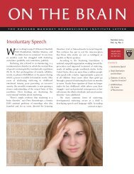
Anger and <strong>the</strong> Brain<br />
The physical signs of anger are unmistakable.<br />
Our heart rate increases, sometimes<br />
climbing from a normal 80 beats per minute to<br />
more than 180; our blood pressure elevates to<br />
often dangerously high levels. Breathing becomes<br />
more rapid as we try to get additi<strong>on</strong>al oxygen into<br />
our bodies, <strong>the</strong> muscles used for <strong>the</strong> fight-or-flight<br />
reacti<strong>on</strong> tighten, and <strong>the</strong> energy outburst can<br />
cause a deficiency in our blood sugar that makes<br />
us literally “shake” from anger. But, what is going<br />
<strong>on</strong> in our <strong>brain</strong> when we get angry?<br />
“A lot goes into anger,” says Roy Perlis, MD, an<br />
assistant professor of psychiatry at <strong>Harvard</strong> <strong>Medical</strong><br />
<strong>School</strong> who studies anger and <strong>the</strong> <strong>brain</strong>. “There’s<br />
<strong>the</strong> physical: <strong>the</strong> pounding heart, sweating,<br />
gestures: and <strong>the</strong> mental: thinking about why<br />
you’re angry and what you’re going to do about it.<br />
So, as far as <strong>the</strong> <strong>brain</strong> is c<strong>on</strong>cerned, anger is both<br />
cortical [higher cognitive aspects] and subcortical<br />
[physiological aspects].”<br />
Imaging studies have shown that in normal<br />
individuals (those without underlying mental or<br />
physical illnesses), <strong>the</strong>re is increased activity in <strong>the</strong><br />
orbitofr<strong>on</strong>tal cortex (OFC), <strong>the</strong> area of <strong>the</strong> <strong>brain</strong><br />
behind <strong>the</strong> forehead that c<strong>on</strong>trols reas<strong>on</strong>ing and<br />
o<strong>the</strong>r higher cognitive functi<strong>on</strong>s. At <strong>the</strong> same time,<br />
blood flow to <strong>the</strong> amygdala, <strong>the</strong> alm<strong>on</strong>d-shaped<br />
regi<strong>on</strong> deep in <strong>the</strong> <strong>brain</strong> that c<strong>on</strong>trols emoti<strong>on</strong>s,<br />
also increases its activity. Angry emoti<strong>on</strong>s in <strong>the</strong><br />
amgydala are thus cooled by activity in <strong>the</strong> OFC,<br />
inhibiting our thoughts of rage.<br />
But, why <strong>the</strong>n, do we not fly into a fit of rage<br />
every time we become angry?<br />
“Our, and o<strong>the</strong>rs’, hypo<strong>the</strong>sis is that <strong>the</strong> OFC<br />
plays a crucial role as a brake when our limbic<br />
system [which includes <strong>the</strong> amygdala] is active; that<br />
is, when we are emoti<strong>on</strong>al,” says Darin Dougherty,<br />
ON THE BRAIN<br />
MD, a psychiatrist at Massachusetts General Hospital<br />
and associate professor at HMS. “Most people have<br />
intact, functi<strong>on</strong>al OFCs, so <strong>the</strong>y are able to avoid<br />
frequent bouts of rage. Of course, our findings are<br />
that patients who do have frequent bouts of rage,<br />
do not activate <strong>the</strong>ir OFCs to <strong>the</strong> degree that healthy<br />
volunteers do.”<br />
Perlis, whose work focuses <strong>on</strong> regi<strong>on</strong>s of <strong>the</strong><br />
<strong>brain</strong> influenced by genetics, says <strong>the</strong>re is likely a<br />
genetic comp<strong>on</strong>ent that influences how we resp<strong>on</strong>d<br />
to anger, ei<strong>the</strong>r c<strong>on</strong>trolling our own or resp<strong>on</strong>ding<br />
to o<strong>the</strong>rs who are angry. Some studies suggest that<br />
twins are more likely to act similarly to hostility<br />
and anger than are unrelated people.<br />
“There is some inherited factor [that predisposes<br />
<strong>on</strong>e to angry outbursts],” he says. “There’s some<br />
normal variati<strong>on</strong> in <strong>the</strong> populati<strong>on</strong>, so we know it’s<br />
not pathological, just like we know <strong>the</strong>re are some<br />
people who are 6 feet tall and o<strong>the</strong>rs who are 5’9”.”<br />
Psychiatric disorders, including depressi<strong>on</strong> and<br />
bipolar disorder, influence how pr<strong>on</strong>e <strong>on</strong>e is to anger.<br />
More than half of those with major depressi<strong>on</strong><br />
report significant problems with irritability, says<br />
Perlis, so “as often as depressi<strong>on</strong> is about sadness,<br />
it can also be an angry state.” Bipolar disorder is also<br />
tied to anger, with irritability often being a primary<br />
feature of <strong>the</strong> illness. In additi<strong>on</strong>, substance abuse<br />
has a str<strong>on</strong>g effect <strong>on</strong> anger and its c<strong>on</strong>sequences.<br />
In general, Perlis adds, anger is a normal,<br />
sometimes even healthy, emoti<strong>on</strong>. “People shouldn’t<br />
be afraid to get angry,” he says. “We worry about<br />
it when it impacts <strong>the</strong> individual’s or o<strong>the</strong>rs’ lives<br />
or becomes pervasive.”<br />
Emoti<strong>on</strong> and c<strong>on</strong>trol<br />
In 2004, Dougherty and his colleagues published<br />
findings in <strong>the</strong> Archives of General Psychiatry, showing<br />
that people suffering from major depressive<br />
disorder (MDD) and anger attacks have decreased<br />
blood flow to <strong>the</strong> OFC and amygdala, which<br />
reduces <strong>the</strong>ir ability to c<strong>on</strong>trol impulsive acts and<br />
<strong>the</strong> feelings about <strong>the</strong> c<strong>on</strong>sequences of <strong>the</strong>ir acti<strong>on</strong>s.<br />
Thus, <strong>the</strong>y suffer both a lack of emoti<strong>on</strong> and a lack<br />
of c<strong>on</strong>trol. Dougherty’s team found significantly less<br />
activati<strong>on</strong> of <strong>the</strong> OFC in patients with MDD with<br />
anger attacks when compared to a c<strong>on</strong>trol group,<br />
but did not find a difference in amygdala activati<strong>on</strong><br />
between <strong>the</strong> groups. The amygdala came into play<br />
when <strong>the</strong>y looked at areas of <strong>the</strong> <strong>brain</strong> that<br />
correlated with OFC activati<strong>on</strong>. In normal patients,<br />
<strong>the</strong>y found a healthy inverse reciprocal relati<strong>on</strong>ship<br />
in functi<strong>on</strong> between <strong>the</strong> two structures, whereas
this relati<strong>on</strong>ship was exactly <strong>the</strong> opposite in MDD<br />
patients with anger attacks (with MDD patients<br />
without anger attacks falling in <strong>the</strong> middle).<br />
“We d<strong>on</strong>’t know that <strong>the</strong> underlying neurobiology<br />
of frequent rage attacks in patients with major<br />
depressi<strong>on</strong> with anger attacks is any different than<br />
that in o<strong>the</strong>r populati<strong>on</strong>s who have frequent rage<br />
attacks,” says Dougherty. “In fact, studies of patients<br />
with borderline pers<strong>on</strong>ality disorder (BPD) or<br />
intermittent explosive disorder (IED) exhibit results<br />
very similar to ours. We chose patients with major<br />
depressi<strong>on</strong> with anger attacks as a c<strong>on</strong>venient<br />
vehicle for studying rage attacks, whereas o<strong>the</strong>rs<br />
have studied BPD and IED.”<br />
<strong>Harvard</strong> researchers, including R<strong>on</strong>ald Kessler,<br />
PhD, a professor of health-care policy at HMS, found<br />
that IED, a disorder characterized by frequent bouts<br />
of angry and potentially violent outbursts, affects<br />
nearly 16 milli<strong>on</strong> adults and may predispose <strong>the</strong>m<br />
to o<strong>the</strong>r mental illnesses and substance abuse.<br />
Small amyloid assemblies Provide new target for alzheimer’s<br />
c<strong>on</strong>tinued from page 5<br />
into <strong>the</strong> synaptic cleft, bind to membranes, and<br />
cause local damage.”<br />
The HMS team also collaborated with scientists<br />
at <strong>University</strong> College Dublin to test human Abeta’s<br />
effect <strong>on</strong> behavior. They injected soluble Abeta<br />
dimers isolated from patients’ <strong>brain</strong>s into <strong>the</strong><br />
<strong>brain</strong>s of adult rats and found that <strong>the</strong> dimers<br />
induced certain characteristics of Alzheimer’s in<br />
<strong>the</strong> rats, specifically disrupting <strong>the</strong> memories of<br />
newly learned behaviors. The dimers, <strong>the</strong>y fur<strong>the</strong>r<br />
determined, acted directly <strong>on</strong> synapses, damaging<br />
<strong>the</strong> c<strong>on</strong>necti<strong>on</strong>s that are essential for neur<strong>on</strong>-t<strong>on</strong>eur<strong>on</strong><br />
communicati<strong>on</strong>.<br />
Neutralizing Abeta<br />
The findings provide a potential new target for<br />
anti-Alzheimer’s drugs, including antibodies that<br />
effectively neutralize Abeta dimers, says Selkoe.<br />
His team found that antibodies that target <strong>the</strong> first<br />
amino acid at <strong>the</strong> beginning of <strong>the</strong> Abeta protein<br />
worked best at neutralizing <strong>the</strong> protein.<br />
Changing <strong>the</strong> way we think, feel, act<br />
While it is hard to change a habitual behavior such<br />
as anger, formal anger management programs that<br />
focus <strong>on</strong> reducing both emoti<strong>on</strong>al feelings and <strong>the</strong><br />
physiological arousal that c<strong>on</strong>tributes to anger can<br />
help individuals c<strong>on</strong>trol <strong>the</strong>ir reacti<strong>on</strong>s. Perlis says<br />
that anger management programs that are based<br />
<strong>on</strong> cognitive behavioral <strong>the</strong>rapy, or CBT, are often<br />
<strong>the</strong> most successful.<br />
CBT is an umbrella term for several types of<br />
<strong>the</strong>rapies. These are all based <strong>on</strong> <strong>the</strong> idea that our<br />
thoughts, not external things like o<strong>the</strong>r people,<br />
situati<strong>on</strong>s and events, cause our feelings and<br />
behaviors. The benefit of this is that we can change<br />
<strong>the</strong> way we think, feel and act even if <strong>the</strong> situati<strong>on</strong><br />
doesn’t change.<br />
“The approach to anger would be to ei<strong>the</strong>r lessen<br />
<strong>the</strong> limbic resp<strong>on</strong>se, streng<strong>the</strong>n <strong>the</strong> brakes in <strong>the</strong><br />
OFC, or a combinati<strong>on</strong> of <strong>the</strong> two,” says Dougherty.<br />
“CBT utilizes cortical regi<strong>on</strong>s, such as <strong>the</strong> OFC and<br />
o<strong>the</strong>r prefr<strong>on</strong>tal areas, and streng<strong>the</strong>ns <strong>the</strong>m so<br />
<strong>the</strong>y are better able to suppress limbic resp<strong>on</strong>ses.”<br />
Four related clinical trials are currently being<br />
c<strong>on</strong>ducted in <strong>the</strong> United States and Europe to<br />
determine if an N-terminus antibody can neutralize<br />
<strong>the</strong> adverse effects of Abeta dimers in Alzheimer’s<br />
patients. Unpublished results of a Phase II study,<br />
c<strong>on</strong>ducted by Elan Corporati<strong>on</strong> and Wyeth<br />
Pharma ceuticals, showed some encouraging results<br />
in patients with mild to moderate stages of<br />
Alzheimer’s.<br />
The HMS research has implicati<strong>on</strong>s bey<strong>on</strong>d<br />
Alzheimer’s disease. The same Abeta process that<br />
causes Alzheimer’s, Selkoe says, can also c<strong>on</strong>tribute<br />
to age-related mild memory loss. “There are o<strong>the</strong>r<br />
reas<strong>on</strong>s [for age-related memory loss] than Abeta,”<br />
he says, “but some percentage is represented by<br />
Abeta attacks <strong>on</strong> neur<strong>on</strong>al synapses.”<br />
Selkoe says <strong>the</strong> next step in <strong>the</strong> research is to<br />
fully purify Abeta dimers and trimers from patients’<br />
<strong>brain</strong>s, label <strong>the</strong>m with radioactive chemicals, and<br />
bind <strong>the</strong>m to slices of mouse <strong>brain</strong> to determine<br />
which specific neur<strong>on</strong>al receptors are impaired by<br />
human Abeta. These receptors may provide<br />
additi<strong>on</strong>al targets to attack with drugs.<br />
<strong>on</strong> <strong>the</strong> <strong>brain</strong><br />
c<strong>on</strong>tinued <strong>on</strong> page 8