National best practice and evidence based guidelines for wound ...
National best practice and evidence based guidelines for wound ...
National best practice and evidence based guidelines for wound ...

You also want an ePaper? Increase the reach of your titles
YUMPU automatically turns print PDFs into web optimized ePapers that Google loves.
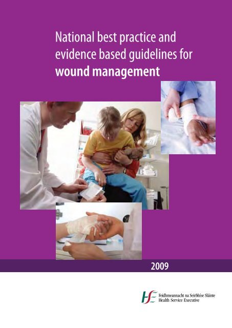
<strong>National</strong> <strong>best</strong> <strong>practice</strong> <strong>and</strong><br />
<strong>evidence</strong> <strong>based</strong> <strong>guidelines</strong> <strong>for</strong><br />
<strong>wound</strong> management<br />
2009
ISBN 978-1-906218-29-4<br />
© Health Service Executive<br />
October 2009<br />
Health Service Executive,<br />
Dr. Steevens’ Hospital,<br />
Dublin 8<br />
Irel<strong>and</strong><br />
Phone +353 1 6352000<br />
http://www.hse.ie
Foreword<br />
The development of HSE national <strong>guidelines</strong> <strong>for</strong> <strong>wound</strong> management are designed to support the<br />
st<strong>and</strong>ardisation of care <strong>and</strong> encourage <strong>best</strong> clinical <strong>practice</strong>. These <strong>guidelines</strong> constitute a general guide<br />
to be followed, subject to the medical practioners judgement in each individual case.<br />
These <strong>guidelines</strong> are <strong>based</strong> upon up to date scientific <strong>evidence</strong> <strong>and</strong> expert opinion <strong>and</strong> will serve to<br />
support consistency of treatment <strong>and</strong> contribute to improved patient outcomes.<br />
It is estimated that 1.5% of the population are affected by a <strong>wound</strong> at any one point in time. Wounds<br />
have a major personal, social, <strong>and</strong> economic impact. Wounds not only impact on the individual <strong>and</strong> their<br />
quality of life, they also have a significant impact on our health service <strong>and</strong> our society as a whole. Studies<br />
in the UK indicate that up to 4% of total health care expenditure is spent on the provision of <strong>wound</strong><br />
management while in Irel<strong>and</strong> it is estimated that two thirds of community nursing time is spent on the<br />
provision of <strong>wound</strong> management.<br />
As part of the HSE ef<strong>for</strong>ts to improve healthcare, it is hoped that these national <strong>guidelines</strong> will assist<br />
all clinicians in the decision making process <strong>and</strong> help to st<strong>and</strong>ardise the management of <strong>wound</strong>s at<br />
primary, secondary <strong>and</strong> tertiary levels. The availability of national <strong>guidelines</strong> will also provide guidance<br />
to policy makers.<br />
Healthcare is an ever changing science <strong>and</strong> advances <strong>and</strong> new developments in <strong>wound</strong> care will continue<br />
to take place. Thus, revision of these <strong>guidelines</strong> will be necessary as new knowledge is gained.<br />
The HSE wish to sincerely express their gratitude to those who reviewed the <strong>guidelines</strong> <strong>and</strong> in particular<br />
to the <strong>guidelines</strong> development group as this work, <strong>for</strong> some members, was per<strong>for</strong>med on an honorary<br />
basis <strong>and</strong> in addition to their usual work commitments.<br />
________________<br />
Dr Barry White<br />
<strong>National</strong> Director Clinical <strong>and</strong> Quality Care.<br />
1
NatioNal <strong>best</strong> <strong>practice</strong> aNd evideNce <strong>based</strong> guideliNes <strong>for</strong> wouNd maNagemeNt<br />
2<br />
Executive Summary<br />
Approximately 1.5% of the population will have a <strong>wound</strong> of some type at any one point in time.<br />
Fortunately, many of these are minor or acute <strong>and</strong> will heal without incident. The remaining <strong>wound</strong>s,<br />
the majority of which are chronic ulcers are a significant source of patient morbidity <strong>and</strong> in some cases<br />
mortality. Chronic <strong>wound</strong>s affect the individual’s quality of life <strong>and</strong> reduce their ability to optimise<br />
their contribution to society. The management of <strong>wound</strong>s is also very costly to the health service with<br />
the largest portion of that cost being nursing time. The protracted course of treatment, potential <strong>for</strong><br />
infection, together with the knowledge <strong>and</strong> skills required <strong>for</strong> optimal management supports the need<br />
<strong>for</strong> national <strong>guidelines</strong> to promote <strong>evidence</strong> <strong>based</strong> <strong>practice</strong>.<br />
The approach to optimal <strong>wound</strong> management centers on a comprehensive assessment of the patient<br />
<strong>and</strong> the <strong>wound</strong>. This should be completed by a person trained in such assessment. The aetiology of the<br />
<strong>wound</strong> should be determined with referral to appropriate members of the multi-disciplinary team when<br />
further investigation or intervention is required. All aspects of care from initial presentation through to<br />
treatment <strong>and</strong> evaluation should be documented. Following assessment, treatment goals should be<br />
agreed with the patient <strong>and</strong> a time frame <strong>for</strong> their achievement set. Underlying factors which could<br />
influence the potential <strong>for</strong> <strong>wound</strong> healing should be addressed. As <strong>wound</strong> healing is a complex multifactorial<br />
process, the input of several members of the multi-disciplinary team may be required to achieve<br />
the objectives. Evaluation is an on-going process. Each clinician involved in the provision of care must<br />
work within their Scope of Practice <strong>and</strong> is accountable <strong>for</strong> their <strong>practice</strong>.<br />
When cleansing the <strong>wound</strong>, potable tap water is suited <strong>for</strong> chronic <strong>wound</strong>s <strong>and</strong> in adults with lacerations.<br />
An aseptic technique is required when the individual is immuno-compromised <strong>and</strong>/or the <strong>wound</strong> enters<br />
a sterile body cavity. All dressings used in <strong>wound</strong> management should be used in accordance with<br />
manufacturer’s instructions <strong>and</strong> the integrity of such products must be ensured through proper storage<br />
<strong>and</strong> use. The choice of dressing is influenced by the type of <strong>wound</strong>, the amount of exudate, location of<br />
<strong>wound</strong>, skin condition, presence or absence of infection, condition of the <strong>wound</strong> bed, the characteristics<br />
of dressings available <strong>and</strong> treatment goals. Surgical <strong>wound</strong> dressings should be left dry <strong>and</strong> untouched<br />
<strong>for</strong> a minimum of 48 hours post-operatively to allow <strong>for</strong> re-establishment of the natural bacteria-proof<br />
barrier, unless otherwise clinically indicated.<br />
Patients presenting with lower limb ulceration should have assessment <strong>and</strong> investigation undertaken<br />
by health care professionals trained in leg ulcer management. All such patients should be screened <strong>for</strong><br />
<strong>evidence</strong> of arterial disease by measurement of ABPI by a person trained in such measurement. ABPI<br />
should be conducted when: an ulcer is deteriorating, is not fully healed by 12 weeks, is recurrent, prior<br />
to commencing compression therapy, when there is sudden increase in <strong>wound</strong> size, sudden increase<br />
in <strong>wound</strong> pain, change in colour <strong>and</strong>/or temperature of the foot or as part of on-going assessment.<br />
Graduated compression therapy with adequate padding, capable of sustaining compression <strong>for</strong> at least<br />
one week should be the first line of treatment <strong>for</strong> uncomplicated venous leg ulcers. This should be<br />
applied by a practitioner trained in its application.<br />
Removal of devitalised tissue will promote <strong>wound</strong> healing. However, in arterial ulcers with dry gangrene<br />
or eschar, debridement should not be per<strong>for</strong>med until arterial flow has been established. Routine use of<br />
antibiotics is unnecessary unless there are signs of infection.
The management of diabetic foot disease centres on identification of the ‘at risk’ limb <strong>and</strong> prevention<br />
of onset <strong>and</strong> management of the ulcerated limb. All people with diabetes should be examined at least<br />
once a year <strong>for</strong> potential foot problems. Patients with demonstrated risk factors should be examined<br />
more often – every 1-6 months. In a high risk patient, callus <strong>and</strong> nail <strong>and</strong> skin pathology should be<br />
treated regularly, preferably by a trained foot care specialist. Patients <strong>and</strong> their family or carer, if they wish,<br />
should be educated on the importance of foot care <strong>and</strong> regular foot inspection. Infection in a diabetic<br />
foot presents a direct threat to the affected limb <strong>and</strong> should be treated promptly <strong>and</strong> actively. Patients<br />
with an ulcer deeper than subcutaneous tissues should be treated intensively <strong>and</strong> depending on local<br />
resources <strong>and</strong> infrastructure, hospitalisation must be considered. Ill fitting shoes are a frequent cause of<br />
ulceration <strong>and</strong> there<strong>for</strong>e shoes should be examined meticulously in all patients.<br />
Each health care setting should have a pressure ulcer prevention policy in place. This should include<br />
recommendations <strong>for</strong> the structured approach to risk assessment relevant to the health care setting,<br />
the timing of risk assessment <strong>and</strong> reassessment, clear recommendations <strong>for</strong> documentation of risk<br />
assessment <strong>and</strong> communication to the wider healthcare team.<br />
To assist in documentation of care <strong>and</strong> evaluation of <strong>practice</strong> using clinical audit, these <strong>guidelines</strong> provide<br />
a comprehensive glossary of terms, examples of documentation <strong>and</strong> assessment tools <strong>and</strong> an audit <strong>for</strong>m<br />
<strong>for</strong> use by clinicians in their own working environment.<br />
3
NatioNal <strong>best</strong> <strong>practice</strong> aNd evideNce <strong>based</strong> guideliNes <strong>for</strong> wouNd maNagemeNt<br />
4<br />
Acknowledgements<br />
Canadian Wound Management Association<br />
Australian Wound Management Association<br />
Royal College of Nursing, London<br />
International Working Group on Diabetic Foot<br />
European Pressure Ulcer Advisory Panel<br />
Joanna Briggs Institute, Australia<br />
Gillian Mannion, HSE NMPDU Dublin Mid Leinster – <strong>for</strong> secretarial support.<br />
Dr. Niamh Macey, HSE West, <strong>for</strong> assistance with development of audit tool.<br />
Reviewers<br />
Ms. Eileen Kelly, RGN, RM, RNT, Dip Nursing Studies, MSc, Director Nurse Education Centre,<br />
Cork University Hospital<br />
Prof Sean Tierney, BSc Mch FRCSI(gen Surg), Prof of Surgical In<strong>for</strong>matics, RCSI <strong>and</strong> Consultant Vascular<br />
Surgeon, AMNCH<br />
Prof Jan Apelqvist, MD, PhD, Snr Consultant Department of Endocrinology, University Hospital of Malmo,<br />
Sweden <strong>and</strong> Assoc. Prof. Division <strong>for</strong> clinical studies, University of Lund, Sweden<br />
Dr Carol Dealey, Senior Research Fellow. Research & Education - University Hospitals Birmingham NHS<br />
Foundation Trust Queen Elizabeth Hospital, Queen Elizabeth Medical Centre, Birmingham, B15 2TH
These <strong>guidelines</strong> have been endorsed by:<br />
5
NatioNal <strong>best</strong> <strong>practice</strong> aNd evideNce <strong>based</strong> guideliNes <strong>for</strong> wouNd maNagemeNt<br />
6<br />
Contents<br />
Foreword 1<br />
Executive Summary 2<br />
Acknowledgements 4<br />
Reviewers 4<br />
Guideline endorsement 5<br />
Section 1<br />
1.0 Background <strong>and</strong> justification <strong>for</strong> Guidelines 11-12<br />
Introduction 12<br />
Prevalence 12<br />
Leg ulceration 14<br />
Diabetic foot ulceration 14<br />
Pressure ulcers 15<br />
Impact of <strong>wound</strong>s 15<br />
- On the individual 16<br />
- On the health service (the financial impact) 16<br />
- On society 17<br />
The need <strong>for</strong> <strong>guidelines</strong> 17<br />
Limitations to these <strong>guidelines</strong> 17<br />
1.1 Scope <strong>and</strong> Purpose of the Guidelines 18<br />
1.2 Guideline Development Team 18<br />
1.3 Terms of Reference 19<br />
1.4 Layout of Document 19<br />
Section 2 – Methology<br />
2.1 Guideline Development Process 22<br />
Search Strategy 22<br />
2.2 Outline of the grading method used 23<br />
table 1: Level of Evidence 23<br />
table 2: Level of Recommendation 23<br />
2.3 Decision Framework in Wound Management 23<br />
figure 2: Decision framework 24
Section 3 – Clinical Guidelines 25-26<br />
3.1 Corporate <strong>and</strong> individual responsibilities <strong>and</strong> accountability 26<br />
Corporate responsibilities 26<br />
Individual responsibilities 26<br />
3.2 Principals of Wound Management 27<br />
Assessment 27<br />
Objectives of <strong>wound</strong> management 27<br />
Treatment <strong>and</strong> management 28<br />
Wound cleansing 28<br />
Aseptic technique 28<br />
Clean <strong>wound</strong> management technique 28<br />
Cleansing solutions 28<br />
Pressure <strong>for</strong> <strong>wound</strong> cleansing 28<br />
Wound dressings 29<br />
Documentation/education 29<br />
Evaluation 29<br />
3.3 Guidelines <strong>for</strong> the management of venous leg ulceration<br />
– key points (level of <strong>evidence</strong>) 30<br />
Assessment 30<br />
Objectives 30<br />
Treatment 30<br />
Documentation 31<br />
Evaluation 31<br />
table 3: Patient factors to be recorded at baseline 32<br />
table 4: Wound factors to be recorded at baseline 32<br />
table 5: Limb <strong>and</strong> peri-<strong>wound</strong> assessment 32<br />
3.4 Guidelines <strong>for</strong> management of arterial ulcers<br />
– key points (level of <strong>evidence</strong>) 33<br />
Assessment 33<br />
Objectives 33<br />
Treatment 33<br />
Documentation 34<br />
Evaluation 34<br />
3.5 Guidelines <strong>for</strong> the prevention <strong>and</strong> management of Diabetic foot ulcerations 35<br />
Introduction 35<br />
Part A: The non-ulcerated limb 35<br />
Assessment 35<br />
table 6: Key elements of diabetic foot management<br />
table 7: History & examination 35<br />
table 8: Assessing neuropathy 35<br />
Objectives 36<br />
table 9: Progression of risk categories 36<br />
7
NatioNal <strong>best</strong> <strong>practice</strong> aNd evideNce <strong>based</strong> guideliNes <strong>for</strong> wouNd maNagemeNt<br />
8<br />
Treatment 36<br />
Evaluation <strong>and</strong> education <strong>for</strong> patients, family <strong>and</strong> healthcare provider 36<br />
table 10: Patient education 37<br />
Appropriate footwear 37<br />
part b: Active ulceration 37<br />
Assessment 37<br />
Objectives 38<br />
Treatment 38<br />
Evaluation 39<br />
Documentation 39<br />
3.6 Guidelines <strong>for</strong> the prevention <strong>and</strong> management of pressure ulcers 40<br />
Introduction 40<br />
3.6.1 Risk assessment 40<br />
Risk assessment <strong>practice</strong> 40<br />
3.6.2 Skin assessment 41<br />
Skin care 41<br />
3.6.3 Nutrition <strong>for</strong> pressure ulcer prevention 42<br />
General recommendations 42<br />
3.7 Repositioning <strong>for</strong> the prevention of pressure ulcers 43<br />
Repositioning frequency 43<br />
Repositioning technique 43<br />
Repositioning the seated individual 44<br />
Repositioning documentation 44<br />
Repositioning education <strong>and</strong> training 44<br />
3.8 Support Surfaces 45<br />
General statements 45<br />
Mattress <strong>and</strong> bed use in pressure ulcer prevention 45<br />
The use of support surfaces to prevent heel pressure ulcers 45<br />
Use of support surfaces to prevent pressure ulcers while seated 46<br />
The use of other support surfaces in pressure ulcer prevention 46<br />
Special population: operating room patients 46
Section 4 References <strong>and</strong> Appendices 47-48<br />
References 48<br />
Bibliography 50<br />
Appendices 51<br />
appendix 1: Glossary of terms 52<br />
appendix 2: Wound Assessment Forms 59<br />
appendix 3: Pressure ulcer risk assessment tools 68<br />
appendix 4: MUST tool 72<br />
appendix 5: Diabetic foot screening assessment sheet 78<br />
appendix 6: Audit tool 79<br />
9
NatioNal <strong>best</strong> <strong>practice</strong> aNd evideNce <strong>based</strong> guideliNes <strong>for</strong> wouNd maNagemeNt 10
SECTION 1:<br />
Background <strong>and</strong> Justification <strong>for</strong> Guidelines<br />
11
NatioNal <strong>best</strong> <strong>practice</strong> aNd evideNce <strong>based</strong> guideliNes <strong>for</strong> wouNd maNagemeNt<br />
12<br />
Section 1: Background <strong>and</strong> Justification <strong>for</strong> Guidelines<br />
Introduction<br />
Owing to the diversity of <strong>wound</strong> aetiologies <strong>and</strong> their associated co-morbidities, a range of health care<br />
professionals across all health settings, each with a varied knowledge base related to <strong>wound</strong> healing<br />
deliver <strong>wound</strong> care. In order to st<strong>and</strong>ardise care <strong>and</strong> encourage optimal <strong>practice</strong> in <strong>wound</strong> management,<br />
with the goal of improving patient outcomes, there is a need to develop national <strong>guidelines</strong> <strong>for</strong> their<br />
management. The background <strong>and</strong> justification <strong>for</strong> these <strong>guidelines</strong> will provide the health professional<br />
with the rationale <strong>for</strong> their development together with in<strong>for</strong>mation relating to prevalence, potential<br />
patient outcomes, <strong>and</strong> resource issues <strong>for</strong> the health service.<br />
A <strong>wound</strong> is defined as a break in the continuity of the skin (Schultz et al. 2003). It may arise from an<br />
underlying altered physiological state or be primary in origin. As the largest organ in the body, damage to<br />
the skin <strong>and</strong> alteration in its functions can have catastrophic consequence <strong>for</strong> the individual. Reassuringly,<br />
the vast majority of insults to the integrity of the skin heal uneventfully. However, whether it is due to the<br />
nature of the injury, or the health of the individual, some <strong>wound</strong>s have a delayed <strong>and</strong> protracted course<br />
of healing (Falanga 2001).<br />
Wounds can be broadly classified as acute or chronic. Acute <strong>wound</strong>s usually heal in an ordered, timely<br />
fashion, <strong>and</strong> are typically seen as post-operative <strong>wound</strong>s, minor lacerations, abrasions, minor burns <strong>and</strong><br />
scalds <strong>and</strong> some trauma <strong>wound</strong>s (Falanga 2002, Schultz et al. 2003). Conversely, chronic <strong>wound</strong>s do not<br />
follow this ordered sequence of events <strong>and</strong> are characterised by delayed healing, cellular senescence,<br />
<strong>and</strong> recurrent infections (Schultz et al. 2003). Chronic <strong>wound</strong>s in particular are common across all health<br />
care settings <strong>and</strong> there is growing <strong>evidence</strong> that the burden of chronic <strong>wound</strong>s in Irel<strong>and</strong> is already high<br />
<strong>and</strong> likely to increase (O’Brien et al. 2000, McDermott-Scales et al. 2009).<br />
Prevalence<br />
Although it is often assumed that skin breakdown is confined to the frail older person, the problem of<br />
prevalence is seen at both ends of the age spectrum (Voegeli 2007). Critically ill neonates are prone to<br />
skin damage due to intrinsic factors such as having a thinner immature skin (Chung et al. 2002). Older<br />
persons have a thinner epidermis with a flattened interface between the epidermis <strong>and</strong> dermis, making it<br />
less resistant to shearing <strong>for</strong>ces (Chung et al. 2002). However, the prevalence of chronic <strong>wound</strong>s is strongly<br />
related to increasing age <strong>and</strong> <strong>for</strong>ecast trends in Irel<strong>and</strong> indicate that the number of people with chronic<br />
<strong>wound</strong>s is likely to increase substantially in the future (Callam et al. 1985, Jeffcoate <strong>and</strong> Harding 2003,<br />
Moffatt et al. 2004, V<strong>and</strong>erwee et al. 2007). The number of persons aged 65 years <strong>and</strong> above is expected<br />
to increase from 430,000 today (2008) to 811,000 by 2025 (CSO 2002). By that time those aged 65 years<br />
<strong>and</strong> above will account <strong>for</strong> 16.7% of the total population compared with 11.3 % today (CSO 2002).<br />
It is estimated that 1 - 1.5% of the population are affected by a <strong>wound</strong> at any point in time (Gottrup<br />
2004). While there are no Irish figures directly related to all <strong>wound</strong>s, Hospital In-Patient Enquiry (HIPE)<br />
data <strong>for</strong> 2003 show that of all diagnosis on discharge from acute hospitals, disorders of the skin <strong>and</strong><br />
subcutaneous tissue accounted <strong>for</strong> 48,466 cases with cellulitis <strong>and</strong> abscess accounting <strong>for</strong> 7,806 of<br />
these (ESRI 2007). Wound debridement, <strong>wound</strong> infection or burns accounted <strong>for</strong> 7,342 cases <strong>and</strong> 2,375<br />
cases of skin grafts were registered. There were 313 burns cases referred to the <strong>National</strong> paediatric burns<br />
service in Our Lady’s Hospital, Crumlin in 2006. Of these 75% were in children under 5 years. In 2003, HIPE
anked operations of the skin <strong>and</strong> subcutaneous tissue 11th out of the top 20 principal procedures <strong>for</strong><br />
all in-patients representing 13,247 cases. Open <strong>wound</strong>s represented 9,097 of all discharges by principle<br />
diagnosis from acute hospitals in 2003. The average length-of-stay <strong>for</strong> all patients with an open <strong>wound</strong><br />
was 2.4 days, but this increased to 5.5 days in those over 65 years of age (ESRI 2007).<br />
For day procedures, operations on the skin <strong>and</strong> subcutaneous tissue were ranked third highest accounting<br />
<strong>for</strong> 33,569 procedures <strong>and</strong> 9.8% of day-care patients (ESRI 2007). Cellulitis <strong>and</strong> abscess accounted <strong>for</strong><br />
7,806 of all diagnoses <strong>and</strong> open <strong>wound</strong>s accounted <strong>for</strong> 14,119. Neither Out Patient Department nor<br />
Emergency department attendances are recorded on HIPE. As many persons with <strong>wound</strong>s attend these<br />
departments, the numbers stated are potentially an under representation of the impact of <strong>wound</strong>s in<br />
acute services.<br />
Few Irish researchers have quantified the prevalence of <strong>wound</strong>s in the non-acute setting. A prevalence<br />
of 4% was identified in one study on the active caseload of community nurses (McDermott-Scales et<br />
al. 2009). This is in contrast to a Canadian study in which 50% of patients on the active caseload of<br />
community nurses working in a community area had a <strong>wound</strong> (Hurd et al. 2008). Differences in sampling<br />
methods may account <strong>for</strong> the wide variation in these figures. Of note in the latter study was that nonhealing<br />
surgical <strong>wound</strong>s accounted <strong>for</strong> 31-38% of all <strong>wound</strong>s being managed (Hurd et al. 2008). Point<br />
prevalence of 0.37% with a mean of 1.44 <strong>wound</strong>s per patient has been reported in health districts<br />
covering both acute <strong>and</strong> community care (Hurd et al. 2008).<br />
A recent pan-European review of prevalence of <strong>wound</strong>s has estimated that 3.7 per 1000 population have<br />
at least one <strong>wound</strong> under treatment (Posnett et al. 2009). Researchers have reported that <strong>for</strong> patients<br />
with advanced illness 53% of those with cancer <strong>and</strong> 80% of patients with non-cancer related advanced<br />
illness had a <strong>wound</strong>, with an average of 2 <strong>wound</strong>s per patient (Maida et al. 2008).<br />
Chronic <strong>wound</strong>s are associated with at least one co-morbidity (Olin et al. 1999, Oien et al. 2000). These<br />
co-morbidities are frequently hypertension, diabetes, cardio-vascular disease, <strong>and</strong> neurological disorders.<br />
The risk factors <strong>for</strong> chronic illnesses are well recognised <strong>and</strong> include; hypertension, obesity, poor<br />
nutrition, tobacco, alcohol <strong>and</strong> high cholesterol (DoHC 2007a, DoHC 2007b). Recent Irish researchers<br />
have clearly demonstrated that the prevalence of such risk factors shows no signs of abating (Whelton<br />
et al. 2007, Morgan et al. 2008). This clearly demonstrates that the prevalence of <strong>wound</strong>s with associated<br />
co-morbidities will be evident into the future.<br />
While these <strong>guidelines</strong> can apply to all <strong>wound</strong>s, particular emphasis in this document is on categories of<br />
<strong>wound</strong>s most commonly encountered in routine clinical <strong>practice</strong> <strong>and</strong> which provide many challenges to<br />
practitioners. These include venous ulceration, arterial ulceration, diabetic foot ulceration <strong>and</strong> pressure<br />
ulceration.<br />
13
NatioNal <strong>best</strong> <strong>practice</strong> aNd evideNce <strong>based</strong> guideliNes <strong>for</strong> wouNd maNagemeNt<br />
14<br />
Leg ulceration<br />
Leg ulcer is defined as a breakdown of the epidermal <strong>and</strong> dermal tissue below the knee on the leg<br />
or foot, due to any cause, which fails to heal (Moffatt <strong>and</strong> Harper 1997). Leg ulceration has multiple<br />
causes, the most common being venous disease accounting <strong>for</strong> 37% - 81% of all cases depending on the<br />
methods used <strong>for</strong> diagnosis (Briggs <strong>and</strong> Closs 2003). Other causes include rheumatoid arthritis, diabetes,<br />
arterial disease, trauma <strong>and</strong> malignancy. Importantly, patients can have leg ulcers with a single aetiology<br />
or with multiple causes (Briggs <strong>and</strong> Closs 2003).<br />
Irish studies have reported a leg ulceration prevalence of 0.12% in the adult population increasing to<br />
1.03% in those over 70 years of age (O’Brien et al. 2000). These results are supported by some international<br />
research as the prevalence of patients with open leg ulcers receiving treatment from health professionals<br />
ranged from 0.11% -3.6% (Graham et al. 2003). However, the range in prevalence rates might have been<br />
due to the variety of methodologies <strong>and</strong> in particular the inclusion criteria used (O’Brien et al. 2000, Briggs<br />
<strong>and</strong> Closs 2003, Graham et al. 2003, Moffatt et al. 2004). Age specific prevalence rates are comparable<br />
between the sexes but women predominate in the older age group with higher st<strong>and</strong>ardised prevalence<br />
rates (Callam et al. 1985, Graham et al. 2003).<br />
True prevalence is arguably higher as people of working age are under represented in the published<br />
studies, because they are more likely to be self-caring (Nelzen et al. 1996). Of note it was reported in many<br />
studies that while prevalence increases with age, the age of onset was below 65 years <strong>for</strong> approximately<br />
half of the populations under consideration (Moffatt et al. 1992, Moffatt et al. 2004).<br />
Based on reported prevalence rates to date <strong>and</strong> the current Irish population of 4,000,000 it can be<br />
estimated that 4,800 persons in Irel<strong>and</strong> may suffer from active open ulceration at any one point in time.<br />
The true prevalence rates are even higher if one is cognisant of the proposal that only 20-25% of venous<br />
ulcers are open at any point in time (Nelzen et al. 1996). Thus it is likely that 24,000 persons in Irel<strong>and</strong> are<br />
affected by leg ulceration. It is estimated that 490,000 to 1.3 million EU citizens have an open lower-limb<br />
ulcer at any one time (Posnett et al. 2009).<br />
The chronicity of lower limb ulceration is manifested by the high recurrence rates, protracted courses<br />
of treatment with a mean of only 50% of those in receipt of compression therapy <strong>for</strong> venous ulceration<br />
healing after 12 weeks of therapy (Moffatt <strong>and</strong> Dorman 1995, Peters 1998, Gethin <strong>and</strong> Cowman 2009).<br />
Duration of ulceration is a cause <strong>for</strong> concern with studies frequently reporting open ulceration <strong>for</strong> more<br />
than one year while there are reports of ulceration spanning 60 years (Clarke-Moloney et al. 2006, Gethin<br />
<strong>and</strong> Cowman 2009, McDermott-Scales et al. 2009).<br />
There is <strong>evidence</strong> of a change in trend in ulcer aetiology <strong>and</strong> that prevalence of more chronic mixed<br />
aetiology ulcers <strong>and</strong> arterial ulcers is increasing (Moffatt et al. 2004). Increased life expectancy <strong>and</strong> increased<br />
prevalence of arterial disease in the population may account <strong>for</strong> these results (Moffatt et al. 2004).<br />
Diabetic Foot Ulceration<br />
While in<strong>for</strong>mation on specific <strong>wound</strong> types cannot be extrapolated from the HIPE system, diabetes<br />
accounted <strong>for</strong> 36,642 of all listed total discharges by principal diagnosis in 2003 (ESRI 2007). It is estimated<br />
that one in every seven persons with diabetes will develop a foot ulcer (Boulton et al. 2005, IWGDF 2007);<br />
there<strong>for</strong>e, potentially 5234 cases of diabetic foot ulceration were treated in Irish hospitals in 2003.
The number of adults diagnosed with diabetes in Irel<strong>and</strong> has been estimated at 141,063 in 2006 (Bal<strong>and</strong>a<br />
et al. 2005). Prevalence rates among children were estimated at 0.2% or 2,229 persons (Bal<strong>and</strong>a et al.<br />
2005). Additionally, it is estimated that <strong>for</strong> every person with diabetes there is another as yet undiagnosed<br />
(Bal<strong>and</strong>a et al. 2005, Boulton et al. 2005, IWGDF 2007).<br />
Foot ulceration is a frequent complication of diabetes <strong>and</strong> <strong>based</strong> on international research prevalence<br />
data (Wraight et al. 2004, IWGDF 2007)it can be estimated that there are 20,470 – 41, 020 cases of diabetic<br />
foot ulceration in Irel<strong>and</strong>. Persons with diabetes are fifteen times more likely to have an amputation than<br />
those without <strong>and</strong> 85% of all such amputations are preceeded by ulceration (Boulton et al. 2005, IWGDF<br />
2007). It has been reported that once an individual has undergone an amputation there is a 50% risk of<br />
an amputation of the remaining limb within 5 years (Boulton et al. 2005, IWGDF 2007). Many individuals<br />
still do not receive optimal preventative care <strong>and</strong> the number of patients with diabetes who required<br />
admission <strong>for</strong> treatment of acute foot pathology remains high (Wraight et al. 2004).<br />
Pressure ulcers<br />
A pressure ulcer is defined as an area of localised damage to the skin <strong>and</strong> underlying tissue caused by<br />
pressure or shear or a combination of these (EPUAP 2002). Depth of ulceration is documented using a<br />
classification system, Category 1 through to Category 4. Category 1 represents superficial skin damage<br />
without a break in the continuity of the skin, commonly referred to as non-blanchable erythema. Category<br />
4 indicates extensive destruction, tissue necrosis or damage to muscle, bone or supporting structures<br />
with or without full thickness skin loss (EPUAP 2002). The prevalence of these <strong>wound</strong>s in the Irish acute<br />
setting is consistent with international studies ranging from 12-38% (Moore <strong>and</strong> Pitman 2000, Gethin<br />
et al. 2005, Gallagher et al. 2008). A trans-European survey identified that one in every 5 hospitalised<br />
patients had a pressure ulcer, while 50% of patients were at risk (EPUAP 2002). Similar to other studies,<br />
the higher prevalence in Irish studies was recorded in spinal injury units <strong>and</strong> intensive care units (Sheerin<br />
et al. 2005, deLaat et al. 2006).<br />
Pressure ulcer prevalence <strong>and</strong> incidence among hip fracture patients in five European countries reported<br />
that 10% had a pressure ulcer on arrival to the hospital while 22% had one on discharge (Lindholm et<br />
al. 2008). The majority were category one with no category four ulcers (Lindholm et al. 2008). In Irel<strong>and</strong>,<br />
fractured neck of femur is one of the most common reasons <strong>for</strong> hospital admission in the elderly with<br />
3,585 such patients over 65 years of age admitted to Irish hospitals in 2002 (ESRI 2007). Worldwide,<br />
elderly people represent the fastest growing age-group <strong>and</strong> the yearly number of fractures is likely to<br />
rise substantially with continued ageing of the population (Sambrook <strong>and</strong> Cooper 2006). There<strong>for</strong>e, there<br />
is a potential <strong>for</strong> increase in the incidence of pressure ulceration in this group.<br />
Prevalence in the non-acute sector is harder to quantify due to the diversity of care settings. However,<br />
researchers have reported that pressure ulcers were the <strong>wound</strong> most frequently encountered by<br />
community nurses with prevalence rates of 4 % (McDermott-Scales et al. 2009). Prevalence rates increased<br />
significantly with the age of the individual, as 75% of pressure ulcers occurred in those over 60 years of<br />
age (McDermott-Scales et al. 2009).<br />
Impact of <strong>wound</strong>s<br />
The impact of <strong>wound</strong>s, <strong>and</strong> in particular chronic <strong>wound</strong>s, on patient health <strong>and</strong> well being, <strong>and</strong> the<br />
substantial burden <strong>wound</strong> care places on health care staff, organisations <strong>and</strong> resources provides an<br />
opportunity to improve prevention <strong>and</strong> management strategies (Posnett et al. 2009). Wounds do not<br />
have a one-dimensional impact but rather can impact under three domains; that is, to the individual, the<br />
health service <strong>and</strong> to society.<br />
15
NatioNal <strong>best</strong> <strong>practice</strong> aNd evideNce <strong>based</strong> guideliNes <strong>for</strong> wouNd maNagemeNt<br />
16<br />
On the individual<br />
Quality of life studies have clearly demonstrated that persons with <strong>wound</strong>s have lower quality-of-life<br />
scores than their age <strong>and</strong> sex-matched counterparts (Price <strong>and</strong> Harding 1996, Price <strong>and</strong> Harding 1997,<br />
Rich <strong>and</strong> McLachlan 2003). Wounds cause pain, suffering, sepsis, infection, nausea, fatigue, depression,<br />
psychological disturbances, loss of function, loss of mobility <strong>and</strong> personal financial cost (Price <strong>and</strong> Harding<br />
1996, Price <strong>and</strong> Harding 1997, Rich <strong>and</strong> McLachlan 2003). In some cases <strong>wound</strong>s may lead to amputation<br />
<strong>and</strong> even death. For many patients <strong>wound</strong>s are a significant <strong>and</strong> preventable barrier to the successful<br />
recovery or management of, a wide range of medical conditions (RCN 2006). These range from, routine<br />
surgical interventions to chronic conditions such as diabetes. Pain is frequently associated with <strong>wound</strong>s,<br />
with some patients describing it as horrible or excruciating <strong>and</strong> it may be associated with the <strong>wound</strong><br />
aetiology, dressing change or infection (Price <strong>and</strong> Harding 1997, Rich <strong>and</strong> McLachlan 2003).<br />
On the health service (the financial impact)<br />
Wound care is very labour intensive <strong>and</strong> up to 66% of community nursing time is spent on the provision<br />
of <strong>wound</strong> care with patients receiving an average of 2.4 dressing changes per week (Clarke-Moloney et al.<br />
2006, O’Keeffe 2006, Clarke-Moloney et al. 2008). In the United Kingdom it was reported that up to 4% of<br />
total health care expenditure is spent on the provision of <strong>wound</strong> care (Bennett et al. 2004, Gottrup 2004,<br />
Drew et al. 2007, Posnett <strong>and</strong> Franks 2007, Hurd et al 2008. Developments such as the establishment of leg<br />
ulcer clinics has resulted in improved outcomes <strong>for</strong> patients with lower limb ulceration <strong>and</strong> in particular<br />
venous ulcers (Clarke-Moloney et al. 2008). Nurse led leg ulcer clinics have improved the management of<br />
venous ulcers with more assessments taking place in the community versus the hospital setting (Clarke-<br />
Moloney et al. 2008). A reduction in dressing change frequency, particularly in the home, is significant<br />
given the high percentage of nursing visits that involve <strong>wound</strong> care <strong>and</strong> the travel time required (Clarke-<br />
Moloney et al. 2008, Hurd et al. 2008).<br />
The appointment of tissue viability Clinical Nurse Specialists has raised the profile of <strong>wound</strong> management<br />
with 14 such posts in Irel<strong>and</strong> (www.ncnm.ie). However, to date only two of these are in primary care with<br />
the majority in acute care setting.<br />
Recent Irish researchers have reported that the cost of treating one patient with 3 grade 4 pressure ulcers<br />
in 2003 was €119,000 <strong>for</strong> a period of 129 days (Gethin et al. 2005). Researchers itemised all costs of care<br />
<strong>and</strong> the patient was discharged with a healed <strong>wound</strong>. This was a positive outcome in a relatively short<br />
period, but such is not always the case. Indeed, such costs are potentially much higher today due to<br />
inflationary price increases. It is easy to focus on dressings <strong>and</strong> other materials as being the major cost<br />
factor in <strong>wound</strong> care. However, this component accounts <strong>for</strong> only 10-15% of costs with nursing time<br />
<strong>and</strong> hospitalisation being the main drivers of cost (Carr et al. 1999, Posnett <strong>and</strong> Franks 2007). A recent UK<br />
audit covering a population of 590,000 persons revealed that 3% of total local health budget, 151,000<br />
nursing hours <strong>and</strong> the equivalent of 52-87 acute bed beds were spent annually specifically on <strong>wound</strong><br />
care (Hurd et al. 2008).<br />
The implications <strong>for</strong> health care in Irel<strong>and</strong> are particularly significant whether individuals are cared <strong>for</strong> in<br />
the primary or secondary care setting. Community care providers are attempting to deliver services to<br />
an ageing population facing a growing prevalence of chronic disease <strong>and</strong> disability. Most community<br />
care organisations in Irel<strong>and</strong> face challenges as acute care facilities attempt to reduce the length-of -stays<br />
in hospital <strong>and</strong> are relying more heavily on community services. Overall community care services must<br />
cater <strong>for</strong> patients who are older, with more serious <strong>and</strong> complex health issues <strong>and</strong> there<strong>for</strong>e at greater<br />
risk of <strong>wound</strong>s.
On Society<br />
The loss to society due to individuals being unable to engage in their normal activities is hard to quantify.<br />
Loss of time from work <strong>for</strong> the individual <strong>and</strong> their carer can have financial implications. Feelings of<br />
social isolation, anxiety <strong>and</strong> depression have potential to reduce the contribution the individual makes to<br />
society, whether is at a local or national level. It has been reported that problematic <strong>wound</strong>s frequently<br />
result in a loss of productivity; extended hospital stays <strong>and</strong> increased expenditure (Zhan <strong>and</strong> Miller, 2003;<br />
Tennevall et al., 2005).<br />
The need <strong>for</strong> <strong>guidelines</strong><br />
Clinical <strong>practice</strong> <strong>guidelines</strong> have been broadly defined as ”providing guidance in decision making at<br />
each level of interaction; between health professional <strong>and</strong> consumer, between purchaser <strong>and</strong> provider, <strong>and</strong><br />
between ‘funder’ <strong>and</strong> ‘purchaser’’. (http://www.nzgg.org.nz). There are five different types of <strong>guidelines</strong><br />
but those related to <strong>best</strong> <strong>practice</strong> are defined as ‘systematically developed statements to assist practitioner<br />
<strong>and</strong> consumer decisions about appropriate health or disability care <strong>for</strong> specific circumstances, taking into<br />
account <strong>evidence</strong> <strong>for</strong> effectiveness <strong>and</strong> competing claims, <strong>and</strong> <strong>for</strong>m a fundamental basis <strong>for</strong> planning’ (NZGG<br />
2001). The HSE hopes that these <strong>National</strong> <strong>wound</strong> management <strong>guidelines</strong> will assist professionals in the<br />
decision making process as they are <strong>based</strong> on the most current <strong>and</strong> <strong>best</strong> available <strong>evidence</strong> <strong>and</strong> aim to<br />
bring consistency to the provision of <strong>wound</strong> care in Irel<strong>and</strong>.<br />
The provision of <strong>wound</strong> care falls within the remit of a wide range of disciplines. The knowledge, skills,<br />
<strong>and</strong> underst<strong>and</strong>ing of each of these disciplines can vary, <strong>and</strong> may depend on the type <strong>and</strong> frequency<br />
of the <strong>wound</strong> aetiology encountered <strong>and</strong> the level of expertise available. There is a growing body of<br />
<strong>evidence</strong> that a structured, organised <strong>and</strong> planned approach to <strong>wound</strong> management whether <strong>for</strong><br />
specific <strong>wound</strong> aetiologies or <strong>for</strong> <strong>wound</strong>s in general improves patient outcomes <strong>and</strong> is cost effective <strong>for</strong><br />
the health service.<br />
It is anticipated that these <strong>guidelines</strong> will promote <strong>and</strong> enhance <strong>evidence</strong> <strong>based</strong> <strong>practice</strong> in <strong>wound</strong> care<br />
in Irel<strong>and</strong>. In addition, the provision of an audit tool should help to provide <strong>evidence</strong> to support the<br />
use of the <strong>guidelines</strong> as services <strong>and</strong> professionals can assess <strong>wound</strong> care management <strong>practice</strong>s <strong>and</strong><br />
patient outcomes against defined st<strong>and</strong>ards of care.<br />
Limitations of these <strong>guidelines</strong><br />
These <strong>guidelines</strong> have been developed following systematic search of the literature together with a<br />
review of current published <strong>guidelines</strong> using the AGREE <strong>guidelines</strong> review tool. They represent <strong>best</strong><br />
<strong>practice</strong> as it relates to current knowledge. It is anticipated that as new in<strong>for</strong>mation becomes available<br />
that some aspects of these <strong>guidelines</strong> will no longer be valid <strong>and</strong> will require updating.<br />
Some specific <strong>wound</strong> aetiologies such as burns <strong>and</strong> malignant <strong>wound</strong>s are frequently managed in<br />
specialist centres <strong>and</strong> thus are not included here.<br />
17
NatioNal <strong>best</strong> <strong>practice</strong> aNd evideNce <strong>based</strong> guideliNes <strong>for</strong> wouNd maNagemeNt<br />
18<br />
1.1 Scope <strong>and</strong> Purpose of the Guidelines<br />
These <strong>guidelines</strong> have been developed by the Health Service Executive (HSE) in collaboration with<br />
academic institutions <strong>and</strong> professional organisations involved in <strong>wound</strong> management in Irel<strong>and</strong>. The<br />
aim of these <strong>guidelines</strong> is to progress towards achieving the HSE’s commitment to delivering better<br />
services <strong>for</strong> the individual through the provision of <strong>evidence</strong>d <strong>based</strong> <strong>practice</strong> (HSE 2007). The <strong>guidelines</strong><br />
are applicable to all professionals involved in <strong>wound</strong> management.<br />
1.2 Guideline Development Team<br />
Table 1: Guideline Development Team (alphabetical order)<br />
Ms. Eithne Cusack (Co-chairperson) Director, Nursing & Midwifery Planning &<br />
Development, HSE<br />
Dr. Davida DelaHarpe (Co-chair) Assistant <strong>National</strong> Director, Population Health, HSE<br />
Dr. Georgina Gethin (lead researcher) Lecturer /Research co-ordinator, Research Centre,<br />
Faculty of Nursing <strong>and</strong> Midwifery, RCSI (WMAOI)<br />
TEAM MEMBERS<br />
Ms. Maura Belton Assistant Direct of Public Health Nursing representing<br />
Dublin Mid Leinster PCCC<br />
Ms. Caroline Connolly Irish Nursing Homes Association (replaced by<br />
Sinead Fitzpatrick)<br />
Ms. Brigid Considine Asst Director of Public Health Nursing representing<br />
Dublin North East PCCC<br />
Ms. Gerardine Craig CNS Tissue Viability, Drogheda representing Tissue<br />
Viability Nurses Association of Irel<strong>and</strong><br />
Ms. Sinead Fitzpatrick Representing Nursing Homes Association<br />
Ms. Ann Higgins Director of Infection Control, Mater Private Hospital,<br />
Dublin. Representing Infection Control Nurses<br />
Association<br />
Ms. Brenda Kelly <strong>National</strong> Hospitals Office, HSE<br />
Ms. Pat McCluskey CNS <strong>wound</strong> care, CUH: representing WMAOI<br />
Ms. Raphael McMullen Nursing Practice Development Co-ordinator<br />
representing Irish Nursing & Midwifery Practice<br />
Development Assoc. <strong>and</strong> DATH’s<br />
Ms. Patricia McQuillan Professional Development Co-ordinator <strong>for</strong> PNs<br />
representing Irish Practice Nurses Ass.<br />
Dr. Zena Moore Lecturer RCSI<br />
Ms. Alice O’Connor CNS St Johns Hospital, Limerick representing NHO<br />
HSE West<br />
Ms. Mary Parker PHN, representing PCCC HSE West<br />
Ms. Martina Rafter CNS Tissue Viability Water<strong>for</strong>d Regional Hospital<br />
representing NHO HSE South<br />
Ms. Helen Strapp CNS Tissue Viability AMNCH representing Dublin<br />
Academic Teaching Hospitals (DATH’s)<br />
Ms. Catherine Tunney Public Health Nurse representing Institute of<br />
Community Health Nursing<br />
Ms. Eileen Walsh Public Health Nurse: representing HSE Southern area/<br />
Cork & Kerry
1.3 Terms of Reference<br />
● To ensure/facilitate the development of <strong>National</strong> Wound Management Guidelines which represent<br />
up to date <strong>best</strong> <strong>practice</strong>.<br />
● To agree an approach to this work <strong>and</strong> secure funding as required.<br />
● To provide guidance on a st<strong>and</strong>ardised approach to <strong>wound</strong> management across all care settings<br />
in the interest of <strong>best</strong> <strong>practice</strong> <strong>and</strong> quality patient care.<br />
● To establish <strong>and</strong> support a guideline development team that is representative geographically <strong>and</strong><br />
across care settings.<br />
● To develop content <strong>and</strong> <strong>for</strong>mat of Wound Management Guidelines.<br />
● To liaise <strong>and</strong> work with approved research support.<br />
● To recommend a process <strong>for</strong> dissemination, implementation <strong>and</strong> evaluation of these Guidelines.<br />
● To support the dissemination of these Guidelines.<br />
1.4 Layout of Document<br />
This document has been divided into four sections:<br />
Section One deals with the administrative <strong>and</strong> corporate issues related to their development <strong>and</strong><br />
intended use.<br />
Section Two outlines the search strategies which lead to the <strong>guidelines</strong> <strong>and</strong> the levels of <strong>evidence</strong><br />
associated with guideline statements. This section also presents a decision framework to guide the<br />
clinician in the necessary steps to optimise <strong>best</strong> <strong>practice</strong> in <strong>wound</strong> management.<br />
Section Three is dedicated to the clinical aspects of <strong>wound</strong> management. This section contains 5<br />
parts;<br />
- general principles in <strong>wound</strong> management;<br />
- venous ulceration;<br />
- arterial ulceration;<br />
- diabetic foot;<br />
- pressure ulceration.<br />
Section Four contains audit tool, references, bibliography details, <strong>and</strong> appendices relevant to<br />
the document.<br />
A glossary of terms used throughout the document is provided <strong>for</strong> the reader.<br />
A Quick reference guide is provided at the end of this document.<br />
The term ‘clinician’ is used throughout to denote any professional involved in <strong>wound</strong> management.<br />
19
NatioNal <strong>best</strong> <strong>practice</strong> aNd evideNce <strong>based</strong> guideliNes <strong>for</strong> wouNd maNagemeNt<br />
20
SECTION 2:<br />
Methodology<br />
21
NatioNal <strong>best</strong> <strong>practice</strong> aNd evideNce <strong>based</strong> guideliNes <strong>for</strong> wouNd maNagemeNt<br />
22<br />
Section 2: Methodology<br />
2.1 Guideline Development Process.<br />
Following recommendations to the HSE from individual hospitals, tissue viability nurses, <strong>and</strong> <strong>wound</strong><br />
management organisations in Irel<strong>and</strong>, a guideline development group was <strong>for</strong>med by the HSE to oversee<br />
the development of <strong>guidelines</strong> <strong>for</strong> <strong>wound</strong> management in Irel<strong>and</strong>. This group invited representatives from<br />
professional bodies, academic institutions, <strong>and</strong> representatives of <strong>National</strong> Hospitals Office, Population<br />
Health, Primary Community <strong>and</strong> Continuing Care (PCCC), voluntary hospitals, private healthcare providers<br />
<strong>and</strong> <strong>wound</strong> management organisations to participate in the process.<br />
From its inception, it was agreed that the proposed <strong>guidelines</strong> would be multi-disciplinary in nature <strong>and</strong><br />
applicable to all professionals involved in the management of <strong>wound</strong>s. Following a literature search to<br />
guide the process of guideline development the framework as set out by the New Zeal<strong>and</strong> Guideline<br />
Group (www.nzgg.org) was deemed the most appropriate to meet the objectives of this document.<br />
(NZGG 2001).<br />
The <strong>guidelines</strong> are divided into sections which include general principles of <strong>wound</strong> management,<br />
management of chronic <strong>wound</strong>s including venous ulcers, arterial ulcers, diabetic foot ulcers <strong>and</strong><br />
pressure ulcers. The search strategy is set out in this section. When <strong>guidelines</strong> were sourced that met<br />
the search criteria, they were appraised by the group using the Appraisal of Guidelines <strong>for</strong> Research<br />
<strong>and</strong> Evaluation tool (AGREE) (www.agreecollaboration.org) (NZGG 2001). This tool assesses both the<br />
quality of the reporting <strong>and</strong> the quality of some aspects of recommendations. It provides an assessment<br />
of the likelihood that the <strong>guidelines</strong> will achieve their intended outcome.<br />
Once <strong>guidelines</strong> were identified <strong>and</strong> appraised, having achieved a st<strong>and</strong>ard suited <strong>for</strong> implementation in<br />
the Irish setting, they were adapted <strong>for</strong> use here. The process involved printing in draft <strong>for</strong>mat, review by<br />
the guideline development group, redrafting, review by professionals outside of the group, editing, <strong>and</strong><br />
finally endorsement by national <strong>and</strong> international professional groups <strong>and</strong> organisations.<br />
Search strategy<br />
The New Zeal<strong>and</strong> Guideline Group recommends a specific process <strong>for</strong> guideline development (NZGG<br />
2001). The process recommends identifying the need <strong>for</strong> <strong>guidelines</strong> <strong>and</strong> then conducting an extensive<br />
search of relevant databases <strong>for</strong> any pre-existing <strong>guidelines</strong>.<br />
All existing <strong>guidelines</strong> related to <strong>wound</strong> management published in the years 2001-2007 were identified.<br />
This search was restricted to the English language <strong>and</strong> to <strong>guidelines</strong> which were compiled by multidisciplinary<br />
or uni-disciplinary groups which were independent of any ‘<strong>for</strong>-profit’ organisations. During<br />
the course of the guideline development process other <strong>guidelines</strong> became available <strong>and</strong> these were<br />
later evaluated.<br />
Previous <strong>guidelines</strong> both local <strong>and</strong> national, the Cochrane database of systematic reviews, PubMed,<br />
Clinical Evidence, TRIP, <strong>National</strong> Guidelines Clearing House, NICE, RCN, CREST, MEDLINE, EMBASE, CINAHL<br />
were also searched. In addition international <strong>wound</strong> management organisations <strong>for</strong> current <strong>guidelines</strong><br />
including those in European, Australia, New Zeal<strong>and</strong>, Canada, <strong>and</strong> USA were aslo contacted.
2.2 Outline of the grading method used<br />
The grading systems related to strength of <strong>evidence</strong> <strong>and</strong> levels of recommendation are presented in<br />
Tables 1 <strong>and</strong> 2 below.<br />
Table 1 : Level of Evidence<br />
Level Definition<br />
Level 1<br />
Level 2<br />
Level 3<br />
Table 2: Level of Recommendation<br />
Level Definition<br />
The <strong>evidence</strong> consists of results from studies of strong<br />
design <strong>for</strong> answering the question addressed<br />
Either <strong>based</strong> on a single acceptable study, or a weak<br />
or inconsistent finding in multiple, acceptable studies<br />
Limited scientific <strong>evidence</strong> that does not meet all the<br />
criteria of acceptable studies or absence of directly<br />
applicable studies of good quality. This includes<br />
published or unpublished, expert opinion<br />
Level A Strongly recommended/likely to be of benefit<br />
Level B Recommended<br />
Level C Recommended but not essential<br />
Level D NOT recommended<br />
2.3 Decision Framework in <strong>wound</strong> management<br />
This framework was developed by the guideline development group <strong>and</strong> <strong>for</strong>ms the basis <strong>for</strong> the structure<br />
<strong>and</strong> layout of the <strong>guidelines</strong>.<br />
23
NatioNal <strong>best</strong> <strong>practice</strong> aNd evideNce <strong>based</strong> guideliNes <strong>for</strong> wouNd maNagemeNt<br />
24<br />
Figure 1: Decision Framework<br />
D<br />
O<br />
C<br />
U<br />
M<br />
E<br />
N<br />
T<br />
A L<br />
L<br />
A SPECT<br />
S<br />
O F<br />
C ARE<br />
Assess the patient<br />
Assess <strong>wound</strong> speci�c factors e.g. pain, slough<br />
Diagnose the aetiology of the <strong>wound</strong><br />
Identify modi�able risk factors <strong>for</strong> poor healing potential<br />
Identify <strong>and</strong> agree short <strong>and</strong> long-term<br />
management / prevention objectives.<br />
Address<br />
modi�able<br />
factors<br />
Treat the<br />
underlying <strong>wound</strong><br />
aetiology<br />
Support patient <strong>and</strong><br />
carer through health<br />
promotion <strong>and</strong> education.<br />
Treat the<br />
<strong>wound</strong> speci�c<br />
factors<br />
Evaluate objectives/Assess outcomes/<br />
Plan prevention strategies<br />
A<br />
S<br />
S<br />
E<br />
S<br />
S<br />
M<br />
E<br />
N<br />
T<br />
O<br />
B<br />
J<br />
E<br />
C<br />
T<br />
I<br />
V<br />
E<br />
S<br />
T<br />
R<br />
E<br />
A<br />
T<br />
M<br />
E<br />
N<br />
T<br />
E<br />
V<br />
A<br />
L<br />
U<br />
A<br />
T<br />
I<br />
O<br />
N
SECTION 3:<br />
Clinical Guidelines<br />
25
NatioNal <strong>best</strong> <strong>practice</strong> aNd evideNce <strong>based</strong> guideliNes <strong>for</strong> wouNd maNagemeNt<br />
26<br />
Section 3: Clinical Guidelines<br />
3.1 Corporate <strong>and</strong> individual responsibilities <strong>and</strong> accountability<br />
Corporate Responsibilities<br />
● A collaborative <strong>and</strong> interdisciplinary approach to <strong>wound</strong> management is recognised as the<br />
optimal approach in the prevention <strong>and</strong> management of <strong>wound</strong>s.<br />
● Clinical <strong>practice</strong> in <strong>wound</strong> management should comply with, <strong>and</strong> respect; legislation, codes of<br />
<strong>practice</strong>, scope of <strong>practice</strong>, clinical <strong>practice</strong> <strong>guidelines</strong>, <strong>and</strong> organisational policies.<br />
● Compliance with the above ensures the safety <strong>and</strong> facilitates the <strong>wound</strong> healing potential of the<br />
individual.<br />
Individual Responsibilities<br />
● The clinician will ensure optimal <strong>wound</strong> healing is facilitated by an ongoing process of clinical<br />
decision making in order to determine the risk of <strong>wound</strong>ing, the <strong>wound</strong> aetiology, <strong>and</strong> <strong>wound</strong><br />
healing responses.<br />
● The clinician will acknowledge the need <strong>for</strong> a partnership in <strong>practice</strong> between interdisciplinary<br />
team members in all aspects of the <strong>wound</strong> management process.<br />
● Documentation in the individuals’ notes must facilitate communication <strong>and</strong> continuity of care<br />
between interdisciplinary team members <strong>and</strong> fulfil legal requirements. The clinician must ensure<br />
that all relevant documentation is maintained.<br />
● The clinician is accountable <strong>for</strong> his/her clinical <strong>practice</strong>.<br />
● The clinician will endeavour to implement <strong>wound</strong> management <strong>practice</strong>s <strong>based</strong> on valid research<br />
findings or <strong>best</strong> <strong>practice</strong>.<br />
● The clinician must execute his/her responsibilities according to their scope of <strong>practice</strong>.
3.2 Principles of Wound Management<br />
Assessment<br />
● The individual will be in<strong>for</strong>med of the need <strong>and</strong> options <strong>for</strong> comprehensive <strong>and</strong> multidisciplinary<br />
assessment.<br />
● The individual will receive a comprehensive assessment that reflects the intrinsic <strong>and</strong> extrinsic<br />
factors which have the potential to impact on <strong>wound</strong> healing or potential <strong>wound</strong>ing.<br />
● The individual should be provided with in<strong>for</strong>mation relating to proposed assessment <strong>and</strong> planned<br />
care options in a manner that is considerate of their age <strong>and</strong> cognitive status <strong>and</strong> which will<br />
facilitate their underst<strong>and</strong>ing <strong>and</strong> in<strong>for</strong>med consent to assessment <strong>and</strong> planned care.<br />
● Patient assessment should include at a minimum:<br />
● Past medical history<br />
● Current <strong>and</strong> past drug therapies<br />
● Identification of factors which have the potential to increase the risk of <strong>wound</strong>ing; increase the<br />
risk of non-healing or delayed healing; promote <strong>wound</strong> healing. This may include <strong>for</strong> example<br />
pressure ulcer risk assessment (see examples of risk assessment tools in appendix) <strong>and</strong><br />
nutrition screening tool (see examples of validated nutrition screening tools in appendix).<br />
● Wound bed assessment should include at a minimum:<br />
● Type of <strong>wound</strong> <strong>and</strong> aetiology of <strong>wound</strong>ing<br />
● Location of <strong>wound</strong><br />
● Size of <strong>wound</strong><br />
● Condition of the <strong>wound</strong> bed.<br />
● Description of exudate<br />
● Presence of infection, pain, malodour or <strong>for</strong>eign body.<br />
● State of surrounding skin <strong>and</strong> alterations in sensation.<br />
● Ongoing assessment should be per<strong>for</strong>med <strong>and</strong> provide <strong>evidence</strong> of <strong>wound</strong> healing or<br />
deterioration in <strong>wound</strong> healing.<br />
● The timing of on-going assessment should be <strong>based</strong> on the <strong>wound</strong> type <strong>and</strong> patient factors.<br />
● On-going assessment should include assessment of nutritional status through the use of a<br />
nutritional screening tool (see appendix).<br />
● The individual <strong>and</strong> their carer, if they permit, will be in<strong>for</strong>med of the outcomes of the assessment<br />
<strong>and</strong> will be supported in the decision making <strong>for</strong> potential management options.<br />
Objectives of <strong>wound</strong> management<br />
● The <strong>wound</strong> should be allowed to heal in a moist environment, unless the clinical goal is to<br />
maintain eschar in a dry <strong>and</strong> non-infected condition.<br />
27
NatioNal <strong>best</strong> <strong>practice</strong> aNd evideNce <strong>based</strong> guideliNes <strong>for</strong> wouNd maNagemeNt<br />
28<br />
Treatment <strong>and</strong> Management<br />
● The patient should be actively involved <strong>and</strong> supported in setting treatment goals.<br />
● Treatment <strong>and</strong> management regimes should address the issues raised in the assessment<br />
process e.g. poor mobility, poor nutrition status, pressure re-distributing devices.<br />
● Routine use of antibiotics is unnecessary unless there are signs of infection. (level 2).<br />
● All <strong>wound</strong>s are potentially painful. An approach to pain management should address the<br />
cause of pain <strong>and</strong> implementation of local, regional or systemic patient factors to control it.<br />
(Level 3)<br />
Wound Cleansing<br />
The primary objective of <strong>wound</strong> cleansing is to remove <strong>for</strong>eign materials <strong>and</strong> reduce the bioburden,<br />
in the hope of treating or preventing <strong>wound</strong> infection, preparing the <strong>wound</strong> <strong>for</strong> grafting <strong>and</strong> reducing<br />
exudate <strong>and</strong> odour.<br />
Aseptic Technique<br />
● An aseptic <strong>wound</strong> technique should be used when:<br />
● The individual is immuno-compromised,<br />
● The <strong>wound</strong> enters a sterile body cavity (i.e nephrostomy or central venous line),<br />
● Irrigation with single use sachets or pods of normal saline stored at room temperature is the<br />
method of choice <strong>for</strong> <strong>wound</strong>s when aseptic technique is considered appropriate.<br />
Clean <strong>wound</strong> management technique<br />
● A clean <strong>wound</strong> management technique i.e. washing or showering of <strong>wound</strong>s, may be implemented<br />
when the criterion <strong>for</strong> aseptic technique is not demonstrated or when policies <strong>and</strong> procedures<br />
dictate.<br />
● Wounds should not be cleansed with products that potentially leaves fibres in the <strong>wound</strong> e.g.<br />
cotton wool or cotton wool containing products.<br />
Cleansing Solutions<br />
● For adults with lacerations, potable tap water is effective (level 1).<br />
● Potable tap water is suitable <strong>for</strong> adults with chronic <strong>wound</strong>s (Level 2).<br />
● When using a clean <strong>wound</strong> management technique, potable tap water or normal saline may be<br />
used <strong>for</strong> irrigation.<br />
● For patients with chronic <strong>wound</strong>s such as venous leg ulcers, immersion of the limb in a bucket<br />
lined with disposable plastic bag <strong>and</strong> filled with potable tap water or showering is acceptable.<br />
Pressure <strong>for</strong> <strong>wound</strong> cleansing<br />
● Cleansing solutions must be delivered with sufficient volume <strong>and</strong> <strong>for</strong>ce to loosen <strong>and</strong> wash away<br />
microorganisms <strong>and</strong> debris but caution must be exercised as excessive <strong>for</strong>ce may drive loosened<br />
material into viable tissue.
Wound Dressings<br />
● The integrity of <strong>wound</strong> management products <strong>and</strong> devices must be ensured through proper<br />
storage <strong>and</strong> use.<br />
● Products <strong>and</strong> devices must be used in accordance with licensing acts <strong>and</strong>/or regulatory bodies<br />
<strong>and</strong> manufacturers <strong>guidelines</strong><br />
● The choice of dressing will be influenced by type of <strong>wound</strong>, amount of exudate, location of the<br />
<strong>wound</strong>, skin condition of the patient, presence/absence of infection, condition of <strong>wound</strong> bed,<br />
characteristics of dressings available <strong>and</strong> treatment goals.<br />
● Surgical <strong>wound</strong> dressings should be left dry <strong>and</strong> untouched <strong>for</strong> a minimum of 48 hrs post-op<br />
to allow <strong>for</strong> re-establishment of the natural bacteria-proof barrier, unless otherwise clinically<br />
indicated.<br />
Documentation / Education<br />
● Documentation in the individuals’ notes must facilitate communication <strong>and</strong> continuity of care<br />
between interdisciplinary team members <strong>and</strong> fulfil legal requirements.<br />
● The clinician should provide relevant in<strong>for</strong>mation to individuals <strong>for</strong> the prevention of <strong>wound</strong>ing<br />
<strong>and</strong> promotion of healing.<br />
● The clinician should maximise opportunities <strong>for</strong> teaching <strong>and</strong> learning <strong>for</strong> the individual <strong>and</strong> /or<br />
their carer.<br />
Evaluation<br />
● On-going evaluation of <strong>wound</strong> healing should be per<strong>for</strong>med through comprehensive <strong>wound</strong><br />
assessment <strong>and</strong> documentation of findings.<br />
● Patients should be referred to members of the multi-disciplinary team or <strong>for</strong> more detailed<br />
diagnostic assessment <strong>based</strong> on the findings of the initial assessment process or following<br />
evaluation of response to current management strategies.<br />
Wound healing is a dynamic process, <strong>and</strong> it is anticipated that <strong>wound</strong> management <strong>practice</strong>s will change,<br />
as new scientific <strong>evidence</strong> becomes available.<br />
29
NatioNal <strong>best</strong> <strong>practice</strong> aNd evideNce <strong>based</strong> guideliNes <strong>for</strong> wouNd maNagemeNt<br />
30<br />
3.3 Guidelines <strong>for</strong> the management of venous leg ulceration- key Points<br />
(Level of <strong>evidence</strong>)<br />
Assessment<br />
● Patients presenting with leg ulceration should have assessment <strong>and</strong> investigation undertaken by<br />
a health care professional trained in leg ulcer management. (Level 3).<br />
● All patients presenting with either a new or recurrent ulceration should have a complete clinical<br />
history <strong>and</strong> physical examination which includes the factors outlined in Table 3 conducted <strong>and</strong><br />
assessment should be on-going thereafter. (Level 3).<br />
● The assessor should be aware that leg ulcers may be due completely or in part to arterial disease,<br />
Type 1 or Type 2 diabetes, Rheumatoid arthritis, malignancy or other conditions. Practitioners<br />
should record any unusual presentation of the ulcer <strong>and</strong> if there is any doubt or concern about<br />
the aetiology the patient should be referred <strong>for</strong> specialist medical assessment. (level 3).<br />
● All patients presenting with leg ulceration should be screened <strong>for</strong> <strong>evidence</strong> of arterial disease<br />
by measurement of Ankle Bracial Pressure Index (ABPI). This should be conducted by a person<br />
trained in such measurement. (level 1).<br />
● ABPI should be conducted when: an ulcer is deteriorating, is not fully healed by 12 weeks, is<br />
recurrent, prior to recommencing compression therapy, when there is a sudden increase in<br />
<strong>wound</strong> size, sudden increase in <strong>wound</strong> pain, change in colour <strong>and</strong> /or temperature of the foot, as<br />
part of ongoing assessment (three monthly). (Level 2).<br />
● Factors associated with failure of <strong>wound</strong> to heal within 24 weeks as outlined in Table 4 should be<br />
recorded at baseline. (Level 2).<br />
● Condition of the limb <strong>and</strong> peri-<strong>wound</strong> area as outlined in Table 5 will aid in differential diagnosis<br />
<strong>and</strong> provides in<strong>for</strong>mation <strong>for</strong> evaluating treatment outcomes <strong>and</strong> should be recorded at baseline<br />
<strong>and</strong> weekly thereafter. (Level 3).<br />
● Routine bacteriological swabbing is unnecessary unless there is <strong>evidence</strong> of infection. (Level 2).<br />
● Formal assessment of ulcer size should be recorded at baseline <strong>and</strong> at least monthly thereafter.<br />
(Level 3).<br />
Objectives<br />
● Discuss <strong>and</strong> agree a treatment plan with the patient <strong>and</strong>/or carer if they wish. This should include<br />
management of co-morbidities <strong>and</strong> factors which may delay healing.<br />
● Identify short <strong>and</strong> long term treatment goals <strong>and</strong> provide a time frame to review these goals.<br />
Treatment<br />
● Graduated multi-layer high compression systems (including short stretch regimes), with adequate<br />
padding, capable of sustaining compression <strong>for</strong> at least one week should be the first line of<br />
treatment <strong>for</strong> uncomplicated venous leg ulcers (APBI ≥ 0.8) in all settings. (Level 1).<br />
● The most important aspect of treatment <strong>for</strong> uncomplicated venous ulcers is the application of<br />
high compression. The compression therapy should be applied by a practitioner trained in its<br />
application. (Level 1).<br />
● Irrigation of the ulcer when necessary, with warmed potable tap water or saline is usually sufficient.<br />
Strict asepsis is unnecessary. (Level 2).
● Removal of devitalised tissue can aid <strong>wound</strong> healing. The method chosen is dependent on patient<br />
<strong>and</strong> <strong>wound</strong> treatment goals <strong>and</strong> will be influenced by the resources, skills <strong>and</strong> knowledge of the<br />
clinician, <strong>and</strong> condition of the <strong>wound</strong> bed. (Level 2).<br />
● Pentoxifylline may be a cost-effective adjunvct to compression b<strong>and</strong>aging <strong>for</strong> treating venous<br />
ulcers, <strong>and</strong> may be considered <strong>for</strong> prescription in appropriate clinical circumstances. (Level 1).<br />
● Dressings <strong>for</strong> uncomplicated venous ulcers should be simple, low adherent, cost-effective, able to<br />
maintain a moist <strong>wound</strong> environment <strong>and</strong> acceptable to the patient. (Level 1).<br />
● Cellulitis surrounding the venous ulcer should be treated with systemic antibiotics. (Level 2).<br />
● Minimize the tissue level of bacteria, preferably to ≤ 10 5 CFU/g of tissue, with no beta haemolytic<br />
streptococci in the venous ulcer be<strong>for</strong>e attempting surgical closure by skin graft, skin equivalent,<br />
pedicled or free flap. (Level 2).<br />
● Routine use of antibiotics is unnecessary unless there are signs of infection. (level 2).<br />
● All <strong>wound</strong>s are potentially painful. An approach to pain management should address the cause<br />
of pain <strong>and</strong> implementation of local, regional or systemic patient factors to control it. (Level 3)<br />
● Less extensive surgery on the venous system such as superficial venous ablation, endovenous<br />
laser ablation, or valvuloplasty, especially when combined with compression therapy, can be<br />
useful in decreasing the recurrence of venous ulcer. (Level 1).<br />
Documentation<br />
● All aspects of care, including assessment, treatment plan, implementation <strong>and</strong> evaluation should<br />
be documented clearly, comprehensively <strong>and</strong> meet legal requirements <strong>and</strong> local policies <strong>and</strong>/or<br />
<strong>guidelines</strong>.<br />
● An example of a documentation <strong>for</strong>mat is attached as a <strong>wound</strong> assessment inventory in appendices.<br />
This may be copied <strong>and</strong> used in your clinical <strong>practice</strong>.<br />
Evaluation<br />
● Regular monitoring of pain associated with venous ulceration is recommended. (Level 2).<br />
● Use of compression stocking reduces venous ulcer recurrence rates <strong>and</strong> is cost-effective. Patients<br />
should be encouraged to wear the highest level of compression they will tolerate, unless<br />
contraindicated (Level 1).<br />
● Venous ulcers that have been open continuously without signs of healing <strong>for</strong> 3 months or that do<br />
not demonstrate any response to treatment after 6 weeks should be reassessed <strong>and</strong> a biopsy <strong>for</strong><br />
histological diagnosis considered. (Level 3).<br />
31
NatioNal <strong>best</strong> <strong>practice</strong> aNd evideNce <strong>based</strong> guideliNes <strong>for</strong> wouNd maNagemeNt<br />
32<br />
Table 3: Patient factors to be recorded at baseline<br />
Record the following at initial assessment:<br />
Family history of leg ulceration, venous or non-venous.<br />
Varicose veins (record whether or not treated, type of treatment <strong>and</strong> year)<br />
History of DVT in the affected leg. State whether proven, not proven or suspected.<br />
History of phlebitis in the affected leg (diagnosed by a clinician).<br />
History of surgery/fractures to the leg<br />
History of episodes of chest pain, haemoptosis, or pulmonary embolus.<br />
History of heart disease, stroke, transient ischaemia attach, diabetes mellitus, peripheral vascular<br />
disease/intermittent claudication, cigarette smoking (current or past), rheumatoid arthritis.<br />
Nutrition status.<br />
Table 4: Wound factors to be recorded at baseline<br />
Baseline <strong>wound</strong> factors to be recorded:<br />
Year of first ulcer<br />
Site of ulcer <strong>and</strong> any previous episodes<br />
Number of previous episodes of ulceration<br />
Time to healing in previous episodes of ulceration<br />
Time free of ulcers<br />
Past treatment methods – both successful <strong>and</strong> unsuccessful<br />
Previous operations on venous system<br />
Previous <strong>and</strong> current use of compression<br />
Table 5: Limb <strong>and</strong> peri-<strong>wound</strong> assessment<br />
Condition of peri-<strong>wound</strong> skin – factors to consider:<br />
Oedema – note if bilateral or unilateral<br />
Eczema<br />
Ankle flare<br />
Lipodermatosclerosis<br />
Varicose veins<br />
Hyperpigmentation<br />
Atrophie blanche<br />
Depth of ulcer (note if shallow or ‘punch out’ in appearance).<br />
Note whether feet are cold, pale or blue<br />
Is skin shiny <strong>and</strong> taut<br />
Assess <strong>for</strong> blackened or gangrenous toes.
3.4 Guidelines <strong>for</strong> management of arterial ulcers – Key Points (level of <strong>evidence</strong>)<br />
Assessment<br />
● All patients with lower extremity ulcers should be assessed by a person trained in leg ulcer<br />
assessment (level 1).<br />
● Patients presenting with rest pain or gangrene should be promptly referred to a vascular specialist<br />
(level 1).<br />
● An arterial ulcer is a component of a pool of diseases. It is paramount to evaluate the patient as<br />
a whole, identifying <strong>and</strong> addressing the causes of tissue damage. This includes observation <strong>and</strong><br />
assessment of systemic diseases <strong>and</strong> medications, nutrition, tissue perfusion <strong>and</strong> oxygenation<br />
(level 2).<br />
● Patients presenting with risk factors <strong>for</strong> atherosclerosis (smoking, diabetes, hypertension,<br />
hypercholesterolemia, advanced age, obesity, hypothyroidism) <strong>and</strong> who have ulcers, are more<br />
likely to have arterial disease ulcers <strong>and</strong> should be carefully <strong>and</strong> broadly evaluated (level 1).<br />
● In arterial ulcers, evaluate <strong>for</strong> contributing factors other than atherosclerosis that involve the<br />
arterial system (microvascular vs. macrovascular) such as thromboangiitis, vasculitis, Raynauds,<br />
pyoderma gangrenosum, thalassemia, or sickle cell disease (level 1).<br />
Objectives<br />
● Discuss the outcome of assessment with the patient. Identify <strong>and</strong> agree short <strong>and</strong> long term<br />
treatment objectives.<br />
● Refer as appropriate to members of the multi-disciplinary team <strong>for</strong> assessments <strong>and</strong> appropriate<br />
interventions.<br />
● Identify a time frame to review the objectives.<br />
Treatment<br />
● In the presence of an arterial ulceration, adjuvant therapies may improve healing of the ulcer<br />
but do not correct the underlying vascular disease. They cannot replace revascularisation.<br />
Revascularisation is not always successful <strong>and</strong> durable. Thus adjuvant therapy may improve the<br />
outcome if combined with revascularisation (level 2).<br />
● In general, removal of all necrotic or devitalised tissue by sharp, enzymatic, mechanical, biological,<br />
or autolytic debridement leads to a more normal <strong>wound</strong> –healing process (level 2). In arterial<br />
ulcers with dry gangrene or eschar, however, debridement should not be used until<br />
arterial inflow has been established (level 3).<br />
● Routine use of antibiotics is unnecessary unless there are signs of infection. (level 2)<br />
● Wound healing potential is enhanced <strong>and</strong> infection potential is reduced in a <strong>wound</strong> environment<br />
that is adequately oxygenated (level 1).<br />
● Compression therapy may be beneficial in ulcers of mixed aetiology but should only be undertaken<br />
with close supervision by an individual trained in management of patients with arterial leg ulcers.<br />
(level 3).<br />
33
NatioNal <strong>best</strong> <strong>practice</strong> aNd evideNce <strong>based</strong> guideliNes <strong>for</strong> wouNd maNagemeNt<br />
34<br />
● In arterial ulcers with sufficient arterial inflow to support healing, use a dressing that will maintain<br />
a moist <strong>wound</strong>-healing environment (level 2). Dry gangrene or eschar is <strong>best</strong> left dry until<br />
revascularisation is successful (level 2).<br />
● Select a dressing that is cost effective <strong>and</strong> appropriate to the ulcer aetiology. (level 2).<br />
● Arterial ulcers are painful, <strong>and</strong> an approach to control pain in patients with arterial ulceration<br />
should address the cause <strong>and</strong> use local, regional, or/<strong>and</strong> systemic measures. (level 3).<br />
Documentation<br />
● All aspects of care, including assessment, treatment plan, implementation <strong>and</strong> evaluation should<br />
be documented clearly, comprehensively <strong>and</strong> meet legal requirements <strong>and</strong> local policies <strong>and</strong>/or<br />
<strong>guidelines</strong>.<br />
Evaluation<br />
● Evaluate <strong>and</strong> re-assess treatment objectives at the agreed time frame according to the initial<br />
assessment. Patient re-assessment <strong>and</strong> new treatment objectives may need to be set <strong>and</strong> agreed<br />
with the patient following evaluation.<br />
● Exercise to increase blood flow has been demonstrated to be helpful in long-term maintenance<br />
<strong>and</strong> arterial ulcer prevention (level 1).<br />
● Risk factor reduction is the most significant issue to be addressed. It includes cigarette smoking<br />
cessation, control of diabetes mellitus, control of elevated homocysteine levels, control of<br />
hyperlipidaemia <strong>and</strong> hypertension. (level 1).
3.5 Guidelines <strong>for</strong> the prevention <strong>and</strong> management of Diabetic foot ulceration<br />
Introduction<br />
The basic principles of prevention <strong>and</strong> management of diabetic foot ulceration described here are <strong>based</strong><br />
on the international consensus <strong>and</strong> practical <strong>guidelines</strong> on the management <strong>and</strong> prevention of<br />
the diabetic foot (IWGDF 2007). They are aimed at health care workers involved in the care of people<br />
with diabetes. It should be noted that the full set of <strong>guidelines</strong> are available through the International<br />
Working Group on Diabetic Foot (www.iwgdf.org).<br />
This section is divided into two distinct parts:<br />
Part A deals with the non-ulcerated limb<br />
Part B deals with the ulcerated limb<br />
This consensus identifies 5 key elements which underpin foot management:<br />
Table 6: Key elements in DF management<br />
1 Regular inspection <strong>and</strong> examination of the at-risk foot<br />
2 Identification of the at-risk foot<br />
3 Education of patient, family <strong>and</strong> healthcare providers<br />
4 Appropriate footwear<br />
5 Treatment of non-ulcerative pathology<br />
Part A: the non-ulcerated limb<br />
Assessment<br />
All people with diabetes should be examined at least once a year <strong>for</strong> potential foot problems. Patients<br />
with demonstrated risk factor(s) should be examined more often – every 1 -6 months (see tables 7,8,9).<br />
The patient’s feet should be examined with the patient lying down <strong>and</strong> st<strong>and</strong>ing up, <strong>and</strong> their shoes <strong>and</strong><br />
socks should be inspected.<br />
History <strong>and</strong> Examination should include items in table 7:<br />
Table 7: History <strong>and</strong> Examination<br />
History<br />
Previous ulcer/amputation, previous foot education, social isolation, poor access to<br />
healthcare, bare-foot walking<br />
Neuropathy Symptoms, such as tingling or pain in the lower limb, especially at night<br />
Vascular status Claudication, rest pain, pedal pulses<br />
Skin Colour, temperature, oedema<br />
Bone/Joint De<strong>for</strong>mities (eg claw toes, hammer toes) or bony prominences<br />
Footwear / socks Assessment of both inside <strong>and</strong> outside<br />
35
NatioNal <strong>best</strong> <strong>practice</strong> aNd evideNce <strong>based</strong> guideliNes <strong>for</strong> wouNd maNagemeNt<br />
36<br />
Sensory loss. Sensory loss due to diabetic polyneuropathy can be assessed using the techniques set out<br />
in table 8:<br />
Table 8: Assessing neuropathy<br />
Pressure perception<br />
Semmes-Weinstein monofilaments. The risk of<br />
future ulceration can be determined with a 10g<br />
monofilament<br />
Vibration perception 128 Hz tuning <strong>for</strong>k (hallux)<br />
Discrimination<br />
Pin prick (dorsum of foot, without penetrating<br />
the skin<br />
Tactile sensation Cotton wool (dorsum of foot)<br />
Reflexes Achilles tendon reflexes<br />
Objectives<br />
● Identify the at-risk foot<br />
● Following examination of the foot, each patient can be assigned to a risk category, which should<br />
guide subsequent management. Table 9 identifies the progression of risk categories.<br />
Table 9: Progression of risk categories:<br />
Sensory neuropathy <strong>and</strong>/or foot de<strong>for</strong>mities or bony prominences <strong>and</strong>/or signs of peripheral<br />
ischaemia <strong>and</strong> /or previous ulcer or amputation<br />
Sensory neuropathy<br />
Non-sensory neuropathy<br />
Treatment<br />
In a high-risk patient, callus, <strong>and</strong> nail <strong>and</strong> skin pathology should be treated regularly, preferably by a<br />
trained foot care specialist.<br />
Evaluation <strong>and</strong> education <strong>for</strong> patients, family <strong>and</strong> healthcare providers<br />
Education, presented in a structured <strong>and</strong> organised manner, plays an important role in the prevention<br />
of foot problems. Healthcare professionals involved in the management of diabetic foot disease<br />
should receive periodic education to improve care <strong>for</strong> high-risk individuals.<br />
Items which should be addressed when instructing the high-risk patient are set out in table 10:
Table 10: Patient Education<br />
Inspect feet daily, including areas between the toes<br />
Emphasise the need <strong>for</strong> another person with appropriate skills to inspect feet, should the individual<br />
with diabetes be unable to do so.<br />
Regular washing of feet with careful drying, especially between the toes is recommended.<br />
Water temperature – always below 37 0 <strong>for</strong> washing.<br />
Using a heater or hot-water bottle to warm feet is not recommended.<br />
Avoidance of barefoot walking indoor or outdoor <strong>and</strong> of wearing shoes without socks should<br />
be promoted<br />
Chemical agents or plasters to remove corn <strong>and</strong> calluses- should not be used.<br />
Recommend daily inspection <strong>and</strong> palpation of the inside of the shoes.<br />
Tight shoes or shoes with rough edges <strong>and</strong> uneven seams should not be worn.<br />
Lubricating oils or creams <strong>for</strong> dry skin may be used - but not between the toes.<br />
Socks should be changed daily.<br />
Wearing of stocking with seams inside our or preferably without any seams is most appropriate.<br />
Tight or knee-high socks should be avoided.<br />
Nails should be cut straight across<br />
Corns <strong>and</strong> calluses – should be cut by a healthcare provider trained in such procedures<br />
Promote patient awareness of the need to ensure that feet are examined regularly by a healthcare<br />
provider.<br />
Recommend that the healthcare provider should be notified at once if a blister, cut, scratch or sore<br />
has developed.<br />
Appropriate footwear<br />
Inappropriate footwear is a major cause of ulceration. Specific <strong>guidelines</strong> on foot wear <strong>and</strong> off-loading<br />
are available in the international consensus document. The main points are:<br />
● Appropriate footwear should be used both indoors <strong>and</strong> outdoors, <strong>and</strong> should be adapted to the<br />
altered biomechanics <strong>and</strong> de<strong>for</strong>mities- essential <strong>for</strong> prevention.<br />
● Patients without loss of protective sensation can select off-the-shelf foot wear.<br />
● In patients with neuropathy <strong>and</strong>/or ischaemia, extra care must be taken when fitting footwear –<br />
particularly when foot de<strong>for</strong>mities are also present.<br />
● The inside of the shoe should be 1-2cm longer than the foot itself. The internal width should be<br />
equal to the width of the foot at the site of the metatarsal phalangeal joints, <strong>and</strong> height should<br />
allow enough room <strong>for</strong> the toes.<br />
Part B: Active ulceration<br />
Assessment<br />
There are four core principles which guide management of active ulceration:<br />
1. Treatment of any associated infections<br />
2. Revascularisation if possible <strong>and</strong> feasible<br />
3. Off-loading in order to minimise trauma to the ulcer site<br />
4. Management of the <strong>wound</strong> <strong>and</strong> <strong>wound</strong> bed in order to promote healing.<br />
37
NatioNal <strong>best</strong> <strong>practice</strong> aNd evideNce <strong>based</strong> guideliNes <strong>for</strong> wouNd maNagemeNt<br />
38<br />
Principles of <strong>wound</strong> <strong>and</strong> <strong>wound</strong> bed management are:<br />
● Regular inspection<br />
● Cleansing<br />
● Removal of surface debris<br />
● Protection of the regenerating tissue from the environment.<br />
● The risk of osteomyeltitis should be determined. After initial debridement, if it is possible to touch<br />
bone with a sterile probe, it is likely that the underlying bone is infected.<br />
● Comprehensive assessment of the patient including the <strong>wound</strong> bed should be conducted by<br />
persons trained in such assessment. It is recognised that such assessment will require knowledge<br />
<strong>and</strong> skills of more than one professional discipline.<br />
● Most ulcers can be classified as neuropathic, ischaemic or neuro-ischaemic. This will guide further<br />
therapy. Assessment of the vascular tree is essential in the management of a foot ulcer.<br />
Objectives<br />
● Discuss the outcome of assessment with the patient, identify, <strong>and</strong> agree short <strong>and</strong> long term<br />
treatment objectives.<br />
● Identify a time frame to review the objectives.<br />
Treatment<br />
● The <strong>wound</strong> should be cleansed regularly with clean water or saline.<br />
● Exudate should be controlled in order to maintain a moist <strong>wound</strong> environment.<br />
● In addition to regular debridement with a scalpel, other agents may be used in an attempt to<br />
clean the <strong>wound</strong> bed. The <strong>best</strong> <strong>evidence</strong> supports the use of hydrogels although contraindication<br />
should be considered, such as infection, excessive exudate, or critical limb ischaemia but other<br />
debriding agents may also be effective.<br />
● Plantar neuropathic ulcers which do not heal readily with appropriate off-loading can be<br />
considered (provided the arterial blood supply is adequate) <strong>for</strong> management by excision of the<br />
whole ulcer bed <strong>and</strong> (if indicated to reduce abnormal pressure loading) of underlying bone.<br />
● Neuropathic ulcers should be debrided as soon as possible by a person trained in debridement.<br />
This debridement should not be per<strong>for</strong>med in ischaemic or neuro-ischaemic ulcers without signs<br />
of infection.<br />
● Infection in a diabetic foot presents a direct threat to the affected limb <strong>and</strong> should be<br />
treated promptly <strong>and</strong> actively. Signs <strong>and</strong>/or symptoms of infection, such as fever, pain or<br />
increased white cell count, increased ESR is often absent.<br />
● Patients with an ulcer deeper than subcutaneous tissues should be treated intensively <strong>and</strong><br />
depending on local resources <strong>and</strong> infrastructure, hospitalisation must be considered.
Evaluation<br />
● If one or more pedal pulses are absent or if an ulcer does not improve despite optimal treatment,<br />
more extensive vascular evaluation should be per<strong>for</strong>med. As a first step, the ABPI can be measured.<br />
ABPI may be falsely elevated due to calcification of the arteries. Preferably other tests such as<br />
measurement of the toes pressure or transcutaneous pressure of oxygen should be used.<br />
● Ill fitting shoes are a frequent cause of ulceration <strong>and</strong> there<strong>for</strong>e shoes should be examined<br />
meticulously in all patients.<br />
Documentation<br />
All aspects of care, including assessment, treatment plan, implementation <strong>and</strong> evaluation should<br />
be documented clearly, comprehensively <strong>and</strong> meet legal requirements <strong>and</strong> local policies <strong>and</strong>/or<br />
<strong>guidelines</strong>.<br />
39
NatioNal <strong>best</strong> <strong>practice</strong> aNd evideNce <strong>based</strong> guideliNes <strong>for</strong> wouNd maNagemeNt<br />
40<br />
3.6 Guidelines <strong>for</strong> the prevention of pressure ulcers<br />
Introduction<br />
The <strong>guidelines</strong> <strong>for</strong> prevention of pressure ulceration are adapted from the joint NPUAP/EPUAP <strong>guidelines</strong><br />
published in 2009 <strong>and</strong> are reproduced with kind permission of EPUAP. European Pressure Ulcer Advisory<br />
Panel <strong>and</strong> <strong>National</strong> Pressure Ulcer Advisory Panel. Prevention <strong>and</strong> treatment of pressure ulcers: quick<br />
reference guide <strong>for</strong> clinicians. Washington DC: <strong>National</strong> Pressure Ulcer Advisory Panel; 2009.<br />
We are providing a summary of these <strong>guidelines</strong> in the <strong>for</strong>m of a quick reference guide <strong>and</strong> the complete<br />
set of <strong>guidelines</strong> are available at www.epuap.org. The <strong>for</strong>mat of this section is adapted from that of<br />
NPUAP/EPUAP <strong>and</strong> differs slightly from previous sections.<br />
A Recommendation supported by direct scientific <strong>evidence</strong> from properly designed & implemented<br />
controlled trials on PU in humans providing statistical results that consistently support the<br />
guideline statement. (Level I studies)<br />
B Recommendation supported by direct scientific <strong>evidence</strong> from properly designed & implemented<br />
clinical series on PU in humans providing statistical results that consistently support the<br />
recommendation. (Level II, III, IV, V studies)<br />
C The recommendation is supported by expert opinion or indirect <strong>evidence</strong> (e.g. studies in animal<br />
models & other types of chronic <strong>wound</strong>s).<br />
3.6.1 Risk Assessment<br />
Examples of risk assessment tools are available in appendices.<br />
● Establish a risk assessment policy in all health care settings. (Strength of Evidence = C.)<br />
● Educate healthcare professionals on how to achieve an accurate <strong>and</strong> reliable risk assessment.<br />
(Strength of Evidence = B.)<br />
● Document all risk assessments. (Strength of Evidence = C.)<br />
Risk Assessment Practice<br />
● Use a structured approach to risk assessment to identify individuals at risk of pressure ulcer<br />
development. (Strength of Evidence = C.)<br />
A structured approach may be achieved through the use of a risk assessment scale in conjunction with a<br />
comprehensive skin assessment <strong>and</strong> clinical judgment. Evidence suggests that their introduction in conjunction<br />
with the establishment of skin care teams, education programs <strong>and</strong> care protocols can reduce the incidence<br />
of pressure ulcers.<br />
● Use a structured approach to risk assessment which includes assessment of activity <strong>and</strong> mobility.<br />
(Strength of <strong>evidence</strong> = C.)<br />
● Consider individuals who are bedfast <strong>and</strong>/or chairfast to be at risk of pressure ulcer<br />
development.<br />
● Use a structured approach to risk assessment which includes a comprehensive skin assessment<br />
including alterations to intact skin. (Strength of <strong>evidence</strong> = C.)<br />
● Consider individuals with alterations to intact skin to be at risk of pressure ulcer development.
● Alteration in skin condition includes dry skin, erythema <strong>and</strong> other alterations. The presence of<br />
non-blanching erythema also increases the risk of further pressure ulcer development.<br />
● Use a structured approach to risk assessment which is refined by using clinical judgment<br />
in<strong>for</strong>med by knowledge of key risk factors. (Strength of Evidence = C.)<br />
● Consider the impact of the following risk factors on an individual’s risk of pressure ulcer<br />
development.<br />
a) Nutritional indicators<br />
Nutritional indicators include haemoglobin, anaemia, <strong>and</strong> serum albumin, measures of nutritional<br />
intake, <strong>and</strong> weight.<br />
b) Factors affecting perfusion <strong>and</strong> oxygenation<br />
c) Skin moisture<br />
d) Increased age<br />
● Consider the potential impact of the following risk factors on an individual’s risk of pressure ulcer<br />
development.<br />
a) Friction <strong>and</strong> shear (Subscale Braden Scale)<br />
b) Sensory perception (Subscale Braden Scale)<br />
c) General health status<br />
d) Body temperature<br />
● Conduct a structured risk assessment on admission, <strong>and</strong> repeat as regularly <strong>and</strong> as frequently<br />
as required by patient acuity. Reassessment should also be undertaken if there is any change in<br />
patient condition. (Strength of Evidence = C.)<br />
● Develop <strong>and</strong> implement a prevention plan when individuals have been identified as being at risk<br />
of pressure ulcer development. (Strength of Evidence = C.)<br />
● Risk factors identified in a risk assessment should lead to an individualized plan of care to<br />
minimize the impact or risk factors.<br />
3.6.2 Skin Assessment<br />
● Ensure that a complete skin assessment is included in the risk assessment screening policy in<br />
place in all health care settings. (Strength of Evidence = C.)<br />
● Educate the professional on how to undertake a comprehensive skin assessment that includes the<br />
techniques in identifying blanching response, localized heat, oedema <strong>and</strong> induration (hardness).<br />
(Strength of Evidence = B.)<br />
These additional assessment techniques can be used <strong>for</strong> all individuals. However, there is <strong>evidence</strong> that<br />
Category I pressure ulcers are under-detected in individuals with darkly pigmented skin because areas<br />
of redness are not as easily seen.<br />
41
NatioNal <strong>best</strong> <strong>practice</strong> aNd evideNce <strong>based</strong> guideliNes <strong>for</strong> wouNd maNagemeNt<br />
42<br />
● Inspect skin regularly <strong>for</strong> signs of redness in individuals identified as being at risk of pressure<br />
ulceration. The frequency of inspection may need to be increased in response to any deterioration<br />
in overall condition. (Strength of Evidence = B.)<br />
● Skin inspection should include assessment <strong>for</strong> localized heat, oedema or induration (hardness),<br />
especially in individuals with darkly pigmented skin. (Strength of Evidence = C.)<br />
Localized heat, oedema <strong>and</strong> induration have all been identified as warning signs <strong>for</strong> pressure ulcer<br />
development. As it is not always possible to see signs of redness on darkly pigmented skin these<br />
additional signs should be used <strong>for</strong> assessment.<br />
● Ask individuals to identify any areas of discom<strong>for</strong>t or pain that could be attributed to pressure<br />
damage. (Strength of Evidence = C.)<br />
In several studies there is also some indication that pain over the site was a precursor to tissue<br />
breakdown.<br />
● Observe the skin <strong>for</strong> pressure damage due to medical devices. (Strength of Evidence =C.)<br />
● Document all skin assessments including details of any pain possibly related to pressure damage.<br />
(Strength of Evidence = C.)<br />
Skin Care<br />
● Do not turn the individual onto a body surface that is still reddened from a previous episode of<br />
pressure loading whenever possible. (Strength of Evidence =C.)<br />
● Do not use massage <strong>for</strong> pressure ulcer prevention (Strength of Evidence = B.)<br />
● Do not vigorously rub skin at risk <strong>for</strong> pressure ulceration. (Strength of Evidence = C.)<br />
As well as being painful, rubbing the skin can also cause mild tissue destruction or provoke an<br />
inflammatory reaction, particularly in the frail elderly.<br />
● Use skin emollients to hydrate dry skin in order to reduce risk of skin damage. (Strength of Evidence<br />
= B.)<br />
● Protect the skin from exposure to excessive moisture with a barrier product in order to reduce the<br />
risk of pressure damage. (Strength of Evidence = C.)<br />
3.6.3 Nutrition For Pressure Ulcer Prevention<br />
GENERAL RECOMMENDATIONS<br />
● Screen <strong>and</strong> assess nutritional status <strong>for</strong> every individual at risk of pressure ulcers in each health<br />
care setting (see examples in appendices).<br />
● Refer individuals with nutritional risk <strong>and</strong> pressure ulcer risk to a registered dietician <strong>and</strong> also, if<br />
needed to a multidisciplinary nutritional team including a registered dietician, a nurse specialized<br />
in nutrition, physician, speech & language therapist, occupational therapist <strong>and</strong>/or dentist.
3.7 Repositioning <strong>for</strong> the Prevention of Pressure Ulcers<br />
● Repositioning should be undertaken to reduce the duration <strong>and</strong> magnitude of pressure over<br />
vulnerable areas of the body. (Strength of Evidence = A.)<br />
● The use of repositioning as a prevention strategy must take into consideration the condition of<br />
the patient <strong>and</strong> the support surface in use. (Strength of Evidence = C.)<br />
Repositioning Frequency<br />
● Repositioning frequency will be influenced by the individual (Strength of Evidence = C.) <strong>and</strong> the<br />
support surface in use (Strength of Evidence = A.).<br />
● Repositioning frequency will be determined by the individual’s tissue tolerance, their level of<br />
activity <strong>and</strong> mobility, their general medical condition, the overall treatment objectives <strong>and</strong> an<br />
assessment of the individual’s skin condition. (Strength of Evidence = C.)<br />
● Assess the individual’s skin <strong>and</strong> general com<strong>for</strong>t. If the individual is not responding as expected<br />
to the repositioning regime, reconsider the frequency <strong>and</strong> method of repositioning. (Strength<br />
of Evidence = C.)<br />
● Repositioning frequency will be influenced by the suppor t sur face used.<br />
(Strength of Evidence = A.)<br />
Repositioning Technique<br />
● Repositioning will maintain the individual’s com<strong>for</strong> t, dignity <strong>and</strong> functional ability.<br />
(Strength of Evidence = C)<br />
● Reposition the individual in such a way that pressure is relieved or redistributed. (Strength of<br />
Evidence = C.)<br />
● Avoid subjecting the skin to pressure <strong>and</strong> shear <strong>for</strong>ces. (Strength of Evidence = C.)<br />
● Use transfer aids to reduce friction <strong>and</strong> shear. Lift, don’t drag the individual while repositioning.<br />
(Strength of Evidence = C.)<br />
● Avoid positioning the individual directly onto medical devices, such as tubes or drainage<br />
systems. (Strength of Evidence = C.)<br />
● Avoid positioning the individual on bony prominences with existing non-blanchable<br />
erythema. (Strength of Evidence = C.)<br />
● Repositioning should be undertaken using the 30 degree semi Fowler position or the prone<br />
position <strong>and</strong> the 30 degree-tilted side lying position (alternately right side, back, left side) if<br />
the individual can tolerate this position <strong>and</strong> the medical condition allows. Avoid postures that<br />
increase pressure, such as the Fowler’s over 30 degree or the 90 degree side lying position, or<br />
the semi-recumbent position. (Strength of Evidence = C.)<br />
● If sitting in bed is necessary, avoid head of bed elevation <strong>and</strong> a slouched position that places<br />
pressure <strong>and</strong> shear on the sacrum <strong>and</strong> coccyx. (Strength of Evidence = C.)<br />
43
NatioNal <strong>best</strong> <strong>practice</strong> aNd evideNce <strong>based</strong> guideliNes <strong>for</strong> wouNd maNagemeNt<br />
44<br />
Repositioning the seated individual<br />
● Position the individual so as to maintain their full range of activities. (Strength of Evidence = C.)<br />
● Select a posture that is acceptable <strong>for</strong> the individual <strong>and</strong> minimizes the pressures <strong>and</strong> shear<br />
exerted on the skin <strong>and</strong> soft tissues. (Strength of Evidence = C.)<br />
● Place the feet of the individual on a foot stool or foot rest when the feet do not reach the floor.<br />
(Strength of Evidence = C.)<br />
● Limit the time an individual spends seated in a chair without pressure relief. (Strength of<br />
Evidence = B)<br />
Repositioning Documentation<br />
● Record repositioning regimes, specifying the frequency, position adopted <strong>and</strong> the evaluation of<br />
the outcome of the repositioning regime. (Strength of Evidence = C)<br />
Repositioning Education <strong>and</strong> Training<br />
● Education in the role of repositioning in pressure ulcer prevention should be offered to all persons<br />
involved in the care of individuals at risk of pressure ulcer development, including the individual<br />
<strong>and</strong> significant others (where possible). (Strength of Evidence = C)<br />
● Training in the correct methods of repositioning <strong>and</strong> use of equipment should be offered to all<br />
persons involved in the care of individuals at risk of pressure ulcer development, including the<br />
individual <strong>and</strong> significant others (where possible <strong>and</strong> appropriate). (Strength of Evidence = C.)
3.8 Support Surfaces<br />
General Statements<br />
● Prevention in individuals at risk should be provided on a continuous basis during the time that<br />
they are at risk. (Strength of Evidence = C.)<br />
● Do not base the selection of a support surface solely on the perceived level of risk or the Category/<br />
Stage of pressure ulcer. (Strength of Evidence = C.)<br />
● Selection of an appropriate support surface should also take into consideration factors such<br />
as the individual’s level of mobility within the bed, com<strong>for</strong>t, need <strong>for</strong> microclimate control <strong>and</strong><br />
the place <strong>and</strong> circumstances of care provision.<br />
● Choose a support surface compatible with the care setting. (Strength of Evidence = C.)<br />
● Examine the appropriateness <strong>and</strong> functionality of the support surfaces on every encounter.<br />
(Strength of Evidence = C.)<br />
● Verify that the support surface is within its functional life span, through the specific manufacturer’s<br />
recommended test method (or other industry recognized test method) be<strong>for</strong>e use of the support<br />
surface. (Strength of Evidence = C.)<br />
Mattress <strong>and</strong> Bed Use in Pressure Ulcer Prevention<br />
● At a minimum a higher specification foam mattresses rather than st<strong>and</strong>ard hospital foam<br />
mattresses should be used <strong>for</strong> all individuals assessed as at risk <strong>for</strong> pressure ulcer development.<br />
(Strength of Evidence = A.)<br />
● There is no <strong>evidence</strong> of the superiority of one high specification foam mattress over alternative<br />
high specification foam mattresses. (Strength of Evidence = A.)<br />
● Use an active support surface (overlay or mattress) <strong>for</strong> patients at higher risk of pressure ulcer<br />
development which is appropriate with the clinical assessment of the risk status of the patient.<br />
● Overlay or mattress replacement alternating pressure active support surfaces have a similar<br />
efficacy in terms of pressure ulcer incidence. (Strength of Evidence =A.)<br />
● Continue to turn <strong>and</strong> reposition where possible all individuals at risk of pressure ulcers. (Strength<br />
of Evidence = C.)<br />
The use of support surfaces to prevent heel pressure ulcers<br />
● Ensure that heels are free of the surface of the bed. (Strength of Evidence = C.)<br />
● Heel protection devices should elevate the heel completely (off load) in such a way as to distribute<br />
the weight of the leg along the calf without putting pressure on the Achilles tendon. The knee<br />
should be in slight flexion. (Strength of Evidence = C.)<br />
Hyperextension of the knee may cause obstruction of the popliteal vein <strong>and</strong> this could predispose to<br />
deep vein thrombosis.<br />
45
NatioNal <strong>best</strong> <strong>practice</strong> aNd evideNce <strong>based</strong> guideliNes <strong>for</strong> wouNd maNagemeNt<br />
46<br />
● Use a pillow under the calves to elevate the heels (floating heels). (Strength of Evidence = B.)<br />
● Inspect the skin of the heels regularly. (Strength of Evidence = C.)<br />
Use of support surfaces to prevent pressure ulcers while seated<br />
● Use a pressure-redistributing seat cushion <strong>for</strong> individuals sitting in a chair whose mobility is<br />
reduced <strong>and</strong> who are thus at risk of pressure ulcer development. (Strength of Evidence = B.)<br />
● Limit the time an individual spends seated in a chair without pressure relief (Strength of<br />
Evidence = B.)<br />
● Give special attention to individuals with spinal cord injury. (Strength of Evidence = C.)<br />
The use of other support surfaces in pressure ulcer prevention<br />
● Avoid use of synthetic sheepskin; cut-out, ring or doughnut type devices; <strong>and</strong> water-filled gloves.<br />
(Strength of Evidence = C.)<br />
● Natural sheepskin might assist the prevention of pressure ulcers. (Strength of Evidence = B.)<br />
Special Population: Operating Room Patients<br />
● Refine risk assessment of individuals undergoing surgery by examining other factors which are<br />
likely to occur <strong>and</strong> will increase risk of pressure ulcer development including:<br />
a) Length of the operation<br />
b) Increased hypotensive episodes intra-operatively<br />
c) Low core temperature during surgery<br />
d) Reduced mobility on day 1 post-operatively<br />
● Use a pressure-redistributing mattress on the operating table <strong>for</strong> all individuals identified as being<br />
at risk of pressure ulcer development. (Strength of Evidence = B).<br />
● Position the patient in such a way as to reduce the risk of pressure ulcer development during<br />
surgery. (Strength of Evidence = C.)<br />
● Elevate the heel completely (off load) in such a way as to distribute the weight of the leg along<br />
the calf without putting all the pressure on the Achilles tendon. The knee should be in slight<br />
flexion. (Strength of Evidence = C.)<br />
● Elevate the individual’s heels during surgery to reduce the risk of pressure ulcer occurrence on the<br />
heel. (Strength of Evidence = C.)<br />
● Pay attention to pressure redistribution prior to <strong>and</strong> after surgery. (Strength of Evidence = C.)<br />
● Place individuals on a pressure-redistributing mattress both prior to <strong>and</strong> after surgery. (Strength<br />
of Evidence = C.)<br />
● Position the individual in a different posture preoperatively <strong>and</strong> postoperatively to the posture<br />
during surgery. (Strength of Evidence = C.)
SECTION 4:<br />
References <strong>and</strong> Appendices<br />
47
NatioNal <strong>best</strong> <strong>practice</strong> aNd evideNce <strong>based</strong> guideliNes <strong>for</strong> wouNd maNagemeNt<br />
48<br />
Section 4: References <strong>and</strong> Appendices<br />
References<br />
Bal<strong>and</strong>a, K., Fahy, L., Jordan, A. & McArdle, E. (2005) Making diabetes count. A systematic approach to<br />
estimating population prevalence on the isl<strong>and</strong> of Irel<strong>and</strong>. Institute of Public Health in Irel<strong>and</strong>,<br />
Dublin.<br />
Bennett, G., Dealy, C. & Posnett, J. (2004) The cost of pressure ulcers in the UK. Age <strong>and</strong> Ageing, 33(3),<br />
230-235.<br />
Boulton, A., Vileikyte, L., Ragnarson-Tennvall, G. & Apelqvist, J. (2005) The Global Burden of Diabetic Foot<br />
Disease. Lancet, 366(9498), 1719-1724.<br />
Briggs, M. & Closs, S.J. (2003) The prevalence of leg ulceration: a review of the literature. EWMA Journal,<br />
3(2), 14-20.<br />
Callam, M.J., Ruckley, C.V., Harper, D.R. & Dale, J.J. (1985) Chronic ulceration of the leg: extent of the<br />
problem <strong>and</strong> provision of care. Br Med J, 290, 1855-1856.<br />
Carr, L., Phillips, Z. & Posnett, J. (1999) Comparative cost-effectiveness of four-layer b<strong>and</strong>aging in the<br />
treatment of venous leg ulceration. Journal of Wound Care, 8(5), 243-248.<br />
Chung, J., Yano, K., Lee, M., Youn, C., Seo, J., Kim, K., Cho, K., Eun, H. & Detmatr, M. (2002) Differential effects<br />
of photoaging vs. intrinsic aging on the vascularisation of human skin. Archives of Dermatology,<br />
138(11), 1437-1442.<br />
Clarke-Moloney, M., Keane, N. & Kavanagh, E. (2006) An exploration of current leg ulcer management<br />
<strong>practice</strong>s in an Irish community setting. Journal of Wound Care, 15(9), 407-410.<br />
Clarke-Moloney, M., Keane, N. & Kavanagh, E. (2008) Changes in Leg Ulcer Management Practice following<br />
training in an Irish Community Setting. Journal of Wound Care, 17(3), 116-121.<br />
CSO (2002) Statistics: Population by age 2002. Central Statistics Office.<br />
deLaat, E., Schoonhoven, L., Pickkers, P. & vanAchterberg, T. (2006) Epidemiology, risk <strong>and</strong> prevention of<br />
pressure ulcers in critically ill patients: a literature review. Journal of Wound Care, 15(6), 269-275.<br />
DoHC (2007a) Health in Irel<strong>and</strong> : Key Trends 2007. Department of Health <strong>and</strong> Children, Dublin.<br />
DoHC (2007b) Tackling Chronic Disease; A Policy Framework <strong>for</strong> the Management of Chronic Disease.<br />
Department of Health <strong>and</strong> Children, Dublin.<br />
Drew, P., Posnett, J. & Rusling, L. (2007) The cost of <strong>wound</strong> care <strong>for</strong> a local population in Engl<strong>and</strong>.<br />
International Wound Journal, 4(2), 149-155.<br />
EPUAP (2002) Guide to pressure ulcer grading. In EPUAP review 3 (on-line), Vol. 75 www.epuap.org.<br />
ESRI (2007) Activity in Acute Public Hospitals in Irel<strong>and</strong>, 2002 Annual Report. Department of Health <strong>and</strong><br />
Children, Dublin.<br />
Falanga, V. (2001) The Chronic Wounds: failure to heal. In Cutaneous <strong>wound</strong> healing (Falanga, V. ed. Martin<br />
Dunitz Publishers, London.<br />
Falanga, V. (2002) The clinical relevance of <strong>wound</strong> bed preparation. In The clinical relevance of <strong>wound</strong> bed<br />
preparation (Falanga, V. <strong>and</strong> Harding, K. eds.) Springer Publication, Germany.<br />
Gallagher, P., Barry, P., Hartigan, I., McCluskey, P., O’Connor, K. & O’Connor, M. (2008) Prevalence of pressure<br />
ulcers in three university teaching hospitals in Irel<strong>and</strong>. Journal of Tissue Viability, 17, 103-109.<br />
Gethin, G. & Cowman, S. (2009) Manuka honey vs Hydrogel, a prospective, open label, multicentre<br />
r<strong>and</strong>omised controlled trial to compare desloughing efficacy <strong>and</strong> healing outcomes in venous<br />
ulcers. Journal of Clinical Nursing, 18(3), 466-474.<br />
Gethin, G., Jordan O-Brien, J. & Moore, Z. (2005) Estimating the costs of pressure area management <strong>based</strong><br />
on a survey of ulcer care in one Irish hospital. Journal of Wound Care, 14(4), 162-165.<br />
Gottrup, F. (2004) A specialised <strong>wound</strong>-healing center concepts: importance of a multidisciplinary<br />
department structure <strong>and</strong> surgical treatment facilities in the treatment of chronic <strong>wound</strong>s.<br />
American Journal of Surgery, 187, 38S-43S.
Graham, I.D., Harrison, M.B., Nelson, E.A., Lorimer, K. & Fisher, A. (2003) Prevalence of lower-limb ulceration:<br />
a systematic review of prevalence studies. Adv Skin Wound Care, 16(6), 305-316.<br />
HSE (2007) Health Trans<strong>for</strong>mation Programme 2007-2011. Health Service Executive, Dublin.<br />
Hurd, T., Zuiliani, N. & Posnett, J. (2008) Evaluation of the impact of restructuring <strong>wound</strong> management<br />
<strong>practice</strong>s in a community care provider in Niagara, Canada. International Wound Journal, 5(2),<br />
296-304.<br />
IWGDF (2007) International Consensus <strong>and</strong> Practical Guidelines on the Management <strong>and</strong> Prevention of<br />
the Diabetic Foot. (Baker, K. ed. International Diabetes Federation, Belgium.<br />
Jeffcoate, W. & Harding, K. (2003) Diabetic Foot Ulcers. Lancet, 361, 1545-1551.<br />
Lindholm, C., Sterner, E., Romanelli, M., Pina, E., Torra-Y-Bou, J., Hietanen, H., Livanainen, A., Gunningberg, L.,<br />
Hommel, A., Klang, B. & Dealy, C. (2008) Hip fracture <strong>and</strong> pressure ulcers - the Pan-European Pressure<br />
Ulcer Study - intrinsic <strong>and</strong> extrinsic risk factors. International Wound Journal, 5(2), 315 - 328.<br />
Maida, V., Corbo, M., Dolzhykov, M., Ennis, M., Irani, S. & Trozzolo, L. (2008) Wounds in advanced illness: a<br />
prevalence <strong>and</strong> incidence study <strong>based</strong> on a prospective case series. International Wound Journal,<br />
5(2), 305-314.<br />
McDermott-Scales, L., Cowman, S. & Gethin, G. (2009) The prevalence of <strong>wound</strong>s <strong>and</strong> their nursing<br />
management in a community setting in Irel<strong>and</strong>. Journal of Wound care (in press).<br />
Moffatt, C. & Dorman, M. (1995) Recurrence of leg ulcers within a community ulcer service. Journal of<br />
Wound Care, 4(2), 57-61.<br />
Moffatt, C. & Harper, P. (1997) Origins. In access to clinical education: Leg Ulcers (Moffatt, C. <strong>and</strong> Harper, P.<br />
eds.) Churchill Livingstone, London.<br />
Moffatt, C.J., Franks, P., Oldroyd, M., Bosanquet, N., Brown, P., Greenhalgh, R.M. & McCollum, C.N. (1992)<br />
Community clinics <strong>for</strong> leg ulcers <strong>and</strong> impact on healing. BMJ, 305, 1389-1392.<br />
Moffatt, C.J., Franks, P.J., Doherty, D.C., Martin, R., Blewett, R. & Ross, F. (2004) Prevalence of leg ulceration<br />
in a London population. Q J Med, 97, 431-437.<br />
Moore, Z. & Pitman, S. (2000) Towards establishing a pressure sore prevention <strong>and</strong> management policy in<br />
an acute hospital setting. All Irel<strong>and</strong> Journal of Nursing <strong>and</strong> Midwifery, 1(1), 7-11.<br />
Morgan, K., Mcgee, H., Watson, D., Perry, I., Barry, M., Shelley, E., Harrington, J., Molcho, J., Layte, R., Tully, N.,<br />
van Lente, E., ward, M., Lutomski, J., Conroy, R. & Brugha, R. (2008) SLAN 2007: Survey of Lifestyle,<br />
Attitudes <strong>and</strong> Nutrition in Irel<strong>and</strong>. Department of Health <strong>and</strong> Children, Dublin.<br />
Nelzen, O., Bergqvist, D. & Lindhagen, A. (1996) The prevalence of chronic lower-limb ulceration has been<br />
underestimated: results of a validated population questionnaire. British Journal of Surgery, 83,<br />
255-258.<br />
NZGG (2001) H<strong>and</strong>book <strong>for</strong> the preparation of explicit <strong>evidence</strong>-<strong>based</strong> clinical <strong>practice</strong> <strong>guidelines</strong>. In<br />
www.nzgg.org.nz.<br />
O’Brien, J.F., Grace, P.A., Perry, I.J. & Burke, P.E. (2000) Prevalence <strong>and</strong> aetiology of leg ulcers in Irel<strong>and</strong>. Irish<br />
Journal of Medical science, 169, 110-112.<br />
O’Keeffe, M. (2006) Evaluation of a community <strong>based</strong> <strong>wound</strong> care programme in an urban area. Poster<br />
presented at: Innovation Education Implementation. (EWMA Conference, Prague - Checz Republic,<br />
pp. 127.<br />
Oien, R.F., Hakansson, A., Ovhed, I. & Hansen, B.U. (2000) Wound management <strong>for</strong> 287 patients with<br />
chronic leg ulcers dem<strong>and</strong>s 12 full-time nurses. Sc<strong>and</strong> J Prim Health Care, 18, 220-225.<br />
Olin, J., Beusterien, K., Childs, M.B., Seavey, C., McHugh, L. & Griffiths, R. (1999) Medical costs of treating<br />
venous stasis ulcers: <strong>evidence</strong> from a retrospective cohort study. Vascular Medicine, 4, 1-7.<br />
Peters, J. (1998) A review of the factors influencing non-recurrence of venous leg ulcers. Journal of Clinical<br />
Nursing, 7, 3 - 9.<br />
Posnett, J. & Franks, P. (2007) The cost of skin breakdown <strong>and</strong> ulceration in the UK. In The silent epidemic<br />
Smith <strong>and</strong> Nephew Foundation, Hull.<br />
49
NatioNal <strong>best</strong> <strong>practice</strong> aNd evideNce <strong>based</strong> guideliNes <strong>for</strong> wouNd maNagemeNt<br />
50<br />
Posnett, J., Gottrup, F., Lundgren, H. & Saal, G. (2009) The resource impact of <strong>wound</strong>s on health-care<br />
providers in Europe. Journal of Wound Care, 18(4), 154-161.<br />
Price, P. & Harding, K. (1996) Measuring health related quality of life in patients with chronic leg ulcers.<br />
Wounds, 8, 91-94.<br />
Price, P. & Harding, K. (1997) Pain as an indicator of quality-of-life status. In 7th European Conference of<br />
Advances in Skin <strong>and</strong> Wound Care EMAP Publications, Harrogate.<br />
RCN (2006) Clinical Practice Guidelines, The nursing management of patients with venous leg ulcers,<br />
recommendations. In Improving Practice: Improving Care Royal College of Nursing, London.<br />
Rich, A. & McLachlan, L. (2003) How living with a leg ulcer affects people’s daily life: a nurse-led study.<br />
Journal of Wound Care, 12(2), 51-54.<br />
Sambrook, P. & Cooper, C. (2006) Osteoporosis. the Lancet, 367, 2010-2018.<br />
Schultz, G., Sibbald, G., Falanga, V., Ayello, E., Dowsett, C., Harding, K., Romanelli, M., Stacey, M., Teot, L. &<br />
Vanscheidt, W. (2003) Wound bed preparation: a systematic approach to <strong>wound</strong> management.<br />
Wound Repair <strong>and</strong> Regeneration, 11, 1-28.<br />
Sheerin, F., Gillick, A. & Doyle, B. (2005) Pressure ulcers <strong>and</strong> spinal-cord injury: incidence among admissions<br />
to the Irish national specialist unit. Journal of Wound Care, 14(3), 112-115.<br />
V<strong>and</strong>erwee, K., Clarke, M., Dealy, C., Gunningberg, L. & Defloor, T. (2007) Pressure ulcer prevalence in<br />
Europe: a pilot study. Journal Evaluating Clinical Practice, 13(2), 227-235.<br />
Voegeli, D. (2007) Factors that exacerbate skin breakdown <strong>and</strong> ulceration. In Skin Breakdown: the silent<br />
epidemic (Nephew, S. ed. Smith & Nephew, Hull, pp. 17-20.<br />
Whelton, H., Harrington, J., Crowley, E., Kelleher, V., Cronin, M. & Perry, I. (2007) Prevalence of overweight<br />
<strong>and</strong> obesity on the isl<strong>and</strong> of Irel<strong>and</strong>: results from the North South Survey of Children’s Height,<br />
Weight <strong>and</strong> Body Mass Index, 2002. BMC Public Health, 7(187).<br />
Wraight, P., Lawrence, S., Campbell, D. & Colman, P. (2004) Creation of a multidisciplinary, <strong>evidence</strong> <strong>based</strong>,<br />
clinical guideline <strong>for</strong> the assessment, investigation <strong>and</strong> management of acute diabetes related<br />
foot complications. Diabetes UK, Diabetic Medicine, 22, 127-136.<br />
Bibliography;<br />
www.nursingconsult.com<br />
www.cdc.org<br />
The Royal College of Surgeons, Edinburgh.<br />
Crest 1998<br />
NHS Quality Improvement.<br />
European Pressure Ulcer Advisory panel<br />
North eastern Health Board Wound Management Guidelines, 2004.<br />
Northern Health & Social Services Board Wound management Manual, 2005.<br />
Sligo General Hospital Wound Management Guidelines, 2001.<br />
Ox<strong>for</strong>d Medical Dictionary<br />
Balliers Nurse Dictionary<br />
file;//E/pdf/addendum-glossary-all.html (1van 4) 1-6-2007 17.10.59<br />
A-Z Dictionary of Wound Care, 2002, Collins, Hampton, White editors, Mark Allen Publishing, London.
Appendices<br />
1. Glossary of Terms<br />
2. Assessment Tools.<br />
3. Braden Pressure Ulcer Risk Assessment Tool.<br />
4. Waterlow Pressure Ulcer Risk Assessment Tool.<br />
5. MUST – Nutritional screening tool.<br />
6. Screening assessment sheet <strong>for</strong> clinical examination of diabetic foot.<br />
7. Audit Tool<br />
51
NatioNal <strong>best</strong> <strong>practice</strong> aNd evideNce <strong>based</strong> guideliNes <strong>for</strong> wouNd maNagemeNt<br />
52<br />
Appendix 1: Glossary of Terms<br />
● Abrasion: Produced by a rough surface striking the body tangentially removing part of the outer<br />
layer of skin.<br />
● Abscess: A collection of purulent material.<br />
● Aerobes: Organisms requiring oxygen <strong>for</strong> survival.<br />
● Aetiology: Cause.<br />
● Amputation: Resection of a terminal part of a limb.<br />
● Anaerobes: Organisms that do not require oxygen <strong>for</strong> survival.<br />
● Angiogenesis: This occurs during the proliferative phase of healing when new blood vessels<br />
infiltrate the <strong>wound</strong> <strong>and</strong> endothelial budding <strong>for</strong>ms capillaries.<br />
● Angiography: method to visualise blood vessels.<br />
● Angioplasty: Re- establishment of an arterial lumen by percutaneous transluminal<br />
instrumentation/technique.<br />
● Ankle Brachial Pressure Index (ABPI): The ratio of ankle to brachial systolic blood pressure <strong>and</strong><br />
assesses lower extremity arterial perfusion. Measurement can be per<strong>for</strong>med with a h<strong>and</strong>-held<br />
Doppler ultrasound.<br />
● Ankle-flare: The presence of small thread like venules particularly in the medial aspect of the<br />
ankle signifying valve incompetence of the per<strong>for</strong>ator veins.<br />
● Antibiotic: A chemical substance produced by a micro-organism which has the capacity to dilute<br />
solutions, to inhibit selectively the growth (static) of micro-organisms or to kill (cidal) them.<br />
● Apoptosis: Programmed self destruction of cells or cell death which is part of the normal process<br />
of growth control.<br />
● Arthritis: Inflammation of a joint.<br />
● Asepsis: Without pathogens, infections, or toxins.<br />
● Aseptic Technique: Absence of micro-organisms in the surgical environment to reduce the risk<br />
of infection.<br />
● Athletes foot: Fungal infection between the toes.<br />
● Atrophie Blanche: A smooth ivory white plaque of sclerosis stippled with telangiectasis <strong>and</strong><br />
surrounded by hyper-pigmentation occurring on the lower limb.<br />
● Autolysis: Natural, spontaneous process of devitalised tissue being separated from viable<br />
tissue. Together with proteolytic enzymes, macrophage activity is thought to be responsible <strong>for</strong><br />
autolysis.<br />
● Biosurgery: Removal of slough or debridement of necrotic tissue by larval (maggot) therapy.<br />
● Blanchable erythema: Reddened areas on the skin that temporarily turn white or pale when<br />
pressure is applied with a fingertip. It is usually due to a normal reactive hyperaemia.<br />
● Bottoming out: Expression used to describe inadequate support from a mattress or seat cushion<br />
as determined by a h<strong>and</strong> check. If, when a fist is pressed into the surface of a mattress or seat<br />
cushion the supporting base can be felt the item is said to have ‘bottomed out’ <strong>and</strong> is no longer<br />
able to provide pressure relief.<br />
● Bridging: Epithelial tissue <strong>for</strong>ms a bridge from one side of <strong>wound</strong> to the other with a cavity<br />
underneath. Usually seen in an infected cavity <strong>wound</strong>.
● Bulla/Bullae: Another term <strong>for</strong> blisters. Circumscribed, elevated, palpable mass >0.5 cm,<br />
containing serous fluid.<br />
● Burger’s test: In the presence of critical arterial disease where the A.B.P.I is < 0.5 or the perfusion<br />
pressure of the lower limb is severely impaired, Burger’s test can be demonstrated. The patient lies<br />
supine <strong>and</strong> the limb is elevated to about 45 degrees <strong>for</strong> a period of not less than 2 minutes. The<br />
foot may go pale with no demonstrable capillary return <strong>and</strong> the presence of venous guttering in<br />
the foot can be seen. To complete a positive test the patient then sits with the leg dependent over<br />
the edge of the bed <strong>and</strong> after a further 1-2 minutes an intense dusky red hyperaemic response is<br />
seen.<br />
● Callus: A build up of keratinised skin. This is a reaction to persistent pressure.<br />
● Cellulitis: A spreading non-suppurative infection of the soft tissue. Inflammation <strong>and</strong> infection of<br />
the skin <strong>and</strong> subcutaneous tissue most commonly due to streptococci or staphylococci.<br />
● Charcot-foot: Non-infectious destruction of bone <strong>and</strong> joint associated with neuropathy, neuroosteoarthropathy.<br />
● Chronic Venous Disease (CVD): Defined as ambulatory venous hypertension with an abnormally<br />
high pressure in the superficial lower leg veins, generated by st<strong>and</strong>ing or walking. The fundamental<br />
mechanism is damage to vein valves rendering them incompetent particularly the per<strong>for</strong>ator<br />
veins. Clinical signs of C.V.D. include oedema, pigmentation, eczema, lipodermatosclerosis,<br />
atrophie blanche <strong>and</strong> ulceration.<br />
● Chronic Wound: A <strong>wound</strong> that has failed to proceed through an orderly <strong>and</strong> timely reparative<br />
process to produce anatomic <strong>and</strong> functional integrity or that has proceeded through the repair<br />
process without establishing a sustained anatomic <strong>and</strong> functional result.<br />
● Claudication: Severe pain in the legs associated with spasm of the arteries.<br />
● Clean technique: Modified aseptic technique per<strong>for</strong>med by one person where sterile gloves are<br />
not required <strong>and</strong> potable tap water or shower can be used <strong>for</strong> cleansing.<br />
● Co morbidity: The presence of co-existing or additional disease with reference to either an initial<br />
diagnosis or to the index condition that is the subject of study. Co-morbidity may affect the ability<br />
of affected individuals to function <strong>and</strong> their survival. It may be used as a prognostic indicator <strong>for</strong><br />
length of hospital stay, cost factors <strong>and</strong> outcome or survival.<br />
● Colonisation: Micro-organisms present in or on a host, without host interference or interaction<br />
<strong>and</strong> without eliciting symptoms in the host.<br />
● Contact Dermatitis: Is an exogenous eczema caused by external factors that have either irritated<br />
the skin or caused an allergic reaction. The eczema normally occurs in areas of direct contact but if<br />
sufficiently severe the eczema may become generalised. Researchers have observed that patients<br />
with eczema around their leg ulcers have more allergies than those without.<br />
● Contamination: Presence of micro-organisms but without multiplication.<br />
● Contraction: A function of the healing process in granulating <strong>wound</strong>s whereby the edges of the<br />
<strong>wound</strong> are drawn towards each other in <strong>wound</strong>s healing by secondary intention.<br />
● Contusion: Rupture of small blood vessels sustained from a blow with a blunt instrument <strong>and</strong><br />
causing localised bleeding into the tissue.<br />
● Critical colonisation: Delayed healing with malodour, raised levels of exudate <strong>and</strong> slough<br />
present in the <strong>wound</strong> but without clinical infection <strong>and</strong> surrounding cellulites.<br />
● Cytokine: A chemical messenger. See also growth factors.<br />
● Cytotoxic: Chemical that is directly toxic to cells preventing their reproduction or growth.<br />
53
NatioNal <strong>best</strong> <strong>practice</strong> aNd evideNce <strong>based</strong> guideliNes <strong>for</strong> wouNd maNagemeNt<br />
54<br />
● Debridement: The removal of devitalised or contaminated tissue.<br />
● Deep infection: Evidence of abscess, septic arthritis, osteomyelitis or septic tenosnovitis.<br />
● Dehiscence: The breaking down of surgically closed <strong>wound</strong>.<br />
● Demarcation: When devitalised tissue begins to separate from the viable <strong>wound</strong> tissue <strong>and</strong> the<br />
<strong>wound</strong> bed becomes apparent.<br />
● Dermatitis: Inflammation of the skin, either due to direct contact with an irritating substance, or<br />
to an allergic reaction.<br />
● Devitalised: Tissue that is no longer viable.<br />
● Diabetic foot: Infection, ulceration <strong>and</strong>/or destruction of deep tissue associated with neurological<br />
abnormalities <strong>and</strong> various degrees of peripheral vascular disease in the lower limb in a person<br />
with diabetes.<br />
● Diabetic neuropathy: Presence of symptoms <strong>and</strong>/or signs of peripheral nerve dysfunction in<br />
people with diabetes after exclusion of other causes.<br />
● DoHC: Department of Health <strong>and</strong> Children.<br />
● Doppler: A machine that can detect the movement of blood cells within the blood vessel <strong>and</strong><br />
measure blood flow. Used to measure the ABPI.<br />
● Epithelium or Epithelial tissue: The tissue that migrates across the <strong>wound</strong> in the final stage of<br />
<strong>wound</strong> healing. These epidermal cells are pink/white in colour at the <strong>wound</strong> edges or in isl<strong>and</strong>s<br />
over granulation tissue.<br />
● EPUAP: European Pressure Ulcer Advisory Panel.<br />
● Erythema: A redness of the skin caused by congestion of capillaries due to injury, infection,<br />
inflammation or hyperaemia.<br />
● Eschar: Hard necrotic tissue. It often appears black <strong>and</strong> leathery.<br />
● Excoriation: Stripping of the skin.<br />
● Exudate: Serous fluid which has passed through the walls of a damaged or overextended vein.<br />
Contains growth factors in the acute <strong>wound</strong> <strong>and</strong> may contain bacteria, dead white cells, <strong>and</strong><br />
chronic inflammatory cytokines if the <strong>wound</strong> is chronic.<br />
● Fissures: Cracks, splits <strong>and</strong> small cuts.<br />
● Fistula: An abnormal passage that has <strong>for</strong>med between two organs e.g. bowel <strong>and</strong> skin. Fistulas<br />
may be congenital or caused by injury, infection or the spread of malignant disease.<br />
● Foot de<strong>for</strong>mity: Structural de<strong>for</strong>mities in the foot such as presence of hammertoes, claw- toes,<br />
hallux valgus, prominent metatarsal heads, status after neuro-osteoarthropathy, amputation or<br />
other foot surgery.<br />
● Foot lesion: Blister, erosion, minor cut or ulcer on the foot.<br />
● Formulary: A <strong>wound</strong> dressing <strong>for</strong>mulary consists of an agreed, regularly revised, limited list of<br />
dressings by a group of practitioners.<br />
● Friable: Easily damaged- <strong>wound</strong> bleeds easily when touched.<br />
● Fulminant: Fast spread of a disease.<br />
● Gaiter Area: the area of the lower limb in which most venous ulcers occur. Extends from the<br />
lower third of the lower limb to the ankle.<br />
● Gangrene: Death of tissue generally associated with loss of vascular supply <strong>and</strong> followed by<br />
bacterial invasion <strong>and</strong> putrefaction.
● Granulation: During the proliferative phase of healing, this is the bright red tissue <strong>for</strong>med from<br />
new capillary loops which are red/deep pink <strong>and</strong> moist. They have a granular appearance.<br />
● Growth Factors: Peptides which are a sub-set of cytokines vital <strong>for</strong> cell proliferation.<br />
● Haematoma: A bruise or collection of blood in tissues.<br />
● Haemostasis: The arrest of bleeding either by the physiological properties of vasoconstriction,<br />
coagulation or by surgical means.<br />
● Hallux Valgus: De<strong>for</strong>mity of the big toe.<br />
● HSE: Health Service Executive<br />
● Hydrophobic: Water repellent.<br />
● Hydrophyllic: Can readily absorb water.<br />
● Hyperaemia: The presence of excess blood in the vessels supplying part of the body.<br />
● Hyperglycaemia: Elevated serum glucose levels.<br />
● Hypergranulation: Overgranulation- excessive laying down of new blood vessels creating<br />
a bulge of highly vascular tissue which bleeds easily. The tissue <strong>for</strong>ms beyond the level of the<br />
surface level of the <strong>wound</strong> <strong>and</strong> prevents epithelialisation from occurring.<br />
● Hyperkeratosis: thickening of the epidermis.<br />
● Hypertension: High blood pressure.<br />
● Hypertrophic scar: Develops soon after injury as a result of a <strong>wound</strong>ing from <strong>for</strong> example,<br />
vaccination, acne or surgery. More common in large scars such as burns <strong>and</strong> unlike keloid these<br />
scars do not invade the skin beyond the <strong>wound</strong> margins.<br />
● Hypoglycaemia: Low serum glucose levels.<br />
● Induration: Hard (indurated) pigmented skin (lipodermatosclerosis) may be suggestive of<br />
venous disease.<br />
● Infection: Condition in which the host interacts physiologically <strong>and</strong> immunologically with a<br />
micro-organism. Clinical <strong>evidence</strong> of redness, heat <strong>and</strong> pain are prominent. See also surgical site<br />
infection.<br />
● Inflammation: Defensive reaction to tissue injury; involves increased blood flow <strong>and</strong> capillary<br />
permeability <strong>and</strong> facilitates physiologic cleanup of the <strong>wound</strong>; accompanied by increased heat,<br />
redness, swelling <strong>and</strong> pain in the affected area.<br />
● Insulation: Maintenance of <strong>wound</strong> temperature close to body temperature.<br />
● Intermittent Claudication: Pain experienced in the calf, thigh, or buttock muscles after walking<br />
<strong>for</strong> a distance <strong>and</strong> which disappears following a few minutes rest. By implication arterial disease<br />
should be suspected <strong>and</strong> investigated.<br />
● Intertriginous: An area where apposing skin surfaces are in prolonged contact such as in the groin<br />
or axilla <strong>and</strong> under the breasts; friction <strong>and</strong> moisture entrapment are common complications.<br />
● Intertrigo: A mild inflammatory process that occurs on apposing skin surfaces because of friction<br />
<strong>and</strong> moisture; characterised by erythema, superficial linear erosions at the base of the skin fold, or<br />
circular erosion between the buttocks.<br />
● Ischaemia: Deficiency of blood caused by functional constriction or obstruction of a<br />
blood vessel.<br />
● Keloid: A thick proturberance of scar tissue. This out-growth of excessive collagen continues to<br />
grow <strong>for</strong> a considerable time, in some cases years <strong>and</strong> can invade the healthy peri-<strong>wound</strong> skin.<br />
55
NatioNal <strong>best</strong> <strong>practice</strong> aNd evideNce <strong>based</strong> guideliNes <strong>for</strong> wouNd maNagemeNt<br />
56<br />
● Ketoacidosis: A subsequent build up of acids (keto bodies) in the blood, resulting in a condition<br />
of acidosis <strong>and</strong> associated with hyperglycaemia.<br />
● Laceration: Produced when a blunt object strikes the skin with sufficient <strong>for</strong>ce to stretch <strong>and</strong> tear<br />
it. A crushing injury ensues <strong>and</strong> the margins of the <strong>wound</strong> may be ragged, abraded <strong>and</strong> bruised.<br />
● Lesion: A broad term referring to abnormalities in tissues, may be visible as tissue injury, sores<br />
or ulcers.<br />
● Maceration: A softening or sogginess of the tissue owing to retention of excessive moisture.<br />
Usually presents as moist red/white <strong>and</strong> wrinkled.<br />
● Macrophage: Blood cells which destroy bacteria <strong>and</strong> devitalised tissue <strong>and</strong> produce a variety of<br />
growth factors.<br />
● Methicillin-Resistant Staphylococcus aureus (MRSA): Staphylococcus aureus bacterium<br />
that is not succeptible to extended-penicillin antibiotic <strong>for</strong>mulas, such as Methicillin, oxacillin<br />
or nafcillin.<br />
● MMP Matrix metalloproteinase: enzymatic compound capable of connective tissue<br />
degradation; classified as collagenases, gelatinases <strong>and</strong> stromelysins.<br />
● MVTR: Moisture vapour transmission rate; measured in units of weight of moisture vapour per<br />
area of material per time period.<br />
● Necrosis: The local death of tissue. This tissue is often black/brown in colour <strong>and</strong> leathery in<br />
texture.<br />
● Neuro-ischaemic: The combination of diabetic neuropathy <strong>and</strong> peripheral arterial disease.<br />
● Neuropathy: Nerve damage leading to numb or sometimes painful feet.<br />
● NPUAP: <strong>National</strong> Pressure Ulcer Advisory Panel.<br />
● Onychauxis: Thickening of the nails.<br />
● Onychocryptosis: Nail de<strong>for</strong>mities such as ingrown toenails.<br />
● Onychogryphosis: De<strong>for</strong>mities of the nails.<br />
● Orthosis: An appliance which controls, corrects or accommodates a structural or functional<br />
abnormality.<br />
● Osteomyelitis: Inflammation of bone <strong>and</strong> marrow usually caused by pathogens that enter the<br />
bone during an injury or surgery.<br />
● Overgranulation: See Hypergranulation.<br />
● Pathogen: Any disease- producing agent or micro-organism.<br />
● Pemphigus: A group of serious diseases of the skin characterises by the appearance of bullae<br />
(blisters) of various sizes on apparently normal skin <strong>and</strong> mucous membrane. It is thought to be an<br />
autoimmune disease <strong>and</strong> occurs in men <strong>and</strong> women in middle <strong>and</strong> late adulthood.<br />
● Pemphigus: An uncommon chronic intra-epidermal blistering disease characterised by thin<br />
walled bullae.<br />
● Perfusion: Blood flow to the skin.<br />
● Perioperative: Literally, around (the time of ) surgery. More specifically, the period of time<br />
extending from when the patient goes into the hospital, clinic, or doctor’s office <strong>for</strong> surgery until<br />
the time the patient is discharged home.<br />
● Peripheral arterial disease (PAD): Disease of mostly small blood vessels in the extremities<br />
(h<strong>and</strong>s <strong>and</strong> feet), as narrowing of arteries.
● Peri<strong>wound</strong>: The area immediately around the <strong>wound</strong>.<br />
● Phlebitis: Inflammation of a vein.<br />
● Potable: water of sufficient quality to be served as drinking water.<br />
● Pressure Ulcer: Area of localised tissue damage caused by ischaemia due to pressure, friction,<br />
or shear.<br />
● PSI: Pounds per square inch. A measure used to determine pressure applied to a <strong>wound</strong> when<br />
various <strong>wound</strong> cleansing techniques are used.<br />
● Purpura: Bleeding beneath the skin or mucous membranes; it causes black <strong>and</strong> blue spots<br />
(ecchymosis) or pinpoint bleeding.<br />
● Pus: Thick fluid indicative of infection containing leukocytes, bacteria <strong>and</strong> cellular debris.<br />
● Pyogenic: Producing pus.<br />
● Reactive hyperaemia: Extra blood in vessels occurring in response to a period of blocked<br />
blood flow.<br />
● Revascularisation: Improving blood supply through vascular surgery. A bypass graft will be<br />
inserted into the blocked or narrowed blood vessel.<br />
● Senescent cells: An age-related decrease in the proliferation potential in dermal fibroblasts; an<br />
occurrence observed in chronic <strong>wound</strong>s in which fibroblasts have an impaired responsiveness to<br />
growth hormone; a response that may be due to the increased number of senescent cells.<br />
● Shear: Trauma caused by tissue layers sliding against each other; results in disruption or angulation<br />
of blood vessels.<br />
● Sinus: Course or pathway that can extend in any direction from the <strong>wound</strong> surface; results in<br />
dead space with potential <strong>for</strong> abscess <strong>for</strong>mation.<br />
● Slough: The term used to describe the thick yellow layer which often covers the <strong>wound</strong> <strong>and</strong><br />
is strongly adherent to it. Its presence can be related to the end of the inflammatory stage of<br />
healing when dead cells have accumulated in the exudate.<br />
● Stasis: Stagnation of blood caused by venous congestion.<br />
● Stemmer sign: Thickened skin fold at the base of the second toe or second finger that is an early<br />
diagnostic sign of lymphoedema. A positive result occurs when this tissue cannot be lifted only<br />
grasped as a lump of tissue. In a negative result it is possible to lift the tissue normally.<br />
● Strike- through: Evidence of <strong>wound</strong> exudate appearing on the outer surface of the <strong>wound</strong><br />
dressing, indicating a need <strong>for</strong> dressing change. Exudate saturating non-occlusive dressing which<br />
does not have a bacterial barrier is believed to act as a portal <strong>for</strong> the entrance of pathogens.<br />
● Surgical Site Infection: classed according to the Centre <strong>for</strong> Disease Control (www.cdc.org)<br />
classification. That is, only skin <strong>and</strong> subcutaneous tissue (superficial incisional SSI), <strong>and</strong> those<br />
involving deeper soft tissues of the incision (deep incisional SSI).<br />
● Tenosynovitis: Inflammation of the tendon sheath.<br />
● Theraputic footwear: Footwear designed to relieve biochemical stress on an ulcer <strong>and</strong> which<br />
can accommodate dressings.<br />
● Thrombosis: Intravascular <strong>for</strong>mation of a blood clot (thrombus)<br />
● Tinea pedis: Fungal infection of the foot.<br />
● Transcutaneous: Through the skin.<br />
● Trophic: Thinning of skin <strong>and</strong> ridging of nails.<br />
57
NatioNal <strong>best</strong> <strong>practice</strong> aNd evideNce <strong>based</strong> guideliNes <strong>for</strong> wouNd maNagemeNt<br />
58<br />
● Ulcer: Open sore.<br />
● Undermine: Tissue destruction to underlying intact skin along <strong>wound</strong> margins.<br />
● Unstageable pressure ulcer: Covered with eschar or slough which prohibits complete<br />
assessment of the <strong>wound</strong>.<br />
● Varicosities: dilated tortuous superficial veins.<br />
● Vasculitis: Inflammation of small arteries or veins with resulting fibrosis <strong>and</strong> thrombi <strong>for</strong>mation.<br />
It is usually associated with rheumatoid disease.<br />
● Vasoconstriction: Constriction of the blood vessels.<br />
● Vasodilation: Dilation of blood vessels especially small arteries <strong>and</strong> arterioles.<br />
● Venous Insufficiency: Deep or superficial veins become incompetent permitting reverse flow<br />
<strong>and</strong> resulting in raised pressure in the superficial veins during ambulation.<br />
● Venous: Pertaining to veins.<br />
● Verruca: Small hard benign growths on the skin: warts.<br />
● Vesicle: Circumscribed, elevated, palpable mass < 0.5 cm, containing serous fluid..<br />
● Virulence: degree of pathogenicity of an organism.<br />
● Wound Bed Preparation (WBP): is the global management of the <strong>wound</strong> to accelerate<br />
endogenous healing or to facilitate the effectiveness of other therapeutic measures.<br />
● Wound: A cut or break in the continuity of the skin caused by injury or operation.
Appendix 2: Examples of Wound Assessment Forms<br />
Name address<br />
consultant Nurse gp<br />
contact details<br />
medications (current <strong>and</strong> recent)<br />
Past Medical History<br />
peripheral vascular disease _________<br />
myocardial infarction _________<br />
c.o.a.d. _________<br />
c.v.a. _________<br />
Hypertension _________<br />
osteroartritis _________<br />
rheumatoid arthritis _________<br />
diabetes type 1 _________ type 2 _________<br />
r<strong>and</strong>om blood sugar _________<br />
phlebitis _________<br />
d.v.t. _________<br />
injury to ankle or leg _________<br />
varicose veins _________<br />
v.v. injections or surgery __________<br />
appetite poor ___ fair ___ good ___<br />
smoking Yes ___ No ___<br />
<strong>wound</strong> assessment<br />
inventory<br />
pcN d.o.b. phone Number<br />
Allergies<br />
do you have any known allergies to :<br />
dressings _________<br />
medication _________<br />
other _________<br />
59
NatioNal <strong>best</strong> <strong>practice</strong> aNd evideNce <strong>based</strong> guideliNes <strong>for</strong> wouNd maNagemeNt<br />
60<br />
Appendix 2: Examples of Wound Assessment Forms (continued)<br />
Name pcN<br />
date<br />
<strong>wound</strong> size<br />
(in cm2)<br />
measurement of<br />
slough; % of <strong>wound</strong><br />
it covers<br />
<strong>wound</strong> bed (indicate if present or absent)<br />
epithelization<br />
granulation<br />
Necrosis<br />
slough<br />
exudate levels<br />
low<br />
moderate<br />
High<br />
very high<br />
did last dressing<br />
hold exudate?
date<br />
odour<br />
None<br />
on dressing change<br />
all the time<br />
peri <strong>wound</strong> area<br />
maceration<br />
oedema<br />
erythema<br />
fragile<br />
dry /scaling<br />
Healthy / intact<br />
pain score (0-5) assess prior to<br />
dressing change using visual<br />
analogue score<br />
indicate if <strong>wound</strong><br />
swabs were taken<br />
dressing applied<br />
signature<br />
61
NatioNal <strong>best</strong> <strong>practice</strong> aNd evideNce <strong>based</strong> guideliNes <strong>for</strong> wouNd maNagemeNt<br />
62<br />
Appendix 2: Examples of Wound Assessment Forms (continued)<br />
date of review<br />
patients blood pressure<br />
limb assessment<br />
stasis oedema<br />
eczema<br />
brown pigmentation<br />
atrophe blanche<br />
visible varicose veins<br />
induration/<br />
lipodermatosclerosis<br />
ankle flare<br />
ankle pulses<br />
palpable<br />
reduced<br />
absent<br />
measurements<br />
ankle circumference<br />
calf circumference<br />
ankle movements<br />
full/ limited/ fixed<br />
doppler assessment<br />
bracial systolic<br />
central bracial systolic<br />
dorsalis pedis pulse<br />
posterior tibialis<br />
Highest ankle systolic<br />
a.b.p.i.<br />
Right Left<br />
Right Left<br />
Wound Aetiology<br />
1. venous ulcer<br />
2. mixed venous/ arterial<br />
3. arterial<br />
4. pressure ulcer<br />
5. pretibial laceration<br />
6. surgical <strong>wound</strong><br />
7. burn<br />
8. diabetic neuropathic<br />
9. diabetic ischaemic<br />
10. Neuro-ischaemic<br />
11. trauma<br />
12. other_____________<br />
Location<br />
please state<br />
________________________<br />
Wound History<br />
recurrence<br />
length of time since previous<br />
ulcer<br />
Number of occurrences<br />
duration of <strong>wound</strong><br />
size at assessment<br />
current dressing regime<br />
frequency of dressing changes.
Anatomical chart<br />
Please mark the area of lower limb ulceration on chart.<br />
63
N at i o N a l b e s t p r ac t i c e a N d e v i d e N c e b a s e d g u i d e l i N e s f o r w o u N d m a N ag e m e N t<br />
64<br />
Appendix 2: Wound Assessment Forms from AMNCH Tallaght<br />
THE ADELAIDE & MEATH<br />
HOSPITAL, DUBLIN<br />
INCORPORATING<br />
THE NATIONAL CHILDREN’S HOSPITAL<br />
Name: ....................................................................................<br />
Address: ..................................................................................<br />
................................................................................................<br />
M.R.N.: ......................................... D.O.B.: ..........................<br />
Phone No.: ..............................................................................<br />
Consultant: .............................................................................<br />
WOUND ASSESSMENT & MANAGEMENT CHART<br />
INITIAL ASSESSMENT DETAILS<br />
Type of Wound: ..............................................................................................................................................................<br />
Duration of Wound:........................................................................................................................................................<br />
Location of Wound: ........................................................................................................................................................<br />
Nutrition: Poor / Adequate / Good ...................................... Referred to Nutritionalist: Yes / No ..........................<br />
FACTORS WHICH MAY DELAY HEALING (TICK IF PRESENT)<br />
(THIS IS NOT A DEFINITIVE LIST)<br />
Autoimmune Diseases DRUGS<br />
Diabetes Mellitus<br />
Cardiac Disease<br />
Anaemia<br />
Reduced Tissue Perfusion<br />
Chronic Breathing Difficulties<br />
Malabsorption Syndrome<br />
Wound Infection Present<br />
Foreign Body<br />
Radiotherapy<br />
Immobility<br />
Steroids<br />
N.S.A.I.D.S<br />
Cytotoxics<br />
Immunosuppresents<br />
Anticoagulants<br />
Smoker<br />
Other<br />
Allergies:........................................................................................................................................................................<br />
B.P. ................................................................. Urinalysis:....................................................................................<br />
.....................................................................................................<br />
Blood Sugar: Yes ■ No ■ Result: ..........................................................................................<br />
Ankle Brachial Index Per<strong>for</strong>med: Yes ■ No ■<br />
RESULT: Right......................................................... Left.....................................................................................<br />
335706163<br />
Compiled developed by Tissue Viability Clinical Nurse Specialists AMNCH & SJH. & Area 4 Community R520
Date<br />
WOUND DIMENSIONS<br />
Max. length mm.<br />
Max. width mm.<br />
Depth mm.<br />
Undermining mm.<br />
WOUND BED<br />
Necrotic (black)<br />
Sloughy (yellow)<br />
Granulating (red)<br />
Epithelialising (pink)<br />
Other<br />
Exudate—Content:<br />
High/Moderate/Low<br />
Odour:<br />
Condition of<br />
surrounding skin<br />
Intact<br />
Blisters<br />
Erythema<br />
Macerated/Excoriated<br />
Eczema<br />
Odema<br />
Pain in <strong>wound</strong>:<br />
Intermittent<br />
Continuous<br />
At Dressing<br />
*VRS:1-10<br />
Infection:<br />
Yes/No<br />
Wound swab taken<br />
Signature:<br />
Ward:<br />
*VRS= Verbal Rating Scale<br />
THE ADELAIDE & MEATH<br />
HOSPITAL, DUBLIN<br />
INCORPORATING<br />
THE NATIONAL CHILDREN’S HOSPITAL<br />
Surname: ................................................................................<br />
Forenames: .............................................................................<br />
Address: ..................................................................................<br />
................................................................................................<br />
Hospital No.: ................................ D.O.B.: ..........................<br />
Consultant: .............................................................................<br />
WOUND ASSESSMENT CHART<br />
To be completed at each dressing change/measure weekly<br />
ESTIMATE IN %<br />
65
N at i o N a l b e s t p r ac t i c e a N d e v i d e N c e b a s e d g u i d e l i N e s f o r w o u N d m a N ag e m e N t<br />
66<br />
Appendix 2: Wound Assessment Forms from AMNCH Tallaght (continued)<br />
Treatment Objective<br />
Type of cleansing<br />
solution<br />
Treatment of<br />
surrounding skin<br />
Type of primary<br />
dressing<br />
Type of secondary<br />
dressing<br />
Method of securing<br />
dressing<br />
Type of compression<br />
therapy<br />
Frequency of<br />
dressing change<br />
Patient Education<br />
Removal of:<br />
Sutures / Staples<br />
Steristrips<br />
Signature:<br />
Ward:<br />
THE ADELAIDE & MEATH<br />
HOSPITAL, DUBLIN<br />
INCORPORATING<br />
THE NATIONAL CHILDREN’S HOSPITAL<br />
DATE<br />
WOUND MANAGEMENT PROGRAMME<br />
Only re-write if plan changes<br />
OUTCOME:<br />
Wound healed ■ Discharged ■ Did not continue to attend ■<br />
Surname: ................................................................................<br />
Forenames: .............................................................................<br />
Address: ..................................................................................<br />
................................................................................................<br />
Hospital No.: ................................ D.O.B.: ..........................<br />
Consultant: .............................................................................<br />
DATE DATE DATE DATE<br />
FOLLOW-UP REFERRAL:<br />
G.P. ■ P. H.N. ■ OPD ■ A & E ■ Day Hosp. ■ Other ■<br />
Signature: ........................................................................................................ Date: .................................................................................................................
Date Time<br />
THE ADELAIDE & MEATH<br />
HOSPITAL, DUBLIN<br />
INCORPORATING<br />
THE NATIONAL CHILDREN’S HOSPITAL<br />
NURSING NOTES<br />
Surname: ................................................................................<br />
Forenames: .............................................................................<br />
Address: ..................................................................................<br />
................................................................................................<br />
Hospital No.: ................................ D.O.B.: ..........................<br />
Consultant: .............................................................................<br />
Signature<br />
RGN<br />
Signature<br />
Student Nurse<br />
67
N at i o N a l b e s t p r ac t i c e a N d e v i d e N c e b a s e d g u i d e l i N e s f o r w o u N d m a N ag e m e N t<br />
68<br />
Appendix 3: Examples of Pressure Ulcer Risk Assessment tools - Waterlow<br />
Name: Date of Birth: Room No / Unit:<br />
Waterlow Pressure Ulcer Prevention/Treatment Policy<br />
Ring Scores in Table, Add Total, more than 1 Score/Category Can Be Used<br />
Sex<br />
Malnutrition Screening Tool (MST)<br />
Age<br />
Nutrition Vol. 15, No.6 1999-Australia Date Score Signature<br />
Skin type<br />
Visual Risk<br />
Areas<br />
Build/Weight<br />
<strong>for</strong> Height<br />
B – Weight Loss Score<br />
0.5 - 5 kg = 1<br />
5 - 10kg = 2<br />
10 - 15kg = 3<br />
>15kg = 4<br />
unsure = 2<br />
Nutrition Score<br />
If > 2 refer <strong>for</strong> Nutrition<br />
assessment / intervention<br />
A – Has Patient Lost<br />
Weight Recently<br />
Yes - go to B<br />
No - go to C<br />
Unsure - go to C<br />
And score 2<br />
C – Patient Eating Poorly<br />
Or Lack of Appetite<br />
‘No’ = 0; ‘Yes’ Score = 1<br />
1<br />
2<br />
1<br />
2<br />
3<br />
4<br />
5<br />
Male<br />
Female<br />
14-49<br />
50-64<br />
65-74<br />
75-80<br />
81+<br />
0<br />
1<br />
1<br />
1<br />
1<br />
Healthy<br />
Tissue Paper<br />
Dry<br />
0<br />
Oedematous<br />
Clammy, Pyrexia<br />
Discoloured<br />
Grade 1<br />
Broken/Spots<br />
Grade 2-4<br />
1<br />
2<br />
2<br />
3<br />
3<br />
Average<br />
BMI – 20 – 24.9<br />
Above Average<br />
BMI – 25 – 29.9<br />
Obese<br />
BMI > 30<br />
Below Average<br />
BMI < 20<br />
BMI= Wt(kg)/Ht(m)2<br />
Continence Mobility Special Risks<br />
Tissue Malnutrition Neurological Deficit<br />
0<br />
Fully<br />
4-6<br />
4-6<br />
4-6<br />
Diabetes, MS, CVA<br />
Motor/Sensory<br />
Paraplegia (Max of 6)<br />
0<br />
Complete/<br />
Catherised<br />
8<br />
Restless/Fidgety<br />
Terminal Cachexia<br />
1<br />
2<br />
Apathetic<br />
1<br />
Urine incont.<br />
Major Surgery or Trauma<br />
8<br />
Multiple Organ Failure<br />
3<br />
Restricted<br />
2<br />
Faecel incon.<br />
5<br />
Orthopaedic / Spinal<br />
Single Organ Failure<br />
(Resp, Renal, Cardiac,)<br />
5<br />
4<br />
Bedbound<br />
e.g. Traction<br />
3<br />
Urinary + Faecel<br />
Incontinence<br />
5<br />
On Table > 2 hr#<br />
5<br />
Peripheral Vascular<br />
Disease<br />
5<br />
Chairbound<br />
e.g. Wheelchair<br />
8<br />
On Table > 6 hr#<br />
2<br />
Anaemia (Hb
BRADEN SCALE FOR PREDICTING PRESSURE SORE RISK<br />
Appendix 3: Examples of Pressure Ulcer Risk Assessment tools - Braden Scale<br />
Patient••s Name _____________________________________ Evaluator••s Name________________________________ Date of Assessment<br />
4. No Impairment<br />
Responds to verbal<br />
comm<strong>and</strong>s. Has no<br />
sensory deficit which would<br />
limit ability to feel or voice<br />
pain or discom<strong>for</strong>t..<br />
3. Slightly Limited<br />
Responds to verbal comm<strong>and</strong>s,<br />
but cannot always<br />
communicate discom<strong>for</strong>t or the<br />
need to be turned.<br />
OR<br />
has some sensory impairment<br />
which limits ability to feel pain<br />
or discom<strong>for</strong>t in 1 or 2 extremities.<br />
2. Very Limited<br />
Responds only to painful<br />
stimuli. Cannot communicate<br />
discom<strong>for</strong>t except by moaning<br />
or restlessness<br />
OR<br />
has a sensory impairment which<br />
limits the ability to feel pain or<br />
discom<strong>for</strong>t over • • of body.<br />
1. Completely Limited<br />
Unresponsive (does not moan,<br />
flinch, or grasp) to painful<br />
stimuli, due to diminished level of<br />
con-sciousness or sedation.<br />
OR<br />
limited ability to feel<br />
pain over most of body<br />
SENSORY PERCEPTION<br />
ability to respond meaningfully<br />
to pressure-related<br />
discom<strong>for</strong>t<br />
4. Rarely Moist<br />
Skin is usually dry, linen<br />
only requires changing at<br />
routine intervals.<br />
3. Occasionally Moist:<br />
Skin is occasionally moist, requiring<br />
an extra linen change approximately<br />
once a day.<br />
2. Very Moist<br />
Skin is often, but not always moist.<br />
Linen must be changed at least<br />
once a shift.<br />
1. Constantly Moist<br />
Skin is kept moist almost<br />
constantly by perspiration, urine,<br />
etc. Dampness is detected<br />
every time patient is moved or<br />
turned.<br />
MOISTURE<br />
degree to which skin is<br />
exposed to moisture<br />
4. Walks Frequently<br />
Walks outside room at least<br />
twice a day <strong>and</strong> inside room<br />
at least once every two<br />
hours during waking hours<br />
3. Walks Occasionally<br />
Walks occasionally during day, but<br />
<strong>for</strong> very short distances, with or<br />
without assistance. Spends<br />
majority of each shift in bed or chair<br />
2. Chairfast<br />
Ability to walk severely limited or<br />
non-existent. Cannot bear own<br />
weight <strong>and</strong>/or must be assisted into<br />
chair or wheelchair.<br />
1. Bedfast<br />
Confined to bed.<br />
ACTIVITY<br />
degree of physical activity<br />
4. No Limitation<br />
Makes major <strong>and</strong> frequent<br />
changes in position without<br />
assistance.<br />
3. Slightly Limited<br />
Makes frequent though slight<br />
changes in body or extremity<br />
position independently.<br />
2. Very Limited<br />
Makes occasional slight changes in<br />
body or extremity position but<br />
unable to make frequent or<br />
significant changes independently.<br />
1. Completely Immobile<br />
Does not make even slight<br />
changes in body or extremity<br />
position without assistance<br />
MOBILITY<br />
ability to change <strong>and</strong> control<br />
body position<br />
4. Excellent<br />
Eats most of every meal.<br />
Never refuses a meal.<br />
Usually eats a total of 4 or<br />
more servings of meat <strong>and</strong><br />
dairy products.<br />
Occasionally eats between<br />
meals. Does not require<br />
supplementation.<br />
3. Adequate<br />
Eats over half of most meals. Eats<br />
a total of 4 servings of protein<br />
(meat, dairy products per day.<br />
Occasionally will refuse a meal, but<br />
will usually take a supplement when<br />
offered<br />
OR<br />
is on a tube feeding or TPN<br />
regimen which probably meets<br />
most of nutritional needs<br />
2. Probably Inadequate<br />
Rarely eats a complete meal <strong>and</strong><br />
generally eats only about • • of any<br />
food offered. Protein intake<br />
includes only 3 servings of meat or<br />
dairy products per day.<br />
Occasionally will take a dietary<br />
supplement.<br />
OR<br />
receives less than optimum amount<br />
of liquid diet or tube feeding<br />
1. Very Poor<br />
Never eats a complete meal.<br />
Rarely eats more than • • of any<br />
food offered. Eats 2 servings or<br />
less of protein (meat or dairy<br />
products) per day. Takes fluids<br />
poorly. Does not take a liquid<br />
dietary supplement<br />
OR<br />
is NPO <strong>and</strong>/or maintained on<br />
clear liquids or IV••s <strong>for</strong> more<br />
than 5 days.<br />
NUTRITION<br />
usual food intake pattern<br />
3. No Apparent Problem<br />
Moves in bed <strong>and</strong> in chair<br />
independently <strong>and</strong> has sufficient<br />
muscle strength to lift up<br />
completely during move. Maintains<br />
good position in bed or chair.<br />
2. Potential Problem<br />
Moves feebly or requires minimum<br />
assistance. During a move skin<br />
probably slides to some extent<br />
against sheets, chair, restraints or<br />
other devices. Maintains relatively<br />
good position in chair or bed most<br />
of the time but occasionally slides<br />
down.<br />
FRICTION & SHEAR 1. Problem<br />
Requires moderate to maximum<br />
assistance in moving. Complete<br />
lifting without sliding against<br />
sheets is impossible. Frequently<br />
slides down in bed or chair,<br />
requiring frequent repositioning<br />
with maximum assistance.<br />
Spasticity, contractures or<br />
agitation leads to almost<br />
constant friction<br />
0205-52 (Page 1 of 3)<br />
• • Copyright Barbara Braden <strong>and</strong> Nancy Bergstrom, 1988 All rights reserved Obtain permission <strong>for</strong> use at www.bradenscale.com/copyright.asp Total Score<br />
69
N at i o N a l b e s t p r ac t i c e a N d e v i d e N c e b a s e d g u i d e l i N e s f o r w o u N d m a N ag e m e N t<br />
70<br />
Appendix 3: Examples of Pressure Ulcer Risk Assessment tools - Braden Scale<br />
(continued)
I need permission<br />
<strong>for</strong>:<br />
Name:<br />
Title:<br />
Company:<br />
Address:<br />
City / State / Zip:<br />
Intended Use:<br />
E-Mail:<br />
Phone:<br />
Legal Agreement:<br />
The Braden Scale<br />
(Required)<br />
(Required)<br />
(Required)<br />
(Required)<br />
(Required)<br />
(Required)<br />
(Required)<br />
(Required)<br />
(Required)<br />
By typing AGREE in this box, you Agree to the following:<br />
Use these <strong>for</strong>ms as written without changing the wording or scoring of the document. Include the full<br />
name of the tool on the <strong>for</strong>m if it is reproduced. Acknowledge the copyright by including this phrase on<br />
all reprints of the material (Copyright. Barbara Braden <strong>and</strong> Nancy Bergstrom, 1988. Reprinted with<br />
permission.) Use the <strong>for</strong>m only <strong>for</strong> the approved purpose. Any use of the <strong>for</strong>m in publications (other<br />
than internal policy manuals <strong>and</strong> training material) or <strong>for</strong> profit-making ventures requires additional<br />
permission <strong>and</strong>/or negotiation.<br />
Submit Form Reset<br />
Home | Products | Free Products | Copyright Permission Form | Site Map | Contact Us<br />
©2000, 2001 Prevention Plus<br />
All Rights Reserved<br />
Site developed <strong>and</strong> maintained by<br />
eTopia Technologies<br />
www.bradenscale.com/copyright.asp 0205-52 (Page 3 of 3)<br />
71
N at i o N a l b e s t p r ac t i c e a N d e v i d e N c e b a s e d g u i d e l i N e s f o r w o u N d m a N ag e m e N t<br />
72<br />
Appendix 4: MUST Tool<br />
BAPEN<br />
Advancing Clinical Nutrition<br />
'MUST'<br />
'Malnutrition Universal Screening Tool' (‘MUST’)<br />
‘MUST’ is a five-step screening tool to identify adults, who are malnourished, at risk of malnutrition<br />
(undernutrition), or obese. It also includes management <strong>guidelines</strong> which can be used to develop<br />
a care plan.<br />
It is <strong>for</strong> use in hospitals, community <strong>and</strong> other care settings <strong>and</strong> can be used<br />
by all care workers.<br />
This guide contains:<br />
A flow chart showing the 5 steps to use <strong>for</strong> screening <strong>and</strong> management<br />
BMI chart<br />
Weight loss tables<br />
Alternative measurements when BMI cannot be obtained by measuring weight <strong>and</strong> height.<br />
The 5 'MUST' Steps<br />
Step 1<br />
Measure height <strong>and</strong> weight to get a BMI score using chart provided. If unable to obtain<br />
height <strong>and</strong> weight, use the alternative procedures shown in this guide.<br />
Step 2<br />
Note percentage unplanned weight loss <strong>and</strong> score using tables provided.<br />
Step 3<br />
Establish acute disease effect <strong>and</strong> score.<br />
Step 4<br />
Add scores from steps 1, 2 <strong>and</strong> 3 together to obtain overall risk of malnutrition.<br />
Step 5<br />
Use management <strong>guidelines</strong> <strong>and</strong>/or local policy to develop care plan.<br />
Please refer to The ‘MUST’ Explanatory Booklet <strong>for</strong> more in<strong>for</strong>mation when weight <strong>and</strong> height cannot be measured, <strong>and</strong><br />
when screening patient groups in which extra care in interpretation is needed (e.g. those with fluid disturbances,<br />
plaster casts, amputations, critical illness <strong>and</strong> pregnant or lactating women). The booklet can also be used <strong>for</strong> training.<br />
See The ‘MUST’ Report <strong>for</strong> supporting <strong>evidence</strong>. Please note that 'MUST' has not been designed to detect deficiencies<br />
or excessive intakes of vitamins <strong>and</strong> minerals <strong>and</strong> is of use only in adults.<br />
MAG<br />
Malnutrition Advisory Group<br />
A St<strong>and</strong>ing Committee of BAPEN<br />
BAPEN is registered charity number 1023927 www.bapen.org.uk
BAPEN<br />
'Malnutrition Universal Screening Tool' (‘MUST’)<br />
Step 1 – BMI score (& BMI)<br />
MAG<br />
Malnutrition Advisory Group<br />
A St<strong>and</strong>ing Committee of BAPEN<br />
Advancing Clinical Nutrition BAPEN is registered charity number 1023927 www.bapen.org.uk<br />
Weight (kg)<br />
Height (feet <strong>and</strong> inches)<br />
Height (m)<br />
Note : The black lines denote the exact cut off points ( 30,20 <strong>and</strong> 18.5 kg/m 2 ), figures on the chart have been rounded to the nearest whole number.<br />
Weight (stones <strong>and</strong> pounds)<br />
73
N at i o N a l b e s t p r ac t i c e a N d e v i d e N c e b a s e d g u i d e l i N e s f o r w o u N d m a N ag e m e N t<br />
74<br />
Appendix 4: MUST Tool<br />
(continued)<br />
BAPEN<br />
Advancing Clinical Nutrition<br />
'Malnutrition Universal Screening Tool' (‘MUST’)<br />
Step 1<br />
BMI score<br />
BMI kg/m 2 Score<br />
>20(>30 Obese) = 0<br />
18.5 -20 = 1<br />
75 yrs<br />
Treat underlying condition <strong>and</strong> provide help <strong>and</strong><br />
advice on food choices, eating <strong>and</strong> drinking when<br />
necessary.<br />
Record malnutrition risk category.<br />
Record need <strong>for</strong> special diets <strong>and</strong> follow local policy.<br />
Unplanned<br />
weight loss in<br />
past 3-6 months<br />
% Score<br />
10 = 2<br />
Obesity:<br />
If patient is acutely ill <strong>and</strong><br />
there has been or is likely<br />
to be no nutritional<br />
intake <strong>for</strong> >5 days<br />
Score 2<br />
2 or more<br />
High Risk<br />
Treat *<br />
Refer to dietitian, Nutritional<br />
Support Team or implement<br />
local policy<br />
Improve <strong>and</strong> increase<br />
overall nutritional intake<br />
Monitor <strong>and</strong> review care plan<br />
Hospital – weekly<br />
Care Home – monthly<br />
Community – monthly<br />
* Unless detrimental or no benefit<br />
is expected from nutritional<br />
support e.g. imminent death.<br />
Record presence of obesity. For those with<br />
underlying conditions, these are generally<br />
controlled be<strong>for</strong>e the treatment of obesity.<br />
Re-assess subjects identified at risk as they move through care settings<br />
See The ‘MUST’ Explanatory Booklet <strong>for</strong> further details <strong>and</strong> The ‘MUST’ Report <strong>for</strong> supporting <strong>evidence</strong>.<br />
MAG<br />
Malnutrition Advisory Group<br />
A St<strong>and</strong>ing Committee of BAPEN<br />
BAPEN is registered charity number 1023927 www.bapen.org.uk
BAPEN<br />
Weight be<strong>for</strong>e weight loss (kg)<br />
'Malnutrition Universal Screening Tool' (‘MUST’) Malnutrition<br />
SCORE 0<br />
Wt Loss < 5%<br />
SCORE 1<br />
Wt Loss 5-10%<br />
SCORE 2<br />
Wt Loss > 10%<br />
34 kg < 1.70 1.70 – 3.40 >3.40<br />
36 kg < 1.80 1.80 – 3.60 >3.60<br />
38 kg < 1.90 1.90 – 3.80 >3.80<br />
40 kg < 2.00 2.00 – 4.00 >4.00<br />
42 kg < 2.10 2.10 – 4.20 >4.20<br />
44 kg < 2.20 2.20 – 4.40 >4.40<br />
46 kg < 2.30 2.30 – 4.60 >4.60<br />
48 kg < 2.40 2.40 – 4.80 >4.80<br />
50 kg < 2.50 2.50 – 5.00 >5.00<br />
52 kg < 2.60 2.60 – 5.20 >5.20<br />
54 kg < 2.70 2.70 – 5.40 >5.40<br />
56 kg < 2.80 2.80 – 5.60 >5.60<br />
58 kg < 2.90 2.90 – 5.80 >5.80<br />
60 kg < 3.00 3.00 – 6.00 >6.00<br />
62 kg < 3.10 3.10 – 6.20 >6.20<br />
64 kg < 3.20 3.20 – 6.40 >6.40<br />
66 kg < 3.30 3.30 – 6.60 >6.60<br />
68 kg < 3.40 3.40 – 6.80 >6.80<br />
70 kg < 3.50 3.50 – 7.00 >7.00<br />
72 kg < 3.60 3.60 – 7.20 >7.20<br />
74 kg < 3.70 3.70 – 7.40 >7.40<br />
76 kg 7.60<br />
78 kg 7.80<br />
80 kg 8.00<br />
82 kg 8.20<br />
84 kg 8.40<br />
86 kg 8.60<br />
88 kg 8.80<br />
90 kg 9.00<br />
92 kg 9.20<br />
94 kg 9.40<br />
96 kg 9.60<br />
98 kg 9.80<br />
100 kg 10.00<br />
102 kg 10.20<br />
104 kg 10.40<br />
106 kg 10.60<br />
108 kg 10.80<br />
110 kg 11.00<br />
112 kg 11.20<br />
114 kg 11.40<br />
116 kg 11.60<br />
118 kg 11.80<br />
120 kg 12.00<br />
122 kg 12.20<br />
124 kg 12.40<br />
126 kg 12.60<br />
Weight be<strong>for</strong>e weight loss (st lb)<br />
MAG<br />
Advisory Group<br />
A St<strong>and</strong>ing Committee of BAPEN<br />
Advancing Clinical Nutrition BAPEN is registered charity number 1023927 www.bapen.org.uk<br />
Step 2 – Weight loss score<br />
SCORE 0 SCORE 1 SCORE 2<br />
Wt Loss < 5% Wt Loss 5-10% Wt Loss > 10%<br />
5st 4lb 7lb<br />
5st 7lb 8lb<br />
5st 11lb 8lb<br />
6st 8lb<br />
6st 4lb 9lb<br />
6st 7lb 9lb<br />
6st 11lb 10lb<br />
7st 10lb<br />
7st 4lb 10lb<br />
7st 7lb 11lb<br />
7st 11lb 11lb<br />
8st 11lb<br />
8st 4lb 12lb<br />
8st 7lb 12lb<br />
8st 11lb 12lb<br />
9st 13lb<br />
9st 4lb 13lb<br />
9st 7lb 13lb<br />
9st 11lb 1st 0lb<br />
10st 1st 0lb<br />
10st 4lb 1st 0lb<br />
10st 7lb 1st 1lb<br />
10st 11lb 1st 1lb<br />
11st 1st 1lb<br />
11st 4lb 1st 2lb<br />
11st 7lb 1st 2lb<br />
11st 11lb 1st 3lb<br />
12st 1st 3lb<br />
12st 4lb 1st 3lb<br />
12st 7lb 1st 4lb<br />
12st 11lb 1st 4lb<br />
13st 1st 4lb<br />
13st 4lb 1st 5lb<br />
13st 7lb 1st 5lb<br />
13st 11lb 1st 5lb<br />
14st 1st 6lb<br />
14st 4lb 1st 6lb<br />
14st 7lb 1st 6lb<br />
14st 11lb 1st 7lb<br />
15st 1st 7lb<br />
15st 4lb 1st 7lb<br />
15st 7lb 1st 8lb<br />
15st 11lb 1st 8lb<br />
16st 1st 8lb<br />
16st 4lb 1st 9lb<br />
16st 7lb 1st 9lb<br />
75
N at i o N a l b e s t p r ac t i c e a N d e v i d e N c e b a s e d g u i d e l i N e s f o r w o u N d m a N ag e m e N t<br />
76<br />
Appendix 4: MUST Tool<br />
(continued)<br />
BAPEN<br />
Advancing Clinical Nutrition<br />
'Malnutrition Universal Screening Tool' (‘MUST’)<br />
Alternative measurements <strong>and</strong> considerations<br />
Step 1: BMI (body mass index)<br />
If height cannot be measured<br />
Use recently documented or self-reported height (if reliable <strong>and</strong> realistic).<br />
If the subject does not know or is unable to report their height, use one of the alternative measurements<br />
to estimate height (ulna, knee height or demispan).<br />
If height & weight cannot be obtained<br />
Use mid upper arm circumference (MUAC) measurement to estimate BMI category.<br />
Step 2: Recent unplanned weight loss<br />
If recent weight loss cannot be calculated, use self-reported weight loss (if reliable <strong>and</strong> realistic).<br />
Subjective criteria<br />
If height, weight or BMI cannot be obtained, the following criteria which relate to them can assist your<br />
professional judgement of the subject’s nutritional risk.<br />
1. BMI<br />
Clinical impression – thin, acceptable weight, overweight. Obvious wasting (very thin) <strong>and</strong> obesity (very<br />
overweight) can also be noted.<br />
2. Unplanned weight loss<br />
Clothes <strong>and</strong>/or jewellery have become loose fitting (weight loss).<br />
History of decreased food intake, reduced appetite or swallowing problems over 3-6 months <strong>and</strong> underlying<br />
disease or psycho-social/physical disabilities likely to cause weight loss.<br />
3. Acute disease effect<br />
No nutritional intake or likelihood of no intake <strong>for</strong> more than 5 days.<br />
Further details on taking alternative measurements, special circumstances <strong>and</strong> subjective criteria can be found<br />
in The ‘MUST’ Explanatory Booklet. A copy can be downloaded at www.bapen.org.uk or purchased from the<br />
BAPEN office. The full <strong>evidence</strong>-base <strong>for</strong> ‘MUST’ is contained in The ‘MUST’ Report <strong>and</strong> is also available <strong>for</strong><br />
purchase from the BAPEN office.<br />
BAPEN Office, Secure Hold Business Centre, Studley Road, Redditch, Worcs, B98 7LG. Tel: 01527 457 850. Fax: 01527 458 718.<br />
bapen@sovereignconference.co.uk BAPEN is registered charity number 1023927. www.bapen.org.uk<br />
MAG<br />
Malnutrition Advisory Group<br />
A St<strong>and</strong>ing Committee of BAPEN<br />
BAPEN is registered charity number 1023927 www.bapen.org.uk<br />
© BAPEN 2003 ISBN 1 899467 85 8 Price £2.00<br />
All rights reserved. This document may be photocopied <strong>for</strong> dissemination <strong>and</strong> training purposes as long as the source is credited <strong>and</strong> recognised.<br />
Copy may be reproduced <strong>for</strong> the purposes of purposes of publicity <strong>and</strong> promotion. Written permission must be sought from BAPEN if substantial<br />
reproduction or adaptation is required.<br />
Published November 2003 by MAG the Malnutrition Advisory Group, a St<strong>and</strong>ing Committee of BAPEN. Review date December 2004 <strong>and</strong> annually thereafter.<br />
‘MUST’ is supported by the British Dietetic Association, the Royal College of Nursing <strong>and</strong> the Registered Nursing Home Association.
BAPEN<br />
Advancing Clinical Nutrition<br />
'Malnutrition Universal Screening Tool' (‘MUST’)<br />
Alternative measurements: instructions <strong>and</strong> tables<br />
MAG<br />
Malnutrition Advisory Group<br />
A St<strong>and</strong>ing Committee of BAPEN<br />
If height cannot be obtained, use length of <strong>for</strong>earm (ulna) to calculate height using tables below.<br />
(See The ‘MUST’ Explanatory Booklet <strong>for</strong> details of other alternative measurements (knee height <strong>and</strong> demispan)<br />
that can also be used to estimate height).<br />
Estimating height from ulna length<br />
HEIGHT<br />
(m)<br />
HEIGHT<br />
(m)<br />
HEIGHT<br />
(m)<br />
HEIGHT<br />
(m)<br />
Measure between the point of the elbow<br />
(olecranon process) <strong>and</strong> the midpoint of the prominent<br />
bone of the wrist (styloid process) (left side if possible).<br />
Men (65 years) 1.87 1.86 1.84 1.82 1.81 1.79 1.78 1.76 1.75 1.73 1.71 1.70<br />
Ulna length (cm) 32.0 31. 5 31.0 30. 5 30.0<br />
29. 5 29.0<br />
28.5 28.0 27.5 27.0 26.5<br />
Women (65 years) 1.84 1.83 1.81 1.79 1.78 1.76 1.75 1.73 1.71 1.70 1.68 1.66<br />
Estimating BMI category from mid upper arm circumference (MUAC)<br />
The subject’s left arm should be bent at the elbow at a 90 degree angle,<br />
with the upper arm held parallel to the side of the body. Measure the<br />
distance between the bony protrusion on the shoulder (acromion) <strong>and</strong><br />
the point of the elbow (olecranon process). Mark the mid-point.<br />
Ask the subject to let arm hang loose <strong>and</strong> measure around<br />
the upper arm at the mid-point, making sure that the tape<br />
measure is snug but not tight.<br />
If MUAC is < 23.5 cm, BMI is likely to be 32.0 cm, BMI is likely to be >30 kg/m 2 .<br />
BAPEN is registered charity number 1023927 www.bapen.org.uk<br />
1.73 1.71<br />
1.68 1.67<br />
26.0 25.5<br />
1.68 1.66<br />
1.65 1.63<br />
Men (65 years) 1.65 1.63 1.62 1.60 1.59 1.57 1.56 1.54 1.52 1.51 1.49 1.48 1.46 1.45<br />
Ulna length (cm) 25.0 24. 5 24.0<br />
23. 5 23.0<br />
22. 5 22.0 21. 5 21.0 20. 5 20.0<br />
19. 5 19.0<br />
18.5<br />
Women (65 years) 1.61 1.60 1.58 1.56 1.55 1.53 1.52 1.50 1.48 1.47 1.45 1.44 1.42 1.40<br />
77
NatioNal <strong>best</strong> <strong>practice</strong> aNd evideNce <strong>based</strong> guideliNes <strong>for</strong> wouNd maNagemeNt<br />
78<br />
Appendix 5: Diabetic Foot screening assessment sheet <strong>for</strong> clinical examination<br />
The foot is at risk if any of the below are present<br />
De<strong>for</strong>mity of bony prominence Yes/no<br />
Skin not intact (ulcer) Yes/no<br />
Neuropathy<br />
Monofilament undetectable<br />
Tuning <strong>for</strong>k undetectable<br />
Cotton wool undetectable<br />
Yes / no<br />
Yes / no<br />
Yes / no<br />
Abnormal pressure, callus Yes / no<br />
Loss of joint mobility Yes/ no<br />
Foot pulses<br />
Tibial posterior artery absent<br />
Dorsal pedal artery absent<br />
Yes/ no<br />
Yes/no<br />
Discolouration on dependency Yes / no<br />
Any others<br />
Previous ulcer<br />
Amputation<br />
Yes / no<br />
Yes / no<br />
Inappropriate footwear Yes / no
AUDIT TOOL<br />
<strong>for</strong><br />
NATIONAL WOUND MANAGEMENT<br />
GUIDELINES<br />
Appendix 6: Clinical Audit Tool<br />
CLINICAL AUDIT<br />
Definition:<br />
‘Clinical audit is a quality improvement process that seeks to improve patient care <strong>and</strong> outcomes<br />
through systematic review of care against explicit criteria <strong>and</strong> the review of change. Aspects of the<br />
structures, process, <strong>and</strong> outcomes of care are selected <strong>and</strong> systematically evaluated against explicit<br />
criteria. Where indicated changes are implemented at an individual, team, a service level <strong>and</strong> further<br />
monitoring is used to confirm improvement in health care delivery’<br />
(Best Practice in Clinical Audit, 2002 www.clinicalauditsupport.com)<br />
Instructions <strong>for</strong> use<br />
Each statement in the audit tool has been taken from the accompanying national <strong>wound</strong> management<br />
<strong>guidelines</strong>. Each care setting can assess to what degree they comply with the statements in their own<br />
area of <strong>practice</strong>. It is intended that this audit tool will provide each care setting with a baseline tool<br />
through which they can assess their own <strong>practice</strong> <strong>and</strong> identify areas which require improvements.<br />
Users of this audit tool are free to add in additional statements, as they deem appropriate <strong>and</strong> adopt<br />
this tool <strong>for</strong> use in their own setting.<br />
For further support <strong>and</strong> in<strong>for</strong>mation please contact your local clinical audit team or to find out more<br />
in<strong>for</strong>mation please see:<br />
www.clinicalauditsupport.com www.hiqa.ie<br />
79
NatioNal <strong>best</strong> <strong>practice</strong> aNd evideNce <strong>based</strong> guideliNes <strong>for</strong> wouNd maNagemeNt<br />
80<br />
General Principles of Wound Management<br />
Assessment<br />
Statement 1<br />
the individual will receive a comprehensive assessment that reflects the intrinsic <strong>and</strong> extrinsic<br />
factors that have the potential to impact on <strong>wound</strong> healing or potential <strong>wound</strong>ing.<br />
Quality Objective Outcome Measure Target<br />
To ensure that decisions <strong>and</strong><br />
interventions are <strong>based</strong> on<br />
documented clinical findings<br />
There is <strong>evidence</strong> of a<br />
documented assessment that<br />
reflects intrinsic <strong>and</strong> extrinsic<br />
factors<br />
100%<br />
comment: There should be <strong>evidence</strong> that the factors listed <strong>for</strong> patient assessment <strong>and</strong> <strong>wound</strong> assessment<br />
(see page 27) are recorded.<br />
Statement 2<br />
on-going assessment should be per<strong>for</strong>med <strong>and</strong> provide <strong>evidence</strong> of <strong>wound</strong> healing or<br />
deterioration in <strong>wound</strong> healing.<br />
Quality Objective Outcome Measure Target<br />
To ensure that any changes in<br />
treatment are <strong>based</strong> on changes<br />
in patient factors <strong>and</strong>/or <strong>wound</strong><br />
factors.<br />
There is documented <strong>evidence</strong><br />
that provides <strong>evidence</strong> of <strong>wound</strong><br />
healing / deterioration?<br />
100%<br />
comment: On-going assessment should reflect the factors listed <strong>for</strong> patient assessment <strong>and</strong> <strong>wound</strong><br />
assessment (see page 27).<br />
Statement 3<br />
the individual <strong>and</strong> carer, if they permit, will be in<strong>for</strong>med of the outcomes of the assessment <strong>and</strong><br />
will be supported in the decision making <strong>for</strong> potential management options.<br />
Quality Objective Outcome Measure Target<br />
To promote concordance with<br />
treatment regimes the individual<br />
should be assisted to make<br />
in<strong>for</strong>med decisions because of the<br />
assessment.<br />
comment: See page 27.<br />
Documented statements that the<br />
individual (+/- carer) has been<br />
in<strong>for</strong>med of the outcomes of the<br />
assessment.<br />
100%
Objective<br />
Statement 4<br />
identify short <strong>and</strong> long term treatment goals <strong>and</strong> provide a time frame to review these goals.<br />
Quality Objective Outcome Measure Target<br />
To measure the outcomes of<br />
<strong>wound</strong> management, by stating<br />
the short <strong>and</strong> long term goals.<br />
Healing may not always be<br />
possible <strong>and</strong> treatment objectives<br />
may focus solely on pain <strong>and</strong><br />
exudate control. Short-term<br />
goals may have to be reached to<br />
maximise potential <strong>for</strong> healing, <strong>for</strong><br />
example debridement.<br />
comment: See page 28.<br />
Treatment<br />
There is <strong>evidence</strong> of documented<br />
treatment goals.<br />
Statement 5<br />
all <strong>wound</strong>s are potentially painful. an approach to pain management should address the cause<br />
of pain <strong>and</strong> implementation of local, regional or systemic patient factors to control it.<br />
100%<br />
Quality Objective Outcome Measure Target<br />
To promote <strong>wound</strong> healing, pain<br />
experienced by a patient should<br />
be assessed. Pain assessment<br />
ensures that factors which can<br />
identify deterioration of a <strong>wound</strong><br />
or aid in diagnosis of <strong>wound</strong><br />
aetiology or complications are<br />
identified.<br />
comment: See page 28.<br />
There is <strong>evidence</strong> of documented<br />
pain assessment. 100%<br />
Statement 6<br />
an aseptic <strong>wound</strong> cleansing technique should be used when a)the individual is immunocompromised,<br />
b) the <strong>wound</strong> enters a sterile body cavity.<br />
Quality Objective Outcome Measure Target<br />
To minimise the potential <strong>for</strong><br />
<strong>wound</strong> infection in surgical <strong>wound</strong><br />
<strong>and</strong> <strong>for</strong> immuno-compromised<br />
individuals an aseptic technique<br />
<strong>for</strong> <strong>wound</strong> cleansing should be<br />
adopted.<br />
comment: See page 28.<br />
There is documented <strong>evidence</strong><br />
that an aseptic technique <strong>for</strong><br />
<strong>wound</strong> cleansing was taken <strong>for</strong><br />
these two patient groups.<br />
100%<br />
81
NatioNal <strong>best</strong> <strong>practice</strong> aNd evideNce <strong>based</strong> guideliNes <strong>for</strong> wouNd maNagemeNt<br />
82<br />
Evaluation<br />
Statement 7<br />
patients should be referred to members of the multi-disciplinary team or <strong>for</strong> more detailed<br />
diagnostic assessment <strong>based</strong> on the findings of the initial assessment process or following<br />
evaluation of response to current management strategies.<br />
Quality Objective Outcome Measure Target<br />
To ensure that patients receive<br />
optimal opportunity <strong>for</strong><br />
assessment <strong>and</strong> treatment <strong>and</strong><br />
<strong>wound</strong> healing in a timely manner.<br />
comment: See page 30.<br />
Leg Ulceration<br />
Assessment<br />
There is documented <strong>evidence</strong><br />
of referral <strong>for</strong> further evaluation<br />
when appropriate.<br />
Statement 8<br />
patients presenting with leg ulceration should have an assessment <strong>and</strong> investigations undertaken<br />
by a health care professional.<br />
100%<br />
Quality Objective Outcome Measure Target<br />
To ensure that individuals have<br />
assessments conducted by health<br />
professionals<br />
Comprehensive leg ulcer<br />
assessments are conducted.<br />
100%<br />
comment: See table 3, table 4 <strong>and</strong> table 5 <strong>for</strong> patient <strong>and</strong> <strong>wound</strong> factors that should be recorded<br />
at baseline.<br />
Statement 9<br />
all patients presenting with leg ulceration should be screened <strong>for</strong> <strong>evidence</strong> of arterial disease by<br />
measurement of abpi. this should be conducted by a person trained in such measurement.<br />
Quality Objective Outcome Measure Target<br />
To ensure that screening <strong>for</strong><br />
<strong>evidence</strong> of arterial disease is<br />
per<strong>for</strong>med.<br />
ABPI is per<strong>for</strong>med. 100%
Objective<br />
Statement 10<br />
identify short <strong>and</strong> long term treatment goals <strong>and</strong> provide a time frame to review these goals.<br />
Quality Objective Outcome Measure Target<br />
To measure the outcomes of<br />
<strong>wound</strong> management, short<br />
<strong>and</strong> long term goals should be<br />
stated. Healing may not always be<br />
possible <strong>and</strong> treatment objectives<br />
may focus solely on pain <strong>and</strong><br />
exudate control. Short-term<br />
goals may have to be reached to<br />
maximise potential <strong>for</strong> healing, <strong>for</strong><br />
example debridement.<br />
comment:<br />
Treatment<br />
There is <strong>evidence</strong> of documented<br />
treatment goals.<br />
Statement 11<br />
graduated multi-layer compression systems with adequate padding, or alternate <strong>for</strong>ms of<br />
compression therapy, capable of sustaining compression <strong>for</strong> at least one week should be the first<br />
line of treatment <strong>for</strong> uncomplicated venous leg ulcers (abpi >0.8) in all settings.<br />
100%<br />
Quality Objective Outcome Measure Target<br />
To ensure that compression<br />
therapy is the first line of<br />
treatment <strong>for</strong> uncomplicated<br />
venous leg ulcer (ABPI > 0.8).<br />
Compression therapy applied,<br />
consistent with patient consent<br />
agreed treatment goals.<br />
100%<br />
comment: To ensure that all patients considered suitable <strong>for</strong> compression therapy receive treatment unless<br />
the patient considers it unacceptable or intolerable.<br />
Evaluation<br />
Statement 12<br />
venous ulcers that have been open continuously without signs of healing <strong>for</strong> 3 months or that do<br />
not demonstrate any response to treatment after 6 weeks should be reassessed <strong>and</strong> considered<br />
<strong>for</strong> biopsy <strong>for</strong> histological diagnosis.<br />
Quality Objective Outcome Measure Target<br />
To ensure the underlying factors<br />
which may impede healing or<br />
alternative diagnosis are identified.<br />
comment:<br />
There is documented <strong>evidence</strong><br />
of reassessment when there<br />
are no signs of healing within<br />
6weeks which includes, unless<br />
contraindicated, a biopsy <strong>for</strong><br />
histological diagnosis.<br />
100%<br />
83
NatioNal <strong>best</strong> <strong>practice</strong> aNd evideNce <strong>based</strong> guideliNes <strong>for</strong> wouNd maNagemeNt<br />
84<br />
Diabetic Foot Disease<br />
Assessment<br />
Statement 13<br />
all persons with diabetes should be examined at least once a year <strong>for</strong> potential foot problems by<br />
health care professionals.<br />
Quality Objective Outcome Measure Target<br />
To ensure early detection of<br />
individuals ‘at – risk’ <strong>for</strong> foot<br />
ulceration.<br />
comment:<br />
Annual documented foot<br />
assessment<br />
Statement 14<br />
persons with diabetes who demonstrate risk factors <strong>for</strong> ulceration should have foot examination<br />
completed every 1-6 months.<br />
100%<br />
Quality Objective Outcome Measure Target<br />
To ensure close monitoring <strong>for</strong><br />
foot changes or early signs of<br />
ulceration.<br />
Comment:<br />
Objective<br />
Foot examination is conducted<br />
every 1-6 months in persons with<br />
diabetes <strong>and</strong> high risk factors <strong>for</strong><br />
ulceration.<br />
Statement 15<br />
identify short <strong>and</strong> long term treatment goals <strong>and</strong> provide a time frame to review these goals.<br />
100%<br />
Quality Objective Outcome Measure Target<br />
To measure the outcomes of<br />
<strong>wound</strong> management, short<br />
<strong>and</strong> long term goals should be<br />
stated. Healing may not always be<br />
possible <strong>and</strong> treatment objectives<br />
may focus solely on pain <strong>and</strong><br />
exudate control. Short-term<br />
goals may have to be reached to<br />
maximise potential <strong>for</strong> healing, <strong>for</strong><br />
example debridement.<br />
comment:<br />
There is <strong>evidence</strong> of documented<br />
treatment goals.<br />
100%
Treatment<br />
Statement 16<br />
in persons at high risk <strong>for</strong> diabetic foot ulceration, callus <strong>and</strong> nail <strong>and</strong> skin pathology should be<br />
treated regularly, preferably by a trained foot care specialist.<br />
Quality Objective Outcome Measure Target<br />
To promote identification <strong>and</strong><br />
reduction of risk factors in persons<br />
at high risk of Diabetic foot<br />
ulceration the individual should<br />
be assessed by a trained foot care<br />
specialist<br />
comment:<br />
Documented <strong>evidence</strong> of<br />
complete foot assessment.<br />
Statement 17<br />
persons with a diabetic foot ulcer deeper than subcutaneous tissues should be treated intensively<br />
<strong>and</strong> depending on local resources <strong>and</strong> infrastructure, hospitalisation should be considered.<br />
100%<br />
Quality Objective Outcome Measure Target<br />
To promote the <strong>best</strong> opportunity<br />
<strong>for</strong> healing intensive therapies<br />
should be provided if required.<br />
comment:<br />
Evaluation<br />
Documented <strong>evidence</strong> of<br />
comprehensive <strong>wound</strong><br />
assessment <strong>and</strong> treatment.<br />
Statement 18<br />
ill-fitting shoes are a frequent cause of ulceration in persons with diabetes. there<strong>for</strong>e shoes<br />
should be examined meticulously in all patients.<br />
100%<br />
Quality Objective Outcome Measure Target<br />
To ensure that risk factors <strong>for</strong><br />
ulceration such as worn or illfitting<br />
shoes are identified early in<br />
persons with diabetes.<br />
comment:<br />
Documented <strong>evidence</strong> that<br />
footwear has been examined<br />
100%<br />
85
NatioNal <strong>best</strong> <strong>practice</strong> aNd evideNce <strong>based</strong> guideliNes <strong>for</strong> wouNd maNagemeNt<br />
86<br />
Statement 19<br />
all aspects of care, including assessment, treatment plan, implementation <strong>and</strong> evaluation should<br />
be documented clearly, comprehensively, <strong>and</strong> meet legal requirements <strong>and</strong> local policies <strong>and</strong>/<br />
or <strong>guidelines</strong>.<br />
Quality Objective Outcome Measure Target<br />
To ensure communication<br />
of <strong>wound</strong> status between<br />
professionals, accurate <strong>and</strong><br />
comprehensive documentation of<br />
<strong>wound</strong> assessment <strong>and</strong> evaluation<br />
must be recorded.<br />
Pressure Ulceration<br />
Assessment<br />
Documentation of assessment,<br />
implementation, <strong>and</strong> treatment<br />
regimes in medical notes.<br />
Statement 20<br />
inspect skin regularly <strong>for</strong> signs of redness in individuals identified as being at risk of<br />
pressure ulceration.<br />
100%<br />
Quality Objective Outcome Measure Target<br />
To ensure early detection of<br />
individuals ‘at – risk’ <strong>for</strong> pressure<br />
ulceration.<br />
Documented skin assessment 100%<br />
comment: See page 41 <strong>for</strong> in<strong>for</strong>mation relating to skin assessment<br />
Statement 21<br />
screen <strong>and</strong> assess nutritional status <strong>for</strong> every individual at risk of pressure ulcers in each health<br />
care setting.<br />
Quality Objective Outcome Measure Target<br />
To ensure early identification <strong>and</strong><br />
management of under-nutrition is<br />
completed.<br />
comment:<br />
Documented nutritional<br />
assessment<br />
Statement 22<br />
conduct a structured risk assessment on admission <strong>and</strong> repeat as regularly as required by<br />
patient acuity<br />
100%<br />
Quality Objective Outcome Measure Target<br />
To ensure early identification of<br />
patients at risk of pressure damage<br />
a structured risk assessment<br />
should be<br />
carried out.<br />
Documented structured risk<br />
assessment (ie using a recognised<br />
risk assessment scale).<br />
comment: See page 38 <strong>for</strong> in<strong>for</strong>mation on risk assessment <strong>practice</strong>.<br />
100%
Objectives<br />
Statement 23<br />
the use of repositioning should be considered <strong>for</strong> all at risk individuals .<br />
Quality Objective Outcome Measure Target<br />
To reduce the duration <strong>and</strong><br />
magnitude of pressure over<br />
vulnerable areas of the body.<br />
comment:<br />
Treatment<br />
Documented <strong>evidence</strong> of<br />
repositioning.<br />
Statement 24<br />
as a minimum, a higher specification foam mattress rather than st<strong>and</strong>ard hospital foam mattress<br />
should be used <strong>for</strong> all individuals assessed as at risk <strong>for</strong> pressure ulcer development.<br />
100%<br />
Quality Objective Outcome Measure Target<br />
To ensure early intervention in<br />
preventing pressure ulcers.<br />
comment:<br />
The mattress should be<br />
appropriate to the clinical status<br />
<strong>and</strong> pressure ulcer risk status of<br />
the individual<br />
Statement 25<br />
use an active support surface (overlay or mattress) <strong>for</strong> patients at higher risk of pressure ulcer<br />
development which is appropriate with the clinical assessment of the risk status of the patient.<br />
100%<br />
Quality Objective Outcome Measure Target<br />
To ensure early intervention in<br />
prevention of pressure ulcers.<br />
comment:<br />
The mattress should be<br />
appropriate to the clinical status of<br />
the patient, the pressure ulcer risk<br />
status <strong>and</strong> the care setting.<br />
100%<br />
Suggestion 26<br />
limit the time an individual spends in a chair without pressure relief<br />
Quality Objective Outcome Measure Target<br />
To promote pressure relief ensure<br />
seating is limited to a time frame<br />
consistent with the patient’s<br />
clinical condition.<br />
comment: See p ( 78)<br />
Documented <strong>evidence</strong> that<br />
pressure relief was provided while<br />
patient was seated<br />
100%<br />
87
NatioNal <strong>best</strong> <strong>practice</strong> aNd evideNce <strong>based</strong> guideliNes <strong>for</strong> wouNd maNagemeNt<br />
88<br />
Evaluation<br />
Statement 27<br />
document all skin assessments including details of any pain possibly related to<br />
pressure damage.<br />
Quality Objective Outcome Measure Target<br />
Accurate documentation<br />
is essential to monitor the<br />
progress of the individual <strong>and</strong><br />
to aid communication between<br />
professionals.<br />
comment:<br />
Documented of skin assessment. 100%<br />
Statement 27<br />
record reposition regimes, specifying the frequency, position adopted <strong>and</strong> the evaluation of the<br />
outcomes of the repositioning regime.<br />
Quality Objective Outcome Measure Target<br />
Accurate documentation supports<br />
inter-professional communication<br />
<strong>and</strong> appropriate patient<br />
management.<br />
comment:<br />
Accurate documentation of<br />
repositioning regimes.<br />
100%