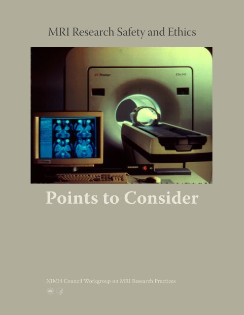
MRI Research Safety and Ethics: Points to Consider - NIMH
MRI Research Safety and Ethics: Points to Consider - NIMH
MRI Research Safety and Ethics: Points to Consider - NIMH

You also want an ePaper? Increase the reach of your titles
YUMPU automatically turns print PDFs into web optimized ePapers that Google loves.
<strong>MRI</strong> <strong>Research</strong> <strong>Safety</strong> <strong>and</strong> <strong>Ethics</strong>: <strong>Points</strong> <strong>to</strong> <strong>Consider</strong>Background <strong>and</strong> PurposeSeptember 14, 2005National Institute of Mental HealthBethesda, MDSponsored by:<strong>NIMH</strong> Council Workgroup on <strong>MRI</strong> <strong>Research</strong> PracticesSummaryAs with any complex <strong>and</strong> evolving technology, the use of magnetic resonance imaging (<strong>MRI</strong>) for researchraises important issues concerning the protection of human subjects or participants. In view of theincreasing involvement of <strong>MRI</strong> technology in human subjects (HS) research, particularly in non-clinical (i.e., university rather than mainly medical research) settings, <strong>NIMH</strong> recognizes the need <strong>to</strong> consider safety<strong>and</strong> ethical issues related <strong>to</strong> both the administration of MR (magnetic resonance) facilities <strong>and</strong> the use ofthese facilities for research.What was once a <strong>to</strong>ol used primarily for medical diagnosis has become a valuable <strong>to</strong>ol for clinical <strong>and</strong>basic cognitive <strong>and</strong> affective neuroscience research. This evolution raises questions regarding how <strong>to</strong>protect the safety of participants without unduly impeding important research. Although the protection ofhuman participants must remain the paramount consideration, regardless of the setting in which <strong>MRI</strong>research occurs (e.g., university settings vs. medical centers), approaches <strong>to</strong> <strong>and</strong> use of <strong>MRI</strong> in researchvary by context or environment. <strong>NIMH</strong> recognizes the lack across various research settings of anycomprehensive guidance <strong>to</strong> assist investiga<strong>to</strong>rs in reviewing the issues posed by <strong>MRI</strong> research concerningthe safety <strong>and</strong> protection of human participants.To address the above-noted concerns, the National Advisory Mental Health Council (NAMHC)Workgroup on <strong>MRI</strong> <strong>Research</strong> Practices was convened on September 14, 2005. Participants included<strong>NIMH</strong> Council members engaged in <strong>MRI</strong> research, intramural <strong>and</strong> extramural scientists including thoseinvolved in pediatric work, MR safety experts, including MR physiologists, MR physicists <strong>and</strong>neuroradiologists, <strong>and</strong> an at<strong>to</strong>rney with expertise in health-related research issues. The major goal of theWorkgroup was <strong>to</strong> enhance the protection of human participants by developing a set of "points <strong>to</strong>consider" for institutions <strong>and</strong> investiga<strong>to</strong>rs conducting or considering <strong>MRI</strong> research. The <strong>NIMH</strong> believesthat investiga<strong>to</strong>rs, institutions <strong>and</strong> facilities can use this document as a resource for the development,administration, evaluation, <strong>and</strong> use of <strong>MRI</strong> research facilities.The development of this document was guided by peer-reviewed publications <strong>and</strong> empirical data <strong>to</strong> thefullest extent possible. Where sufficient data were lacking, the Workgroup sought <strong>to</strong> acknowledge this
limitation <strong>and</strong> <strong>to</strong> identify the need for additional data. Furthermore, because technology is evolvingrapidly <strong>and</strong> new applications of MR continue <strong>to</strong> be discovered, it is expected that new questions will arise.Thus, this list should be updated periodically if it is <strong>to</strong> be kept current.Many of the issues discussed here have been considered previously in some form in the American Collegeof Radiology (ACR) White Paper on MR <strong>Safety</strong>, published in 2002 <strong>and</strong> revised in 2004 (Kanal et al.,2002, 2004) <strong>and</strong> in other recent publications (Shellock, 2001, 2006; Shellock <strong>and</strong> Crues, 2004).<strong>Research</strong>ers are strongly encouraged <strong>to</strong> consult these documents, <strong>and</strong> wherever relevant <strong>and</strong> appropriate,<strong>to</strong> follow the recommendations contained therein. The ACR reports were focused primarily, however, onthe use of MR in medical settings. The present considerations are intended <strong>to</strong> encompass the broader useof <strong>MRI</strong> in human neuroscience research, including studies conducted at facilities that exist outside ofmedical settings. These research settings raise additional issues that are complementary <strong>to</strong> those addressedby the ACR report. <strong>Consider</strong>ation of these research-specific issues was a primary focus of the <strong>NIMH</strong>Council Workgroup.This document summarizes the "points <strong>to</strong> consider" discussed by the NAMHC Workgroup. Examples ofsafe <strong>and</strong> ethical practices are discussed in relation <strong>to</strong> several issues. These examples are intended <strong>to</strong> beillustrative <strong>and</strong> should not be interpreted as an exhaustive or exclusive list. This document was presented<strong>to</strong> the full <strong>NIMH</strong> Council on September 15, 2006 <strong>and</strong> approved unanimously. By making the "points <strong>to</strong>consider" document available publicly, <strong>NIMH</strong> intends <strong>to</strong> provide a resource for researchers <strong>and</strong>institutions that use <strong>MRI</strong> in research.Organization of Meeting <strong>and</strong> ReportThe agenda was organized in<strong>to</strong> six <strong>to</strong>pics, which provide the organization for the points <strong>to</strong> consider thatfollow:A. <strong>MRI</strong> screeningB. Training, operating, <strong>and</strong> emergency proceduresC. Physical facilitiesD. Scanning/participant health variablesE. Context- Specific <strong>Consider</strong>ations: University vs. medical settingsF. Additional data needs <strong>and</strong> updating<strong>Points</strong> <strong>to</strong> <strong>Consider</strong>A. <strong>MRI</strong> ScreeningAre there procedures in place <strong>to</strong> ensure the adequate screening of participants prior <strong>to</strong> scanning?A-1. Is a screening form designed for maintaining MR safety in use at the facility as part of theresearch?
The Workgroup agreed on the need for careful <strong>and</strong> comprehensive screening of all individuals who enterthe <strong>MRI</strong> suite, i.e., all participants <strong>and</strong> any caretakers, such as parents, who accompany children in<strong>to</strong> the<strong>MRI</strong> suite, as well as anyone who routinely enters or has access <strong>to</strong> the facility (e.g., maintenance workers,security, as well as cleaning <strong>and</strong> emergency personnel). The use of a screening form was felt <strong>to</strong> constitutea useful approach. The Workgroup felt that it was good practice <strong>to</strong> document all screening procedures inwriting <strong>and</strong> <strong>to</strong> make this information available <strong>to</strong> <strong>MRI</strong> facility researchers <strong>and</strong> staff.A sample screening form is included in the American College of Radiology White Paper on MR <strong>Safety</strong>,along with the accompanying MR Safe Practice Guidelines (Kanal et al., 2002). Another samplescreening form for patients <strong>and</strong> research subjects may be downloaded from www.mrisafety.com or www.IMRSER.org. A Spanish version of this screening form is available from the ACR website: www.acr.org.A-2. Are there concerns about the participant's comprehension of questions related <strong>to</strong> issues ofsafety?The Workgroup recognized the need for complete <strong>and</strong> trustworthy information from participants <strong>and</strong>/ortheir caregivers <strong>and</strong> was concerned that incomplete or inaccurate responses <strong>to</strong> the items included on thescreening form would be problematic. The participant should comprehend the questions on the screeningform <strong>and</strong> sign an informed consent form. In situations where there are questions or concerns about theparticipant's comprehension (due <strong>to</strong> fac<strong>to</strong>rs such as questionable decision-making capacity, etc.), but theparticipant is nevertheless considered able <strong>to</strong> provide informed consent, the Workgroup recognized thevalue of having a knowledgeable companion, caretaker or family member present, with the consent of theparticipant, for the consent process. If there is a language barrier, an interpreter who is not a member ofthe participant's family is recommended. Also appropriate in specific circumstances is the participation ofan independent physician, consent moni<strong>to</strong>r, or other responsible party <strong>to</strong> help determine that theparticipant satisfies all criteria for MR safety clearance prior <strong>to</strong> entering the <strong>MRI</strong> suite. Clearly theresponsibility for ensuring that it is safe for the participant <strong>and</strong>/or accompanying family/other members <strong>to</strong>enter areas in the <strong>MRI</strong> environment that pose risks due <strong>to</strong> the presence of the magnetic field (describedbelow as "zones 3 or 4", see Section C-1) rests solely with the Principal Investiga<strong>to</strong>r <strong>and</strong> research facility.A-3. Once the participant completes the screening form, is the information reviewed in a screeninginterview?The Workgroup recognized the value of having the screening form for the participant <strong>and</strong>/or any caretakerwho might accompany the participant in<strong>to</strong> the scanner suite reviewed by a qualified interviewer who hasthe authority or access <strong>to</strong> the authority <strong>to</strong> deny entry in<strong>to</strong> the <strong>MRI</strong> suite <strong>and</strong> <strong>to</strong> decline <strong>to</strong> scan. Thefollowing credentials were considered by the Workgroup as qualifying the screening interviewer for thisrole: (a) Defined training <strong>and</strong> experience in MR safety <strong>and</strong> an underst<strong>and</strong>ing of the potential hazardsinvolved in both the MR environment <strong>and</strong> the MR imaging process; (b) knowledge of medical <strong>and</strong> dentaldevices <strong>and</strong> implants <strong>and</strong> of foreign metallic materials <strong>and</strong> conditions that pose hazards <strong>to</strong> the health <strong>and</strong>safety of individuals in the MR environment; <strong>and</strong>, (c) knowledge of how <strong>to</strong> assess the MR safety aspectsof implants <strong>and</strong> medical <strong>and</strong> dental devices <strong>and</strong> foreign metallic materials or how <strong>to</strong> access appropriate
<strong>and</strong> authoritative information. (See also B-1 concerning training.)The Workgroup also recognized the value of having the interviewer administer <strong>and</strong> review the screeningform with the participant item by item, <strong>to</strong> satisfy himself/herself that the participant has thoughtfullyconsidered each item <strong>and</strong> that there are no contraindications <strong>to</strong> scanning. This procedure would providegreater protection than relying solely on the participant independently filling out a written screening form.Similarly, having the interviewer sign <strong>and</strong> date the screening form was recognized as adding value.The importance of the interviewer's access <strong>to</strong> appropriate resources for resolving any questions related <strong>to</strong>the safety of questionable implants, medical or dental devices or foreign metals or objects wasacknowledged. Avenues for checking the safety of devices are numerous, <strong>and</strong> include, first, writtencontact with the manufacturer of the device, peer-reviewed published information, web searches <strong>and</strong>/orconsultation with designated safety experts, <strong>and</strong> discussion with an MR physicist or MR-trainedradiologist, among others.The Workgroup also recognized that, within the research context, MR safety information may be lessavailable for higher field strengths (e.g., 3 Tesla (T)) <strong>and</strong> that the safety aspects of devices may varydepending on how they are used. Thus, ready access <strong>to</strong> an identified individual with the expertise <strong>to</strong>evaluate safety in specific situations is invaluable.In summary, the Workgroup recognized the value of a qualified interviewer taking responsibility forensuring that the screening has been completed, that the screening is documented in writing with theinterviewer's signature <strong>and</strong> date, <strong>and</strong> that any questionable devices are considered safe based on theconsultation of an appropriate expert source.A-4. Have potential contraindications <strong>to</strong> scanning been identified?The Workgroup agreed that if the screening yields information that raises a question concerning safety,steps should be taken <strong>to</strong> resolve the question prior <strong>to</strong> proceeding. An example would be a metalworkerwho vaguely recalls an accident in which metal filings may have entered an injured the eye. In thissituation, screening, e.g., orbital film, should be used <strong>to</strong> rule out this contraindication <strong>to</strong> scanning.Institutional Review Board (IRB) permission <strong>to</strong> acquire screening studies involving ionizing radiationneeds <strong>to</strong> be prospectively obtained. If this type of follow-up screening is not definitive or unavailable (e.g., due <strong>to</strong> cost), the safest action would be <strong>to</strong> exclude the participant.If information indicates a foreign body is present, the Workgroup recognized the importance ofdetermining its MR safety prior <strong>to</strong> scanning.A-5. Do the screening procedures provide for redundancy?The Workgroup recognized that a second approach <strong>to</strong> screening would provide redundancy <strong>and</strong> thereforemight increase safety. This second approach might involve a second interview just prior <strong>to</strong> scanning <strong>to</strong>decrease the probability of missing an item that might pose a threat <strong>to</strong> safety.
As an alternative <strong>to</strong> two full interviews, a researcher could choose <strong>to</strong> include MR contraindications as par<strong>to</strong>f the screening for recruitment <strong>to</strong> the study. This brief interview could be performed prior <strong>to</strong> a participantcoming in for testing. This pre-screening should supplement, but not replace, the full MR screeninginterview. On the day of testing, a qualified interviewer at the MR center would provide a thorough inpersonscreening interview.Another alternative is the use of a second approach <strong>to</strong> ensure that the participant is free of surface objectsthat may be unsafe for <strong>MRI</strong> (such as pens, coins, other metals). For example, this may consist of having achild turn all his/her pockets inside out <strong>to</strong> ensure that they are empty or a check of all pockets <strong>and</strong> thebody using an appropriately sensitive ferromagnetic-detec<strong>to</strong>r w<strong>and</strong> (see Section A-6 below). As a thirdalternative, participants could be required <strong>to</strong> remove all clothes <strong>and</strong> jewelry <strong>and</strong> wear a gown. It remainsthe responsibility of the principal investiga<strong>to</strong>r <strong>and</strong> the research facility <strong>to</strong> ensure <strong>to</strong> the best of their abilitythat the participant does not have any ferromagnetic or other potential contraindicated devices/itemswithin them as a result of prior trauma, surgery, etc.A-6. Is a h<strong>and</strong>-held high-strength magnet available for screening purposes?The Workgroup also recognized the value of having a high strength magnet (1000 gauss or more)available for possible use as a supplement <strong>to</strong> screening, but recognized that its use should not replace thescreening interview. Whereas such a device might be useful for assessing the ferromagnetic status of, <strong>and</strong>screening out, potentially dangerous objects, it would not ensure that objects are safe in the MRenvironment (false negatives). Thus, the Workgroup expressed a need <strong>to</strong> recognize that the use of thismagnet was supplementary <strong>to</strong> a thorough screening process <strong>and</strong> not meant in any way <strong>to</strong> replace it.A-7. What role do ferromagnetic detec<strong>to</strong>rs play in the screening process?Ferromagnetic detec<strong>to</strong>rs include h<strong>and</strong> held, wall-mounted <strong>and</strong> walk-through models designed <strong>to</strong> sound analarm when ferrous objects are detected, but <strong>to</strong> allow ferrous free, metallic objects through. Sufficientlysensitive ferromagnetic detec<strong>to</strong>rs have the potential <strong>to</strong> reduce the risk of projectile incidents, patient injury<strong>and</strong> damage <strong>to</strong> equipment. However, such devices have not yet been systematically tested with respect <strong>to</strong>this potential. Therefore, the Workgroup opined that, at this time, ferromagnetic detec<strong>to</strong>rs cannot replace aconscientious screening <strong>and</strong>/or direct physical inspection, but constitute a potentially useful supplement <strong>to</strong>other screening measures.A-8. What steps are in place <strong>to</strong> screen for pregnancy?At present, there is no known risk of MR brain scanning of a pregnant woman <strong>to</strong> the developing fetus forscanning at 4T or less, <strong>and</strong> no known mechanism of potential risk under normal operating procedures.Nonetheless, the possibility that risks may be discovered in the future cannot be ruled out. Therefore,exposure of fetuses <strong>to</strong> MR scanning without any prospect of direct benefit may not be ethically justifiable.Indeed, the general policy in many clinical Radiology Departments is not <strong>to</strong> scan anyone who may bepregnant, absent compelling clinical need. Thus, it is appropriate <strong>to</strong> screen for pregnancy <strong>and</strong> <strong>to</strong> exclude
pregnant participants for the sake of caution.A somewhat separate, but related, issue is the fact that there may be potential risks associated with theexposure of fetuses <strong>to</strong> some intravenously administered MR contrast agents.It appears that most local IRBs intend <strong>to</strong> exclude potential participants who are pregnant from research<strong>MRI</strong> procedures. Based on Federal HS protection regulations (see below), the Workgroup expects thatpregnant females will not knowingly be scanned for research purposes unless the pregnant mother <strong>and</strong>/orfetus are the subject of an IRB approved pro<strong>to</strong>col that specifically provides for the inclusion of pregnantwomen <strong>and</strong> fetuses in the research. Such research would need <strong>to</strong> utilize appropriate informed consentprocedures consistent with the requirements of the Code of Federal Regulations (CFR) Title 45 (PublicWelfare) Part 46 (Protection of Human Subjects), Subpart B (Additional Protections for PregnantWomen, Human Fetuses <strong>and</strong> Neonates Involved in <strong>Research</strong>) (45 CFR Part 46, Subpart B), as applicable.A variety of approaches are used across centers <strong>to</strong> screen for <strong>and</strong>/or exclude pregnant or possibly pregnantparticipants. Some sites simply note, during the consent/assent process, that the individual should notparticipate if there is a possibility she may be pregnant. Other sites use questions that include the date ofthe last menstrual period <strong>and</strong>/or whether there is any chance the potential participant might be pregnant.Still others test for pregnancy in all females who have begun menstruation unless they are postmenopausalor have undergone surgical procedures after which pregnancy is not a possibility. Theapproach which will be used <strong>to</strong> screen for pregnancy should be described in the pro<strong>to</strong>col in order for theIRB <strong>to</strong> assess the risks <strong>and</strong> benefits of the pro<strong>to</strong>col.Pregnancy testing has the benefit of providing new information in cases where a female may not yetrealize she has conceived. Without such testing, a female may be scanned while unknowingly pregnant.At the same time, pregnancy testing holds implications for the disclosure of such results. For example,having a parent first learn of a child's sexual activity <strong>and</strong>/or pregnancy during the consent/assent orscreening process may be harmful for the adolescent female <strong>and</strong> her family; sensitivity <strong>to</strong> culturalinfluences is warranted here. Caution is warranted <strong>to</strong> avoid accidental disclosures of pregnancy <strong>to</strong>individuals who might be accompanying the participant.If disclosing a pregnancy can have potential negative consequences/risks under certain circumstances,IRBs will want <strong>to</strong> consider this issue. Investiga<strong>to</strong>rs should consider, in advance, how an "incidentalfinding" of pregnancy will be managed, i.e., whether appropriate staff are available <strong>to</strong> provide counseling<strong>and</strong> how such findings will be reported <strong>and</strong> participants counseled. Thus, it is important that thisinformation be provided <strong>to</strong> the IRB so that it may carefully balance the risks <strong>and</strong> benefits pertaining <strong>to</strong> thespecific population, research procedures, <strong>and</strong> methods used <strong>to</strong> screen for pregnancy when reviewingpro<strong>to</strong>cols <strong>and</strong> consent/assent procedures.In the case of minors, the Workgroup acknowledged that sensitivity <strong>to</strong> parent-child issues was important.To minimize the risks of placing under-age females in a potentially conflictual situation at the time oftesting, pregnancy might be mentioned as a reason not <strong>to</strong> participate at the time of recruitment. Thisallows the young woman <strong>to</strong> decline <strong>to</strong> participate without providing specific information bearing on this
issue. <strong>Consider</strong>ations for screening should take in<strong>to</strong> account whether <strong>to</strong> screen the minor privately or withthe parent or guardian present <strong>and</strong> whether <strong>to</strong> inform parents or guardians of the results of suchscreenings. As part of the assent process, the adolescent should be informed of what will be done with theresults of the screening, including any interview data <strong>and</strong>/or pregnancy testing (consistent with state <strong>and</strong>local statutes). Sites testing for pregnancy should consider in advance how participants <strong>and</strong>/or parents willbe informed of results, <strong>and</strong> whether there are personnel on site who are adequately trained <strong>to</strong> providecounseling. Such considerations should be discussed with the local IRB(s) in advance.Special consideration is warranted for vulnerable populations, whose members may be less accurate inreporting the possibility of pregnancy, e.g., minors, or potential participants with psychosis or majordepression. Added safeguards should be considered for such participants. Leaving decisions aboutwhether <strong>to</strong> participate up <strong>to</strong> potentially pregnant adolescents <strong>and</strong>/or people with severe mentalimpairments may be unacceptable <strong>to</strong> IRBs.Note that the issue of when a minor's health information, including pregnancy screening results, may ormust be disclosed <strong>to</strong> parents or guardians is governed by the Department of Health <strong>and</strong> Human Service'sHealth Insurance Portability <strong>and</strong> Accountability Act (HIPAA) <strong>and</strong>/or other applicable privacy <strong>and</strong> statelaws. The application of HIPAA would depend upon whether the screening is performed by a HIPAAcoveredentity (such as a healthcare provider that conducts certain transactions electronically). HIPAAmay also interact with other applicable privacy <strong>and</strong> state laws. With some exceptions, HIPAA generallyrequires covered entities <strong>to</strong> treat parents <strong>and</strong> guardians of un-emancipated minors as "personalrepresentatives" <strong>to</strong> whom the minor's records must be disclosed upon request, if the parent or guardian hasauthority under applicable law (e.g., state law) <strong>to</strong> act on behalf of the minor with respect <strong>to</strong> health caredecisions. However, where a state law permits a minor <strong>to</strong> consent <strong>to</strong> pregnancy treatment without a paren<strong>to</strong>r guardian's consent <strong>and</strong> the minor does so without parental involvement, this HIPAA requirement maynot apply. Note further that state laws may govern m<strong>and</strong>a<strong>to</strong>ry reporting of pregnancy <strong>to</strong> Child ProtectiveServices, depending on the age of the adolescent, the age of the sexual partner who fathered the child, <strong>and</strong>the difference in their ages. HIPAA would permit HIPAA-covered entities <strong>to</strong> disclose patient healthinformation as required by other laws. <strong>Research</strong>ers should consult their institutional legal counsel forguidance on the parental <strong>and</strong> other disclosure requirements, if any, that apply <strong>to</strong> a particular researchsetting, <strong>and</strong> the applicable requirements for reporting should be made clear <strong>to</strong> the adolescent.Providing the results of a pregnancy test <strong>to</strong> participants or parents implicates the federal ClinicalLabora<strong>to</strong>ries Improvement Act (CLIA) <strong>and</strong> state laws regulating labora<strong>to</strong>ry testing.<strong>Research</strong>ers should consult with legal counsel <strong>to</strong> ensure that screening involving pregnancy testing isperformed in compliance with applicable law.A-9. Are there approaches in place for screening for repeat scans?The question of screening for repeated scanning arises most commonly in cognitive neuroscience researchinvolving normal volunteers, some of whom may be scanned daily or weekly for some period of time.
The Workgroup discussed whether participants should complete a full screening questionnaire <strong>and</strong>interview for each scanning session. The Workgroup recognized the value of completing the fullscreening, i.e., full st<strong>and</strong>ard written questionnaire <strong>and</strong> interview, with appropriate signatures for each scan.B. Training, Operating, <strong>and</strong> Emergency ProceduresAre training, operating <strong>and</strong> emergency procedures in place <strong>to</strong> help ensure participant safety?B-1. Are specific policies <strong>and</strong> procedures for training personnel associated with the <strong>MRI</strong> facilitydeveloped <strong>and</strong> documented?Given that the <strong>MRI</strong> environment presents many potential dangers <strong>to</strong> untrained or improperly screenedindividuals, the Workgroup recognized the need for appropriate levels of training for all individuals whooperate the scanner <strong>and</strong>/or have routine access <strong>to</strong> the <strong>MRI</strong> suite <strong>and</strong> for a clearly specified scheme fortraining <strong>and</strong> certifying individuals for each level of authorization. A range of options was mentioned forcertification, including didactic training, mastery of written materials, <strong>and</strong> terms of apprenticeship, as wellas written <strong>and</strong>/or practical tests.The following was discussed as one example of a multi-level training <strong>and</strong> certification scheme:Level I personnel are defined as those who are authorized <strong>to</strong> have unsupervised access <strong>to</strong> the <strong>MRI</strong> suite,but who lack authority <strong>to</strong> screen other individuals or <strong>to</strong> bring other individuals in<strong>to</strong> the scanner facility.Certification at Level I requires training in how <strong>to</strong> screen oneself, a clear underst<strong>and</strong>ing of what is <strong>and</strong> isnot safe in the <strong>MRI</strong> environment, <strong>and</strong> knowledge of safety procedures for entering the <strong>MRI</strong> suite <strong>and</strong> verybasic emergency procedures, e.g., knowledge of whom <strong>to</strong> call <strong>and</strong> how <strong>to</strong> rapidly access appropriatephone numbers. Level I may include cleaning, transportation, security, <strong>and</strong> anesthesia staff, <strong>and</strong> anyothers who may have legitimate reasons <strong>to</strong> enter the <strong>MRI</strong> suite unsupervised <strong>and</strong> thus need <strong>to</strong> beappropriately trained in safety <strong>and</strong> emergency procedures.Level II personnel are those who, in addition <strong>to</strong> Level I certification, are also certified <strong>to</strong> operate the MRequipment, as well as <strong>to</strong> screen others for entry in<strong>to</strong> the <strong>MRI</strong> suite <strong>and</strong> oversee their presence in the suite<strong>and</strong> during scanning. They would have more in depth knowledge of MR safety issues including the safetyof different materials for the particular environment, the safety guidelines of the applicable IRBs, <strong>and</strong>where <strong>to</strong> get additional information if needed.Level III personnel are faculty or senior staff who, in addition <strong>to</strong> having achieved levels I <strong>and</strong> IIcertification, are also certified <strong>to</strong> train <strong>and</strong> certify Level I <strong>and</strong> Level II personnel <strong>and</strong> are authorized by thefacility direc<strong>to</strong>r <strong>to</strong> do so. These individuals would have knowledge of the requirements needed <strong>to</strong> run asafe <strong>MRI</strong> environment <strong>and</strong> would be responsible for keeping these requirements updated.It was felt that researchers should adhere <strong>to</strong> an accepted national st<strong>and</strong>ard of care consistent with safetyprovisions, such as those of the ACR. Personnel should be trained on all elements relevant <strong>to</strong> theirresearch practices. In the terms of the ACR guidelines, those operating the scanner should be trained <strong>to</strong>
Level II status insofar as the guidelines are relevant <strong>to</strong> their research practices. For many universitysettings, this may not include use of contrast agents, but will include training in regard <strong>to</strong> radiofrequency(RF) thermal safety issues, gradient neurostimulation, <strong>and</strong> audi<strong>to</strong>ry concerns.Sites may design their own training procedures so long as these adhere <strong>to</strong> nationally established st<strong>and</strong>ardsof care <strong>and</strong> encompass the MR potential risks <strong>to</strong> which participants might be exposed. For example, if asite expects <strong>to</strong> have a participant undergo MR scanning of the head, MR safety training requirements forthe opera<strong>to</strong>r of this study would cover biological <strong>and</strong> mechanical effects of static magnetic fields, safetyaspects related <strong>to</strong> gradient <strong>and</strong> RF magnetic fields, cryogen usage <strong>and</strong> safety issues, safety aspects ofclaustrophobia <strong>and</strong> other anxiety management, etc. If no sedation, intravenous medication, or MR contrastagents are <strong>to</strong> be administered, training regarding these areas may not be required.The Workgroup recognized that principal investiga<strong>to</strong>rs might have certification at any one of these levelsdepending on their role in running the study. Regardless of their level of certification, all responsibility forthe safety of the <strong>MRI</strong> examination will at all times rest with the principal investiga<strong>to</strong>r <strong>and</strong> the designatedsafety officer.The Workgroup noted the importance of the facility having clearly documented procedures for training<strong>and</strong> certifying personnel at all levels, for keeping certifications current, <strong>and</strong> for maintainingcomprehensive, up-<strong>to</strong>-date records regarding the certification status of all personnel associated with, orwho have access <strong>to</strong>, the facility.B-2. Are staffing patterns adequate for dealing with emergencies?The Workgroup discussed the need <strong>to</strong> anticipate potential emergencies <strong>and</strong> <strong>to</strong> ensure the availability ofadequate personnel coverage with which <strong>to</strong> affect a quick response. The Workgroup felt that the presenceof at least two trained staff (at least one of whom had attained Level II status), at all times that aparticipant or accompanying family member/escort was within an area in which the magnetic field poseda risk (e.g., zones 3 <strong>and</strong>/or 4 as described below), would help ensure an adequate response <strong>to</strong> anemergency.Furthermore, for all cases in which an MR contrast agent was being administered via any route other thanby mouth, a duly licensed physician must be on site <strong>and</strong> readily accessible <strong>to</strong> h<strong>and</strong>le possible adversereactions <strong>to</strong> the administered MR contrast agent. <strong>Research</strong> involving participants of course necessitateswhatever additional (nonspecific <strong>to</strong> the MR environment) protections are needed <strong>to</strong> deal with any medicalissues, e.g., sedation, cardiac issues, psychiatric issues, etc.B-3. Are there clear lines of authority for dealing with safety issues?The Workgroup recognized the need for clear lines of authority for dealing with safety issues <strong>and</strong> felt thata good approach for ensuring this was <strong>to</strong> designate an on-site safety officer for each scan. The designationof a single safety officer for each scan avoids ambiguity or diffusion of responsibility during anemergency. The Workgroup felt that the equivalent of Level II certification described above was
appropriate for such an individual.Personnel involved in the study session should be made aware of the designated safety officer for eachscan. For example, if there is more than one potential safety officer present, e.g., more than one Level IIstaff member at a scan, it is important that a single individual be recognized as in charge of safety issuesprior <strong>to</strong> entry in<strong>to</strong> the facility.B-4. Are st<strong>and</strong>ard procedures for dealing with emergencies developed <strong>and</strong> documented at the <strong>MRI</strong>facility <strong>and</strong> are staff prepared <strong>to</strong> implement these procedures?The Workgroup recognized the importance of having procedures in place for dealing with emergencies,<strong>and</strong> maintaining staff readiness <strong>to</strong> deal with potential emergencies especially in non-medical settings. TheWorkgroup felt that aspects of this preparedness included knowledge of whom <strong>to</strong> call in the case ofemergencies of various sorts, immediate access <strong>to</strong> the appropriate phone numbers, how <strong>to</strong> remove aparticipant from the scanner, transfer the participant on<strong>to</strong> a stretcher if necessary, evacuate him/her fromthe scanning suite, <strong>and</strong> when <strong>and</strong> how <strong>to</strong> quench the magnet.In addition <strong>to</strong> the importance of emergency preparedness, it is equally important for researchers in nonmedicalsettings <strong>to</strong> make it explicitly clear <strong>to</strong> participants-as part of the informed consent procedure-thatemergency medical services may not be available onsite. This precaution is <strong>to</strong> prevent participants frommistakenly interpreting the presence of the MR scanner as evidence that they are in a medical setting withassociated emergency medical services.Related <strong>to</strong> this point, the Workgroup discussed the merits of additional safety training, such ascardiopulmonary resuscitation (CPR) techniques. However, because there are no known risks of acardiovascular event specifically associated with exposure <strong>to</strong> the <strong>MRI</strong> environment or with being scanned(other than the presence of a pacemaker or an implantable cardioverter defibrilla<strong>to</strong>r), the Workgroup didnot find any reason <strong>to</strong> require that personnel associated with a research-dedicated <strong>MRI</strong> facility be certifiedas having been successfully trained in CPR. Exceptions <strong>to</strong> this would include examinations of participantswho are otherwise at risk for cardiopulmonary events.B-5. Has the <strong>MRI</strong> site established relationships with emergency resources in the facility <strong>and</strong>community?The Workgroup recognized the importance of establishing relationships with emergency resources withinthe host institution (e.g., university or medical center), as well as in the community. The Workgroup feltthat particularly in non-medical (e.g., university) settings, where emergency workers may not expect <strong>to</strong>find an <strong>MRI</strong> facility, the relevant services (such as the fire <strong>and</strong> police departments) should be made awareof the existence of the facility <strong>and</strong> its special safety concerns (e.g., personnel cannot enter with air tank).Regular meetings with such personnel were thought <strong>to</strong> be helpful for ensuring familiarity with the facility<strong>and</strong> its safety considerations <strong>and</strong> for reviewing its emergency procedures. Also thought <strong>to</strong> be helpful wasthe practice of educating public safety <strong>and</strong> risk management groups at the institution that houses the <strong>MRI</strong>facility with respect <strong>to</strong> the <strong>MRI</strong> facility's special considerations.
B-6. What centralized system exists at the <strong>MRI</strong> facility for reporting, managing <strong>and</strong> archivingincidents <strong>and</strong> adverse events associated with scanning?The group discussed the need for a system for reporting <strong>and</strong> recording incidents <strong>and</strong> adverse eventsassociated with scanning <strong>and</strong> its value for improving safety. Such a system would allow researchers <strong>to</strong> fileincident reports <strong>and</strong> make the archive available for consultative purposes. It was also felt important thatall breaches of safety procedures <strong>and</strong> other safety-related incidents should be reported, not just thoseinvolving actual adverse outcomes. This would provide useful information about the frequency of "nearmisses" <strong>and</strong> other occurrences that might reveal weaknesses in safety procedures <strong>and</strong>/or signal a declinein safe practices.<strong>Research</strong>ers in clinical facilities must also be aware of any requirements imposed by the facility's riskmanagement reporting process <strong>and</strong> of any legal constraints upon the disclosure of incident information <strong>to</strong>individuals outside of the institution.C. Physical FacilitiesWhat protections are in place in the facility <strong>to</strong> ensure safety in those areas affected by the magneticfield? What special restrictions, equipment <strong>and</strong> experts for evaluating the MR safety of devices doesthe facility provide?C-1. What methods are in place <strong>to</strong> control <strong>and</strong> regulate access <strong>to</strong> the <strong>MRI</strong> suite (control room <strong>and</strong>magnet)?Some, but not all, facilities use a st<strong>and</strong>ard "zoning system" such as that outlined in the ACR guidelines, <strong>to</strong>control <strong>and</strong> regulate access <strong>to</strong> the control room <strong>and</strong> magnet for safety purposes. The ACR zoning systemis described below as one example of a system that can be used. Whatever system is adopted shouldprovide protections comparable <strong>to</strong> those described below.Zone 1 includes all areas that are freely accessible <strong>to</strong> the general public, <strong>and</strong> is typically outside of the<strong>MRI</strong> environment (i.e., that portion of the environment in which the magnetic field poses a risk).Zone 2 is the interface between the publicly accessible, uncontrolled zone 1 <strong>and</strong> the strictly controlledzones 3 <strong>and</strong> 4 (which pose a risk). Typically, participants are greeted in zone 2 <strong>and</strong> their movementthroughout zone 2 is under at least intermittent supervision of MR personnel. This area is typically usedfor initial contact <strong>and</strong> screening.Zone 3 is the region through which there is free, physically unrestricted access <strong>to</strong> areas that pose a riskdue <strong>to</strong> the presence of the <strong>MRI</strong> scanner itself (which is in "zone 4" - see below). Zone 3 will alwaysinclude, but may extend beyond, areas that pose a risk. For example, zone 3 may include the consoleroom, equipment room(s), <strong>and</strong> prepara<strong>to</strong>ry areas, which may or may not fall within the fringe fields (five
gauss line) of the magnet, but all of which have physically unrestricted access <strong>to</strong> areas that are affected bythe magnet's electrical <strong>and</strong>/or magnetic fields. The boundaries of the five gauss line should be clearly <strong>and</strong>visibly marked within zone 3 (e.g., with a red line on the floor <strong>and</strong> with signs indicating the presence ofthe field <strong>and</strong> associated risks), so that it is clear whenever someone is approaching an area of a five gaussor greater exposure level.In some cases, zone 3 may involve discontinuous regions. For example, the static fringe fields of themagnet may extend beyond the physical confines of the scanning suite, in<strong>to</strong> areas that are discontinuousfrom the scanner facility (e.g., in<strong>to</strong> courtyards, roofs, or other areas neighboring the building). Zone 3encompasses these additional areas. When zone 3 involves areas that fall outside of the scanning facility,it is imperative that these additional areas <strong>and</strong> the risks they present be marked clearly <strong>and</strong> visibly.Access <strong>to</strong> zone 3 should be strictly physically restricted, ensuring that there is fully regulated access <strong>to</strong> allareas that pose any risk due <strong>to</strong> the presence of the magnet or its associated electromagnetic fields. Thisneed <strong>to</strong> restrict access applies equally <strong>to</strong> the sorts of discontinuous areas mentioned above. It is imperativethat these additional areas <strong>and</strong> the risks they present be marked clearly <strong>and</strong> visibly <strong>and</strong> that withoutexception (<strong>and</strong> in accordance with their status as zone 3 areas), they be physically restricted frominadvertent access from any non-MR personnel.Access <strong>to</strong> zone 3 should be controlled by Level II MR personnel (with the exception that Level Ipersonnel are permitted <strong>to</strong> screen themselves <strong>and</strong> enter without supervision; see B-1 above). Nounscreened non-MR personnel are allowed access <strong>to</strong> zone 3. "Non-MR personnel" are those not trained orcertified with respect <strong>to</strong> <strong>MRI</strong> facilities at any level. Access should be restricted with key locks, pass keylocking systems or other reliable, physically restricting methods that can differentiate between MRpersonnel <strong>and</strong> non-MR personnel. Combination locks are discouraged, since combinations can be <strong>to</strong>oeasily shared with unauthorized personnel.Zone 4 is the physical confines of the room housing the <strong>MRI</strong> scanner itself. By definition, zone 4 alwaysfalls within zone 3. The primary significance of zone 4 pertains <strong>to</strong> the risks due the attractive forces of thestatic field of the magnet (inducing projectiles), <strong>and</strong> the effects of the time varying gradient <strong>and</strong> RF fieldswhen imaging is in progress. The entry <strong>to</strong> zone 4 should be clearly marked, with a sign indicating thepresence of <strong>and</strong> potential danger due <strong>to</strong> the magnet.C-2. How are areas affected by the MR system demarcated? Do danger signs clarify for whom theseareas pose risks?The Workgroup recognized the importance of clearly demarcating the areas affected by the magnet <strong>and</strong>for warning persons for whom the magnetic fields pose health risks. Examples offered of appropriatesigns are those indicating "Danger" <strong>and</strong> "The magnet is always on" that are typically used <strong>to</strong> demarcateaffected areas <strong>and</strong> make clear what types of persons are at risk (e.g., those with pacemakers). Otherexamples are floor markings <strong>and</strong>/or barriers clearly delimiting the five gauss line.C-3. Is MR-safe fire extinguishing equipment available <strong>and</strong> appropriately located in the <strong>MRI</strong> suite?
The Workgroup acknowledged that s<strong>to</strong>ring MR-safe fire extinguishing equipment in the <strong>MRI</strong> suite (i.e.,in either zones 3 <strong>and</strong>/or 4 as described above) ensures immediate access <strong>and</strong> therefore enhances safety.Although the risk of a fire is small, the ready availability of such equipment would prove extremely usefulin such a rare event.The Workgroup further felt that s<strong>to</strong>ring such equipment in the suite helps protect against the risk ofhaving unsafe equipment enter the suite.C-4. How will the MR safety of devices for the <strong>MRI</strong> research facility be assured?Most devices used as supplementary equipment in MR settings (e.g., surface coils, cardiac recordingdevices, etc.) are Food <strong>and</strong> Drug Administration (FDA)-approved. However, in cases where this is not thecase (e.g., non-FDA-approved devices developed for research, such as devices for displaying stimuli orrecording behavioral responses from the participant), the Workgroup recognized the importance ofspecifying <strong>and</strong> following clear guidelines for ensuring the safety of their use in research settings.Consistent with such practices would be a careful review by individuals competent <strong>to</strong> evaluate the safetyof such devices <strong>and</strong> the provision of a description of these devices <strong>and</strong> procedures for evaluating theirsafety <strong>to</strong> the IRB.Toward these ends, the Workgroup recognized the value of having access <strong>to</strong> one or more consultants withthe expertise needed for evaluating the MR safety of all devices used in the facility (e.g., non-st<strong>and</strong>ardcoils, ancillary devices for presenting stimuli <strong>and</strong> recording responses, etc.). This technical expert orgroup should be competent <strong>to</strong> decide whether IRB, FDA <strong>and</strong>/or external safety consultation are needed,that were not provided by the manufacturer of the scanner, for devices that are <strong>to</strong> be used with it.Among the safety issues that the Workgroup felt were important <strong>to</strong> consider were the magnetic propertiesof the device, as well as the potential for creating current loops that can overheat <strong>and</strong> produce burns. Theproper operational aspects of a given device relative <strong>to</strong> the <strong>MRI</strong> environment should also be considered.C-5. Are adequate oxygen concentrations in the <strong>MRI</strong> suite ensured?The FDA states that: "The oxygen concentration in accessible areas should not be allowed <strong>to</strong> go belowacceptable levels." Oxygen concentration can be moni<strong>to</strong>red using oxygen sensors in the <strong>MRI</strong> suite, suchas those installed by the scanner manufacturers.D. Scanning/Participant Health VariablesWhat st<strong>and</strong>ard procedures are in place in the <strong>MRI</strong> facility for protecting the participant's health,the safe operation of the scanner, <strong>and</strong> the reporting <strong>and</strong> archiving of incidents relevant <strong>to</strong> theseissues?
D-1. What st<strong>and</strong>ard procedures are documented <strong>and</strong> in place for ensuring safety?The Workgroup recognized the value of clearly documenting all procedures for the safe operation of thescanner <strong>and</strong> the h<strong>and</strong>ling of adverse events <strong>and</strong> for making this documentation readily accessible <strong>to</strong> allpersonnel associated with the facility. These procedures <strong>and</strong> their documentation would be an importantfocus of training for Level II personnel (see Section B-1 above).The Workgroup felt that st<strong>and</strong>ard procedures should also be in place for the detection <strong>and</strong> reporting ofincidental findings <strong>and</strong> other specific health <strong>and</strong> safety-related issues as described below.D-2. Are participants always visible <strong>to</strong> <strong>and</strong> in hearing contact of the <strong>MRI</strong> opera<strong>to</strong>r <strong>and</strong> allowed <strong>to</strong>terminate the scan <strong>and</strong> exit the scanner at any time?The Workgroup felt it important that the participant be visible <strong>to</strong> <strong>and</strong> in hearing contact of the <strong>MRI</strong>opera<strong>to</strong>r at all times. The Workgroup further acknowledged that the ability <strong>to</strong> voluntarily terminate astudy at any time during scanning is in accord with the federal regulations for the protection of humansubjects (Code of Federal Regulations Basic Health <strong>and</strong> Human Services Policy for Protection of Human<strong>Research</strong> Subjects: 45 CFR 46.116 (a) (8)).D-3. Is hearing adequately protected?The Workgroup acknowledged the importance of using earplugs or other hearing protection duringscanning <strong>to</strong> attenuate noise levels.Acoustic noise levels during scanning generally fall between 65 <strong>and</strong> 95 decibels (dB), but can reach morethan 120 dB. Acoustic noise levels vary depending on the pulse sequence, field strength, <strong>and</strong> other fac<strong>to</strong>rs.Hearing protection may lower these exposures by about 20 dB, but this may vary according <strong>to</strong> the type ofprotection being used. Risks will differ given single versus repeated exposures, scan duration <strong>and</strong> theeffectiveness of the protection.The Workgroup recognized the importance of facilities working within st<strong>and</strong>ards specified by the U.S.Occupational <strong>Safety</strong> <strong>and</strong> Health Administration (OSHA 29 CFR 1910.95), which take in<strong>to</strong> account thelength <strong>and</strong> level of exposures. If noise levels exceed those specified below, attenuation must be used <strong>to</strong>decrease exposures <strong>to</strong> within these limits. The term dBA, or A-weighted decibels, refers <strong>to</strong> the sound levelwhen measured on an A scale of a st<strong>and</strong>ard sound level meter at slow response.Hours per daySound level dBA slow response8 906 924 953 972 100
1 1/2 1021 1051/2 1101/4 or less 115OSHA also specifies that exposure <strong>to</strong> impulsive or impact noise should not exceed 140 dB peak soundpressure level. In addition, apart from these guidelines, a weighted root mean square sound pressure levelgreater than 99 dBA with hearing protection in place poses a significant risk (according <strong>to</strong> a FDAdocument: Criteria for Significant Risk Investigations of Magnetic Resonance Diagnostic Devices, issuedon July 14, 2003).For any research participant, any MR imaging sequence that has not received FDA approval should onlybe executed with hearing protection in place unless it can be documented that the anticipated audi<strong>to</strong>rylevels would be clearly below established <strong>and</strong> acceptable thresholds <strong>and</strong> guidelines.The issue of medical conditions that might increase risk for hearing loss as a result of MR-generatedacoustic noise was discussed. The Workgroup was not aware of any data <strong>to</strong> indicate that conditions suchas tinnitus or the loss of stapedial reflexes might increase susceptibility <strong>to</strong> noise-induced hearing loss due<strong>to</strong> MR scanning, if adequate hearing protection is used <strong>to</strong> attenuate acoustic noise below allowable limits.D-4. Do the research scans fall within FDA guidelines for magnet strength, specific absorption rate(SAR), gradient fields' rates of change, <strong>and</strong> acoustic noise?In 2003, the FDA specified the following nonbinding guidelines for <strong>MRI</strong> safety (http://www.fda.gov/cdrh/ode/guidance/793.html):Main static magnetic field: For adults, children, <strong>and</strong> infants greater than one month of age, field strengthsgreater than 8T are presently considered <strong>to</strong> pose a potential significant risk. For neonates (infants underone month of age), field strengths greater than 4T are presently considered <strong>to</strong> pose a potential significantrisk.Specific absorption rate (SAR): The following are considered significant risks (expressed in watts perkilogram):Whole body average dose over 15 or more minutes: 4 W/kgHead average dose over 10 or more minutes: 3 W/kgHead or <strong>to</strong>rso dose per gram of tissue over 5 or more minutes: 8 W/kgExtremities dose per gram of tissue over 15 or more minutes: 12 W/kgGradient fields' rates of change: Any rate of change of gradient fields (dB/dt) sufficient <strong>to</strong> produce severediscomfort or painful nerve stimulation is considered a significant risk.
Acoustic noise: A peak acoustic noise level over 140 dB is considered significant risk, as is a weightedroot-meansound pressure level greater than 99 dBA with hearing protection in place. (In addition <strong>to</strong> theFDA guidelines, OSHA guidelines specify the lengths of time at which various noise levels present risks<strong>to</strong> hearing [see D-3]).Because the FDA considers <strong>MRI</strong>/functional <strong>MRI</strong> research studies a nonsignificant risk, no investigationaldevice exemption (IDE) is required. (See Information Sheet Guidance for IRBs, Clinical Investiga<strong>to</strong>rs,<strong>and</strong> Sponsors; Significant Risk <strong>and</strong> Nonsignificant Risk Medical Device Studies: http://www.fda.gov/oc/ohrt/irbs/devrisk.pdf). The regulations for studies using devices such as MR scanners do not requirephysician prescriptions for clinical procedures that are done as part of clinical research. The responsibilityfor safety <strong>and</strong> the protection of human participants rests with the local IRB <strong>and</strong> the informed consentprocess (21 CFR Part 812.5, http://www.accessdata.fda.gov/scripts/cdrh/cfdocs/cfcfr/cfrsearch.cfm).The Workgroup felt that no special actions were needed for research scans that fall within the FDAguidelines concerning magnet strength, pulse sequences (gradient fields' rates of change), SAR, <strong>and</strong> peaksound levels (except see D-3 concerning acoustic noise). Under conditions in which research scans falloutside of FDA guidelines, the Workgroup felt it important <strong>to</strong> make the IRB aware of this <strong>and</strong> informparticipants accordingly. In such instances, the Workgroup recognized the need for consent language thatwould need <strong>to</strong> be very different from that used when working within the FDA ranges.D-5. What special precautions are in place for infant scanning?The Workgroup discussed the need <strong>to</strong> protect against loss of body heat (hypothermia) <strong>and</strong> for physiologicmoni<strong>to</strong>ring of heart rate <strong>and</strong> oxygen saturation when scanning infants.To protect against hypothermia, the Workgroup recognized the value of moni<strong>to</strong>ring core temperature,swathing the infant tightly, using warming packs, turning off fans, <strong>and</strong> increasing the temperature in thescanning room, as well as limiting the scan time <strong>to</strong> one hour <strong>to</strong> help prevent heat loss. The Workgroupalso recognized that significant changes from baseline in heart rate <strong>and</strong> oxygen saturation warrantterminating the scan.D-6. Is there a clear pro<strong>to</strong>col in place for dealing with incidental findings?The Workgroup recognized the importance of having a clear pro<strong>to</strong>col for dealing with incidental findingsprior <strong>to</strong> the start of the research. Important aspects of this pro<strong>to</strong>col were felt <strong>to</strong> include a consent form thatclearly distinguishes research from clinical scans, that explicitly discusses the potential for incidentalfindings <strong>and</strong> associated risks, that informs the participant as <strong>to</strong> whether or not the scans will be reviewedby a clinician qualified <strong>to</strong> render a radiological interpretation, <strong>and</strong> that describes the path that will betaken in the event that an incidental finding occurs. The Workgroup viewed these as consistent with theneed for having a pro<strong>to</strong>col in place for dealing with incidental findings in general, including non-imagingrelatedincidental findings.The Workgroup recognized the importance of not having participants confuse a research scan with a
clinical scan. This might be addressed with consent language that states that the research scan they arereceiving is not a clinical scan (e.g., may not be of clinical quality or thoroughness) <strong>to</strong> avoid any inferencethat the scan rules out a medical problem. Consent language used in a non-medical setting might make itclear that the setting is not a medical one.Further, despite the fact that the scan may not be of clinical caliber <strong>and</strong>/or that there may be no clinicallytrained investiga<strong>to</strong>rs involved in the research, the Workgroup felt it important that participants beinformed of the possibility that an abnormality may be detected or suspected in the process of theresearch, the clinical significance of which may not be clear. In this regard, the risks associated with falsepositives are important <strong>to</strong> recognize <strong>and</strong> acknowledge, including unnecessary worry, expense for medicalfollow-up, <strong>and</strong> possibly unpleasant or invasive medical tests.The Workgroup considered that a clear pro<strong>to</strong>col for dealing with incidental findings should be outlined<strong>and</strong> in place (as discussed above) including the stipulation that the participant will be fully informedconcerning the policies <strong>and</strong> procedures in place concerning potential incidental findings. It will be up <strong>to</strong>the IRB <strong>to</strong> review <strong>and</strong> approve the pro<strong>to</strong>col.In instances where the plan does not include a clinical review, if a researcher does notice somethingbelieved <strong>to</strong> be out of the ordinary on a scan, the Workgroup considered that the researcher could alert theparticipant <strong>to</strong> it. The researcher could either share the information with a physician with the participant's(or parent/legal guardian's) consent, or encourage the participant <strong>to</strong> seek a clinical referral.D-7. Has a qualified individual been identified who will report any incidental findings <strong>to</strong>participants?As stated above, there should be a pro<strong>to</strong>col for dealing with incidental findings that includes a plan forreporting suspected findings <strong>to</strong> the participant. The pro<strong>to</strong>col should identify appropriate personnel orconsultants who will report such findings <strong>to</strong> the participant.An individual responsible for reporting incidental findings should be knowledgeable of the facility'spolicies with regard <strong>to</strong> the detection <strong>and</strong> reporting of incidental findings <strong>and</strong> capable of conveying thepotential significance of the findings <strong>and</strong> possible need for clinical follow-up <strong>to</strong> the participant effectively<strong>and</strong> with sensitivity.The Workgroup concluded that if a physician were involved in the study, he/she would be an appropriateinformant. In non-medical settings, a principal investiga<strong>to</strong>r or other responsible <strong>and</strong> qualified investiga<strong>to</strong>rmay be an appropriate person <strong>to</strong> serve in this role.D-8. Has the need for additional safeguards for vulnerable populations, such as children, beenconsidered <strong>and</strong> resolved?Members of the Workgroup expressed some concern that vulnerable populations, such as children, mightwarrant additional protections. For example, the Workgroup considered cases in which an incidental
finding of potential clinical significance is merely shared with a parent who may not fully appreciate theneed for follow-up (e.g., asymp<strong>to</strong>matic malformation that was causing sub-clinical seizures <strong>and</strong> thusimpacting the child's learning). In anticipating this type of situation, one might consider obtainingpermission <strong>to</strong> inform the child's primary care physician as part of the consent process.Other issues, not unique <strong>to</strong> MR research, include suspected child abuse or neglect, e.g., the latterpotentially associated with a parent's failure <strong>to</strong> address a child's medical needs. During a neuroimagingstudy, an <strong>MRI</strong> procedure may yield findings suggestive of abuse. In such instances, state laws m<strong>and</strong>atingthe reporting of suspected abuse or neglect may become relevant.D-9. Does the research involve repeated scanning?The Workgroup agreed that safety concerns do not dictate any set limits on the duration or frequency ofscanning. The duration of a scanning session was felt <strong>to</strong> be limited only by the participant's ability <strong>to</strong>remain still <strong>and</strong> <strong>to</strong>lerate the scanning conditions. The appropriateness of pro<strong>to</strong>cols involving long orfrequent scans will be determined by the local IRB.The Workgroup knew of no harmful biological effects of short- or long-term exposure <strong>to</strong> static magneticfields. The National Council on Radiation Protection <strong>and</strong> Measurements Commentary No. 18 (2003) onthe biological effects of modulated radiofrequency fields concludes "that the scientific literature related <strong>to</strong>modulation-dependence of biological effects of RF energy is not sufficient <strong>to</strong> draw any conclusions aboutpossible modulation-dependence of health hazards of RF fields nor is there any apparent biophysical basisfrom which <strong>to</strong> anticipate such hazards apart from very intense RF pulses produced by some specializedmilitary equipment."However, guidelines <strong>to</strong> prevent excessive heating <strong>and</strong> burns associated with magnetic resonanceprocedures can be found at http://www.imrser.org/PaperPDFlist.asp?pgname=Guidelines.D-10. Are subjects with major contraindications <strong>to</strong> scanning being studied?The Workgroup agreed that MR scanning of participants with major contraindications <strong>to</strong> scanning is onlyappropriate when there is a clinical need or the research is targeted at a special population in whichpotential benefits have been appropriately weighed against the risks. Major contraindications mightinclude, but are not limited <strong>to</strong>, metal in the eyes, cardiac pacemakers, implanted cardioverterdefibrilla<strong>to</strong>rs, neurostimulation systems, <strong>and</strong> cochlear implants. In such instances, specific risk/benefitratios would, of course, be addressed by the relevant IRB, based on review of inclusion/exclusion criteriaincluded in study pro<strong>to</strong>cols.D-11. Are participants with "minor" contraindications <strong>to</strong> MR scanning being studied?The Workgroup discussed the situation in which participants with "minor" contraindications <strong>to</strong> scanningare studied. A working definition of a "minor" contraindication is one which is unlikely <strong>to</strong> pose a risk, but
which may, albeit infrequently <strong>and</strong> if not carefully moni<strong>to</strong>red, result in injury.The Workgroup felt that the participant should be informed of the potential risks involved in such "minor"contraindications <strong>and</strong> recognized the value of appropriate precautions being instituted.The example most thoroughly discussed was that of tat<strong>to</strong>os. Given that there is some risk of burns, thegroup considered the value of carefully moni<strong>to</strong>ring the participant, although it has also been noted thatburns can occur before the participant notices them or develop after the participant is removed from thescanner. Alternatively or in addition, ice packs or cold compresses might be prospectively placed on anytat<strong>to</strong>o in the volume about <strong>to</strong> undergo RF irradiation in the MR imaging process, although there is noconsensus on the efficacy of this measure.D-12. What should be done when a participant presents with an implant or device for which safetyinformation is unclear or unavailable?The Workgroup acknowledged that participants with implants or devices for which safety information isunclear or unavailable should not be scanned.Ready access <strong>to</strong> a trustworthy <strong>and</strong> up-<strong>to</strong>-date list of devices <strong>and</strong> information on their MR safety was felt<strong>to</strong> be most useful (see www.<strong>MRI</strong>safety.com). For participants with devices for which information isunavailable, the group agreed that the safest thing <strong>to</strong> do is <strong>to</strong> exclude the participant from study.D-13. Are participants being scanned at field strengths for which devices in question have beendetermined <strong>to</strong> be acceptable for MR procedures?The group recognized that implants <strong>and</strong> devices that have acceptable MR conditions established at 1.5Tmay not be safe at 3T. Given that field strengths as high as 7T <strong>and</strong> 8T are in use in research settings, thismay mean excluding subjects with implants <strong>and</strong> devices due <strong>to</strong> lack of safety data at higher fields.D-14. What procedures for documenting <strong>and</strong> reporting incidents pertaining <strong>to</strong> the safe operation ofthe facility are in place?As noted in Section B-6, the Workgroup felt that a facility should have clearly defined procedures fordocumenting <strong>and</strong> reporting all incidents or near incidents that pertain <strong>to</strong> the safe operation of the facility.Such incidents include, but are not limited <strong>to</strong>, adverse events involving the health of a participant that arerequired <strong>to</strong> be reported <strong>to</strong> the IRB, institutional officials, funding agencies, FDA or others. For example,identified failures of participant screening or incidents in which objects have been drawn in<strong>to</strong> the magnetshould be documented, even when these have not resulted in an injury. This helps ensure an adequatedatabase of information upon which existing procedures (such as screening <strong>and</strong> training) can be evaluated<strong>and</strong> potential revisions of policy can be based.E. Context-Specific <strong>Consider</strong>ations: Medical vs. Non-Medical Settings
E-1. Are there any context-specific considerations or modifications of safety procedures applicable<strong>to</strong> the setting?The Workgroup recognized that while safety considerations are paramount in any setting, theimplementation of safety procedures might differ somewhat based on the context or environment.There are generally three contexts for <strong>MRI</strong>, but particularly for functional <strong>MRI</strong> (f<strong>MRI</strong>) scanning: theclinical environment, the dedicated research facility in a medical setting, <strong>and</strong> the non-medicalenvironment (e.g., a psychology or neuroscience department at a university without a medical school).The Workgroup recognized that the last of these presents the greatest challenge in terms of devisingappropriate procedures for ensuring safety, as fewer procedures are likely <strong>to</strong> be in place independent ofthe research environment.In considering the above categories of safety issues, the Workgroup felt that:For <strong>MRI</strong> screening, all of the same recommendations <strong>and</strong> procedures apply <strong>to</strong> all settings.Regarding staffing <strong>and</strong> personnel issues, in the non-medical setting, the pro<strong>to</strong>col <strong>and</strong> procedures <strong>to</strong> dealwith medical emergencies will need <strong>to</strong> be in place, <strong>and</strong> any relationship with a medical partner will need<strong>to</strong> be explicated.While Level II personnel in the medical setting will generally include ACR-certified technologists, thismay not generally be the case in non-medical research settings. Some facilities may use a hybrid model,such as MR technologists during regular working hours <strong>and</strong> other suitably trained <strong>and</strong> certified personnelfor evening, nights <strong>and</strong> weekend operations.Similarly, the procedures for certification may differ by setting. In non-medical settings, senior staff <strong>and</strong>principal investiga<strong>to</strong>rs may do the certification.Regarding physical facilities in non-medical settings, institutional officials <strong>and</strong> personnel not affiliatedwith the <strong>MRI</strong> facility may be unaware of the existence of the <strong>MRI</strong> scanner within their institution orfacility <strong>and</strong> may not be knowledgeable concerning <strong>MRI</strong> facilities <strong>and</strong> their safety requirements. This mayplace a greater burden on the <strong>MRI</strong> facilities manager for identifying <strong>and</strong> managing training needs <strong>and</strong>organizing emergency services.Concerning scanning/participant health variables, <strong>to</strong> the extent that participants are patients orindividuals with medical conditions, the IRB <strong>and</strong> principal investiga<strong>to</strong>r assume responsibility forreviewing inclusion/exclusion criteria <strong>and</strong> ensuring the safety of participants, as in any research.Participants with medical conditions that may precipitate an emergency during scanning (e.g., epilepsy)require additional planning, as in any research environment. This includes a clear specification of theprocedures <strong>to</strong> be followed in the scanning environment in the event of an emergency.
F. Additional Data Needs <strong>and</strong> UpdatingThe following questions <strong>and</strong> issues were deemed by the Workgroup <strong>to</strong> be in need of additional data,attention, <strong>and</strong>/or guidance.F-1. Incidental findings:What is the base rate of incidental findings on a sufficiently large sample size <strong>to</strong> ensure reliable estimates<strong>and</strong>, of these, how many are clinically significant? What is the likelihood of missing a clinicallysignificant finding when radiologists or other physicians do not routinely review research scans?What is the likelihood of detecting a finding that eventually is determined <strong>to</strong> be clinically insignificant(though it may have caused anxiety <strong>and</strong> expense due <strong>to</strong> further testing)?It is possible that relevant actuarial data exist but are not properly assembled. A retrospective study, inwhich a neuroradiologist read scans from a university facility in which staff have been documentingincidental findings detected by non-physicians <strong>and</strong> the participant informed, may yield an answer.F-2. Pregnancy:Does MR scanning pose a risk <strong>to</strong> the fetus? Are there data from women who were scanned when unawarethey were pregnant <strong>and</strong> the consequences, if any, that can be identified?Data on this issue bears on the issue of risks associated with testing adolescents for pregnancy vs. risks ofscanning pregnant adolescents. Testing <strong>and</strong> interviewing adolescents creates ethical issues concerning thepossible disclosure of results <strong>to</strong> parents <strong>and</strong> counseling in the event of positive tests.F-3. MR incident reporting:What are the rates of incidents in which metallic objects have entered the <strong>MRI</strong> suite in medical facilitiesvs. non-medical facilities?How should these incidents impact safety recommendations?How might a centralized system for reporting, managing <strong>and</strong> archiving incidents <strong>and</strong> adverse eventsassociated with scanning be developed? A centralized system would be useful for collecting data withwhich <strong>to</strong> improve safety. <strong>Research</strong>ers could then systematically file incident reports <strong>and</strong> access this sort ofarchive for consultative purposes.How can existing systems for reporting adverse events contribute <strong>to</strong> a centralized archive <strong>and</strong> safetyrecommendations?
Although IRBs currently have reporting requirements for adverse events, these are non-uniform. Underthe Common Rule (45 CFR Part 46, Subpart A), an IRB must require reporting of all unanticipatedproblems resulting in risks <strong>to</strong> participants or others, even if there is no actual injury, as these holdimplications for safety. However, IRBs may not have procedures in place <strong>to</strong> adequately informinvestiga<strong>to</strong>rs of this reporting requirement. A breach (i.e., metal ends up where it should not be) withoutresulting injury may well not be documented.Technical breaches (e.g., a paperclip enters the scanner, but no participant is present <strong>and</strong> therefore there isno risk <strong>to</strong> a participant) remain distinct from those required <strong>to</strong> be reported <strong>to</strong> the IRB. Nonetheless, somefacilities may choose <strong>to</strong> have a mechanism in place for reporting these technical breaches for analyticpurposes, both because such data may prove useful in maintaining the quality of the equipment <strong>and</strong> also infurther improving safety.Actuarial data provided on the web might balance the graphic nature of accidents currently posted there.Attention generally focuses on projectiles, but burns are a more frequent adverse event. Data of this sortmay help <strong>to</strong> focus sites on areas in need of increased attention <strong>and</strong> precautions.F-4. Repeated scanning:What data exist concerning the biosafety of repeated scanning?What data are needed <strong>to</strong> establish a lack of threshold effects, i.e., adverse health effects seen only whensome critical threshold of exposure is crossed as compared <strong>to</strong> effects seen with incremental exposures?F-5. Use of high field magnets in pediatric neuroimaging:Is there a need for additional data on the effects of high field magnets on pediatric participants (e.g.,potential for heating in children at 3T or higher)?F-6. Experimental devices:What guidelines exist for the technical testing of non-FDA approved coils <strong>and</strong> devices, including ancillarydevices designed <strong>to</strong> present stimuli <strong>and</strong> record responses in f<strong>MRI</strong> research?Do individual sites, e.g., non-medical sites, have the authority <strong>to</strong> test <strong>and</strong> approve coils <strong>and</strong> other devicesfor use in their facilities? Are they required <strong>to</strong> adhere <strong>to</strong> commercial FDA st<strong>and</strong>ards?F-7. Dental appliances:The types of dental braces <strong>and</strong> retainers frequently worn by adolescents appear <strong>to</strong> be changing fairlyrapidly with advances in orthodontics. Participant reports of heating in some instances raise issues ofsafety. In addition, the issue of which dental appliances create artifacts in brain images is also of interest.
Longitudinal pediatric research frequently runs in<strong>to</strong> delays caused by dental braces <strong>and</strong> other appliances,as researchers often assume that these devices will create significant artifacts. Yet, scanning somechildren <strong>and</strong> adolescents with dental braces has yielded high quality MR brain images. Access <strong>to</strong> data forthe MR conditions of various dental devices would be helpful.ReferencesAmerican Society for Testing <strong>and</strong> Materials (ASTM) International. (2005). Designation: F 2503-05.St<strong>and</strong>ard practice for marking medical devices <strong>and</strong> other items for safety in the magnetic resonanceenvironment. West Conshohocken, PA: ASTM International.Kanal, E., Borgstede, J.P., Barkovich, A.J., Bell, C., Bradley, W.G., Etheridge, S., Felmlee, J.P., Froelich,J.W., Kaminski, E.M., Keeler, E.K., Lester, J.W., Scoumis, E.A., Zaremba, L.A., & Zinninger, M.D.(2002). American College of Radiology white paper on MR safety. American Journal of Roentgenology,178, 1335-1347.Kanal, E., Borgstede, J.P., Barkovich, A.J., Bell, C., Bradley, W.G., Etheridge, S., Felmlee, J.P., Froelich,J.W., Hayden, J., Kaminski, E.M., Lester, J.W., Scoumis, E.A., Zaremba, L.A., & Zinninger, M.D.(2004). American College of Radiology white paper on MR safety: 2004 update <strong>and</strong> revisions. AmericanJournal of Roentgenology, 182, 1111-1114.National Council on Radiation Protection <strong>and</strong> Measurements (NCRP) (2003). Commentary No. 18:Biological effects of modulated radiofrequency fields. Retrieved December 6, 2006, from www.ncrponline.org.Shellock F.G. (2001). Magnetic resonance procedure: health effects <strong>and</strong> safety. Boca Ra<strong>to</strong>n, FL: CRCpress.Shellock F.G. (2006). Magnetic resonance safety, implants, <strong>and</strong> devices (2006 ed.). Los Angeles, CA:Biomedical <strong>Research</strong> Publishing Group.Shellock, F.G.,& Crues, J.V. (2004). MR procedures: Biologic effects, safety, <strong>and</strong> patient care. Radiology,232, 635-652.MR <strong>Safety</strong> <strong>and</strong> <strong>Ethics</strong> ResourcesPublications:Illes, J., Kirschen, M.P., Edwards, E., Stanford, L.R., B<strong>and</strong>ettini, P., Cho, M.K., Ford, P.J., Glover, G.H.,Kulynych, J., Macklin, R., Michael, D.B., & Wolf, S.M. (2006). <strong>Ethics</strong>. Incidental findings in brainimaging research: What should happen when a researcher sees a potential health problem in a brain scanfrom a research subject? Science, 311 (5672), 783-784.
Illes, J., Kirschen, M.P., Karetsky, K., Kelly, M., Saha, A., Desmond, J.E., Raffin, T.A., Glover, G.H., &Atlas, S.W. (2004). Discovery <strong>and</strong> disclosure of incidental findings in neuroimaging research. Journal ofMagnetic Resonance Imaging, 20 (5), 743-747.Kanal, E., Borgstede, J.P., Barkovich, A.J., Bell, C., Bradley, W.G., Etheridge, S., Felmlee, J.P., Froelich,J.W., Hayden, J., Kaminski, E.M., Lester, J.W., Scoumis, E.A., Zaremba, L.A., & Zinninger, M.D.(2004). American College of Radiology white paper on MR safety: 2004 update <strong>and</strong> revisions. AmericanJournal of Roentgenology, 182, 1111-1114.Kanal, E. (2004). Clinical utility of the American College of Radiology MR safe practices guidelines.Journal of Magnetic Resonance Imaging, 19, 2-5.Kanal, E., Borgstede, J.P., Barkovich, A.J., Bell, C., Bradley, W.G., Etheridge, S., Felmlee, J.P., Froelich,J.W., Kaminski, E.M., Keeler, E.K., Lester, J.W., Scoumis, E.A., Zaremba, L.A., & Zinninger, M.D.(2003). ACR blue ribbon panel response <strong>to</strong> the ADJR commentary by Shellock <strong>and</strong> Crues on the ACRwhite paper on MR safety. American Journal of Roentgenology, 180, 31-35.Kanal, E., Borgstede, J.P., Barkovich, A.J., Bell, C., Bradley, W.G., Etheridge, S., Felmlee, J.P., Froelich,J.W., Kaminski, E.M., Keeler, E.K., Lester, J.W., Scoumis, E.A., Zaremba, L.A., & Zinninger, M.D.(2002). American College of Radiology white paper on MR safety. American Journal of Roentgenology,178, 1335-1347.Katzman, G.L., Dagher, A.P., & Patronas, N.J. (1999). Incidental findings on brain magnetic resonanceimaging from 1000 asymp<strong>to</strong>matic volunteers. Journal of the American Medical Association, 282, 36-39.Shellock, F. G. Screening Patients for MR Procedures <strong>and</strong> Individuals for the MR Environment.(Screening Questionnaires included). Retrieved on December 6, 2006 from www.<strong>MRI</strong>safety.comShellock, F.G. & Crues, J.V. (2004). MR Procedures: biologic effects, safety, <strong>and</strong> patient care. Radiology,635-652.Shellock, F.G. & Crues, J.V. (2002). MR safety <strong>and</strong> the American College of Radiology white paper.American Journal of Roentgenology, 178, 1349-1352.Websites:●●●●Food <strong>and</strong> Drug Administration: (www.fda.gov/cdrh/ode/guidance/793.pdf)International Society for Magnetic Resonance in Medicine (ISMRM): (www.ismrm.org)Institute for Magnetic Resonance <strong>Safety</strong>, Education, <strong>and</strong> <strong>Research</strong>: (www.IMRSER.org)Information Resource for <strong>MRI</strong> <strong>Safety</strong>: (www.mrisafety.com)
AcknowledgmentsThe <strong>NIMH</strong> wishes <strong>to</strong> acknowledge the contributions of the Workgroup <strong>and</strong> <strong>MRI</strong> safety experts <strong>to</strong> thisreport <strong>and</strong> <strong>to</strong> thank them for their efforts. Dr. Jonathan Cohen, M.D., Ph.D. (Prince<strong>to</strong>n) headed theWorkgroup. Members included Susan Bookheimer, Ph.D. (UCLA), John Gabrieli, Ph.D. (MIT), P. EllenGrant, M.D. (Harvard), Raquel Gur, M.D. (University of Pennsylvania), Jennifer Kulynych, J.D., Ph.D.(Johns Hopkins), Beatriz Luna, Ph.D. (University of Pittsburgh), Sean Marrett, Ph.D. (Intramural <strong>NIMH</strong>),Maryl<strong>and</strong> Pao, M.D. (Intramural <strong>NIMH</strong>), Bruce Rosen, M.D., Ph.D. (Massachusetts General), LeslieUngerleider, Ph.D. (Intramural <strong>NIMH</strong>), Gilbert Vezina, M.D. (Children's Hospital National MedicalCenter), <strong>and</strong> Loren Zaremba, Ph.D. (FDA). MR safety experts included Emanuel Kanal, M.D. (Universityof Pennsylvania) <strong>and</strong> Frank Shellock, Ph.D. (University of Southern California <strong>and</strong> the Institute forMagnetic Resonance <strong>Safety</strong>, Education <strong>and</strong> <strong>Research</strong>). Judith Rumsey, Ph.D. led <strong>NIMH</strong> Extramural staffcoordination with assistance from David Shore M.D. Liaison with other NIH Institutes was provided byDeborah Babcock, M.D., Ph.D <strong>and</strong> Emmeline Edwards, Ph.D. (NINDS), Lisa Freund, Ph.D. (NICHD),<strong>and</strong> Steven Grant, Ph.D. <strong>and</strong> Laurence Stanford, Ph.D. (NIDA).Appendix 1.New Terminology With Regard <strong>to</strong> Magnetic Resonance Imaging (<strong>MRI</strong>) <strong>and</strong> Implants <strong>and</strong> DevicesIn 1997, the Food <strong>and</strong> Drug Administration (FDA), Center for Devices <strong>and</strong> Radiological Health proposeddefinitions for the terms "MR safe," "MR compatible," etc. defined below. However, the FDA did notm<strong>and</strong>ate retesting <strong>and</strong> re-labeling of implants <strong>and</strong> devices that had already received approved labelingusing older terminology. Thus, while the new terms will be useful for all present <strong>and</strong> future MR safetytesting <strong>and</strong> labeling, more general terminology has been used in the preceding report.Reference: ASTM International, Designation: F 2503-05. St<strong>and</strong>ard Practice for Marking Medical Devices<strong>and</strong> Other Items for <strong>Safety</strong> in the Magnetic Resonance Environment. ASTM International, 100 BarrHarbor Drive, PO Box C700, West Conshohocken, Pennsylvania, 19428, 2005.The new terms, MR Safe, MR Conditional, <strong>and</strong> MR Unsafe are defined by the above ASTM document, asfollows.MR Safe: an item that poses no known hazards in all <strong>MRI</strong> environments. Safe items include nonconducting,non-magnetic items such as a plastic Petri dish. An item may be determined <strong>to</strong> be MR Safe byproviding a scientifically based rationale rather than test data.MR Conditional: an item that has been demonstrated <strong>to</strong> pose no known hazards in a specified <strong>MRI</strong>environment with specified conditions of use. Field conditions that define the specified <strong>MRI</strong> environmentinclude static magnetic field strength, spatial gradient, dB/dt (time varying magnetic fields), radiofrequency (RF) fields, <strong>and</strong> specific absorption rate (SAR). Additional conditions, including specificconfigurations of the item, may be required.http://wwwntb.nimh.nih.gov/about/mripdf.htm (25 of 28)3/12/2007 12:48:17 PM
For MR Conditional items, the item labeling will include results of testing sufficient <strong>to</strong> characterize thebehavior of the item in the <strong>MRI</strong> environment. In particular, testing for items that may be placed in the<strong>MRI</strong> environment should address magnetically induced displacement force <strong>and</strong> <strong>to</strong>rque, <strong>and</strong> RF heating.Other possible safety issues include but are not limited <strong>to</strong>, thermal injury, induced currents/voltages,electromagnetic compatibility, neurostimulation, acoustic noise, interaction among devices, <strong>and</strong> the safefunctioning of the item <strong>and</strong> the safe operation of the MR system. Any parameter that affects the safety ofthe item should be listed <strong>and</strong> any condition that is known <strong>to</strong> produce an unsafe condition must bedescribed.MR Unsafe: an item that is known <strong>to</strong> pose hazards in all <strong>MRI</strong> environments. MR Unsafe items includemagnetic items such as a pair of ferromagnetic scissors.September 14, 2005Bethesda, MDChairJonathan D. Cohen, M.D., Ph.D.Eugene Higgins Professor of PsychologyDirec<strong>to</strong>r, Center for the Study of Brain, Mind <strong>and</strong> BehaviorDirec<strong>to</strong>r, Prince<strong>to</strong>n Neuroscience InstitutePrince<strong>to</strong>n UniversityPrince<strong>to</strong>n, NJParticipant ListSusan Bookheimer, Ph.D.Professor, Psychiatry <strong>and</strong> Biobehavioral SciencesUCLA School of MedicineLos Angeles, CAJohn D. E. Gabrieli, Ph.D.Grover Hermann Professor in Health Sciences <strong>and</strong> Technology <strong>and</strong> Cognitive NeuroscienceBrain <strong>and</strong> Cognitive SciencesMassachusetts Institute of TechnologyCambridge, MA
P. Ellen Grant, M.D.Division Head, Pediatric RadiologyMassachusetts General Hospital for ChildrenAssistant Professor, Harvard Medical SchoolAthinoula A. Martions Center for Biomedical ImagingBos<strong>to</strong>n, MARaquel E. Gur, M.D., Ph.D.The Karl <strong>and</strong> Linda Rickels ProfessorVice Chair for <strong>Research</strong> DevelopmentDepartments of Psychiatry, Neurology <strong>and</strong> RadiologyDirec<strong>to</strong>r, Neuropsychiatry SectionUniversity of Pennsylvania Medical CenterPhiladelphia, PAJennifer Kulynych, J.D., Ph.D.Legal DepartmentThe Johns Hopkins Hospital <strong>and</strong> Health SystemBaltimore, MDBeatriz Luna, Ph.D.Labora<strong>to</strong>ry of Neurocognitive DevelopmentWestern Psychiatric Institute <strong>and</strong> ClinicUniversity of Pittsburgh Medical CenterPittsburgh, PASean Marrett, Ph.D.National Institute of Mental HealthBethesda, MDMaryl<strong>and</strong> Pao, M.D.Deputy Clinical Direc<strong>to</strong>r<strong>NIMH</strong> Intramural <strong>Research</strong> ProgramNational Institute of Mental HealthBethesda, MDBruce Rosen, M.D., Ph.D.Direc<strong>to</strong>r, Athinoula A. Martinos Center for Biomedical ImagingMassachusetts General HospitalProfessor, Harvard Medical SchoolVisiting Associate ProfessorMassachusetts Institute of TechnologyCharles<strong>to</strong>wn, MA
Leslie Ungerleider, Ph.D.Chief, Lab of Brain <strong>and</strong> CognitionNational Institute of Mental HealthBethesda, MDGilbert Vézina, M.D. Direc<strong>to</strong>r, Program in NeuroradiologyProfessor, Radiology <strong>and</strong> PediatricsThe George Washing<strong>to</strong>n University School of Medicine <strong>and</strong> Health SciencesChildren's National Medical CenterWashing<strong>to</strong>n, DCLoren A. Zaremba, Ph.D.Radiology Devices BranchOffice of Device EvaluationCenter for Devices <strong>and</strong> Radiological HealthFood <strong>and</strong> Drug AdministrationRockville, MD<strong>MRI</strong> <strong>Safety</strong> ConsultantsEmanuel Kanal, M.D.Direc<strong>to</strong>r, MR Services, Division of RadiologyUniversity of PennsylvaniaPittsburgh, PA.Frank Shellock, Ph.D.University of Southern California <strong>and</strong>Institute for Magnetic Resonance <strong>Safety</strong>, Education <strong>and</strong> <strong>Research</strong>Los Angeles, CA.<strong>NIMH</strong> Staff RepresentativesJudith M. Rumsey, Ph.D.Division of Pediatric Translational <strong>Research</strong> <strong>and</strong> Treatment DevelopmentDavid Shore, M.D.Associate Direc<strong>to</strong>r for Clinical <strong>Research</strong>