
You also want an ePaper? Increase the reach of your titles
YUMPU automatically turns print PDFs into web optimized ePapers that Google loves.
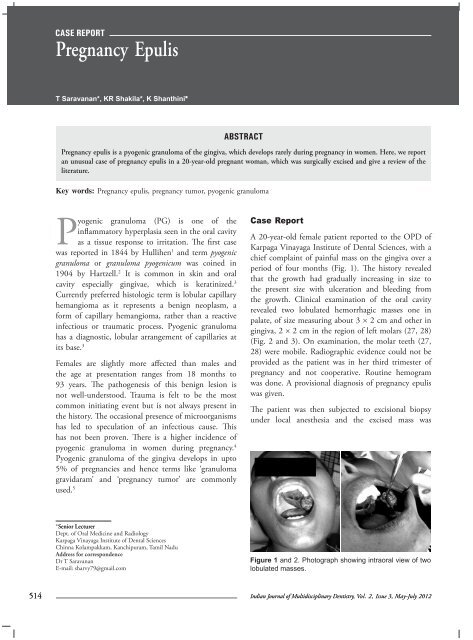
case report<strong>Pregnancy</strong> <strong>Epulis</strong>T Saravanan*, KR Shakila*, K Shanthini*Abstract<strong>Pregnancy</strong> epulis is a pyogenic granuloma of the gingiva, which develops rarely during pregnancy in women. Here, we reportan unusual case of pregnancy epulis in a 20-year-old pregnant woman, which was surgically excised and give a review of theliterature.Key words: <strong>Pregnancy</strong> epulis, pregnancy tumor, pyogenic granulomaPyogenic granuloma (PG) is one of theinflammatory hyperplasia seen in the oral cavityas a tissue response to irritation. The first casewas reported in 1844 by Hullihen 1 and term pyogenicgranuloma or granuloma pyogenicum was coined in1904 by Hartzell. 2 It is common in skin and oralcavity especially gingivae, which is keratinized. 3Currently preferred histologic term is lobular capillaryhemangioma as it represents a benign neoplasm, aform of capillary hemangioma, rather than a reactiveinfectious or traumatic process. Pyogenic granulomahas a diagnostic, lobular arrangement of capillaries atits base. 3Females are slightly more affected than males andthe age at presentation ranges from 18 months to93 years. The pathogenesis of this benign lesion isnot well-understood. Trauma is felt to be the mostcommon initiating event but is not always present inthe history. The occasional presence of microorganismshas led to speculation of an infectious cause. Thishas not been proven. There is a higher incidence ofpyogenic granuloma in women during pregnancy. 4Pyogenic granuloma of the gingiva develops in upto5% of pregnancies and hence terms like ‘granulomagravidaram’ and ‘pregnancy tumor’ are commonlyused. 5Case ReportA 20-year-old female patient reported to the OPD ofKarpaga Vinayaga Institute of Dental Sciences, with achief complaint of painful mass on the gingiva over aperiod of four months (Fig. 1). The history revealedthat the growth had gradually increasing in size tothe present size with ulceration and bleeding fromthe growth. Clinical examination of the oral cavityrevealed two lobulated hemorrhagic masses one inpalate, of size measuring about 3 × 2 cm and other ingingiva, 2 × 2 cm in the region of left molars (27, 28)(Fig. 2 and 3). On examination, the molar teeth (27,28) were mobile. Radiographic evidence could not beprovided as the patient was in her third trimester ofpregnancy and not cooperative. Routine hemogramwas done. A provisional diagnosis of pregnancy epuliswas given.The patient was then subjected to excisional biopsyunder local anesthesia and the excised mass was*Senior LecturerDept. of Oral Medicine and RadiologyKarpaga Vinayaga Institute of Dental SciencesChinna Kolampakkam, Kanchipuram, Tamil NaduAddress for correspondenceDr T SaravananE-mail: sharvy79@gmail.comFigure 1 and 2. Photograph showing intraoral view of twolobulated masses.514Indian Journal of Multidisciplinary Dentistry, Vol. 2, Issue 3, May-July 2012
Case ReportFigure 3. Photograph of intraoperative excisional biopsy.Figure 4. Photograph of excisional biopsy with extractedtooth.Figure 5. Photomicrograph (40x) showing parakeratinizedstratified squamous epithelium associated with fibrovascularconnective tissue.Figure 6. Postoperative photograph shows good healingafter one day.sent for histopathological examination (Fig. 4).Histopathological examination revealed parakeratinizedstratified squamous epithelium associated withfibrovascular connective tissue. In most of the areasepithelium was ulcerated. The underlying connectivetissue exhibited numerous dilated blood vessels,proliferating endothelial cells and extravasated red bloodcells (RBCs). There was diffuse chronic inflammatorycell infiltration throughout the tissue (Fig.5). Thus, thefinal diagnosis of ‘pyogenic granuloma’ was confirmed.There was a uneventful healing on next day (Fig. 6).DiscussionGingiva is often the site of localized growths that areconsidered to be reactive rather than neoplastic innature. Most of the lesions in the gingiva are reactivechronic inflammatory hyperplasia’s with minor traumaand chronic irritation being the main etiologic factors.They found an almost equal distribution of lesionsbetween the maxilla and mandible, with the anteriormaxilla the most prevalent site. 6 It predominantlyoccurs in young females in their 2nd and 3rd decadesdue to hormonal influences on vasculature.There is a higher incidence of pyogenic granulomain women during pregnancy termed as pregnancyepulis. Clinically, the pregnancy epulis appears as asmooth or lobulated and ulcerated mass that is usuallypedunculated or sometimes sessile. Younger tumors aresoft in consistency, progressing to a rubbery textureon maturation. The color may range from pink tobright red to purple or brown. 4 Such lesions begin todevelop in first trimester and their incidence increasesIndian Journal of Multidisciplinary Dentistry, Vol. 2, Issue 3, May-July 2012515
Case Reportupto 7th month of pregnancy. The cause for thepyogenic granuloma in pregnancy is the raised levelsof progesterone and estrogen and it is seen that thetumor usually regresses postparturition. 4The hormonal imbalance coincident with pregnancyheightens the organism’s response to irritation 7however, bacterial plaque and gingival inflammationare necessary for subclinical hormone alterationsleading to gingivitis. 8 The development of thisparticular kind of gingivitis, typical in pregnancy, notdifferent from that appearing in nonpregnant women,suggests the existence of a relationship between thegingival lesion and the hormonal condition observed inpregnancy. Sometimes pregnancy gingivitis can show atendency towards localized hyperplasia, which is calledpregnancy granuloma. Generally, it appears in the2nd - 3rd month of pregnancy, the persistent influenceof plaque induces catarrhal inflammation of the gingivathat serves as a base for development of hyperplasticgingivitis during the last months, modulated by thecumulating hormonal stimuli. In uncontrolled cases,pyogenic granuloma may arise. This lesion is rarelyobserved in women with poor oral hygiene in areaswith local irritating factors such as improperly fittingrestorations or dental calculus. During pregnancy,pyogenic grenuloma when treated by surgical excisionmay reappear due to incomplete excision or inadequateoral hygiene. 9The molecular mechanism behind the development andregression of pyogenic granuloma during pregnancyis due to changes associated with the functions andstructure of the blood and lymph microvasculatureof the skin and mucosa due to profound endocrineupheaval. 10 Recent studies have revealed that sexhormones manifest a variety of biological andimmunological effects. Estrogen accelerates woundhealing by stimulating nerve growth factor (NGF)production in macrophages, granulocyte-macrophagecolonystimulating factor (GM-CSF) production inkeratinocytes and basic fibroblast growth factor (bFGF)and transforming growth factor beta 1 (TGF-β1)production in fibroblasts, leading to granulation tissueformation. Estrogen enhances vascular endothelialgrowth factor (VEGF) production in macrophages, aneffect that is antagonized by androgens and which maybe related to the development of pyogenic grenulomaduring pregnancy. The molecular mechanism for theregression of pyogenic granuloma after the pregnancyis not clear. It is proposed that in the absence of VEGF,the Angiopoietin (Ang-2) causes the blood vessels toregress and VEGF, which was found high in pregnancywas found undetectable after parturition.There are two histological types of pyogenicgranuloma. One type is characterized by proliferatingblood vessels that are organized in lobular aggregatesalthough superficially the lesion frequently undergoesno specific change like edema, capillary dilation orinflammatory granulation tissue reaction. This isknown as lobular capillary hemangioma type, whereasthe second type nonlobular capillary hemangiomatype consists of highly vascular proliferation thatresembles granulation tissue. In the case presented, thehistological picture was that of chronic inflammatorycell infiltration, which showed that it was nonlobularcapillary hemangioma.Differential diagnosis includes pyogenic granuloma,peripheral giant cell granuloma, peripheral ossifyingfibroma and metastatic cancer. The clinical featuresof growth with ulceration and bleeding presentinterdentally during the period of pregnancy made usgive a provisional diagnosis of pregnancy epulis.Possible treatment modalities are excision, curettage,cryotherapy, chemical and electric cauterization,and the use of lasers. The lasers commonly used areargon lasers, continuous wave (CW) Nd:YAG laser,pulsed dye laser and CW carbon dioxide laser, whichpermits rapid, minimally invasive surgical treatment,but the nonspecific coagulation may lead to scars. 11The management of pyogenic granuloma dependson the severity of symptoms. Excisional biopsy isindicated for treatment of pyogenic granuloma,except when the procedure would produce markeddeformity. 12 Recurrence rate after excision ranges from0% to 16%. Pyogenic granuloma of pregnancy oftenregresses postparturition, they need not be excisedunless symptomatic. 4 As the patient presented withhuge painful mass, which was ulcerated and bleedingwe decided to excise completely.Treatment considerations during pregnancy are veryimportant as it is considered that there is a biologicalplausibility that periodontal diseases in pregnancy areassociated with pregnancy complications like pretermbirths, preterm low birth weight (LBW) babies or even516Indian Journal of Multidisciplinary Dentistry, Vol. 2, Issue 3, May-July 2012
Case Reportpre-eclampsia. 13 Surgical and periodontal treatmentshould be completed, when possible.Precautions to be taken for teeth and gums duringpregnancy are:• More frequent visits to your dentist are advisable.• Try to reduce snacking on food high in sugarcontent.References1.2.3.4.5.6.Hullihen SP. Case of aneurysm by anastomosis of thesuperior maxillae. Am J Dent Sc 1844;4:160-2.Hartzell MB. Granuloma pyogenicum. J Cutan DisSymph 1904;22:520-5.Willies-Jacobo LJ, Isaacs H Jr, Stein MT. Pyogenicgranuloma presenting as a congenital epulis. Arch PediatrAdolesc Med 2000;154(6):603-5.Sheth SN, Gomez C, Josephson GD. Pathologicalcase of the month: diagnosis and discussion; pyogenicgranuloma of the tongue. Arch Pediatr Adolesc Med2001;155:1065-6.Sills ES, Zegarelli DJ, Hoschander MM, Strider WE.Clinical diagnosis and management of hormonallyresponsive oral pregnancy tumor (pyogenic granuloma).J Reprod Med 1996;41(7):467-70.Buchner A, Shnaiderman-Shapiro A, Vered M. Relativefrequency of localized reactive hyperplastic lesions of the7.8.9.10.11.12.13.gingiva: a retrospective study of 1675 cases from Israel.J Oral Pathol Med 2010;39(8):631-8.Eversole LR. Clinical outline of oral pathology: diagnosisand treatment. 3rd edition, Decker BC (Ed.), Hamilton2002:p.141-2.Sooriyamoorthy M, Gower DB. Hormonal influenceson gingival tissue: relationship to periodontal disease.J Clin Periodontol 1989;16(4):201-8.Boyarova TV, Dryankova MM, Bobeva AI, Genadiev GI.<strong>Pregnancy</strong> and gingival hyperplasia. Folia Med (Plovdiv)2001;43(1-2):53-6.Henry F, Quatresooz P, Valverde-Lopez JC, Piérard GE.Blood vessel changes during pregnancy: a review. Am JClin Dermatol 2006;7(1):65-9.Raulin C, Greve B, Hammes S. The combined continuouswave/pulsedcarbon dioxide laser for treatment ofpyogenic granuloma. Arch Dermatol 2002;138(1):33-7.Jafarzadeh H, Sanatkhani M, Mohtasham N. Oralpyogenic granuloma: a review. J Oral Sci 2006;48(4):167-75.Bobetsis YA, Barros SP, Offenbacher S. Exploring therelationship between periodontal disease and pregnancycomplications. J Am Dent Assoc 2006;137 Suppl:7S-13S.Indian Journal of Multidisciplinary Dentistry, Vol. 2, Issue 3, May-July 2012517