Paediatric Surgery: A Comprehensive Text For Africa ... - Global HELP
Paediatric Surgery: A Comprehensive Text For Africa ... - Global HELP
Paediatric Surgery: A Comprehensive Text For Africa ... - Global HELP
- No tags were found...

Create successful ePaper yourself
Turn your PDF publications into a flip-book with our unique Google optimized e-Paper software.
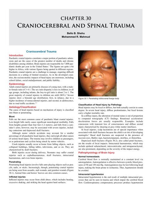
Craniocerebral and Spinal Trauma 191(A) (B) (C)Figure 30.2: CT scans of (A) depressed skull fracture; (B) epidural haematoma; (C) chronic subdural haematoma.and bradycardia, as well as irregular breathing—this is known asCushing’s triad. Therefore, in head injury, it is important to followthe cerebral blood flow (CBF). Because it is difficult to measure CBFdirectly, cerebral perfusion pressure (CPP) is used, which is calculatedas: CPP = ICP – MAP, where ICP is intracranial pressure and MAP isthe mean arterial pressure. 3,5Physiology of injuryFollowing the initial injury at impact, known as the primary injury, biochemicalalterations occur, in particular, the release of glutamate, whichis an excitatory neurotransmitter. This initiates a cascade of cytotoxicreactions, resulting in alterations in cellular energy metabolism, cerebralblood flow, transmembrane ion concentration gradients, free radicalproduction, and cytokine release. Gross secondary changes, suchas haematomas, cerebral oedema, hypotension, seizures, and hypoxia,further worsen the neurologic injury.Clinical FeaturesHistoryDetails of the mechanism of injury, such as distance of fall, the surfacestruck, and the velocity of striking objects, are important. In motorvehicular trauma, the speed of the vehicle and use of restraints shouldbe determined. A careful history regarding immediate posttraumaticevents, such as loss of consciousness, its duration, seizures, and vomiting,should be sought. In the older child, specific questions about neckpain, numbness, and weakness are asked. The possibility of child abuseshould also be kept in mind.Physical assessmentObservation of the mildly head injured child provides a great deal ofinformation. The level of consciousness is determined. Examinationof the head and scalp are done. Scalp abrasions, lacerations, andhaematomas are carefully examined. The skull is palpated for areasof tenderness and fractures without inflicting pain. In older childrenwith moderate to severe injuries, age-specific behavior is a greatguide to neurological assessment. They may appropriately respond tonoxious stimuli by grimacing, crying, or exhibiting a facial expressionof distress. Palpation of an open fontanelle provides a good idea ofintracranial pressure.Assessment of injury severityThe Glasgow Coma Scale (GCS) is a good measure of acute injuryseverity and has been modified using age-appropriate parameters asindicated in Table 30.1. The table shows the best score achievable by anormal child for each parameter at various age groups.Laboratory AssessmentInfants and small children can develop acute anaemia with relativelylittle blood loss. Haemogram and baseline serum electrolytes levels andblood gasses are assessed.Radiological AssessmentSkull x-rayA skull x-ray is useful as an initial assessment tool, particularly in <strong>Africa</strong>,where CT scans are not readily available. Skull fracture sites may heraldpotential intracranial pathologies. The x-ray may also show otherpathologies, such as pneumocephalus (Figure 30.3), and linear fracturesparallel to the slice plane, which may be missed by a CT scan. 7,8CT scanA CT scan is the most useful tool for acute assessment of traumatic headinjury. Bony and parenchymal lesions are usually well seen. Haematomasare clearly seen and can easily be categorised based on age. 9Magnetic resonance imagingMagnetic resonance imaging (MRI) offers superior resolution in visualisingsmall lesions, such as is seen in diffuse axonal injury, but isnot as widely available and affordable as the CT scan. It is also notan investigation of choice in terms of skull fractures and intracranialhaematomas. 10,11Cranial ultrasoundCranial ultrasound (US) is usually a bedside technique used to monitorintracranial collections and ventricular size following trauma. This usefultool is underutilised for the child with an open fontanelle, largely asa result of lack of experience by radiologists and unavailability of USto the neurosurgeons. 10ManagementInitial managementAdequate resuscitation and stabilisation must be given priority. Theairway is the highest management priority. A child with severe headinjury will require control of the airway with intubation. This helps toprevent secondary injury from hypoxia and hypercarbia. 12 The cervicalspine must be assumed to be unstable until proven otherwise by plainradiographs later. Meanwhile, the breathing, circulation, and the stabilisationof vital signs are then attended to. It is the postresuscitation GCSscore that is useful.A focused neurological examination is performed to determinelife-threatening intracranial pathology and assess the child’s baselineneurological level; the papillary examination and the GCS scoreare most important for this purpose. Efforts are made to look forlateralising signs, such as hemiparesis, pupillary dilatation, facialnerve palsy, and so on. The next priority in a child who is unresponsiveis to assess brainstem function by means of the corneal and gagreflexes. Corticosteroids and routine administration of anticonvulsantsare not recommended.Measures to treat raised intracranial pressureWhere ICP can be monitored, the treatment threshold for raised ICP is20–25 mm Hg. 13
192 Craniocerebral and Spinal TraumaTable 30.1: <strong>Paediatric</strong> (Adelaide) Scale.Normal scoreat ageNormal eyeopeningScoreNormal motorresponseScoreNormal verbalresponseScore0–6 months Spontaneous 4 Flexion 3 Cries 2 96–12 months Spontaneous 4 Localises 4 Vocalises 3 111–2 years Spontaneous 4 Localises 4 Words 4 122–5 years Spontaneous 4 Obeys 5 Words 4 13TotalFigure 30.3: Skull x-ray showing intraventricular pneumocephalus.PositioningElevation of the head of the bed to 15–30 degrees, when not contraindicated,optimises arterial flow and venous drainage.OxygenationOxygenation leads to improved cerebral oxygenation and a reductionin cerebral oedema.MannitolMannitol is very effective and can be life saving. It reduces blood viscosityand acts as an osmotic diuretic. It is given only after adequatevolume resuscitation. The dose is 0.5–1g/kg body weight over 20 minutes(bolus). However, because of its rebound effect, it should be administeredonly when the patient is being prepared for an indicated surgery.Controlled hyperventilationHyperventilation has a very rapid effect and is aimed at reducing thePaCO 2to about 40 mm Hg. Lowering the PaCO 2further is associated witha risk of cerebral vasoconstriction, leading to cerebral ischaemic injury.<strong>Surgery</strong>Intracranial haematomas in children are treated more aggressively.Specific head injuriesScalp injuriesIn young children, blood loss from scalp laceration can lead to shockand should be promptly controlled. Scalp loss is managed with skingrafts or rotational flaps. 6Skull fracturesLinear, diastatic, and stellate fractures occur from focal contact forces.They may occur over a venous sinus with resultant tear haemorrhage.A CT scan is advised in the setting of stellate fractures due to the highincidence of underlying contusion.Ping-pong fractures are managed most often nonoperatively becausethey often resolve spontaneously.Depressed skull fractures are treated with elevation when indicated.Fractures at the base of the skull are common in children, butcerebrospinal fluid (CSF) leaks often resolve spontaneously. Persistentleaks, however, require surgical intervention. Accompanying cranialnerve deficits may require surgical decompression and corticosteroid.Growing skull fractures are peculiar and are characterised by apulsatile scalp swelling overlying an enlarged bony defect with anassociated underlying leptomeningeal cyst. They are repaired via cystdrainage, dura graft, and autologous skull graft.HaematomasSubgaleal haematomas can be very massive in children to the extent ofinducing hypovolaemic shock.Prompt recognition and evacuation of epidural haematomas via acraniotomy leads to a good and rapid neurological recovery.Acute subdural haematomas carry a high mortality. Therecommended surgery is craniotomy and evacuation. Chronic subduralhaematomas are approached via burr-hole evacuation.Intraparenchymal haemorrhage may be large enough to causeneurologic deficit or midline shift, and surgical evacuation is indicatedin such children as they tend to recover from neurologic deficits.Intraventricular haematoma is managed with external ventriculardrainage.Penetrating injuriesIn the presence of a protruding penetrating foreign body, the offendingobject should not be removed instantaneously. Following clinical evaluation,a plain radiograph in two views and a brain CT scan should berequested. Appropriate consultation should be sought, such as from anophthalmologist for orbitocranial injuries. Appropriate broad spectrumantibiotics and tetanus prophylaxis are to be instituted. The aim ofsurgery is removal of the foreign body in a controlled condition in theoperating room. 14 In the absence of a retained foreign body, the goalof surgery is to debride the tract and repair the dura and bony defects.Basic Neurosurgical ProceduresBurr-hole evacuation of chronic subdural haematoma1. The patient is positioned supine, head turned laterally and elevated15 degrees, with shoulder support.2. The site of surgery is shaved and cleaned, and the patient is thendraped.3. A vertical incision is made over the site of the haematoma.4. Usually, the first burr hole is temporal, 2.5 cm above the zygomaticarch, just anterior to the ear.5. Scalp bleeding is controlled with cautery and self-retaining mastoidretractors.6. The periosteum is incised and retracted.7. A burr hole is made with a drill.8. The dura is coagulated and incised in a cruciate fashion with a size11 blade.9. The haematoma is evacuated with gentle suction and irrigation,taking care not to injure any bridging vessels. Any bleeding point iscontrolled with bipolar cautery.10. The subdural space is irrigated with normal saline.11. A subdural drain may be left in place for 24–48 hours.12. The incision is closed.
194 Craniocerebral and Spinal TraumaPosttraumatic seizuresSeizures that occur in the first 7 days of injury are termed early posttraumaticseizures, and those that occur after 1 week are late posttraumaticseizures. The incidence of early posttraumatic seizures is1–5%. It can precipitate adverse events as the result of an elevationof intracranial pressure, alteration in blood pressure, and changes inoxygenation. The estimated incidence of late posttraumatic seizure is10–13% within 2 years after head injury. The risk of seizure is higherin patients with acute intracranial haematoma, open depressed skullfractures, parenchymal injury, seizures within the first 24 hours ofinjury, GCS
Craniocerebral and Spinal Trauma 195Clinical PresentationThe area of spinal cord damage and nerve root involvement determinethe clinical presentation. In complete spinal cord injuries, there is a lossof voluntary nervous function below the level of injury. There is an initialtemporary phase of spinal shock, with loss of all reflexes below theinjured segment that may last for minutes or days. About 3% of patientswith complete injuries on initial examination will develop some recoverywithin 24 hours. In incomplete spinal cord injuries, some nervousfunction is present in the form of some muscle power or sensationbelow the level of injury; these injuries carry a better prognosis forrecovery. Frankel grading is used to categorise spinal cord injuries, asshown in Table 30.3.Table 30.3: Frankel grading of spinal cord injuriesClass Functional status DescriptionA Complete Total motor and sensory lossB Sensory only Sensory sparingC Motor useless Motor sparing of no functional valueD Motor useful Motor sparing of functional valueE Recovery No functional deficitThe various spinal cord syndromes include:• Anterior cord syndrome: Damage to the spinothalamic and corticospinaltracts with resultant predominant motor weakness.• Brown–Sequard’s syndrome: Hemicord injury with ipsilateral motorweakness and loss of proprioception and contralateral loss of painand temperature below the level of injury.• Central cord syndrome: Injury to the central portions of the cervicalspinal cord with resultant predominant motor affectation of theupper limb.• Conus medullaris syndrome: Injury towards the end of the spinalcord results in a mixed upper motor neurone and lower motor neuronedysfunction.Spinal cord injury without radiographic abnormality (SCIWORA)is a unique type of spinal cord injury common to children characterisedby posttraumatic neurological deficits with normal plain radiographsor tomographs. It occurs mostly in children younger than 8–10 yearsof age. The mechanism of occurrence is thought to be vascular orischaemic in origin, resulting in spinal cord infarction.InvestigationsRadiographic evaluation is done after adequate resuscitation. A lateralplain x-ray is the most informative and may show fractures, subluxation,or angulation of the spine. Soft tissue swellings may indicateligamentous injury. In suspected odontoid fractures, an open mouthview can be done for the older child. In infants, a CT scan is recommended.At least 75% of patients with spinal cord injury have injury tothe vertebral column and thus some degree of radiographic abnormalities.Therefore, initial plain films are indispensable.Dynamic studies can be done to search for occult instability in theolder cooperative child with neck pain but no neurologic deficit.CT scans and MRI could further elucidate the extent of the injury(Figure 30.4).Radiographic signs of cervical spine trauma include:• soft tissue in retropharyngeal space >22 mm ( child not crying);• displaced prevertebral fat stripe;• tracheal deviation and laryngeal dislocation;• vertebral malalignment;• loss of lordosis;Figure 30.4: CT scan, saggital reconstruction. Slide shows retropulsed thoracicvertebra into spinal canal.• acute kyphotic angulation;• widened interspinous space;• axial rotation of vertebra;• discontinuity in contour lines;• abnormal joints;• atlanto-dental interval of more than 5 mm;• narrow or widened disc space; and• widening of apophyseal joints.ManagementThe goal of management of spinal cord injuries is to prevent furtherinjury and reduce neurological deficits.Initial management and evaluationIdeally, initial management and evaluation are commenced at the sceneof the injury. In most <strong>Africa</strong>n settings, however, prehospital managementis not well established, and the initial management is usuallycommenced at the receiving hospital. The initial management includesresuscitation, immobilisation, constant monitoring, and assessment ofthe injured child. 24ResuscitationThe main causes of death of in a child with spinal cord injury areaspiration and shock, and so the “ABC” of life support is commenced.Early airway control with endotracheal intubation and oxygen administrationmay be indicated in respiratory insufficiency. Manual in-lineimmobilisation of the cervical spine is mandatory during intubation.Hypotension accompanied by bradycardia may be present due to autonomicparalysis. Therefore, adequate hydration with systolic bloodpressure maintained at or above 90 mm Hg prevents shock. 25 Volumeresuscitation suffices, but occasionally ionotropes such as ephedrinemay be indicated.Nasogastric tube decompression of the stomach is instituted becausegastric distention can interfere with respiration or lead to gastricmucosal ulceration.The loss of sympathetic tone may also lead to urinary retention andhypothermia. An indwelling urethral catheter is passed, and attentionpaid to the temperature of the child with constant monitoring.ImmobilisationThe entire spine of the child with suspected spinal injury should beimmobilised. Whole-body braces usually are not readily available, sothe cervical spine is immobilised with collars, particularly in the olderchild. Infants can be immobilised with sand bags or intravenous fluidbags secured at both sides of the head, with the head taped to the board.
196 Craniocerebral and Spinal TraumaEvaluationFollowing resuscitation, a detailed history should be taken as soon aspossible, including mechanism and time of injury, severity of injury, thefirst aid given, and mode of transportation. Examination should includeall motor functions of the major muscle groups as well as a rectal examinationto assess sphincteric tone. Sensory functions, reflexes, and motorfunctions of the diaphragm and intercostal muscles should be assessed.TreatmentMedicalHigh-dose methylprednisolone administration within 8 hours of injuryis said to be beneficial to long-term outcomes. The patient is given30 mg/kg bolus over 15 minutes, followed by a 45-minute pause.Maintenance infusion of 5.4 mg/kg per hour over 23 or 47 hours isgiven. However, the efficacy has not been fully evaluated in childrenyounger than 13 years of age. Gastric erosion is prevented by the use ofH 2-receptor antagonists such as ranitidine.Attention is paid to the prevention of pressure ulcers, chronicurinary tract infection, and contracture and deformities of the limbs.Cervical injuryBesides collars, bracings immobilise the cervical spine (Table 30.4).Cervicothoracic orthosis (CTO) incorporates a body vest to immobilizethe cervical spine and includes the Guilford brace, sterno-occipitomandibularImmobilisation (SOMI), and Yale brace.Table 30.4: Recommended bracing for various cervical spine injuries.ConditionCervical strainJefferson fracturestableunstableOdontoid fracturetype Itypes II & IIIHangman’s fracturestableunstableFlexion injuriesmid cervical (C3-C5)low cervical (C5-T1)Extension injuriesmid cervical (C3-C5)low cervical (C5- T1)Recommended bracePhiladelphia collarCervicothoracic orthosisHaloCervicothoracic orthosisHaloSOMIHaloSOMI, cervicothoracic orthosisCervicothoracic orthosisHalo, cervicothoracic orthosisHaloTractionSkull traction is aimed at reducing cervical fracture or dislocation,maintaining normal alignment, immobilising the spine, and decompressingthe spinal cord and nerve roots. It also facilitates bone healing.Traction includes Crutchfield tongs, Gardner-Wells’ tongs, or halo traction.The traction weight should be increased slowly under the guidanceof an image intensifier to achieve reduction. Three pounds per cervicalvertebral level is recommended (but not more than 10 pounds should beused in children younger than 14 years of age).Thoracolumbar injuryPerhaps the most popular theory in terms of spinal stability is the threecolumntheory of Dennis. In this model, the anterior column includesthe anterior longitudinal ligament, anterior portion of disc, and vertebra.The middle column incorporates the posterior portion of disc andvertebra, posterior longitudinal ligament, and the pedicle. The posteriorcolumn includes the posterior ligamentous complex and arch. The ribcage-sternum complex serves as a fourth column of support unique tothe thoracic spine.Damage to more than one column of the spine renders it unstable.Thoracolumbar spine instability can be categorised into (1) first-degreeinstability, which is mainly mechanical; (2) second-degree instability, inwhich there is neurological instability; and (3) third-degree instability,in which there is both neurological and mechanical instability.Thosewith stable and first-degree instability can be managed with bed rest for1–6 weeks followed by ambulation in an orthosis (e.g., thoracolumbarsacral orthosis (TLSO) or Jewett brace) for 3 to 5 months. Second- andthird-degree instability may require instrumentation.<strong>Surgery</strong>Operative management of spinal cord injury aims at decompressionand stability. Emergency decompression has been associated with neurologicaldeterioration, although it is indicated in incomplete lesions.Other indications are as follows:• progressive neurological deterioration;• complete spinal block (on MRI or myelogram);• bone fragment within the spinal canal;• cervical root compression;• compound fracture or penetrating spinal trauma;• acute anterior cord syndrome; and• nonreducible, locked facet causing compression.Complications of Spinal Cord InjuryRespiratory complicationsRespiratory insufficiency is common in patients with injuries of the cervicalcord. If the neurological lesion is complete, the patient will haveparalysed intercostals muscles and will have to rely on diaphragmaticrespiration. Partial diaphragmatic paralysis may also be present ab initioor after 24–48 hours if ascending posttraumatic oedema develops.In thoracic spine injuries, there may be associated rib fractures, haemopneumothorax,ventilation perfusion, mismatch, and so on.Patients need to be nursed in the recumbent position even afterspinal stabilisation to ensure that diaphragmatic excursion is notcompromised. Regular chest physiotherapy and respiratory functionmonitoring should be done.A patient whose respiratory function is initially satisfactory afterinjury but then deteriorates should regain satisfactory ventilatorycapacity once spinal cord oedema subsides. Artificial ventilation shouldtherefore not be withheld.Cardiovascular complicationsHaemorrhage from associated injuries is the most common causeof posttraumatic shock and must be treated vigorously. In traumaticquadriplegia, the thoracolumbar (T1–L2) sympathetic outflow paralysisgives rise to hypotension and bradycardia. Pharyngeal suction andtracheal intubation stimulate the vagus, and in high spinal cord injuries,these can produce bradycardia and cadiac arrest. Hence, atropine andglycopyrronium should be used before such procedures or when heartrates fall below 50 per minute.Cardiac arrest from sudden hyperkalaemia following the use ofdepolarising agents such as suxamethonium is a risk in these patientsbetween 3 days and 9 months after injury. Hence, nondepolarisingagents are preferred.ThromboembolismNewly injured quadriplegics or paraplegics are at risk of thromboembolism.Antiembolism stockings and anticoagulants must be started immediatelyonce medical contraindications and head injury are ruled out.Bladder complicationsAfter severe cord injury, the urinary bladder is initially acontractile,and if untreated this leads to acute urinary retention. A Foley cathetershould be passed.Gastrointestinal tract complicationsParalytic ileus is a common accompaniment of severe spinal cord injury
Craniocerebral and Spinal Trauma 197and should be treated with intravenous fluids, nil per os (NPO), andnasogastric tube decompression. Uncommonly, ulceration, haemorrhageand perforation may occur. Therefore, proton pump inhibitors orH 2-receptor antagonists should be started immediately.Skin and pressure areasThe patient must be turned every 2 hours manually or automatically,and all pressure areas must be padded to prevent pressure sores. 26Joints and limbsJoints must be passively moved and splinted if necessary to preventcontractures.Autonomic dysreflexiaAutonomic dysreflexia is seen particularly in patients with cervicalinjury above the sympathetic outflow. It occurs after spinal shock andusually is due to distended bladder or detrusor-sphincter dyssynergia.Distended bladder causes reflex sympathetic overactivity below thelevel of the spinal cord lesion, causing vasoconstriction and severehypertension. The carotid and aortic baroreceptors respond via thevasomotor centre with increased vagal tone and bradycardia, but thesestimuli cannot pass distally through the injured cord.Patient suffers headache, profuse sweating, and flushing abovethe level of the cord lesion. Without prompt treatment, intracranialhaemorrhage may occur.Another cause of autonomic dysreflexia includes urinary tract infection.Treatment is by removing the cause, sitting the patient up, andadministering nifedipine or glycryl trinitrate; spinal or epiduralanaesthetics are used occasionally.HyponatraemiaHyponatraemia is usually caused by fluid overload, diuretic usage, andthe sodium-depleting effect of some drugs such as carbamazepine andinappropriate ADH secretion. It is treated by treating sepsis, fluid restriction,and administration of frusamide with potassium supplements.HypercalcaemiaHypercalcaemia is caused by prolonged immobility and manifestswith constipation, abdominal pain, and headache. Treatment involveshydration, achieving diuresis, and the use of sodium etidronate ordisodium pamidronate.Para-articular heterotopic ossificationNew bone is often deposited in soft tissue around paralysed joints. Besttreatment is surgical excision after 18 months when the bone is matured.SpasticitySpasticity is seen only in patients with an upper motor neurone lesionwhose intact spinal reflex arcs below the level of the lesion are isolatedfrom the higher centres. It enhances the tendency to contractures.Aggravating factors are detected and treated, pain is managed,and spastic muscles are passively stretched. Oral baclofen is usuallyhelpful. In intractable cases, however, the use of butulinum toxin, motorpoint injection, intrathecal baclofen pump, tenotomy, neurectomy, orIntrathecal block may be employed.Urologic complicationsAfter spinal cord injury, dysfunctional voiding patterns may soonemerge. These are associated with serious sequelae. Therefore, as part ofearly management, intermittent catheterisation, tapping and expression,indwelling catheterisation, suprapubic cystostomy, or intermittent selfcatheterisationcan be used. Later management may involve augmentationcystoplasty, neuromodulation and sacral anterior root stimulation(SARS), or intermittent self-catheterisation (Mitrofanoff’s technique).Prognosis and OutcomeThe neurological examination and age of the patient are the most criticalprognostic factors for short- and long-term recovery. Children withcomplete lesions rarely improve, whereas those with incomplete butsevere lesions improve with time but hardly regain normal function.Only children with mild to moderate deficits can hope for full recovery.Mortality in the acute setting is 20%.PreventionThe majority of spinal cord injury is caused by pedestrian-motorvehicle accidents and falls. Enforcement of traffic rules cannot be overemphasized.Prevention of children from engaging in activities that aredetrimental, such as climbing trees and unprotected heights or depthsmust be ensured. 27,28Ethical IssuesThose who survive spinal cord trauma develop life-long disabilities.Treatment is often not curative. The decision to operate must be carefullydiscussed with the family, and the prospective multidisplinarymode of subsequent management should be instituted.Evidenced-Based ResearchTable 30.5 is a review of paediatric severe head injuries that comparestwo age groups. Table 30.6 is a review of paediatric spine fractures thatcompares parameters of the injuries.Table 30.5: Evidence-based research.TitleAuthorsInstitutionSevere head injury in children: early prognosis and outcomeZuccarello M, Facco E, Zaampieri P, Zanardi L, Andrioli GCDepartment of Neurosurgery and Institute of Anesthesiologyand Intensive Care, University Hospital, Padova, ItalyReference Child’s Nervous System 1985; 1:158–162ProblemInterventionComparison/control(quality ofevidence)Outcome/effectHistoricalsignificance/commentsIdentifying indicators for early prognosis and outcome inchildren with severe head injury.Controlled hyperventilation and bolus infusion of hypertonic(20%) mannitol, surgical removal of any mass lesions.Sixty-two children with severe head injury were divided intotwo groups: infants aged
198 Craniocerebral and Spinal TraumaTable 30.6: Evidence-based research.TitleAuthorsInstitutionPediatric spine fractures: a review of 137 hospitaladmissionsCarreon LY, Glassman ST, Campbell MJLeatherman Spine Center, Louisville, Kentucky, USAReference J Spinal Disorders Techniques 2004; 17: 477–482ProblemInterventionComparison/control(quality ofevidence)In children with spinal injury, prevention of further neurologicdamage and deformity, as well as good potential forrecovery, makes timely identification and appropriatetreatment of the injury critical.Decompression, fusion, instrumentation.The 137 patients were divided into three groups for analysis:0–9 years of age (36 patients), 10–14 (49 patients) and15–17 (52 patients). This allowed for comparison of age withmechanism of injury, injury pattern and level, incidence ofcord injury, treatment, and outcomes.Outcome/effectHistoricalsignificance/commentsThirty-six (1%) of 3,685 injured children aged 0–9 yearssustained spine injury, compared to 49 (3%) of 1,609 injuredchildren aged 10–14 years, and 52 (5%) of 921 injuredchildren aged 15–17 years (p < 0.001). Motor vehicleaccidents were the most common cause of injury across allages, followed by falls, sports, and pedestrian accidents.The incidence of multilevel and noncontiguous injuries in thedifferent age groups was not significantly different.Twenty-four patients (19%) had spinal cord injury; 21(87%) were complete cord injuries, and 3 (13%) wereincomplete. Cord injury was more common in the 0–9 yearage group. Four of five patients with spinal cord injurywithout radiographic abnormality (SCIWORA) were in the0–9 age group and had complete neurologic injuries. Youngchildren with cervical injuries were more likely to die thanolder children. Fifty-three percent had associated injuries.Eighteen percent underwent decompression, fusion, andinstrumentation. Two patients developed scoliosis. Thecomplication rate in surgical patients was higher than inpatients treated nonsurgically and in polytrauma patients.This retrospective clinical case series has presented importantand useful data from a large series of paediatric patients withspine injuries from a single regional trauma center.Craniocerebral Trauma1. <strong>Paediatric</strong> head injury is common in our environment.2. Motor vehicle accidents cause the severest form of injury.3. Child abuse is increasingly becoming recognised as a cause ofpaediatric head injury.4. Prompt resuscitation and cervical spine protection are key tosurvival.5. Scalp bleeding may easily cause anaemia because of smallintravascular volume.6. The postresuscitation GCS score is an important prognosticfactor.7. Intracranial haematomas are aggressively managed.8. Education and enforcement of legislation on vehicle safetyrules are important in preventive strategies.Key Summary PointsSpinal Cord Injury1. <strong>Paediatric</strong> spinal cord injury presents an enormous challenge,not only to the neurosurgeon but to the health and economicresources of any nation.2. The care of the spinally disabled child is far from ideal in ourenvironment.3. Late presentation is the rule in most <strong>Africa</strong>n settings,precluding those who would have benefitted from the institutionof early treatment modalities.4. Prompt resuscitation and optimal fluid administration limitfurther cord injury.5. More personnel, facilities, and dedicated centres for spinalcare are in arrears, needing urgent attention.
Craniocerebral and Spinal Trauma 199References1. Winn HR, ed. <strong>Paediatric</strong> head injury In: Youman’s Neurological<strong>Surgery</strong>, 5th ed. Saunders, 2004.2. Tindall GT,Cooper PR, Barrow DL, eds. Head Injury in the<strong>Paediatric</strong> Patient. In: The Practice of Neurosurgery, Version4.18.4; Williams and Wilkins, 1996.3. Basso A, Previglioano I, Duarte,JM, Ferrari, N. Advances inManagement of Neurosurgical Trauma in Different Continents.World J Surg 2001; 25:1174–1178.4. Shokunbi MT, Solagberu BA. Mortality in Childhood Head Injury inIbadan. Afri J Med Sci 1995; 24:159–163.5. Solagberu BA, Adekanye AO, Ofoegbu CP, Udoffa US, Abdur-Rahman LO, Taiwo JO. Epidemiology of trauma deaths. West Afr JMed 2003; 22:177–181.6. Legbo JN, Shehu BB. Managing scalp defects in Sub Saharan<strong>Africa</strong>. East Afri Med J 2004; 81:87–91.7. Thanni L. Evaluation of guidelines for skull radiography in headinjury. Niger Postgrad Med J 2003; 10:231–233.8. Obisesan AA, Bohrer SP. The uses and abuses of skull x-ray inhead injury. Niger Med J 1979; 9:65–69.9. Nagy KK, Joseph KT, Krosner SM, et al. The utility of headcomputed tomography after minimal head injury. J Trauma 1999;46:268–270.10. Umerah B. Cerebral angiography in Central <strong>Africa</strong>. Diagn Imaging1981; 50:225–228.11. Otieno T, Woodfield JC, Bird P, et al. Trauma in rural Kenya. Injury2004; 35:1228–1233.12. Muhammad I. Management of head injuries at the ABU Hospital,Zaria. East Afr Med J 1990; 67:447–451.13. Brain Trauma Foundation AAON. Guidelines for the acute medicalmanagement of severe traumatic brain injury in infants, childrenand adolescents. Paediatr Crit Care Med 2003; 4:S1–S75.14. Bullock MR, Chestnut R, Ghajar J, et al. Guidelines for the surgicalmanagement of traumatic brain injury. Neurosurgery 2006; 58(3Suppl.):S1–S60.15. Shokunbi MT, Olurin OI. Childhood head injuries in Nigeria:causes, neurologic complications and outcome. West Afr Med J1994; 13:38–42.16. Mwang’ombe NJ, Kiboi J. Factors influencing the outcome ofsevere head injury at Kenyatta National Hospital. East Afr Med J2001; 78:238–241.17. Bahloul M, Chelly H, Ben Hmida MS, et al. Prognosis of traumatichead injury in south Tunisia: a multivariate analysis of 437 cases.J Trauma 2004; 57:255–561.18. Winn HR, ed. <strong>Paediatric</strong> vertebral column and spinal cord injuries,In: Youman’s Neurological <strong>Surgery</strong>, 5 ed. Saunders, 2004.19. David P. Spinal cord injury in children. In: Principles and Practiceof <strong>Paediatric</strong> Neurosurgery. Thieme, 1999, Pp. 955–969.20. Brockmeyer DL. Spinal cord and spinal column injuries in children: current management options. In: <strong>Paediatric</strong> Neurosurgery for theGeneral Neurosurgeon. Thieme, 2002, Pp. 36–48.21. Hamilton MG, Myles ST. <strong>Paediatric</strong> spinal injury: review of 174hospital admissions. J Neurosurg 1992; 77:700–704.22. Rauzzino MJ, Grabb PA, Hadley MN. <strong>Paediatric</strong> vertebral columnand spinal cord injuries. Persp Neurolog Surg 1997; 8:27–60.23. Scher AT. Trauma of the spinal cord in children. S Afr Med J 1976;50:2023–2025.24. Adeolu AA, Shokunbi MT, Malomo AO, Komolafe EO, Adeloye EA.Cervical spine injury in children with head injury in Ibadan, Nigeria.Afr J Paediatr Surg 2005; 1:76–79.25. Mayuri G, Maharajh J. Case report of cranio-cervical dislocation ina child. S Afr Radiographer 2006; 44(2):1826. Chen Y, DeVivo MJ, Jackson AB. Pressure ulcer prevalence inpeople with spinal cord injury: age-period-duration effects. ArchPhys Med Rehabil 2005; 86:1208–1213.27. Odeku EL, Richard DR. Peculiarities of spinal trauma in Nigeria.West Afr Med J 1971; 20:211–225.28. Rhodes N. Focus on parents: teen’s perceptions of parentalsafety messages. Workshop presentation at the 2007 LifesaversConference. Chicago, IL, March 2007.

![Clubfoot: Ponseti Management [Vietnamese] - Global HELP](https://img.yumpu.com/51276842/1/184x260/clubfoot-ponseti-management-vietnamese-global-help.jpg?quality=85)

![Steenbeek Brace For Clubfoot [2nd Edition] - Global HELP](https://img.yumpu.com/46612972/1/190x245/steenbeek-brace-for-clubfoot-2nd-edition-global-help.jpg?quality=85)

![Basics Of Wound Care [Indonesia] - Global HELP](https://img.yumpu.com/41566370/1/190x245/basics-of-wound-care-indonesia-global-help.jpg?quality=85)