Gastric Ulcers and Carcinoma - Pathology
Gastric Ulcers and Carcinoma - Pathology
Gastric Ulcers and Carcinoma - Pathology

Create successful ePaper yourself
Turn your PDF publications into a flip-book with our unique Google optimized e-Paper software.
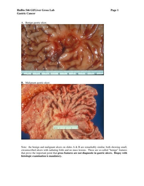
HuBio 546 GI/Liver Gross Lab Page 1<strong>Gastric</strong> CancerA. Benign gastric ulcer.B. Malignant gastric ulcer.Note: the benign <strong>and</strong> malignant ulcers on slides A & B are remarkably similar, both showing small,circumscribed ulcers with radiating folds <strong>and</strong> no mass lesions. These are so-called "benign" featuresthat prove the important point that gross features are not diagnostic in gastric ulcers. Biopsy withhistologic examination is m<strong>and</strong>atory.
HuBio 546 GI/Liver Gross Lab Page 2<strong>Gastric</strong> CancerC. Leather bottle stomach of linitis plastica-type gastric adenocarcinoma.D. Gross cross section of linitis plastica gastric adenocarcinoma showing wall thickening <strong>and</strong> diffusegross infiltration by tumor into all layers of the gastric wall, including the mucosa, submucosa,muscularis propria (white streaks) <strong>and</strong> subserosa.
HuBio 546 GI/Liver Gross Lab Page 3<strong>Gastric</strong> CancerE. Mass lesion/polypoid <strong>and</strong> ulcerated gastric adenocarcinoma, seen in this en face mucosal orsurface view.F. Gross cross section of the mass lesion/polypoid <strong>and</strong> ulcerated gastric adenocarcinoma seen inslide E above. Note the transmural invasion of the tumor--important for staging.