Tinea capitis favosa due to Trichophyton schoenleinii
Tinea capitis favosa due to Trichophyton schoenleinii
Tinea capitis favosa due to Trichophyton schoenleinii

You also want an ePaper? Increase the reach of your titles
YUMPU automatically turns print PDFs into web optimized ePapers that Google loves.
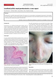
<strong>Tinea</strong> <strong>capitis</strong> <strong>favosa</strong> <strong>due</strong> <strong>to</strong> Trichophy<strong>to</strong>n <strong>schoenleinii</strong>C a s e r e p o r t<strong>Tinea</strong> <strong>capitis</strong> <strong>favosa</strong> <strong>due</strong> <strong>to</strong> Trichophy<strong>to</strong>n<strong>schoenleinii</strong>A. Khaled, L. Ben Mbarek, M. Kharfi, F. Zeglaoui, A. Bouratbine, B. Fazaa,and M. R. Kamoun BarekS U M M A R YA case of a tinea <strong>capitis</strong> caused by Trichophy<strong>to</strong>n <strong>schoenleinii</strong> is presented. It involves a 6-year oldTunisian boy that had presented with diffuse scaling of the scalp misdiagnosed as psoriasis and wastreated unsuccessfully with kera<strong>to</strong>lytic shampoos for two years. <strong>Tinea</strong> <strong>favosa</strong> <strong>due</strong> <strong>to</strong> Trichophy<strong>to</strong>n<strong>schoenleinii</strong> was confirmed by mycological examination. He was successfully treated with griseofulvinfor 6 weeks and <strong>to</strong>pical application of imidazole.Trichophy<strong>to</strong>n <strong>schoenleinii</strong> is an important anthropophilic derma<strong>to</strong>phyte that causes tinea <strong>favosa</strong>. It istransmitted by contagion between humans and is currently endemic in Africa. Ringworm is still frequentin Tunisia, but favus is becoming exceptional <strong>due</strong> <strong>to</strong> improvements in living conditions and hygiene.IntroductionTrichophy<strong>to</strong>n <strong>schoenleinii</strong> is an important anthropophilicderma<strong>to</strong>phyte that causes tinea <strong>favosa</strong> and istransmitted by contact between humans. It is currentlyendemic in Africa. Improvements in living conditionsand hygiene in developing countries after the SecondWorld War have been associated with the almost completedisappearance of many anthropophilic species,including Trichophy<strong>to</strong>n <strong>schoenleinii</strong>. In Tunisia, favusis becoming exceptional.We describe a patient that had an unusual variant oftinea <strong>capitis</strong> caused by Trichophy<strong>to</strong>n <strong>schoenleinii</strong>.Case reportA 6-year old Tunisian boy, living in the city of Tunis,was seen at the Department of Derma<strong>to</strong>logy for largeadherent scales surrounding the hair shafts. Examinationrevealed multiple scaly hairless patches (Figure 1).He had been treated unsuccessfully for psoriasis withsalicylic acid shampoos for two years. No other derma<strong>to</strong>logicalabnormality was observed; he was otherwisea healthy boy. A Wood’s light examination revealed agreen fluorescence. Scales and altered hairs were collected.Direct microscopic examination of the hairs in10% potassium hydroxide revealed an endothrix inva-K E YWORDSTrichophy<strong>to</strong>n<strong>schoenleinii</strong>,favus,Tunisia34 Acta Derma<strong>to</strong>ven APA Vol 16, 2007, No 1
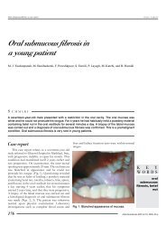
C a s e r e p o r t<strong>Tinea</strong> <strong>capitis</strong> <strong>favosa</strong> <strong>due</strong> <strong>to</strong> Trichophy<strong>to</strong>n <strong>schoenleinii</strong>sion, with hyphae and air spaces in the hair shafts. Cultureon Sabouraud glucose agar with antibiotics producedwaxy colonies that later became velvety andwhitish. They consisted of irregular, dicho<strong>to</strong>mouslybranched hyphae with favic chandeliers and a few intercalatechlamydospores (Figure 2). A diagnosis of tinea<strong>capitis</strong> with Trichophy<strong>to</strong>n <strong>schoenleinii</strong> was made. Thepatient was treated with 20 mg/kg/day of oral griseofulvin(400 mg twice daily) for 6 weeks and <strong>to</strong>pical imidazolesfor 8 weeks. The follow-up direct examinationand culture were negative at the end of the treatment.No alopecia was observed. All the family members (parents,1 brother) had apparently healthy scalps.DiscussionFavus is a chronic fungal infection of the scalp, glabrousskin, and/or nails caused by Trichophy<strong>to</strong>n <strong>schoenleinii</strong>.Occasionally Trichophy<strong>to</strong>n violaceum or Microsporumgypseum may cause similar lesions.Although favus occurred worldwide in the past, <strong>due</strong><strong>to</strong> the great improvement in socio-economic conditionsit is now limited <strong>to</strong> some endemic regions (1). It canstill be found in areas where the population suffers frompoor hygiene and malnutrition.Favus has been observed worldwide, including Southernand Northern Africa, Pakistan, the United Kingdom,Australia, South America, the Middle East, andPoland (2).Trichophy<strong>to</strong>n <strong>schoenleinii</strong> is now rare throughoutEurope, as mentioned in Korstanje’s paper on tinea<strong>capitis</strong> in northwestern Europe from 1963 <strong>to</strong> 1993 (1).In Poland, T. <strong>schoenleinii</strong> was isolated in 0.2% of 1,045specimens taken from cases of tinea <strong>capitis</strong> during aperiod of 20 years (3). One case of T. <strong>schoenleinii</strong> wasisolated out the 190 cases of tinea <strong>capitis</strong> in Spain between1977 and 1997 (4). In Greece, only 35 cases oftinea <strong>capitis</strong> were identified between 1981 and 1995,and 5.7% of them were <strong>due</strong> <strong>to</strong> T. <strong>schoenleinii</strong> (5). Therehas been a shift in organisms associated with tinea <strong>capitis</strong>in the Netherlands from T. <strong>schoenleinii</strong> <strong>to</strong> T. violaceum;this can be explained by the increase of immigrantsfrom Mediterranean countries (1).In a survey on derma<strong>to</strong>phytes isolated from 1979 <strong>to</strong>1981 in the United States, less than 1% of the <strong>to</strong>tal wereM. gypseum, M. fulvum, M. ferrugineum, or T. <strong>schoenleinii</strong>(6). No isolation of T. <strong>schoenleinii</strong> was reported in asurvey of derma<strong>to</strong>phytes isolated from 1982 <strong>to</strong> 1984 (7).There has been a dramatic decrease in the incidenceof favus, with complete disappearance of T. <strong>schoenleinii</strong>as a causative agent of tinea <strong>capitis</strong> in Benghazi, Libya (8).A <strong>to</strong>tal of 12,150 cases of suspected derma<strong>to</strong>phy<strong>to</strong>sesin different areas of Iran were studied between1986 and 1991, where 5.5% of the 9,345 labora<strong>to</strong>ry-examinedcases were caused by T. <strong>schoenleinii</strong> (9).Figure 1. Multiple scaly hairless patches.At the department of Derma<strong>to</strong>logy and Venerologyin Tunis, tinea <strong>capitis</strong> was diagnosed in 349 patientsduring a period of 10 years (1995 <strong>to</strong> 2005). <strong>Tinea</strong> <strong>favosa</strong>was diagnosed in only 4 instances (1.14%).<strong>Tinea</strong> <strong>favosa</strong> caused by T. <strong>schoenleinii</strong> is a chronicdisease, characterized by the presence of yellowish,cup-shaped crusts called “scutula” on the scalp and glabrousskin, with severe alopecia (10). In addition <strong>to</strong> theclassic clinical type, there are erythema<strong>to</strong>us follicularforms without alopecia, similar <strong>to</strong> seborrheic lesions,psoriasis, or tinea amiantacea (11). Because of its noninflamma<strong>to</strong>ryappearance, the disorder may persist undiagnosedfor many years with an ultimate evolutionin<strong>to</strong> scarring alopecia (11).Our patient presented a diffuse scaling of the scalp,which was misdiagnosed as psoriasis and treated un-Figure 2. Irregular dicho<strong>to</strong>mously branchedhyphae with favic chandeliers.Acta Derma<strong>to</strong>ven APA Vol 16, 2007, No 1 35
<strong>Tinea</strong> <strong>capitis</strong> <strong>favosa</strong> <strong>due</strong> <strong>to</strong> Trichophy<strong>to</strong>n <strong>schoenleinii</strong>C a s e r e p o r tsuccessfully with kera<strong>to</strong>lytic shampoos for two years.We emphasize the importance of mycological examinationin similar cases.The specific type of hair invasion (endothrix <strong>favosa</strong>)contributes <strong>to</strong> the chronic course of favus, which maypersist in<strong>to</strong> adulthood (12). Endothrix infections are morelikely <strong>to</strong> result in outbreaks among family members orintimate friends (13). Continuous contact with affectedfamily members seems <strong>to</strong> be the most important fac<strong>to</strong>rfor anthropophilic tinea <strong>capitis</strong> infection. Trichophy<strong>to</strong>n<strong>schoenleinii</strong> can also be transmitted from person <strong>to</strong> personby sharing <strong>to</strong>wels or clothing (14).It has been shown that asymp<strong>to</strong>matic adult carriersact as reservoirs of infection and are responsible for thespread and persistence of the scalp ringworm within acommunity (12).The treatment of tinea <strong>favosa</strong> consisted of griseofulvinand micronazole in our patient (13). Fungal resistance<strong>to</strong> griseofulvin has been described (14). The multiple-layered,thick cell wall of the fungi may act as abarrier impermeable <strong>to</strong> griseofulvin (14).ConclusionThe patient presented an atypical variant of tinea <strong>capitis</strong>caused by Trichophy<strong>to</strong>n <strong>schoenleinii</strong> without displayingthe characteristic scutula. In any derma<strong>to</strong>sis of thescalp in a child, the clinician should investigate the possibilityof a mycotic infection by means of mycological examination.Prompt treatment is necessary in order <strong>to</strong> avoidevolution <strong>to</strong> a definitive cicatricial alopecia.R E F E R E N C E SA U T H O R S 'ADDRESSES1. Korstanje MJ, Staats CCG. <strong>Tinea</strong> <strong>capitis</strong> in Northwestern Europe 1963–1993: etiologic agents and theirchanging prevalence. Int J Derma<strong>to</strong>l. 1994;33:548–9.2. Matte SM, Lopes JO, Beber AA. A focus <strong>due</strong> <strong>to</strong> Trichophy<strong>to</strong>n <strong>schoenleinii</strong> in Rio Grande do Sul, Brasil.Rev Inst Med Trop São Paulo. 1997;39:1–3.3. Niczyporuk W, Krajewska-Kulak E, Lukaszuk C. <strong>Tinea</strong> <strong>capitis</strong> <strong>favosa</strong> in Poland. Mycoses. 2004;47:257–60.4. Rubio-Calvo C, Gill-Thomas J, Rezusta-Lopez A, Beni<strong>to</strong>-Ruesca R. The aetiological agents of tinea<strong>capitis</strong> in Zaragosa (Spain). Mycoses. 2001;44:55–8.5. Marcelou-Kinti, U. L’épidémiologie des teignes en Greece. Bull Soc Myc Med. 1996;11:20–1.6. Stinski JT, Flouras K. A survey of derma<strong>to</strong>phytes isolated from human patients in the United States from1979 <strong>to</strong> 1981 with chronological listings of worldwide incidence of five derma<strong>to</strong>phytes often isolated inthe United States. Mycopathologia. 1984;15:97–120.7. Stinski JT, Kelly LM. A survey of derma<strong>to</strong>phytes isolated from human patients in the United States from1982 <strong>to</strong> 1984. Mycopathologia. 1987;98(1):35–40.8. Gargoom AM, Elyazachi MB, Al-Ani SM, Duweb GA. <strong>Tinea</strong> <strong>capitis</strong> in Benghazi, Libya. Int J Derma<strong>to</strong>l.2000;39(4):263–5.9. Khosravi AR, Aghamirian MR, Mahmoudi M. Derma<strong>to</strong>phy<strong>to</strong>ses in Iran. Mycoses. 1994;37:43–8.10. Weitzman I, Summerbell RC. The derma<strong>to</strong>phytes. Clin Microbiol Rev. 1995;240–59.11. Esteves JA, Cabrita JD, Nobre GN. Micologia Médica. 2nd ed. Lisbon: Fundação Calouste Gulbenkian; 1990.12. Greer DL. Treatment of the symp<strong>to</strong>m-free carriers in the management of tinea <strong>capitis</strong>. Lancet.1996;348:350–1.13. Bradley FA, Bickford AA, Walker RL. Efficacy of miconazole nitrate against favus in oriental breedchickens. Avian Dis. 1995;39:900–1.14. Zheng YC. Morphology of griseofulvin-resistant isolates of Mongolian variant Trichophy<strong>to</strong>n <strong>schoenleinii</strong>.Chin Med J (Engl). 1990;103:489–92.Aida Khaled, MD, Department of Derma<strong>to</strong>logy, Charles Nicolle Hospitalof Tunis, E-mail: aida.khaled@rns.tnLilia Ben Mbarek, MD, same addressMonia Kharfi, MD, same addressFaten Zeglaoui, MD, same addressAida Bouratbine, MD, Department of Mycology, Pasteur Institute of TunisBecima Fazaa, MD, Department of Derma<strong>to</strong>logy, Charles NicolleHospital of TunisMohamed Ridha Kamoun, MD, same address36 Acta Derma<strong>to</strong>ven APA Vol 16, 2007, No 1