On the larval development of Eubranchipus grubii (Crustacea ...
On the larval development of Eubranchipus grubii (Crustacea ...
On the larval development of Eubranchipus grubii (Crustacea ...
You also want an ePaper? Increase the reach of your titles
YUMPU automatically turns print PDFs into web optimized ePapers that Google loves.
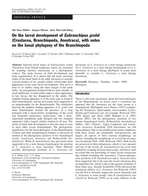
Zoomorphology (2004) 123:107–123DOI 10.1007/s00435-003-0093-0ORIGINAL ARTICLEOle Sten Møller · Jørgen Olesen · Jens Thorvald Høeg<strong>On</strong> <strong>the</strong> <strong>larval</strong> <strong>development</strong> <strong>of</strong> <strong>Eubranchipus</strong> <strong>grubii</strong>(<strong>Crustacea</strong>, Branchiopoda, Anostraca), with noteson <strong>the</strong> basal phylogeny <strong>of</strong> <strong>the</strong> BranchiopodaReceived: 19 March 2003 / Accepted: 15 October 2003 / Published online: 11 December 2003 Springer-Verlag 2003Abstract Selected <strong>larval</strong> stages <strong>of</strong> <strong>Eubranchipus</strong> <strong>grubii</strong>(Anostraca) from Danish temporary waters are examinedby scanning electron microscopy in a phylogeneticcontext. The study focuses on limb <strong>development</strong> andbody segmentation. It is shown that <strong>the</strong> large, proximalendite <strong>of</strong> <strong>the</strong> trunk limbs in <strong>the</strong> adult Anostraca is actuallya fusion product <strong>of</strong> two smaller endites which make <strong>the</strong>irappearance in <strong>the</strong> early <strong>larval</strong> <strong>development</strong>. This gives atotal <strong>of</strong> six endites along <strong>the</strong> inner margin <strong>of</strong> <strong>the</strong> trunklimbs. An unsegmented endopod follows more distally. Asmall additional, seventh endite makes a short appearancein late larvae, but has disappeared in <strong>the</strong> adults. Thenaupliar feeding apparatus is <strong>of</strong> <strong>the</strong> same type as found ino<strong>the</strong>r branchiopods, and has previously been suggested asan autapomorphy for <strong>the</strong> Branchiopoda. The similaritiesbetween <strong>the</strong> naupliar feeding apparatus <strong>of</strong> E. <strong>grubii</strong> ando<strong>the</strong>r branchiopods include <strong>the</strong> presence <strong>of</strong> a longprotopod with a characteristic morphology <strong>of</strong> <strong>the</strong> coxaland basipodal masticatory spines/setae, and a threesegmentedmandibular palp (basipod and two endopodsegments) with a largely similar setation in all taxa. Themode <strong>of</strong> trunk limb <strong>development</strong> is also <strong>the</strong> same as seenin most o<strong>the</strong>r recent branchiopods. The phylogeneticsignificance for <strong>the</strong> basal phylogeny <strong>of</strong> <strong>the</strong> Branchiopoda<strong>of</strong> <strong>the</strong>se and o<strong>the</strong>r morphological features is discussed inrelation to <strong>the</strong> phylogenetic position <strong>of</strong> two branchiopodfossils, Lepidocaris rhyniensis and Rehbachiella kinnekullensis.While R. kinnekullensis has previously beensuggested to be a stem lineage branchiopod, <strong>the</strong> position<strong>of</strong> L. rhyniensis is more uncertain. Three differentpossible phylogenetic positions <strong>of</strong> L. rhyniensis areO. S. Møller ()) · J. T. HøegDepartment <strong>of</strong> Zoomorphology, Zoological Institute,University <strong>of</strong> Copenhagen,Universitetsparken 15, 2100 Copenhagen OE, Denmarke-mail: osmoller@zi.ku.dkFax: +45-35321200J. OlesenZoological Museum,University <strong>of</strong> Copenhagen,Universitetsparken 15, 2100 Copenhagen OE, Denmarkdiscussed: (a) L. rhyniensis as a stem lineage anostracan,(b) L. rhyniensis as a stem lineage branchiopod or (c) L.rhyniensis as a stem lineage phyllopod. It seems mostplausible to consider L. rhyniensis a stem lineageanostracan.Keywords <strong>On</strong>togeny · Nauplius · Limbs · SEM ·PhylogenyIntroductionThere is still some uncertainty about <strong>the</strong> basal phylogeny<strong>of</strong> <strong>the</strong> Branchiopoda. In recent years a consensus hasappeared that <strong>the</strong> Anostraca are <strong>the</strong> sister group to amonophyletic Phyllopoda sensu Preuss (1951) (comprising<strong>the</strong> Notostraca, Spinicaudata, Laevicaudata, Cycles<strong>the</strong>ridaand Cladocera) (see, for example, Walossek 1993,1998; Spears and Abele 2000; Braband et al. 2002;Olesen 2003), but <strong>the</strong> phylogenetic positions <strong>of</strong> twominute branchiopod fossils, Rehbachiella kinnekullensisMüller, 1983 (“Orsten”, Upper Cambrian) and Lepidocarisrhyniensis Scourfield, 1926 (Devonian), are stillbeing debated. Due to favourable preservation conditions,both <strong>the</strong>se fossils are remarkably well-preserved, andlargely all aspects <strong>of</strong> <strong>the</strong>ir external morphology areknown, including <strong>the</strong> morphology <strong>of</strong> several <strong>larval</strong> stages(see Scourfield 1926, 1940; Walossek 1993). Both fossilsare preserved in three dimensions, which is very unusual,and one <strong>of</strong> <strong>the</strong> fossils (R. kinnekullensis) has even beenstudied by scanning electron microscopy, and this to suchan extent that our knowledge <strong>of</strong> many recent crustaceanslags behind (including <strong>the</strong> Anostraca). Various phylogeneticpositions <strong>of</strong> R. kinnekullensis and L. rhyniensis havebeen suggested, but in all cases <strong>the</strong> recent Anostraca playan important role, which is due to <strong>the</strong> general similarity <strong>of</strong><strong>the</strong>se three taxa. Already Scourfield (1926) discussed <strong>the</strong>anostracan affinity <strong>of</strong> L. rhyniensis, but Walossek (1993)was <strong>the</strong> first to formally suggest <strong>the</strong> recent Anostraca andL. rhyniensis as sister groups. Also R. kinnekullensis wasviewed by Walossek (1993) as an early <strong>of</strong>fshoot <strong>of</strong> <strong>the</strong>
108anostracan line. Olesen (1999) questioned <strong>the</strong> anostracanrelationship <strong>of</strong> R. kinnekullensis and suggested a positionone step down as a stem lineage branchiopod. Schram andKoenemann (2001) had <strong>the</strong> same view and placed L.rhyniensis in a similar position. Olesen (2003) followedup on <strong>the</strong> discussion and provided some characters insupport <strong>of</strong> <strong>the</strong> position <strong>of</strong> R. kinnekullensis as a stemgroup branchiopod, but left <strong>the</strong> position <strong>of</strong> L. rhyniensisunresolved.Due to <strong>the</strong> pivotal role <strong>of</strong> <strong>the</strong> Anostraca in <strong>the</strong>understanding <strong>of</strong> <strong>the</strong> phylogenetic position <strong>of</strong> R. kinnekullensisand L. rhyniensis, along with <strong>the</strong> lack <strong>of</strong>important morphological information for <strong>the</strong> Anostraca, itis <strong>of</strong> interest to examine aspects <strong>of</strong> <strong>the</strong> external morphology<strong>of</strong> an anostracan species in more detail to facilitatecomparison to <strong>the</strong> two fossils. To address questions <strong>of</strong>limb homologies and body segmentation in a comparativecontext, we chose to study <strong>the</strong> external morphology <strong>of</strong>three different stages <strong>of</strong> <strong>Eubranchipus</strong> <strong>grubii</strong> (Dybowski,1860). This species has been chosen for conveniencesince it is <strong>the</strong> most common species in Denmark(Damgaard and Olesen 1998), and since it provides analternative to <strong>the</strong> <strong>of</strong>ten-studied species <strong>of</strong> Artemia. Thespecies has previously been briefly dealt with byOehmichen (1921) in light microscopy. As summarisedby Walossek (1993), <strong>the</strong> Anostraca have <strong>the</strong> longest andmost gradual (anamorphic) <strong>development</strong> among recent<strong>Crustacea</strong> (see also Heath 1924; Benesch 1969; Bernice1972; Fryer 1983; Schrehardt 1987), and we have chosenlarvae <strong>of</strong> E. <strong>grubii</strong> that represent <strong>the</strong> early, intermediateand late <strong>development</strong>. O<strong>the</strong>r important studies on <strong>the</strong><strong>larval</strong> <strong>development</strong> <strong>of</strong> various anostracan species includethose <strong>of</strong> Claus (1873, 1886), Cannon (1926), Anderson(1967), Barlow and Sleigh (1980) and Jurasz et al. (1983).Comparative studies <strong>of</strong> ontogeny have previously providedclear information useful for phylogenetic considerationswithin <strong>the</strong> Branchiopoda (Fryer 1988; Walossek1993; Olesen 2003).The end <strong>of</strong> <strong>the</strong> discussion will focus on <strong>the</strong> phylogeneticposition <strong>of</strong> L. rhyniensis since this is an importantaspect <strong>of</strong> branchiopod phylogeny that remains to beclarified. As a basis for <strong>the</strong> discussion, we assume that:(1) R. kinnekullensis is not a stem lineage anostracan, butra<strong>the</strong>r a stem lineage branchiopod, (2) within <strong>the</strong> Branchiopoda,all branchiopods, except <strong>the</strong> Anostraca, can begrouped in <strong>the</strong> Phyllopoda and (3) L. rhyniensis forms amonophylum with <strong>the</strong> recent Branchiopoda based onsimilarities in <strong>the</strong> nauplius feeding apparatus and <strong>the</strong>morphology <strong>of</strong> <strong>the</strong> adult trunk limbs, <strong>the</strong> details <strong>of</strong> whichwere summarised by Olesen (2003). Accepting <strong>the</strong> listedassumptions for <strong>the</strong> basal branchiopod phylogeny, threepossibilities exist for <strong>the</strong> phylogenetic position <strong>of</strong> L.rhyniensis: (a) a stem lineage anostracan, (b) a stemlineage branchiopod or (c) a stem lineage phyllopod.Hence, <strong>the</strong> purpose <strong>of</strong> this paper is tw<strong>of</strong>old:1. To provide new information on <strong>the</strong> external morphology<strong>of</strong> a species <strong>of</strong> <strong>the</strong> Anostraca, with special focuson limb <strong>development</strong> and limb homologies2. To use <strong>the</strong> new information and <strong>the</strong> available literatureto evaluate a debated aspect <strong>of</strong> basal branchiopodphylogeny: <strong>the</strong> phylogenetic position <strong>of</strong> <strong>the</strong> Devonianbranchiopod fossil L. rhyniensisMaterials and methodsLarvae <strong>of</strong> E. <strong>grubii</strong> were collected in <strong>the</strong> early spring <strong>of</strong> 2001(January to March) in one <strong>of</strong> <strong>the</strong> classic Danish localities for “largebranchiopods” close to “Trepilelågen” in <strong>the</strong> deer park, (JaegersborgDyrehave), north <strong>of</strong> Copenhagen (more information about <strong>the</strong>locality in Damgaard and Olesen 1998 and Mossin 1986). Thematerial was fixed in a 1% osmium tetroxide solution overnight atroom temperature. Fur<strong>the</strong>r preparation was by dehydration throughacetone and critical-point drying using CO 2 . The specimens weresputter coated with a mixture <strong>of</strong> palladium and platinum. The SEMsused were a Jeol JSM-840 and a Jeol JSM 6335-F, placed at <strong>the</strong>Zoological Museum, University <strong>of</strong> Copenhagen.ResultsIn <strong>the</strong> following description, we include larvae in <strong>the</strong>following three categories largely representing <strong>the</strong> whole<strong>larval</strong> sequence: (1) stage without well-developed limbbuds, (2) stage with five to seven well-developed limbbuds and (3) stage with 12–13 well-developed limb buds.Stage without well-developed limb budsThe earliest stage observed is a nauplius larva <strong>of</strong>approximately 670 mm in length (without antennules).The three naupliar appendages, antennules, antennae andmandibles, are developed and partly functional, whiletrunk limbs are only weakly demarcated (Fig. 1).The labrum is large, inflated and evenly roundeddistally (U-shaped); it overhangs <strong>the</strong> median tips <strong>of</strong> <strong>the</strong>mandibular coxae and lacks setation (Fig. 1A–C). Theantennules are inserted anteriorly and somewhat laterally,separated by a space corresponding to <strong>the</strong> width <strong>of</strong> <strong>the</strong>labrum (Fig. 1A, C). They are long (nearly same length astrunk), slender, unsegmented and bear three setae at <strong>the</strong>tips.The antennae are inserted more laterally at <strong>the</strong> bodythan are <strong>the</strong> antennules (Fig. 1A, B). They are large(almost <strong>the</strong> same length as <strong>the</strong> whole animal) andbiramous. The protopod is only weakly divided into acoxal and basipodal part. The distal portion <strong>of</strong> <strong>the</strong>protopod, <strong>the</strong> prospective basipod, is about twice <strong>the</strong>length <strong>of</strong> <strong>the</strong> proximal portion, <strong>the</strong> prospective coxa. Thecoxal portion <strong>of</strong> <strong>the</strong> antenna has a large spine orientedtowards <strong>the</strong> mouth (“proximal masticatory spine”, sensuFryer 1983); here we intentionally use <strong>the</strong> neutral term“spine”, leaving it open whe<strong>the</strong>r this structure, or parts <strong>of</strong>it, should be a seta. The basipod also bears ano<strong>the</strong>r spinedirected somewhat against <strong>the</strong> midline <strong>of</strong> <strong>the</strong> animal(“distal masticatory spine”, sensu Fryer 1983, in <strong>the</strong>following referred to as <strong>the</strong> “basipodal masticatoryspine”). The endopod is only about <strong>the</strong> length <strong>of</strong> <strong>the</strong>
109Fig. 1A–D <strong>Eubranchipus</strong> <strong>grubii</strong>, stage without well-developedlimb buds. A Ventral view. B Lateral view. C View from ventral/posterior. D Ventral view on trunk region. a1 Antennule, a2antenna, en endopod, ex exopod, la labrum, md mandible, tl1 trunklimb 1, tl12 trunk limb 12basipod, which corresponds to one quarter <strong>the</strong> length <strong>of</strong><strong>the</strong> exopod; <strong>the</strong> segmentation is indistinct and it has threelong setae distally. The exopod has about <strong>the</strong> length <strong>of</strong> <strong>the</strong>protopod, is multiannulated and has 20 long, natatorysetae on <strong>the</strong> posterior/median face and two setae distally.The mandibles are inserted laterally on <strong>the</strong> body(Fig. 1A, C). They each consist <strong>of</strong> an enlarged coxa with athree-segmented “palp” (basipod and two presumedendopodal segments distally). The coxal processes areonly weakly developed and seem unable to meet mediallywhere <strong>the</strong>y are covered ventrally by <strong>the</strong> fleshy U-shapedlabrum. The median edge <strong>of</strong> <strong>the</strong> coxal process has a smallseta (termed “gnathobasic seta” by Fryer 1983). The palphas seven setae: two setae on <strong>the</strong> basipod, two on <strong>the</strong> firstendopodal segment and three setae terminally on <strong>the</strong>distalmost second segment.Nei<strong>the</strong>r maxillules nor maxillae can be clearly seen inthis stage. The head region <strong>of</strong> <strong>the</strong> larva is generally
110“narrower” (i.e. <strong>the</strong> circumference is smaller) than <strong>the</strong>thorax with <strong>the</strong> maxillary region as a cone-shapedtransition zone.The trunk is almost twice as long as <strong>the</strong> head region.No clearly recognisable trunk limbs are yet present, but12–14 weakly demarcated pairs <strong>of</strong> buds are presentventrally, posteriorly lying progressively closer to eacho<strong>the</strong>r (Fig. 1C). A pair <strong>of</strong> minute precursors <strong>of</strong> <strong>the</strong> furcalrami, with an anal opening in between, completes <strong>the</strong>larva posteriorly (Fig. 1D).Stage with five to seven developed limb budsThe length <strong>of</strong> this stage is about 750 mm (withoutantennules). Apart from an increase in size, <strong>the</strong> mostsignificant differences between this and <strong>the</strong> previouslydescribed stage are that <strong>the</strong> buds <strong>of</strong> <strong>the</strong> maxillae andmaxillules have appeared (Fig. 2A, E) and that <strong>the</strong> trunklimb buds have become differentiated into various limbportions (Fig. 2A).Frontally a pair <strong>of</strong> separate buds represents <strong>the</strong>primordial compound eyes (Fig. 3B). Slightly moreventrally, closer toge<strong>the</strong>r, a pair <strong>of</strong> small depressions isseen that probably represent <strong>the</strong> “frontal organs”(Fig. 3B). Dorsally a large dorsal organ covers <strong>the</strong> regioncorresponding to <strong>the</strong> antennae and <strong>the</strong> mandibles(Figs. 3A–F, 4A).Behind <strong>the</strong> naupliar portion <strong>of</strong> <strong>the</strong> head is a short,segment-like section <strong>the</strong> same size as posterior trunksegments (Fig. 4A), corresponding to <strong>the</strong> maxillules andmaxillae, but without external demarcation into twoseparate segments. There are 14–15 postmaxillary segments,decreasing gradually in width posteriorly(Fig. 4A, D). The labrum is large, fleshy and U-shaped(Fig. 2A, C).The naupliar appendages (antennules, antennae andmandibles) are very much like those <strong>of</strong> <strong>the</strong> previouslydescribed stage. In this stage <strong>the</strong>y have been examinedsomewhat closer, and since <strong>the</strong> setae are more developed,more details will be presented here. The antennules arenow long and slender, with three distal setae (Figs. 2A,3A). The antennal protopods are more clearly divided intoa coxa and a basipod (Fig. 2A). The coxa has a longenditic process, <strong>the</strong> “coxal masticatory spine”, directedtowards <strong>the</strong> mouth region (Fig. 2A). This process is armedwith setules distally which <strong>the</strong>mselves bear small secondordersetules (Fig. 2B). The basipod is about twice as longas wide and is superficially divided into four portions(Fig. 2A). Distally <strong>the</strong> basipod carries a long setaequipped with long setules also directed towards <strong>the</strong>mouth region (Fig. 2A). The endopod is short and itssegmentation is somewhat unclear but is probablyrevealed by several transverse rows <strong>of</strong> minute spines thatindicate a total <strong>of</strong> four portions (probable segment bordersmarked by arrows on Fig. 4C). The exopod is unchangedfrom <strong>the</strong> previously described stage and it has 20 setaealong <strong>the</strong> posteromedian margin, one for each annulation.The mandibular coxa has become larger compared to<strong>the</strong> previously described stage (Figs. 2A, 3F, 4A) and <strong>the</strong>gnathal edges <strong>of</strong> <strong>the</strong> coxal processes are now able to meeteach o<strong>the</strong>r. The gnathobasic setae are also larger andalmost hidden by <strong>the</strong> labrum. The mandibular palp hasmore distinct borders between <strong>the</strong> three segments (basipodand two endopod segments; Fig. 4D, E). The setation <strong>of</strong> <strong>the</strong>mandibular palp is <strong>the</strong> same as earlier—basipod with twosetae, <strong>the</strong> first endopod segment with two setae, and <strong>the</strong>second endopod segment with three setae—but <strong>the</strong> setaeare longer. The two setae on <strong>the</strong> basipod and endopodsegment 1, respectively, have a number <strong>of</strong> unevenlydistributed long setules, arranged pairwise along <strong>the</strong> seta towhich <strong>the</strong>y are attached in a “V”-like arrangement(Fig. 4D). These setules point towards <strong>the</strong> mouth region.The three setae on <strong>the</strong> second endopod segment each havea row <strong>of</strong> short, closely placed setules along <strong>the</strong>m.A pair <strong>of</strong> primordial paragnaths, densely covered withsetae, is visible just below and slightly posterior to <strong>the</strong>labrum (Fig. 2E). Between <strong>the</strong> primordial paragnaths is aparagnath channel. Both <strong>the</strong> maxillules and <strong>the</strong> maxillaeare now visible and armed with fine setae on <strong>the</strong>ir ventralface (Fig. 2E). The maxillulary and maxillary limb budsare attached ventrally at <strong>the</strong> short, segment-like sectionbetween <strong>the</strong> naupliar portion <strong>of</strong> <strong>the</strong> head and <strong>the</strong> firsttrunk segment (Figs. 2E, 4A, 5D). The maxillules areelongate limb buds placed laterally, immediately behind<strong>the</strong> paragnaths and oriented towards <strong>the</strong> ventral midline,placing <strong>the</strong>m on <strong>the</strong> “outside” <strong>of</strong> <strong>the</strong> maxillae(Fig. 2A, E). The maxillae are small rounded buds,placed slightly behind <strong>the</strong> elongate maxillules, and muchcloser toge<strong>the</strong>r than <strong>the</strong>se (Figs. 2A, E, 5D).The trunk has two rows <strong>of</strong> limb buds placed ventrolaterally(Figs. 2A, 4A, B, 5); no limb has yet attained <strong>the</strong>adult, vertical (“upright”) orientation. A gap separates <strong>the</strong>two rows <strong>of</strong> limb buds in <strong>the</strong> ventral midline <strong>of</strong> <strong>the</strong> trunk(Figs. 2A, 5D). <strong>On</strong> each side seven to eight limb buds aredeveloped with various limb portions demarcated to avarying degree (Figs. 2A, 3F, 4A, B, 5). More posteriorly<strong>the</strong>re are seven to eight additional limb buds or trunksegments, with no portions indicated. The anterior limbbuds are <strong>the</strong> largest and most developed ones. Each limbbud curves or bends slightly posteriorly and overlaps itsmore posterior neighbour, thus reflecting <strong>the</strong> situation in<strong>the</strong> adult (Figs. 3F, 4A, B). The complete and mostdeveloped limb buds have as many as 11 lobes <strong>of</strong> varyingsizes, to which we henceforward apply <strong>the</strong> terminologyused for adult trunk limbs. Ventrally (median in adult)each limb bud is divided into seven portions: threemedian (proximal) large ones, called endites in <strong>the</strong> adult,three smaller lobes more laterally, which are also calledendites in <strong>the</strong> adult, and one large lobe most laterally,called an endopod in <strong>the</strong> adult. Lateral to <strong>the</strong> endopodfollows first a large lobe, <strong>the</strong> exopod, and <strong>the</strong>reafter threeadditional lobes that will develop into <strong>the</strong> adult epipods.The exopod has a distinct setation consisting <strong>of</strong> two t<strong>of</strong>our setae on <strong>the</strong> four most anterior limbs. Small setae arepresent on <strong>the</strong> three proximal ventral lobes (endites) ontrunk limbs 1 and 2.
111Fig. 2A–E <strong>Eubranchipus</strong> <strong>grubii</strong>, stage with five to seven welldevelopedtrunk limbs. A Ventral view. B Tip <strong>of</strong> antenna coxalmasticatory spine. C Distal margin <strong>of</strong> labrum. D Left antenna. EClose-up <strong>of</strong> area behind labrum showing paragnaths (pgn),maxillules (mx1) and maxillae (mx2). a1 Antennule, a2 antenna,bas basis, cox coxa, en endopod, ex exopod, tl1 trunk limb 1
112
113Fig. 4A–E <strong>Eubranchipus</strong> <strong>grubii</strong>, stage with five to seven welldevelopedtrunk limbs. A Lateral view. B Lateral view <strong>of</strong> trunklimb buds. C Lateral view <strong>of</strong> left side antennal endopod (a2 en).Arrows point at possible rudiments <strong>of</strong> segmentation as indicated byrows <strong>of</strong> short spines. D Median side <strong>of</strong> left side mandibular palp. EAnterolateral view <strong>of</strong> right side mandible. a2 Antenna, bas basis, cecompound eye, cox coxa, do dorsal organ, en endopod, en1, 2endopodal segments 1, 2, ep epipod, ex exopod, md cox mandibularcoxa, mx1 maxillule, tl1–9 trunk limbs 1–9Dorsally each postmaxillary segment carries tworelatively long setae placed some distance apart(Figs. 3E, F, 4A). In contrast, <strong>the</strong>re is no such pair <strong>of</strong>setae on <strong>the</strong> maxillulary/maxillary segment-like section.In total, 11 postmaxillary segments have a pair <strong>of</strong> setae.These setae are placed in <strong>the</strong> same position on mostsegments, but on segment 9, <strong>the</strong>y are placed significantlyFig. 3A–F <strong>Eubranchipus</strong> <strong>grubii</strong>, stage with five to seven welldevelopedtrunk limbs. A Dorsal view. B Frontal view showingprimordial compound eyes (ce) and dorsal organ (do). C Close-up<strong>of</strong> dorsal organ. D Pair <strong>of</strong> caudal protrusions each with a seta. EDorsal side <strong>of</strong> trunk. Arrows point to a pair <strong>of</strong> setae placed closertoge<strong>the</strong>r than <strong>the</strong> remaining dorsal pairs <strong>of</strong> setae. F Lateral view. a1Antennule, a2 antenna, md mandible, md cox mandibular coxa, tl1trunk limb 1closer toge<strong>the</strong>r (Fig. 3E arrows). The telson region tapersinto two conical protrusions each carrying a seta, denselyarmed with setules along <strong>the</strong> distal half (Fig. 3D, E).Stage with 12–13 well-developed limb budsThis stage is about 1.2 mm long and, in most respects, isquite different from <strong>the</strong> previously described stages.Among <strong>the</strong> most significant changes are <strong>the</strong> tips <strong>of</strong> <strong>the</strong>coxal masticatory spines <strong>of</strong> <strong>the</strong> antennae have becomebifid and <strong>the</strong> anterior trunk limbs have attained an“upright”, adult orientation.The frontally placed primordial buds <strong>of</strong> <strong>the</strong> compoundeyes have become larger and <strong>the</strong> separate facets arevisible through <strong>the</strong> cuticle (Fig. 6B). Between <strong>the</strong> eyes is
114
a pair <strong>of</strong> small elevations probably being frontal organs(Fig. 6B).The antennules, with three distal setae, are relativelylong and slender (Fig. 6A). The antennae are ra<strong>the</strong>rsimilar to those in <strong>the</strong> previously described stage, but <strong>the</strong>yare relatively smaller and, most significantly, <strong>the</strong> tips <strong>of</strong><strong>the</strong> coxal masticatory spines have become bifid(Fig. 7A, D, H). Both branches <strong>of</strong> <strong>the</strong> bifid tip arecovered with robust setules, arranged semiregularly inrows along each spine. The setules <strong>the</strong>mselves also bearsome second-order setules. The posterior branch <strong>of</strong> <strong>the</strong>bifid tip has an annulation close to its basis (Fig. 7E).As earlier, <strong>the</strong> basipodal masticatory spine has <strong>the</strong>same arrangement <strong>of</strong> mostly pairwise attached, longsetules, directed towards <strong>the</strong> mouth region and <strong>the</strong>mselvesbearing small, second-order setules (Fig. 7A, F). Along itsposterior margin, <strong>the</strong> basipodal masticatory spine carries arow <strong>of</strong> short, densely placed setules (Fig. 7F). Distally,<strong>the</strong> endopod carries four ra<strong>the</strong>r long setae (Fig. 7C) incontrast to <strong>the</strong> previously described stage, where one wasra<strong>the</strong>r short. The endopod is apparently composed <strong>of</strong> fourportions as indicated by rows <strong>of</strong> spines and articulationlikeconstrictions (Fig. 7C arrows). The exopod isunchanged and carries 19–20 long setae (Figs. 6A, 7A).The coxae <strong>of</strong> <strong>the</strong> mandibles have increased in size. Theprimordial paragnaths with a paragnath channel in betweenare found behind <strong>the</strong> mandibles (Fig. 7B). The paragnathsare covered by a dense layer <strong>of</strong> setae. Placed immediatelybehind <strong>the</strong> paragnaths, <strong>the</strong> elongate buds <strong>of</strong> <strong>the</strong> maxillulesare also covered by fine setae. The maxillules have becomemore prominent than in <strong>the</strong> previously described stage(Fig. 7B) and, pointing more or less medially, <strong>the</strong>y have afew setae directed towards <strong>the</strong> mouth region. The smallerand circular buds <strong>of</strong> <strong>the</strong> maxillae are placed closer toge<strong>the</strong>rthan <strong>the</strong> maxillules, slightly posterior to <strong>the</strong>se, and are alsocovered with small setae.The region <strong>of</strong> <strong>the</strong> trunk limbs has developed significantly,carrying 13 pairs <strong>of</strong> limb buds, which is <strong>the</strong> fullnumber achieved in adults, including <strong>the</strong> modified limbsassociated with <strong>the</strong> genital segments (Fig. 6A, F). Trunklimb 1 is almost fully developed, but <strong>the</strong> degree <strong>of</strong><strong>development</strong> decreases posteriorly, and trunk limbs 9–11are elongate limb buds with only incipient subdivisions(Fig. 6A, F). Buds <strong>of</strong> limb pairs 11 and 12 on <strong>the</strong> futuregenital segments show no clear division into substructures(Fig. 6F). The anterior six to seven pairs <strong>of</strong> trunk limbshave enlarged very much and attained a more verticalorientation; <strong>the</strong>se limbs have a medial row <strong>of</strong> six to sevenenditic lobes (Fig. 6F, G). In <strong>the</strong> three anterior trunk limbs<strong>the</strong> two proximal enditic lobes have begun to fuse (as isFig. 5A–D <strong>Eubranchipus</strong> <strong>grubii</strong>, stage with five to seven welldevelopedtrunk limbs. A Ventral view <strong>of</strong> buds <strong>of</strong> trunk limbs 1–6,right side. B Lateral view <strong>of</strong> buds <strong>of</strong> trunk limbs 2–4, right side,showing future distal parts. C Lateral view <strong>of</strong> buds on right side <strong>of</strong>trunk limbs 4–11. D Ventral view <strong>of</strong> approximately 12 limb buds indifferent degrees <strong>of</strong> <strong>development</strong>. e1/pe Endite 1/proximal endite,e2–6 endites 2–6, en endopod, ep epipod, ex exopod, mx2 maxilla,tl1–7 trunk limbs 1–7<strong>the</strong> adult condition; Fig. 6F). At least six endites arepresent, but possible rudiments <strong>of</strong> a seventh endite can berecognised in trunk limbs 4–6 between <strong>the</strong> sixth enditeand <strong>the</strong> endopod (Fig. 6F, G). The exopod has attained aconsiderable size in <strong>the</strong> anterior four to six trunk limbs(Fig. 6A, D, F). Laterally, <strong>the</strong> epipod and <strong>the</strong> twoadditional more proximal exites (or epipods) haveenlarged significantly (Fig. 6D, E). The distalmost epipodis <strong>the</strong> largest, and it appears to be incipiently articulated to<strong>the</strong> stem <strong>of</strong> <strong>the</strong> limb. Setation is most developed in <strong>the</strong>anterior trunk limbs (Fig. 6A, F, G). The proximal endites<strong>of</strong> <strong>the</strong> trunk limbs have a row <strong>of</strong> curved, well-developedsetae at <strong>the</strong> posterior edges <strong>of</strong> <strong>the</strong> endites. The remainingfive endites also have setae along <strong>the</strong> posterior edge.Slightly more anteriorly, on endites 1–4, one or two shortspines are found (Fig. 6F, G). Setae are present on <strong>the</strong>exopods <strong>of</strong> trunk limbs 1–9. Setation is only present on<strong>the</strong> endopods and <strong>the</strong> most proximal endites on trunklimbs 1–7. The telsonic region tapers into two conicalprotrusions that are more elongate than in <strong>the</strong> previouslydescribed stage (Fig. 6A); each carries a long seta armedwith setules.DiscussionThis study deals with three different <strong>larval</strong> instars <strong>of</strong> <strong>the</strong>anostracan E. <strong>grubii</strong> chosen specifically to cover most <strong>of</strong><strong>the</strong> variation throughout <strong>the</strong> <strong>development</strong>al sequence <strong>of</strong><strong>the</strong> species. Based on what is known, <strong>the</strong> <strong>larval</strong>morphology <strong>of</strong> various anostracan species appears to bevery similar, which leads us to assume that E. <strong>grubii</strong>, in<strong>the</strong> context <strong>of</strong> comparative <strong>larval</strong> morphology <strong>of</strong> <strong>the</strong>Branchiopoda, is a typical representative <strong>of</strong> <strong>the</strong> Anostraca.First, selected structures <strong>of</strong> body segments and limbs<strong>of</strong> E. <strong>grubii</strong> will be summarised and compared to similarstructures in relevant branchiopods, especially those <strong>of</strong>two early branchiopod fossils (R. kinnekullensis and L.rhynieniens). Then some <strong>of</strong> this information and o<strong>the</strong>rinformation from <strong>the</strong> literature will be used in a discussion<strong>of</strong> <strong>the</strong> basal phylogeny <strong>of</strong> <strong>the</strong> Branchiopoda where<strong>the</strong> Anostraca play a central role.Body segmentation, dorsal setation <strong>of</strong> trunkand o<strong>the</strong>r details115In <strong>the</strong> stage with five to seven limb buds <strong>the</strong>re is a short,segment-like body section between <strong>the</strong> mandibular regionand <strong>the</strong> segment <strong>of</strong> <strong>the</strong> first trunk limbs. This sectioncorresponds ventrally to <strong>the</strong> maxillulary and maxillarylimb buds. Although two limb pairs (maxillules andmaxillae) belong to this section <strong>the</strong>re is no clear externalindication <strong>of</strong> its division into two separate segments.Probably this segment-like section is primarily maxillularysince <strong>the</strong> maxillules in size dominate over <strong>the</strong>maxillae (Figs. 4A, 5D). It appears likely that <strong>the</strong> section/segment in question is, at least in part, a fusion product <strong>of</strong>
116
<strong>the</strong> maxillary and maxillulary segments. The presence <strong>of</strong>such a “free” body portion immediately behind <strong>the</strong>mandibular region (not fused with naupliar portion t<strong>of</strong>orm a continuous head) prevails in <strong>the</strong> adults. Also adults<strong>of</strong> <strong>the</strong> Devonian fossil L. rhyniensis have such a freeportion behind <strong>the</strong> mandibles. Since this situation isunique within <strong>the</strong> Branchiopoda, and apparently differentfrom <strong>the</strong> condition in o<strong>the</strong>r <strong>Crustacea</strong>, it is suggestedbelow as a possible synapomorphy <strong>of</strong> L. rhyniensis and<strong>the</strong> Anostraca.In E. <strong>grubii</strong> each trunk segment carries a pair <strong>of</strong>relatively long setae dorsally. They are placed in a quiteregular pattern, in which <strong>the</strong> distance between setae <strong>of</strong> apair decreases posteriorly. In all members <strong>of</strong> <strong>the</strong> Phyllopodasensu Preuss (1951), even in <strong>the</strong> aberrant Leptodorakindtii (Focke, 1844) (see Olesen et al. 2003), a dorsalpair <strong>of</strong> setae is present posteriorly on <strong>the</strong> trunk (homologiesfirst discussed by Martin and Cash-Clark 1995). In <strong>the</strong>Cladocera <strong>the</strong>y are traditionally termed postabdominalsetae and in <strong>the</strong> “Conchostraca” sometimes telson filaments.Because <strong>of</strong> <strong>the</strong> presumed close relationshipbetween all extant Branchiopoda, it is tempting to considerhomology between <strong>the</strong> single pair present in <strong>the</strong> Phyllopodaand one <strong>of</strong> <strong>the</strong> setae pairs in <strong>the</strong> Anostraca (seeMartin and Cash-Clark 1995 on <strong>the</strong> same subject).However, such a homology can only be established withmajor difficulties since <strong>the</strong> position <strong>of</strong> <strong>the</strong> setae is quitedifferent. Embryological studies <strong>of</strong> Cycles<strong>the</strong>ria hislopi(Baird, 1859), (Olesen 1999) and L. kindtii (Cladocera)(Olesen et al. 2003), and <strong>the</strong>reby most certainly in o<strong>the</strong>rPhyllopoda, have shown that <strong>the</strong> single dorsal pair <strong>of</strong> setaeis telsonal, while none <strong>of</strong> <strong>the</strong> dorsal setae pairs present inE. <strong>grubii</strong> can be found that far posteriorly.The latest <strong>larval</strong> stage examined has 13 pairs <strong>of</strong> trunklimb buds (Fig. 6A, F), while adults only have 11 truetrunk limbs. We interpret <strong>the</strong> two posterior buds (Fig. 6F)as ei<strong>the</strong>r <strong>the</strong> anlagen to <strong>the</strong> penis in <strong>the</strong> male or <strong>the</strong> ovisacin <strong>the</strong> female, which indicate a limb-origin for <strong>the</strong>sestructures.Comparison <strong>of</strong> larvae <strong>of</strong> E. <strong>grubii</strong> with larvae<strong>of</strong> two branchiopod fossilsNaupliar feeding apparatus <strong>of</strong> <strong>Eubranchipus</strong>, Lepidocarisand RehbachiellaThe naupliar feeding systems <strong>of</strong> <strong>the</strong>se three taxa are ra<strong>the</strong>rsimilar, but those <strong>of</strong> E. <strong>grubii</strong> and L. rhyniensis sharesome specific similarities not found in R. kinnekullensis(if not o<strong>the</strong>rwise specified, <strong>the</strong> information on L.Fig. 6A–G <strong>Eubranchipus</strong> <strong>grubii</strong>, stage with 12–13 well-developedtrunk limbs. A Ventral view. B Frontal view showing compoundeyes (ce). C Distal margin <strong>of</strong> labrum. D Ventral view <strong>of</strong> trunklimbs. E Close-up <strong>of</strong> exites <strong>of</strong> left side trunk limbs 1–3. F Medianview <strong>of</strong> left side trunk limbs. G Median view <strong>of</strong> left side trunklimbs 5–8. a1 Antennule, a2 antenna, e1/pe endite 1/proximalendite, e2–7 endites 2–7, en endopod, ep epipod, ex exopod, lalabrum, md mandible, tl1–13 trunk limbs 1–13117rhyniensis and R. kinnekullensis is from Scourfield1926, 1940 and Walossek 1993). These similaritiesinclude a long antennal protopod, a similar arrangement<strong>of</strong> <strong>the</strong> coxal and basipodal masticatory setae <strong>of</strong> <strong>the</strong>antennae, as well as <strong>the</strong> segmentation and setation <strong>of</strong> <strong>the</strong>mandibular palp. Since o<strong>the</strong>r large branchiopods (Notostraca,Spinicaudata and Laevicaudata) for <strong>the</strong> most parthave a similar naupliar feeding apparatus, not found ino<strong>the</strong>r <strong>Crustacea</strong>, Olesen (2003) treated it as a synapomorphyfor <strong>the</strong> Branchiopoda (or Branchiopoda s. str.),excluding R. kinnekullensis.In <strong>the</strong> antennae, <strong>the</strong> specific similarities <strong>of</strong> <strong>the</strong> coxalmasticatory spines <strong>of</strong> E. <strong>grubii</strong> (Fig. 7D) and L. rhyniensisinclude that <strong>the</strong>y, in certain parts <strong>of</strong> <strong>the</strong> <strong>development</strong>, arepincer-like, divided into two branches <strong>of</strong> equal length andcovered with setules arranged more or less like <strong>the</strong> hairson a bottle-brush. This pattern is also present in <strong>the</strong>Spinicaudata and Laevicaudata (Olesen 2003; Olesen andGrygier 2003). It should be noted, however, that R.kinnekullensis also has long median coxal spines <strong>of</strong> <strong>the</strong>antennae in early larvae (see, for example, Walossek 1993plate 2:6), but never with <strong>the</strong> morphology describedabove. The most import difference is that <strong>the</strong> antennalcoxae have more than one spine in R. kinnekullensis,while only one is present in <strong>the</strong> remaining Branchiopoda.Ano<strong>the</strong>r difference concerns <strong>the</strong> branched tip <strong>of</strong> <strong>the</strong> coxalspine which, in contrast to L. rhyniensis and <strong>the</strong> recentBranchiopoda, has not been documented for any stage <strong>of</strong>R. kinnekullensis, but it is difficult to exclude that thiscould be explained by <strong>the</strong> fact that setae and spines tendto be broken <strong>of</strong>f in R. kinnekullensis (personal communicationD. Waloszek).The specific similarities <strong>of</strong> <strong>the</strong> <strong>larval</strong> mandible in <strong>the</strong>free-living larvae <strong>of</strong> all large branchiopods (including L.rhyniensis and E. <strong>grubii</strong>) concern <strong>the</strong> uniramous condition<strong>of</strong> <strong>the</strong> palp and <strong>the</strong> identical segmentation pattern(basipod and two endopod segments) (Olesen 2003). Itfur<strong>the</strong>r supports a segmental homology <strong>of</strong> <strong>the</strong> mandiblesbetween <strong>the</strong> groups that <strong>the</strong> setation pattern is <strong>the</strong> same inanostracans, notostracans and L. rhyniensis and largely<strong>the</strong> same in <strong>the</strong> spinicaudatans and laevicaudatans (seeOlesen 2003 for more details). R. kinnekullensis isdifferent in having more segments in <strong>the</strong> endopod andmore coxal and basipodal masticatory spines (seeWalossek 1993). Fur<strong>the</strong>rmore, <strong>the</strong> mandibular palp in R.kinnekullensis is biramous and consists <strong>of</strong> a basipod, anendopod and an exopod, in accordance with <strong>the</strong> situationin <strong>the</strong> larvae <strong>of</strong> <strong>the</strong> Cephalocarida and in many “maxillopodan”taxa (see, for example, Sanders 1963; Moyse1984, 1987; Olesen 2001). This makes <strong>the</strong> uniramous palpa putative synapomorphy <strong>of</strong> <strong>the</strong> Branchiopoda excludingR. kinnekullensis.Development <strong>of</strong> trunk limbs in <strong>Eubranchipus</strong>, Lepidocarisand Rehbachiella and some o<strong>the</strong>r branchiopodsDespite <strong>the</strong> general similarity between adult limbs <strong>of</strong> E.<strong>grubii</strong> and those <strong>of</strong> <strong>the</strong> Devonian fossil L. rhyniensis,
118
including a leaf-like appearance with a largely identicalarrangement <strong>of</strong> median and lateral lobes (endites andexopod/epipods; see, for example, Sars 1896; Scourfield1926), <strong>the</strong> mode <strong>of</strong> limb <strong>development</strong> is different. Themain difference is that while <strong>the</strong> long axis <strong>of</strong> <strong>the</strong> limb(proximal/distal axis in <strong>the</strong> adult) is displaced laterally in<strong>the</strong> larvae <strong>of</strong> E. <strong>grubii</strong>, L. rhyniensis has <strong>the</strong> adultdorsoventral orientation <strong>of</strong> this axis from <strong>the</strong> onset <strong>of</strong> <strong>the</strong><strong>larval</strong> <strong>development</strong> (Scourfield 1940). In this respect E.<strong>grubii</strong> is similar to o<strong>the</strong>r large branchiopods, as summarisedby Olesen (2003), while L. rhyniensis is similarto R. kinnekullensis and to o<strong>the</strong>r crustaceans.The serial displacement <strong>of</strong> <strong>the</strong> degree <strong>of</strong> <strong>development</strong><strong>of</strong> <strong>the</strong> trunk limbs in E. <strong>grubii</strong> allows <strong>the</strong> ontogeny <strong>of</strong> <strong>the</strong>various limb parts to be followed. The trunk limbs firstappear as elongate buds placed ventrolaterally on <strong>the</strong>body, following <strong>the</strong> curvature <strong>of</strong> <strong>the</strong> trunk, withoutexternal subdivision. This division appears first as a cleftpractically in <strong>the</strong> middle <strong>of</strong> <strong>the</strong> limb bud that divides itinto a ventral and laterodorsal part between <strong>the</strong> futureendopod and exopod (Møller et al. 2003 describe a similarcondition in <strong>the</strong> <strong>development</strong> <strong>of</strong> <strong>the</strong> Notostraca). The nextsubdivision appears fur<strong>the</strong>r laterally between <strong>the</strong> exopodand <strong>the</strong> future epipod. Approximately at <strong>the</strong> same time <strong>the</strong>first indication <strong>of</strong> <strong>the</strong> three to four most proximal enditesappears as well as <strong>the</strong> primordia <strong>of</strong> two more laterallyplaced epipods. The differentiation <strong>of</strong> <strong>the</strong> (future) medianedge (directed ventrally in <strong>the</strong> larvae) takes placesgradually from proximal towards distal. The proximalendites appear first and <strong>the</strong> distal endites and <strong>the</strong>unsegmented endopod appear last.In general terms, <strong>the</strong> <strong>development</strong> <strong>of</strong> <strong>the</strong> trunk limbs inE. <strong>grubii</strong> is very similar to that <strong>of</strong> two o<strong>the</strong>r branchiopodsviz., C. hislopi and Eulimnadia braueriana Ishikawa,1895 which have been studied in some detail by Olesen(1999) and Olesen and Grygier (2003). In those works,<strong>the</strong> similarities in <strong>development</strong> have been discussedbriefly as a possible synapomorphy <strong>of</strong> <strong>the</strong> branchiopods.Some important similarities are: <strong>the</strong> developing limb budsare placed as two rows lateroventrally on <strong>the</strong> trunk with<strong>the</strong> future median edge <strong>of</strong> <strong>the</strong> limbs facing ventrally, and<strong>the</strong> limbs in all mentioned species differentiate firstdistally (directed laterally in larvae) and <strong>the</strong> median edge(directed ventrally in larvae) differentiates into enditiclobes from proximal towards distal. Significant differencesbetween <strong>the</strong> species include that <strong>the</strong> developingtrunk limb buds <strong>of</strong> E. <strong>grubii</strong> are divided into 11 portions(six endites, an endopod, an exopod, three “epipods”),while only eight are present in <strong>the</strong> two “conchostracans”.Homologies <strong>of</strong> <strong>the</strong> endites and endopod<strong>of</strong> anostracans and o<strong>the</strong>r branchiopods119In early trunk limbs <strong>of</strong> E. <strong>grubii</strong> at least six endites and anunsegmented endopod are found along <strong>the</strong> medial edge(ventral in larvae). The three proximal, relatively largeenditic lobes are followed by three smaller ones. Largerlarvae occasionally carry a rudiment <strong>of</strong> a seventh enditeimmediately proximal to <strong>the</strong> endopod on some <strong>of</strong> <strong>the</strong>limbs but it disappears before adulthood (indicated by e7?on Fig. 6F).Our observations agree with those <strong>of</strong> Fryer (1983:Fig. 36) that <strong>the</strong> most proximal, large enditic lobe in <strong>the</strong>adult trunk limbs <strong>of</strong> anostracans is <strong>the</strong> product <strong>of</strong> a fusionbetween <strong>the</strong> <strong>larval</strong> enditic lobes 1 and 2 (Fig. 8). Thepresence <strong>of</strong> six enditic lobes means that <strong>the</strong>re is noequivalence in <strong>the</strong> number <strong>of</strong> endites between anostracantrunk limbs <strong>of</strong> and those <strong>of</strong> notostracans and “conchostracans”where only five enditic lobes are present (seeMøller et al. 2003 for Notostraca). The large proximalendite in <strong>the</strong> trunk limbs <strong>of</strong> <strong>the</strong> Anostraca probablycorresponds to endites 1 and 2 in <strong>the</strong> Notostraca and <strong>the</strong>“Conchostraca”. With respect to <strong>the</strong> number <strong>of</strong> endites,anostracans are more similar to L. rhyniensis, where <strong>the</strong>anterior trunk limbs also have six enditic lobes (Scourfield1926). Interestingly, previous authors have beenuncertain on <strong>the</strong> status <strong>of</strong> <strong>the</strong> large, proximal endite <strong>of</strong> <strong>the</strong>trunk limbs <strong>of</strong> <strong>the</strong> anostracans. Most workers mark <strong>the</strong>large proximal endite as endite 1. Based on work withPolyartemia forcipata Fischer, 1851, Ekman (1902)suggested that this endite is formed by a fusion <strong>of</strong> twoendites. Borradaile (1926) left <strong>the</strong> question unresolved,while Baqai (1963) explicitly stated (wrongly) that <strong>the</strong>most proximal, large endite does not consist <strong>of</strong> twoendites. Fryer (1983), on <strong>the</strong> o<strong>the</strong>r hand, correctly labelled<strong>the</strong> proximal large endite in adults <strong>of</strong> Branchinecta ferox(Milne-Edwards, 1840) as being composed <strong>of</strong> twoendites, but did not comment much on <strong>the</strong> issue.Walossek (1993) also favoured this interpretation.Fig. 7A–J <strong>Eubranchipus</strong> <strong>grubii</strong>, stage with 12–13 well-developedtrunk limbs. A Left side antenna (a2) and mandible (md). B Atriumoris: paragnaths (pgn), maxillules (mx1) and maxillae (mx2). CAntennae endopod <strong>of</strong> left side. D Bifurcated antennal coxalmasticatory spine <strong>of</strong> left side. E Detail <strong>of</strong> D showing a distinctannulus <strong>of</strong> <strong>the</strong> posterior branch. F Close-up <strong>of</strong> <strong>the</strong> basipodalmasticatory spine/seta <strong>of</strong> <strong>the</strong> antenna, showing its characteristicsetulation. G Mandible <strong>of</strong> right side. H Close-up <strong>of</strong> distal branches<strong>of</strong> coxal masticatory spine <strong>of</strong> <strong>the</strong> antenna. I Close-up <strong>of</strong> mandibleendopod segment 1, showing its armature <strong>of</strong> two setae. J Close-up<strong>of</strong> distal setae <strong>of</strong> mandible endopod segment 2. a2 en Antennalendopod, a2 ex antennal exopod, ba, bas basis, bas msp basipodalmasticatory spine, cox coxa, cox msp coxal masticatory spine,en1, 2 endopodal segments 1, 2, la labrum, md cox mandibularcoxa, tl1 trunk limb 1Phylogenetic evaluationIn recent years a growing body <strong>of</strong> evidence has indicatedthat <strong>the</strong> Branchiopoda is monophyletic based on informationfrom different sources, such as <strong>larval</strong> morphology,sperm morphology and molecular data (Sanders1963; Wingstrand 1978; Walossek 1993; Spears andAbele 2000; Olesen 2003). Various authors support <strong>the</strong>Anostraca as <strong>the</strong> sister group <strong>of</strong> <strong>the</strong> remaining Branchiopoda,and Walossek (1993) and Braband et al.(2002) suggested that <strong>the</strong> latter should be grouped under<strong>the</strong> name Phyllopoda sensu Preuss (1951). This was also
120Fig. 8A–C <strong>Eubranchipus</strong> <strong>grubii</strong>. A Ventral view <strong>of</strong> subadult. B Trunk limb <strong>of</strong> adult. C Close-up <strong>of</strong> B showing endites along <strong>the</strong> medianmargin. e1/pe Endite 1/proximal endite, e2–6 endites 2–6, en endopod, ep epipod, ex exopodfavoured in <strong>the</strong> comprehensive account <strong>of</strong> crustaceantaxonomy provided by Martin and Davis (2001). Hence,<strong>the</strong> basal split in branchiopod phylogeny now seems wellunderstood, but due to <strong>the</strong> generally primitive appearance<strong>of</strong> <strong>the</strong> Devonian L. rhyniensis and <strong>the</strong> UpperCambrian R. kinnekullensis, <strong>the</strong>se remarkably wellpreservedcrustaceans are obviously important for ourunderstanding <strong>of</strong> early branchiopod evolution. It is
121Fig. 9 Basal phylogeny <strong>of</strong> <strong>the</strong>Branchiopoda. The DevonianLepidocaris rhyniensis is placedas a stem lineage anostracan(position A), but two o<strong>the</strong>rpossible positions are also indicated(B and C)<strong>the</strong>refore crucial to evaluate <strong>the</strong> phylogenetic position <strong>of</strong><strong>the</strong>se fossils.Olesen (1999), Schram and Koenemann (2001) andOlesen (2003) suggested that at least R. kinnekullensis is astem lineage branchiopod ra<strong>the</strong>r than a stem lineageanostracan. In o<strong>the</strong>r words, <strong>the</strong>se authors suggested that R.kinnekullensis is not closer related to <strong>the</strong> Anostraca than itis to o<strong>the</strong>r branchiopods. The Branchiopoda toge<strong>the</strong>r withits stem group were treated as Pan-Branchiopoda byOlesen (2003). What remains to be established is <strong>the</strong>phylogenetic position <strong>of</strong> L. rhyniensis which, as notedpreviously, has been suggested to be a fossil anostracan(Scourfield 1926; Walossek 1993).Below is listed a number <strong>of</strong> putative synapomorphies<strong>of</strong> L. rhyniensis and <strong>the</strong> Anostraca which support that L.rhyniensis is a stem lineage anostracan (see Fig. 9). Thisis <strong>the</strong> preferred phylogenetic position <strong>of</strong> L. rhyniensis inthis study, but o<strong>the</strong>r possible positions <strong>of</strong> L. rhyniensis arediscussed more briefly below.– Ventral brood pouch formed from trunk segments 12and 13 (if Scourfield’s 1926 original counting isfollowed)– Median edge <strong>of</strong> trunk limbs with six endites (onlyanterior limbs in L. rhyniensis)– Carapace lacking– Thirteen limb-bearing segments (including <strong>the</strong> genitalsegments)– Maxillular/maxillar segment not fused to cephalon(this character may not be independent from carapacelacking)– Maxillar limb buds in larvae placed close toge<strong>the</strong>rScourfield (1926) also listed some “points <strong>of</strong> agreement”between L. rhyniensis and <strong>the</strong> Anostraca, but onlytwo <strong>of</strong> <strong>the</strong>se (“elongated form and general appearance”and “absence <strong>of</strong> dorsal shield or bivalve shell”) are noteasily dismissible as symplesiomorphies also present inmany o<strong>the</strong>r branchiopods (like structure <strong>of</strong> mandibularpalp and foliaceous structure <strong>of</strong> trunk limbs).A second possibility is L. rhyniensis as a stem lineagebranchiopod (position B in Fig. 9; suggested by Schramand Koenemann 2001), a hypo<strong>the</strong>sis which can be basedon <strong>the</strong> following putative synapomorphies for <strong>the</strong> recentBranchiopoda (excluding L. rhyniensis): (1) early limbbuds have future median edge directed ventrally, <strong>the</strong>median limb portions (endites and endopod) becomedifferentiated before <strong>the</strong> limbs attain a dorsoventralorientation, (2) maxillule reduced in size and (3) antennulesunsegmented in adults (with few exceptions which<strong>the</strong>n must be considered secondary).A third possibility would be L. rhyniensis being a stemlineage phyllopod (position C in Fig. 9). At least <strong>the</strong>re is asimilarity in <strong>the</strong> presence <strong>of</strong> only five to seven setae (incontrast to many more) on <strong>the</strong> antennal exopod <strong>of</strong> <strong>the</strong>larvae.However, due to <strong>the</strong> limited support for L. rhyniensisas a stem lineage phyllopod, we believe that only <strong>the</strong> tw<strong>of</strong>irst-mentioned possible phylogenetic positions <strong>of</strong> L.rhyniensis are worth considering: (a) L. rhyniensis as astem lineage anostracan or (b) L. rhyniensis as a stemlineage branchiopod. An anostracan stem lineage position<strong>of</strong> L. rhyniensis would mean that <strong>the</strong> laterally displaceddorsoventral axis <strong>of</strong> <strong>the</strong> trunk limbs (enditic portionsfacing ventrally) in <strong>the</strong> Anostraca and <strong>the</strong> Phyllopodawould have appeared independently in <strong>the</strong>se two groups(or it is an evolutionary reversal <strong>of</strong> <strong>the</strong> taxon L.
122rhyniensis), as would <strong>the</strong> reduced maxillules and <strong>the</strong> o<strong>the</strong>rcharacters listed above. If L. rhyniensis on <strong>the</strong> o<strong>the</strong>r hand,is considered to be a branchiopod stem lineage, <strong>the</strong>n all<strong>the</strong> characters listed in support for an anostracan stemlineage position (brood pouch, six median endites, samenumber <strong>of</strong> body somites, etc.) would be ei<strong>the</strong>r convergentor symplesiomorphies. We conclude, as did Walossek(1993), that an anostracan stem lineage position <strong>of</strong> L.rhyniensis is <strong>the</strong> most likely.Acknowledgements We thank Jesper T. Stenderup (Copenhagen)for assistance when collecting and sorting <strong>the</strong> material for thisstudy. Two anonymous reviewers and <strong>the</strong> editor <strong>of</strong> Zoomorphology,Pr<strong>of</strong>. Dr. Thomas Bartolomaeus (Berlin, Germany), arethanked for providing many useful suggestions for improvement<strong>of</strong> <strong>the</strong> paper. Pr<strong>of</strong>. Dr. Dieter Waloszek (Ulm, Germany) is greatlyacknowledged for many stimulating discussions with all threeauthors over <strong>the</strong> years.ReferencesAnderson DT (1967) Larval <strong>development</strong> and segment formation in<strong>the</strong> branchiopod crustaceans Limnadia stanleyana King (Conchostraca)and Artemia salina (L.) (Anostraca). Aust J Zool15:47–91Baqai IU (1963) Studies on <strong>the</strong> postembryonic <strong>development</strong> on <strong>the</strong>fairy shrimp Streptocephalus seali Ryder. Tulane Stud Zool10:91–120Barlow DI, Sleigh MA (1980) The propulsion and use <strong>of</strong> watercurrents for swimming and feeding in <strong>larval</strong> and adult Artemia.In: Persoone G, Sorgeloos P, Roels O, Jaspers E (eds) The brineshrimp Artemia, vol 1. Morphology, genetics, radiobiology,toxicology. Universa, Wetteren, Belgium, pp 61–73Benesch R (1969) Zur <strong>On</strong>togenie und Morphologie von Artemiasalina L. Zool Jahrb Abt Anat <strong>On</strong>tog Tiere 86:307–458Bernice R (1972) Hatching and postembryonic <strong>development</strong> <strong>of</strong>Streptocephalus dichotomus Baird (<strong>Crustacea</strong>: Anostraca).Hydrobiologia 40:251–278Borradaile LA (1926) Notes upon crustacean limbs. Ann Mag NatHist Ser 9 17:193–213Braband A, Richter S, Diesel R, Scholtz G (2002) Phylogeneticrelationships within <strong>the</strong> Phyllopoda (<strong>Crustacea</strong>, Branchiopoda)based on mitochondrial and nuclear markers. Mol PhylogenetEvol 25:229–244Cannon HG (1926) <strong>On</strong> <strong>the</strong> post-embryonic <strong>development</strong> <strong>of</strong> <strong>the</strong>fairy-shrimp (Chirocephalus diaphanus). J Linn Soc 36:401–416Claus C (1873) Zur Kenntniss des Baues und der Entwicklung vonBranchipus stagnalis und Apus cancriformis. Abh K Ges WissGöttingen 18:93–140Claus C (1886) Untersuchungen über die Organisation undEntwicklung von Branchipus und Artemia nebst vergleichendenBemerkungen über andere Phyllopoden. Arb Zool InstUniv Wien 6:1–104Damgaard J, Olesen J (1998) Distribution, phenology and status for<strong>the</strong> larger Branchiopoda (<strong>Crustacea</strong>: Anostraca, Notostraca,Spinicaudata and Laevicaudata) in Denmark. Hydrobiologia377:9–13Ekman S (1902) Beiträge zur Kenntnis der PhyllopodenfamiliePolyartemiidae. Bih K Svenska VetensAkad Handl 28:3–38Fryer G (1983) Functional ontogenetic changes in Branchinectaferox (Milne-Edwards) (<strong>Crustacea</strong>, Anostraca). Philos Trans RSoc Lond B Biol Sci 303:229–343Fryer G (1988) Studies on <strong>the</strong> functional morphology and biology<strong>of</strong> <strong>the</strong> Notostraca (<strong>Crustacea</strong>: Branchiopoda). Philos Trans RSoc Lond B Biol Sci 321:27–124Heath H (1924) The external <strong>development</strong> <strong>of</strong> certain phyllopods. JMorphol 38:453–483Jurasz W, Kittel W, Presler P (1983) Life cycle <strong>of</strong> Branchinectagaini, Daday, 1910 (Branchiopoda, Anostraca) from KingGeorge Island, South Shetland Islands. Pol Polar Res 4:143–154Martin JW, Cash-Clark C (1995) The external morphology <strong>of</strong> <strong>the</strong>onychopod “cladoceran” genus Bythotrephes (<strong>Crustacea</strong>, Branchiopoda,<strong>On</strong>ychopoda, Cercopagididae), with notes on <strong>the</strong>morphology and phylogeny <strong>of</strong> <strong>the</strong> order <strong>On</strong>ychopoda. Zool Scr24:61–90Martin JW, Davis GE (2001) An updated classification <strong>of</strong> <strong>the</strong>recent <strong>Crustacea</strong>. Nat Hist Mus Los Angeles, Sci Ser 39Møller OS, Olesen J, Høeg JT (2003) SEM studies on <strong>the</strong> early<strong>larval</strong> <strong>development</strong> <strong>of</strong> Triops cancriformis (Bosc) (<strong>Crustacea</strong>:Branchiopoda, Notostraca). Acta Zool (Stockh) 84:267–284Mossin J (1986) Physiochemical factors inducing embryonic<strong>development</strong> and spring hatching <strong>of</strong> <strong>the</strong> European fairy shrimpSiphonophanes grubei (Dybowsky) (<strong>Crustacea</strong>: Anostraca). JCrustac Biol 6:693–704Moyse J (1984) Some observations on <strong>the</strong> swimming and feeding<strong>of</strong> <strong>the</strong> nauplius larvae <strong>of</strong> Lepas pectinata (Cirripedia: <strong>Crustacea</strong>).Zool J Linn Soc 80:323–336Moyse J (1987) Larvae <strong>of</strong> lepadomorph barnacles. In: SouthwardAJ (ed) <strong>Crustacea</strong>n issues 5. Barnacle biology. Balkema,Rotterdam, pp 329–362Oehmichen A (1921) Die Entwicklung der äußeren Form desBranchipus grubei Dyb. Zool Anz 53:241–253Olesen J (1999) Larval and post-<strong>larval</strong> <strong>development</strong> <strong>of</strong> <strong>the</strong>branchiopod clam shrimp Cycles<strong>the</strong>ria hislopi (Baird, 1859)(<strong>Crustacea</strong>, Branchiopoda, Conchostraca, Spinicaudata). ActaZool (Stockh) 80:163–184Olesen J (2001) External morphology and <strong>larval</strong> <strong>development</strong> <strong>of</strong>Derocheilocaris remanei Delamare-Deboutteville and Chappuis,1951 (<strong>Crustacea</strong>, Mystacocarida), with a comparison <strong>of</strong>crustacean segmentation and tagmosis patterns. K Dan VidenskSelsk Biol Skr 53:1–59Olesen J (2003) <strong>On</strong> <strong>the</strong> ontogeny <strong>of</strong> <strong>the</strong> Branchiopoda (<strong>Crustacea</strong>):contribution <strong>of</strong> <strong>development</strong> to phylogeny and systematics. In:Scholtz G (ed) <strong>Crustacea</strong>n issues 15. Evolutionary <strong>development</strong>albiology <strong>of</strong> <strong>Crustacea</strong>. Balkema, Lisse (in press)Olesen J, Grygier MJ (2003) Larval <strong>development</strong> <strong>of</strong> Japanese“conchostracans”: part 1, <strong>larval</strong> <strong>development</strong> <strong>of</strong> Eulimnadiabraueriana (<strong>Crustacea</strong>, Branchiopoda, Spinicaudata, Limnadiidae)compared to that <strong>of</strong> o<strong>the</strong>r limnadiids. Acta Zool (Stockh)84:41–61Olesen J, Richter S, Scholtz G (2003) <strong>On</strong> <strong>the</strong> ontogeny <strong>of</strong>Leptodora kindtii (<strong>Crustacea</strong>, Branchiopoda, Cladocera), withnotes on <strong>the</strong> phylogeny <strong>of</strong> <strong>the</strong> Cladocera. J Morphol 256:235–259Preuss G (1951) Die Verwandtschaft der Anostraca und Phyllopoda.Zool Anz 147:49–64Sanders HL (1963) The Cephalocarida. Functional morphology,<strong>larval</strong> <strong>development</strong>, comparative external anatomy. Mem ConnAcad Arts Sci 15:1–80Sars GO (1896) Development <strong>of</strong> Es<strong>the</strong>ria packardi, as shown byartificial hatching from dried mud. Arch Math Naturvidensk B18:3–27Schram FR, Koenemann S (2001) Developmental genetics andarthropod evolution: part 1, on legs. Evol Dev 3:343–354Schrehardt A (1987) A scanning electron-microscope study <strong>of</strong> <strong>the</strong>post-embryonic <strong>development</strong> <strong>of</strong> Artemia. In: Sorgeloos P,Bengtson DA, Declair W, Jasper E (eds) Artemia research andits applications. 1. Morphology, genetics, strain characterization,toxicology. Universa, Wetteren, Belgium, pp 5–32Scourfield DJ (1926) <strong>On</strong> a new type <strong>of</strong> crustacean from <strong>the</strong> Old RedSandstone (Rhynie Chert Bed, Aberdeenshire): Lepidocarisrhyniensis gen. et sp. nov. Philos Trans R Soc B Biol Sci214:153–187Scourfield DJ (1940) Two new and nearly complete specimens <strong>of</strong>young stages <strong>of</strong> <strong>the</strong> Devonian fossil crustacean Lepidocarisrhyniensis. Zool J Linn Soc 152:290–298
123Spears T, Abele LG (2000) Branchiopod monophyly and interordinalphylogeny inferred from 18S ribosomal DNA. J CrustacBiol 20:1–24Walossek D (1993) The Upper Cambrian Rehbachiella and <strong>the</strong>phylogeny <strong>of</strong> Branchiopoda and <strong>Crustacea</strong>. Fossils Strata 32:1–202Walossek D (1998) Early arthropod phylogeny in light <strong>of</strong> <strong>the</strong>Cambrian “Orsten” fossils. In: Edgecombe GD (ed) Arthropodfossils and phylogeny. Columbia University Press, New York,pp 185–231Wingstrand KG (1978) Comparative spermatology <strong>of</strong> <strong>the</strong> <strong>Crustacea</strong>Entomostraca. 1. Subclass Branchiopoda. K Dan Vidensk SelskBiol Skr 22:1–67


