
You also want an ePaper? Increase the reach of your titles
YUMPU automatically turns print PDFs into web optimized ePapers that Google loves.
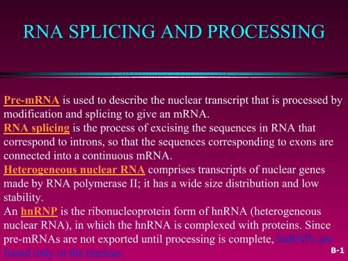
<strong>RNA</strong> <strong>SPLICING</strong> <strong>AND</strong> <strong>PROCESSING</strong>Pre-m<strong>RNA</strong> is used to describe the nuclear transcript that is processed bymodification and splicing to give an m<strong>RNA</strong>.<strong>RNA</strong> splicing is the process of excising the sequences in <strong>RNA</strong> thatcorrespond to introns, so that the sequences corresponding to exons areconnected into a continuous m<strong>RNA</strong>.Heterogeneous nuclear <strong>RNA</strong> comprises transcripts of nuclear genesmade by <strong>RNA</strong> polymerase II; it has a wide size distribution and lowstability.An hnRNP is the ribonucleoprotein form of hn<strong>RNA</strong> (heterogeneousnuclear <strong>RNA</strong>), in which the hn<strong>RNA</strong> is complexed with proteins. Sincepre-m<strong>RNA</strong>s are not exported until processing is complete, hnRNPs arefound only in the nucleus.B-1
• Typical mammalian gene has 7-8 exons spread outover ~16 kb. The exons are relatively short (~100-200 bp), and the introns are relatively long (>1 kb).• The process by which the introns are removed iscalled <strong>RNA</strong> splicing. Typical messenger of ~2.2 kb.B-2
The physical form of hn<strong>RNA</strong> is hnRNP.hnRNP= Core proteins plus others = ~20 proteinsTypically proteins are present at ~10 8 copies per nucleus, compared with~10 6 molecules of hn<strong>RNA</strong>.The most abundant proteins in the particleare the core proteins, but other proteinsare present at lower stoichiometry, makinga total of ~20 proteins. The proteinstypically are present at ~108 copies pernucleus, compared with ~106 molecules ofhn<strong>RNA</strong>. Some of the proteins may have astructural role in packaging the hn<strong>RNA</strong>;several are known to shuttle between thenucleus and cytoplasm, and play roles inexporting the <strong>RNA</strong> or otherwise controllingits activityFigure 24.1 hn<strong>RNA</strong> exists as a ribo-nucleoprotein particle organized asa series of beads.B-3
Splicing systems1. Involves spliceosomes2. Self-splicing introns3. Endonuclease and ligase arerequired (i.e. Yeast t<strong>RNA</strong>)Figure 24.2 <strong>RNA</strong> is modified in the nucleus by additions to the 5 and 3ends and by splicing to remove the introns. The splicing event requiresbreakage of the exon-intron junctions and joining of the ends of theexons. Mature m<strong>RNA</strong> is transported through nuclear pores to thecytoplasm, where it is translated.B-4
Nuclear splice junctions are short sequencesSplice sites are the sequences immediately surrounding theexon-intron boundaries.GT-AG rule describes the presence of these constantdinucleotides at the first two and last two positions of introns ofnuclear genes.Figure 24.3 The ends of nuclear introns are defined by the GU-AGrule.B-5
Splicing depends only on recognition of pairs of splice junctions.•All 5’ splice sites are functionally equivalent, and all 3’ splice sites arefunctionally equivalent.Splice sites are generic:The apparatus for splicing is not tissue specificFigure 24.4 Splicing junctions are recognized only in the correctpairwise combinations.B-6
What ensures that the correct pairs of sites arespliced together? The corresponding GU-AG pairsmust be connected across great distances (someintrons are >10 kb long). Two types of principlemight be responsible for pairing the appropriate 5’and 3’ sites:•It could be an intrinsic property of the <strong>RNA</strong> toconnect the sites at the ends of a particular intron.This would require matching of specific sequencesor structures.•Or all 5’ sites may be functionally equivalent andall 3’ sites may be similarly indistinguishable, butsplicing could follow rules that ensure a 5’ site isalways connected to the 3’ site that comes next inthe <strong>RNA</strong>.Are introns removed in a specific order from a particular<strong>RNA</strong>? Conformation of the <strong>RNA</strong> influences theaccessibility of the splice sites.Figure 24.5 Northern blotting of nuclear <strong>RNA</strong>with an ovomucoid probe identifies discreteprecursors to m<strong>RNA</strong>. The contents of the moreprominent bands are indicated, which suggeststhat splicing occurs via definite pathways.B-7
Nuclear extracts can splice purified <strong>RNA</strong>precursors, which shows that the actionof splicing is not linked to the process oftranscription.The lariat is an intermediate in <strong>RNA</strong>splicing in which a circular structure witha tail is created by a 5 -2 bond.The branch site is a short sequence justbefore the end of an intron at which thelariat intermediate is formed in splicing byjoining the 5 nucleotide of the intron tothe 2 position of an Adenosine.A transesterification reaction breaksand makes chemical bonds in acoordinated transfer so that no energy isrequired.Figure 24.6 Splicing occurs in twostages. First the 5’ exon is cleaved off;then it is joined to the 3’ exon.B-8
The branch site in yeast ishighly conserved, and hasthe consensus sequenceUACUAAC (Fig. 24.6).The branch site in highereukaryotes is not wellconserved, but has apreference for purines orpyrimidines at eachposition and retains thetarget A nucleotide.Figure 24.7 Nuclear splicingoccurs by twotrans-esterification reactions inwhich an OH group attacks aphosphodiester bond.B-9
A small nuclear <strong>RNA</strong> is one of many small<strong>RNA</strong> species confined to the nucleus;several of the sn<strong>RNA</strong>s are involved insplicing or other <strong>RNA</strong> processing reactions.Small cytoplasmic <strong>RNA</strong>s are present in thecytoplasm and (sometimes are also found inthe nucleus).snRNPs are small nuclear ribonucleoproteins(sn<strong>RNA</strong>s associated with proteins).scRNPs are small cytoplasmicribonucleoproteins (sc<strong>RNA</strong>s associated withproteins).The spliceosome is a complex formed bythe snRNPs that are required for splicingtogether with additional protein factors.Anti-Sm is an autoimmune antiserum thatdefines the Sm epitope that is common to agroup of proteins found in snRNPs that areinvolved in <strong>RNA</strong> splicing.Composition of SpliceosomeFigure 24.8 The spliceosome is ~12 MDa. 5 sn<strong>RNA</strong>Ps account for almost half of themass. The remaining proteins include known splicing factors and also proteins that areinvolved in other stages of gene expression.B-10
The five snRNPs involved in splicing are U1, U2, U5, U4, and U6.Together with some additional proteins, the snRNPs form thespliceosome.Each snRNP contains a single sn<strong>RNA</strong> and several (
An SR protein has a variable lengthof n Arg-Ser-rich region and isinvolved in splicing.•U1 snRNP initiates splicing bybinding to the 5’ splice site bymeans of an <strong>RNA</strong>-<strong>RNA</strong> pairingreaction.•The E complex contains U1snRNP bound at the 5’ splice site,the protein U2AF bound to apyrimidine tract between thebranch site and the 3’ splice site,and SR proteins connecting U1snRNP to U2AF.Figure 24.9 U1 sn<strong>RNA</strong> has a base paired structure that creates severaldomains. The 5’ end remains single stranded and can base pair with the5, splicing site.B-12
Binding of U1 snRNP to the 5′ splice site is the firststep in splicing. The recruitment of U1 snRNPinvolves an interaction between one of its proteins(U1-70k) and the protein ASF/SF2 (a generalsplicing factor in the SR class: see below). U1sn<strong>RNA</strong> base pairs with the 5′ site by means of asingle-stranded region at its 5′–terminus whichusually includes a stretch of 4-6 bases that iscomplementary with the splice site.The wild-type sequence of the splice site of the 12Sadenovirus pre-m<strong>RNA</strong> pairs at 5 out of 6 positionswith U1 sn<strong>RNA</strong>. A mutant in the 12S <strong>RNA</strong> thatcannot be spliced has two sequence changes; theGG residues at positions 5-6 in the intron arechanged to AU.Figure 24.10 Mutations that abolish function of the 5’ splicing site canbe suppressed by compensating mutations in U1 sn<strong>RNA</strong> that restore basepairing.B-13
E complex =early presplicingcomplexSplicing can be broadlydivided into two stages:•First the consensussequences at the 5′ splicesite, branch sequence, andadjacent pyrimidine tract arerecognized. A complexassembles that contains allof the splicing components.•Then the cleavage andligation reactions change thestructure of the substrate<strong>RNA</strong>. Components of thecomplex are released orreorganized as it proceedsthrough the splicingreactions.Figure 24.11 The commitment (E) complex forms by the successive additionof U1 snRNP to the 5 splice site, U2AF to the pyrimidine tract/3 splice site,and the bridging protein SF1/BBP (mammalian/yeat).B-14
The direct way of forming an E complex is for U1 snRNP to bindat the 5’ splice site and U2AF to bind at a pyrimidine tractbetween the branch site and the 3’ splice site.Another possibility is for the complex to form between U2AF at thepyrimidine tract and U1 snRNP at a downstream 5’ splice site.The E complex is converted to the A complex when U2 snRNPbinds at the branch site.If an E complex forms using a downstream 5’ splice site, thissplice site is replaced by the appropriate upstream 5’ splice sitewhen the E complex is converted to the A complex.Weak 3’ splice sites may require a splicing enhancer located inthe exon downstream to bind SR proteins directly.B-15
Intron definition describes the process when a pair of splicing sites arerecognized by interactions involving only the 5′ site and the branchpoint/3′ site.Exon definition describes the process when a pair of splicing sites arerecognized by interactions involving the 5′ site of the intron and also the 5′ of thenext intron downstream.Figure 24.12 There may be multiple routes for initial recognition of 5’and 3’ splice sites.B-16
Following formation of the E complex, theother snRNPs and factors involved insplicing associate with the complex in adefined order.The B1 complex is formed when a trimercontaining the U5 and U4/U6 snRNPsbinds to the A complex containing U1 andU2 snRNPs. This complex is regarded as aspliceosome, since it contains thecomponents needed for the splicingreaction. It is converted to the B2 complexafter U1 is released. The dissociation of U1is necessary to allow other components tocome into juxtaposition with the 5′ splicesite, most notably U6 sn<strong>RNA</strong>. At this pointU5 sn<strong>RNA</strong> changes its position; initially it isclose to exon sequences at the 5′ splicesite, but it shifts to the vicinity of the intronsequencesFigure 24.13 The splicing reactionproceeds through discrete stages inwhich spliceosome formation involvesthe interaction of components thatrecognize the consensus sequences.B-17
The catalytic reaction is triggered by therelease of U4; this requires hydrolysis ofATP. The role of U4 sn<strong>RNA</strong> may be tosequester U6 sn<strong>RNA</strong> until it is needed.In the U6/U4 snRNP, a continuous length of26 bases of U6 is paired with two separatedregions of U4. When U4 dissociates, theregion in U6 that is released becomes freeto take up another structure. The first part ofit pairs with U2; the second part forms anintramolecular hairpin. The interactionbetween U4 and U6 is mutuallyincompatible with the interaction betweenU2 and U6, so the release of U4 controlsthe ability of the spliceosome to proceedFigure 24.14 U6-U4 pairing is incompatible with U6-U2 pairing. When U6joins the spliceosome it is paired with U4. Release of U4 allows aconformational change in U6; one part of the released sequence forms ahairpin (dark grey), and the other part (black) pairs with U2. Because anadjacent region of U2 is already paired with the branch site, this brings U6 intojuxtaposition with the branch. Note that the substrate <strong>RNA</strong> is reversed from theusual orientation and is shown 3’-5’ .B-18
•Binding of U5 and U4/U6 snRNPs convertsthe A complex to the B1 spliceosome, whichcontains all the components necessary forsplicing.•The spliceosome passes through a series offurther complexes as splicing proceeds.•Release of U1 snRNP allows U6 sn<strong>RNA</strong> tointeract with the 5’ splice site, andconverts the B1 spliceosome to the B2spliceosome.•When U4 dissociates from U6 snRNP, U6sn<strong>RNA</strong> can pair with U2 sn<strong>RNA</strong> to form thecatalytic active site.Figure 24.15 Splicing utilizes a series ofbase pairing reactions between sn<strong>RNA</strong>sand splice sites.B-19
An alternative splicing pathway uses another set of snRNPs thatcomprise the U12 spliceosome.The target introns are defined by longer consensus sequences atthe splice junctions, but usually include the same GU-AGjunctions.Some introns have the splice junctions AU-AC, including somethat are U1-dependent and some that are U12-dependent.The REF proteins bind to splicing junctions by associating with thespliceosome.After splicing, they remain attached to the <strong>RNA</strong> at the exon-exonjunction.They interact with the transport protein TAP/Mex that exports the<strong>RNA</strong> through the nuclear pore.B-20
Figure 24.16 The EJC (exon junction complex) binds to <strong>RNA</strong> byrecognizing the splicing complex.The EJC complex consists of >9 proteins.The EJC includes a group of proteinscalled the REF family (the bestcharacterized member is called Aly).TheREF proteins in turn interact with atransport protein (variously called TAPand Mex) which has direct responsibilityfor interaction with the nuclear poreB-21
Export of m<strong>RNA</strong>The EJC includes a group ofproteins called the REF family(the best characterized member iscalled Aly).The REF proteins inturn interact with a transportprotein (variously called TAP andMex) which has directresponsibility for interaction withthe nuclear poreFigure 24.17 A REF protein binds to a splicing factor andremains with the spliced <strong>RNA</strong> product. REF binds to an exportfactor that binds to the nuclear pore.B-22
Figure 24.18 Three classes of splicing reactions proceed by twotrans-esterifications. First, a free OH group attacks the exon 1–intron junction.Second, the OH created at the end of exon 1 attacks the intron–exon 2 junction.The group I and group II introns have ability to excisethemselves from an <strong>RNA</strong>. This is called autosplicing.Group I introns are more common than group II introns.In both cases the <strong>RNA</strong> can perform the splicing reaction invitro by itself, without requiring enzymatic activitiesprovided by proteins; however, proteins are almostcertainly required in vivo to assist with folding.Group II introns excise themselves from <strong>RNA</strong> by anautocatalytic splicing event.The splice junctions and mechanism of splicing of groupII introns are similar to splicing of nuclear introns.A group II intron folds into a secondary structure thatgenerates a catalytic site resembling the structure of U6-U2-nuclear intron.The 5′ exon-intron junction is attacked by a free hydroxylgroup provided by an internal 2′–OH position in nuclearand group II introns, and by a free guanine nucleotidein group I introns.B-23
Figure 24.19 Splicing releases a mitochondrial group II intron inthe form of a stable lariat. Photograph kindly provided by LeslieGrivell and Annika Arnberg.B-24
Figure 24.20 Nuclear splicing and groupII splicing involve the formation of similarsecondary structures. The sequences aremore specific in nuclear splicing; group IIsplicing uses positions that may beoccupied by either purine (R) or eitherpyrimidine (Y).A group II intron forms into a secondary structurethat contains several domains formed by basepairedstems and single-stranded loops. Domain 5is separated by 2 bases from domain 6, whichcontains an A residue that donates the 2′–OHgroup for the first transesterification. Thisconstitutes a catalytic domain in the <strong>RNA</strong>.B-25
Figure 24.21 Alternative forms of splicing may generate a variety ofprotein products from an individual gene. Changing the splice sites mayintroduce termination codons (shown by asterisks) or change readingframes.The large T/ small t antigens of SV40 and theproducts of the adenovirus E1A region are generatedby connecting a varying 5′ site to a constant 3′ site.In T/t antigens - the 5′ site used for T antigenremoves a termination codon that is present in the tantigen m<strong>RNA</strong>, so that T antigen is larger than tantigen.In E1A transcripts, one of the 5′ sites connects to thelast exon in a different reading frame, again makinga significant change in the C-terminal part of theprotein.B-26
The ratio of X chromosomes toautosomes determines theexpression of sxl, and changes inexpression are passedsequentially through the othergenes to dsx, the last in thepathway.Presence of Sxl protein changesthe splicing of the transformer(tra) geneFigure 24.22 Sex determination in D. melanogaster involves a pathway inwhich different splicing events occur in females. Blocks at any stage of thepathway result in male development.B-27
Specific exons may be excluded or included inthe <strong>RNA</strong> product by using or failing to use a pairof splicing junctions.Exons may be extended by changing one of thesplice junctions to use an alternative junction.Sex determination in Drosophila involves a seriesof alternative splicing events in genes coding forsuccessive products of a pathway.P elements of Drosophila show germline-specificalternative splicing (producing a repressor (66 kdor transposase protein, as discussed in chapter16).Figure 24.23 Alternative splicing events that involve both sites maycause exons to be added or substituted.B-28
SL <strong>RNA</strong> is a small <strong>RNA</strong> that donates an exon in the trans-splicingreaction of trypanosomes and nematodes.•Splicing reactions usually occur only in cis between splice junctions on thesame molecule of <strong>RNA</strong>.•trans-splicing occurs in trypanosomes and worms where a short sequence(SL <strong>RNA</strong>) is spliced to the 5 ends of many precursor m<strong>RNA</strong>s.•SL <strong>RNA</strong> has a structure resembling the Sm-binding site of U sn<strong>RNA</strong>s andmay play an analogous role in the reaction.Figure 24.24 Splicingusually occurs only in cisbetween exons carried on thesame physical <strong>RNA</strong>molecule, but trans splicingcan occur when specialconstructs are made thatsupport base pairing betweenintrons.1 2 1 4B-29
In actin genes in C.Elegans, the leadersequence <strong>RNA</strong> (100 b)is coded elsewhereSL (spliced leader<strong>RNA</strong>)Figure 24.25 The SL <strong>RNA</strong> provides an exon that is connected to thefirst exon of an m<strong>RNA</strong> by trans-splicing. The reaction involves the sameinteractions as nuclear cis-splicing, but generates a Y-shaped <strong>RNA</strong>instead of a lariat.B-30
Figure 24.26 The intron in yeast t<strong>RNA</strong>Phe base pairs with theanticodon to change the structure of the anticodon arm. Pairing betweenan excluded base in the stem and the intron loop in the precursor may berequired for splicing.t<strong>RNA</strong> splicing occurs by successive cleavage and ligation reactions.B-31
Figure 24.27 Splicing of yeast t<strong>RNA</strong> in vitro can be followed byassaying the <strong>RNA</strong> precursor and products by gel electro-phoresis.B-32
Figure 24.28 The 3’ and 5’ cleavages in S. cerevisiae pre-t<strong>RNA</strong> arecatalyzed by different subunits of the endonuclease. Another subunitmay determine location of the cleavage sites by measuring distance fromthe mature structure. The AI base pair is also important.B-33
Figure 24.29 Archaeal t<strong>RNA</strong> splicing endonuclease cleaves each strandat a bulge in a bulge-helix-bulge motif.An endonuclease cleaves thet<strong>RNA</strong> precursors at bothends of the intron.The yeast endonuclease is aheterotetramer, with two(related) catalytic subunits.It uses a measuringmechanism to determine thesites of cleavage by theirpositions relative to a pointin the t<strong>RNA</strong> structure.The archaeal nuclease has asimpler structure andrecognizes a bulge-helixbulgestructural motif in thesubstrate.B-34
Figure 24.30 Splicing of t<strong>RNA</strong> requires separate nuclease and ligaseactivities. The exon-intron boundaries are cleaved by the nuclease to generate2 -3 cyclic phosphate and 5’ OH termini. The cyclic phosphate is opened togenerate 3’ -OH and 2’ phosphate groups. The 5’ -OH is phosphorylated. Afterreleasing the intron, the t<strong>RNA</strong> half molecules fold into a t<strong>RNA</strong>-like structure thatnow has a 3’ -OH, 5’ -P break. This is sealed by a ligase.B-35
Release of the intron generates two half-t<strong>RNA</strong>s that pair to formthe mature structure.The halves have the unusual ends 5’ hydroxyl and 2 –3 cyclicphosphate.The 5’ –OH end is phosphorylated by a polynucleotide kinase, thecyclic phosphate group is opened by phosphodiesterase togenerate a 2’ –phosphate terminus and 3’ –OH group, exon endsare joined by an <strong>RNA</strong> ligase, and the 2’ –phosphate is removed bya phosphatase.B-36
45S <strong>RNA</strong> is a precursor that containsthe sequences of both major ribosomal<strong>RNA</strong>s (28S and 18S r<strong>RNA</strong>s).Figure 24.37 Mature eukaryotic r<strong>RNA</strong>s are generated by cleavage andtrimming events from a primary transcript.B-37
Ire1p is an inner nuclear membrane protein with itsN-terminal domain in the ER lumen, and its C-terminal domain in the nucleus.Binding of an unfolded protein to the N-terminaldomain activates the C-terminal nuclease byautophosphorylation.The activated nuclease cleaves Hac1 m<strong>RNA</strong> torelease an intron and generate exons that are ligatedby a t<strong>RNA</strong> ligase.The spliced Hac1 m<strong>RNA</strong> codes for a transcriptionfactor that activates genes coding for chaperonesthat help to fold unfolded proteins.Figure 24.31 The unfolded protein response occurs by activatingspecial splicing of HAC1 m<strong>RNA</strong> to produce a transcription factorthat recognizes the UPRE.B-38
<strong>RNA</strong> polymerase I terminates transcription at an 18 base terminatorsequence.<strong>RNA</strong> polymerase III terminates transcription in poly(U)4 sequenceembedded in a G·C-rich sequence.Figure 24.32 When a 3 end isgenerated by termination, <strong>RNA</strong>polymerase and <strong>RNA</strong> arereleased at a discrete(terminator) sequence in DNA.B-39
Figure 24.33 When a3 end is generated bycleavage, <strong>RNA</strong>polymerase continuestranscription while anendonuclease cleavesat a defined sequencein the <strong>RNA</strong>.B-40
•The sequence AAUAAA is a signal for cleavage to generate a3 end of m<strong>RNA</strong> that is polyadenylated.•The reaction requires a protein complex that contains a specificityfactor, an endonuclease, and poly(A) polymerase.•The specificity factor and endonuclease cleave <strong>RNA</strong> downstreamof AAUAAA.•The specificity factor and poly(A) polymerase add ~200 Aresidues processively to the 3 end.•A·U-rich sequences in the 3 tail control cytoplasmicpolyadenylation or deadenylation during Xenopus embryonicdevelopment.B-41
Figure 24.34 The sequence AAUAAA is necessary for cleavage togenerate a 3 end for poly-adenylation.B-42
Figure 24.35 The 3 processing complex consists of several activities.CPSF and CstF each consist of several subunits; the other componentsare monomeric. The total mass is >900 kD.B-43
•Cordycepin is 3’ deoxyadenosine, an inhibitor of polyadenylation of<strong>RNA</strong>.•Endonucleases cleave bonds within a nucleic acid chain; they maybe specific for <strong>RNA</strong> or for single-stranded or double-stranded DNA.•The sequence AAUAAA is a signal for cleavage to generate a 3’ endof m<strong>RNA</strong> that is polyadenylated.•The reaction requires a protein complex that contains a specificityfactor, an endonuclease, and poly(A) polymerase.•The specificity factor and endonuclease cleave <strong>RNA</strong> downstream ofAAUAAA.•The specificity factor and poly(A) polymerase add ~200 A residuesprocessively to the 3 end.•A·U-rich sequences in the 3 tail control cytoplasmicpolyadenylation or deadenylation during Xenopus embryonicdevelopment.B-44
•Histone m<strong>RNA</strong>s are not polyadenylated; their 3’ ends are generatedby a cleavage reaction that depends on the structure of the m<strong>RNA</strong>.The cleavage reactionrequires the SLBP tobind to a stem-loopstructure, and the U7sn<strong>RNA</strong> to pair with anadjacent singlestrandedregion.Figure 24.36 Generation of the 3’ end of histone H3 m<strong>RNA</strong> dependson a conserved hairpin and a sequence that base pairs with U7 sn<strong>RNA</strong>.B-45
• 45S <strong>RNA</strong> is a precursor that contains the sequences of bothmajor ribosomal <strong>RNA</strong>s (28S and 18S r<strong>RNA</strong>s).Figure 24.37r<strong>RNA</strong>s are cleaved from acommon precursorB-46
Figure 24.38 The rrn operons in E. coli (7) contain genes for both r<strong>RNA</strong> andt<strong>RNA</strong>. The exact lengths of the transcripts depend on which promoters (P) andterminators (t) are used. Each <strong>RNA</strong> product must be released from thetranscript by cuts on either side.B-47
Figure 24.39 A sno<strong>RNA</strong> base pairs with a region of r<strong>RNA</strong> that is to bemethylated.B-48
Figure 24.40 Uridine is converted to pseudouridine by replacing theN1-sugar bond with a C5-sugar bond and rotating the base relative to thesugar.B-49
Processing and modification of r<strong>RNA</strong> requires a class of small <strong>RNA</strong>s calledsno<strong>RNA</strong>s (small nucleolar <strong>RNA</strong>s). There are 71 sno<strong>RNA</strong>s in the yeast (S.cerevisiae) genome.The C/D group of sno<strong>RNA</strong>s is requiredfor modifying the 2′ position of ribosewith a methyl group.The H/ACA group of sno<strong>RNA</strong>s isrequired for converting uridine topseudouridine.In each case the sno<strong>RNA</strong> base pairswith a sequence of r<strong>RNA</strong> that containsthe target base to generate a typicalstructure that is the substrate formodification.Figure 24.41 H/ACA sno<strong>RNA</strong>s have two short conserved sequencesand two hairpin structures, each of which has regions in the stem that arecomplementary to r<strong>RNA</strong>. Pseudouridine is formed by converting anunpaired uridine within the complementary region of the r<strong>RNA</strong>.B-50
B-51