Anatomy Review: Skeletal Muscle Tissue - Sinoe medical homepage.
Anatomy Review: Skeletal Muscle Tissue - Sinoe medical homepage.
Anatomy Review: Skeletal Muscle Tissue - Sinoe medical homepage.

Create successful ePaper yourself
Turn your PDF publications into a flip-book with our unique Google optimized e-Paper software.
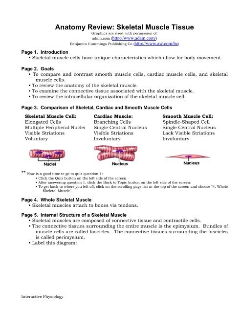
<strong>Anatomy</strong> <strong>Review</strong>: <strong>Skeletal</strong> <strong>Muscle</strong> <strong>Tissue</strong>Graphics are used with permission of:adam.com (http://www.adam.com/)Benjamin Cummings Publishing Co (http://www.aw.com/bc)Page 1. Introduction• <strong>Skeletal</strong> muscle cells have unique characteristics which allow for body movement.Page 2. Goals• To compare and contrast smooth muscle cells, cardiac muscle cells, and skeletalmuscle cells.• To review the anatomy of the skeletal muscle.• To examine the connective tissue associated with the skeletal muscle.• To review the intracellular organization of the skeletal muscle cell.Page 3. Comparison of <strong>Skeletal</strong>, Cardiac and Smooth <strong>Muscle</strong> Cells<strong>Skeletal</strong> <strong>Muscle</strong> Cell: Cardiac <strong>Muscle</strong>: Smooth <strong>Muscle</strong> Cell:Elongated Cells Branching Cells Spindle-Shaped CellMultiple Peripheral Nuclei Single Central Nucleus Single Central NucleusVisible Striations Visible Striations Lack Visible StriationsVoluntary Involuntary Involuntary** Now is a good time to go to quiz question 1:• Click the Quiz button on the left side of the screen.• After answering question 1, click the Back to Topic button on the left side of the screen.• To get back to where you left off, click on the scrolling page list at the top of the screen and choose "4. Whole<strong>Skeletal</strong> <strong>Muscle</strong>".Page 4. Whole <strong>Skeletal</strong> <strong>Muscle</strong>• <strong>Skeletal</strong> muscles attach to bones via tendons.Page 5. Internal Structure of a <strong>Skeletal</strong> <strong>Muscle</strong>• <strong>Skeletal</strong> muscles are composed of connective tissue and contractile cells.• The connective tissues surrounding the entire muscle is the epimysium. Bundles ofmuscle cells are called fascicles. The connective tissues surrounding the fasciclesis called perimysium.• Label this diagram:Interactive Physiology
Page 6. Internal Structure of a Fascicle• Important Points About Endomysium:• Made of connective tissue.• Surrounds individual muscle cells.• Functions to electrically insulates muscle cells from one another.• Three connective tissue layers of the muscle (endomysium, perimysium, andepimysium):• Bind the muscle cells together.• Provide strength and support to the entire muscle.• Are continuous with the tendons at the ends of the muscle.• Label this diagram:Page 7. Internal Structure of a <strong>Skeletal</strong> <strong>Muscle</strong> Cell• Label this diagram:Interactive Physiology 2
<strong>Muscle</strong> fibers: Alternative name for skeletal muscle cells.• Nucleus: Contains the genetic material.• Sarcolemma: Plasma membrane of the muscle cell.• Sarcoplasmic reticulum (SR): Interconnecting tubules of endoplasmic reticulumthat surround each myofibril.• Terminal cisternae: Sac-like regions of the sarcoplasmic reticulum that containcalcium ions.• T tubules: Invaginations of the sarcolemma that project deep into the cell.• Triad: A group of one T tubule lying between two adjacent terminal cisternae.• Cytosol: Intracellular fluid.• Mitochondria: Sites of ATP synthesis.• Myofibril: Contains the contractile filaments within the skeletal muscle cell.** Now is a good time to go to quiz questions 2 and 3:• Click the Quiz button on the left side of the screen.• Click on the scrolling page list at the top of the screen and choose "2. Labeling <strong>Muscle</strong> Cell Structures".• After answering question 3, click the Back to Topic button on the left side of the screen.• To get back to where you left off, click on the scrolling page list at the top of the screen and choose "8.Structure of a Myofibril".Page 8. Structure of a Myofibril• Myofibrils: Contractile units within muscle cells.• Made of myofilaments called thin filaments and thick filaments.• Thin and thick filaments are made mainly of the proteins actin and myosin.** Now is a good time to go to quiz question 4:• Click the Quiz button on the left side of the screen.• Click on the scrolling page list at the top of the screen and choose "4. Myofibril Puzzle".• After answering question 4, click the Back to Topic button on the left side of the screen.• To get back to where you left off, click on the scrolling page list at the top of the screen and choose "9.Arrangement of Myofilaments".Page 9. Arrangement of Myofilaments• Label the diagram:Interactive Physiology 3
• A bands: Dark areas that correspond to the areas where thick filaments arepresent.• I bands: Light areas that contains only thin filaments.• Z line: A protein disk within the I band that anchors the thin filaments andconnects adjacent myofibrils.• H zone: Located in the middle of each A band, this lighter stripe appearscorresponding to the region between the thin filaments.• M line: Protein fibers that connect neighboring thick filaments.• Sarcomere: The region of the myofibril between two Z lines.** Keep in mind, this information is crucial for your understanding of the sliding filament theory.** Now is a good time to go to quiz question 5:• Click the Quiz button on the left side of the screen.• Click on the scrolling page list at the top of the screen and choose "5. Labeling a Myofibril".• After answering question 5, click the Back to Topic button on the left side of the screen.• To get back to where you left off, click on the scrolling page list at the top of the screen and choose "10.<strong>Review</strong>: Organizational Levels of <strong>Skeletal</strong> <strong>Muscle</strong>".Interactive Physiology 4
Page 10. <strong>Review</strong>: Organizational Levels of <strong>Skeletal</strong> <strong>Muscle</strong>• "Bundle-within-a-bundle" organization of skeletal muscle:myofilaments↓myofibril↓muscle cell or muscle fiber↓fascicles↓whole skeletal musclePage 11. Pyramid of Subunits• Whole muscle as a pyramid of subunits:Fascicles<strong>Muscle</strong> cells (<strong>Muscle</strong> Fibers)MyofibrilsMyofilamentsPage 12. Summary• The three types of muscle cells in the body are skeletal, cardiac, and smooth.• <strong>Skeletal</strong> muscle has three layers of connective tissue: epimysium, perimysium, andendomysium.• The striations of skeletal muscle cells are due to the organized arrangement ofcontractile proteins called thick and thin filaments.• A whole muscle demonstrates a bundle-within-a-bundle organization:myofilaments → myofibrils → muscle cells (muscle fibers) → fascicles → wholemuscle** Now is a good time to go to quiz question 6:• Click the Quiz button on the left side of the screen.• Click on the scrolling page list at the top of the screen and choose "6. Organizational Levels of <strong>Skeletal</strong><strong>Muscle</strong>".Notes on Quiz Questions:Quiz Question #1: Three Types of <strong>Muscle</strong> Cells• This question allows you to fill in a table which contains the characteristics ofthe various muscle cells.Quiz Question #2: Labeling <strong>Muscle</strong> Cell Structures• This question allows you to label the parts of a skeletal muscle cell.Quiz Question #3: Definition of <strong>Muscle</strong> Cell Structures• This question allows you to match the parts of the skeletal muscle cells to theirdefinitions.Quiz Question #4: Myofibril Puzzle• This question allows you to assemble a myofibril.Quiz Question #5: Labeling a Myofibril• This question allows you to label the parts of a myofibril.Quiz Question #6: Organizational Levels of <strong>Skeletal</strong> <strong>Muscle</strong>• This question allows you to label the muscle at various levels of organization.Interactive Physiology 5
Study Questions on <strong>Anatomy</strong> <strong>Review</strong>: <strong>Skeletal</strong> <strong>Muscle</strong> <strong>Tissue</strong>1. (Page 1.) What is the main function of skeletal muscles?2. (Page 3.) List the three types of contractile cells of the body.3. (Page 3.) Match the following types of contractile cells to their shape (branching,elongated, spindle-shaped):___________________ a. <strong>Skeletal</strong> muscle cells___________________ b. Cardiac muscle cells___________________ c. Smooth muscle cells4. (Page 3.) Match the following types of contractile cells to the characteristics of theirnuclei and presence or absence of striations:Cardiac <strong>Muscle</strong> Cells Smooth <strong>Muscle</strong> Cells <strong>Skeletal</strong> <strong>Muscle</strong> Cells___________________ a. presence of visible striations & single, centrally-locatednuclei___________________ b. presence of visible striations & multiple peripheral nuclei___________________ c. absence of visible striations & single, centrally-locatednuclei number of nuclei5. (Page 4.) What is the name of the structure that attaches skeletal muscles to bones?6. (Page 5.) Bundles of skeletal muscle cells are called ________________.7. (Page 5.) The connective tissue which immediately surrounds a muscle is called_______________ and the connective tissue around the fascicles is called________________.8. (Page 6.) What is the function of endomysium?9. (Page 7.) Match these terms to their description:TriadT tubulesTerminal cisternaeSarcolemma<strong>Muscle</strong> fibersMitochondriaSarcoplasmic reticulumMyofibril___________________a. Sac-like regions of the sarcoplasmic reticulum thatcontain calcium ions.___________________ b. Sites of ATP synthesis.Interactive Physiology 6
___________________ c. Plasma membrane of the muscle cell.___________________ d. Alternative name for skeletal muscle cells.___________________ e. Interconnecting tubules of endoplasmic reticulum thatsurround each myofibril.___________________ f. A group of one T tubule lying between two adjacentterminal cisternae.___________________ g. Invaginations of the sarcolemma that projecting deep intothe cell.___________________ h. Contains the contractile filaments within the skeletalmuscle cell.10. (Page 8.) What are the names for the two types of filament in a myofibril?11. (Page 9.) What creates the skeletal muscle cell's striated appearance?12. (Page 9.) Match the following:A band I band H zone______________ a. Contains only thin filaments.______________ b. Contains only thick filaments.______________ c. Contains both thin and thick filaments.13. (Page 9.) Perpendicular to the myofilaments are the Z lines and the M lines. The Zlines connect the _____________ filaments and the M lines connect the _____________filaments.14. (Page 9.) The region of the myofibril between two Z lines that is the contractile unitof a muscle cell is called a _____________ .15. (Page 10.) Arrange the following from smallest structure to largest structure:<strong>Muscle</strong> cell or muscle fiberFascicleMyofilamentsWhole skeletal muscleMyofibrilInteractive Physiology 7