The Ocular Hypertension Treatment Study - Vision Research ...
The Ocular Hypertension Treatment Study - Vision Research ...
The Ocular Hypertension Treatment Study - Vision Research ...
Create successful ePaper yourself
Turn your PDF publications into a flip-book with our unique Google optimized e-Paper software.
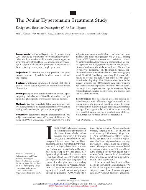
tous damage using current clinical tests. 15 Thus, theseindividuals are at an increased risk for developingPOAG. 13,16,17 Up to now there has been no consensus onhow to manage this large group of people, who arereferred to as ocular hypertensives or glaucoma suspects.Quigley et al 18,19 reported that 12% to 63% ofoptic nerve fibers can be lost before glaucomatousvisual field defects are detected by routine kineticperimetry. <strong>The</strong> findings of Quigley et al, the high prevalenceof glaucoma, and the potentially serious consequencesof this disease suggest the need for widespreadglaucoma screening and early treatment. This aggressiveapproach is buttressed by evidence from randomizedclinical studies 20-24 as well as by the almost universalclinical impression that treatment initiated early inthe course of glaucoma is far more effective in preventingprogressive visual loss than is treatment initiatedlate in the course of the disease. However, the approachof widespread glaucoma screening and early treatmenthas been challenged by investigators who point out thatthere is insufficient scientific information to support amajor health initiative. 23 One of the prerequisites forany screening program is that there must be an effectivetreatment for the disease. Surprisingly, there is no consensuson the efficacy of medical treatment in preventingor delaying the onset of damage due to POAG. 25-27<strong>The</strong>refore, we designed the <strong>Ocular</strong> <strong>Hypertension</strong> <strong>Treatment</strong><strong>Study</strong> (OHTS) to evaluate the safety and efficacy oftopical ocular hypotensive medication in preventing or delayingthe onset of visual field loss and/or optic nerve damagein subjects with ocular hypertension at moderate riskfor developing POAG. <strong>The</strong> secondary aim of OHTS is toidentify risk factors that predict which subjects with ocularhypertension are most likely to develop visual field lossand/or optic nerve damage due to POAG. Potential risk factorsinclude age, cup-disc ratio, IOP, myopia, systemic vasculardisease, family history of glaucoma, and race.This article describes the study protocol and baselinecharacteristics of study participants and serves as areference for future publications of the OHTS.DESIGN AND METHODSSTUDY SYNOPSIS<strong>The</strong> <strong>Ocular</strong> <strong>Hypertension</strong> <strong>Treatment</strong> <strong>Study</strong> is a multicenterrandomized controlled clinical trial. See the boxon page 581 for a description of the study organizationand a list of participating clinics. A data and safety monitoringcommittee monitors the ethical conduct of the studyand the accumulating data for evidence of adverse andbeneficial treatment effects.<strong>The</strong> study protocol is described in detail in the studymanual 28 and is summarized in Table 1. Eligibility wasdetermined by a comprehensive eye examination, medicaland ocular history, masked evaluation of Humphrey 30-2visual fields by the Visual Field Reading Center, and maskedevaluation of stereoscopic optic disc photographs by theOptic Disc Reading Center. Following a discussion of thestudy, subjects were requested to sign an informed consentform. Eligibility and exclusion criteria are summarizedin Table 2. Eligible subjects were randomized at theirbaseline/randomization visit over the telephone by the CoordinatingCenter to either the close observation group ormedication group. Subjects randomized to the medicationgroup began a stepped medical regimen to reduce IOPby at least 20% from the average of the IOPs measured atthe qualifying and baseline visits, to 24 mm Hg or less. Follow-upvisits are at 6-month intervals for a minimum of 5years or until a closure date determined by the Data andSafety Monitoring Committee (Table 3). Visual field testingis performed every 6 months and optic disc photographyis performed every 12 months.<strong>The</strong> primary study end point is the development ofeither a reproducible visual field abnormality or reproducibleprogressive optic disc cupping due to POAG. <strong>The</strong>presence of a visual field abnormality is determined bymasked readers at the Visual Field Reading Center, andthe presence of optic disc progression is determined bymasked, certified readers at the Optic Disc Reading Center.When an abnormality is detected by the reading center,the subject is recalled for retesting to confirm the abnormality.When an abnormality is confirmed on thesecond test, the Endpoint Committee reviews the subject’socular and medical history in a masked fashion todetermine if the abnormality is attributable to POAG. Independent,masked determination of a reproducible visualfield abnormality and/or reproducible progressive opticdisc cupping and attribution of the abnormality to POAGis required, as neither the subject nor the clinician is maskedas to randomization assignment. Subjects with a reproducibleabnormality due to POAG continue the same follow-upvisit schedule and receive the same tests and measures.<strong>The</strong> treatment course for this group of subjects isat the discretion of the treating clinician.ELIGIBILITY ASSESSMENTIntraocular PressureTo reduce potential bias, IOP is measured by 2 certifiedstudy personnel (an operator and a recorder) using a calibratedGoldmann applanation tonometer. <strong>The</strong> operatorinitially sets the dial at 10 mm Hg, then looks throughthe slitlamp and adjusts the dial while the recorder readsand records the results. <strong>The</strong> procedure is repeated on thesame eye. If the 2 readings differ by 2 mm Hg or less, theaverage of the 2 readings serves as the IOP measurement.If the 2 readings differ by more than 2 mm Hg, thena third reading is performed and the median reading servesas the IOP measurement. This IOP measurement protocolwas adapted from the Advanced Glaucoma Intervention<strong>Study</strong> 29 and the Glaucoma Laser Trial. 30<strong>The</strong> OHTS eligibility criteria were intended to selectocular hypertensive subjects at moderate risk for developingglaucoma because this group presents the greatestclinical uncertainty to the clinician. <strong>The</strong> qualifyingIOP must be at least 24 mm Hg but less than 32 mm Hgin at least 1 eye calculated from 2 separate consecutivemeasurements taken at least 2 hours but not more than12 weeks apart. <strong>The</strong> fellow eye must have an IOP of atleast 21 mm Hg but less than 32 mm Hg on 2 separateIOP measurements. All IOP readings for eligibility as-ARCH OPHTHALMOL / VOL 117, MAY 1999574Downloaded from www.archophthalmol.com at Washington University - St Louis, on February 9, 2010©1999 American Medical Association. All rights reserved.
Table 1. Design SynopsisType of trial<strong>Treatment</strong> assignment<strong>The</strong>rapeuticRandomCentersPatient is randomization and treatment unit22 Clinical centers Stratified by clinic and raceChairman’s officeBias controlCoordinating centerMasked evaluation of visual fieldsOptic disc photography reading centerMasked evaluation of optic disc stereophotographsVisual field reading centerMasked ascertainment of cause of abnormality by<strong>Treatment</strong>sEndpoint CommitteeClose observationPatient recruitmentTopical ocular hypotensive medication according to a stepped medical regimen1500 subjects (calculated)Outcome measures1637 subjects (achieved)Reproducible visual field abnormality due to primary open angle glaucomaFollow-upReproducible optic disc cupping due to primary open angle glaucomaMinimum 5 years (planned)sessment were performed after appropriate washout ofthe following prestudy topical ocular medications: parasympathomimeticagents (1 week), -adrenergic blockers(4 weeks), dipivefrin and epinephrine products (4weeks), 2 -agonists (4 weeks), and carbonic anhydraseinhibitors (2 weeks).For purposes of analyses, the baseline IOP of each eyeis the IOP measurement at the baseline/randomization visit.Humphrey 30-2 Visual FieldsQualifying Humphrey 30-2 visual fields must be normaland reliable in both eyes as determined by the Visual FieldReading Center prior to randomization. A reliable visualfield is defined as less than 33% false positives, false negatives,and fixation losses. A normal field is determined byclinical review at the Visual Field Reading Center and bySTATPAC 2 criteria for global indices within the 95% agespecificpopulation norms and the glaucoma hemifield testwithin the 97% age-specific population norm. Two of amaximum of 3 visual fields for each eye must meet all entrycriteria. <strong>The</strong> visual field testing sessions during qualifyingassessment must be separated by a minimum of 1hour and a maximum of 12 weeks.Stereoscopic Optic Disc PhotographsStereoscopic optic disc photographs must be judged normalby 2 independent, masked, certified readers at theOptic Disc Reading Center prior to randomization. Subjectswere excluded from the study if the photographsdocument an optic disc hemorrhage, a localized notchor thinning of the rim, a diffuse or localized area of pallor,or a difference in the cup-disc ratios between the 2eyes greater than 0.2 disc diameters. Subjects were alsoexcluded if they had optic disc drusen, pits, colobomas,atrophy, or other severe anomalies.TREATMENT ASSIGNMENTHalf of the subjects were randomized to the close observationgroup and half were randomized to the medicationgroup. Randomization was stratified by clinic andrace so that in any given clinic there was an approximatelyequal number of African Americans allocated toTable 2. Major Inclusion and Exclusion Criteria*Inclusion CriteriaIOP in at least 1 eye of each patient 24 mm Hg and 32 mm HgIOP in fellow eye 21 mm Hg and 32 mm HgAge 40 to 80 years, inclusiveNormal and reliable Humphrey 30-2 visual fields for both eyes asdetermined by the Visual Field Reading CenterNormal optic discs in both eyes on clinical examination and onstereoscopic photographs as determined by the Optic DiscReading CenterInformed consentExclusion CriteriaBest-corrected visual acuity worse than 20/40 in either eyePrevious intraocular surgery, except for uncomplicated extracapsularcataract extraction with posterior chamber–intraocular lens implantand no escape of vitreous to the anterior chamber, strabismus,cosmetic eyelid surgery, and radial keratotomyA life-threatening or debilitating diseaseSecondary causes of elevated IOP, including ocular and systemiccorticosteroid useAngle closure glaucoma or anatomically narrow angles—75% of thecircumference of the angle must be grade 2 or more by ShaffercriteriaOther diseases that cause visual field loss or optic disc abnormalitiesDifference in cup-disc ratios (horizontal by contour) of the 2 eyes 0.2Background diabetic retinopathy, defined as at least 1 microaneurysmseen on direct ophthalmoscopy with dilated pupil. Retinalhemorrhage is not an exclusion unless associated with backgroundor proliferative diabetic retinopathyInability to visualize or photograph the optic discsPregnant or nursing women as determined by patient self-report andtesting*IOP indicates intraocular pressure.the close observation and medication groups irrespectiveof the actual number enrolled. <strong>The</strong> randomizationunit is the subject. Randomization was completed by theCoordinating Center over the telephone during the subject’srandomization visit. <strong>The</strong> date of randomizationserves as the official date of entry into the study.TESTS AND MEASURESRandomized subjects complete regularly scheduled follow-upvisits at 6-month intervals. Semiannual visits(months 6,18, 30, 42, 54, etc) include a patientcompletedsymptom checklist, ocular and medical his-ARCH OPHTHALMOL / VOL 117, MAY 1999575Downloaded from www.archophthalmol.com at Washington University - St Louis, on February 9, 2010©1999 American Medical Association. All rights reserved.
Table 3. Measurement and Examination Procedures for Scheduled Visits*VisitQualityof LifeSymptom MedicalVisual VisualChecklist History Refraction Acuity FieldEyeDilatedExamination IOP Ophthalmoscopy Gonioscopy FundusStereoscopicOptic DiscPhotographyQualifying assessment X X X X X X X X X XBaseline/randomization X X XIOP confirmation X X X XFollow-upSemiannual (6 months,X X X X X X X X18 months, etc)Annual (12 months,24 months, etc)X X X X X X X X X X*IOP indicates intraocular pressure.tory, refraction, best-corrected visual acuity, Humphrey 30-2visual fields, slitlamp examination, direct ophthalmoscopy,and IOP measurement. Annual visits (months 12, 24,36, 48, 60, etc) include most of the measures at semiannualvisits plus completion of the 36-item short form healthsurvey (SF-36), 31 dilated fundus examination, and stereoscopicoptic disc photographs. A listing of regularly scheduledtests and measures is given in Table 3. Pachymetry isperformed once during follow-up and repeated only if thepatient has refractive or intraocular surgery.TREATMENT REGIMENSubjects randomized to the medication group began thestepped medical regimen with a therapeutic trial in theeye with the higher IOP. Subjects returned in 4 ± 2 weeksfor evaluation of therapeutic response. If drug therapywas ineffective or minimally effective (IOP reduction10% from the average of the qualifying and baselineIOP), it was stopped and another drug was substituted.If drug therapy was moderately effective (IOP reduction10% to 20% from the average of the qualifying and baselineIOP), the clinician had the choice of substituting anotherdrug or adding a drug. <strong>The</strong> drug regimen includesall commercially available topical ocular hypotensivemedications and reflects standard clinical practice in theUnited States. As new drugs become commercially availablein the United States, they are added to the treatmentoptions. All study drugs are provided free of chargeto the subjects and are distributed to participating clinicsfrom the study’s central pharmacy.TREATMENT GOAL<strong>The</strong> treatment goals for subjects randomized to the medicationgroup are an IOP of 24 mm Hg or less and a 20%reduction in IOP from the average of the qualifying andbaseline IOP. <strong>The</strong> 20% reduction is not necessary if theIOP is 18 mm Hg or less. Topical medical therapy ischanged and/or added until both of these goals are metor until the subject is receiving maximum tolerated topicalmedical therapy. Subjects in the medication group whodo not meet these goals despite maximum tolerated topicalmedical therapy continue to be evaluated in the trialand continue following the same schedule of tests andmeasurements. We believe these treatment goals constitutean adequate test of the primary hypothesis and reflectcurrent clinical practice.CONFIRMATION OF VISUAL FIELDABNORMALITYA technically acceptable visual field is consideredabnormal if P.05 for the corrected pattern SD or if theresults of the glaucoma hemifield test are outside normallimits (P.01). <strong>The</strong> study originally defined 2 consecutiveabnormal visual fields as confirmation of visualfield abnormality; however, a high percentage of abnormalvisual fields on the first test were found to be normalon retesting. Accordingly, a more stringent criterionfor the confirmation of visual field abnormality wasadopted effective January 1, 1998, at the recommendationof the Data and Safety Monitoring Committee,Steering Committee, and Full Investigative Group. <strong>The</strong>protocol was changed so that confirmation of a visualfield abnormality requires 3 consecutive visual fieldtests. Thus, a patient with an abnormal visual field istested at the next regularly scheduled follow-up visit in6 months. If the Visual Field Reading Center considersthe second visual field abnormal, a third visual field testis scheduled for 1 day to 8 weeks after the second visualfield test.When such a sequence of abnormal follow-up visualfields is received, the visual fields are evaluated by 2 independentsenior readers who are masked as to clinic and randomizationstatus. <strong>The</strong>y decide whether the visual field abnormalityis of the same character and in the same locationon all 3 visual fields. If the visual field abnormality is confirmed,the Visual Field Reading Center prepares a briefnarrative description of the abnormality and sends all visualfields for the affected eye to the Coordinating Centerfor review by the Endpoint Committee.CONFIRMATION OF OPTIC DISC PROGRESSIONWe defined optic disc progression as generalized or localizedthinning of the optic disc rim compared with baselineas judged by 1 or more of the following characteristics:change in the position of the vessels greater thanexpected from a shift in the position of the eye, developmentof a notch, development of an acquired pita, thinningof the rim, or development of localized or diffuseARCH OPHTHALMOL / VOL 117, MAY 1999576Downloaded from www.archophthalmol.com at Washington University - St Louis, on February 9, 2010©1999 American Medical Association. All rights reserved.
pallor. Disc hemorrhage, nerve fiber layer dropout, andchange in the depth of the cup are not considered opticdisc end points.<strong>The</strong> follow-up optic disc photographs and baselinephotographs are compared by 2 independent, certifiedreaders at the Optic Disc Reading Center who are maskedas to the order of the photographs, randomization assignmentof the patient, clinic, and previous optic discassessments. If one or both of the initial readers detect adifference between the 2 sets of photographs and identifytheir correct order, the photographs are reviewed ina similar masked fashion by a senior reader at the OpticDisc Reading Center. If the senior reader’s assessmentagrees with that of the initial reader or readers, the OpticDisc Reading Center requests the clinic to recall thepatient for a second set of photographs for the affectedeye within 4 ± 2 weeks. <strong>The</strong> second set of photographsis compared with baseline photographs in the same manneras the initial review. If a difference is confirmed andcorrectly sequenced, the senior reader reviews the photographsin a masked fashion and makes the final decisionas to the occurrence of progressive damage. If thesecond set of photographs is judged to confirm the occurrenceof progressive optic disc cupping, the Optic DiscReading Center prepares a brief narrative description ofthe findings and sends all optic disc photographs of theaffected eye to the Coordinating Center for review by theEndpoint Committee.DETERMINATION OF POAG END POINTWhen visual fields and/or optic disc photographs confirmthe presence of an abnormality, the appropriate readingcenter completes a report describing the abnormality.<strong>The</strong> Endpoint Committee, which is masked as to therandomization assignment of the patient and the end pointstatus of the fellow eye, reviews the reading center report,the patient’s medical and ocular history from thestudy forms, and the visual fields and stereoscopic opticdisc photographs of the affected eye to determine if theabnormality is attributable to POAG.On determination of a POAG end point by theEndpoint Committee, either visual field or optic discprogression, a patient randomized to close observationbegins the stepped medical regimen and continuesscheduled follow-up tests and measures until studycloseout. Similarly, a patient randomized to the medicationgroup continues scheduled follow-up tests andmeasures until study closeout. In such subjects, additionalmedications are added as necessary, includingsystemic carbonic anhydrase inhibitors. In some cases,clinicians may advise laser trabeculoplasty or filteringsurgery.ADVERSE EVENTS AND PATIENT SAFETYAt 6-month intervals, a medical and ocular history is takenand subjects complete a “symptom checklist” for possibleocular and systemic side effects of medication. At12-month intervals, subjects complete a health-relatedquality of life survey (SF-36). Hospital discharge summariesfor inpatient hospitalizations are retrieved.STATISTICAL ANALYSIS<strong>The</strong> necessary sample size was estimated to be 1500 totalsubjects (750 subjects per group) on the basis of thefollowing assumptions: a 40% reduction in the 5-year incidenceof POAG, from 15% in the close observation groupto 9% in the medication group; 2-sided error of .05 andpower of .90; a comparison of 2 proportions using an arcsinetransformation; 15% of subjects unavailable for follow-up;and 10% crossover between randomizationgroups. Because the sample size calculations assume acomparison at a fixed time, the statistical power couldbe slightly higher in analyses that take failure time intoconsideration. <strong>The</strong> sample size estimates were based onstudies that did not use strategies to enhance adherenceto the medication regimen. Thus, estimates of the efficacyof treatment do not assume optimization of adherenceto the medication regimen. Recruitment was extended6 months to ensure adequate representation ofAfrican American participants. <strong>The</strong> primary analysis isan intent-to-treat analysis in which study outcomes areanalyzed by randomization assignment.ANCILLARY STUDIES<strong>The</strong> OHTS provides a unique opportunity to conduct ancillarystudies on the early diagnosis of glaucoma damageand the effect of medical treatment on the eye. <strong>The</strong>seancillary studies are described briefly below. <strong>The</strong>ir dataare monitored by the OHTS Data and Safety MonitoringCommittee. Clinical center investigators are masked tothe results of ancillary study measures and have agreednot to use ancillary study data to make treatment decisionsfor OHTS subjects.CONFOCAL SCANNING LASEROPHTHALMOSCOPY OF THE OPTIC DISC<strong>The</strong> purpose of the confocal scanning laser ophthalmoscopyancillary study is to determine the effectiveness ofthe laser in detecting the presence and progression of glaucomatousoptic disc damage and to determine whether opticdisc measurement with this instrument is an accuratepredictor of visual field loss. Confocal scanning laser ophthalmoscopyusing the Heidelberg Retina Tomograph(Heidelberg Engineering, Heidelberg, Germany), is performedat the annual OHTS examinations to coincide withthe dilated examination and stereoscopic optic disc photography.<strong>The</strong> organization of the confocal scanning laserophthalmoscopy ancillary study consists of a readingcenter and 7 participating OHTS clinical centers. As of June30, 1998, 453 subjects were enrolled in this ancillary study.SHORT WAVELENGTH AUTOMATED PERIMETRY<strong>The</strong> purpose of the short wavelength automated perimetryancillary study is to determine if this visual field test,which measures the sensitivity of short wavelength (blue)sensitive visual mechanisms, is more responsive to earlyfunctional losses in glaucoma than conventional whiteon-whiteautomated perimetry. Short wavelength perimetryis performed at the OHTS 6-month follow-up visits onARCH OPHTHALMOL / VOL 117, MAY 1999577Downloaded from www.archophthalmol.com at Washington University - St Louis, on February 9, 2010©1999 American Medical Association. All rights reserved.
Table 6. Baseline Clinical CharacteristicsAfrican AmericanOtherRight Eye Left Eye Right Eye Left EyeOverall*Intraocular pressure, mm Hg†‡ 25.2 ± 3.0 25.0 ± 3.1 25.0 ± 3.0 24.7 ± 2.9 24.9 ± 2.7Refractive error, D† −0.35 ± 2.01 −0.32 ± 1.99 −0.72 ± 2.46 −0.74 ± 2.49 −0.63 ± 2.33Cup-disc ratio†‡ 0.42 ± 0.18 0.42 ± 0.17 0.34 ± 0.19 0.35 ± 0.19 0.36 ± 0.18Visual field mean deviation, dB†‡§ 0.06 ± 1.11 0.06 ± 1.14 0.29 ± 1.07 0.31 ± 1.13 0.24 ± 1.05Visual field pattern SD, dB†‡ 1.93 ± 0.25 1.91 ± 0.24 1.92 ± 0.24 1.90 ± 0.26 1.91 ± 0.21Visual field corrected pattern SD, dB†‡ 1.06 ± 0.46 1.10 ± 0.45 1.14 ± 0.45 1.12 ± 0.46 1.12 ± 0.35African American Other OverallPrevious topical ocular hypotensive medication, % 38 37 37Family history of glaucoma, % 43 44 44High blood pressure, %§ 56 32 38Heart disease, % 9 5 6Diabetes, %§ 19 10 12*For eye-specific variables the overall value represents the average of the mean for the right and left eyes.†Values are expressed as mean ± SD.‡Eligibility criterion.§Significant difference between African American and other ( P.05).History of high blood pressure was tested controlling for age, sex, marital status, education, and history of diabetes.History of diabetes was tested controlling for age, sex, marital status, education, and history of high blood pressure.Table 7. Subjects Reporting <strong>Ocular</strong> SymptomsWithin 4 Weeks Prior to Baseline Visit<strong>Ocular</strong>SymptomAfricanAmerican, % Other, % Overall, %Blurry/dim vision* 33 21 24Burning, smarting, stinging 24 19 21Dryness 19 23 22Halos around lights* 9 12 11Hard to see in dark 21 19 19Hard to see in daylight 8 6 6Itching* 41 30 33Something in eye* 33 19 22Soreness, tiredness* 24 26 25Tearing 40 26 29*Significant difference between African American and other ( P.05),tested controlling for age, sex, marital status, education, history of diabetes,and history of high blood pressure.Table 8. Subjects Reporting Systemic Symptoms Within4 Weeks Prior to Baseline VisitSystemic Symptom African American, % Other, % Overall, %Difficulty sleeping* 49 54 53Upset stomach 30 30 30Diarrhea 18 20 20Headache 52 53 53Headache above eyes 43 38 39Breathing difficult 20 18 19Shortness of breath 28 24 25Irregular heart beat 19 18 18Trouble concentrating 33 28 29Feeling depressed 39 36 36Less interest in sex* 36 28 30Food tastes metallic* 9 4 6Numbness in limbs 37 29 31Weakness 23 20 20*Significant difference between African American and other ( P.05),tested controlling for age, sex, marital status, education, history of diabetes,and history of high blood pressure.fered by race, the mean deviation (average of values for theright and left eyes) for African American subjects(0.06 ± 1.05) was significantly different from that for othersubjects (0.30 ± 1.04, P.005).Overall, 44% of the subjects reported a family historyof glaucoma and 37% reported previous use of topicalocular hypotensive medication prior to study enrollment.Neither of these factors differed by race.Substantially more African American subjects than othersubjects reported diabetes and high blood pressure (Table6). African Americans differed from others in this samplewith regard to age, sex, marital status, education, historyof diabetes, and high blood pressure; therefore, statisticalanalyses to estimate possible racial differences wereadjusted for these factors (Table 6).A high percentage of all subjects reported 1 or moreocular symptoms (Table 7) or systemic symptoms (Table8) during the 4-week period prior to randomization. Becausesubjects with a history of ocular hypotensive medicationsmay report residual ocular symptoms even afterappropriate washout, we compared ocular symptoms reportedby subjects with vs without a history of ocular hypotensivemedication. Seventy percent of the subjects witha history of medication usage reported ocular symptomsin at least 1 eye, compared with 65% of subjectswith no history. African American subjects reported ahigher frequency of some ocular symptoms (blurry vision,itching, foreign body sensation, and tearing). <strong>The</strong>rewere few differences between African Americans and othersin the systemic symptoms reported.Table 9 gives the SF-36 profile for African Americanand other subjects. 28 <strong>The</strong> SF-36 profile for the entireOHTS sample was better than age- and sex-matchedpopulation-based norms (P.001 for all subscales). <strong>The</strong>SF-36 profile for African American and other subjects didARCH OPHTHALMOL / VOL 117, MAY 1999579Downloaded from www.archophthalmol.com at Washington University - St Louis, on February 9, 2010©1999 American Medical Association. All rights reserved.
25African AmericanOther25African AmericanOther202020%19%21% 22% 14%20%17%Subjects, %1510Subjects, %151013%13%12%8%8%554%5%3% 3%016 17 18 19 20 21 22 23 24 25 26 27 28 29 30 31 32 33 34 35 36 37Mean IOP, mm Hg0≤0.11%0%0.2 0.3 0.4 0.5 0.6 0.7 0.8 0.9Baseline Cup-Disc RatioFigure 1. Distribution of baseline IOP by race. <strong>The</strong> IOPs of the right and lefteyes of subjects are averaged.Figure 2. Distribution of baseline cup-disc ratio (horizontal by contour) byrace. <strong>The</strong> cup-disc ratios of the right and left eyes are averaged.Table 9. Mean Baseline SF-36 Scale Scores*African AmericanOtherMean ± SD Adjusted Mean† Mean ± SD Adjusted Mean† Overall, Mean ± SDPhysical function‡ 81 ± 24 84 87 ± 18 86 86 ± 20Role—physical 84 ± 32 86 89 ± 27 88 88 ± 28Bodily pain 77 ± 23 80 79 ± 21 78 79 ± 22General health 74 ± 20 77 78 ± 18 77 77 ± 18Vitality 68 ± 18 70 70 ± 17 69 69 ± 18Social functioning 88 ± 19 90 92 ± 17 91 91 ± 17Role—emotional 86 ± 29 88 91 ± 23 90 90 ± 25Mental health 80 ± 16 82 81 ± 15 80 81 ± 15*SF-36 indicates 36-item short form health survey, with scores ranging from 0 (worst) to 100 (best).†Least squares means, calculated using the observed marginals option of the general linear models procedure of SAS software (SAS Inc, Cary, NC). Meanswere adjusted for age, sex, marital status, education, history of diabetes, and history of high blood pressure.‡Significant difference between African American and other ( P.05), tested controlling for age, sex, marital status, education, history of diabetes, and historyof high blood pressure.not differ after adjustment for demographic factors andsystemic comorbid conditions, except for the physicalfunction scale score, which was lower for African Americansthan for others (P.03).To evaluate the equivalency of the medication andclose observation groups achieved by randomization, wecompared baseline values for the following prognosticfactors: age, sex, race, IOP, visual field indices, cup-discratio, myopia (spherical equivalent), history of hypertensionand diabetes, and family history of glaucoma. <strong>The</strong>2 groups were compared using t tests, Wilcoxon rank sumtests, and 2 tests. No statistically significant differences(P.05) were found between the medication and closeobservation groups for these prognostic factors.COMMENT<strong>The</strong> efficacy of topical ocular hypotensive medication inpreventing or delaying the onset of glaucomatous damagehas never been proven conclusively. In fact, the publishedstudies are divided almost evenly between thosethat find prophylactic treatment to be of benefit and thosethat find no benefit. 24,26 Clearly, this issue must be resolvedbefore embarking on a campaign to screen anddetect large numbers of glaucoma suspects. <strong>The</strong> OHTSwas designed to settle this important issue and has metand exceeded its recruitment goals within the proposedrecruitment period; however, the subjects in the studyare ocular hypertensives at moderate risk for developingPOAG. <strong>The</strong>refore, the OHTS sample should provideadequate statistical power to answer the question of theefficacy of prophylactic medical treatment.Primary open angle glaucoma is the leading causeof blindness in African Americans. 2 Because of the highprevalence of POAG in the African American populationand the seriousness of the disease, OHTS set a goalof 25% African American recruitment. Since we wereable to reach this recruitment goal, the study may beable to answer important questions about glaucomatreatment in African Americans, including the protectiveeffect of topical ocular hypotensive medication, theincidence of POAG in individuals with ocular hypertension,and the IOP response to topical ocular hypotensivemedication.ARCH OPHTHALMOL / VOL 117, MAY 1999580Downloaded from www.archophthalmol.com at Washington University - St Louis, on February 9, 2010©1999 American Medical Association. All rights reserved.
Participating Clinics, Committees, and Resource Centers in the <strong>Ocular</strong> <strong>Hypertension</strong> <strong>Treatment</strong> <strong>Study</strong> From 9/1/97Clinical CentersBascom Palmer Eye Institute, University of Miami, Miami, Fla: Richard K. Parrish II, MD, Donald L. Budenz, MD,Francisco E. Fantes, MD, Steven J. Gedde, MD, John J. McSoley, OD, James R. Davis, BS, OD, Madeline L. Del Calvo, BS,Elena F. Ferrer.Professional Corporation, Atlanta, Ga: M. Angela Vela, MD, Thomas S. Harbin, Jr, MD, Paul McManus, MD, RandallR. Ozment, MD, Charles J. Patorgis, OD, Ron Tilford, MD, Montana L. Hooper, COT, Stacey S. Goldstein, COMT, June M.LaSalle, COA, Debbie L. Lee, COT, Michelle D. Mondshein, Shelly R. Smith, Julie M. Wright, COT, Linda K. Butler, COT,Carla F. Crissey, Mary Pat Hubert, Teresa A. Long, COT.Cullen Eye Institute, Baylor College of Medicine, Houston, Tex: Ronald L. Gross, MD, Silvia Orengo-Nania, MD,Pamela M. Frady, COMT, CCRC, Sandy A. Ellis, COA, Benita D. Slight, COT, EMT-P.Devers Eye Institute, Portland, Ore: George A. (Jack) Cioffi, MD, E. Michael Van Buskirk, MD, Julia Whiteside-Michel, MD, Kathryn Sherman, JoAnne M. Fraser, COT, Linda Diehl Boly, RN, Vanora Volk.Emory University Eye Center, Atlanta, Ga: Reay H. Brown, MD, Allen D. Beck, MD, Mary G. Lynch, MD, John Rieser,MD, Donna Leef, MMSc, COMT, David Jones, COT, Lillie Reyes, COT.Henry Ford Medical Center, Troy Mich: G. Robert Lesser, MD, Deborah Darnley-Fisch, MD, James Klein, MD, TalyaKupin, MD, Rhett Schiffman, MD, Melanie Gutkowski, COMT, CO, Jim Bryant, COT, Amy Draghiceanu, COMT, Ingrid CrystalFugmann, COMT, Jeannine Gartner, Wendy Gilroy, COMT, Melina Mazurk, COT, George Ponka, COMT, Colleen Wojtala.Johns Hopkins University School of Medicine, Baltimore, Md: Donald J. Zack, MD, PhD, Donald A. Abrams, MD,Robert A. Copeland, MD, Ramzi Hemady, MD, Eve J. Higginbotham, MD, Henry D. Jampel, MD, MHS, Omofolasade B. Kosoko,MD, Stuart J. McKinnon, MD, PhD, Arlene L. Murray, MD, Irvin P. Pollack, MD, Sreedhar V. Potarazu, MD, Harry A.Quigley, MD, Alan L. Robin, MD, Agnes Huang, MD, Eugene C. Salvo, MD, Karen F. Shelton, MD, Scott Drew Smith, MD,Nancy E. Williams, MD, Rachel Scott, BS, COA, Kathleen A. Hoffman, Rani Kalsi, Felicia Keel, COA, Robyn Priest-Reed,MMSc, Mary Ellen Flaks, COA, Claudia L. Johns, Nicole Laviniere, Patricia Zwaska, BS.Charles R. Drew University University of Medicine and Science, Jules Stein Eye Institute, University of California–Los Angeles, Los Angeles: M. Roy Wilson, MD, Richard S. Baker, MD, Hyong S. Choe, MD, Anne L. Coleman, MD, Y. P.Dang, MD, Simon K. Law, MD, Salvador Murillo, MD, Mary R. Chang, Nichola X. Hamush, MD, Leonidas A. Johnson, OD,Michael S. Kook, MD, Hung H. Le, MD, David A. Lee, MD, John C. Marsh, MD, Irene Fong Sasaki, MD, Jackie R. Sanguinet,BS, COT, Rudolfo Garcia, Manju Sharma, Rebecca A. Rudenko.W. K. Kellogg Eye Center, Ann Arbor, Mich: Terry J. Bergstrom, MD, Maria S. Gottfreds, MD, Sayoko E. Moroi, MD,PhD, Robert M. Schertzer, MD, Andrew N. Bainnson, MD, Bruce D. Cameron, MD, Pam R. Henderson, MD, A. Tim Johnson,MD, Mariannette Miller-Meeks, MD, Carol J. Pollack-Rundle, BS, COT, Michelle A. Tehranisa, COA.Kresge Eye Institute, Wayne State University, Detroit, Mich: Dong H. Shin, MD, PhD, Robert V. Finlay, OD, Bret A.Hughes, MD, Mark S. Juzych, MD, John M. O’Grady, MD, John M. Ramocki, MD, Stephen Y. Reed, MD, Dian Shi, MD, BeverlyD. McCarty, LPN, ST, COA, Mary B. Hall, E. Joie Manning DeGiulio, Laura L. Schoff, CNA, Patricia Toia, Linda A. VanConett, COT, Chris R. Foster, Tina R. Seltzer, COT.University of Louisville, Louisville, Ky: Robert D. Fechtner, MD, Albert Khouri, MD, Anthony D. Realini, MD, RobbShrader, MD, Thom Zimmerman, MD, PhD, Richard M. Fenton, MD, Gustavo Gamero, MD, Nicholas Karunaratne, MD,Mahmud A. Naser, MD, Michelle Y. Robison, MD, Michael R. Willman, MD, Sandy Lear, RN, Kathleen Coons, COT, Jane H.Fenton, COT, Nancy Mahoney, Linda Upton, COA.Mayo Clinic/Foundation, Rochester, Minn: David C. Herman, MD, Matthew G. Hattenhauer, MD, Douglas H. Johnson,MD, Paul H. Kalina, MD, Becky A. Nielsen, LPN, Nancy J. Tvedt.New York Eye and Ear Infirmary, New York, NY: Jeffrey M. Liebmann, MD, Robert O. Ritch, MD, Ronald M. Caronia,MD, David S. Greenfield, MD, Alyson L. Hall, MD, Elisa N. Morinelli, MD, Jean E. Denaro, David A. Steinberger, Debra L.Beck, BA, COA.Ohio State University, Columbus: Robert J. Derick, MD, N. Douglas Baker, MD, David Lehmann, MD, Paul Weber,MD, Becky Gloeckner, COT, Mary Cassady, COA, Crystal Hendricks, COT, Kathyrne McKinney, COMT, Diane Moore, COA,Billi Romans, COT.Pennsylvania College of Optometry/Allegheny University of the Health Sciences, Philadelphia: G. Richard Bennett,MS, OD, Sara Foster, OD, Elliot Werner, MD, Myron Yanoff, MD, Lindsay C. Bennett, BA, Mary Jameson, OPT, TR, MariaMassini, Kim M. Yoakum, BS.Scheie Eye Institute, University of Pennsylvania, Philadelphia: Jody R. Piltz-Seymour, MD, Michelle R. Piccone, MD,Donald L. Budenz, MD, Lydia Matkovich, MD, Frank S. Parisi, MD, Jane L. Anderson, MS, Janice T. Petner, COA, Diane L.McDonald, COT.University of California–Davis, Sacramento: James D. Brandt, MD, Richard A. Bernheimer, MD, Marcia V. Beveridge,MD, Jeffrey J. Casper, MD, Edward V. Hernandez, MD, Alan M. Roth, MD, Ivan R. Schwab, MD, Loan T. Tran, MD, MarinaS. Chechelnitsky, MD, Richard L. Nguyen, MD, Ingrid J. Clark, COA, Vachiraporn X. Jaicheun, COA, Denise M. Owensby,BS, COA.University of California–San Diego, La Jolla: Robert N. Weinreb, MD, J. Rigby Slight, MD, Rivak Hoffman, COT,Kimberly N. Kebabjian, Marina Madrid, BA.University of California–San Francisco: Michael V. Drake, MD, Allan J. Flach, MD, Lou Anne Aber, COA, Peggy Yamada,COT.University Suburban Health Center, South Euclid, Ohio: Kathleen A. Lamping, MD, Laurence D. Kaye, MD, BeveryC. Forcier, MD, Sheri Burkett-Porter, COA, Angela K. McKean, Laura Brevard, COT, Elizabeth Laux, COT, Dina DeLisio,Kimberly Purkey, COA.ARCH OPHTHALMOL / VOL 117, MAY 1999581Downloaded from www.archophthalmol.com at Washington University - St Louis, on February 9, 2010©1999 American Medical Association. All rights reserved.
Participating Clinics, Committees and Resource Centers in the <strong>Ocular</strong> <strong>Hypertension</strong> <strong>Treatment</strong> <strong>Study</strong> (cont)Washington <strong>Ocular</strong> <strong>Hypertension</strong> <strong>Treatment</strong> <strong>Study</strong> Center, Washington, DC: Douglas E. Gaasterland, MD, Frank S. Ashburn,MD, Howard S. Weiss, MD, Sherri L. Berman, MD, Alice T. Gasch, MD, Jane R. Hughes, MD, John D. Mitchell, MD, Guy S.Mullin, MD, Pedro M. Rivera, MD, Arthur L. Schwartz, MD, Soo Y. Shin, MD, Thomas H. Yau, MD, Melissa M. Kellogg, COA, KarenD. Schacht, COT, Anne M. Boeckl, MS, Karen Carmody, COT, Dina Rothlin, Jing Cheng Zhao, Ellen T. Coyle, COMT, Jennifer A.Gloor, Jocelyn M. Kotey, Diane Latham, Vikki L. Monks, Suzanne M. Plavnieks, COT, Lynn S. Vayer, BS, COT, Cindy V. Witol, CO.Washington University School of Medicine, St Louis, Mo: Martin B. Wax, MD, Rebecca S. Heaps, MD, Michael A.Kass, MD, Allan E. Kolker, MD, Pierre G. Mardelli, MD, Carla J. Siegfried, MD, John C. Burchfield, MD, Deepak P. Edward,MD, Marshall W. Stafford, MD, Paul M. Tesser, MD, Arnold D. Jones, COA, Lori A. Clark, COT.CommitteesExecutive/Steering Committee: Douglas R. Anderson, MD, Donald F. Everett, MA, Robert D. Fechtner, MD, Mae E. Gordon,PhD, Dale K. Heuer, MD, Eve J. Higginbotham, MD, Chris A. Johnson, PhD, Michael A. Kass, MD, John L. Keltner, MD,Richard K. Parrish II, MD, Arthur Shedden, MD, M. Roy Wilson, MD, Jody Piltz-Seymour, MD, Richard L. Mowery, PhD, IngridJ. Clark, COA, Patricia A. Morris, Ann K. Wilder, RN, BSN, Donna Leef, MMSc, COMT, Debra L. Beck, BA, COA.Data and Safety Monitoring Committee: Roy Beck, MD, PhD, John Connett, PhD, Claude Cowan, MD, Barry Davis, MD,PhD, Donald F. Everett, MA (nonvoting), Mae O. Gordon, PhD (nonvoting), Michael A. Kass, MD (nonvoting), Ronald Munson,PhD, Arthur Shedden, MD (nonvoting), Mark Sherwood, MD, Gregory L. Skuta, MD, Rev Kevin O’Rourke, OP, JCD, STM.Endpoint Committee: Dale Heuer, MD, Eve Higginbotham, MD, Richard K. Parrish II, MD, Mae O. Gordon, PhD (nonvoting).Resource CentersCoordinating Center, Washington University School of Medicine, St Louis, Mo: Mae O. Gordon, PhD, J. Philip Miller,Kenneth Schechtman, PhD, Joel Achtenberg, MSW, Mary Bednarski, MAS, Julia Beiser, MS, Karen Clark, Ellen C. Fischbach,Patricia Morris, Ann K. Wilder, RN, BSN.Chairman’s Office, Washington University School of Medicine, St Louis, Mo: Michael A. Kass, MD, Deborah Dunn,Dawn Tourville.Project Office, National Eye Institute, Rockville, Md: Donald F. Everett, MA.Optic Disc Reading Center, Bascom Palmer Eye Institute, University of Miami, Miami, Fla: Richard K. Parrish II,MD, Douglas R. Anderson, MD, Donald L. Budenz, MD, Maria-Cristina Wells, MPH, William Feuer, MS, Ditte Hess, CRA,Heather McNish, Joyce Schiffman, MS, Ruth Vandenbroucke.Visual Field Reading Centers, University of California–Davis, Sacramento, and Discoveries in Sight, Devers Eye Institute,Portland, Ore: John L. Keltner, MD, Chris A. Johnson, PhD, John O. Spurr, MA, MBA, Kimberly E. Cello, BS, BhupinderS. Dhillon, BSc, Peter S. Gunther, JD, Denise M. Owensby, BS, Jacqueline M. Quigg, BS, David A. Claunch.Ancillary <strong>Study</strong> Reading CentersConfocal Scanning Laser Ophthalmoscopy Reading Center, University of California–San Diego, La Jolla: Robert N.Weinreb, MD, Linda Zangwill, PhD, Isabela Niculae.Short Wavelength Automated Perimetry Reading Center, Devers Eye Institute, Legacy Portland Hospitals, Portland,Ore: Chris A. Johnson, PhD, Rory Bartok, John O. Spurr, MA, MBA, Kimberly E. Cell, BS, Jacqueline M. Quigg, BS.Corneal Endothelial Cell Density Reading Center, Mayo Clinic/Foundation, Rochester, Minn: William M. Bourne,MD, Becky Nielsen, LPN, Thomas P. Link, CRA, BA, Jay A. Rostvold.It is well known that volunteers for some studies maynot resemble the general population. It is of note that thevolunteers for the OHTS seem to be more educated, havehigher socioeconomic status, and report higher healthrelatedquality of life on the SF-36 than the general population.<strong>The</strong> African American and non–African Americangroups are similar in most demographic characteristicsand almost all health-related quality of life scales; however,there are a few clinically significant differences betweenthe African Americans and non–African Americansrecruited for this study, including a larger baselinecup-disc ratio (horizontal by contour) and higher reportedrates of high blood pressure and diabetes amongAfrican Americans.An important function of the OHTS will be to refinemodels of risk for POAG in a national sample. A numberof investigators have attempted to quantify risk factorsin ocular hypertensive populations; however, noneof the previous attempts have looked at large nationalsamples. Possible risk factors include age, cup-disc ratio,IOP, myopia, systemic vascular disease, family historyof glaucoma, and race.A high proportion of the OHTS subjects reported ocularsymptoms at baseline. More than 66% of the subjectsreported 1 or more ocular symptoms, including tearing,soreness/tiredness, blurry/dim vision, itching, foreign bodysensation, and burning/smarting/stinging. This indicatesthe need for appropriate control groups when evaluatingthe impact of medications on patient symptoms.Another important function of the OHTS may be toredefine early damage in POAG. A subset of subjects inthe OHTS are participating in ancillary studies to evaluatethe prognostic value of scanning laser ophthalmoscopyof the optic disc and short wavelength automated perimetry.<strong>The</strong>se tests may help us to define early structuraland functional damage for POAG. <strong>The</strong> availability of a largenumber of visual field tests and stereoscopic optic disc photographsin this carefully studied sample should allow usto assess these tests of early glaucomatous damage.Accepted for publication January 5, 1999.Supported by grants EY09341 (Dr Gordon) andEY09307 (Dr Kass) from the National Eye Institute, NationalInstitutes of Health, Bethesda, Md; by the Office ofARCH OPHTHALMOL / VOL 117, MAY 1999582Downloaded from www.archophthalmol.com at Washington University - St Louis, on February 9, 2010©1999 American Medical Association. All rights reserved.
<strong>Research</strong> on Minority Health, National Institutes of Health;by Merck <strong>Research</strong> Laboratories, West Point, Pa (Dr Kass);and by an unrestricted grant from <strong>Research</strong> to Prevent BlindnessInc, New York, NY.Presented at the Association for <strong>Research</strong> in <strong>Vision</strong> andOphthalmology meeting, Fort Lauderdale, Fla, May 14, 1998.Topical ocular hypotensive medications are providedfree of charge to the study by Alcon Laboratories Inc, FortWorth, Tex; Allergan <strong>The</strong>rapeutics Group, Irvine, Calif;Bausch & Lomb Inc, Rochester, NY; Merck <strong>Research</strong> Laboratories,West Point, Pa; Otsuka America PharmaceuticalInc, Rockville, Md; and Pharmacia & Upjohn, Bridgewater,NJ.Reprints: Mae O. Gordon, PhD, OHTS CoordinatingCenter, Department of Ophthalmology and Visual Sciences,Washington University School of Medicine, Box 8203,660 S Euclid, St Louis, MO 63110.REFERENCES1. Quigley HA. Number of people with glaucoma worldwide. Br J Ophthalmol. 1996;80:389-393.2. Sommer A, Tielsch JM, Katz J, et al. Racial differences in the cause-specific prevalenceof blindness in East Baltimore. N Engl J Med. 1991;325:1412-1422.3. Statistics on Blindness in the Model Reporting Area 1969-70. Washington, DC:US Dept of Health, Education, and Welfare; 1973. Publication NIH 73-427.4. Quigley HA, Vitale S. Models of open-angle glaucoma prevalence and incidencein the United States. Invest Ophthalmol Vis Sci. 1997;38:83-91.5. Rahmani B, Tielsch JM, Katz J, et al. <strong>The</strong> cause-specific prevalence of visual impairmentin urban population: the Baltimore Eye Survey. Ophthalmology. 1996;103:1721-1725.6. Hattenhauer MG, Johnson DH, Ing HH, et al. <strong>The</strong> probability of blindness fromopen-angle glaucoma. Ophthalmology. 1998;105:2099-2104.7. Dielemans I, Vingerling JR, Wolfs RC, Hofman A, Grobbee DE, de Jong PTVM.<strong>The</strong> prevalence of primary open-angle glaucoma in a population-based study inthe Netherlands: the Rotterdam <strong>Study</strong>. Ophthalmogy. 1994;101:1851-1855.8. Leske MC, Connell AM, Schachat AP, Hyman L. <strong>The</strong> Barbados Eye <strong>Study</strong>: prevalenceof open-angle glaucoma. Arch Ophthalmol. 1994;112:821-829.9. Mitchell P, Smith W, Attebo K, Healey PR. Prevalence of open-angle glaucoma inAustralia: the Blue Mountains Eye <strong>Study</strong>. Ophthalmology. 1996;103:1661-1669.10. Mason RP, Kosoko O, Wilson MR, et al. National survey of the prevalence andrisk factors of glaucoma in St. Lucia, West Indies, I: prevalence findings. Ophthalmology.1989;96:1363-1368.11. Wallace J, Lovell HG. Glaucoma and intraocular pressure in Jamaica. Am J Ophthalmol.1969;67:93-100.12. Tielsch JM, Sommer A, Katz J, Royall RM, Quigley HA, Javitt J. Racial variationsin the prevalence of primary open-angle glaucoma: the Baltimore Eye Survey.JAMA. 1991;266:369-374.13. Leske MC, Connell AMS, Wu SY, et al. Four-year incidence and progression ofopen-angle glaucoma: preliminary data from the Barbados Incidence <strong>Study</strong> ofEye Diseases. Invest Ophthalmol Vis Sci. 1997;38:S728.14. Podgor MJ, Leske MC, Ederer F. Incidence estimates for lens changes, macularchanges, open-angle glaucoma and diabetic retinopathy. Am J Epidemiol. 1983;118:206-212.15. Leibowitz HM, Krueger DE, Maunder LR, et al. <strong>The</strong> Framingham Eye <strong>Study</strong> monograph:an ophthalmological and epidemiological study of cataract, glaucoma, diabeticretinopathy, macular degeneration, and visual acuity in a general populationof 2631 adults, 1973-1975. Surv Ophthalmol. 1980;24(suppl):355-610.16. Armaly MF, Krueger DE, Maunder L, et al. Biostatistical analysis of the collaborativeglaucoma study, I: summary report of glaucomatous visual-field defects.Arch Ophthalmol. 1980;98:2163-2171.17. Quigley HA, Enger C, Katz J, Sommer A, Scott R, Gilbert D. Risk factors for thedevelopment of glaucomatous visual field loss in ocular hypertension. Arch Ophthalmol.1994;112:644-649.18. Quigley HA, Addicks EM, Green WR, Maumenee AE. Optic nerve damage in humanglaucoma, II: the site of injury and susceptibility to damage. Arch Ophthalmol.1981;99:635-649.19. Quigley HA, Dunkelberger GR, Green WR. Chronic human glaucoma causing selectivelygreater loss of large optic nerve fibers. Ophthalmology. 1988;95:357-363.20. Becker B, Morton WR. Topical epinephrine in glaucoma suspects. Am J Ophthalmol.1966;62:272.21. Shin DH, Kolker AE, Kass MA, Kaback MB, Becker B. Long-term epinephrine therapyof ocular hypertension. Arch Ophthalmol. 1976;94:2059-2060.22. Kitazawa Y. Prophylactic therapy of ocular hypertension: a prospective study.Trans Ophthalmol Soc N Z. 1981;33:30-32.23. Epstein DL, Krug JH Jr, Hertzmark E, Remis LL, Edelstein DJ. A long-term clinicaltrial of timolol therapy versus no treatment in the management of glaucomasuspects. Ophthalmology. 1989;96:1460-1467.24. Kass MA, Gordon MO, Hoff MR, et al. Topical timolol administration reduces theincidence of glaucomatous damage in ocular hypertensive individuals: a randomized,double-masked, long-term clinical trial. Arch Ophthalmol. 1989;107:1590-1598.25. Eddy DM, Billings J. <strong>The</strong> Quality of Medical Evidence and Medical Practice. Washington,DC: National Leadership Commission on Health Care; 1987.26. Boivin JF, McGregor M, Archer C. Cost effectiveness of screening for primaryopen angle glaucoma. J Med Screen. 1996;3:154-164.27. Rossetti L, Marchetti I, Orzalesi N, Scorpiglione N, Torri V, Liberati A. Randomizedclinical trials on medical treatment of glaucoma: are they appropriate to guideclinical practice? Arch Ophthalmol. 1993;111:96-103.28. Gordon MO, Kass MA, the <strong>Ocular</strong> <strong>Hypertension</strong> <strong>Study</strong> Group (OHTS). Manual ofProcedures. Washington, DC: National Technical Information Service; 1997. PublicationPB97-148308NZ.29. Ederer F, Gaasterland DE, Sullivan EK, the Advanced Glaucoma Intervention <strong>Study</strong>(AGIS) Investigators, I: study design and methods and baseline characteristicsof study subjects. Control Clin Trials. 1994;15:299-325.30. Glaucoma Laser Trial <strong>Research</strong> Group. <strong>The</strong> Glaucoma Laser Trial (GLT), 3: designand methods. Control Clin Trials. 1991;12:504-524.31. Ware JE, Snow KK, Kosinski M, Gandek B. SF-36 Health Survey: Manual and InterpretationGuide. Boston, Mass: <strong>The</strong> Health Institute, New England Medical Center;1993.32. SAS/STAT User’s Guide, Version 6, 4th ed. Cary, NC: SAS Institute Inc; 1989:2.ARCH OPHTHALMOL / VOL 117, MAY 1999583Downloaded from www.archophthalmol.com at Washington University - St Louis, on February 9, 2010©1999 American Medical Association. All rights reserved.


