Fall 2008 - Cleveland Clinic Lerner Research Institute
Fall 2008 - Cleveland Clinic Lerner Research Institute
Fall 2008 - Cleveland Clinic Lerner Research Institute
Create successful ePaper yourself
Turn your PDF publications into a flip-book with our unique Google optimized e-Paper software.
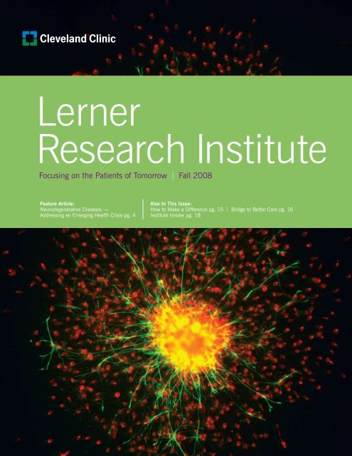
Focusing on the patients of tomorrow FALL <strong>2008</strong>“These are going to be the biggestmedical issues facing us in the next20 years,” said Bruce D. Trapp, PhD,Chair, Neurosciences.a patient in an assisted living facility is $34,860, and the costof a private room in a nursing home is $74,095 annually.Parkinson’s disease Each patient spends an average of $2,500yearly for medications; medical care, disability payments andlost income exceed $5.6 billion annually.ALS In the advanced stages, care can cost up to $200,000yearly, and there are 5,000 newly diagnosed cases annually.Multiple sclerosis (MS) The annual cost associated with MS hasbeen estimated at more than $34,000 per person, translatinginto a conservative estimate of national annual cost of $6.8billion, and a total lifetime cost per case of $2.2 million.To address this looming economic and public-health crisis,<strong>Institute</strong> researchers are targeting the principal neurodegenerativediseases with the goal of finding new ways to monitor, delay,prevent and perhaps cure them. “We have been fortunate toattract some of the best translational researchers in the world,”said Dr. Trapp. “We have developed very strong programsfocused on human neurodegenerative diseases.”The Neuroinflammation <strong>Research</strong> Center (NRC) allows researchersto place special emphasis on inflammation as a commondenominator among a variety of devastating neurological diseases.The Center’s researchers investigate a range of diseases — fromMS and Alzheimer’s disease to muscular dystrophy and stroke.“The role of inflammation in neurodegenerative diseases isrelatively newly appreciated,” said Richard Ransohoff, MD,Neurosciences, who is Director of the Center. “We knowinflammation always impacts neurological disorders. We alsoknow that neuroinflammation is complex — it’s different in eachdisease and almost always includes both helpful and harmfulelements. Medicines that modulate inflammation are available.So if we knew which element of inflammation is causing harm,there would probably be an existing medicine that could be used.This is something we’re desperate to understand so that we canapply this knowledge to benefit our patients.”One unique characteristic of the NRC is the breadth of researchexpertise within one unit. <strong>Research</strong>ers interested in such diversediseases are seldom, if ever, part of a single center. Anothersalient element of the group is its collaborative nature. Much ofthe primary expertise of the NRC scientists lies in neurologicaldisease, with knowledge about inflammation acquired duringdisease-related research.ALS research received substantialsupport in 2007 when the Bright Sideof the Road Foundation created theBarry Winovich Endowed Chair in ALS<strong>Research</strong>. The endowment enablesdedicated research of this progressiveneurodegenerative disease that affectsnerve cells in the brain and the spinalRichard Ransohoff, MDcord. Erik Pioro, MD, PhD, is theinaugural holder of the chair. His research focuses on themolecular basis for degeneration of motor neurons, using imagingtechniques to identify abnormalities in nervous system pathways5
<strong>Lerner</strong> research instituteThe Self-Repairing BrainMore than 15 years ago, Bruce Trapp, PhD, Chair, Neurosciences, created an approach to studying multiple sclerosis(MS). Rather than being confined to investigating the disease in animals, Dr. Trapp wanted to see how MS actuallydevelops and progresses in the human brain. He started an innovative autopsy program to collect the brains andcentral nervous systems of multiple sclerosis patients after they die.“The rapid autopsy program allowed us to study MS by studyingthe disease itself, not a ‘near second’ in animals. This is muchmore relevant than investigating aspects of the disease in animalmodels,” he said. “In essence, what’s happening in the brainsof MS patients is the only thing that matters.”The approach yielded surprising insights that at the timequestioned conventional wisdom about the cause of permanentneurological disability in MS patients, how neuronal tissue isdamaged in MS brains, and whether or not brain cells andtissues can regenerate.White matter neurons are increased in a subset ofchronic multiple sclerosis lesions. Image shows a highmagnification view of post mortem tissue from amultiple sclerosis patient stained with an antibodythat recognizes neurons (microtubule associatedprotein 2, MAP2). The green line delimits a lesionwith increased neurons (top) from the surrounding,non-lesion white matter (bottom). (Photo: BruceTrapp, PhD, Chair, Neurosciences)“Some of our initialobservations were controversialat the time, but they’ve becomeaccepted by neurologists. Ourwork really created a paradigmshift regarding the pathogenesisof permanent neurologicaldisability, tissue damage andregeneration in MS brains,”Dr. Trapp said.Multiple sclerosis is aninflammatory disease thatdamages the insulation (myelin)that surrounds the nerve fibersof the central nervous systemand is the most common causeof neurologic disability in youngadults. Most patients have progressive neurologic deteriorationthat was thought to be caused by the loss of myelin. The axons,or nerve fibers that transmit electrical impulses, were thoughtto be spared this destructive process.A decade ago, Dr. Trapp examined tissues donated throughthe rapid autopsy program and found that a consistent featureof multiple sclerosis lesions was axons that had been cut (ortransected). Additionally, the frequency of these transected axonsrelated to the degree of inflammation within the lesion.“We were able to show initially that transected axons are commonin the lesions of multiple sclerosis, and eventually established arelationship between axonal loss and neurodegeneration withirreversible neurologic impairment in individuals with MS,”Dr. Trapp said. “This was an understanding of the diseaseprocess that contributed to new research in the area.”Other breakthroughs would emerge from the rapid autopsyprogram. For example, in addition to myelin and axons,inflammation associated with MS also destroys cells calledoligodendrocytes that produce myelin. Most multiple sclerosislesions typically do not regenerate myelin, a process calledremyelination. To remyelinate, the MS brain must generate newoligodendrocytes. Dr. Trapp, in collaboration with Case WesternReserve University and the University of Toledo, recently received$3 million by the Ohio Third Frontier Commission to develop smallmolecules that can enhance repair of the brain in MS, with thegoal of not only delaying the progression of disability but reversingit. The leading compounds will be chemically optimized to obtaincompounds suitable for licensing by major pharmaceuticalcompanies as candidate drugs for future clinical trials in patients.Dr. Trapp found that cells capable of producing myelin — the“premyelinating” oligodendrocytes — are present in chroniclesions of multiple sclerosis. “Our expanding knowledge of thecellular interactions between premyelinating oligodendrocytes,axons and the microenvironment of lesions may lead to effectivestrategies for enhancing remyelination.“For example, a possible therapeutic strategy could perhaps beachieved by transplanting oligodendrocyte-producing cells intothe lesions or developing drugs that will promote an MS brain torepair itself,” Dr. Trapp said.More recently, Dr. Trapp’s research team made an unexpectedand exciting finding: the adult human brain can replace neuronsthat were thought to be destroyed by multiple sclerosis.“Proving that the adult human brain produces new neurons isa tough egg to crack because we cannot perform the definitiveexperiments of injecting radioisotopes in the living patients,” hesaid. “The objective, therefore, was to identify areas of the MSbrain which had increased densities of neurons when comparedto normal brains.”8
Focusing on the patients of tomorrow FALL <strong>2008</strong>Because it’s difficult to identify increased neuron density in thegray matter of brains, Dr. Trapp’s study team took a closer look atthe relatively low-density neurons that control blood flow in whitebrain matter. They found that these neurons are destroyed as themyelin is destroyed, forming the white-matter lesions typical ofMS brains. Surprisingly, in tissue sections from some older andmore chronic MS lesions, the concentrations of “stained” neuronswere so dense that they were visible to the naked eye. And these“new neurons” shared characteristics with the mature whitematterneurons in normal brains.“These observations beg the question, ‘How did the new neuronsget there?’ We know the normal adult brain can generate newneurons in certain regions. But it has been controversial andvery difficult to prove that neurogenesis [regeneration of neuronaltissue] occurs in the adult brain in response to neuronaldestruction,” Dr. Trapp said.“Some researchers believe that these new concentrations ofneurons might be caused by migration of mature neuronal cells tothe lesions. But our observations suggest new neuron production,”he said. “For example, we also found a significant increasein immature neurons in the same regions that had increasednumbers of mature neurons. Demyelinated white matter couldcreate a great hospitable environment for neurogenesis.“Our research provides a positive message for MS patients,” saidDr. Trapp. The multiple sclerosis brain never gives up trying toreplace the myelin and neurons that are destroyed by the disease.The findings could lead other investigators to search for signs ofneurogenesis in other brain diseases. Dr. Trapp and his colleaguesare now looking for molecules that could regulate neurogenesis inthe MS brain. In addition, Dr. Trapp’s research team just receiveda $1.5 million grant from the National <strong>Institute</strong>s of Health toinvestigate the cause of cognitive dysfunction in MS patients.Tracking Disappearing Gray Matter in MS PatientsIt’s known that the brain wastes away slowly as multiple sclerosis progresses, just as thesymptoms become more pronounced over time. It seemed natural to conclude that thewhole brain would atrophy during the disease.But new evidence is showing that this might not be the case.Elizabeth Fisher, PhDThe human brain is composed of twobasic components: gray matter (whichconsists mainly of the nerve cellbodies that generate nerve impulses)and white matter (which consists of nerve cell “wires,” calledaxons, that are covered by a sheath made of myelin and thatcarry nerve impulses).Think of a computer network: regions of gray matter in the brainand nervous system are like computers, and the white matter isthe network that links them. This network allows gray matter toshare and process information and stimuli and to generate nerveimpulses, which are also transmitted by white matter.Throughout the progression of multiple sclerosis, the brain wastesaway an average of 0.5% to 1% yearly. Both white and graymatter seem to diminish equally during the early stages of thedisease, meaning the “whole” brain is involved.But brain imaging of MS patients conducted during the last fouryears by Elizabeth Fisher, PhD, Biomedical Engineering, foundthat gray matter starts to waste away at a faster rate than whitematter as the disease progresses. Her research is in collaborationwith Richard Rudick, MD, Vice Chair of <strong>Research</strong> at <strong>Cleveland</strong><strong>Clinic</strong>’s Neurological <strong>Institute</strong> and Director of its Mellen Centerfor Multiple Sclerosis Treatment and <strong>Research</strong>.“We found that atrophy of gray matter accounted entirely for theincreasing rate of atrophy as the disease advanced,” Dr. Fishersaid. “This is the first study to demonstrate that gray matteratrophy speeds up as multiple sclerosis progresses. Initially, whiteand gray matter are affected similarly, but over time atrophy ofthe brain is dominated by the wasting away of gray matter.”If a multiple sclerosis patient’s brain is wasting away, does itreally matter which components are affected the most? Definitely.“Gray matter makes up more than half of the functioning portionof the brain, and tissue damage in gray matter is a large part ofthe overall burden of multiple sclerosis,” Dr. Fisher said. “But,unlike damage in white matter, we can’t see it. ConventionalMRI [magnetic resonance imaging] techniques are not sensitiveto lesions that develop in gray matter, so gray matter atrophymeasurement is one of the only available methods to monitorgray matter tissue damage in multiple sclerosis patients usingstandard MRIs.“Gray matter atrophy may be a clinically relevant way to measuremultiple sclerosis progression,” she said. “Measuring atrophy isimportant because we don’t know yet how and why the brainwastes away at different rates. It can also be useful to test newtreatments — to show whether or not new therapies can actuallyprotect the brain from wasting away.”9
<strong>Lerner</strong> research instituteTag-Teaming Alzheimer’sWe don’t know what causes Alzheimer’s disease.There are tantalizing clues. There are hints of what genetic, lifestyle and environmentalcauses might lead to it. But a definitive explanation for why and how it develops?Not yet.But the disease has many paradoxes. For example, scientistsfound a group of German nuns many of whom had amyloidplaques typical of Alzheimer’s, but who didn’t exhibit dementia.Conversely, some people develop dementia in the absence ofamyloid plaques.“It’s much more of a complicated equation than we thoughtbefore,” Dr. Lamb said. “The disease starts and progresses overthe course of 20 years, and not everyone with amyloid plaqueshas dementia. There’s something we’re missing.”“Everyone,” Dr. Pimplikar added, “generates the precursors toamyloid plaques. Beta-Amyloid is made as soon as we’re born,perhaps even earlier, and plaques are found in about 40% ofnormal people. Why does it lead to a disease in some peopleand not in others?”Left to right: Bruce Lamb, PhD, Riqiang Yan, PhD, Sanjay Pimplikar, PhDIt’s a vexing health issue that promises only to grow. It’sestimated that 5.2 million people have Alzheimer’s today — anumber that will explode to as many as 9 million by the year2020. As the Baby Boom generation ages and the ranks ofpatients swells, increasing pressure will be placed on patients,caregivers and the healthcare industry.A team of <strong>Institute</strong> researchers — Bruce Lamb, PhD, Sanjay W.Pimplikar, PhD, and Riqiang Yan, PhD, all in Neurosciences — isworking to unravel the mysteries of a disease that robs people oftheir memories and identities.For many years, the onset of Alzheimer’s was attributed largelyto the formation of distinct alterations within the brain, namely,dense clumps of proteins outside brain cells called amyloidplaques and insoluble twisted fibers inside brains cells calledneurofibrillary tangles. While some consider Alzheimer’s diseasethe result of normal aging, increasing evidence suggests thatit is instead part of a complex, age-related disease process.Each researcher is looking at a different aspect of Alzheimer’s,yet their work is complementary. A common denominator isamyloid precursor protein, or APP, the precursor to the amyloidplaques, which seems to be involved intimately with theinitiation of Alzheimer’s.The genetics of Alzheimer’sToo much APP — what researchers call overexpression of APP— has been linked to elevated production of beta-amyloidpeptides, which are the principal components of amyloidplaques in the brains of Alzheimer’s patients. Are some peoplegenetically predisposed to this APP overexpression?Dr. Lamb has identified several regions of specific mousechromosomes that prevent the formation of amyloid plaquesin animal models of AD that overexpress APP and is in theprocess of narrowing in on the specific gene responsible.He also is looking at possible therapies, specifically nonsteroidalanti-inflammatory drugs, which could decrease the risk ofAlzheimer’s. Most studies have studied these therapies later inlife, after the onset of Alzheimer’s — when the damage is doneand is irreversible. “But there is some evidence that if you start10
Focusing on the patients of tomorrow FALL <strong>2008</strong>In this image, beta-amyloid peptides — which form amyloidplaque in Alzheimer’s patients — are stained red. Microglia(the primary immune cell of the brain) is green, and nuclei arestained blue. The image shows how microglia surround theamyloid plaques. (Photo: Bruce Lamb, PhD, Neurosciences)certain nonsteroidal anti-inflammatories early in life, you candramatically lessen how Alzheimer’s progresses. There’s awindow where it might be effective, but it’s an early window,”Dr. Lamb said.Finally, he is studying what influence diet might play. “We placedseveral different strains of mice on high-fat, high-cholesteroldiets. One strain had an increase in beta-amyloid and severalother strains had no effect. So there might be some geneticpredisposition to dietary effects on beta-amyloid,” he said.The unusual suspectThere are many suspected causes of and contributors toAlzheimer’s. Targeting the right ones is “like shooting in thedark. You know they’re there; you just cannot see them,”said Dr. Pimplikar.Dr. Pimplikar focuses on a relatively new area of Alzheimer’sdealing with APP intracellular domain, or AICD. For years, thegenerally accepted progression of Alzheimer’s went like this:APP is cleaved, or “cut,” by the BACE1 enzyme. One of thepieces is then cleaved again into two still smaller pieces. Onepiece is beta-amyloid, which many consider a cause of thedisease. The second piece is AICD, and it never attractedmuch attention until recently.“We made mice that overexpress only AICD and not betaamyloid,”Dr. Pimplikar said. “And guess what? Our mice startto show lots of symptoms of Alzheimer’s. Why and how is AICDinvolved? Our research seems to indicate that even if you canget rid of beta-amyloid, but still have AICD, you may still getAlzheimer’s. Ultimately, we want to see to what extent AICDcontributes to how Alzheimer’s develops.“Plaques by themselves will induce inflammation, butinflammation starts early or without plaques in our mice.Does AICD play a role in this inflammation process, andcan we develop drugs that are effective against AICD?”Dr. Pimplikar said.Which comes first?The hallmark brain plaque is actually composed of beta-amyloidsurrounded by inflammatory glia (the network of cells thatsupport the nervous system) and abnormal or swollen nerve cellextensions called dystrophic neurites. What hasn’t been knowndefinitively is what role, if any, is played by these dystrophicneurites. Are they a by-product of the formation of beta-amyloid?Could they influence its development? Do they even contributedirectly to the dysfunction associated with Alzheimer’s?Dr. Yan found that increased levels of the protein reticulon 3(RTN3) cause the formation of dystrophic neurites and that havingdystrophic neuritis impairs spatial learning and memory — evenwithout beta-amyloid.“The presence of dystrophic neurites has been identified asone of the distinguishing features in the brains of patients withAlzheimer’s. However, the molecular nature wasn’t understood,until now,” Dr. Yan said.Dr. Yan’s research shows that even a modest increase in RTN3— perhaps only one- or two-fold — is sufficient to cause RTN3aggregation and formation of dystrophic neurites. And whenthese neurites are created, even absent beta-amyloid, cognitiveimpairment still results.“The research suggests that suppressing RTN3 aggregation maynot only delay dysfunction, but also inhibit the BACE1 enzymethat creates the beta-amyloid plaque,” he said. “RTN3 could bea new therapeutic target.”Have we made any progress and is there hope?“There are a lot more unanswered questions, but I believewithin 10 or 15 years we will have therapies that will delay theprogression of Alzheimer’s once it’s started and may even be ableto stop it developing in the first place,” Dr. Pimplikar said. “Justlike President Nixon’s ‘War on Cancer’ led to tremendous resultswithin a decade, we need a similar focus on Alzheimer’s if we’regoing to effectively deal with it.”11
<strong>Lerner</strong> research instituteGetting to the Heartof Alzheimer’sA few years ago, Jonathan Smith, PhD, Cell Biology, was studying how a particular gene helps to transportfatty compounds called lipids throughout the body. Lipids are a major contributor to the plaque that formson the inside of arteries that can lead to heart attacks or strokes, so learning how to control or reverse thisbuildup is a critical healthcare goal.Jonathan Smith, PhDThen Dr. Smith heard a bit ofinteresting news. The same genealso was found to be linked to thehallmark senile plaque that formsin the brains of patients withAlzheimer’s disease (AD). Couldhis work in cardiovascular geneticsalso contribute to AD research?The focus is on a gene calledapolipoprotein E, or ApoE for short. ApoE produces severalvariations of a protein — the protein helps to transport lipids andis linked to AD. The protein ApoE-ε3 is the most common form,while ApoE-ε2 and ApoE-ε4 are relatively common variants.“If you look at Alzheimer’s cases, about two-thirds of the patientscarry at least one copy of the gene that produces ApoE-ε4, whileonly one quarter of a young control population are ApoE-ε4carriers. People who carry one copy of ApoE-ε4 have a two- tofour-fold risk of AD, mostly among women. About 2% of peoplecarry two copies of ApoE-ε4, and they have a 15-fold risk ofAlzheimer’s,” Dr. Smith said. “It’s the most prevalent geneticsusceptibility factor for AD by far.”Although there is much to still be discovered about howApoE-ε4 plays a role in AD susceptibility, Dr. Smith’s ADresearch has shifted to looking for a drug that could preventor delay the onset of AD. A protein called amyloid precursorprotein produces a substance called beta-amyloid peptide (Aβ).Aβ is the major component of the plaque that forms in thebrains of Alzheimer’s patients.“By the time a person is diagnosed with Alzheimer’s, it’s toolate. Too much damage has been done,” he said. “The bestapproach is to work toward therapies that prevent or delay theage of onset of the disease. If you delay the age of onset by evenfive or 10 years, you could reduce the incidence of AD by halfand significantly improve the burden on patients and society.”To achieve this goal, Dr. Smith and his colleague EnakshiChakrabarti, PhD, Cell Biology, screened a library of smallmolecule compounds at the <strong>Institute</strong>’s Small Molecule ScreeningCore. Small molecules are small organic compounds that arebiologically active and can be used as “tools” in molecular biologyor as drugs in medicine. The goal is to find compounds that couldinhibit Aβ secretion from cells of the human nervous system.Although they have identified a compound of interest that isvery effective in reducing Aβ production, this drug does nothave the properties that will allow it to pass through theblood-brain barrier, a membrane that primarily protects thebrain from chemicals in the blood, while still allowing essentialmetabolic function.Dr. Smith is now working with Lawrence Sayre, PhD, a chemistat Case Western Reserve University, to modify this compoundso that it can enter the brain. Once a drug that can inhibit Aβproduction has been shown to be both safe and effective, aperson with a family history of Alzheimer’s or someone whocarries the ApoE-ε4 variant could start a daily drug regimen intheir 40s or 50s — something as simple as a daily pill — thatmight be able to prevent the disease.12
Focusing on the patients of tomorrow FALL <strong>2008</strong>Beyond the LimitsJay Alberts, PhD, Biomedical Engineering, had justcompleted a 450-mile weeklong bicycle ride across Iowato raise awareness of Parkinson’s disease (PD). Quite bychance, a PD patient on a tandem bicycle mentioned thatshe didn’t have Parkinson’s symptoms during the rideand that the tremor in her hand was gone afterward.Jay Alberts, PhDDr. Alberts’ curiosity was roused.Could exercise — specifically, “forcedexercise,” in which people are pushedbeyond their normal limits — betherapeutic for PD patients?The PD patient in Iowa had beenpaired with a rider who set a cadencethat was faster than what the patientwould have pedaled alone. Dr. Albertsused this as a starting point to see if such forced exercise wouldimprove motor function in other PD patients.physical activity — the manual tasks that range from buttoningshirts to tying shoe laces to handwriting. Dr. Alberts has beenworking on the project with Mark Lowe, PhD, <strong>Cleveland</strong> <strong>Clinic</strong>’sImaging <strong>Institute</strong>, and Micheal Phillips, MD, Section Headof Imaging Sciences in the <strong>Cleveland</strong> <strong>Clinic</strong> Department ofDiagnostic Radiology.“There’s an increase in activation after just one forced exercisesession. We’ve seen improvements remain for four weeks aftera patient stopped forced exercising,” Dr. Alberts said.“We believe driving of the central nervous system may benecessary to produce the underlying biochemical changes whichneed to occur to improve motor function,” he said. “The nextsteps are to understand the mechanisms underlying improvedfunction, what is the minimum dose necessary for improvementsto occur, are these effects long-lasting, and does forced exerciseof this kind slow the progression of Parkinson’s.”Most previous interventions using exercise allowed PD patientsto set their own exercise rates, which yielded mixed results. Dr.Alberts designed a tandem bicycle that measures and monitorspatients’ performance, power and pedaling rate. The bicycleforces PD patients to pedal at rates 40% to 60% faster thanthey can achieve on their own.The results have been promising.“One encouraging aspect is the improvement in motor functionin patients’ arms even though they’re only using their legs duringthe exercising. We’ve seen as much as a 35% improvementin motor function,” Dr. Alberts said. “This suggests that forcedexercise is impacting higher brain function and improvingcentral motor function.”Scans of PD patients’ brains show that exercise seems tostimulate the supplementary motor area of the brain, the regionthat controls general movement and governs 60% of our dailyThe image on the left is a scan of a PD patient’s brain priorto exercise. The image on the right shows the brain areasinvolved in constant, forced tasks involving the thumb andindex finger of the dominant hand. After exercise, it wasclear that additional motor regions — particularly thesupplementary motor area (in the circle) — were recruitedfor task performance. (Photos: Mark J. Lowe, PhD, Directorof High-Field MRI, <strong>Cleveland</strong> <strong>Clinic</strong> Imaging <strong>Institute</strong>)13
Focusing on the patients of tomorrow FALL <strong>2008</strong>Want to Make a Difference?Start with the <strong>Lerner</strong> <strong>Research</strong> <strong>Institute</strong>Dear Friends,improve motor skills can “spread” and in some cases diminishother skills.Today, Dr. Vitek is looking at using DBS in an area of the braincalled the globus pallidus interna (Gpi), which, like the STN, is partof the “circuitry” that controls movement, thought and emotions.“One positive aspect about the Gpiis that the areas that influence motorskills, thought and emotion occupy alarge area and allows one to minimizethe effect of DBS in the motor areaon non-motor areas,” Dr. Vitek said.“Focusing DBS in that area achievesMicheal Phillips, MDthe same motor benefits with lessrisk of affecting cognition or mood.”Precision is paramount when it comes to implanting electrodesin a brain. That’s where work with Dr. McIntyre comes in.“For the most part, there’s not much opportunity for surgeons tosee where electrodes are or the spread of electrical stimulationin the brain,” he said. “Surgeons generally aren’t schooledin electrical engineering and physics, so a lot that has beenlearned and gained has been trial and error. Because there’sso much ambiguity in the process, it’s led to side effects [likethe motor/cognitive skills problem].”Dr. McIntyre’s work has two goals. First, map the brain anddetermine parameters for electrode implantation — where theyshould be placed and how much electrical stimulation is neededto improve motor skills without sacrificing cognitive abilities.Second, draw the “map” for others.“Right now it’s an art, not a science,” Dr. McIntyre said. “Eachdisease [Parkinson’s, dystonia, depression, etc.] has its owncenter that’s a different part of the brain, and we have a goodidea of the general locations of the regions. What we wantto provide is essentially a road map that will tell surgeonsexactly where they want the electrode to be and the levelof stimulation that’s appropriate.”It’s a proverbial question ponderedby many. “Why do people donatemoney?” In 2005, a group ofneuroscientists found that activationof a particular brain region canpredict whether or not people tendAlicia Hoose, MPA to be selfish or altruistic. They hadfound the brain’s “charity spot:” aregion that determines whether we put others before ourselves.Please activate the region of your brain called the posteriorsuperior temporal sulcus (if you’re curious, this region lies in theback and top portion of the brain) prior to reading any further.Now that I have your attention (and the attention of yourposterior superior temporal sulcus), I can proceed!We at the <strong>Institute</strong> are proud to present the current <strong>Lerner</strong><strong>Research</strong> <strong>Institute</strong> Magazine. The cover story discussesneurodegenerative diseases. In the United States, more than6 million people live with some type of neurological condition,ranging from Alzheimer’s and Parkinson’s disease to multiplesclerosis and amyotrophic lateral sclerosis (Lou Gehrig’sdisease). These disorders are devastating and expensive,with annual costs currently exceeding several hundred billiondollars in the United States alone, and current treatments areinadequate. Adding to the urgency is the fact that the incidenceof these disorders is increasing rapidly as our population ages.Although private donations play more of a role in fundingresearch into neurological conditions, a great need remains.Because laboratory scientists have markedly feweropportunities for contact with patients, they are less likelyto form the kind of lasting relationships with a patient —and potential donor — than clinicians routinely form. Basedon this fact, your donations, whether they’re a result of theactivation of your brain’s “charity spot” or other reasons,are very important to the biomedical research that we aredoing. If you truly want to make a difference, a gift to the<strong>Lerner</strong> <strong>Research</strong> <strong>Institute</strong> is the place to start.Alicia Hoose, MPADirector of Development15
<strong>Lerner</strong> research instituteA Bridge to Better Patient CareSerpil Erzurum, MDAs Chair of the <strong>Institute</strong>’s Departmentof Pathobiology, Serpil Erzurum, MD,is a well-known researcher andclinician involved in the causesand treatment of asthma. She alsoinvestigates the causes of pulmonaryhypertension, a debilitating respiratorydisease that’s particularly deadlyamong women.She is among the ranks of clinician-scientists — medicalprofessionals with one foot planted firmly in the laboratory andthe other next to patients’ bedsides. In addition to supervisingPathobiology and its laboratories, Dr. Erzurum is Co-Director of<strong>Cleveland</strong> <strong>Clinic</strong>’s Asthma Center and sees patients through itsDepartment of Pulmonary, Allergy and Critical Care Medicine.“I really see the benefit of an environment that lets clinicians andresearchers talk and share their experiences. You can learn somuch from one another in informal settings,” she said. “There areso many times when I hear what a researcher is doing that couldapply to my patients, or what I’m seeing with patients that couldhave an impact on laboratory projects.”CTSA by the Numbers• 2,359: Outpatient visits• 310: Overnight research admissions• 15,000: Samples processed in laboratory• 139: <strong>Research</strong> projects• 35: Departments involved• 225: Principal Investigators who use the<strong>Clinic</strong>al <strong>Research</strong> Unit• 41: Refereed journal publications fromstudies done on the Unit12-month periodDr. Erzurum is well positioned to encourage that interactionas the Director of the <strong>Clinic</strong>al <strong>Research</strong> Unit of the <strong>Cleveland</strong><strong>Clinic</strong>al Science and Translational Consortium. The collaborativeis possible through a <strong>Clinic</strong>al and Translational Science Award,a $64 million National <strong>Institute</strong>s of Health grant providingresources for the coordinated development of clinical andtranslational sciences. One of 38 academic health centerawards nationally, CTSA is a partnership of <strong>Cleveland</strong> <strong>Clinic</strong>,Case Western Reserve University, University Hospitals CaseMedical Center, MetroHealth Medical Center, and the LouisStokes <strong>Cleveland</strong> VA Medical Center.The <strong>Clinic</strong>al <strong>Research</strong> Unit provides the equipment, nursingsupport and support staff for more than 200 investigatorscarrying out more than 125 clinical research projects. Mostsuch projects are NIH-supported and are under way at<strong>Cleveland</strong> <strong>Clinic</strong>, from diabetes research and exercise studiesto storing and shipping samples and investigating acute heartattacks. W.H. Wilson Tang, MD, Cell Biology and <strong>Cleveland</strong><strong>Clinic</strong>’s Department of Cardiovascular Medicine, directs theinpatient research services, and Lara Danziger-Isakov, MD,MPH, <strong>Cleveland</strong> <strong>Clinic</strong>’s Pediatric Infectious Diseases, directsthe pediatric studies in the research unit.The services range from the ordinary (e.g., echocardiogramsof people’s hearts) to the extraordinary, such as “apheresisand elutriation” — collecting blood from volunteer donors andseparating out the billions of white blood cells, then returning thered blood cells to the volunteer. With the support of the CTSA,Peter Cohen, MD, formerly of Immunology, and ClemenciaColmenares, PhD, Cancer Biology and Co-Director of the<strong>Institute</strong>’s <strong>Research</strong> Core Services, who leads the translationaltechnologies component of the CTSA at <strong>Cleveland</strong> <strong>Clinic</strong>,launched this new research service, in which healthy humanvolunteers undergo collection of their blood to obtain humancirculating cells, which are then separated into the variouscomponents for use by basic science researchers.16
Focusing on the patients of tomorrow FALL <strong>2008</strong>“Bridging the gap between laboratory and clinical research isintegral to patient care, and it would be much more challengingto do without the CTSA,” said Dr. Erzurum, who credits RichardRudick, MD, for spearheading the application for the CTSA.Dr. Rudick is Vice Chair of <strong>Research</strong> at <strong>Cleveland</strong> <strong>Clinic</strong>’sNeurological <strong>Institute</strong>. All projects in the CTSA undergo reviewby the Protocol Review Committee, which is led by DavidVan Wagoner, PhD, Molecular Cardiology and Chair of thecommittee, and Bret Lashner, MD, Gastroenterology andHepatology, and Co-Chair of the committee.“Our <strong>Clinic</strong>al <strong>Research</strong> Unit enables us to bring our research tothe patient in a larger way than before. It allows rapid translationof our findings in the lab to the patient. Our staff — led byCharlotte Bhasin, our unit administrator, and Kay Stelmach, ournurse administrator — is dedicated to helping the investigatorson our unit advance patient-oriented research,” Dr. Erzurum said.Just as important, the unit serves as an advocate for themembers of the public who volunteer for the various researchprojects. “Our participants are equal partners in the clinicalresearch enterprise. Without them, we couldn’t advance medicalcare, and in the CTSA structure, they are partners in the researchin the CTSA,” she said.As much as the <strong>Clinic</strong>al <strong>Research</strong> Unit provides to investigators,Dr. Erzurum sees potential for growth.“We have a need to expand the clinical unit, in particular intonutritional and metabolic areas,” she said. “We are starting toorganize for the development of a nutritional research center tofocus on that facet of the disease process, which is so importantfor diabetes and obesity research. We have also begun to create aresearch participant membership that will allow greater and moremeaningful involvement of our volunteers in our ongoing studies,including the sharing of research findings.”<strong>Clinic</strong>al <strong>Research</strong>Unit ResourcesThe <strong>Clinic</strong>al <strong>Research</strong> Unit provides molecularbiology, specimen processing and analytical work.Its primary functions are to provide technicalsupport for sophisticated chemical analyses calledassays and to develop or validate new laboratorymethods. The unit provides staffing for specificlaboratory procedures.Specimen ProcessingThe unit performs minimal to complex specimenprocessing. Among its services are blood, serum,plasma or urine collection; highly consistent,project-specific processing; specimen shipping;and large-volume blood cell separation.<strong>Research</strong> AssaysThe unit can provide chemical analysis assaysthat offer a complete chemical analysis of bloodand the detection of antibodies or antigens, amongother services.Specimen Storage<strong>Research</strong>ers have access to a 4°C refrigerator, oneupright -20°C and one -70°C freezer for short-termspecimen storage. All refrigerators and freezers areequipped with centrally monitored alarm systemsfor efficient and accurate temperature control.Specimen ShipmentThe unit has the capability to ship ambient orfrozen specimens to other laboratories.17
Focusing on the patients of tomorrow FALL <strong>2008</strong>said Dr. Muschler. “We believe these consortia offer a lot ofpotential for us to realize the value of advanced therapies totreat the increasingly complex health issues facing the Americanhealthcare system. Our sense of responsibility in bringing theselife-restoring therapies to practice is profound, but so is oursense of hope for the future of military and civilian traumapatients in this country.”Known Hormone Could Be Mystery Key toHypertensive DiseasesMore than 20 years ago, researchers discovered a hormone inthe cells that make up heart tissue that is essential to regulatingblood pressure. It remained unclear, however, what wasresponsible for activating the hormone.Qingyu Wu, MD, PhD, Molecular Cardiology, has identified oneof the mystery “keys” — a discovery that could help people withcardiac hypertrophy and high blood pressure, as well as pregnantwomen with life-threatening hypertensive disorders.Cardiac hypertrophy occurs when the muscle of the leftventricle (the chamber that pumps blood to the body) enlargesand obstructs blood flow. High blood pressure, in additionto contributing to strokes and other diseases, can causepreeclasmpsia and eclampsia — hypertensive disordersaffecting about 10% of pregnant women and accounting foran estimated 18% of maternal deaths and 15% of prematurebirths, according to the Preeclasmpsia Foundation.Regulating blood pressure is a delicate balancing act for thehuman body. Any disruption or inefficiency of how cardiachormones behave can lead to cardiac hypertrophy and otherhypertensive disorders.Atrial natriuretic peptide (ANP) is a hormone that regulates bloodpressure by encouraging the kidneys to remove salt and water andthe blood vessels to relax. It’s found in heart cells as an inactiveform (“pro-ANP”) until it is activated. Enzymes are catalysts inbiochemical reactions like those that activate hormones.Prior to Dr. Wu’s discovery, it had been unclear what enzyme orenzymes are responsible for converting pro-ANP into an activehormone necessary for blood pressure regulation.Dr. Wu found that corin — an enzyme readily made and foundin heart tissue — is the only enzyme able to convert pro-ANPinto active ANP.For example, mice without corin are more likely to develophypertension later in life and during pregnancy (similar to whatpregnant women experience during preeclasmpsia or eclampsia).Additionally, mice without corin develop cardiac hypertrophy,or enlarged hearts.“What we call the ‘ANP-mediated pathway’ plays an importantrole in regulating blood pressure, and corin is a key part ofthat pathway. Without it, patients can experience high bloodpressure, enlarged hearts and hypertension that could put atrisk the lives of expectant mothers and their unborn child,”said Dr. Wu.“Knowing how this pathway works and which enzymesand hormones are essential to its success can lead to newtreatments or therapies for people who are corin-deficient,”he said. “It might be therapies to preemptively correct thecorin deficiency, or it might be treatments to encouragecorin production after diagnosis as a way to treat orprevent diseases.”19
<strong>Lerner</strong> research institute<strong>Institute</strong> Insider (continued)New Key to BloodClots Found?A protein normally associated with the development of bloodvessels might also render platelets more active and makepeople more susceptible to dangerous blood clots. Thisdiscovery could lead to new therapies against unwantedblood clotting.At the center of the research is a protein called CD36. Thisprotein is found on the surface membrane of a variety of cellsin the cardiovascular system. It plays a significant role ina number of biological processes, including blood vesselformation (as in tumors), inflammation, and plaque formationon the inside of arteries (atherosclerosis), among others.<strong>Research</strong> led by Roy Silverstein, MD, Chair, Cell Biology,found that this protein can also activate blood platelets.These hyperactive platelets then collect to form blood clots,called thrombus, which can cause heart attacks or strokes.The research focuses on the interaction of two components:platelets and microparticles.Platelets are very small cells that float in blood plasma andare responsible for blood clotting. If you get a cut, plateletsbecome activated and stick to the injury site to form amesh-like structure and signal for more platelets. Thisgrowing structure attracts and traps other cells to create aplug to stop bleeding. But although you want a plug at thesite of a cut or injury, you don’t want one forming in theblood vessels that feed and nourish the heart or brain.Microparticles, small fragments that bud off from normalcells, must become activated or they die. Microparticlescome from a variety of cells in the vascular system:platelets, white blood cells called monocytes, andendothelial cells that line the inside of blood vessels. Thesefragments usually occur when there is a vascular injury.Dr. Silverstein’s research found that the CD36 proteinon platelets responds to vascular injury by recognizingand binding to microparticles that arise from endothelialcells. This aggregation of microparticles and plateletsfacilitates formation of blood clots. Conversely, reducingor eliminating the level of CD36 reduces thrombosis,or the formation of clots.“For the first time, to our knowledge, we demonstrateCD36’s essential role in contributing to the formation ofblood clots,” Dr. Silverstein said. “Our research suggeststhat vascular injury generates microparticles and othermolecules that, in turn, bind platelets via CD36 andenhance the formation of blood clots.“That means that CD36 could be a target for anti-clottingtherapies. If we can find a way to reduce the amount ofCD36 present after a vascular injury, we could help to avoidthe thrombosis that had been a person’s normal responseto some types of vascular injury,” he said.20
Focusing on the patients of tomorrow FALL <strong>2008</strong>First Cross-Race Link in Arterial Disease, Heart Attack FoundA year ago, researchers found that a cluster of genetic variantson a specific region of chromosome 9 is linked to coronaryartery disease (CAD) in white people in northern Europe andNorth America. People who have that genetic quirk are moresusceptible to developing CAD or having a heart attack.Now, <strong>Institute</strong> researchers have shown the same geneticmaterial also is associated with coronary artery diseases inthe South Korean population — the first evidence of cross-racesusceptibility to CAD associated with the same specificcombination of genetic variants.CAD is a condition in which plaque builds up inside thecoronary arteries that supply heart muscle with oxygen-richblood. Plaque narrows the coronary arteries and reduces bloodflow to heart muscle. It also makes it more likely that bloodclots will form in arteries. CAD can lead to angina, heart attack,heart failure and arrhythmias (irregular heartbeats). It is themost common type of heart disease and the leading causeof death in the United States for both men and women. Bothgenetic and environmental factors — and the interplay betweenthe two — have been identified as major risk factors for peopledeveloping CAD. But the molecules and genes that contributeto the genetic risk have remained largely unidentified.At the center of the research are four single-nucleotidepolymorphisms (or SNPs) that are at a specific location on the 9p21piece of chromosome 9. Each SNP is a single DNA building block.SNPs occur normally throughout a person’s DNA, which meansthere are roughly 10 million SNPs in the human genome. MostSNPs have no effect on health or development. But when SNPsoccur within a gene or in a regulatory region near a gene, they mayplay a more direct role in disease by affecting the gene’s function.In this case, the presence of these four SNPs on 9p21 seem tobe associated with CAD, meaning people who have these fourspecific SNPs on their 9p21 gene have a higher risk of developingcoronary disease or suffering a heart attack.<strong>Research</strong> led by Qing Wang, PhD, Molecular Cardiology andDirector of the Center for Cardiovascular Genetics, took what isknown about the 9p21 SNPs in the white population and appliedit to 611 South Koreans who were diagnosed with CAD and 294healthy control patients.The researchers found a similar association between the 9p21SNPs and a higher risk of developing coronary disease andsuffering heart attacks among Asians.“Identifying specific ‘hot spots’ that might harbor one or moregenes that could influence development of CAD and showing thatit is a cross-race risk is an important advance in being able togenetically screen for coronary and arterial diseases or heartattacks,” Dr. Wang said. “Proving this association across diversehuman populations can lead to new clinical markers for diseasewhich can alert people about their risk of CAD or heart attackyears before any actual diseases develop. Early screening canlead to people making lifestyles changes to reduce the risk andearlier monitoring for potential health problems.”Magnets Could Save Stitch in Time for SomeHeart Valve Patients<strong>Research</strong>ers are investigating a replaceable heart valve that usesmagnets rather than stitches to keep the valve in the properposition, an innovation that will mean shorter surgeries, less timefor patients on heart-lung bypass machines, and reducing therisk of postoperative complications.During the last 40 years, there’s been significant progressin heart valve design and replacement procedures, with lifeexpectancy of about 20 years for the valves themselves. Butfailed valves require replacement surgery. In some cases, hearttissue can interfere with the replacement valve’s performanceby growing into it, a condition known as ingrowth.Kiyotaka Fukamachi, MD, PhD, Biomedical Engineering, isdeveloping a new heart valve that uses magnetic coupling to keepthe valve in the proper position. The prototype has two parts: abase magnet that is connected to a heart tissue with sutures, anda magnetic ring with the actual valve. Both are neodymium ironboron magnets encased in very thin stainless steel.During the initial surgery, sutures are used to connect the basemagnet to the fibrous tissue that encircles the opening with thefaulty natural valve. The magnet ring with the new valve is then“mated” to the base magnet without sutures or other permanentattachment techniques — two opposites being attracted to eachother. The two magnets remain connected even under greaterthan-normalpressure.The research team also designed a special separation toolcapable of detaching the magnets. Each ring has a beveled edgearound the outside edges. Should a repeat replacement beneeded, the blades of the tool can be placed in this groove —and squeezing the tool like a pair of scissors exerts enough forceto separate the rings. A new ring valve can then be “mated”quickly and easily to the base magnet that remains suturedto the heart tissue.21
<strong>Lerner</strong> research institute<strong>Institute</strong> Insider (continued)First, a primer in basic genetics. Cells are the building blocks ofthe body. Each cell in your body contains instructions encodedin your DNA that are parceled into 23 pairs of chromosomes.Approximately 39,000 genes, which are the instruction bookletsmade of DNA, are found dotted along all the chromosomes.Differences in people come from slight variations in these genes,which determine everything from hair and eye color to whetheror not a person is more or less susceptible to certain diseases.“There are several advantages to our innovation. First, the tight fitof the two rings make it extremely unlikely that tissue ingrowthwould occur to prevent the safe removal of the valve ring,” Dr.Fukamachi said. “The valve opening remains nearly normal sizebecause the base magnet is small and a simple design, so bloodflow is not obstructed as much. Finally, the coupling method hasthe potential for long-term durability.“If trials continue to be as successful as the first ones, weexpect repeat valve replacement surgeries to be easier and lesstime consuming compared to conventional valve replacements,”Dr. Fukamachi said. “Reducing the length of surgeries andthe amount of time on heart bypass machines is especiallyimportant for high-risk patients whose risk of death is increasedin those situations.”Corralling Enablers of Genetic MutationsGenetic mutations by themselves may not necessarily increasethe risk of developing certain cancers. Some genetic mutationsmay need enablers — little helpers that increase an individual’srisk of a genetic disorder.<strong>Institute</strong> researchers may have identified two of these enablersinvolved in cancer, a finding that could lead to advances in bothdiagnostic and preventive care for people who have certaingenetic mutations.Charis Eng, MD, PhD, Chair, Genomic Medicine <strong>Institute</strong>, hasidentified two tiny components of the human genome calledmicroRNA (miRNA) that appear to play an out-of-the-box rolein turning off a tumor-suppressing gene. With the tumor-fightinggene inactive, cells are more likely to grow uncontrollably andto cause cancers.The DNA in genes is translated or decoded into another geneticmaterial called messenger RNA. In turn, messenger RNA producesproteins that are the basic building blocks and workers of eachcell in the body. This conversion, of DNA to RNA to protein, isa tightly regulated process. Recent work, by many groups, hasdemonstrated that one regulatory player is the miRNAs. EachmiRNA provides a level of regulation to ensure that genes do notproduce too much or too little messenger RNA, which wouldresult in creating too much or too little cellular protein.Tumor suppressors are like the brakes of a cell. They tell thecell to stop dividing and growing at the right moment. Whena tumor suppressor gene is “turned off,” the cell’s brakes nolonger work, and unchecked growth and multiplication of cellscan occur, which often results in cancer. Dr. Eng’s group hasfound that two specific miRNAs — miR-19a and miR-21 —are associated with deactivating the tumor-suppressing genePTEN, which is involved in several types of cancer, as well asin Cowden syndrome. Cowden syndrome is an underdiagnosedheritable disorder associated with an increased risk ofdeveloping breast, thyroid and uterine cancers. In the firstpatient-oriented study of the two miRNA, Dr. Eng’s groupfound that the levels of these miRNAs are correlated to theamount of PTEN protein the cell produces.This is an exciting observation because it shows, for the firsttime, that despite having identical PTEN mutations, the PTENprotein levels can vary from individual to individual, dependingon their levels of these two miRNAs. And depending on themiRNA and PTEN protein levels, the severity of disease andthe cancer types can vary.“These miRNAs could serve as biomarkers that signal thepossibility of development of specific disease features. We thinkthat by understanding how miRNAs and the chromosomal genemutations talk to each other, we are one step closer to predictingprecisely what cancers develop and therefore personalizinghealthcare,” Dr. Eng said.22
The Future of Medicine Unfolds Dailyat the <strong>Lerner</strong> <strong>Research</strong> <strong>Institute</strong>.To contribute to the<strong>Lerner</strong> <strong>Research</strong> <strong>Institute</strong>,visit www.lerner.ccf.org/givingHome to <strong>Cleveland</strong> <strong>Clinic</strong>’s laboratory-based, translational, and clinicalresearch, the <strong>Lerner</strong> <strong>Research</strong> <strong>Institute</strong> is instrumental in improvingpatient care and introducing new therapies. Your support will directlyimpact the development of future treatments here at <strong>Cleveland</strong> <strong>Clinic</strong>.<strong>Cleveland</strong> <strong>Clinic</strong> is ranked one of America’s top hospitals byU.S.News & World Report.
The latest information about researchachievements, educational opportunities andphilanthropic options can now be delivereddirectly to your computer. You can nowsubscribe at www.lerner.ccf.org.9500 Euclid Avenue/NB21, <strong>Cleveland</strong>, OH 441958


