A B C - PULSION Medical Systems SE
A B C - PULSION Medical Systems SE
A B C - PULSION Medical Systems SE

You also want an ePaper? Increase the reach of your titles
YUMPU automatically turns print PDFs into web optimized ePapers that Google loves.
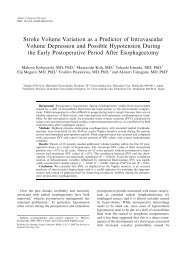
Goal-Directed Fluid Management by Bedside Transpulmonary Hemodynamic<br />
Monitoring After Subarachnoid Hemorrhage<br />
Tatsushi Mutoh, Ken Kazumata, Minoru Ajiki, Satoshi Ushikoshi and Shunsuke<br />
Terasaka<br />
Stroke published online Nov 8, 2007;<br />
DOI: 10.1161/STROKEAHA.107.484634<br />
Stroke is published by the American Heart Association. 7272 Greenville Avenue, Dallas, TX 72514<br />
Copyright © 2007 American Heart Association. All rights reserved. Print ISSN: 0039-2499. Online<br />
ISSN: 1524-4628<br />
The online version of this article, along with updated information and services, is<br />
located on the World Wide Web at:<br />
http://stroke.ahajournals.org<br />
Subscriptions: Information about subscribing to Stroke is online at<br />
http://stroke.ahajournals.org/subscriptions/<br />
Permissions: Permissions & Rights Desk, Lippincott Williams & Wilkins, a division of Wolters<br />
Kluwer Health, 351 West Camden Street, Baltimore, MD 21202-2436. Phone: 410-528-4050. Fax:<br />
410-528-8550. E-mail:<br />
journalpermissions@lww.com<br />
Reprints: Information about reprints can be found online at<br />
http://www.lww.com/reprints<br />
Downloaded from<br />
stroke.ahajournals.org by guest on November 16, 2007
Goal-Directed Fluid Management by Bedside<br />
Transpulmonary Hemodynamic Monitoring<br />
After Subarachnoid Hemorrhage<br />
Tatsushi Mutoh, MD, DVM, PhD; Ken Kazumata, MD; Minoru Ajiki, MD;<br />
Satoshi Ushikoshi, MD; Shunsuke Terasaka, MD<br />
Background and Purpose—Optimal monitoring of cardiac output and intravascular volume is of paramount importance<br />
for good fluid management of patients with subarachnoid hemorrhage (SAH). The aim of this study was to demonstrate<br />
the feasibility of advanced hemodynamic monitoring with transpulmonary thermodilution and to provide descriptive<br />
data early after SAH.<br />
Methods—Forty-six patients with SAH treated within 24 hours of the ictus were investigated. Specific targets for cardiac<br />
index (�3.0 L � min �1 � m �2 ), global end-diastolic volume index (700 to 900 mL/m 2 ), and extravascular lung water index<br />
(�14 mL/kg) were established by the single-indicator transpulmonary thermodilution technique, and a fluid<br />
management protocol emphasizing supplemental colloid administration was used to attain these targets. Plasma<br />
hormones related to stress and fluid regulation were also measured.<br />
Results—A higher cardiac index (mean value of 5.3 L � min �1 � m �2 ) and a lower global end-diastolic volume index (555<br />
mL/m 2 ) were observed on initial measurement, for which elevations of plasma adrenaline, noradrenaline, and cortisol<br />
were also detected. Cardiac index was progressively decreased (3.5 L � min �1 � m �2 ) and global end-diastolic volume<br />
index was normalized by fluid administration aimed at normovolemia. The extent of the initial hemodynamic and<br />
hormonal profile was greater in patients with a poor clinical status (P�0.05). The extravascular lung water index was<br />
mildly elevated but within the target range throughout the study period. No patients developed pulmonary edema or<br />
congestive heart failure.<br />
Conclusions—The impact of sympathetic hyperactivity after SAH predisposes patients to a hyperdynamic and<br />
hypovolemic state, especially in those whose clinical status is poor. Bedside monitoring with the transpulmonary<br />
thermodilution system may be a powerful tool for the systemic management of such patients. (Stroke.<br />
2007;38:000-000.)<br />
Key Words: hemodynamic monitoring � stress � subarachnoid hemorrhage � transpulmonary thermodilution<br />
Aneurysmal subarachnoid hemorrhage (SAH) is one of<br />
the most striking and devastating neurologic disorders.<br />
SAH affects most central nervous system functions, leading<br />
to deleterious systemic consequences. It is recognized that<br />
SAH is associated with a “catecholamine surge” due to<br />
hypothalamic and brainstem activation. 1 Neurocardiogenic<br />
injuries, such as neurogenic stunned myocardium and neurogenic<br />
pulmonary edema, are thought to result from exaggerated<br />
sympathetic tone and elevated circulating levels of<br />
catecholamines at the site of aneurysm rupture. 2,3 Hypovolemia<br />
accompanied by cerebral salt wasting and a progressive<br />
reduction of extracellular fluid volume also occurs frequently<br />
within a few days after symptom onset, possibly through<br />
central nervous system–mediated mechanisms. 4,5<br />
Brain regions with marginal perfusion and loss of autoregulation,<br />
hypotension, and hypovolemia, all of which can<br />
exacerbate decreases or reductions in cerebral blood flow,<br />
have been shown to increase the incidence of cerebral<br />
vasospasm and the related delayed ischemic neurologic deficits<br />
resulting from the earlier detrimental effects on blood<br />
pressure and cardiac output (CO). In this context, hypervolemia,<br />
hypertension, and hemodilution therapy is believed to<br />
prevent ischemic events and improve outcome and is central<br />
to the medical management of symptomatic vasospasm. 6<br />
To ensure appropriate intravascular volume and CO for<br />
good fluid management in patients after SAH, an optimal<br />
hemodynamic monitoring technique is of paramount importance.<br />
However, no bedside monitoring system has been<br />
practicable for estimating both CO and volume status simultaneously,<br />
other than the pulmonary artery catheter in the<br />
intensive care unit setting. Moreover, few studies concerning<br />
the serial changes in cardiac performance and circulating<br />
Received April 18, 2007; accepted May 4, 2007.<br />
From the Department of Neurosurgery, Teine Keijinkai <strong>Medical</strong> Center, Sapporo, Japan.<br />
Correspondence to Tatsushi Mutoh, MD, DVM, PhD, Department of Strokology, Research Institute of Brain and Blood Vessels, Akita, 6-10<br />
Senshu-Kubota-machi, Akita 010-0874, Japan. E-mail tmutoh@tiara.ocn.ne.jp<br />
© 2007 American Heart Association, Inc.<br />
Stroke is available at http://stroke.ahajournals.org DOI: 10.1161/STROKEAHA.107.484634<br />
Downloaded from<br />
stroke.ahajournals.org 1 by guest on November 16, 2007
2 Stroke December 2007<br />
blood volume early after the insult of SAH, which may<br />
predispose these patients to volumetric and hemodynamic<br />
impairments, have been documented.<br />
The single thermal indicator transpulmonary dilution system<br />
is a device for continuous CO measurement combined<br />
with cardiac preload volume and extravascular lung water<br />
(EVLW) monitoring. It computes CO with an arterial pulsecontour<br />
analysis algorithm after calibration by means of the<br />
transpulmonary thermodilution method to measure the volumetric<br />
preload parameter that includes the total volumes of<br />
the cardiac atria and ventricles, as well as part of the systemic<br />
vascular blood volume. Bedside monitoring with this system<br />
offers the facility to measure CO with only central venous and<br />
arterial catheters; it has numerous clinical advantages in<br />
patients with hemodynamic instability or critical illness in<br />
whom continuous measurement of CO is required, and it<br />
avoids the well-documented risks associated with the use of<br />
the pulmonary artery catheter. 7<br />
We hypothesized that compared with conventional<br />
pressure-derived preload assessment, volumetric preload determination<br />
by the transpulmonary thermodilution system<br />
would better reflect left ventricular filling, thereby giving a<br />
better estimate of the safety and efficacy of volume therapy to<br />
minimize associated cardiopulmonary complications, such as<br />
pulmonary edema or congestive heart failure. Hence, the aim<br />
of this study was to investigate the serial changes in cardiac<br />
performance and volume status in patients with SAH treated<br />
postoperatively with normovolemia guided by the transpulmonary<br />
thermodilution technique. The mechanism of change<br />
in CO and circulating blood volume early after SAH was<br />
examined by measuring hormones related to stress and fluid<br />
regulation.<br />
Methods<br />
Patients<br />
Forty-six consecutive patients (14 men and 32 women; mean�SD<br />
age, 65�11 years) with aneurysmal SAH were investigated in the<br />
Department of Neurosurgery, Teine Keijinkai <strong>Medical</strong> Center. The<br />
study protocol was approved by the institutional ethics committee,<br />
and informed consent was obtained from each patient or appropriate<br />
designee. Patients entered the study on the day after surgical clipping<br />
or intravascular coiling for aneurysm within 24 hours of the onset of<br />
symptoms (designated study day 0). Exclusion criteria included the<br />
following conditions: (1) both good clinical grade (World Federation<br />
of Neurological Surgery [WFNS] grade I) and modest bleeds (Fisher<br />
CT grade �2); (2) renal disease (creatinine level �2.0 mg/dL); or (3)<br />
death within 7 days of bleeding. Fifty-six patients were screened, of<br />
whom 46 met the inclusion criteria and were enrolled between April<br />
2005 and October 2006. Neurologic outcome was assessed by the<br />
modified Rankin scale score for all patients after 1 month. A<br />
summary of clinical data is given in the Table.<br />
Experimental Procedures<br />
General Management<br />
All patients had a 7F central venous catheter inserted into the femoral<br />
vein postoperatively and received a baseline infusion of crystalloid<br />
(1500 to 3000 mL/d) for up to 14 days after onset of SAH. The<br />
patients were maintained on bed rest with intravenous fluids and oral<br />
food intake if possible. Intracranial hypertension was treated with<br />
glycerol and/or cerebrospinal drainage. Hyponatremia (defined as a<br />
serum sodium level of �135 mEq/L for at least 2 consecutive days)<br />
was corrected by adding an ampule(s) of 10% NaCl (20 mL) to the<br />
main fluid bag. If hyponatremia persisted, fludrocortisone (0.3 mg/d)<br />
Table. Clinical Characteristics of 46 Patients With SAH<br />
Characteristics No. of Patients<br />
Sex, M/F 14/32<br />
Age, y<br />
�49 2<br />
50–59 15<br />
60–69 11<br />
70� 18<br />
WFNS grade<br />
I 12<br />
II 9<br />
III 2<br />
IV 14<br />
V 9<br />
Fisher CT grade<br />
3 32<br />
4 14<br />
Aneurysm location<br />
ACoA/ACA 15<br />
MCA 13<br />
ICA 12<br />
Other 6<br />
Treatment<br />
Aneurysmal clipping/coiling 31/15<br />
Outcome at 1 month, mRS score<br />
0 6<br />
1 6<br />
2 8<br />
3 7<br />
4 11<br />
5 6<br />
6 2<br />
mRS indicates modified Rankin Scale; ACA, anterior cerebral artery; AcoA,<br />
anterior communicating artery; MCA, middle cerebral artery; and ICA, internal<br />
carotid artery.<br />
or hydrocortisone (1200 mg/d) was given as necessary. 8 Blood<br />
transfusion was performed only when the hematocrit level was<br />
�30%. If the maximal systolic blood pressure was �200 mm Hg, a<br />
calcium antagonist was administered. Nimodipine was not used (this<br />
drug is unavailable in Japan).<br />
Patients were followed up by transcranial Doppler sonography<br />
daily or every other day according to the standard criteria for<br />
angiographic vasospasm. 9 Delayed ischemic neurologic deficit was<br />
defined as a worsening of the neurologic condition that could not be<br />
attributed to rebleeding or systemic or postoperative complications.<br />
Surveillance angiography was performed when patients did not<br />
respond to medical treatment. When the caliber of any artery was<br />
�50% of that observed on the admission angiogram, ischemia due to<br />
cerebral vasospasm was diagnosed. 10<br />
Single-Indicator Transpulmonary Thermodilution<br />
A 4F thermistor-tipped arterial catheter (PV2014L16, Pulsion <strong>Medical</strong><br />
<strong>Systems</strong>, Munich, Germany) was inserted into the brachial<br />
artery. The arterial catheter and a central venous catheter were<br />
connected to pressure transducers and to the single thermal indicator<br />
dilution system (PiCCO, Pulsion <strong>Medical</strong> <strong>Systems</strong>) for monitoring.<br />
Continuous CO calibration, global end-diastolic volume (GEDV),<br />
Downloaded from<br />
stroke.ahajournals.org by guest on November 16, 2007
and EVLW were determined by triplicate central venous injections<br />
of 15 mL of ice-cold saline (�8°C). CO was calculated by analysis<br />
of the thermodilution curve followed by pulse-contour analysis for<br />
continuous monitoring. GEDV was calculated from the difference of<br />
mean indicator transit time and exponential indicator downslope<br />
time and from CO. EVLW was calculated from GEDV based on a<br />
fixed algorithm established from data obtained from earlier doubleindicator<br />
transpulmonary thermodilution. The basis of this method<br />
can be accessed in the supplemental material, available online at<br />
http://stroke.ahajournals.org.<br />
Hemodynamic values were indexed to body surface area by means<br />
of the DuBois formula: body weight (in kilograms)�body length (in<br />
centimeters) 0.725 �1.84. In this study, cardiac index (CI), GEDV<br />
index (GEDVI), and EVLW index (EVLWI) were calculated.<br />
Measurements<br />
The following measurements were performed during the postoperative<br />
period (days 1 to 14) after the onset of SAH: CI (normal values,<br />
3.0 to 5.0 L � min �1 � m �2 ), GEDVI (680 to 800 mL/m 2 ), EVLWI<br />
(3 to 7 mL/kg), central venous pressure (CVP), plasma adrenaline,<br />
noradrenaline, cortisol, aldosterone, antidiuretic hormone (ADH),<br />
and brain natriuretic peptide (BNP). Water balance was calculated<br />
from the difference between the total amount of water intake (sum of<br />
transvenously infused water and orally ingested water) and water<br />
losses (sum of urine, transpirated water, and various drainage fluids).<br />
Metabolized water and water included in the stool were not included<br />
in this study.<br />
The CI, GEDVI, EVLWI, and CVP were measured at least twice<br />
daily until day 14; plasma adrenaline, noradrenaline, cortisol, ADH,<br />
and BNP were measured initially on admission, at 12 hours, and<br />
daily until day 3 by commercially available radioimmunoassay; and<br />
other blood and chemical parameters were measured daily or every<br />
other day until day 14 by automatic analyzer. Water balance was<br />
measured every 8 hours until day 14.<br />
PiCCO-Guided Fluid Management<br />
Basic fluid management was aimed at the maintenance of CO, which<br />
was our primary method to increase cerebral blood flow medically,<br />
and prevention of hypovolemia and cardiopulmonary complications.<br />
6,11 Hemodynamic stability was defined as a CI �3.0<br />
L � min �1 � m �2 , a GEDVI �700 mL/m 2 (lower limits were defined at<br />
lower values as the normal 680 to 800 mL/m 2 ), and EVLWI �14<br />
mL/kg (upper limits were defined at a higher risk of mortality with<br />
pulmonary edema when EVLW �14 mL/kg.). 12 Patients were<br />
assigned to receive intravascular volume expansion with 6% hydroxyethylstarch<br />
(500 to 1500 mL/d) if CI fell below the target level<br />
(�3.0 L � min �1 � m �2 ) due to hypovolemia (GEDVI �700 mL/m 2 ).<br />
When hydroxyethylstarch was ineffective in raising GEDVI above<br />
the lower target values and low CI persisted for at least 24 hours,<br />
supplemental 25% albumin solution (50 to 100 mL/d) was administered.<br />
If the low CI persisted even under hypervolemia (GEDVI<br />
�900 mL/m 2 , EVLWI �14 mL/kg) along with fluid therapy for at<br />
least 24 hours, inotropic support with dobutamine (3 to 15<br />
�g � kg �1 � min �1 ) or milrinone (0.25 to 0.75 �g � kg �1 � min �1 ) 13 was<br />
started to maintain the CI above target levels. When the patient had<br />
an elevated EVLWI (�15 mL/kg) and any sign of congestive heart<br />
failure or pulmonary edema (eg, bilateral pulmonary infiltrates<br />
and/or cardiomegaly observed with a cardiothoracic ratio �50% on<br />
chest radiography), furosemide (5 to 20 mg/d) was administered until<br />
EVLWI was reduced to �14 mL/kg. This fluid management protocol<br />
was strictly adhered to throughout the entire study period unless<br />
symptomatic vasospasm was diagnosed.<br />
Patients who become symptomatic with delayed ischemic neurologic<br />
defect due to vasospasm were managed by enhancement of<br />
their cardiac contractility (“hyperdynamic therapy”) 14 by the use of<br />
inotropes to titrate the CI above normal limits (�5.0 L � min �1 � m �2 )<br />
to the level at which the deficit resolved or until a maximal systolic<br />
blood pressure of 200 mm Hg was achieved.<br />
Mutoh et al Bedside Hemodynamic Monitoring After SAH 3<br />
Statistical Analysis<br />
All data were stored on a personal computer and analyzed by<br />
commercially available software (SPSS version 15.0, SPSS Inc,<br />
Chicago, Ill). Data that were collected sequentially with a data<br />
acquisition system (PiCCO-VoLEF-Win version 6.0, Pulsion <strong>Medical</strong><br />
<strong>Systems</strong>) were examined by repeated-measures ANOVA with a<br />
post hoc Bonferroni-Dunn correction or paired t test. Categorical<br />
data were analyzed by � 2 test. Comparisons between groups were<br />
examined with an unpaired t test when the dispersions of the 2<br />
groups were equal or by the Mann-Whitney U test. The Pearson<br />
correlation was established for absolute values between stress<br />
hormones and CI. Statistical significance was claimed when the<br />
probability of a type I error was �0.05. All values were expressed as<br />
mean�SD.<br />
Results<br />
Stress Response and Hemodynamic Outcomes<br />
After SAH<br />
The time-course changes of CI, GEDVI, and EVLWI obtained<br />
from the single-indicator transpulmonary thermodilution<br />
technique in 46 patients after SAH and early surgery are<br />
shown in Figure 1. High CI values (5.3�0.4 L � min�1 � m�2 )<br />
were detected on day 1, which progressively fell to a<br />
minimum of 3.5�0.2 L � min�1 � m�2 on day 5 (P�0.05).<br />
Conversely, the GEDVI was low (555�27 mL/m2 ) on day 1<br />
but normalized to 740�47 mL/m2 (P�0.05) by day 3 by<br />
titrating fluid administration aimed at normovolemia.<br />
Throughout the study period, EVLWI remained slightly<br />
raised (10.4�2.3 mL/kg).<br />
Initial plasma noradrenaline concentrations sampled on<br />
admission were 1.6 times higher (0.79�0.15 ng/mL) than the<br />
reference level but then returned to close to the upper normal<br />
limits (0.39�0.05 ng/mL) after 24 hours. Plasma adrenaline<br />
concentrations were mildly elevated (0.11�0.02 ng/mL) on<br />
admission but also returned to within the normal range after<br />
24 hours. Plasma cortisol was also high (24.8�2.0 �g/dL) on<br />
admission but gradually returned to the normal range within<br />
48 hours. There were no statistically significant time-course<br />
changes in plasma aldosterone, ADH, and BNP throughout<br />
the study period (P�0.05). Linear-regression analysis between<br />
the stress hormones and CI showed significant correlations<br />
for adrenaline (r2�0.390, P�0.0001), noradrenaline<br />
(r2�0.453, P�0.0001), and cortisol (r2�0.388, P�0.0001;<br />
Figure 2).<br />
Comparison of Hemodynamic Outcomes Between<br />
Good and Poor Grades<br />
Initial measurements after SAH without the effect of fluid<br />
regulation were compared between clinical grades (WFNS<br />
grades I–III versus grades IV and V; Figure 3). Higher CI and<br />
plasma concentrations of adrenaline (0.06�0.02 versus<br />
0.13�0.02 ng/mL), noradrenaline (0.46�0.10 versus<br />
1.06�0.25 ng/mL), and cortisol (21.8�2.4 versus 29.1�3.0<br />
�g/dL) and lower GEDVI values were present in SAH<br />
patients with a poor clinical grade (P�0.05 for each).<br />
Although in patients with a poor clinical grade the EVLWI<br />
values tended to be higher, the difference was not statistically<br />
significant (P�0.07). There were no statistically significant<br />
differences in plasma aldosterone, ADH, and BNP between<br />
good and poor grades (P�0.05).<br />
Downloaded from<br />
stroke.ahajournals.org by guest on November 16, 2007
4 Stroke December 2007<br />
A<br />
CI (L/min/m 2 )<br />
B<br />
GEDVI (mL/m 2 )<br />
C<br />
EVLWI (mL/kg)<br />
5.5<br />
4.5<br />
3.5<br />
2.5<br />
1000<br />
800<br />
600<br />
400<br />
20<br />
15<br />
10<br />
5<br />
0<br />
1 2 3 4 5 6 7 8 9 10 11 12 13 14<br />
1 2 3 4 5 6 7 8 9 10 11 12 13 14<br />
1 2 3 4 5 6 7 8 9 10 11 12 13 14<br />
Day after SAH onset<br />
Figure 1. Time-course changes of CI, GEDVI, and EVLWI for 14<br />
days in 46 patients after SAH. Lower hatched box on the vertical<br />
axis indicates the normal range of each variable.<br />
Overall Performance of Transpulmonary<br />
Thermodilution System<br />
In 43 patients (93%), the hemodynamic targets (CI �3.0<br />
L � min �1 � m �2 , GEDVI �700 mL/m 2 , and EVLWI �14<br />
mL/kg) were obtained by using the fluid management protocol<br />
until day 3�2 (mean�SD; range, 1 to 8) after SAH onset.<br />
However, 3 patients failed to reach the established target<br />
values. In all cases CI and EVLWI were within the target<br />
range, but a low GEDVI (�700 mL/m 2 ) persisted throughout<br />
the study period.<br />
For fluid management, 4 patients (9%) were given maintenance<br />
fluid infusion only and oral intake; 38 patients (83%)<br />
were administered supplemental hydroxyethylstarch for volume<br />
expansion; and 26 patients (57%) were given 25%<br />
Figure 2. Correlation coefficients between stress hormones and<br />
CI in 46 patients after SAH. Hormonal data were sampled on<br />
admission, and CI was obtained immediately after application of<br />
the transpulmonary thermodilution system.<br />
Downloaded from<br />
stroke.ahajournals.org by guest on November 16, 2007
CI (L/min/m 2 )<br />
A B C<br />
* *<br />
6<br />
5<br />
4<br />
3<br />
I-III IV,V<br />
GEDVI (mL/m 2 )<br />
1100<br />
900<br />
700<br />
500<br />
I-III IV,V<br />
albumin. To obtain a CI �3.0 L � min �1 � m �2 , 3 patients (7%)<br />
required inotropic support with dobutamine (4%, n�2) or<br />
milrinone (2%, n�1). Hyponatremia developed in 24 patients<br />
(52%; n�10 in grades I–III and n�14 in grades IV and V);<br />
in most, their condition was corrected by adding 10% NaCl to<br />
the main fluid, 2 patients needed oral fludrocortisone, and 3<br />
required intravenous hydrocortisone. For the correction of<br />
anemia (hematocrit �30%), 11 patients (24%) required blood<br />
transfusion. Furosemide (10 mg/d for 3 days) was used in 1<br />
patient to treat an elevated EVLWI due to neurogenic<br />
pulmonary edema.<br />
The daily fluid intake of all patients was gradually increased<br />
(eg, 2661�547 mL/d; range, 1516 to 3807 mL/d until<br />
day 3 and 4336�907 mL/d; range, 2324 to 5959 mL/d after<br />
day 4; n�46), and patients with a poor clinical grade required<br />
significantly greater fluid administration on day 5 (P�0.05;<br />
Figure 4). Net daily fluid balance and CVP were not significantly<br />
different throughout the study period nor between<br />
clinical grades (P�0.05).<br />
Of the 17 patients (37%) who met the standard criteria for<br />
angiographic vasospasm, 9 (20%) remained asymptomatic,<br />
whereas 8 (17%) showed a delayed ischemic neurologic<br />
defect and had direct angiographic verification of vasospasm.<br />
Patients who experienced symptomatic neurologic deterioration<br />
due to vasospasm were switched to hyperdynamic<br />
therapy with dobutamine during the study. At the onset of<br />
symptomatic vasospasm (9�2 days; range, 6 to 12 days after<br />
SAH onset), the CI was 3.3�0.3 L � min �1 � m �2 (within the<br />
lower limit of normal), and all of them were started on<br />
dobutamine at a maximum mean�SD dose of 6.7�2.4<br />
�g � kg �1 � min �1 (range, 3 to 12 �g � kg �1 � min �1 ) to attain a<br />
CI of 5.1�0.6 L � min �1 � m �2 until 10�2 days (range, 7 to 13<br />
days) after SAH onset. Cerebral infarctions secondary to a<br />
vasospasm-induced delayed ischemic neurologic defect occurred<br />
in 4 patients (9%).<br />
In this study, no patient developed pulmonary edema or<br />
congestive heart failure during fluid therapy, except for the<br />
presence of neurogenic pulmonary edema in 4 patients (9%)<br />
and neurogenic stunned myocardium (“Tako-tsubo” cardiomyopathy)<br />
in 1 patient (2%) diagnosed on admission.<br />
Discussion<br />
This study provides new evidence for ongoing hemodynamic<br />
monitoring of cardiac performance and volume status with<br />
Mutoh et al Bedside Hemodynamic Monitoring After SAH 5<br />
EVLWI (mL/kg)<br />
15<br />
10<br />
5<br />
0<br />
I-III IV,V<br />
Figure 3. Comparison of CI, GEDVI, and<br />
EVLWI between SAH patients with a<br />
good (WFNS grades I–III; n�23) and a<br />
poor (grades IV and V; n�23) clinical<br />
grade. These hemodynamic variables<br />
were obtained immediately after application<br />
of the transpulmonary thermodilution<br />
system.*P�0.05 vs grades I–III.<br />
the single-indicator transpulmonary thermodilution technique.<br />
We have demonstrated that CO increases progressively<br />
and significantly early after SAH, which was strongly associated<br />
with stress-induced sympathetic hyperactivity at the<br />
site of aneurysmal rupture, especially in patients with a poor<br />
clinical grade. Bedside monitoring with the single-indicator<br />
transpulmonary thermodilution system may be a powerful<br />
tool for systemic management.<br />
An incremental relation was observed between the WFNS<br />
grade, which is widely used in assessing the severity of<br />
neurologic injury after SAH, and the hyperdynamic/hypovolemic<br />
state early after SAH. It is known that the release of<br />
norepinephrine induces peripheral and splanchnic vasoconstriction,<br />
a major contributor to the maintenance of central<br />
organ perfusion, whereas reduced vagal activity increases<br />
heart rate and CO. Our finding is consistent with previous<br />
studies 15,16 that showed that patients with more severe grades<br />
of SAH had a higher incidence of catecholamine release, as<br />
estimated by an increased stress index obtained from a<br />
combination of blood sugar level/serum potassium concentration<br />
or by direct measurement of plasma catecholamine<br />
levels. Those investigations demonstrated that the plasma<br />
catecholamine level was extremely high in the superacute<br />
stage (within an hour of bleeding) but decreased fairly<br />
quickly at 24 hours to the normal range. Given the higher<br />
correlation and similar time-course changes observed between<br />
the stress hormones (epinephrine, norepinephrine, and<br />
cortisol) and CI, acute stress caused by SAH can contribute to<br />
the sympathetic hyperactivation that induces a hyperdynamic<br />
state at an early stage, depending largely on the intensity of<br />
the initial stress. Adverse physical (eg, burn trauma, infection,<br />
or sepsis) and psychological conditions can activate the<br />
sympathetic nervous system, the hypothalamic-pituitaryadrenal<br />
axis, immune cells, and cytokines, which can initiate<br />
the stress-response cascade 16–18 that regulates myocardial and<br />
adrenal transcription/translation genes 19,20 and culminates in<br />
cardiac contraction/relaxation defects or an impairment of<br />
fluid/sodium retention. Such actions and others may negatively<br />
affect the heart in several ways.<br />
Natriuresis was reported to be 1 of the major factors of<br />
volume contraction several days after SAH. 21 Most patients<br />
who demonstrated hyponatremia showed a decrease in<br />
plasma volume by �10% 22 or failed to maintain CVP within<br />
Downloaded from<br />
stroke.ahajournals.org by guest on November 16, 2007
6 Stroke December 2007<br />
A<br />
Fluid Intake-Output (mL/d)<br />
B<br />
Fluid Balance (mL/d)<br />
C<br />
CVP (cmH 2 O)<br />
5000<br />
4000<br />
3000<br />
2000<br />
1000<br />
0<br />
-1000<br />
-2000<br />
-3000<br />
-4000<br />
-5000<br />
1 2 3 4 5 6 7 8 9 1011121314<br />
+1500 Grade I-III<br />
Grade IV,V<br />
+1000<br />
+500<br />
±0<br />
-500<br />
-1000<br />
-1500<br />
16 Grade I-III<br />
Grade IV,V<br />
14<br />
12<br />
10<br />
8<br />
6<br />
4<br />
2<br />
0<br />
*<br />
*<br />
1 2 3 4 5 6 7 8 9 10 11 12 13 14<br />
1 2 3 4 5 6 7 8 9 1011121314<br />
Day after SAH onset<br />
Grade I-III<br />
Grade IV,V<br />
Figure 4. Daily fluid intake/output, fluid balance, and CVP<br />
changes for 14 days in SAH patients with a good (WFNS grades<br />
I–III, n�23; shown in white bars/circles) and a poor (grades IV<br />
and V, n�23; shown in black bars/circles) clinical grade.<br />
*P�0.05 vs grades I–III.<br />
the target range (8 to 12 cm H 2O), 8 suggesting the presence<br />
of hypovolemia, consistent with the cerebral salt-wasting<br />
syndrome. However, we did not find any correlations<br />
between a decrease in GEDVI and hormonal changes, such<br />
as mineralocorticoid deficiency or an excessive release of<br />
BNP. The reduction in GEDVI after SAH may not be a<br />
simple phenomenon that can be explained by a single<br />
hormonal change. A reduction in circulating blood volume<br />
after stress is caused by the shift of fluid to the interstitial<br />
spaces, which may in part explain the initial depression of<br />
GEDVI and the need for optimizing hemodynamics early<br />
after SAH.<br />
Patients with pulmonary complications after SAH are more<br />
prone to vasospasm and are at higher risk for mortality and<br />
neurogenic morbidity. Furthermore, patients who are subjected<br />
to administration of large amounts of fluid as well as<br />
vasopressors to maintain a higher CO associated with hypervolemia,<br />
hypertension, and hemodilution therapy may have a<br />
higher risk of pulmonary edema. 6 The transpulmonary thermodilution<br />
system has been shown to quantify EVLW with a<br />
very high correlation to conventional gravimetric measurement<br />
of EVLW, 23 which allowed us to minimize pulmonary<br />
edema and/or congestive heart failure by directing adequate<br />
volume status after SAH. In the present study, the decreased<br />
intravascular volume that developed at an early stage, largely<br />
in patients with a poor clinical grade, was able to establish<br />
hemodynamic stability safely and appropriately before the<br />
onset of cerebral vasospasm without overloading, according<br />
to the protocol for this method.<br />
Good fluid management of cerebral vasospasm involves<br />
knowing how much hydration patients will tolerate before<br />
developing complications such as pulmonary edema and<br />
congestive heart failure due to fluid overloading. Serial<br />
volume measurements as estimated by GEDVI and EVLWI<br />
with the transpulmonary thermodilution method proved to be<br />
sufficiently practical and accurate and would be useful for<br />
monitoring patients after SAH. The system also allows<br />
continuous assessment of CO, which gave us the opportunity<br />
to respond rapidly to hemodynamic changes. So far, this<br />
system has not been validated in patients with SAH except for<br />
a case report, wherein the catheters was placed a few days<br />
after the occurrence of vasospasm. 24 Although this study<br />
included a small number of patients, we believe that our<br />
initial experience with SAH patients indicates a potentially<br />
effective monitoring technique for the management of cerebral<br />
vasospasm. Future studies should continue to focus on<br />
safety and better definition of the optimal protocol and<br />
overall efficacy of this therapy.<br />
Our study has shown that this methodology is useful and<br />
can be applied repeatedly for estimating cardiac performance<br />
and the volume status of neurosurgical patients with a bolus<br />
injection of cold saline solution. A recent randomized, controlled<br />
trial of CVP–guided volume expansion also failed to<br />
demonstrate an increase in circulating blood volume in<br />
patients after SAH or sepsis. 4 Cardiac filling pressures also<br />
afforded a poor prediction of fluid responsiveness after the<br />
early phase of sepsis. 25,26 Although a comparison between the<br />
transpulmonary thermodilution technique and conventional<br />
monitoring to guide fluid therapy in SAH patients has not<br />
been performed and should be confirmed in the future, the<br />
finding that neither net fluid balance nor CVP changed<br />
significantly throughout the study period (Figure 4) may<br />
support the concept that theses parameters are poor predictors<br />
of volume expansion. In fact, several studies have demonstrated<br />
that volumetric cardiac preload measurements assessed<br />
by transpulmonary thermodilution better reflect left<br />
ventricular filling than pulmonary artery catheter–derived<br />
Downloaded from<br />
stroke.ahajournals.org by guest on November 16, 2007
pulmonary artery occlusion pressure to estimate left ventricular<br />
end-diastolic volume, 27,28 indicating the superiority of<br />
volumetric monitoring of cardiovascular volume status over<br />
conventional pressure monitoring.<br />
Acknowledgments<br />
We are grateful to the entire nursing staff of the Teine Keijinkai<br />
<strong>Medical</strong> Center Neurosurgical Intensive Care Unit for their kind and<br />
generous help, without which this work could not have<br />
been completed.<br />
None.<br />
Disclosures<br />
References<br />
1. Macmillan CS, Grant IS, Andrews PJ. Pulmonary and cardiac sequelae of<br />
subarachnoid haemorrhage: time for active management? Intensive Care<br />
Med. 2002;28:1012–1023.<br />
2. Tung P, Kopelnik A, Banki N, Ong K, Ko N, Lawton MT, Gress D, Drew<br />
B, Foster E, Parmley W, Zaroff J. Predictors of neurocardiogenic injury<br />
after subarachnoid hemorrhage. Stroke. 2004;35:548–551.<br />
3. Lee VH, Connolly HM, Fulgham JR, Manno EM, Brown RD Jr, Wijdicks<br />
EFM. Tako-tsubo cardiomyopathy in aneurysmal subarachnoid hemorrhage:<br />
an underappreciated ventricular dysfunction. J Neurosurg. 2006;<br />
105:264–270.<br />
4. Lennihan L, Mayer SA, Fink ME, Beckford A, Paik MC, Zhang H, Wu<br />
YC, Klebanoff LM, Raps EC, Solomon RA. Effect of hypervolemic<br />
therapy on cerebral blood flow after subarachnoid hemorrhage: a randomized<br />
controlled trial. Stroke. 2000;31:383–391.<br />
5. Mori K, Arai H, Nakajima K, Tajima A, Maeda M. Hemorheological and<br />
hemodynamic analysis of hypervolemic hemodilution therapy for<br />
cerebral vasospasm after aneurysmal subarachnoid hemorrhage. Stroke.<br />
1995;26:1620–1626.<br />
6. Sen J, Belli A, Albon H, Morgan L, Petzold A, Kitchen N. Triple-H<br />
therapy in the management of aneurysmal subarachnoid haemorrhage.<br />
Lancet Neurol. 2003;2:614–621.<br />
7. Chittock DR, Dhingra VK, Ronco JJ, Russell JA, Forrest DM, Tweeddale<br />
M, Fenwick JC. Severity of illness and risk of death associated with<br />
pulmonary artery catheter use. Crit Care Med. 2004;32:911–915.<br />
8. Moro N, Katayama Y, Kojima J, Mori T, Kawamata T. Prophylactic<br />
management of excessive natriuresis with hydrocortisone for efficient<br />
hypervolemic therapy after subarachnoid hemorrhage. Stroke. 2003;34:<br />
2807–2811.<br />
9. Seiler RW, Newell DW. Subarachnoid hemorrhage and vasospasm. In:<br />
Newell DW, Aaslid R, eds. Transcranial Doppler Sonography. New<br />
York: Raven Press; 1992:101–107.<br />
10. Knuckey NW, Fox RA, Surveyor I, Stokes BA. Early cerebral blood flow<br />
and computerized tomography in predicting ischemia after cerebral aneurysm<br />
rupture. J Neurosurg. 1985;62:850–855.<br />
11. Wijdicks EFM, Kallmes DF, Manno EM, Fulgham JR, Piepgras DG.<br />
Subarachnoid hemorrhage: neurointensive care and aneurysm repair.<br />
Mayo Clin Proc. 2005;80:550–559.<br />
12. Kirov MY, Kuzkov VV, Bjertnaes LJ. Extravascular Lung Water in<br />
Sepsis. Berlin: Springer-Verlag; 2005.<br />
13. Naidech A, Du Y, Kreiter KT, Parra A, Fitzsimmons BF, Lavine SD,<br />
Connolly ES, Mayer SA, Commichau C. Dobutamine versus milrinone<br />
after subarachnoid hemorrhage. Neurosurgery. 2005;56:21–27.<br />
14. Hadeishi H, Mizuno M, Suzuki A, Yasui N. Hyperdynamic therapy for<br />
cerebral vasospasm. Neurol Med Chir (Tokyo). 1990;30:317–323.<br />
15. Satoh A, Nakamura H, Kobayashi S, Miyata A, Matsutani M. Management<br />
of severe subarachnoid hemorrhage: significance of assessment<br />
of both neurological and systemic insults at acute stage. Acta Neurochir<br />
Suppl. 2005;94:59–63.<br />
16. Mutoh T, Kazumata K, Ajiki M, Yokoyama Y, Sakurai J, Asaoka K,<br />
Ushikoshi S, Terasaka S. Stress response of subarachnoid hemorrhage to<br />
cardiac performance and intravascular volume evaluated by the pulse contour<br />
cardiac output (PiCCO) system. No Shinkei Geka. 2007;35:163–168.<br />
17. Terazono H, Mutoh T, Yamaguchi S, Kobayashi M, Akiyama M, Udo R,<br />
Ohdo S, Okamura H, Shibata S. Adrenergic regulation of clock gene<br />
Mutoh et al Bedside Hemodynamic Monitoring After SAH 7<br />
expression in mouse liver. Proc Natl Acad Sci U S A. 2003;100:<br />
6795–6800.<br />
18. Johnson JD, Campisi J, Sharkey CM, Kennedy SL, Nickerson M,<br />
Greenwood BN, Fleshner M. Catecholamines mediate stress-induced<br />
increases in peripheral and central inflammatory cytokines. Neuroscience.<br />
2005;135:1295–1307.<br />
19. Ballard-Croft C, Maass DL, Sikes P, White J, Horton J. Activation of<br />
stress-responsive pathways by the sympathetic nervous system in burn<br />
trauma. Shock. 2002;18:38–45.<br />
20. Ishida A, Mutoh T, Ueyama T, Bando H, Masubuchi S, Nakahara D,<br />
Tsujimoto G, Okamura H. Light activates the adrenal gland: timing of<br />
gene expression and glucocorticoid release. Cell Metab. 2005;2:<br />
297–307.<br />
21. Diringer MN, Wu KC, Verbalis JG, Hanley DF. Hypervolemic therapy<br />
prevents volume contraction but not hyponatremia following subarachnoid<br />
hemorrhage. Ann Neurol. 1992;31:543–550.<br />
22. Wijdicks EF, Vermeulen M, Hijdra A, van Gijn J. Hyponatremia and<br />
cerebral infarction in patients with ruptured intracranial aneurysms: is<br />
fluid restriction harmful? Ann Neurol. 1985;17:137–140.<br />
23. Rossi P, Wanecek M, Rudehill A, Konrad D, Weitzberg E, Oldner A.<br />
Comparison of a single indicator and gravimetric technique for estimation<br />
of extravascular lung water in endotoxemic pigs. Crit Care Med. 2006;<br />
34:1437–1443.<br />
24. Segal E, Greenlee JD, Hata SJ, Perel A. Monitoring intravascular<br />
volumes to direct hypertensive, hypervolemic therapy in a patient with<br />
vasospasm. J Neurosurg Anesthesiol. 2004;16:296–298.<br />
25. Sakka SG, Bredle DL, Reinhart K, Meier-Hellmann A. Comparison<br />
between intrathoracic blood volume and cardiac filling pressures in the<br />
early phase of hemodynamic instability of patients with sepsis or septic<br />
shock. J Crit Care. 1999;14:78–83.<br />
26. Osman D, Ridel C, Ray P, Monnet X, Anguel N, Richard C, Teboul<br />
JL. Cardiac filling pressures are not appropriate to predict hemodynamic<br />
response to volume challenge. Crit Care Med. 2007;35:64–68.<br />
27. Bindels AJ, van der Hoeven JG, Graafland AD, de Koning J, Meinders<br />
AE. Relationships between volume and pressure measurements and<br />
stroke volume in critically ill patients. Crit Care. 2000;4:193–199.<br />
28. Lichtwarck-Aschoff M, Zeravik J, Pfeiffer UJ. Intrathoracic blood volume<br />
accurately reflects circulatory volume status in critically ill patients with<br />
mechanical ventilation. Intensive Care Med. 1992;18:142–147.<br />
Supplemental Material<br />
Single-Indicator Transpulmonary Thermodilution<br />
The single-indicator transpulmonary thermodilution system<br />
measures the change in temperature over time induced by a<br />
bolus injection of cold saline (supplemental Figure I, available<br />
online at http://stroke.ahajournals.org). CO is then calculated<br />
by analysis of the thermodilution curve according to<br />
the Stewart-Hamilton equations. 1 The measurement of CO by<br />
transpulmonary thermodilution has been previously validated<br />
against pulmonary thermodilution and the Fick method. By<br />
using standard equations, the system calculates the flow (Q˙ )<br />
and mean transit time (t�) for the thermal indicator. By<br />
multiplying these 2 factors, the system can determine the<br />
thermal distribution volume between the site of injection and<br />
the thermistor. This volume is denoted as intrathoracic<br />
thermal volume (ITTV�Q˙ �t�). Moreover, the system measures<br />
the downslope time of the logarithmically transformed<br />
dilution curve. By multiplying the downslope time by Q˙ , the<br />
system calculates the volume of the largest mixing chamber<br />
in the serial system comprising the chambers of the heart and<br />
the lungs, according to Newman et al. 2 The largest mixing<br />
chamber for the thermal indicator is denoted as pulmonary<br />
thermal volume (PTV�DSt�Q˙ ) and comprises pulmonary<br />
blood volume and lung tissue. By subtracting PTV from<br />
ITTV, the composite extrapulmonary blood volume between<br />
the site of injection and the thermistor can be calculated; this<br />
Downloaded from<br />
stroke.ahajournals.org by guest on November 16, 2007
8 Stroke December 2007<br />
Figure I. Single-indicator transpulmonary thermodilution system.<br />
Schematic illustration of the transpulmonary thermodilution system<br />
(PiCCO) was modified with permission from Pulsion <strong>Medical</strong><br />
<strong>Systems</strong> (Munich, Germany).<br />
is GEDV (GEDV�ITTV�PTV). To calculate EVLW<br />
(EVLW�ITTV�ITBV), intrathoracic blood volume (ITBV)<br />
must be determined so that the system can use the empirically<br />
established linear relation between intrathoracic volume and<br />
GEDV to calculate ITBV. 3 The default relation used by the<br />
PiCCO system, ITBV�1.25�GEDV, is based on a study by<br />
Sakka et al. 4<br />
The PiCCO system operates in such a way that every time<br />
a thermodilution injection is performed, the pulse contour<br />
analysis automatically and immediately self-calibrates from<br />
the shape of the arterial pressure wave with the new value<br />
from the transpulmonary thermodilution method to compute<br />
each single stroke volume (SV). 5 As pulse contour analysis<br />
continuously measures SV and arterial pressure, CO<br />
(CO�SV�heart rate) and systemic vascular resistance (mean<br />
arterial pressure�CVP/CO) are computed simultaneously<br />
and displayed for continuous monitoring.<br />
Supplemental References<br />
1. Profant M, Vyska K, Eckhardt U. The Stewart-Hamilton equations and the<br />
indicator dilution method. SIAM J Appl Math. 1978;34:666–675.<br />
2. Newman EV, Merrell M, Genecin A, Monge C, Milnor WR, McKeever<br />
WP. The dye dilution method for describing the central circulation: an<br />
analysis of factors shaping the time-concentration curves. Circulation.<br />
1951;4:735–746.<br />
3. Kirov MY, Kuzkov VV, Bjertnaes LJ. Extravascular lung water in sepsis.<br />
In: Vincent JL, ed. Yearbook of Intensive Care and Emergency Medicine.<br />
Berlin: Springer-Verlag; 2005:449–461.<br />
4. Sakka SG, Ruhl CC, Pfeiffer UJ. Assessment of cardiac preload and<br />
extravascular lung water by single transpulmonary thermodilution.<br />
Intensive Care Med. 2000;26:180–187.<br />
5. Gödje O, Hoke K, Goetz AE, Felbinger TW, Reuter DA, Reichart B, Friedl<br />
R, Hannekum A, Pfeiffer UJ. Reliability of a new algorithm for continuous<br />
cardiac output determination by pulse-contour analysis during hemodynamic<br />
instability. Crit Care Med. 2002;30:52–58.<br />
Downloaded from<br />
stroke.ahajournals.org by guest on November 16, 2007