pdf - Department of Molecular and Cellular Biology - Harvard ...
pdf - Department of Molecular and Cellular Biology - Harvard ...
pdf - Department of Molecular and Cellular Biology - Harvard ...

Create successful ePaper yourself
Turn your PDF publications into a flip-book with our unique Google optimized e-Paper software.
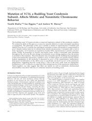
The Centromeric Protein Sgo1 Is Required to SenseLack <strong>of</strong> Tension on Mitotic ChromosomesVahan B. Indjeian, et al.Science 307, 130 (2005);DOI: 10.1126/science.1101366The following resources related to this article are available online atwww.sciencemag.org (this information is current as <strong>of</strong> July 16, 2009 ):Updated information <strong>and</strong> services, including high-resolution figures, can be found in the onlineversion <strong>of</strong> this article at:http://www.sciencemag.org/cgi/content/full/307/5706/130Supporting Online Material can be found at:http://www.sciencemag.org/cgi/content/full/307/5706/130/DC1A list <strong>of</strong> selected additional articles on the Science Web sites related to this article can befound at:http://www.sciencemag.org/cgi/content/full/307/5706/130#related-contentThis article cites 26 articles, 13 <strong>of</strong> which can be accessed for free:http://www.sciencemag.org/cgi/content/full/307/5706/130#otherarticlesThis article has been cited by 40 article(s) on the ISI Web <strong>of</strong> Science.This article has been cited by 14 articles hosted by HighWire Press; see:http://www.sciencemag.org/cgi/content/full/307/5706/130#otherarticlesThis article appears in the following subject collections:Cell <strong>Biology</strong>http://www.sciencemag.org/cgi/collection/cell_biolInformation about obtaining reprints <strong>of</strong> this article or about obtaining permission to reproducethis article in whole or in part can be found at:http://www.sciencemag.org/about/permissions.dtlDownloaded from www.sciencemag.org on July 16, 2009Science (print ISSN 0036-8075; online ISSN 1095-9203) is published weekly, except the last week in December, by theAmerican Association for the Advancement <strong>of</strong> Science, 1200 New York Avenue NW, Washington, DC 20005. Copyright2005 by the American Association for the Advancement <strong>of</strong> Science; all rights reserved. The title Science is aregistered trademark <strong>of</strong> AAAS.
R EPORTSThe Centromeric Protein Sgo1 IsRequired to Sense Lack <strong>of</strong> Tensionon Mitotic ChromosomesVahan B. Indjeian, Bodo M. Stern,* Andrew W. Murray.Chromosome alignment on the mitotic spindle is monitored by the spindlecheckpoint. We identify Sgo1, a protein involved in meiotic chromosomecohesion, as a spindle checkpoint component. Budding yeast cells withmutations in SGO1 respond normally to microtubule depolymerization butnot to lack <strong>of</strong> tension at the kinetochore, <strong>and</strong> they have difficulty attachingsister chromatids to opposite poles <strong>of</strong> the spindle. Sgo1 is thus required forsensing tension between sister chromatids during mitosis, <strong>and</strong> its degradationwhen they separate may prevent cell cycle arrest <strong>and</strong> chromosome loss inanaphase, a time when sister chromatids are no longer under tension.Errors in chromosome segregation lead todisease <strong>and</strong> death. To prevent such errors, theprotein complex cohesin (1) holds replicatedchromosomes together, <strong>and</strong> this linkage isnot broken until every pair <strong>of</strong> sister chromatidsis bi-oriented on the mitotic spindle,with the two sisters attached to microtubulesthat emanate from opposite poles <strong>of</strong> thespindle (2). Bi-orientation generates tensionon the chromosomes because the linksbetween the sister chromatids resist thepulling forces <strong>of</strong> the spindle. Chromosomeorientation is monitored by the spindlecheckpoint, which detects unattached kinetochores(3) or the lack <strong>of</strong> tension betweensister chromatids (4, 5). Either lesion inhibitsthe signal that induces chromosome segregation.Several components <strong>of</strong> the spindlecheckpoint (Mad2, Mad3, <strong>and</strong> Bub3) forma complex that prevents entry into anaphaseby binding to Cdc20, an essential activator <strong>of</strong>the anaphase-promoting complex (APC) (6).The APC triggers the destruction <strong>of</strong> securin(Pds1), the protein that inhibits separase(Esp1), which is the protease that triggerssister separation by cleaving cohesin (1)(Fig. 1A). When the spindle checkpointinhibits the APC, Pds1 is stable, separase isinhibited, <strong>and</strong> the sisters remain unseparated.Little is known about how cells sense theabsence <strong>of</strong> tension between a pair <strong>of</strong> sisterchromatids <strong>and</strong> send this information to thespindle checkpoint. Ipl1, the budding yeastmember <strong>of</strong> the Aurora family <strong>of</strong> mitoticprotein kinases (7), is involved in thisprocess (8) <strong>and</strong> is also needed to detachmicrotubules from kinetochores that are notunder tension (9–11). The only role <strong>of</strong> Ipl1<strong>Department</strong> <strong>of</strong> <strong>Molecular</strong> <strong>and</strong> <strong>Cellular</strong> <strong>Biology</strong>,Biological Laboratories, <strong>Harvard</strong> University, 16 DivinityAvenue, Cambridge, MA 02138, USA.*Present address: Cell Press, 1100 MassachusettsAvenue, Cambridge, MA 02138, USA..To whom correspondence should be addressed.E-mail: amurray@mcb.harvard.edumay be to induce chromosomes to detachfrom microtubules, thus creating nakedkinetochores, which inhibit the APC byrecruiting other checkpoint components.Alternatively, the lack <strong>of</strong> tension at thekinetochore may signal directly to thecheckpoint without requiring microtubuledetachment (8, 12, 13).To identify components <strong>of</strong> the tensionsensingmachinery, we looked for mutantsthat ignored chromosomes that were notunder tension. The screen used a buddingyeast (Saccharomyces cerevisiae) strainharboring linear minichromosomes (LMCs)that segregate poorly <strong>and</strong> activate the spindlecheckpoint (14). In wild-type budding yeast,lack <strong>of</strong> tension delays APC activation, butcells eventually enter anaphase (5, 8). Thisdelay becomes a lethal arrest in cells thatcontain CDC28-VF, a mutation in Cdc28(the budding yeast homolog <strong>of</strong> the cyclindependent kinase Cdk1) that reduces APCactivity (15). This arrest is dependent on thespindle checkpoint <strong>and</strong> can be overcome byexpressing CDC20-127, a dominant Cdc20allele that is refractory to Mad2 inhibition<strong>and</strong> therefore overrides the checkpoint (16)(Fig. 1A). We mutagenized the CDC28-VFstrain carrying the LMCs <strong>and</strong> selected mutantsthat grow in the absence <strong>of</strong> thecheckpoint-resistant Cdc20 <strong>and</strong> thereforebypass the minichromosome-induced mitoticarrest (Fig. 1A). Two <strong>of</strong> the mutants lie inSGO1, the fungal homolog <strong>of</strong> DrosophilaMEI-S332, a gene whose product protectscentromeric cohesion during the first meioticdivision (17–20). Each allele carries a mutationin the most conserved region <strong>of</strong> the protein(Thr 379 Y Ile in sgo1-100 <strong>and</strong> Pro 390 YHis in sgo1-700); the sgo1-700 allele also hasa second substitution (Asp 519 Y Asn) in aregion that is strongly conserved among fungi.Sgo1 is cell cycle regulated. Its expressionincreased at the G 1-S transition <strong>and</strong>decreased during mitosis in a pattern similarto that <strong>of</strong> Pds1 (Fig. 1B). Like Pds1, Sgo1was stabilized in the presence <strong>of</strong> benomyl<strong>and</strong> nocodazole, drugs that destabilizemicrotubules (Fig. 1C). Sgo1 could be asubstrate <strong>of</strong> either the APC or separase.Because Sgo1 was still degraded in cellsthat expressed a nondegradable version <strong>of</strong>Pds1 that inhibits separase but has no effecton the APC (21) (Fig. 1D), Sgo1 is likely adirect target <strong>of</strong> the APC. Four potential APCrecognition motifs (D-boxes) in Sgo1 furthersupport this possibility.Our selection was designed to identifycomponents that signal <strong>and</strong>/or sense tensionat the kinetochores. To examine the responseto chromosomes that lack tension, we placedsgo1 mutants in cells that also express Mcd1(also known as Scc1), a component <strong>of</strong>cohesin (1), under the glucose-repressibleGAL1 promoter. In the presence <strong>of</strong> glucose,Mcd1 is not expressed <strong>and</strong> the sisterchromatids are not linked. The positioning<strong>of</strong> the sisterless chromosomes on the spindleindicates that they attach to the spindle (5),although we cannot prove that every kinetochoreis attached to a microtubule at alltimes. In the absence <strong>of</strong> Mcd1, reducedtension at the kinetochores activates thespindle checkpoint <strong>and</strong> delays APC activation(5, 8), as indicated by stabilization <strong>of</strong>the APC target Pds1 (Fig. 2A). The sgo1mutants, however, failed to delay APCactivation, hence they are unable to senseor respond to lack <strong>of</strong> tension.We also examined cells lacking Sgo1(sgo1D). In the W303 strain background,sgo1D cells showed poor viability <strong>and</strong>acquired suppressor mutations (22). Reducedviability was largely overcome by slowingdown DNA replication, an approach previouslyused to reduce the sensitivity <strong>of</strong>spindle checkpoint mutants to benomyl(23). The exact timing <strong>of</strong> Pds1 degradationwas somewhat variable in sgo1D cells, butwithin an experiment the timing <strong>of</strong> destructionwas similar regardless <strong>of</strong> whether Mcd1was expressed (Fig. 2A); this result impliesthat the sgo1D cells cannot sense the lack <strong>of</strong>tension. The sgo1 point mutants also failed todelay APC activation in cells lacking Cdc6expression (22). Cdc6 is required for DNAreplication, <strong>and</strong> in its absence chromosomesenter mitosis without the sisters needed togenerate tension at the kinetochore (5, 8).The sgo1 mutants shared properties withother spindle checkpoint mutants such asmad2D. The sgo1 mutants showed chromosomeloss rates (24) approximately equal tothose <strong>of</strong> mad2D cells, five times the rate forwild-type cells, <strong>and</strong> one-third the rates forbub1D <strong>and</strong> bub3D strains (table S1) (25).Like mad2D, the growth <strong>of</strong> the sgo1 mutantswas sensitive to benomyl (Fig. 2B). Toexamine their response to microtubule depolymerization,we arrested cells in G 1byDownloaded from www.sciencemag.org on July 16, 20091307 JANUARY 2005 VOL 307 SCIENCE www.sciencemag.org
exposure to mating pheromone (a-factor)<strong>and</strong> then released them into mediumcontaining benomyl <strong>and</strong> nocodazole. Theviability <strong>of</strong> sgo1 mutants dropped markedlyafter microtubule depolymerization (Fig. 2C)even though the sgo1 mutants, like wild-typecells, arrested with high levels <strong>of</strong> Pds1 for atleast 4 hours (Fig. 2D). In contrast, mad2Dcells failed to detect the damaged spindle<strong>and</strong> degraded Pds1 about 2 hours after theirrelease from G 1arrest (Fig. 2D). Therefore,the sgo1 mutants, unlike mad2D, are still ableto arrest in mitosis in response to unattachedkinetochores. Although we cannot exclude thepossibility that weak defects in the spindlecheckpoint allow cells to respond to unattachedkinetochores, but not to those that are notunder tension, the observation that sgo1mutants that have different effects on viabilityare all inactivate to tension sensing withoutaltering the ability to detect unattachedkinetochores argues against this possibility.The behavior <strong>of</strong> sgo1 mutants seemsparadoxical; they die after microtubule depolymerizationeven though they arrest.Because Sgo1 protects centromeric cohesionduring meiosis (17–19), one explanation isthat the sgo1 mutants have mitotic cohesiondefects. We introduced the sgo1 alleles intostrains that have chromosome IV labeledwith 256 t<strong>and</strong>em repeats <strong>of</strong> the Lac operatorinserted at the TRP1 locus (12 kb from thecentromere). This array is seen as a fluorescentdot when cells express a green fluorescentprotein–Lac repressor fusion protein(GFP-LacI) (26). As controls, we used wildtype,mad2D, <strong>and</strong> GAL1-MCD1 strains in thesame background. The mad2D cells lack allknown aspects <strong>of</strong> the spindle checkpoint, <strong>and</strong>GAL1-MCD1 cells lack cohesin when grownin glucose. The strains were released from G 1into medium that contained glucose, benomyl,<strong>and</strong> nocodazole. In wild-type cells, theabsence <strong>of</strong> kinetochore-microtubule attachmentactivates the spindle checkpoint, stabilizingcohesion. As a result, sister chromatidsstay together <strong>and</strong> the GFP-LacI dots correspondingto the two sisters <strong>of</strong> chromosomeIV cannot be resolved, appearing as a singledot under the fluorescent microscope. Roughlyhalf the cells lacking Mad2 or cohesin hadtwo green dots, showing that they had prematurelyseparated their sisters (Fig. 3A). Thesgo1 mutants behaved much more like wildtypecells <strong>and</strong> predominantly arrested as largebuddedcells with a single visible dot.These results show that sgo1 mutants donot have a major mitotic cohesion defect.Their rapid death in the absence <strong>of</strong> microtubulescould be explained in two ways. Oneis that the sgo1 mutants have a minor cohesiondefect that is small enough to differentiatethem from cells lacking cohesin, yetensures that at least one chromosome missegregatesin most benomyl- <strong>and</strong> nocodazoletreatedcells. The other is that cohesion isnormal, but sgo1 mutants make errors inchromosome alignment as the spindle reformsafter benomyl <strong>and</strong> nocodazole have been removed.We distinguished these hypotheses bydelaying sister chromatid segregation untilwell after benomyl <strong>and</strong> nocodazole had beenremoved, a treatment that has no effect onchromosomes that have already separated butallows more time for linked pairs <strong>of</strong> sisters toalign correctly on the spindle.We compared sgo1-100, wild-type, <strong>and</strong>mad2D cells, all containing the GFP-markedchromosome IV. Two versions <strong>of</strong> each strainADLMC-+mad2∆ +sgo1-100 +sgo1-700 +Securin (Pds1)Dominant Cdc204-fold serial dilutionsSeparase (Esp1)ChromosomeseparationDominant Cdc20G1 (αF) GalactosePDS1-db∆ 0 60 75 90 105 120 135 150 165 180 minSgo1Pds1were made, differing only in the presence <strong>of</strong>an extra copy <strong>of</strong> the MPS1 gene driven bythe GAL1 promoter. Mps1 is part <strong>of</strong> the spindlecheckpoint, <strong>and</strong> its overexpression causesa spindle checkpoint <strong>and</strong> APC-dependentmetaphase arrest (27). The APC <strong>and</strong> Mps1mutually oppose each other because the APCinduces the destruction <strong>of</strong> Mps1 (28). Hence,the metaphase arrest induced by overexpressingMps1 can be rapidly reversed by addingglucose. All the strains were released fromG 1into medium that contained galactose,benomyl, <strong>and</strong> nocodazole. Regardless <strong>of</strong>whether they overexpressed Mps1, wild-typeSgo1Pds1BG1 (αF) Rich medium (YPD)0 30 40 50 60 70 80 90 minCR EPORTSSgo1Pds1G1 (αF) Nocodazole0 60 90 120 150 180 210 240 minSgo1Pds1Linear minichromosome(LMC) Fig. 1. Isolation <strong>and</strong> characterizationMad2 complexCdc20 Dom APCCdc20<strong>of</strong> the spindle checkpoint componentSgo1. (A) LMCs separate inpart because <strong>of</strong> their inability towithst<strong>and</strong> spindle forces. As a result,their linkage to microtubules is nolonger under tension, <strong>and</strong> this activatesthe spindle checkpoint (stars)(30). In a CDC28-VF strain withLMCs, the APC is kept inactive <strong>and</strong>the cells arrest in metaphase. Abolishingthe spindle checkpoint by, forexample, deleting mad2 (mad2D)alleviates this arrest, as does theexpression <strong>of</strong> dominant Cdc20 thatcannot be inhibited by the spindlecheckpoint complex. After ethylmethanesulfonate mutagenesis <strong>of</strong>CDC28-VF cells with a tetracyclinerepressibledominant Cdc20, spindlecheckpoint mutants were recoveredon the basis <strong>of</strong> their ability totolerate LMCs in the absence <strong>of</strong>dominant Cdc20. Two alleles <strong>of</strong>sgo1 were isolated, sgo1-100 <strong>and</strong>sgo1-700, <strong>and</strong> their growth (fourfoldserial dilutions) in the presenceor absence <strong>of</strong> dominant Cdc20 isshown. All strains shown are in theCDC28-VF background. (B to D) Astrain with epitope-tagged Pds1(Pds1-18Myc) <strong>and</strong> Sgo1 (Sgo1-13Myc) was released from a G 1(afactor,aF) block; at the indicatedtimes, samples were taken forWestern blot analysis against theMyc epitopes. (B) Sgo1 <strong>and</strong> Pds1expression during the normal cellcycle in rich medium with glucose (YPD) at 30-C, <strong>and</strong> (C) after release into benomyl (30 mg/ml)<strong>and</strong> nocodazole (30 mg/ml) at 23-C; (D) Sgo1 <strong>and</strong> Pds1 expression after release from G 1to galactose at30-C in the presence or absence <strong>of</strong> nondegradable, destruction box–deleted Pds1 (Pds1-dbD) undercontrol <strong>of</strong> the GAL1 promoter. In both cases the APC is activated, as evidenced by the destruction <strong>of</strong>the endogenous Pds1-18Myc. However, in the presence <strong>of</strong> Pds1-dbD the strains remained arrested aslarge budded cells, whereas in its absence the cells rebudded after about 120 min (22).Downloaded from www.sciencemag.org on July 16, 2009www.sciencemag.org SCIENCE VOL 307 7 JANUARY 2005 131
R EPORTSAGAL1-MCD1GAL1-MCD1 mad2∆GAL1-MCD1 sgo1-100GAL1-MCD1 sgo1∆GAL1-MCD1GAL1-MCD1 sgo1∆G1 (αF) Glucose (No Mcd1 expression)0 40 50 60 70 80 90 100 110 120 minG1 (αF)Galactose (Mcd1 expressed)Fig. 2. Sgo1 is involved in tension sensing <strong>and</strong>chromosome segregation. (A) The indicated GAL1-MCD1 strains, all with epitope-tagged Pds1 (Pds1-18Myc), were released from a-factor in rich mediumwith glucose (Mcd1 not expressed) or galactose(Mcd1 expressed) at 30-C. At the indicated times,samples were taken for Western blot analysis againstthe Myc epitopes. (B) The indicated strains underwentfourfold serial dilution <strong>and</strong> were spotted on plateswith or without benomyl (10 mg/ml). (C) The viability(defined as the ability to give rise to colonies on richmedium) <strong>of</strong> the indicated strains was measured at theindicated times after they were released from G 1(ana-factor block) into medium containing benomyl(10 mg/ml) <strong>and</strong> nocodazole (15 mg/ml) at 23-C. Dataare from three experiments; error bars represent SDs.Pds1Dwild typemad2∆sgo1-100sgo1-700sgo1∆B4-fold serial dilutionsCPercent Viable Cells100wild typemad2∆sgo1-100sgo1-700YPDwild typemad2∆sgo1-100sgo1-700+10 µg/ml Ben8060wild typemad2∆40sgo1-1002000 2 4 6Time in Nocodazole, hrG1 (αF) Nocodazole0 60 90 120 150 180 210 240 minPds1(D) The indicated strains all contained epitope-tagged Pds1 (Pds1-18Myc) <strong>and</strong> were released froma-factor in media containing benomyl (30 mg/ml) <strong>and</strong> nocodazole (30 mg/ml) at 23-C. Sampleswere taken at the indicated times for Western blot analysis against the Myc epitopes.APercent Two Dots403020100wildtypeG1 (αF)mad2∆sgo1-100Nocodazole0 hr 3 hrsgo1-700mcd1 sgo1∆Fig. 3. The sgo1 alleles do not have a strong cohesiondefect in mitosis but fail to bi-orient their chromatidsafter spindle reformation. (A) The indicated strains, allwith GFP dots labeling the TRP1 locus on chromosome IV,were released from a-factor block in glucose, benomyl(30 mg/ml), <strong>and</strong> nocodazole (30 mg/ml) at 23-Cfor3hours.Percentages <strong>of</strong> cells with two dots at the time <strong>of</strong> releasefrom a-factor arrest (white bars) <strong>and</strong> at the end <strong>of</strong> 3 hours(black bars) are shown. At least 100 cells were counted ateach time point <strong>of</strong> each experiment; results from threeindependent experiments were combined. Error bars showSDs. (B) Wild-type, mad2D, <strong>and</strong> sgo1-100 strains with(black bars) or without (white bars) GAL1-MPS1 were releasedfrom an a-factor block into galactose (Gal),benomyl (30 mg/ml), <strong>and</strong> nocodazole (Noc) (30 mg/ml)for 5 hours at 23-C. The samples were then treated withgalactose alone for 1.5 hours (experimental) or withBG1 arrestin αFPercent Viable CellsPercent Two Dots100806040205040302010Ia Noc0 wild type mad2∆ sgo1-100IIa NocGal + Noc, 5 hrno spindleMetaphase DelayGAL1-MPS1Metaphase Delay0 wild type mad2∆ sgo1-100ExperimentalGal - Noc, 1.5 hrspindlereformsControlGlc + Noc, 0.5 hrno spindlePercent Viable CellsPercent Two Dots100<strong>and</strong> sgo1-100 cells arrested in metaphase,whereas mad2D cells did not <strong>and</strong> separatedtheir sisters prematurely (22). The cells werethen transferred to medium that contained galactosebut lacked benomyl <strong>and</strong> nocodazolefor 1.5 hours to allow the spindle to reassemblewell before chromosome segregation(Fig. 3B). In this experiment, overexpression<strong>of</strong> Mps1 increased the viability <strong>of</strong> sgo1-100cells but had no effect on mad2D cells whosesisters had already separated. To confirm thatrescue depended on delaying anaphase untilthe spindle had reformed, we transferred sgo1-100 cells that had been treated with galactose,benomyl, <strong>and</strong> nocodazole into medium containingglucose, benomyl, <strong>and</strong> nocodazole for0.5 hours, allowing Mps1 levels to fall beforewashing out the microtubule-destabilizingdrugs. These cells died, indicating that Mps1overexpression was only effective when itcontinued after the benomyl <strong>and</strong> nocodazolehad been removed (Fig. 3B). We concludethat although sgo1 mutants arrest normallywhen they lack a spindle, they die becausethey make but cannot detect errors in chromosomealignment as the spindle reforms.We monitored chromosome segregationas cells recovered from microtubule depolymerization.Cells that had overexpressedMps1 were released from their final mitoticblock into a-factor, which prevents theirescape from G 1, <strong>and</strong> numbers <strong>of</strong> GFP dots(a proxy for the number <strong>of</strong> copies <strong>of</strong>chromosome IV) per cell were counted. Bi-80604020050403020100viability IaGlc + αF, 2 hrviability IbGlc + αF, 2 hrIb Nocwild type mad2∆ sgo1-100IIb NocMitotic ExitMitotic ExitGFP dots IIaGFP dots IIbwild type mad2∆ sgo1-100glucose (Glc), benomyl, <strong>and</strong> nocodazole for 0.5 hours (control). Both sets were then released into glucose <strong>and</strong> a-factor at 23-C for 2 hours. Percentages <strong>of</strong>viable cells <strong>and</strong> cells with two green fluorescent dots were determined. Data from four independent experiments are shown; error bars show SDs.Downloaded from www.sciencemag.org on July 16, 20091327 JANUARY 2005 VOL 307 SCIENCE www.sciencemag.org
Fig. 4. A model <strong>of</strong> Sgo1 functionin sensing <strong>and</strong>/or signaling tension<strong>and</strong> promoting bi-orientation <strong>of</strong>mitotic chromosomes (blue). Spindlemicrotubules (light gray) regulateSgo1 activity, which switchesbetween an active (red) <strong>and</strong> inactive(green) state. In the absence<strong>of</strong> tension, Sgo1 is incontact with spindle microtubules<strong>and</strong> sends signals to promote biorientation<strong>and</strong> delay progressioninto anaphase. The dashed arroworientation <strong>of</strong> a chromosome leads to eachdaughter cell inheriting one sister chromatid<strong>and</strong> thus having a single GFP dot. Failure tobi-orient the sister chromatids leaves onedaughter with two GFP dots <strong>and</strong> its sisterwith none. As expected from the rescue <strong>of</strong>viability, allowing Mps1 overexpression asthe spindle reformed substantially reducedthe fraction <strong>of</strong> sgo1-100 cells that had twodots in the following G 1(Fig. 3B). These resultsargue that sgo1-100 cells cannot properlyalign their sister chromatids on the spindlebut do successfully hold them together.We detected two new defects in sgo1mutants: an inability to arrest the cell cyclein cells whose chromosomes were not undertension, <strong>and</strong> a defect in chromosome segregationthat could be rescued by delaying theonset <strong>of</strong> anaphase. Vertebrate homologs <strong>of</strong>Sgo1 contain a strong microtubule-binding domain(29), <strong>and</strong> Sgo1 is found at the kinetochore(18). Both observations argue that Sgo1is directly involved in interactions betweenchromosomes <strong>and</strong> microtubules, <strong>and</strong> raisethe possibility that it is the tension sensor.Sgo1Sgo1KinetochoreDetachmentSpindle CheckpointActivationrepresents uncertainty aboutwhether the lack <strong>of</strong> tension can inhibit APC activity without generating unattached kinetochores.A separate mechanism (dark gray) may monitor attachment <strong>of</strong> kinetochores to spindlemicrotubules.For example, Sgo1 could be located in aregion <strong>of</strong> the kinetochore that could only bereached by microtubules that were not undertension (Fig. 4). As long as it is boundto a microtubule, Sgo1 would send a signalthat destabilizes the attachment <strong>of</strong> the kinetochoreto its microtubule <strong>and</strong> might alsosend a signal directly to the spindle checkpoint.Sister centromeres separate from eachother as anaphase begins. Once chromosomeshave left their sisters, arresting the cell cyclewould be pointless <strong>and</strong> the destabilization <strong>of</strong>microtubule-kinetochore attachment couldhave disastrous consequences for chromosomesegregation. The degradation <strong>of</strong> Sgo1 asanaphase begins <strong>and</strong> the resulting ablation<strong>of</strong> the tension-signaling pathway may explainwhy sister chromatids remain stably attachedto the spindle once they reach its poles.References <strong>and</strong> Notes1. K. Nasmyth, Science 297, 559 (2002).2. A. Musacchio, K. G. Hardwick, Nature Rev. Mol. CellBiol. 3, 731 (2002).3. C. L. Rieder, R. W. Cole, A. Khodjakov, G. Sluder, J. CellBiol. 130, 941 (1995).4. X. Li, R. B. Nicklas, Nature 373, 630 (1995).5. B. M. Stern, A. W. Murray, Curr. Biol. 11, 1462 (2001).6. H. Yu, Curr. Opin. Cell Biol. 14, 706 (2002).7. M. Carmena, W. C. Earnshaw, Nature Rev. Mol. CellBiol. 4, 842 (2003).8. S. Biggins, A. W. Murray, Genes Dev. 15, 3118 (2001).9. I. M. Cheeseman et al., Cell 111, 163 (2002).10. S. Biggins et al., Genes Dev. 13, 532 (1999).11. T. U. Tanaka et al., Cell 108, 317 (2002).12. S. Hauf et al., J. Cell Biol. 161, 281 (2003).13. C. Ditchfield et al., J. Cell Biol. 161, 267 (2003).14. W. A. Wells, A. W. Murray, J. Cell Biol. 133, 75 (1996).15. A. D. Rudner, K. G. Hardwick, A. W. Murray, J. CellBiol. 149, 1361 (2000).16. L. H. Hwang et al., Science 279, 1041 (1998).17. V.L.Katis,M.Galova,K.P.Rabitsch,J.Gregan,K.Nasmyth, Curr. Biol. 14, 560 (2004).18. T. S. Kitajima, S. A. Kawashima, Y. Watanabe, Nature427, 510 (2004).19. A. L. Marston, W. H. Tham, H. Shah, A. Amon, Science303, 1367 (2004).20.A.W.Kerrebrock,W.Y.Miyazaki,D.Birnby,T.L.Orr-Weaver, Genetics 130, 827 (1992).21. R. L. Tinker-Kulberg, D. O. Morgan, Genes Dev. 13,1936 (1999).22. V. B. Indjeian, A. W. Murray, unpublished data.23. R. Li, A. W. Murray, Cell 66, 519 (1991).24. P. Hieter, C. Mann, M. Snyder, R. W. Davis, Cell 40,381 (1985).25. See supporting data on Science Online.26. A. F. Straight, A. S. Belmont, C. C. Robinett, A. W.Murray, Curr. Biol. 6, 1599 (1996).27. K. G. Hardwick, E. Weiss, F. C. Luca, M. Winey, A. W.Murray, Science 273, 953 (1996).28. W. Palframan, B. M. Stern, A. W. Murray, unpublisheddata.29. A.Salic,J.C.Waters,T.J.Mitchison,Cell 118, 567 (2004).30. B. M. Stern, K. Liem, A. W. Murray, unpublished data.31. We thank S. Davis for invaluable technical assistancewith the selection; all members <strong>of</strong> the Murray labfor helpful comments <strong>and</strong> suggestions; A. Amon<strong>and</strong> T. Mitchison for sharing data before publication;S. Biggins <strong>and</strong> A. Amon for helpful comments; <strong>and</strong>R. Hellmiss-Peralta for advice on graphics. Supportedby NIH grant GM043987 (A.W.M.) <strong>and</strong> by aLeukemia <strong>and</strong> Lymphoma Society fellowship (B.M.S.).Supporting Online Materialwww.sciencemag.org/cgi/content/full/307/5706/130/DC1Materials <strong>and</strong> MethodsTables S1 <strong>and</strong> S214 June 2004; accepted 6 November 200410.1126/science.1101366R EPORTSDownloaded from www.sciencemag.org on July 16, 2009www.sciencemag.org SCIENCE VOL 307 7 JANUARY 2005 133