PATIENT - Abaxis
PATIENT - Abaxis
PATIENT - Abaxis
You also want an ePaper? Increase the reach of your titles
YUMPU automatically turns print PDFs into web optimized ePapers that Google loves.
02 March/April 2010
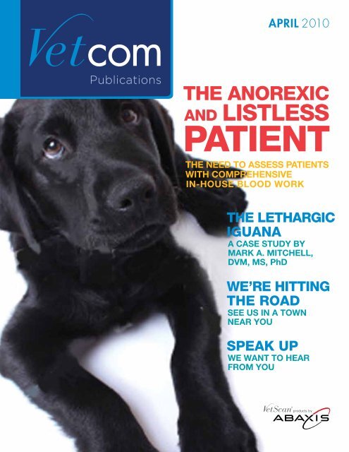
Dateline 4.1.10Welcome to VetCom Publications, a bi-monthly newsletter availableboth in print and online. VetCom Publications offers readers case studies,practice tips from a clinical and cost savings perspective as well aseducational opportunities. This issue also includes a forum for Veterinarianto Manufacturer communication and so much more.As an introductory note to this issue I want to highlight that <strong>Abaxis</strong>,Inc. has experienced incredible growth despite the economic downturnimpacting many companies in a wide variety of industries. <strong>Abaxis</strong>, Inc.,a point of care diagnostic company, has experienced an increase inveterinary instruments and consumable revenue for the nine monthsending December 31, 2009 in the North America Animal Health segmentof 23% year over year. While many companies are downsizing, <strong>Abaxis</strong>has increased our head count by nearly 4% and our facilities at ourworldwide corporate headquarters have grown by 27%.We don’t take our growth for granted; on the contrary, <strong>Abaxis</strong> is focusedon research and development, exemplary products, sales, marketing,services and support in every sense of the word. Because of that focus ourcustomers have come to know <strong>Abaxis</strong> for our superior quality products andservices, ultimately reflected in the company's strong balance sheet.Please note our growth is a direct result of VetScan ® products like thenewly released VetScan i-STAT ® 1, a handheld analyzer, and the muchanticipated Canine Wellness Profile with a built in heartworm antigentest which changes the paradigm of wellness testing.The addition of these products to the portfolio of products that includethe VetScan VS2 chemistry analyzer, VetScan HM2 hematology analyzer,VetScan HM5 hematology analyzer, VetScan VSpro coagulation andspecialty analyzer and the Canine Heartworm Rapid Test allows yourpractice to meet all of your in-house testing needs.Enjoy this issue and I look forward to your feedback, comments andcustomer advice in this exciting field of Animal Health.Sincerely,Valerie Goodwin-AdamsDirector of Marketing<strong>Abaxis</strong>, Inc.IN THIS ISSUEPublicationsAPRIL 2010The Case forCoagA review of coagulationand the need forin-office assessmentTHE LETHARGICIGUANAA CASE STUDY BYMARK A. MITCHELL,DVM, MS, PhDWe’re Hittingthe RoadSee us in a townnear youSpeak UpWe want to hearfrom youThe Need to Assess Patients with ComprehensiveIn-House Blood Work 4Where to Find Us 11Full Recovery for This Foal 12Renal Failure in a Green Iguana 13Let Us Hear From You 16VETSCANDISTRIBUTORSUnited StatesAmerican Veterinary Supply800-869-2510DVM Resources877-828-1026Great Western Animal Supply800-888-7247Equipment Outreach, Inc.888-996-9968Hawaii Mega-Cor, Inc.800-369-7711IVESCO800-457-0118Lextron, Inc.800-333-0853Merritt Veterinary Supply800-845-0411Nelson Laboratories800-843-3322Northeast Veterinary Supply Co.866-638-7265Penn Vet Supply800-233-0210PCI Animal Health800-777-7241TW Medical888-787-4483Western Medical Supply800-242-4415Vet Pharm, Inc.800-735-8387VWR International, LLC800-932-5000CanadaAssociated Vet Purchasing Co.604-856-2146Aventix877-909-2242CDMV450-771-2368MidWest Drug204-233-8155Vet Novations866-382-6937Vie et Sante418-650-7888Western Drug Distribution877-329-93322010 March/April 03
The Needto AssessPatients withComprehensiveIn-HouseBlood WorkContributing Authors:Andrew J. Rosenfeld, DVM, ABVPSharon Dial, DVM, ACVPCase ExampleGeneral and emergency practitioners have all faced the enigma of theanorexic, listless patient. For example, Chance Murphy, a 6-year-old66-pound female spayed black lab mix, walks in the practice door.The owners are faithful clients and areconcerned because Chance is suddenlydepressed, weak, lethargic, and has not eatenfor the last two days. There has been nooverall change in her history, no known toxinsor poisons that she could have gotten into,and the other pets in the household are fine.The veterinarian evaluates Chance and findsthat her body temperature is 102.3 degreesFahrenheit. She has a strong 120 pulse rate,40 respiration rate, light pink mucus membranes,a two second capillary refill time, and normalhydration. She appears weak and possiblyslightly painful in her caudal abdomen; nopalpable abnormalities can be detected. Allother elements of her physical examinationare within normal limits.The veterinarian discusses the concernswith the owners and outlines the diagnosticrecommendations, which include a complete04 April 2010
THE PRACTICAL NEED TO ASSESS COAGULATION IN THE <strong>PATIENT</strong>blood count, chemistry profile, urinalysis andchest and abdominal radiographs. The ownersagree to the initial diagnostic plan, and themedical team takes Chance to the back forradiographs and lab work.Thoracic films show a possible microcardiawith no significant changes in the lungs,diaphragm and trachea. Abdominal filmssuggest a slight loss of contrast in the ventralabdomen. Blood is collected and mild bruisingis noted on Chance’s hind limb venipuncturesite. In-house lab work is completed, andexcept for the following changes in the bloodwork; all other parameters are unremarkable.The differential list for this patient includesDisseminated Intravascular Coagulopathy(DIC) / Atraumatic Bleed of the Abdomen,Intestinal Disease / Chronic GI Ulceration, LiverDisease, Infectious Disease (i.e. Ehrlichiosis),Toxin / Poison, and Neoplasia.The primary concern for this patient is whetherChance has a non-regenerative anemia suchas anemia of chronic disease, has a chronicbleeding issue (i.e. gastrointestinal ulceration),or has an acute blood loss anemia. At this point,an in-house coagulation profile is necessary tohelp determine if there is a coagulation concernsuggesting a life-threatening problem. The clottingprofile shows:Test Finding NormalHCT 26% (l) 35–55%WBC 19,250 (h) 6,000–17,000Platelet 125,000 (l) 200,000–500,000Albumin 2.5 mg/dl 2.5–3.5 mg/dlThe medical team evaluates a bloodfilm and a wet slide prep of Chance’sblood. The technician team memberfinds no obvious agglutination onthe wet prep, and a blood film thatshows normal red and white bloodcell morphology. The medical teamagrees that the platelet numberappears slightly decreased and seesno overall sign of a regenerative redblood cell response.Test Canine NormalPT 25 s 12–17 saPTT 180 s 90–110 sWith the observed prolonged clotting timesand evidence of anemia and thrombocytopenia,acute internal bleeding must be a primaryconcern. Ultrasound evaluation showed amoderate amount of free fluid in the abdomenwhich when tapped produced non-clottingblood with a packed cell volume of 32%. Anemergency exploratory laparotomy revealeda small, irregular spleen with multiple nodules.The spleen was successfully removed and thebleeding resolved.2010 April 05
A Review of CoagulationIn this case, as with many other cases, the need to evaluate the clotting abilityof the patient is essential. There are several principle events that are involvedin coagulation:• Constriction of the injured vessel• Formation of the “primary platelet plug”• Stabilization of the primary platelet plugby deposits of fibrin through activationof clotting factorsThe three primary elements of hemostasis arethe endothelial cells that line the wall of theblood vessels, platelets and clotting factors(See Figure 1). Each of these elements mustfunction appropriately for normal hemostasis.Defects in any of these three participants canresult in abnormal bleeding or inappropriateclot formation.Figure 1Damage to the endothelial cells exposesreceptors (von Willebrand factor),and platelets adhere to the wall of thevessel covering the defect. The plateletsbecome activated, undergo a shapechange and begin to aggregate to forma plug at the injury. The platelets secretesubstances and provide tissue factor foractivation of the clotting factors. Oncethe coagulation pathways are activated,prothrombin is converted to thrombin;thrombin converts fibrinogen to fibrinand the fibrin “glues” the plateletstogether into a stable clot sealing offthe injury 1 .Endothelial cells line all blood vessels.When injured, the endothelial cell retracts toexpose proteins present in the tissue deepto the vessel wall or interstitium that interactwith platelets to promote their adhesion.Further, these sites release molecules thatpromote activation of the clotting factors. Theblood vessel wall plays a major role in bothcoagulation and fibrinolysis.There is no laboratory testing that canevaluate the function of endothelial cells.There are diseases that can interfere withthe function of the endothelial cells. In mostcases, abnormal endothelial cells result inabnormal clot formation because they do notmaintain their anticoagulant nature. Sepsis andmetabolic diseases such as diabetes can resultin pro-coagulant changes in endothelial cellsthroughout the body.Platelets are essential for primary hemostasis;the formation of the initial primary plateletplug. The injured endothelial cells promotethe adhesion, aggregation, swelling andsecretion by platelets (activation). Activatedplatelets also promote the activation of otherplatelets. When platelets are activated, theyrelease factors that promote activation ofthe coagulation cascade. In addition to the1. Image courtesy of Clinical Pathology for the Veterinary Team, Rosenfeld, A & Dial, S.Wiley, Ames Ia, 2010.06 March/April 2010 2010
THE PRACTICAL NEED TO ASSESS COAGULATION IN THE <strong>PATIENT</strong>secreted factors, platelets provide a surface forthe clotting process and the cellular mass thatwill form the clot. Defects in primary hemostasiscan occur due to decreased numbers ofcirculating platelets (thrombocytopenia) orplatelet dysfunction.Thrombocytopenia is the most common defectin primary hemostasis. The platelet count isthe most common test to evaluate plateletnumber. In general an animal will not begin tospontaneously bleed until platelet number isless than 40,000 / µL.Platelet function tests should be performedin the bleeding patient with normal plateletnumbers and coagulation times. There aresensitive tests for platelet function thatrequire submission of a sample to a specialtylaboratory. However, there are in-housetests that can be done to identify patientswith possible defects in platelet function; themost reliable is the buccal mucosal bleedingtime. The buccal mucosal bleeding timeis commonly done to screen dogs for vonWillebrand’s disease. A small shallow cut ismade in the mucosal surface of the upper lipand the time from the initial cut to cessationof bleeding is recorded. It is very important toperform this test in a standard and consistentmanner. A standard procedure for this test isoutlined in Figure 2. Normal buccal bleedingtimes are 1.7 to 4.2 minutes with a mean of2.6 minutes. If there is a prolonged buccalmucosal bleeding time, further laboratorytests are required to determine the cause ofthe platelet dysfunction.Step I Step 2 Step 3Figure 2Step I: With the patient anesthetized, fold the upper lip up and secure loosely with a gauze tie. The tie must be loose to allow normal blood flow.Step 2: Make a reproducible 1/8 – 1/4 inch stab wound using a #11 surgical blade. The depth of the wound can be determined by marking the blade.Step 3: Gently remove the blood drops with filter paper without touching the wound. Measure the time it takes to stop bleeding 2 .Secondary hemostasis is the process ofstabilizing the platelet plug by formation offibrin, the mortar that holds the plateletstogether. The processes of primary andsecondary hemostasis occur simultaneously.The formation of stable fibrin is the result ofactivation of numerous circulating clottingfactors and enzymes or enzyme complexes thatform the coagulation cascade. The coagulationcascade depends on many cofactors suchas calcium and multiple feedback loops thataccelerate and inhibit the process. If thereis a deficiency in the coagulation cascade,the primary plug will degrade prematurelyand bleeding will resume. The common testsfor defects in secondary hemostasis are theProthrombin Time (PT) and the activatedPartial Thromboplastin Time (aPTT).2. Image courtesy of Clinical Pathology for the Veterinary Team, Rosenfeld, A & Dial, S.Wiley, Ames Ia, 2010.2010 April 07
A prolonged PT and normal aPTT indicatea defect in the extrinsic pathway, while aprolonged aPTT with a normal PT indicates adefect in the intrinsic pathway. If both tests areprolonged, there may be a combined pathwaydisorder, such as Vitamin K antagonism dueto warfarin toxicity or liver disease, or DIC, orthere may be a common pathway disordersuch as an inherited coagulation factor defectsuch as Factor X or Factor II (Prothrombin).Indications for Clotting TimesThere are many congenital and acquired disease entities that can produce acoagulopathy, either from dysfunctional endothelial cells, decreased plateletnumber, platelet dysfunction or decreased clotting factors (See Table 1).Recommendations to establish baseline clotting times include:• Pre-surgical blood work evaluation:Evaluating clotting times as part of a firsttime pre-surgical (i.e. kitten/puppy) bloodscreen can help identify patients withhereditary disorders such as HemophiliaA or B. Identification of these patientsprior to surgery is a necessity to preventprolonged surgical bleeding. Althoughthese diseases can affect any pure ormixed breed canine or feline, veterinariansshould screen the following breeds closelyfor hereditary coagulation disorders:Hemophilia A: Beagles, AlaskanMalamutes, Boxers, MiniatureSchnauzers, BulldogsHemophilia B: Cairn Terriers,Airedales, Coonhounds, St.Bernards, American CockerSpaniels, French Bulldogs, AlaskanMalamutes, Scottish Terriers,Shetland Sheepdogs, LabradorRetrievers, Bichon Frise,Old English Sheepdogs, BritishShort Haired Cat• Hospitalized and Ill Patients: Syndromesthat increase clotting times can includediseases that affect liver function,ingestion of toxins which affect Vitamin Kmetabolism, and diseases that promoteDisseminated Intravascular Coagulopathy.These entities can present subtly in an illpatient and develop into a severe acute lifethreatening syndrome. Baseline clotting timesare recommended for the following diseaseconditions:Sepsis/Infectious (Parvo Viral Infection,Pneumonia, Pyometra…)Liver DiseaseHeat StrokeAnemia/HemorrhageToxins or PoisoningsSevere Metabolic Disease (DiabeticKetoacidosis (DKA), Amyloidosis…)NeoplasiaPatients requiring surgical procedure orsurgical biopsy should always have athorough coagulation profile completed priorto the procedure.08 April 2010
THE PRACTICAL NEED TO ASSESS COAGULATION IN THE <strong>PATIENT</strong>ConclusionThe ability to evaluate and treat the at-risk patients withcoagulopathy is becoming a practical and necessarysolution to save patients’ lives.VetScan ® VSproFrom an emergency and general practiceperspective, the use of in-house clottinganalyzers or coagulation tests is an importantaspect of the standard of care. With thetightening economy, clients are expectingmore from the general practitioner and areless responsive to referring cases out to largersecondary and tertiary care centers. The abilityto evaluate in-hospital clotting factors andcomplete blood counts in ill patients allows theveterinarian to treat the patient effectively beforesevere bleeding occurs, and can save lives andhelp maintain the patient’s quality of life.Table 1Pathology that disruptthe clotting processEndothelial DysfunctionDecreased platelet numberDecreased platelet functionDecreased Clotting FactorsCause(Not intended to be a complete list)• Hereditary Disease• Sepsis (i.e. Parvo, Pyometra, Pneumonia)• Endocrine Disease: Diabetes Miletus• Immune Mediated Disease• Infectious Disease (i.e. Ehrlichia)• Consumption• Production Disorder (i.e. bone marrow)• Congenital Disease(i.e. von Willebrand’s disease)• Toxins / Drugs (i.e. Aspirin)• Congenital Disease (i.e. Hemophilia A[deficiency of Factor VIII] & Hemophilia B[deficiency of Factor IX and deficiencyof Factor X])• Hepatic Dysfunction: Infectious, Inflammatory,Toxin, Metabolic, Neoplastic•Toxin – Coumadin / Anti-vitamin Kpoisons (Rodenticides)• DIC: Sepsis, Heat Stroke, Neoplasia,Immune Mediated Disease, Shock, GDV,Advanced Heartworm Disease• Neoplasia: Atraumatic Abdominal Bleeding• Chronic Bleeding DisorderIn-House Clinical DiagnosticsThere are no specific tests for endothelialdysfunction. Diagnosis of this process isseen through elevation of clotting timesbased on prolonged bleeding.Platelet CountBuccal Mucosal Bleeding TimeProthrombin Time (PT) and ActivatedPartial Thromboplastin Time (aPTT)References:Clinical Pathology for the Veterinary Team. Rosenfeld, A. & Dial, S. Wiley, Ames Ia, 2010Textbook of Veterinary Internal Medicine, 6th Edition. Ettinger, S. and Feldman, E., Elsevier, Baltimore, 2004.Blackwell's Five-Minute Veterinary Consult – Canine and Feline, 4th Edition. Tilley, L. and Smith, F. Wiley, Ames, Ia, 2008.Handbook of Small Animal Practice, 5th Edition. Morgan, R. Saunders, Baltimore, 2007.Textbook of Medical Physiology, 11th Edition. Guyton, A. and Hall, J. Saunders. Baltimore, 2005.Jergens, A.E., Turrentine, M.A., Kraus, K.H. and Johnson, G.S. (1987). Buccal mucosal bleeding time of healthy dogs and of dogs in variouspathological states, including thrombocytopenia, uremia and von Willebrand’s disease. American Journal of Veterinary Research 48 p 1337 - 1342.2010 April 09
Coming to aTrade Show Near YouDateConferenceLocation4/8/10–4/10/10Auburn University Vet ConferenceAuburn, AL4/9/10–4/10/10Florida VMATampa, FL4/10/10–4/11/10Special Species SymposiumPhiladelphia, PA4/10/10–4/12/10CVC EastBaltimore, MD4/15/10–4/17/104/16/10–4/18/10North American VeterinaryDermatology ForumABVPPortland, ORDenver, CO4/17/10–4/18/10San Diego VMASan Diego, CA4/18/10–4/21/104/24/10–4/25/10American Association forCancer ResearchUC Davis Vet Tech SeminarWashington, DCIrvine, CA6/10/10–6/13/10Alabama VMASandestin, FL6/10/10–6/12/10ACVIMAnaheim, CA6/17/10Utah VMAMoab, UT6/24/10–6/27/106/26/10Appalachian MountainVeterinary ConferenceArizona Veterinary SpecialistsAsheville, NCTempe, AZABAXIS ANIMAL HEALTH IS HIRING1. MULTIPLE POSITIONS AT CORPORATE HEADQUARTERS IN UNION CITY, CA2. FIELD SALES3. PROFESSIONAL SERVICESSend resumes to careers@abaxis.com2010 April 11
Full Recovery for This FoalTerry C. Gerros,DVM, MS, Diplomate,ACVIM - Large AnimalA 3-day-old filly waspresented for lethargy.Foaling was uneventfuland the foal was observednursing shortly after birth.It is important to perform blood gases to differentiatebetween metabolic and respiratory acidosis. Metabolicacidosis typically results from poor tissue perfusion andlactate accumulation while respiratory acidosis develops asa result of hypoventilation caused by abdominal distention,respiratory disease or pleural effusion. Differentiatingbetween these two is important in treatment. In treatingmetabolic acidosis, rehydration often resolves theproblem. In dealing with respiratory acidosis, removingthe abdominal fluid and mechanical ventilation would beutilized in treating this foal.Physical examination wasperformed and blood drawnfor a CBC and IgG levels atapproximately 24 hours of age.All were found to be withinnormal limits. The foal displayednormal activity until the timeof examination.Physical examination revealedthe foal to be in good bodycondition, lethargic withslightly enlarged abdominalcontours. The foal posturedto urinate during examinationand produced a small volumeof urine; posturing appearedprolonged. She had an increasedbody temperature (102.8°F),was tachycardic (76 bpm), andhad normal respirations. Mucousmembranes and sclera werewithin normal limits. Ballotmentof the abdomen was normal.Blood was drawn for a CBC andEquine Profile Plus. An arterialsample was also obtained fori-STAT ® blood gas analysis. TheCBC showed a decreased WBCcompared to an initial CBC. TheEquine Profile Plus revealedhyperkalemia, hyponatremia,and hypochloremia. The BUNwas normal, but the creatininelevels were mildly elevated.The GGT and ALP wereelevated, but this is a normalfinding in newborn foals. Thearterial blood sample showeda mild metabolic acidosis.Based on the physicalexamination and supported bylaboratory data, the workingdiagnosis was uroperitoneumdue to ruptured bladder. Similarabnormalities may also beobserved in foals which areseptic, suffering from renalor gastrointestinal disease,or urinary obstruction. Allof these problems mayoccur simultaneously.Paracentesis was thenperformed and yielded >500 mlof a clear fluid, which had aprotein concentration
marrow, a replacement needlecan be placed directly intothe insertion site. Surveyradiographs can be usedto ensure the catheter isplaced correctly (Figure 2).Normosol-R fluids (AbbottLaboratories, North Chicago,IL USA) were used to correctthe animal’s fluid deficit andprovide maintenance fluids.The deficit was estimated tobe 10% (pending bloodwork),which meant that the animalrequired 230 ml of lost fluid.Maintenance was determinedto be 15 ml/kg/day. Thedeficit was to be correctedover 72 hours, while themaintenance level would beprovided daily. The fluids weredelivered continuously viaa syringe pump. The animalwas returned to the incubatorpending the diagnostictest results.The results of the CBCsuggested that the iguanahad a chronic inflammatoryresponse (14.6 x 10 3 cells/µL),characterized by a monocytosis(2.6 x 10 3 cells/µL), and wasdehydrated (PCV: 40%)(Mitchell, unpublished data).The results of the plasmabiochemistry panel can be foundin Table 1. The iguana was foundto be severely hypocalcemic,severely hyperphosphatemic,hypernatremic, hyperuricemic,and had elevated AST andCK levels. The inverse calcium:phosphorus ratio is a strongindicator of renal disease.Under normal conditions, thekidneys should conserve calciumand excrete phosphorus. Theelevated sodium and uricacid were considered to beassociated with dehydration.Elevated CK and AST areconsistent with muscledegradation, likely associatedwith the catabolic state ofthe patient. The total proteinvalues suggest an elevationin the globulins, as there isan inverse albumin: globulinratio. It is possible that albuminwas not being retained atthe level of the kidneys, butthe dehydration could alsofalsely elevate the value. It isimportant to consider thatsome patient values may fallwithin a minimum-maximum,but still be abnormal. Manyparameter ranges are quitelarge, and depending on wherethe patient’s value started canhave a large impact on whereit is at the time of testing.The radiographs revealedthat the two coelomic massesoriginated from the pelvis(Figure 3). The primarystructures found in the caudalcoelomic cavity in a green iguanainclude the two fat pads, therectum, urinary bladder andkidneys. The kidneys are actuallylobulated structures locatedwithin the pelvic canal. Thefindings on the radiographsand the bloodwork suggestedthat the animal had chronicrenal disease.Based on these findings, theowners were given a graveprognosis. Renal biopsies wererecommended to confirm adiagnosis, but were declined.A decision was made toinstitute additional therapiesto manage the chronic renalfailure and continue the fluidtherapy over 72 hours. At thattime, the blood work and theanimal would be re-assessed.Over the next 72 hours theiguana was started on calciumgluconate (400 mg/kg/day)IO to correct deficits andensure rapid availability tocells, aluminum hydroxide(25 mg/kg twice daily) toserve as a phosphate binder,and enrofloxacin (10 mg/kg once daily) as a broadspectrum antibiotic with goodpenetration to the kidneys.After 24 hours of treatmentand rehydration, the iguanawas more alert and active.Nutritional support wasinitiated at that time usingRepta-Aid Herbivore (FlukerFarms, Port Allen, LA USA).After 72 hours of treatment,the plasma biochemistrieswere re-tested using an <strong>Abaxis</strong>VetScan Avian/ReptilianProfile Plus. Because thechanges to the calcium (3.9mg/dL) and phosphorus (25.6mg/dL) were minimal, and theiguana had a grave prognosis,the owner elected euthanasia.Renal failure is a commonproblem identified in adultcaptive green iguanas. Thereare several theories as to whythis occurs, including chronicdehydration, high protein diets,and toxic exposure. In thiscase, and with many others,14 April 2010
enal failure in a green lguanathe author finds that iguanaswith free roam in a house andexposure to carnivore diets(e.g., cat diet) tend to developthis disease. Green iguanasare herbivores, and theirgastrointestinal system is notbuilt for the high protein andfat-soluble vitamins found incarnivore diets.A biopsy in this case wouldhave been helpful forconfirming the diagnosis anddetermining if there weremore specific treatments thatcould be used to manage thepatient, but it was declined bythe owner. A necropsy wasalso declined, but the authorhas seen similar cases wherethe post-mortem diagnosisis glomerulonephrosis. Aprognostic indicator that canbe used to predict the potentialsuccess with these cases is thecalcium and phosphorus levels.Under normal conditions theratio of calcium: phosphorusshould be 1.5–2.0:1. When theinverse ratio is slight (1:1–1.5),it is often possible to managethe patient by correctinghusbandry deficiencies (e.g.,no carnivore diets, access towater in an appropriatelysized container), providingfluids and oral calcium, andbinding phosphate. When theratio gets as large as was seen inthis case (1:7.9), it is not possibleto stabilize these patients shortof renal transplant.Table 1. Green iguana plasma biochemistry test results (pre-treatment).Parameter Green Iguana patient Reference 2MeanMinimum–MaximumAlbumin (g/dL) 1.6 2.02 0.91–3.01AST (IU/L) 248 19.80* 11.2–41.08Bile Acids (µmol/L)
What’s OnYour Mind?We’d like to know.Over the years the AnimalHealth industry has told us howmuch they appreciate all of thevaluable information and insightthat they receive from theirVetCom subscription.That’s always great for us to hear because we strive to ensure that each issueof VetCom is packed with case studies and real-world experiences from yourpeers, as well as updates on the latest news from <strong>Abaxis</strong>. But we’d like tomake this a two-way street. So we encourage you to contact VetCom editorswith your thoughts, questions, experiences and concerns. It's another channelto ensure that everyone here at <strong>Abaxis</strong> is surpassing the expectations of ourcustomers and the marketplace.If you have a question we’ll answer it. If you have a tip or valuable experiencewe’ll share it. If you have a suggestion we’ll take it to heart. We’ll be sharingmany of your letters and emails in upcoming editions of VetCom and togetherwe’ll make this an extended and engaging conversation.So let us hear from you. We promise that we’re listening.Sincerely,Valerie Goodwin - AdamsEditor In ChiefVetCom PublicationsCraig Tockman, DVMDirector - Professional ServicesMedical Editor - VetComSend Your Correspondence To: VetCom@abaxis.comVetCom is also available onlineat www.abaxis.com<strong>Abaxis</strong> Worldwide Headquarters3240 Whipple RoadUnion City, CA 94587Tel 800 822 2947Fax 510 441 6150<strong>Abaxis</strong> EuropeOtto-Hesse-Strasse 19T9, 3. OG OstD-64293 Darmstadt GermanyTel +49 6151 350 790Fax +49 6151 350 7911B E T T E R . A C T U A L LY.i-STAT is a registered trademark of the Abbott Group of Companies in various jurisdictions. <strong>Abaxis</strong> and VetScan are registered trademarks of <strong>Abaxis</strong>, Inc. © <strong>Abaxis</strong> 2010888-9300 Rev. V


