Chapter-1 / Physiological Foundations - WHNLive Public Library
Chapter-1 / Physiological Foundations - WHNLive Public Library
Chapter-1 / Physiological Foundations - WHNLive Public Library

You also want an ePaper? Increase the reach of your titles
YUMPU automatically turns print PDFs into web optimized ePapers that Google loves.
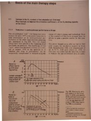
aPI)Ii:lcati?n with a patially very limited inter-Ion In O2 upply (th occlu ion of certainblood v I form an exception to this). Whenth .0 ygen upply i weakened, the extent toWhl h the reaction mechanism discussed in thefollowing sub-paragrap e involved 'whole occurrence depends on the de eeduration of the reduction, the type of til'lIUeaffected, and other factors.1.4.2 Energy gain by means of glycolysis in oxygen deficiencyThe individual cell in the organism needs forthe maintenance of its structure and its r~adines and ability to perform its function aertain energy supply, which it mainly obt;insfrom the oxidative catabolism of nutrients,when the 02 offer is sufficient.When the oxygen is lacking (anaerobic condition)the required energy can simply be gainedby glycolysis. Under in vivo conditions theoxidative breakdown of 1 mol glucose delivers689 kcal (2885 kJ) and is connected to theformation of 38 mol ATP. With 270-380 kcal(1130-1590 kJ) these represent the (physiologically)utilizable free energy which correspondsto an energy yield of between 39 and55 %. By comparison, the transformation of1 mol glucose into 2 mol lactate is connectedwith a reaction energy of only 47 kcal (196 kJ),by which 2 mol ATP are formed. These correspondto only 16-20 kcal (67-84 kJ) and anenergy yield of 3.4-4.3 %. Thus the aerobicdecomposition of glucose supplies a totalamount of energy which is 15 times higher, andATP which is 19 times higher, than glycolysis[175, 176]. It is therefore not surprising thatthe end-product of the glycolysis, lactic acid,still contains a high amount of energy.Special relationships exist in the heart inasmuchas the heart muscle which is sufficiently suppliedwith oxygen, covers more than 50 % of itsenergy requirements from the oxidation offatty acids. Glucose contributes 18 % to t eenergy gain, lactate 17 %, pyruvate approximatelyI % and ketone bodies 5 % [177]. nconditions of hypoxia, however, the breakdoof fatty acids is reduced, and glucose uptakeand breakdown increase, as does the breakdoof glycogen reserves; thus lactate thereby accumulatesto a greater extent [178] and overacidificationoccurs. The joint use of the lac 'cacid as an energy substrate in the myocardi(and perhaps also in the brain with a contrib tion of glycolysis to metabolism of approximately19 %), can be strengthened by g~trophanthin[169, 179-182]. This effect is indicatedby an increase in the pH in 02-deficienareas of the myocardium, roughly 3 min afteradministration of g-strophllnthin. This increaseis found only with g-strophanthin, not withdigitalis preparations. This confrrms the oldconcepts (Sarre, Uhlenbruck 1953) that gstrophanthin, but not digitalis preparations, improvesthe O2 supply to the heart muscle. It hasbeen hardly considered that g-strophanthin (a cording to [148, 183], highly effective and withreproducible potency, even when applied perlingually),in addition to its main effect in theheart muscle, also triggers a peripheral effec .Due to its action on the vasculature the bloodflow is increased [185] and the O2 and gluco esupply is improved, particularly in tissue lyingon the other side of the blood brain barrier[186, 187].1.4.3 Target area in tissue for O 2 deficiency and 02MTThe calculations of the dependence of theoxygen partial pressure P02 on the distance rto the capillary axis in the single-capillarymodel (Fig. 92) referred to the P0 2 level (e.g.45 mmHg) existing in the centre of the capillary.In reality the P0 2 drops, under normalconditions, from the arterial to the venous endof the capillary from 93 to 63-23 mmHg,depending on the organ (cf. Fig. 9). For anexample roughly corresponding to skeletalmu cle, curve A in Fig. 97 shows the course ofthe P02 along the capillary, and cu~e B thecou e af the coaxial cylinder area of thdi tanc r = 30 IJm. he pre ntation howthat the tissue area most at risk from O 2 deficiencyis localized at the venous end of thecapillary and at a great distance r from thecapillary axis. Under normal ondition hrelative frequency i f < 2 % for P0 2•mmHg (change to ferm ntation m tab li mand f < 0.2 % for P0 2 < 1 mmH riti al Pof th mitochondria, irreve ibl 11 d ma can be roughl . n from th di tri u icurve in Fig. 98.Th ti u r a with h' hv nou nd of tha di u d form