Imaging of Pulmonary Viral Pneumonia 1 - SIRM
Imaging of Pulmonary Viral Pneumonia 1 - SIRM
Imaging of Pulmonary Viral Pneumonia 1 - SIRM
- No tags were found...
You also want an ePaper? Increase the reach of your titles
YUMPU automatically turns print PDFs into web optimized ePapers that Google loves.
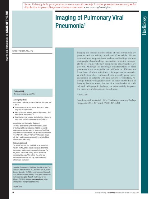
Note: This copy is for your personal,non-com m ercialuse only.To order presentation-ready copies fordistribution to your colleagues or clients,contactus atwww.rsna.org/rsnarights.REVIEWS AND COMMENTARY n STATE OF THE ARTTomás Franquet , MD, PhDOnline CMESee www.rsna.org/ry_cme.html<strong>Imaging</strong> <strong>of</strong> <strong>Pulmonary</strong> <strong>Viral</strong><strong>Pneumonia</strong> 1<strong>Imaging</strong> and clinical manifestations <strong>of</strong> viral pneumonia areprotean and not reliably predictive <strong>of</strong> its origin. All patientswith neutropenic fever and normal findings at chestradiography should undergo thin-section computed tomographyto determine whether parenchyma abnormalities arepresent. Although the radiologic manifestations <strong>of</strong> viralpneumonia are nonspecific and difficult to differentiatefrom those <strong>of</strong> other infections, it is important to considerviral infection when confronted with a rapidly progressivepneumonia in patients with risk factors for infection. Althoughdefinitive diagnosis cannot be made on the basis <strong>of</strong>imaging features alone, the use <strong>of</strong> a combination <strong>of</strong> clinicaland radiographic findings can substantially improvethe accuracy <strong>of</strong> diagnosis in this disease .qRSNA, 2011Learning Objectives:After reading the article and taking the test, the reader willbe able ton Describe the role <strong>of</strong> thin-section thoracic CT in thediagnosis viral pneumonia.n Identify the most common features <strong>of</strong> pulmonary viralinfections at thin-section CT.n Describe the most common viral infections in immuno-competent and in immunocompromised patients.Accreditation and Designation StatementThe RSNA is accredited by the Accreditation Councilfor Continuing Medical Education (ACCME) to providecontinuing medical education for physicians. The RSNAdesignates this journal-based CME activity for a maximum<strong>of</strong> 1.0 AMA PRA Category 1 Credit TM . Physicians shouldonly claim credit commensurate with the extent <strong>of</strong> theirparticipation in the activity.Disclosure StatementThe ACCME requires that the RSNA, as an accreditedprovider <strong>of</strong> CME, obtain signed disclosure statementsfrom authors, editors, and reviewers for this case. Forthis journal-based CME activity, author disclosuresare listed at the end <strong>of</strong> this article. The editor andthe reviewers indicated that they have no relevantrelationships to disclose.Supplemental material: http :// radiology . rsna . org / lookup/ suppl / doi : 10 . 1148 / radiol . 11092149 /-/ DC11From the Department <strong>of</strong> Radiology, Hospital de Sant Pau,Avda Sant Antoni M. Claret 167, Barcelona 08128, Spain.Received November 19, 2009; revision requested January 7,2010; revision received February 4; accepted February 17;fi nal version accepted March 1; fi nal review by T.F.February 24, 2011. Address correspondence to theauthor (e-mail: tfranquet@santpau.cat ) .qRSNA, 201118 radiology.rsna.org n Radiology: Volume 260: Number 1—July 2011
STATE OF THE ART: <strong>Imaging</strong> <strong>of</strong> <strong>Pulmonary</strong> <strong>Viral</strong> <strong>Pneumonia</strong>FranquetMore than 55 million people die eachyear worldwide, and pneu moniais among the leading causes ( 1 ).<strong>Viral</strong> infections are an increasingly frequentcause <strong>of</strong> pulmonary disease worldwide.Unfortunately, the diagnosis <strong>of</strong>viral pneumonia still relies heavily onclinician suspicion in the proper setting,which is based on host risk factors,presentation, and exposures. Thediagnostic approach involves a consideration<strong>of</strong> the likely pathogens on thebasis <strong>of</strong> the patient’s presenting signsand symptoms and his or her immunologiccondition ( 2 , 3 ). The clinical manifestations<strong>of</strong> most viral infections aresimilar. Although most <strong>of</strong> these infectionsin nonimmunocompromised personsare self-limited, some patients candevelop severe pneumonitis ( 4 ). Clinically,viral pneumonia in adults can bedivided into two groups: so-called atypicalpneumonia in otherwise healthy hostsand viral pneumonia in immunocompromisedhosts ( 5 ). Influenza virus typesA and B account for most viral pneumoniasin immunocompetent adults ( 5 ). Ithas been considered that clinical history,results <strong>of</strong> physical examination,and imaging features cannot enable theprediction <strong>of</strong> the etiologic agent. Furthermore,a pathogen is identified in onlyEssentialsn <strong>Viral</strong> pulmonary infections areclinically important in both immunocompromisedand immunocompetentpatients; despite its limitations,CT is currently the imagingmodality <strong>of</strong> choice for the evaluation<strong>of</strong> pulmonary viral infections.n Although the CT features <strong>of</strong> viralpulmonary infections are usuallynonspecific, precise imaging characterizationis essential; knowledge<strong>of</strong> CT findings has a substantialeffect on the treatment <strong>of</strong>patients suspected <strong>of</strong> having pulmonaryviral infections.n Knowledge <strong>of</strong> the underlyingpathologic characteristics <strong>of</strong>these diseases aids in the differentiation<strong>of</strong> viral infections fromother entities that may haveoverlapping imaging features.up to 50%–64% <strong>of</strong> patients admitted tothe hospital with pneumonia. The recentdevelopment <strong>of</strong> multidetector helical computedtomographic (CT) scanners capable<strong>of</strong> rapid scanning and acquisition<strong>of</strong> thin sections has revolutionized thethin-section CT technique. Volumetricthin-section CT with thin detectors (0.5–0.625 mm) has become the routine inmany institutions. New serologic testshave also come to the aid <strong>of</strong> the clinician,and sophisticated molecular methods arebecoming more commonplace in routinediagnosis <strong>of</strong> acute respiratory diseasein both immunocompetent and high-riskpatients ( 1 , 6 – 8 ). Confirmation <strong>of</strong> the diagnosisand identification <strong>of</strong> the specificagent can be accomplished with use <strong>of</strong>tissue cultures, serologic tests, detection<strong>of</strong> viral antigens within respiratory tractsecretions or blood with use <strong>of</strong> monoclonalantibodies, detection <strong>of</strong> virusassociatedmolecules with use <strong>of</strong> in situhybridization or polymerase chain reaction(PCR), and observation <strong>of</strong> virusinducedchanges cytologically or histologically( 9 , 10 ). The purpose <strong>of</strong> this reviewis to provide a framework with which tobetter understand respiratory viral infections,the diverse clinical settings in whichthey occur, and their potential importancein these varying clinical contexts. Whileacknowledging the less-than-definitivediagnostic role <strong>of</strong> CT in their diagnosis,I will also include discussion <strong>of</strong> thoseconditions in which an understanding <strong>of</strong>the pathologic characteristics can be usefulto the interpretation <strong>of</strong> thin-sectionCT findings. The epidemiology and pathogenesis<strong>of</strong> viral infections are discussedin Appendix E1 (online).Histologic Features <strong>of</strong> <strong>Viral</strong> InfectionsVarious histopathologic patterns <strong>of</strong>lung injury have been described in viralpneumonia. Although some <strong>of</strong> thesepatterns may be relatively unique to aspecific clinical context, others are nonspecificwith respect to either the causeor pathogenesis.Viruses can result in several pathologicforms <strong>of</strong> lower respiratory tractinfection, including tracheobronchitis,bronchiolitis, and pneumonia. Organizingpneumonia is a nonspecific reparativereaction that may be seen in a variety<strong>of</strong> clinical contexts. In other words, organizingpneumonia may result from a variety<strong>of</strong> causes or underlying pathologicprocesses, including viral infections.Most respiratory viruses damage cellsdirectly through cytopathic effects mediatedby means <strong>of</strong> either viral-directedcell lysis or the inhibition <strong>of</strong> host cellRNA, protein, and DNA synthesis. Otherviruses (eg, cytomegalovirus [CMV], adenovirus,or herpesvirus) may producespecific nuclear changes or characteristiccytoplasmic inclusions ( 11 ) ( Fig 1 ).Cytopathic respiratory viruses (eg,influenza, adenovirus, and herpesvirusgroup) cause a virus-specific lung injurypattern. Influenza virus affects the epitheliumdiffusely and, in severe cases, resultsin necrotizing bronchitis and/or bronchiolitisand diffuse alveolar damage.The histologic features <strong>of</strong> influenzapneumonia are epithelial necrosis <strong>of</strong> theairways with submucosal chronic inflammation.Fatal influenza pneumonia representsa necrotizing bronchiolitis withdiffuse alveolar damage, which can behemorrhagic.Adenovirus has its greatest effect inthe terminal bronchioles and may producebronchiolitis or even bronchiectasis( 12 – 14 ). The bronchiolitis may be necrotizingand result in a necrotizing bronchopneumoniasimilar to that seen in severeherpes simplex infection ( 13 ) ( Fig 2 ).The herpes group <strong>of</strong> viruses (herpessimplex, varicella-zoster, CMV, Epstein-Barr virus [EBV]) may cause focal cytopathiceffects in either the airway oralveoli. The histologic features <strong>of</strong> herpessimplex lower respiratory tract infectionPublished online10.1148/radiol.11092149Radiology 2011; 260:18– 39Content codes:Abbreviations:AIDS = acquired immunodefi ciency syndromeCMV = cytomegalovirusEBV = Epstein-Barr virusHMPV = human metapneumovirusHTLV-1 = human T-cell lymphotropic virus type 1PCR = polymerase chain reactionRSV = respiratory syncytial virusSARS = severe acute respiratory syndromeAuthor stated no fi nancial relationship to disclose.Radiology: Volume 260: Number 1—July 2011 n radiology.rsna.org 19
STATE OF THE ART: <strong>Imaging</strong> <strong>of</strong> <strong>Pulmonary</strong> <strong>Viral</strong> <strong>Pneumonia</strong>FranquetFigure 1Figure 2Figure 3Figure 1: Histopathologic features <strong>of</strong> pulmonaryCMV infection. Photomicrograph (hematoxylineosinstain; original magnification, 3400 )shows numerous CMV-infected cells; they areenlarged and contain nuclei with prominentamphophilic inclusions (arrowheads). (Imagecourtesy <strong>of</strong> T. Colby , MD, Mayo Clinic, Scottsdale,Ariz.)Figure 2: Histopathologic features <strong>of</strong> respiratorysyncytial virus (RSV) bronchiolitis in a child. Photomicrograph(hematoxylin-eosin stain; original magnification, 3400) shows two adjacent bronchioleswith a marked transmural cellular infi ltrate (bronchiolitis)(arrowheads) and some associated metaplasticchanges in the mucosa. Ulcerative mucosalchanges are also seen (arrow). (Image courtesy <strong>of</strong> T.Colby, MD, Mayo Clinic, Scottsdale, Ariz.)Figure 3: Histopathologic features <strong>of</strong> necrotizingherpes pneumonia. Image from scanning low-powermicroscopy (hematoxylin-eosin stain; original magnification, 320) shows nodular zones <strong>of</strong> necrosis,some <strong>of</strong> which are bronchiolocentric (arrows).(Image courtesy <strong>of</strong> T. Colby, MD, Mayo Clinic,Scottsdale, Ariz.)include necrosis <strong>of</strong> the airway epithelium,necrotizing pneumonia as the reactionspreads from the airways to the adjacentparenchyma, diffuse alveolar damage,miliary nodules, and scattered larger nodules( 15 ) ( Fig 3 ). Multicentric areas <strong>of</strong>hemorrhage may appear centered on airways.An acute lung injury pattern maybe present, with interstitial edema, congestion,and inflammation. Histologicfeatures <strong>of</strong> varicella-zoster virus pneumoniainclude endothelial damage in smallvessels, with focal hemorrhagic necrosis,mononuclear infiltration <strong>of</strong> alveolarwalls, and fibrinous exudates in the alveoli( 16 ). Late sequelae <strong>of</strong> varicella-zosterinfection consist <strong>of</strong> multiple 1–2-mmdiametercalcified nodules ( 17 ).The histologic findings in CMV pneumoniainclude either an acute interstitialpneumonia or a miliary pattern. Acuteinterstitial pneumonia is characterizedby a diffuse mild alveolar widening byedema and mononuclear cells, airspacefibrinous exudate and/or hyaline membraneswith relatively scant neutrophils,and prominent type 2 alveolar lining cells( Fig 4 ); foci <strong>of</strong> organizing pneumoniaare <strong>of</strong>ten found. The miliary pattern issimilar to that <strong>of</strong> other miliary viral infections;nodules contain the infectedcells with the characteristic cytoplasmicinclusions <strong>of</strong> CMV.Coxsackievirus B, reoviruses, andrhinoviruses may also infect the lungs,usually producing interstitial pneumoniaswith diffuse alveolar damage. Humanpapilloma viruses are known to cause recurrentpapillomas and have been linkedto lung cancer.In immunocompetent individuals,RSV and parainfluenza viruses may producenecrotizing bronchiolitis characterizedby exudates within bronchiolarlumen, inflammatory cells in the wall<strong>of</strong> bronchioles, a peribronchiolar reactionwith chronic inflammatory cells, andexudate in alveoli. In immunocompromisedpatients, parainfluenza virusesproduce giant cell pneumonia with diffusealveolar damage indistinguishablefrom that caused by measles pneumonia.Measles virus may involve both airwaysand lung parenchyma, producingbronchitis and/or bronchiolitis and interstitialpneumonia.The predominant pathologic process<strong>of</strong> hantavirus pulmonary syndrome andsevere acute respiratory syndrome (SARS)is diffuse alveolar damage and diffuselung disease characterized histologicallyby interstitial edema and central alveolarfilling ( 18 , 19 ) ( Fig 5 ).The radiologic findings reflect thevariable extent <strong>of</strong> the histopathologicfeatures, which include diffuse alveolardamage (intraalveolar edema, fibrin,and variable cellular infiltrates with ahyaline membrane), intraalveolar hemorrhage,and interstitial (intrapulmonaryor airway) inflammatory cell infiltration( 20 ).Clinical ManifestationsThe clinical signs and symptoms <strong>of</strong> viralpneumonia are <strong>of</strong>ten nonspecific, andthe clinical course <strong>of</strong> infection will behighly dependent on the overall immunestatus <strong>of</strong> the host ( 3 ). Acute bronchiolitisis a term most <strong>of</strong>ten used to describean illness in infants and children characterizedby acute wheezing with concomitantsigns <strong>of</strong> respiratory viral infection( 21 ). RSV is the etiologic agent in mostpatients, but other viruses (adenovirus,influenza, parainfluenza) and nonviralpathogens (mycoplasma, Chlamydia species)can cause a similar syn drome ( 22 ).Symptomatic acute bronchiolitis inadults is relatively rare but can be causedby infectious agents such as RSV. Becausesmall airways in adults contributeless to total pulmonary resistance, acuteinfectious bronchiolitis may spare adultsthe severe symptoms characteristic <strong>of</strong>bronchiolitis in infants. Acute bronchiolitisin adults may also be seen withaspiration, toxic inhalation, connective20 radiology.rsna.org n Radiology: Volume 260: Number 1—July 2011
STATE OF THE ART: <strong>Imaging</strong> <strong>of</strong> <strong>Pulmonary</strong> <strong>Viral</strong> <strong>Pneumonia</strong>FranquetFigure 4Figure 5and not invariably associated with pneumonia;lower respiratory tract symptomsare found in 10% <strong>of</strong> cases ( 3 ).Figure 4: Histopathologic features <strong>of</strong> a CMV interstitialpneumonia. Photomicrograph (hematoxylineosinstain; original magnifi cation, 3100) showsinterstitial widening with formation <strong>of</strong> micronodules(arrows). This is associated with acute lung injuryand some hyaline membranes (arrowheads). (Imagecourtesy <strong>of</strong> T. Colby, MD, Mayo Clinic, Scottsdale,Ariz.)tissue diseases, lung and bone marrowtransplantation, and Stevens-Johnsonsyndrome ( 23 ).Pathologic studies <strong>of</strong> acute infectiousbronchiolitis have shown intenseacute and chronic inflammation <strong>of</strong> smallbronchioles with associated epithelialnecrosis and sloughing. There may beassociated edema as well as inflammatoryexudate and mucus in the bronchiolarlumen ( 24 ).The possibility <strong>of</strong> pneumonia shouldbe considered in any patient who has newrespiratory symptoms (including cough,sputum, or dyspnea), particularly whenthese symptoms are accompanied by feveror abnormalities at physical examination<strong>of</strong> the chest (eg, rhonchi and rales).<strong>Pneumonia</strong> is also increasingly prevalentin patients with specific comorbiditiesor risk factors, including smoking,chronic obstructive pulmonary disease,asthma, diabetes mellitus, malignancy,heart failure, neurologic diseases, narcoticand alcohol use, and chronic liverdisease.A thorough physical examination, posteroanteriorand lateral chest radiography,and a blood leukocyte count witha differential cell count should be performedwhen pneumonia is suspected.Even with intensive laboratory investigation,the specific microbiologicFigure 5: Histopathologic features <strong>of</strong> diffusealveolar damage in a patient with SARS. Photomicrograph(hematoxylin-eosin stain; original magnifi -cation, 3200) demonstrates that alveolar septashow complete loss <strong>of</strong> epithelium and are lined byhyaline membranes (arrowheads); intraalveolaredematous material is also present (arrows). (Imagecourtesy <strong>of</strong> T. Colby, MD, Mayo Clinic, Scottsdale,Ariz.)cause can be established with certaintyonly in approximately 50% <strong>of</strong> patientswith pneumonia. The probable predominantorganism varies with the host’s epidemiologicfactors, the severity <strong>of</strong> illness,and the laboratory approach used to establishthe diagnosis.Although most attention traditionallyfocuses on bacterial causes <strong>of</strong> severecommunity-acquired pneumonia, virusescan also cause serious lower respiratorytract infections. The predominant respiratoryviruses that can cause severepneumonia include influenza and RSV.The clinical manifestations <strong>of</strong> viralinfections <strong>of</strong>ten vary from patient to patientand cannot be reliably used to establisha specific (microbiologic) diagnosis.The initial symptoms and signs maybe categorized into several clinical syndromes( 25 ). A “cold” is characterizedby upper respiratory tract symptomsand includes tonsillopharyngitis, pharyngitis,epiglottitis, sinusitis, otitis media,and conjunctivitis. The “influenza”syndrome, which may be caused by virusesother than influenza, consists primarily<strong>of</strong> abrupt fever, headache, myalgias,and malaise. The disease manifestations<strong>of</strong> several viruses (eg, human parainfluenzavirus, RSV, and influenza) are <strong>of</strong>tenconfined to the upper respiratory tractLaboratory Identification MethodsDifferentiation <strong>of</strong> common respiratoryinfections due to Streptococcus pneumoniaeand those due to Mycoplasmapneumoniae , Chlamydophila pneumoniae, or Legionella pneumophila as wellas those due to viruses is essential toallow correct decisions concerning theantibiotics to be administered ( 3 ).Diagnosis <strong>of</strong> respiratory viruses isstill difficult, with only a small percentage<strong>of</strong> cases being routinely diagnosed.Although viral culture has been the referencestandard, it has considerable limitations(ie, depending on the virus, itmay take 3–14 days for cultures to yieldresults).The development <strong>of</strong> better diagnostictests has markedly improved ourability to detect multiple viral pathogens( 3 ). These techniques include cultureto prove replication <strong>of</strong> completeviral particles, hybridization techniquesperformed with or without PCR to detectviral nucleic acids, immunohistochemistryto detect viral proteins, electronmicroscopy to demonstrate a fullyassembled virus, and measurements <strong>of</strong>a specific host immune response to thevirus in either serum or cells or tissue.The most appropriate specimens arenasopharyngeal aspirates, nasopharyngealand throat swabs, and those frombronchoalveolar lavage ( 26 ).Nucleic Acid Amplification TestsThe introduction <strong>of</strong> highly sensitive nucleicacid amplification tests has dramaticallyimproved the ability to detectmultiple respiratory viruses such as influenza,RSV, rhinovirus, parainfluenza,and adenovirus ( 27 – 29 ).The most familiar formats use DNAor RNA target amplification methods forenhanced sensitivity above that <strong>of</strong> cultureand antigen-based procedures ( 28 ).PCRThe value <strong>of</strong> PCR in identifying respiratoryviruses in clinical samples hasbeen clearly shown, and as much as aRadiology: Volume 260: Number 1—July 2011 n radiology.rsna.org 21
STATE OF THE ART: <strong>Imaging</strong> <strong>of</strong> <strong>Pulmonary</strong> <strong>Viral</strong> <strong>Pneumonia</strong>Franquetthree- to fourfold increase in positive specimenshas been found when PCR testingis added to conventional cell cultureand/or “standard” serologic methods. Thedownside <strong>of</strong> PCR is that it may be too sensitive,in that it can detect small amounts<strong>of</strong> residual viral nucleic acid when thereis no other laboratory or clinical evidencethat a viral infection is present. Factorsthat lead to negative laboratory resultsinclude poor specimen handling and collection,low viral copy num bers, and inhibitorsin the clinical sam ple ( 10 ).Reverse Transcriptase PCRReverse transcriptase PCR is a modification<strong>of</strong> PCR used when the initialtemplate is RNA rather than DNA. Reversetranscriptase PCR can be used to amplifya much higher number <strong>of</strong> DNA copiespresent in bacteria, fungi, virus, orother proteins ( 10 ).Despite the obvious advantages <strong>of</strong>these newer procedures, there may bepotential limitations to DNA amplificationtechnology in the diagnostic microbiologylaboratory, as follows: (a) Falsenegativetest results may occur because<strong>of</strong> the presence <strong>of</strong> substances in thespecimen that inhibit nucleic acid extractionor amplification, (b) careful interpretation<strong>of</strong> results is necessary, (c) theprocedure is technically complex, (d)equipment is expensive, and (e) there isa lack <strong>of</strong> standardization ( 30 , 31 ).<strong>Imaging</strong> <strong>of</strong> <strong>Viral</strong> <strong>Pneumonia</strong>In patients suspected <strong>of</strong> having viral pneumonia,imaging is performed to help detectdisease, assess disease extent, performfollow-up assessment <strong>of</strong> responseto treatment, and guide interventionaldiagnostic procedures (eg, bronchoscopywith bronchoalveolar lavage).It can be difficult to differentiate viralpneumonia from other infectious processes,and the cause <strong>of</strong> infection (eg,viral vs pyogenic or fungal) cannot bereliably ascertained from its imagingappearance.Conventional Radiographic FindingsRadiologic features are dependent onseveral host factors, including age andunderlying immune status; risk factorsassociated with increased frequency andseverity <strong>of</strong> viral infections include veryyoung and old age, malnutrition, andimmunologic disorders.Chest radiographs demonstrate normalfindings or unilateral or patchy bilateralareas <strong>of</strong> consolidation, nodularopacities, bronchial wall thickening, andsmall pleural effusions; lobar consolidationis uncommon in patients with viralpneumonia. Patients may develop acutepneumonia with rapid progression toacute respiratory distress syndrome.Although radiographic findings aloneare not sufficient for the definitive diagnosis<strong>of</strong> viral pneumonia, in combinationwith clinical findings they can substantiallyimprove the accuracy <strong>of</strong> diagnosisin this disease.Thin-Section CT FindingsAn increasing number <strong>of</strong> patients undergothin-section CT when there is highclinical suspicion <strong>of</strong> pneumonia with normalor questionable radiographic findings( 32 – 38 ). In a study <strong>of</strong> 87 consecutivepatients with febrile neutropenia,Heussel et al ( 39 ) noted that the CT scanrevealed a pulmonary lesion not seenon the radiograph in 50% <strong>of</strong> subjects.The CT signs <strong>of</strong> pulmonary viral infectionwill depend on the underlyingpathologic process. They are quite similarsimply because the underlying pathogenicmechanism, depending on thevirulence <strong>of</strong> the virus, is related to variableextents <strong>of</strong> histopathologic featuressuch as diffuse alveolar damage (intraalveolaredema, fibrin, and variable cellularinfiltrates with a hyaline membrane),intraalveolar hemorrhage, and interstitial(intrapulmonary or airway) inflammatorycell infiltration ( 40 , 41 ).The spectrum <strong>of</strong> CT findings encounteredin various pulmonary viral diseasesis not particularly wide and encompassesfive main categories: (a) parenchymalattenuation disturbances; (b) groundglassopacity and consolidation; (c) nodules,micronodules, and tree-in-bud opacities;(d) interlobular septal thickening;and (e) bronchial and/or bronchiolarwall thickening ( 42 , 43 ).Parenchymal attenuation disturbances.—Patchy inhomogeneities in theattenuation <strong>of</strong> lung parenchyma (mosaicattenuation pattern) are a recognizedfinding in some viral infections causedby hypoventilation <strong>of</strong> alveoli distal tobronchiolar obstruction (inflammation orcicatricial scarring <strong>of</strong> many bronchioles),which leads to secondary vasoconstriction(and, consequently, underperfusedlung) and is seen on CT scans as areas<strong>of</strong> decreased attenuation ( 44 – 46 ). PairedCT scans obtained in inspiration andexpiration are useful for differentiatingbronchiolar disease from pulmonary vasculardisease and some diffuse infiltrativediseases that may also cause a mosaicpattern. In bronchiolar disease, the regions<strong>of</strong> decreased attenuation seen inthe lung at inspiration are also seen atexpiration owing to air trapping and showlittle increase in attenuation or decreasein volume as seen with primary vascularlung disease ( 47 , 48 ) ( Fig 6 ).Uninvolved segments <strong>of</strong> lung shownormal or increased perfusion with resultingnormal or increased attenuation,respectively. In the specific context, coexistinginflammatory small airways andparenchyma disease may both contributeto a mosaic attenuation pattern ( 49 ).Nevertheless, differentiation betweenthe various causes <strong>of</strong> a mosaic attenuationpattern on CT scans (diffuse infiltrative,small airways, and occlusive vascular diseases)is not always straightforward andmay, on occasion, cause diagnostic difficulties( 45 , 48 ).Ground-glass opacity and consolidation.—A ground-glass opacity is a hazyincrease in attenuation seen in a variety<strong>of</strong> interstitial and alveolar processes. Inthe context <strong>of</strong> pulmonary infectiousdiseases, coexisting thickening <strong>of</strong> theinterstitium and partial filling <strong>of</strong> the airspacesmay both contribute to groundglassopacity and consolidation ( Fig 7 ).Nevertheless, other noninfectious conditionscharacterized by ground-glassopacity include interstitial pulmonaryedema, pulmonary hemorrhage (in whichthere is thickening <strong>of</strong> the interstitiumand partial filling <strong>of</strong> the airspaces withblood), hypersensitivity pneumonitis, respiratorybronchiolitis, organizing pneumonia,and alveolar proteinosis ( 50 – 54 ).Areas <strong>of</strong> pulmonary consolidation aremost <strong>of</strong>ten patchy and poorly defined(consistent with bronchopneumonia) or22 radiology.rsna.org n Radiology: Volume 260: Number 1—July 2011
STATE OF THE ART: <strong>Imaging</strong> <strong>of</strong> <strong>Pulmonary</strong> <strong>Viral</strong> <strong>Pneumonia</strong>FranquetFigure 6Figure 7Figure 6: Transverse expiratory thin-section CT scan through thelower lobes in a 44-year-old woman with constrictive obliterative bronchiolitissecondary to a severe viral lower respiratory tract infection.There are bilateral well-demarcated patchy areas <strong>of</strong> increased anddecreased lung attenuation. Whereas hypoattenuated areas contain fewand small pulmonary vessels (arrows), hyperattenuated areas containenlarged pulmonary vessels (arrowheads), refl ecting the pulmonaryblood fl ow distribution toward normal ventilated areas.Figure 8Figure 7: Transverse thin-section CT scan at the level <strong>of</strong> the bronchusintermedius in a patient with RSV infection shows a hazy increasein lung opacity without obscuration <strong>of</strong> underlying vessels.Figure 9Figure 8: Transverse thin-section CT scan at the level <strong>of</strong> the bronchusintermedius in a patient with infl uenza A virus shows ill-defi nedcentrilobular nodules (arrows). Peripheral subpleural consolidationin the apical segment <strong>of</strong> left lower lobe (arrowhead) representscoalescence <strong>of</strong> nodules.focal and well-defined (consistent withlobar pneumonia). As with consolidation,a variety <strong>of</strong> acute and chronic lungdiseases may result in lobular areas <strong>of</strong>ground-glass opacity, which give the lunga mosaic appearance. Lobular groundglassopacity may be seen in patientswith infection (eg, bronchopneumonia,viral infections, Pneumocystis jiroveciipneumonia, or M pneumoniae ).Nodules, micronodules, and tree-inbudopacities.— Nodules 1–10 mm in di-ameter may be seen in a number <strong>of</strong> lunginfections. Nodule size is helpful in thedifferential diagnosis <strong>of</strong> infectious causes<strong>of</strong> nodules. Franquet et al ( 37 ) comparedthe CT findings in 78 consecutive immunocompromisedpatients with solitary ormultiple nodular opacities <strong>of</strong> proved infectiousorigin. There was a good correlationbetween the size <strong>of</strong> the nodulesand their origin. Patients whose noduleswere smaller than 10 mm in diameterwere most likely to have a viral infection.Figure 9: Transverse thin-section CT scan through the lower lobes in apatient with infl uenza A virus infection shows bilateral multiple smallbranching opacities (arrows) representing dilated peripheral bronchioles.Centrilobular nodules are most commonlyseen in patients with disease thatprimarily affects centrilobular bronchiolesand results in inflammation, infiltration,or fibrosis <strong>of</strong> the surrounding interstitiumand alveoli ( 55 ) ( Fig 8 ). These nodulesmay be dense and <strong>of</strong> homogeneous attenuationor show ground-glass opacity.The tree-in-bud finding, which is usuallyindicative <strong>of</strong> small airways disease,reflects the presence <strong>of</strong> dilated centrilobularbronchioles with lumina thatare impacted with mucus, fluid, or pus;this finding is <strong>of</strong>ten associated withperibronchiolar inflammation ( 56 ) ( Fig 9 ).Radiology: Volume 260: Number 1—July 2011 n radiology.rsna.org 23
STATE OF THE ART: <strong>Imaging</strong> <strong>of</strong> <strong>Pulmonary</strong> <strong>Viral</strong> <strong>Pneumonia</strong>FranquetFigure 10Figure 11Figure 10: Transverse thin-section CT scan through the lower lobesin a patient with adenovirus infection shows patchy bilateral groundglassopacities with superimposed linear opacities (crazy-pavingpattern) (arrows).Figure 11: Transverse thin-section CT scan at the level <strong>of</strong> the aorticarch in a patient with parainfl uenza virus infection shows bilateralground-glass opacities and bronchial wall thickening (arrows).Furthermore, in most cases, a tree-inbudappearance is associated with airwaysinfection. Infectious diseases associatedwith cellular bronchiolitis andcentrilobular nodules include endobronchialspread <strong>of</strong> tuberculosis, nontuberculousmycobacteria bronchopneumonia( 57 – 62 ), and infectious bronchiolitis( 56 ). Branching or centrilobular nodulesand mosaic perfusion are seen in patientswith viral bronchiolitis ( 63 , 64 ).Similar findings may also be observedwith bacterial and fungal pneumoniaand in patients with allergic bronchopulmonaryaspergillosis ( 65 ).Interlobular septal thickening.— Septalthickening may be seen in the presence<strong>of</strong> interstitial fluid, cellular infiltration,or fibrosis and can have a smooth,nodular, or irregular contour in differentpathologic processes. In the context <strong>of</strong>viral diseases, the most dramatic cause<strong>of</strong> widespread smooth thickening <strong>of</strong> theinterlobular septa is acute respiratorydistress syndrome ( 66 ).Smooth septal thickening may alsobe seen in association with ground-glassopacity, a pattern termed “crazy paving”( Fig 10 ); this pattern, initially describedas typical <strong>of</strong> alveolar proteinosis,has an extensive differential diagnosis( 27 , 67 – 69 ).Bronchial and bronchiolar wall thickening.—Bronchiolar wall thickening mayoccur due to inflammation or fibrosis.In viral bronchitis, obstruction <strong>of</strong> bronchiolesby inflammatory exudates andbronchiolar wall thickening from edemaand smooth muscle hyperplasia producethe thin-section CT features <strong>of</strong>atelectasis, air trapping, centrilobularnodules, and bronchiolar wall thickening.Air trapping may be present because <strong>of</strong>associated bronchiolitis ( Fig 11 ). A summary<strong>of</strong> CT findings in viral pneumoniais shown in Table 1 .In clinical practice, radiologists willhave to consider that there are manynoninfectious or different infectious disordersthat should be differentiated fromviral pneumonia. A combined strategyconsisting <strong>of</strong> thin-section CT and guidedbronchoscopy with bronchoalveolar lavageperformed within the first 24 hoursafter CT could improve the diagnosticyield and subsequent therapeuticchanges in these patients.<strong>Viral</strong> Pathogens <strong>of</strong> the RespiratoryTract<strong>Viral</strong> infections <strong>of</strong> the respiratory tractinclude both those considered to be principalrespiratory viruses, whose replicationis generally restricted to the respiratorytract, and others whose respiratoryinvolvement is part <strong>of</strong> a generalizedinfection. Viruses are classified on thebasis <strong>of</strong> (a) the type and structure <strong>of</strong>the nucleic acid in the viral genome andthe strategy used in its replication (eg,DNA or RNA), (b) the type <strong>of</strong> symmetry<strong>of</strong> the virus capsid (helical vs icosahedral),and (c) the presence or absence<strong>of</strong> a lipid envelope.The respiratory viruses can be dividedinto two large groups accordingto the type <strong>of</strong> nuclei acid they contain( Table 2 ).Selected RNA Virus–related DiseasesInfluenza.— Influenza viruses are singlestrandedRNA viruses that belong tothe family Orthomyxoviridae. Influenzaviruses are important human respiratorypathogens that cause seasonal upperrespiratory tract infections in the community,endemic infections, and periodic,unpredictable pandemics ( 70 ). The greatestinfluenza pandemic, the so-calledSpanish influenza, spread worldwide in1918 and resulted in the deaths <strong>of</strong> approximately50 million people ( 71 , 63 ).Influenza type A is the most important<strong>of</strong> the respiratory viruses with respectto the morbidity and mortality inthe general population. They are morecommon during infancy and <strong>of</strong>ten maylead to severe lower respiratory tractdisease. In adults, infections are usuallymild and restricted to the upper respiratorytract. Influenza A virus is transmittedfrom person to person by aerosolizedor respiratory droplets. In theUnited States, more than 35 000 deathsand 200 000 hospitalizations due to influenzaoccur annually, and the numberis increasing ( 64 ).24 radiology.rsna.org n Radiology: Volume 260: Number 1—July 2011
STATE OF THE ART: <strong>Imaging</strong> <strong>of</strong> <strong>Pulmonary</strong> <strong>Viral</strong> <strong>Pneumonia</strong>FranquetTable 1Summary <strong>of</strong> CT Findings in <strong>Viral</strong> <strong>Pneumonia</strong>Cause <strong>of</strong> <strong>Pneumonia</strong>ParenchymalAttenuationDisturbancesGround-GlassOpacity andConsolidationNodules, Micronodules,and Tree-in-BudOpacitiesInterlobularSeptalThickeningBronchial and/orBronchiolar WallThickeningOtherRNA virusesInfl uenza A … +++ +++ … … …Avian fl u (H5N1) … +++ + … … Pneumatocele, pleural effusionSwine-origin influenza A (H1N1) … +++ … … … …Parainfl uenza 1–4 … +++ +++ … … …RSV … +++ +++ … +++ …HMPV * … +++ +++ … … …Measles … +++ +++ ++ ++ Pleural effusion, lymphadenopathyEnteroviruses … … … … … …Hantavirus … +++ ++ +++ … Acute respiratory distress syndromeCoronavirus (SARS) … +++ … +++ … Crazy-paving patternDNA virusesAdenovirus … ++ … … +++ BronchiectasisHerpes simplex virus … +++ ++ … … Nodules with halo signVaricella … ++ +++ … … Nodules with halo sign or calcifi edCMV … +++ ++ … … Nodules with halo signEPV … +++ + + … Nodules with halo signNote.—Plus signs indicate the relative frequency <strong>of</strong> the fi ndings from lowest (+) to highest (+++).* HMPV = human metapneumovirus.Table 2Selected Viruses Causing Respiratory SyndromesFamilyThe symptoms <strong>of</strong> influenza beginrapidly with fever, usually 101°–102°F(38°–39°C), and is associated with myalgias,headache, lethargy, and respiratorytract symptoms <strong>of</strong> dry cough, rhinorrhea,and sore throat.In recent years, both influenza andparainfluenza viruses have been recognizedas a major cause <strong>of</strong> respiratoryillness in immunocompromised patients,Species and SubtypeRNA virusesOrthomyxoviridaeInfl uenza A (subtype: avian fl u [H5N1], swine-origininfl uenza A [H1N1]), infl uenza BParamyxoviridaeParainfl uenza 1–4, measlesMetapneumoviridaeRSV, HMPVPicornaviridaeEnterovirusesRetroviruses HTLV-1 *BunyaviridaeHantavirusCoronaviridaeCoronaviruses (subtype: SARS coronavirus)DNA virus (Adenoviridae)Adenovirus, herpes simplex virus (HSV), varicella-zostervirus, CMV, EBV, human papilloma virus* HTLV-1 = Human T-cell lymphotropic virus type 1.including solid organ transplant recipients( 72 ). Oikonomou et al ( 65 ) reviewedthe thin-section CT findings infour patients with hematologic malignanciesand influenza A pneumonitisand found that the predominant thinsectionCT findings were ground-glassopacities, consolidation, centrilobularnodules, and branching linear opacities( Fig 12 ).Avian flu (H5N1).— Avian influenzais caused by the H5N1 subtype <strong>of</strong> theinfluenza A virus ( 73 , 74 ). Most humaninfections appear to be the result <strong>of</strong>close contact with infected birds—usuallypoultry or their products ( 73 , 75 , 76 ).In May 1997, an influenza H5N1 viruswas isolated from a previously healthy3-year-old boy in Hong Kong who died<strong>of</strong> severe pneumonia complicated by acuterespiratory distress syndrome ( 77 ). InNovember and December <strong>of</strong> the sameyear, 18 additional cases <strong>of</strong> human H5N1infections were identified—six <strong>of</strong> whichwere fatal ( 78 ). According to World HealthOrganization statistics, as <strong>of</strong> June 19,2008, 385 human infections had beenconfirmed from 15 countries; <strong>of</strong> theseinfections, 243 were fatal. Approximately90% <strong>of</strong> confirmed infections have beenin persons 40 years <strong>of</strong> age and younger.Most <strong>of</strong> those infected were previouslyhealthy individuals. Signs and symptoms<strong>of</strong> infection included fever, cough,diarrhea, shortness <strong>of</strong> breath, lymphocytopenia,and thrombocytopenia. Theoverall case fatality rate for influenzaA (H5N1) infections exceeds 60%( 79 – 81 ).Radiology: Volume 260: Number 1—July 2011 n radiology.rsna.org 25
STATE OF THE ART: <strong>Imaging</strong> <strong>of</strong> <strong>Pulmonary</strong> <strong>Viral</strong> <strong>Pneumonia</strong>FranquetFigure 12Figure 13Figure 12: Transverse thin-section CT scan through the upper lobesin a patient with infl uenza pneumonia shows extensive bilateral groundglassopacities. Areas <strong>of</strong> ground-glass opacity are associated withboth intra- and interlobular septal thickening (crazy-paving pattern).Most chest radiographs are abnormalat presentation, with multifocal consolidationthe most common radiographicfinding. The most common CT findingsconsist <strong>of</strong> focal, multifocal, or diffuseground-glass opacities or areas <strong>of</strong> consolidation( 76 ) ( Fig 13 ). Pseudocavitation,pneumatocoele formation, lymphadenopathy,and centrilobular nodulesare <strong>of</strong>ten seen. During the course <strong>of</strong>disease, pleural effusions and cavitationcan also develop ( 76 ).Reverse transcriptase PCR is themost commonly used test for diagnosingH5N1 infection ( 6 ). The test can bedone on nasal and pharyngeal swabs,respiratory specimens, blood, cerebrospinalfluid, and feces ( 82 ).Swine influenza (H1N1).— In thespring <strong>of</strong> 2009, an outbreak <strong>of</strong> severepneumonia was reported in conjunctionwith the concurrent isolation <strong>of</strong> a novelswine-origin influenza A (H1N1) virus,widely known as swine flu, in Mexico(approximately 1600 cases) ( 83 ).Although it seems that the virulence<strong>of</strong> this microorganism is not currentlyvery high, and in most <strong>of</strong> the infectedpatients it only causes a mild respiratorydisease, some deaths, particularlyin Mexico, have been reported ( 84 , 85 ).The cause <strong>of</strong> these deaths is not yetknown but logically they could be dueto severe pulmonary complications suchas acute respiratory distress syndromeor secondary pneumonia. Perez-Padillaet al ( 86 ) reported bilateral patchy pneu-monia in 18 patients with proved swineorigininfluenza A virus; 12 <strong>of</strong> the 18patients required mechanical ventilationand seven died.On June 11, 2009, the World HealthOrganization declared the first pandemic<strong>of</strong> the 21st century caused by swineorigininfluenza virus A (H1N1) ( 87 ). Actually,the virus continues to spread globallyand its transmission among humansappears to be high; however, its virulenceis not greater than that observedwith seasonal influenza ( 85 , 87 ). The diseasehas spread rapidly since then, with254 206 cases having been documentedworldwide as <strong>of</strong> September 7, 2009, andan estimated 2837 deaths ( 88 ).Reverse transcriptase PCR is the mostcommonly used test for making a clinicaldiagnosis <strong>of</strong> H1N1 infection and differentiatingit from seasonal influenzaviruses ( 89 , 90 ).The predominant CT findings areunilateral or bilateral ground-glass opacitieswith or without associated focal ormultifocal areas <strong>of</strong> consolidation. At multidetectorCT, the ground-glass opacitiesand areas <strong>of</strong> consolidation have a predominantperibronchovascular and subpleuraldistribution, resembling organizingpneumonia ( 91 ) ( Fig 14 ).Parainfluenza virus.— Parainfluenzaviruses belong to the family Paramyxoviridaeand are known to be a common causeFigure 13: Transverse thin-section CT scan at the level <strong>of</strong> the bronchusintermedius in a patient with avian fl u H5N1 pneumonia showsa rounded area <strong>of</strong> consolidation (arrow) surrounded by groundglassopacity in the lingula.<strong>of</strong> seasonal upper respiratory tract infectionin adults and children ( 92 ). Humanparainfluenza virus is genetically and antigenicallydivided into types 1–4. Althoughparainfluenza types 1–4 are all respiratorypathogens in humans, types 1–3 are themost common cause <strong>of</strong> disease ( 93 ).Recently, parainfluenza virus type 3has been recognized as a substantialcause <strong>of</strong> respiratory illness in immunocompromisedpatients, including solidorgan transplant recipients ( 94 ).The CT findings <strong>of</strong> parainfluenza virusinfection are variable, consisting <strong>of</strong> multiplesmall peribronchial nodules, groundglassopacities, and airspace consolidation( 94 – 96 ) ( Fig 15 ).RSV.— RSV is a ubiquitous cause <strong>of</strong>respiratory infection, with a worldwidedistribution and seasonal occurrence.RSV is the most frequent viral cause <strong>of</strong>lower respiratory tract infection in infants( 97 ). The major risk factors forsevere RSV disease in children are prematurity( , 36 weeks gestation), congenitalheart disease, chronic lung disease,immunocompromised status, and multiplecongenital abnormalities ( 21 , 68 ).In immunocompetent adults, RSV infectionusually manifests with rhinorrhea,pharyngitis, cough, bronchitis, headache,fatigue, and fever. In older persons,particularly those with chronic cardiopulmonaryillnesses, severe pneu monia26 radiology.rsna.org n Radiology: Volume 260: Number 1—July 2011
STATE OF THE ART: <strong>Imaging</strong> <strong>of</strong> <strong>Pulmonary</strong> <strong>Viral</strong> <strong>Pneumonia</strong>FranquetFigure 14Figure 15Figure 14: Coronal reformation multidetector CT scan in a 46-yearoldman with swine-origin infl uenza A (H1N1) viral infection shows bilaterallobular and subsegmental areas <strong>of</strong> consolidation involving mainlythe subpleural lung regions (arrowheads).Figure 15: Transverse thin-section CT scan through the lower lobesin a patient with parainfl uenza pneumonia shows a central area <strong>of</strong>consolidation in the right lower lobe and ground-glass opacitiesin the left lower lobe.may occur, leading to respiratory failure.It is a known cause <strong>of</strong> life-threateningpneumonia in hematopoietic stem celltransplant recipients and those with hematologicmalignancies.Gasparetto et al ( 98 ) reviewed thethin-section CT findings in 20 patientswith RSV pneumonitis after hematopoieticstem cell transplantation and foundthat the predominant patterns <strong>of</strong> abnormalitywere small centrilobular nodules(50%), airspace consolidation (35%),ground-glass opacities (30%), and bronchialwall thickening (30%) ( Fig 16 ).The abnormalities were located in thecentral and peripheral areas <strong>of</strong> the lungsand manifested with a predominantlybilateral and asymmetric distribution.HMPV.— HMPV is a recently identifiedRNA virus, genus Metapneumovirus.Like RSV, HMPV is usually associatedwith acute respiratory tract infectionsincluding upper airway disease, lowerairway bronchitis and bronchiolitis, influenza-likesyndrome, and pneumonia.Although HMPV has been implicatedin 4%–21% <strong>of</strong> infants with acute bronchiolitis,their symptoms are clinicallyindistinguishable from those elicited byRSV ( 99 ). Children typically present withrhinorrhea, fever exceeding 100.4°F( . 38°C), nonproductive cough, progressivedyspnea, hypoxemia, and bronchiolitis.An increased risk for severeillness occurs in premature infants withor without chronic lung disease and infantsand young children with congenitalheart disease ( 100 , 101 ).In adults, epidemiologic studies havedemonstrated that HMPV infection accountedfor 4% <strong>of</strong> cases among patientswith community-acquired pneumonia orchronic obstructive pulmonary diseaseexacerbations ( 102 , 103 ) and caused anonspecific respiratory illness in morethan 2% <strong>of</strong> patients with cancer ( 104 ).Moreover, HMPV accounted for 9% <strong>of</strong>all acute respiratory infections in patientswith hematologic malignancies,with a mortality rate <strong>of</strong> 11% in the subset<strong>of</strong> patients who developed lower respiratoryinfection ( 99 ).Recently, Franquet et al ( 105 ) reviewedCT findings in five patients withHMPV pneumonia and found that thepredominant findings were patchy areas<strong>of</strong> ground-glass attenuation, small nodules,and multifocal areas <strong>of</strong> consolidationin a bilateral asymmetric distribution( Fig 17 ). <strong>Pulmonary</strong> parenchymal involvementduring the course <strong>of</strong> HMPVpneumonia infection may result in interstitiallung disease and fibrosis.Measles.— Measles is one <strong>of</strong> the threemajor infectious diseases worldwide andcauses approximately 1.5 million childhooddeaths per year. Measles is a singlestrandedRNA enveloped virus belongingto the family Paramyxoviridae that causesa febrile illness with rash in children.Measles virus is highly contagious andtransmitted from person to person byeither aerosolized droplet nuclei or directcontact with contaminated respiratorysecretions. After an incubation time<strong>of</strong> almost 2 weeks, disease starts with aprodromal phase <strong>of</strong> fever, cough, andcoryza ( 106 ). A few days later, a generalizedmaculopapular skin rash appears—<strong>of</strong>ten in combination with conjunctivitis.Whereas mild pulmonary infection<strong>of</strong>ten occurs in healthy adults, severepneumonia, with an <strong>of</strong>ten protracted andfatal course, may occur in immunocompromisedand debilitated patients. Themortality in adult measles pneumonitisappears to be lower than that in children;the reported mortality rates varyconsiderably, from 1% up to 36% ( 107 ).CT findings <strong>of</strong> measles pneumoniaare nonspecific and include bronchial wallthickening , centrilobular nodules, groundglassopacity, interlobular septal thickening,pleural effusion, and lymphadenopathy( 20 , 107 – 109 ) ( Fig 18 ). Nakanishiet al ( 110 ) reported that centrilobularnodules, ground-glass opacity, and interlobularseptal thickening may indicatea measles-specific, virus-inducedpneumonia.In most measles cases, diagnosis isbased on clinical symptoms only. TheRadiology: Volume 260: Number 1—July 2011 n radiology.rsna.org 27
STATE OF THE ART: <strong>Imaging</strong> <strong>of</strong> <strong>Pulmonary</strong> <strong>Viral</strong> <strong>Pneumonia</strong>FranquetFigure 16Figure 18Figure 16: Transverse thin-section CT scan through the upper lobesin a patient with respiratory syncytial pneumonia shows bilateralill-defi ned centrilobular nodules (arrows) and bronchial wallthickening (arrowhead).Figure 18: Transverse thin-section CT scan at the level <strong>of</strong> the carinain a patient with measles infection shows multiple centrilobular nodules(arrowhead) and bilateral areas <strong>of</strong> lobular consolidation (arrows).Figure 17Figure 17: Close-up view <strong>of</strong> transverse thinsectionCT scan at the level <strong>of</strong> the right lower lobein a patient with HMPV infection after hematopoieticstem cell transplantation shows multiple centrilobularnodules (arrows) and focal areas <strong>of</strong>consolidation (arrowhead).most common method is the demonstration<strong>of</strong> measles virus–specific IgM in asingle serum sample, but a more thanfourfold titer increase in paired serumsamples is also formal pro<strong>of</strong> <strong>of</strong> a recentmeasles virus infection. Virus detectionmay be performed by means <strong>of</strong> virus isolationor reverse transcriptase PCR ( 111 ).Coxsackievirus, echovirus, and enterovirus.—Coxsackievirus is a RNA virus,genus Enterovirus. The human neonateis uniquely susceptible to coxsackievirusand echovirus disease. Enteroviruses accountfor most viruses recovered fromchildren with summertime upper respiratorytract infections ( 112 ). Most infectionsare not associated with pneumonia.Lower respiratory tract infection mayoccur sporadically, and some enterovirusserotypes are capable <strong>of</strong> producingfulminant, frequently fatal disease inthe newborn infant. In the first fewdays <strong>of</strong> life, fatal cases <strong>of</strong> enteroviruspneumonia caused by echovirus types6, 9, and 11 and group A coxsackievirustype 3 have been reported ( 113 – 115 ).Enterovirus 69 has been isolated fromthe throat secretions <strong>of</strong> infants with bronchiolitisand pneumonia ( 116 ).HTLV-1.— HTLV-1 is an RNA retrovirus.It is the first retrovirus to be associatedwith human disease ( 117 ). It istransmitted by sexual contact, by bloodtransfusion, from mother to child transplacentally,and via breast feeding.The lung is a preferential site forHTLV-1 infection ( 118 ). HTLV-1 is anetiologic retrovirus <strong>of</strong> adult T-cell leukemiaor lymphoma ( 117 , 119 ). Myelopathy,Sjögren syndrome, and lymphocyticpneumonitis have been reported in associationwith HTLV-1 infection ( 120 ).Clinical-pathologic and radiologicsimilarities have been demonstrated inpatients with HTLV-1–associated bronchiolitisand in those with diffuse panbronchiolitis( 121 ). CT findings in patientswith HTLV-1 consist mainly <strong>of</strong> centrilobularnodules, ground-glass opacity,and thickening <strong>of</strong> the bronchovascularbundles in the peripheral lung ( 121 ).Hantavirus.— The genus Hantaviruscomprises a genetically homogeneousgroup <strong>of</strong> enveloped, single-stranded RNAviruses belonging to the family Bunyaviridae.During the spring <strong>of</strong> 1993, anemerging rodent-borne zoonotic disease,characterized by severe acute respiratoryfailure, rapid clinical progression,and high case-fatality rates, occurredamong healthy adults in the southwesternUnited States ( 122 ).Hantavirus infection may cause diffuseairspace disease, termed hantaviruspulmonary syndrome ( 123 ). The incubationperiod for hantavirus pulmonarysyndrome is typically 1–2 weeks butranges from 1 to 4 weeks. The prodromalstage is usually 3–5 days (range, 1–10days) ( 124 ). Patients have an abruptonset <strong>of</strong> symptoms, which include fever,myalgia, malaise, chills, anorexia, andheadache.Although hantavirus pulmonary syndromeand acute interstitial pneumoniacan share similar clinical presentations,acute interstitial pneumonia and fatalcases <strong>of</strong> hantavirus pulmonary syndromecan generally be differentiated on clinicaland histologic grounds, and this distinctioncan be further confirmed immunohistochemically( 18 ).Histologically, changes are characteristicfor exudative (eg, focal hyalinemembranes, extensive intraalveolar28 radiology.rsna.org n Radiology: Volume 260: Number 1—July 2011
STATE OF THE ART: <strong>Imaging</strong> <strong>of</strong> <strong>Pulmonary</strong> <strong>Viral</strong> <strong>Pneumonia</strong>FranquetFigure 19Figure 19: (a) Bedside chest radiograph in a patient with severe hantavirus pulmonary syndrome shows extensive bilateral interstitial opacities.(b) Frontal chest radiograph obtained 6 hours later demonstrates rapid progression to diffuse perihilar and lower lung consolidation ,refl ecting associated diffuse alveolar damage. (Images courtesy <strong>of</strong> Loren Ketai, MD, Albuquerque, NM.)edema, fibrin, and variable numbers<strong>of</strong> inflammatory cells) and proliferative(eg, proliferation <strong>of</strong> reparative type IIpneumocytes, fibroblastic thickening <strong>of</strong>the alveolar septa with severe airspacedisorganization, and distortion <strong>of</strong> lungarchitecture) stages <strong>of</strong> diffuse alveolardamage ( 18 ).Chest radiographs may be initiallynormal but progressively worsen, displayingsigns <strong>of</strong> pulmonary edema andacute respiratory distress syndrome( 125 , 126 ). The findings at chest radiographymay represent differences inthe extent <strong>of</strong> alveolar epithelial damageseen in hantavirus pulmonary syndromeand acute respiratory distresssyndrome ( 127 ). The lack <strong>of</strong> peripheraldistribution <strong>of</strong> initial airspace disease,the prominence <strong>of</strong> interstitial edema,and the presence <strong>of</strong> pleural effusionsearly in the disease process are in contrastto the typical radiographic findingsin acute respiratory distress syndrome( 126 ) ( Fig 19 ).The CT appearances <strong>of</strong> hantaviruspulmonary syndrome consist <strong>of</strong> extensivebilateral ground-glass opacities,thickened interlobular septa, few poorlydefined small nodules, bronchial wallthick ening, and small bilateral pleuraleffusions ( 128 ).SARS.— SARS caused by SARSassociated coronavirus is a systemic infectionthat clinically manifests as progressivepneumonia ( 129 – 131 ). SARS was firstdetected in the Guangdong Province <strong>of</strong>China in late 2002, with major outbreaksin Hong Kong, Guangdong, Singapore,and Toronto and Vancouver, Canada( 132 ). More than 8000 people were affected,with a mortality rate <strong>of</strong> 10%.The typical clinical manifestation <strong>of</strong>SARS-associated coronavirus includesan incubation period <strong>of</strong> 2–10 days, earlysystemic symptoms followed within 2–7days by dry cough or shortness <strong>of</strong> breath,the development <strong>of</strong> radiographically confirmedpneumonia by day 7–10, and, inmany cases, lymphocytopenia ( 133 , 134 ).Criteria for the confirmation <strong>of</strong> SARSat laboratory analysis include detection<strong>of</strong> antibodies in a convalescent-phaseblood serum sample, detection <strong>of</strong> SARSassociatedcoronavirus in a clinical specimenwith reverse transcriptase PCR, orisolation <strong>of</strong> SARS-associated coronavirusin a cultured clinical specimen ( 135 ).Histologically, acute diffuse alveolardamage with airspace edema is the mostprominent feature in patients who diebefore the 10th day after onset <strong>of</strong> illness.Hyaline membranes, interstitial edema,interstitial infiltrates <strong>of</strong> inflammatorycells, and bronchiolar injury with loss<strong>of</strong> cilia are other observed features( 136 , 137 ). Patients who die after the 10thday <strong>of</strong> illness present with a mixture <strong>of</strong>acute changes and those <strong>of</strong> the organizingphase <strong>of</strong> diffuse alveolar damage ( 19 )( Fig 5 ).The imaging features <strong>of</strong> SARS-associatedcoronavirus infection consist <strong>of</strong>unilateral or bilateral ground-glass opacities,focal unilateral or bilateral areas<strong>of</strong> consolidation, or a mixture <strong>of</strong> both.In the areas <strong>of</strong> ground-glass opacification,thickening <strong>of</strong> the intralobular interstitiumor interlobular septa may bepresent ( Fig 20 ). If marked septal thickeningoccurs, a crazy-paving appearanceresults ( 134 , 138 – 141 ). Cavitation,calcification, a reticular or nodular pattern<strong>of</strong> opacification, lymphadenopathy,or pleural effusion are not features <strong>of</strong>SARS ( 14 ).Selected DNA VirusesAdenovirus.— Human adenoviruses arenonenveloped, double-stranded DNAviruses belonging to the family Adenoviridae.More than 50 serotypes havebeen described, and approximately half<strong>of</strong> these serotypes are known humanpathogens. Depending on the serotype,adenovirus may cause respiratory diseaseRadiology: Volume 260: Number 1—July 2011 n radiology.rsna.org 29
STATE OF THE ART: <strong>Imaging</strong> <strong>of</strong> <strong>Pulmonary</strong> <strong>Viral</strong> <strong>Pneumonia</strong>FranquetFigure 20Figure 21Figure 20: Transverse thin-section CT scan at the level <strong>of</strong> the lowerpulmonary veins in a patient with SARS infection shows a focal area <strong>of</strong>consolidation in the medial segment <strong>of</strong> the left lower lobe (arrows) andbilateral ground-glass opacities in the lower lobes (arrowheads).(Image courtesy <strong>of</strong> Nestor L. Müller, MD, Vancouver, Canada.)(serotypes 1–3 and 7) or other illnessessuch as gastroenteritis, keratoconjunctivitis,cystitis, meningoencephalitis, andhepatitis. Adenoviral infections are morecommon from fall to spring.Humans are the reservoir for theadenoviruses that cause human disease.Adenovirus accounts for 5%–10% <strong>of</strong>acute respiratory infections in infantsand children but for less than 1% <strong>of</strong> respiratoryillnesses in adults ( 142 ).Swyer-James-MacLeod syndrome isconsidered to be an acquired disease secondaryto adenovirus infection in childhood( 143 – 146 ). A typical infection producescough, nasal congestion, and coryzaand is <strong>of</strong>ten accompanied by systemicsymptoms such as headache, fever, chills,malaise, and myalgia.In a study by Chang et al ( 146 ) <strong>of</strong> 19children with postinfectious bronchiolitisobliterans, chest radiographs showedfive patterns: (a) unilateral hyperlucency<strong>of</strong> increased volume, (b) completecollapse <strong>of</strong> the affected lobe, (c) unilateralhyperlucency <strong>of</strong> a small or normalsizedlung, (d) bilateral hyperlucentlungs, and (e) mixed pattern <strong>of</strong> persistentcollapse and hyperlucent and peribronchialthickening. Thin-section CTfindings in postinfectious bronchiolitisobliterans consist <strong>of</strong> sharply marginatedfocal areas <strong>of</strong> increased and decreasedlung opacity with reduced vessel sizein low-attenuation lung regions, bronchialwall thickening, and bronchiectasis( 14 , 147 , 148 ). Air trapping is commonlyvisible on expiratory CT scans as areas<strong>of</strong> low attenuation that represent regions<strong>of</strong> lung that are poorly ventilated andperfused ( 146 , 147 , 149 – 151 ) ( Fig 21 ).Adenovirus infections in immunocompromisedindividuals (eg, stem celland solid organ transplant recipients)are increasingly recognized as substantialcauses <strong>of</strong> morbidity and mortality.In the stem cell transplantation population,the frequency <strong>of</strong> disease rangesfrom 3% to 47% ( 152 ). Adenovirus pneumoniahas been documented in kidneyand liver transplant recipients ( 144 )but has only been sporadically reportedin lung transplant recipients ( 153 ). Arapidly fatal adenovirus necrotizing pneumonia,early in the posttransplantationcourse, may occur in the pediatric population( 153 ).The radiographic manifestations consist<strong>of</strong> patchy bilateral areas <strong>of</strong> consolidationin a lobular or segmental distributionand/or bilateral ground-glass opacitieswith a random distribution ( 144 , 154 )( Fig 22 ). In children, adenovirus mayresult in lobar collapse, especially <strong>of</strong> theright upper lobe ( 155 ).Figure 21: Transverse thin-section CT scan at end expiration in apatient with Swyer-James-MacLeod syndrome after adenovirus bronchiolitisin childhood shows a decrease in attenuation and vascularity <strong>of</strong>the right lung; a hyperattenuating zone is also visible in the posteriorleft upper lobe. The peripheral pulmonary markings are diminutiveas a result <strong>of</strong> vascular narrowing, and a clear shift <strong>of</strong> themediastinum to the left is also seen (arrow).Herpes simplex virus type 1.—Herpes simplex virus type 1 pneumoniamay be a life-threatening infection seenalmost exclusively in immunocompromisedand/or mechanically ventilatedpatients, usually as a component <strong>of</strong> polymicrobialinfection ( 156 ). Herpes simplexvirus pneumonia is rare in patientswho have undergone solid organ or hematopoieticstem cell transplantationor those who have received cytotoxic orimmunosuppressive agents. Histologically,diagnosis depends on recognition<strong>of</strong> herpes virus cytopathic changes ininfected cells (eg, mild nucleomegalyand formation <strong>of</strong> intranuclear viral particlesthat coalesce, forming eosinophilicviral inclusions) ( Fig 1 ).Umans et al ( 157 ) reviewed the radiographicfindings in 14 patients withherpes simplex virus pneumonia. Theabnormalities in 12 patients manifestedas lung opacification, which was predominantlylobar or more extensive andalways bilateral. Although most patientshad a mixed airspace and interstitialpattern <strong>of</strong> opacity, 11 showed at least anairspace consolidation. Lobar, segmental,or subsegmental atelectasis was presentin seven patients. Pleural effusions30 radiology.rsna.org n Radiology: Volume 260: Number 1—July 2011
STATE OF THE ART: <strong>Imaging</strong> <strong>of</strong> <strong>Pulmonary</strong> <strong>Viral</strong> <strong>Pneumonia</strong>FranquetFigure 22Figure 23Figure 22: Transverse thin-section CT scan at the level <strong>of</strong> the carinain a patient with adenovirus infection shows combination <strong>of</strong> bilateralwidespread areas <strong>of</strong> ground-glass opacity with bilateral subsegmentalareas <strong>of</strong> consolidation (arrows). (Image courtesy <strong>of</strong> Kyung Soo Lee, MD,Seoul, Korea.)Figure 23: Transverse thin-section CT scan at the level <strong>of</strong> the bronchusintermedius in a patient with herpesvirus infection shows multiple,bilateral, and randomly distributed pulmonary nodules surrounded by ahalo <strong>of</strong> ground-glass opacity (arrows).Figure 24Figure 24: Transverse thin-section CT scan at the level <strong>of</strong> the lower lobes ina patient with healed varicella-zoster infection shows multiple, bilateral, andrandomly distributed well-defi ned small pulmonary nodules—some <strong>of</strong> whichare calcifi ed (arrows).can also develop during the course <strong>of</strong>the disease ( 34 , 157 , 158 ).Common findings at CT includepatchy lobular, subsegmental, or segmentalconsolidation and ground-glassopacities; associated small centrilobularnodules and tree-in-bud patternhave been described in patients infectedwith herpes simplex virus type 2( 159 ) ( Fig 23 ).Varicella virus.— Varicella virus(varicella-zoster virus) is a doublestrandedDNA virus and a member <strong>of</strong>the Herpesviridae family. Varicella (chick-enpox) is a common contagious infectionin childhood, with increasing frequency inadults ( 160 ). <strong>Pneumonia</strong>, although rare,is the most serious complication affectingadults with chickenpox. Varicellapneumonia is estimated to occur in one<strong>of</strong> every 400 cases <strong>of</strong> adulthood chickenpoxinfections, being more commonin pregnant and immunosuppressed patients( 16 ).Predisposing conditions include underlyingleukemia and lymphoma andother causes <strong>of</strong> immunodeficiency. Patientswith human immunodeficiencyvirus or acquired immunodeficiency syndrome(AIDS) with chickenpox are athigh risk for developing varicella pneumonia( 161 ).The thin-section CT appearances invaricella pneumonia largely reflect themulticentric hemorrhage and necrosiscentered on airways ( 162 ). Common findingsinclude numerous nodular opacitiesmeasuring 5–10 mm in diameter,some with a surrounding halo <strong>of</strong> groundglassopacity (halo sign), patchy groundglassopacities, and coalescence <strong>of</strong> nodules( 162 ) A miliary distribution may alsooccur ( 162 , 163 ). After antiviral chemotherapy,imaging findings disappear concurrentlywith healing <strong>of</strong> skin lesions( 162 ). Occasionally, lesions may calcifyand persist as well-defined, randomlyscattered, 2–3-mm densely calcified nodules( 17 ) ( Fig 24 ).CMV.— CMV is a DNA virus and amember <strong>of</strong> the Herpesviridae family,which includes varicella-zoster virus ,herpes simplex virus types 1 and 2, andEBV. It is a widely distributed humanpathogen and has the capacity to remainlatent in a variety <strong>of</strong> nucleated cells. CMVdoes not produce clinical disease in mostpeople with an intact immune system.CMV pneumonia is a major cause <strong>of</strong>morbidity and mortality following hematopoieticstem cell and solid organtransplantation and in patients with AIDSin whom CD4 cells are decreased toRadiology: Volume 260: Number 1—July 2011 n radiology.rsna.org 31
STATE OF THE ART: <strong>Imaging</strong> <strong>of</strong> <strong>Pulmonary</strong> <strong>Viral</strong> <strong>Pneumonia</strong>FranquetFigure 25Figure 26Figure 25: Coronal thin-section CT scan obtained with a multidetectorunit in a 41-year-old man with CMV infection after hematopoietic stemcell transplantation shows multiple, bilateral, and randomly distributedsmall ill-defi ned pulmonary nodules, some <strong>of</strong> which are surrounded by ahalo <strong>of</strong> ground-glass opacity (arrow). Note the presence <strong>of</strong> multiple smallbranch opacities representing cellular bronchiolitis (arrowheads). (Imagecourtesy <strong>of</strong> Dante L. Escuissato, MD, Curitiba, Brazil.)fewer than 100 cells per cubic millimeter.CMV infection occurs in up to 70%<strong>of</strong> bone marrow transplant recipients,and approximately one-third developCMV pneumonia ( 164 ). This complicationcharacteristically occurs during thepostengraftment period (30–100 daysafter transplantation), with a median onsettime <strong>of</strong> 50–60 days after transplantation( 165 , 166 ).Although CMV antigenemia assay hasbeen a major advance in the diagnosis<strong>of</strong> CMV infection in organ transplantation( 167 ), recent evaluations <strong>of</strong> reversetranscriptase PCR have revealed thatPCR and antigenemia assay could providefalse-negative results when viruslevels are quite low ( 168 ).Histologically, CMV has four majorpatterns <strong>of</strong> lung involvement: (a) miliarypattern, (b) diffuse interstitial pneumonitis,(c) hemorrhagic pneumonia,and (d) CMV inclusions associated withminimal inflammation <strong>of</strong> lung injury.Because <strong>of</strong> the obvious overlap interms <strong>of</strong> pathologic characteristics, it isnot surprising that the thin-section CTfindings for each pattern will vary andbe modulated by the exact nature <strong>of</strong>the underlying pathologic condition. CTfeatures <strong>of</strong> CMV pneumonia consist <strong>of</strong>lobar consolidation, diffuse and focalground-glass opacities, irregular reticularopacities, and multiple miliary nodulesor small nodules with associated areas<strong>of</strong> ground-glass opacity (halo sign) ( 164 ,169 – 172 ) ( Fig 25 ).Nevertheless, the findings <strong>of</strong> CMVinfection in patients with AIDS seemto differ from those in patients withoutAIDS ( 171 ). McGuinness et al ( 173 ) describedthe thin-section CT findings in21 patients with AIDS and CMV pneumonia.The most common abnormalitiesconsisted <strong>of</strong> ground-glass opacity ,consolidation, and discrete pulmonarynodules or masses that measure between1 and 3 cm. These investigatorspointed out that the possibility <strong>of</strong> CMVpneumonia should be considered in patientswith AIDS, particularly if they haveKaposi sarcoma and areas <strong>of</strong> dense consolidationor masslike opacities. Althoughconsolidation is also relatively commonlyseen in patients without AIDS, itis not dense and does not result in masslikeopacities ( 173 ).EBV.— EBV is a ubiquitous humanherpesvirus with worldwide distribution.Primary infection with EBV occurs earlyin life and manifests as infectious mononucleosiswith the typical triad <strong>of</strong> fever,Figure 26: Close-up view <strong>of</strong> transverse thinsectionCT scan obtained with a multidetector unit ina young patient with tracheobronchial papillomatosisshows a small nodule (papilloma) arising from thetracheal wall (arrowhead). A thin-walled cyst is alsovisible in the right upper lobe (arrow). (Image courtesy<strong>of</strong> Kyung Soo Lee, MD, Seoul, Korea.)pharyngitis, and lymphadenopathy–whichare <strong>of</strong>ten accompanied by splenomegaly( 174 ). EBV pneumonia is rare eitherin immunocompetent or immunocompromisedsubjects. Mild, asymptomaticpneu monitis occurs in about 5%–10%<strong>of</strong> cases <strong>of</strong> infectious mononucleosis( 174 , 175 ).EBV has also been associated withthe development <strong>of</strong> Burkitt lymphoma,Hodgkin lymphoma, nasopharyngeal carcinoma,and other EBV-associated diseasessuch as EBV-associated hemophagocyticlymphoproliferative syndrome andchronic active EBV infection ( 176 ).The CT manifestations <strong>of</strong> EBV pneumoniaare similar to those <strong>of</strong> other viralpneumonias. The findings usually consist<strong>of</strong> lobar consolidation, diffuse and focalparenchymal haziness, irregular reticularopacities, and multiple miliary nodulesor small nodules with associated areas<strong>of</strong> ground-glass opacity (halo sign).Human papilloma virus.— Human papillomavirus is a double-stranded DNAvirus that only infects humans. Recurrentrespiratory papillomatosis is <strong>of</strong>tenassociated with human papilloma virustypes 6 and 11. The papillomas may bemultiple (tracheobronchial papillomatosis)and involve the lung parenchyma. Thecharacteristic radiographic manifestationsconsist <strong>of</strong> bilateral, multiple thin-walled32 radiology.rsna.org n Radiology: Volume 260: Number 1—July 2011
STATE OF THE ART: <strong>Imaging</strong> <strong>of</strong> <strong>Pulmonary</strong> <strong>Viral</strong> <strong>Pneumonia</strong>Franquetcysts and nodules. On CT scans, cystsare round or irregular in shape and usuallyless than 5 cm in diameter; cysts usuallyshow smooth but asymmetric thickwalls (2–3 mm) ( 177 , 178 ) ( Fig 26 ). Thepapillomas, either nodules or masses, <strong>of</strong>tenhave lobulated margins and measureless than 3 cm in diameter ( 179 ).Conclusion<strong>Imaging</strong> and clinical manifestations <strong>of</strong>viral pneumonia are not reliably predictive<strong>of</strong> its origin. Herein, emphasis wasplaced on the commonest imaging features<strong>of</strong> some <strong>of</strong> the most common virusesthat produce pulmonary disease.Thin-section CT is an effective diagnosticmethod when findings at chestradiography are normal or inconclusive.Although the radiologic manifestations<strong>of</strong> viral pneumonia are nonspecific anddifficult to differentiate from those <strong>of</strong>other infections, it is important to considerviral infection when confrontedwith a rapidly progressive pneumoniain patients with risk factors for infection.Although definitive diagnosis cannotbe made on the basis <strong>of</strong> imagingfeatures alone, a combination <strong>of</strong> clinicaland radiographic findings can substantiallyimprove the accuracy <strong>of</strong> diagnosisin this disease.Future challenges include a greaterinsight into the appropriate timing andindication <strong>of</strong> imaging studies, determination<strong>of</strong> the relative value <strong>of</strong> imagingversus molecular analysis for the diagnosis<strong>of</strong> viral infections, and an evaluation<strong>of</strong> the effect <strong>of</strong> surveillance imagingon patient outcome.References1 . Greenberg SB . Respiratory viral infectionsin adults . Curr Opin Pulm Med 2002 ; 8 ( 3 ):201 – 208 .2 . Parienti JJ , Carrat F . <strong>Viral</strong> pneumonia andrespiratory sepsis: association, causation,or it depends? Crit Care Med 2007 ; 35 ( 2 ):639 – 640 .3 . Marcos MA , Esperatti M , Torres A . <strong>Viral</strong>pneumonia . Curr Opin Infect Dis 2009 ;22 ( 2 ): 143 – 147 .4 . de Roux A , Marcos MA , Garcia E , et al .<strong>Viral</strong> community-acquired pneumonia in nonimmunocompromisedadults . Chest 2004 ;125 ( 4 ): 1343 – 1351 .5 . Sullivan CJ , Jordan MC . Diagnosis <strong>of</strong> viralpneumonia . Semin Respir Infect 1988 ;3 ( 2 ): 148 – 161 .6 . Daum LT , Canas LC , Arulanandam BP ,Niemeyer D , Valdes JJ , Chambers JP . RealtimeRT-PCR assays for type and subtypedetection <strong>of</strong> influenza A and B viruses . InfluenzaOther Respir Viruses 2007 ; 1 ( 4 ):167 – 175 .7 . Virkki R , Juven T , Mertsola J , Ruuskanen O .Radiographic follow-up <strong>of</strong> pneumonia inchildren . Pediatr Pulmonol 2005 ; 40 ( 3 ):223 – 227 .8 . Copley SJ . Application <strong>of</strong> computed tomographyin childhood respiratory infections .Br Med Bull 2002 ; 61 : 263 – 279 .9 . Shelhamer JH , Gill VJ , Quinn TC , et al . Thelaboratory evaluation <strong>of</strong> opportunistic pulmonaryinfections . Ann Intern Med 1996 ;124 ( 6 ): 585 – 599 .10 . Cuchacovich R . Clinical applications <strong>of</strong> thepolymerase chain reaction: an update . InfectDis Clin North Am 2006 ; 20 ( 4 ): 735 – 758, v .11 . Torres HA , Aguilera E , Safdar A , et al . Fatalcytomegalovirus pneumonia in patients withhaematological malignancies: an autopsybasedcase-control study . Clin MicrobiolInfect 2008 ; 14 ( 12 ): 1160 – 1166 .12 . Aherne W , Bird T , Court SD , Gardner PS ,McQuillin J . Pathological changes in virusinfections <strong>of</strong> the lower respiratory tract inchildren . J Clin Pathol 1970 ; 23 ( 1 ): 7 – 18 .13 . Becr<strong>of</strong>t DM . Histopathology <strong>of</strong> fatal adenovirusinfection <strong>of</strong> the respiratory tract inyoung children . J Clin Pathol 1967 ; 20 ( 4 ):561 – 569 .14 . Becr<strong>of</strong>t DM . Bronchiolitis obliterans, bronchiectasis,and other sequelae <strong>of</strong> adenovirustype 21 infection in young children .J Clin Pathol 1971 ; 24 ( 1 ): 72 – 82 .15 . Nash G , Foley FD . Herpetic infection <strong>of</strong> themiddle and lower respiratory tract . Am JClin Pathol 1970 ; 54 ( 6 ): 857 – 863 .16 . Mohsen AH , McKendrick M . Varicella pneumoniain adults . Eur Respir J 2003 ; 21 ( 5 ):886 – 891 .17 . Brunton FJ , Moore ME . A survey <strong>of</strong> pulmonarycalcification following adult chickenpox. Br J Radiol 1969 ; 42 ( 496 ): 256 – 259 .18 . Colby TV , Zaki SR , Feddersen RM , Nolte KB .Hantavirus pulmonary syndrome is distinguishablefrom acute interstitial pneumonia. Arch Pathol Lab Med 2000 ; 124 ( 10 ):1463 – 1466 .19 . Cheung OY , Chan JW , Ng CK , Koo CK .The spectrum <strong>of</strong> pathological changes insevere acute respiratory syndrome (SARS) .Histopathology 2004 ; 45 ( 2 ): 119 – 124 .20 . Kim EA , Lee KS , Primack SL , et al . <strong>Viral</strong>pneumonias in adults: radiologic andpathologic findings . RadioGraphics 2002 ;22 ( Spec Issue ): S137 – S149 .21 . Hall CB . Respiratory syncytial virus andparainfluenza virus . N Engl J Med 2001 ;344 ( 25 ): 1917 – 1928 .22 . Penn CC , Liu C . Bronchiolitis following infectionin adults and children . Clin ChestMed 1993 ; 14 ( 4 ): 645 – 654 .23 . Kim CK , Kim SW , Kim JS , et al . Bronchiolitisobliterans in the 1990s in Korea and theUnited States . Chest 2001 ; 120 ( 4 ): 1101 – 1106 .24 . Johnson BA , Iacono AT , Zeevi A , McCurryKR , Duncan SR . Statin use is associatedwith improved function and survival <strong>of</strong>lung allografts . Am J Respir Crit Care Med2003 ; 167 ( 9 ): 1271 – 1278 .25 . Anderson LJ , Patriarca PA , Hierholzer JC ,Noble GR . <strong>Viral</strong> respiratory illnesses . MedClin North Am 1983 ; 67 ( 5 ): 1009 – 1030 .26 . Templeton KE , Scheltinga SA , Beersma MF ,Kroes AC , Claas EC . Rapid and sensitivemethod using multiplex real-time PCR fordiagnosis <strong>of</strong> infections by influenza A and influenzaB viruses, respiratory syncytial virus,and parainfluenza viruses 1, 2, 3, and 4 .J Clin Microbiol 2004 ; 42 ( 4 ): 1564 – 1569 .27 . Fox JD . Nucleic acid amplification testsfor detection <strong>of</strong> respiratory viruses . J ClinVirol 2007 ; 40 ( suppl 1 ): S15 – S23 .28 . Aslanzadeh J , Zheng X , Li H , et al . Prospectiveevaluation <strong>of</strong> rapid antigen testsfor diagnosis <strong>of</strong> respiratory syncytial virusand human metapneumovirus infections .J Clin Microbiol 2008 ; 46 ( 5 ): 1682 – 1685 .29 . Pabbaraju K , Tokaryk KL , Wong S , Fox JD .Comparison <strong>of</strong> the Luminex xTAG respiratoryviral panel with in-house nucleic acidamplification tests for diagnosis <strong>of</strong> respiratoryvirus infections . J Clin Microbiol 2008 ;46 ( 9 ): 3056 – 3062 .30 . Teba L . Polymerase chain reaction: a newchapter in critical care diagnosis . Crit CareMed 1999 ; 27 ( 5 ): 860 – 861 .31 . Lo YM , Mehal WZ , Fleming KA . Falsepositiveresults and the polymerase chainreaction . Lancet 1988 ; 2 ( 8612 ): 679 .32 . Kotl<strong>of</strong>f RM , Ahya VN , Crawford SW . <strong>Pulmonary</strong>complications <strong>of</strong> solid organ andhematopoietic stem cell transplantation .Am J Respir Crit Care Med 2004 ; 170 ( 1 ):22 – 48 .33 . Demirkazik FB , Akin A , Uzun O , AkpinarMG , Ariyürek MO . CT findings in immunocompromisedpatients with pulmonaryRadiology: Volume 260: Number 1—July 2011 n radiology.rsna.org 33
STATE OF THE ART: <strong>Imaging</strong> <strong>of</strong> <strong>Pulmonary</strong> <strong>Viral</strong> <strong>Pneumonia</strong>Franquetinfections . Diagn Interv Radiol 2008 ; 14 ( 2 ):75 – 82 .34 . Aquino SL , Dunagan DP , Chiles C , HaponikEF . Herpes simplex virus 1 pneumonia: patternson CT scans and conventional chestradiographs . J Comput Assist Tomogr 1998 ;22 ( 5 ): 795 – 800 .35 . Gulati M , Kaur R , Jha V , Venkataramu NK ,Gupta D , Suri S . High-resolution CT in renaltransplant patients with suspected pulmonaryinfections . Acta Radiol 2000 ; 41 ( 3 ):237 – 241 .36 . Franquet T . <strong>Imaging</strong> <strong>of</strong> pneumonia: trendsand algorithms . Eur Respir J 2001 ; 18 ( 1 ):196 – 208 .37 . Franquet T , Müller NL , Giménez A , MartínezS , Madrid M , Domingo P . Infectious pulmonarynodules in immunocompromisedpatients: usefulness <strong>of</strong> computed tomographyin predicting their etiology . J ComputAssist Tomogr 2003 ; 27 ( 4 ): 461 – 468 .38 . Logan PM , Primack SL , Staples C , MillerRR , Müller NL . Acute lung disease in theimmunocompromised host: diagnostic accuracy<strong>of</strong> the chest radiograph . Chest 1995 ;108 ( 5 ): 1283 – 1287 .39 . Heussel CP , Kauczor HU , Heussel G ,Fischer B , Mildenberger P , Thelen M . Earlydetection <strong>of</strong> pneumonia in febrile neutropenicpatients: use <strong>of</strong> thin-section CT . AJRAm J Roentgenol 1997 ; 169 ( 5 ): 1347 – 1353 .40 . Franquet T , Rodriguez S , Martino R , GiménezA , Salinas T , Hidalgo A . Thin-section CTfindings in hematopoietic stem cell transplantationrecipients with respiratory viruspneumonia . AJR Am J Roentgenol 2006 ;187 ( 4 ): 1085 – 1090 .41 . Kanne JP , Godwin JD , Franquet T , EscuissatoDL , Müller NL . <strong>Viral</strong> pneumonia after hematopoieticstem cell transplantation: highresolutionCT findings . J Thorac <strong>Imaging</strong>2007 ; 22 ( 3 ): 292 – 299 .42 . Müller NL . High-resolution computed tomography<strong>of</strong> diffuse lung disease . Curr OpinRadiol 1989 ; 1 ( 1 ): 5 – 8 .43 . Müller NL . Differential diagnosis <strong>of</strong> chronicdiffuse infiltrative lung disease on highresolutioncomputed tomography . SeminRoentgenol 1991 ; 26 ( 2 ): 132 – 142 .44 . Sherrick AD , Swensen SJ , Hartman TE .Mosaic pattern <strong>of</strong> lung attenuation on CTscans: frequency among patients with pulmonaryartery hypertension <strong>of</strong> differentcauses . AJR Am J Roentgenol 1997 ; 169 ( 1 ):79 – 82 .45 . Worthy SA , Müller NL , Hartman TE , SwensenSJ , Padley SP , Hansell DM . Mosaic attenuationpattern on thin-section CT scans <strong>of</strong>the lung: differentiation among infiltrativelung, airway, and vascular diseases as acause . Radiology 1997 ; 205 ( 2 ): 465 – 470 .46 . Remy-Jardin M , Remy J , Gosselin B , CopinMC , Wurtz A , Duhamel A . Sliding thin slab,minimum intensity projection technique inthe diagnosis <strong>of</strong> emphysema: histopathologic-CT correlation . Radiology 1996 ; 200 ( 3 ):665 – 671 .47 . Stern EJ , Müller NL , Swensen SJ , HartmanTE . CT mosaic pattern <strong>of</strong> lung attenuation:etiologies and terminology . J Thorac <strong>Imaging</strong>1995 ; 10 ( 4 ): 294 – 297 .48 . Stern EJ , Swensen SJ , Hartman TE , FrankMS . CT mosaic pattern <strong>of</strong> lung attenuation:distinguishing different causes . AJR Am JRoentgenol 1995 ; 165 ( 4 ): 813 – 816 .49 . Martin KW , Sagel SS , Siegel BA . Mosaicoligemia simulating pulmonary infiltrateson CT . AJR Am J Roentgenol 1986 ; 147 ( 4 ):670 – 673 .50 . Shah RM , Miller W Jr . Widespread groundglassopacity <strong>of</strong> the lung in consecutivepatients undergoing CT: does lobular distributionassist diagnosis? AJR Am J Roentgenol2003 ; 180 ( 4 ): 965 – 968 .51 . Erasmus JJ , McAdams HP , Rossi SE . HighresolutionCT <strong>of</strong> drug-induced lung disease .Radiol Clin North Am 2002 ; 40 ( 1 ): 61 – 72 .52 . Flaherty KR . High-resolution computed tomographyand the many faces <strong>of</strong> idiopathicpulmonary fibrosis . Am J Respir Crit CareMed 2008 ; 177 ( 4 ): 367 – 368 .53 . Flaherty KR , Martinez FJ . Nonspecific interstitialpneumonia . Semin Respir CritCare Med 2006 ; 27 ( 6 ): 652 – 658 .54 . Silva CI , Churg A , Müller NL . Hypersensitivitypneumonitis: spectrum <strong>of</strong> high-resolutionCT and pathologic findings . AJR Am JRoentgenol 2007 ; 188 ( 2 ): 334 – 344 .55 . Gruden JF , Webb WR , Warnock M . Centrilobularopacities in the lung on highresolutionCT: diagnostic considerationsand pathologic correlation . AJR Am J Roentgenol1994 ; 162 ( 3 ): 569 – 574 .56 . Müller NL , Miller RR . Diseases <strong>of</strong> the bronchioles:CT and histopathologic findings .Radiology 1995 ; 196 ( 1 ): 3 – 12 .57 . Murata K , Khan A , Herman PG . <strong>Pulmonary</strong>parenchymal disease: evaluation with highresolutionCT . Radiology 1989 ; 170 ( 3 Pt 1 ):629 – 635 .58 . Murata K , Itoh H , Todo G , et al . Centrilobularlesions <strong>of</strong> the lung: demonstrationby high-resolution CT and pathologic correlation. Radiology 1986 ; 161 ( 3 ): 641 – 645 .59 . Im JG , Itoh H , Shim YS , et al . <strong>Pulmonary</strong>tuberculosis: CT findings—early active diseaseand sequential change with antituberculoustherapy . Radiology 1993 ; 186 ( 3 ):653 – 660 .60 . Hartman TE , Swensen SJ , Williams DE .Mycobacterium avium-intracellulare complex:evaluation with CT . Radiology 1993 ;187 ( 1 ): 23 – 26 .61 . Itoh H , Tokunaga S , Asamoto H , et al .Radiologic-pathologic correlations <strong>of</strong> smalllung nodules with special reference to peribronchiolarnodules . AJR Am J Roentgenol1978 ; 130 ( 2 ): 223 – 231 .62 . Lee KS , Kim YH , Kim WS , Hwang SH ,Kim PN , Lee BH . Endobronchial tuberculosis:CT features . J Comput Assist Tomogr1991 ; 15 ( 3 ): 424 – 428 .63 . Taubenberger JK , Reid AH , Krafft AE ,Bijwaard KE , Fanning TG . Initial geneticcharacterization <strong>of</strong> the 1918 “Spanish” influenzavirus . Science 1997 ; 275 ( 5307 ):1793 – 1796 .64 . Rothberg MB , Haessler SD , Brown RB .Complications <strong>of</strong> viral influenza . Am J Med2008 ; 121 ( 4 ): 258 – 264 .65 . Oikonomou A , Müller NL , Nantel S . Radiographicand high-resolution CT findings <strong>of</strong>influenza virus pneumonia in patients withhematologic malignancies . AJR Am J Roentgenol2003 ; 181 ( 2 ): 507 – 511 .66 . Ostapchuk M , Roberts DM , Haddy R .Community-acquired pneumonia in infantsand children . Am Fam Physician 2004 ;70 ( 5 ): 899 – 908 .67 . Lahti E , Peltola V , Virkki R , Ruuskanen O .Influenza pneumonia . Pediatr Infect Dis J2006 ; 25 ( 2 ): 160 – 164 .68 . Shlaes DM . Respiratory syncytial virus infection:an emerging or unappreciated infection?Semin Respir Crit Care Med 2000 ;21 ( 4 ): 305 – 311 .69 . Kuiken T , Taubenberger JK . Pathology <strong>of</strong>human influenza revisited . Vaccine 2008 ;26 ( suppl 4 ): D59 – D66 .70 . Taubenberger JK , Morens DM . The pathology<strong>of</strong> influenza virus infections . Annu RevPathol 2008 ; 3 : 499 – 522 .71 . Reid AH , Taubenberger JK , Fanning TG .The 1918 Spanish influenza: integrating historyand biology . Microbes Infect 2001 ; 3 ( 1 ):81 – 87 .72 . Nichols WG , Guthrie KA , Corey L , BoeckhM . Influenza infections after hematopoieticstem cell transplantation: risk factors, mortality,and the effect <strong>of</strong> antiviral therapy .Clin Infect Dis 2004 ; 39 ( 9 ): 1300 – 1306 .73 . de Jong MD , Hien TT . Avian influenza A(H5N1) . J Clin Virol 2006 ; 35 ( 1 ): 2 – 13 .74 . Uyeki TM . Human infection with highlypathogenic avian influenza A (H5N1) virus:34 radiology.rsna.org n Radiology: Volume 260: Number 1—July 2011
STATE OF THE ART: <strong>Imaging</strong> <strong>of</strong> <strong>Pulmonary</strong> <strong>Viral</strong> <strong>Pneumonia</strong>Franquetreview <strong>of</strong> clinical issues . Clin Infect Dis2009 ; 49 ( 2 ): 279 – 290 .75 . Tran TH , Nguyen TL , Nguyen TD , et al .Avian influenza A (H5N1) in 10 patientsin Vietnam . N Engl J Med 2004 ; 350 ( 12 ):1179 – 1188 .76 . Qureshi NR , Hien TT , Farrar J , Gleeson FV .The radiologic manifestations <strong>of</strong> H5N1 avianinfluenza . J Thorac <strong>Imaging</strong> 2006 ; 21 ( 4 ):259 – 264 .77 . Subbarao K , Klimov A , Katz J , et al . Characterization<strong>of</strong> an avian influenza A (H5N1)virus isolated from a child with a fatal respiratoryillness . Science 1998 ; 279 ( 5349 ):393 – 396 .78 . Chan PK . Outbreak <strong>of</strong> avian influenzaA(H5N1) virus infection in Hong Kong in1997 . Clin Infect Dis 2002 ; 34 (s uppl 2 ):S58 – S64 .79 . Wong SS , Yuen KY . Avian influenza virusinfections in humans . Chest 2006 ; 129 ( 1 ):156 – 168 .80 . Writing Committee <strong>of</strong> the Second WorldHealth Organization Consultation on ClinicalAspects <strong>of</strong> Human Infection with AvianInfluenza A (H5N1) Virus , Abdel-GhafarAN , Chotpitayasunondh T , et al . Update onavian influenza A (H5N1) virus infection inhumans . N Engl J Med 2008 ; 358 ( 3 ): 261 – 273 .81 . Uyeki TM . Global epidemiology <strong>of</strong> humaninfections with highly pathogenic avianinfluenza A (H5N1) viruses . Respirology2008 ; 13 ( suppl 1 ): S2 – S9 .82 . Chotpitayasunondh T , Ungchusak K ,Hanshaoworakul W , et al . Human diseasefrom influenza A (H5N1), Thailand, 2004 .Emerg Infect Dis 2005 ; 11 ( 2 ): 201 – 209 .83 . Chowell G , Bertozzi SM , Colchero MA ,et al . Severe respiratory disease concurrentwith the circulation <strong>of</strong> H1N1 influenza .N Engl J Med 2009 ; 361 ( 7 ): 674 – 679 .84 . Butler D . Swine flu goes global . Nature 2009 ;458 ( 7242 ): 1082 – 1083 .85 . Cohen J . Swine flu outbreak. Out <strong>of</strong> Mexico?Scientists ponder swine flu’s origins . Science2009 ; 324 ( 5928 ): 700 – 702 .86 . Perez-Padilla R , de la Rosa-Zamboni D ,Ponce de Leon S , et al . <strong>Pneumonia</strong> and respiratoryfailure from swine-origin influenzaA (H1N1) in Mexico . N Engl J Med 2009 ;361 ( 7 ): 680 – 689 .87 . Cohen J , Enserink M . Swine flu: after delays,WHO agrees—the 2009 pandemic hasbegun . Science 2009 ; 324 ( 5934 ): 1496 – 1497 .88 . World Health Organization . Global alertand response (GAR): Pandemic (H1N1) 2009.http :// www . who . int / csr / disease / swinefl u/ en / . Accessed April 29, 2009 .89 . Carr MJ , Gunson R , Maclean A , et al . Development<strong>of</strong> a real-time RT-PCR for the detection<strong>of</strong> swine-lineage influenza A (H1N1) virusinfections . J Clin Virol 2009 ; 45 ( 3 ): 196 – 199 .90 . Whiley DM , Bialasiewicz S , Bletchly C ,et al . Detection <strong>of</strong> novel influenza A(H1N1)virus by real-time RT-PCR . J Clin Virol2009 ; 45 ( 3 ): 203 – 204 .91 . Ajlan AMQB , Quiney B , Nicolaou S , MüllerNL . Swine-origin influenza A (H1N1) viralinfection: radiographic and CT findings . AJRAm J Roentgenol 2009 ; 193 ( 6 ): 1494 – 1499 .92 . Reichman RC , Dolin R . <strong>Viral</strong> pneumonias .Med Clin North Am 1980 ; 64 ( 3 ): 491 – 506 .93 . Chien JW , Johnson JL . <strong>Viral</strong> pneumonias:epidemic respiratory viruses . Postgrad Med2000 ; 107 ( 3 ): 41 – 42 , 45–47, 51–52 .94 . Ferguson PE , Sorrell TC , Bradstock KF ,Carr P , Gilroy NM . Parainfluenza virustype 3 pneumonia in bone marrow transplantrecipients: multiple small nodules inhigh-resolution lung computed tomographyscans provide a radiological clue to diagnosis. Clin Infect Dis doi:10.1086/597297. Publishedonline February 17, 2009. AccessedFebruary 19, 2009 .95 . Simmonds A , Munoz J , Montecalvo M ,Clones B , Lagamma EF . Outbreak <strong>of</strong> parainfluenzavirus type 3 in a neonatal intensivecare unit . Am J Perinatol 2009 ; 26 ( 5 ):361 – 364 .96 . Weinberg GA , Hall CB , Iwane MK , et al .Parainfluenza virus infection <strong>of</strong> young children:estimates <strong>of</strong> the population-basedburden <strong>of</strong> hospitalization . J Pediatr 2009 ;154 ( 5 ): 694 – 699 .97 . Sinaniotis CA . <strong>Viral</strong> pneumoniae in children:incidence and aetiology . Paediatr RespirRev 2004 ; 5 ( suppl A ): S197 – S200 .98 . Gasparetto EL , Escuissato DL , Marchiori E ,Ono S , Frare e Silva RL , Müller NL . HighresolutionCT findings <strong>of</strong> respiratory syncytialvirus pneumonia after bone marrow transplantation. AJR Am J Roentgenol 2004 ;182 ( 5 ): 1133 – 1137 .99 . Williams JV , Martino R , Rabella N , et al . Aprospective study comparing human metapneumoviruswith other respiratory virusesin adults with hematologic malignanciesand respiratory tract infections . J InfectDis 2005 ; 192 ( 6 ): 1061 – 1065 .100 . Williams JV , Harris PA , Tollefson SJ , et al .Human metapneumovirus and lower respiratorytract disease in otherwise healthyinfants and children . N Engl J Med 2004 ;350 ( 5 ): 443 – 450 .101 . Williams JV , Wang CK , Yang CF , et al . Therole <strong>of</strong> human metapneumovirus in upperrespiratory tract infections in children: a20-year experience . J Infect Dis 2006 ;193 ( 3 ): 387 – 395 .102 . Hamelin ME , Côté S , Laforge J , et al . Humanmetapneumovirus infection in adults withcommunity-acquired pneumonia and exacerbation<strong>of</strong> chronic obstructive pulmonarydis ease . Clin Infect Dis 2005 ; 41 ( 4 ):498 – 502 .103 . Johnstone J , Majumdar SR , Fox JD , MarrieTJ . Human metapneumovirus pneumoniain adults: results <strong>of</strong> a prospective study .Clin Infect Dis 2008 ; 46 ( 4 ): 571 – 574 .104 . Kamboj M , Gerbin M , Huang CK , et al .Clinical characterization <strong>of</strong> human metapneumovirusinfection among patients withcancer . J Infect 2008 ; 57 ( 6 ): 464 – 471 .105 . Franquet T , Rodríguez S , Martino R , SalinasT , Giménez A , Hidalgo A . Human metapneumovirusinfection in hematopoietic stemcell transplant recipients: high-resolutioncomputed tomography findings . J ComputAssist Tomogr 2005 ; 29 ( 2 ): 223 – 227 .106 . Rima BK , Duprex WP . Morbilliviruses andhuman disease . J Pathol 2006 ; 208 ( 2 ):199 – 214 .107 . Gremillion DH , Crawford GE . Measles pneumoniain young adults: an analysis <strong>of</strong> 106cases . Am J Med 1981 ; 71 ( 4 ): 539 – 542 .108 . Quinn JL III. Measles pneumonia in anadult . Am J Roentgenol Radium Ther NuclMed 1964 ; 91 : 560 – 563 .109 . Young LW , Smith DI , Glasgow LA . <strong>Pneumonia</strong><strong>of</strong> atypical measles: residual nodularlesions . Am J Roentgenol Radium TherNucl Med 1970 ; 110 ( 3 ): 439 – 448 .110 . Nakanishi M , Okamura S , Demura Y ,Ishizaki T , Miyamori I , Itou H . HRCT findingsfor four cases <strong>of</strong> measles pneumonia[in Japanese] . Nihon Kokyuki Gakkai Zasshi2001 ; 39 ( 7 ): 466 – 470 .111 . de Witte L , Abt M , Schneider-Schaulies S ,van Kooyk Y , Geijtenbeek TB . Measles virustargets DC-SIGN to enhance dendritic cellinfection . J Virol 2006 ; 80 ( 7 ): 3477 – 3486 .112 . Johnson KM , Bloom HH , Forsyth B , MufsonMA , Webb PA , Chanock RM . The role <strong>of</strong>enteroviruses in respiratory disease . AmRev Respir Dis 1963 ; 88 ( suppl ): 240 – 245 .113 . Boyd MT , Jordan SW , Davis LE . Fatal pneumonitisfrom congenital echovirus type 6infection . Pediatr Infect Dis J 1987 ; 6 ( 12 ):1138 – 1139 .114 . Toce SS , Keenan WJ . Congenital echovirus11 pneumonia in association with pulmonaryhypertension . Pediatr Infect Dis J1988 ; 7 ( 5 ): 360 – 362 .115 . Cheeseman SH , Hirsch MS , Keller EW ,Keim DE . Fatal neonatal pneumonia causedRadiology: Volume 260: Number 1—July 2011 n radiology.rsna.org 35
STATE OF THE ART: <strong>Imaging</strong> <strong>of</strong> <strong>Pulmonary</strong> <strong>Viral</strong> <strong>Pneumonia</strong>Franquetby echovirus type 9 . Am J Dis Child 1977 ;131 ( 10 ): 1169 .116 . Schieble JH , Fox VL , Lennette EH . A probablenew human picornavirus associatedwith respiratory diseases . Am J Epidemiol1967 ; 85 ( 2 ): 297 – 310 .117 . Watanabe T . HTLV-1-associated diseases .Int J Hematol 1997 ; 66 ( 3 ): 257 – 278 .118 . Sugimoto M , Kitaichi M , Ikeda A , Nagai S ,Izumi T . Chronic bronchioloalveolitis associatedwith human T-cell lymphotrophicvirus type I infection . Curr Opin Pulm Med1998 ; 4 ( 2 ): 98 – 102 .119 . Poiesz BJ , Ruscetti FW , Gazdar AF , Bunn PA ,Minna JD , Gallo RC . Detection and isolation<strong>of</strong> type C retrovirus particles fromfresh and cultured lymphocytes <strong>of</strong> a patientwith cutaneous T-cell lymphoma . Proc NatlAcad Sci U S A 1980 ; 77 ( 12 ): 7415 – 7419 .120 . Kompoliti A , Gage B , Sharma L , DanielsJC . Human T-cell lymphotropic virus type1–associated myelopathy, Sjögren syndrome,and lymphocytic pneumonitis . Arch Neurol1996 ; 53 ( 9 ): 940 – 942 .121 . Okada F , Ando Y , Yoshitake S , et al . <strong>Pulmonary</strong>CT findings in 320 carriers <strong>of</strong> humanT-lymphotropic virus type 1 . Radiology 2006 ;240 ( 2 ): 559 – 564 .122 . Duchin JS , Koster FT , Peters CJ , et al .Hantavirus pulmonary syndrome: a clinicaldescription <strong>of</strong> 17 patients with a newly recognizeddisease. The Hantavirus Study Group .N Engl J Med 1994 ; 330 ( 14 ): 949 – 955 .123 . Khan AS , Khabbaz RF , Armstrong LR , et al .Hantavirus pulmonary syndrome: the first100 US cases . J Infect Dis 1996 ; 173 ( 6 ):1297 – 1303 .124 . Cleri DJ , Ricketti AJ , Porwancher RB ,Ramos-Bonner LS , Vernaleo JR . <strong>Viral</strong>hemorrhagic fevers: current status <strong>of</strong> endemicdis ease and strategies for control .Infect Dis Clin North Am 2006 ; 20 ( 2 ): 359 –393, x .125 . Ketai LH , Williamson MR , Telepak RJ , et al .Hantavirus pulmonary syndrome: radiographicfindings in 16 patients . Radiology1994 ; 191 ( 3 ): 665 – 668 .126 . Ketai LH , Kelsey CA , Jordan K , et al . DistinguishingHantavirus pulmonary syndromefrom acute respiratory distress syndromeby chest radiography: are there differentradiographic manifestations <strong>of</strong> increasedalveolar permeability? J Thorac <strong>Imaging</strong>1998 ; 13 ( 3 ): 172 – 177 .127 . Boroja M , Barrie JR , Raymond GS . Radiographicfindings in 20 patients with Hantaviruspulmonary syndrome correlated withclinical outcome . AJR Am J Roentgenol2002 ; 178 ( 1 ): 159 – 163 .128 . Gasparetto EL , Davaus T , Escuissato DL ,Marchiori E . Hantavirus pulmonary syndrome:high-resolution CT findings in onepatient . Br J Radiol 2007 ; 80 ( 949 ): e21 – e23 .129 . Ketai L , Paul NS , Wong KT . Radiology <strong>of</strong>severe acute respiratory syndrome (SARS):the emerging pathologic-radiologic correlates<strong>of</strong> an emerging disease . J Thorac <strong>Imaging</strong>2006 ; 21 ( 4 ): 276 – 283 .130 . Peiris JS , Yuen KY , Osterhaus AD , Stöhr K .The severe acute respiratory syndrome .N Engl J Med 2003 ; 349 ( 25 ): 2431 – 2441 .131 . Muller MP , McGeer A . Severe acute respiratorysyndrome (SARS) coronavirus . SeminRespir Crit Care Med 2007 ; 28 ( 2 ): 201 – 212 .132 . Centers for Disease Control and Prevention(CDC) . Outbreak <strong>of</strong> severe acute respiratorysyndrome—worldwide, 2003 . MMWRMorb Mortal Wkly Rep 2003 ; 52 ( 11 ):226 – 228 . [Published correction appearsin MMWR Morb Mortal Wkly Rep 2003;52(13):284.]133 . Peiris JS , Chu CM , Cheng VC , et al . Clinicalprogression and viral load in a communityoutbreak <strong>of</strong> coronavirus-associated SARSpneumonia: a prospective study . Lancet2003 ; 361 ( 9371 ): 1767 – 1772 .134 . Antonio GE , Wong KT , Hui DS , et al . ThinsectionCT in patients with severe acute respiratorysyndrome following hospital discharge:preliminary experience . Radiology2003 ; 228 ( 3 ): 810 – 815 .135 . Guidelines for laboratory diagnosis <strong>of</strong> SARS-CoV infection . Centers for Disease Controland Prevention Web site . http :// www . cdc. go / ncidod / sars / lab . htm . Accessed May 3,2005 .136 . Hwang DM , Chamberlain DW , PoutanenSM , Low DE , Asa SL , Butany J . <strong>Pulmonary</strong>pathology <strong>of</strong> severe acute respiratory syndromein Toronto . Mod Pathol 2005 ; 18 ( 1 ):1 – 10 .137 . Nicholls JM , Poon LL , Lee KC , et al . Lungpathology <strong>of</strong> fatal severe acute respiratorysyn drome . Lancet 2003 ; 361 ( 9371 ):1773 – 1778 .138 . Müller NL , Ooi GC , Khong PL , Nicolaou S .Severe acute respiratory syndrome: radiographicand CT findings . AJR Am J Roentgenol2003 ; 181 ( 1 ): 3 – 8 .139 . Wong KT , Antonio GE , Hui DS , et al . ThinsectionCT <strong>of</strong> severe acute respiratory syndrome:evaluation <strong>of</strong> 73 patients exposed toor with the disease . Radiology 2003 ; 228 ( 2 ):395 – 400 .140 . Ooi GC , Khong PL , Müller NL , et al . Severeacute respiratory syndrome: temporal lungchanges at thin-section CT in 30 patients .Radiology 2004 ; 230 ( 3 ): 836 – 844 .141 . Paul NS , Roberts H , Butany J , et al . Radiologicpattern <strong>of</strong> disease in patients withsevere acute respiratory syndrome: theToronto experience . RadioGraphics 2004 ;24 ( 2 ): 553 – 563 .142 . Chen HL , Chiou SS , Hsiao HP , et al . Respiratoryadenoviral infections in children: astudy <strong>of</strong> hospitalized cases in southern Taiwanin 2001–2002 . J Trop Pediatr 2004 ;50 ( 5 ): 279 – 284 .143 . Myers GD , Bollard CM , Wu MF , et al . Reconstitution<strong>of</strong> adenovirus-specific cellmediatedimmunity in pediatric patientsafter hematopoietic stem cell transplantation. Bone Marrow Transplant 2007 ; 39 ( 11 ):677 – 686 .144 . Chong S , Lee KS , Kim TS , Chung MJ ,Chung MP , Han J . Adenovirus pneumoniain adults: radiographic and high-resolutionCT findings in five patients . AJR Am JRoentgenol 2006 ; 186 ( 5 ): 1288 – 1293 .145 . Müller NL . Unilateral hyperlucent lung:MacLeod versus Swyer-James . Clin Radiol2004 ; 59 ( 11 ): 1048 .146 . Chang AB , Masel JP , Masters B . Postinfectiousbronchiolitis obliterans: clinical,radiological and pulmonary function sequelae. Pediatr Radiol 1998 ; 28 ( 1 ): 23 – 29 .147 . Marti-Bonmati L , Ruiz Perales F , Catala F ,Mata JM , Calonge E . CT findings in Swyer-James syndrome . Radiology 1989 ; 172 ( 2 ):477 – 480 .148 . Lynch DA , Brasch RC , Hardy KA , Webb WR .Pediatric pulmonary disease: assessmentwith high-resolution ultrafast CT . Radiology1990 ; 176 ( 1 ): 243 – 248 .149 . Lucaya J , Gartner S , García-Peña P , Cobos N ,Roca I , Liñan S . Spectrum <strong>of</strong> manifestations<strong>of</strong> Swyer-James-MacLeod syndrome . J ComputAssist Tomogr 1998 ; 22 ( 4 ): 592 – 597 .150 . Stern EJ , Samples TL . Dynamic ultrafasthigh resolution CT findings in a case <strong>of</strong>Swyer-James syndrome . Pediatr Radiol 1992 ;22 ( 5 ): 350 – 352 .151 . Moore AD , Godwin JD , Dietrich PA ,Verschakelen JA , Henderson WR Jr . Swyer-James syndrome: CT findings in eight patients. AJR Am J Roentgenol 1992 ; 158 ( 6 ):1211 – 1215 .152 . Bruno B , Gooley T , Hackman RC , Davis C ,Corey L , Boeckh M . Adenovirus infectionin hematopoietic stem cell transplantation:effect <strong>of</strong> ganciclovir and impact on survival .Biol Blood Marrow Transplant 2003 ; 9 ( 5 ):341 – 352 .153 . Ohori NP , Michaels MG , Jaffe R , Williams P ,Yousem SA . Adenovirus pneumonia in lungtransplant recipients . Hum Pathol 1995 ;26 ( 10 ): 1073 – 1079 .36 radiology.rsna.org n Radiology: Volume 260: Number 1—July 2011
STATE OF THE ART: <strong>Imaging</strong> <strong>of</strong> <strong>Pulmonary</strong> <strong>Viral</strong> <strong>Pneumonia</strong>Franquet154 . Motallebi M , Mukunda BN , Ravakhah K .Adenoviral bronchopneumonia in an immunocompetentadult: computed tomographyand pathologic correlations . Am J MedSci 2003 ; 325 ( 5 ): 285 – 287 .155 . Chiu CY , Wong KS , Huang YC , Lin TY . Bronchiolitisobliterans in children: clinical presentation,therapy and long-term follow-up .J Paediatr Child Health 2008 ; 44 ( 3 ): 129 – 133 .156 . Luyt CE , Combes A , Nieszkowska A ,Trouillet JL , Chastre J . <strong>Viral</strong> infections inthe ICU . Curr Opin Crit Care 2008 ; 14 ( 5 ):605 – 608 .157 . Umans U , Golding RP , Duraku S , Manoliu RA .Herpes simplex virus 1 pneumonia: conventionalchest radiograph pattern . EurRadiol 2001 ; 11 ( 6 ): 990 – 994 .158 . Ramsey PG , Fife KH , Hackman RC , MeyersJD , Corey L . Herpes simplex virus pneumonia:clinical, virologic, and pathologicfeatures in 20 patients . Ann Intern Med1982 ; 97 ( 6 ): 813 – 820 .159 . Gasparetto EL , Escuissato DL , Inoue C ,Marchiori E , Müller NL . Herpes simplexvirus type 2 pneumonia after bone marrowtransplantation: high-resolution CT findingsin 3 patients . J Thorac <strong>Imaging</strong> 2005 ; 20 ( 2 ):71 – 73 .160 . Frangides CY , Pneumatikos I . Varicellazostervirus pneumonia in adults: report <strong>of</strong>14 cases and review <strong>of</strong> the literature . Eur JIntern Med 2004 ; 15 ( 6 ): 364 – 370 .161 . Popara M , Pendle S , Sacks L , Smego RA Jr ,Mer M . Varicella pneumonia in patientswith HIV/AIDS . Int J Infect Dis 2002 ; 6 ( 1 ):6 – 8 .162 . Kim JS , Ryu CW , Lee SI , Sung DW , ParkCK . High-resolution CT findings <strong>of</strong> varicellazosterpneumonia . AJR Am J Roentgenol1999 ; 172 ( 1 ): 113 – 116 .163 . Maher TM , Gupta NK , Burke MM , CarbyMR . CT findings <strong>of</strong> varicella pneumoniaafter lung transplantation . AJR Am J Roentgenol2007 ; 188 ( 6 ): W557 – W559 .164 . Gasparetto EL , Ono SE , Escuissato D , et al .Cytomegalovirus pneumonia after bone marrowtransplantation: high-resolution CT findings. Br J Radiol 2004 ; 77 ( 921 ): 724 – 727 .165 . Kang EY , Patz EF Jr , Müller NL . Cytomegaloviruspneumonia in transplant patients:CT findings . J Comput Assist Tomogr 1996 ;20 ( 2 ): 295 – 299 .166 . Worthy SA , Flint JD , Müller NL . <strong>Pulmonary</strong>complications after bone marrowtransplantation: high-resolution CT andpathologic findings . RadioGraphics 1997 ;17 ( 6 ): 1359 – 1371 .167 . Razonable RR , Emery VC ; 11th AnnualMeeting <strong>of</strong> the IHMF (International HerpesManagement Forum) . Management <strong>of</strong> CMVinfection and disease in transplant patients.27–29 February 2004 . Herpes 2004 ; 11 ( 3 ):77 – 86 .168 . Thorne LB , Civalier C , Booker J , Fan H ,Gulley ML . Analytic validation <strong>of</strong> a quantitativereal-time PCR assay to measure CMVviral load in whole blood . Diagn Mol Pathol2007 ; 16 ( 2 ): 73 – 80 .169 . Bonatti H , Pruett TL , Brandacher G , et al .<strong>Pneumonia</strong> in solid organ recipients: spectrum<strong>of</strong> pathogens in 217 episodes . TransplantProc 2009 ; 41 ( 1 ): 371 – 374 .170 . Nomura F , Shimokata K , Sakai S , YamauchiT , Kodera Y , Saito H . Cytomegalovirus pneumonitisoccurring after allogeneic bonemarrow transplantation: a study <strong>of</strong> 106 recipients. Jpn J Med 1990 ; 29 ( 6 ): 595 – 602 .171 . Moon JH , Kim EA , Lee KS , Kim TS , JungKJ , Song JH . Cytomegalovirus pneumonia:high-resolution CT findings in ten non-AIDSimmunocompromised patients . Korean JRadiol 2000 ; 1 ( 2 ): 73 – 78 .172 . Franquet T , Lee KS , Müller NL . Thin-sectionCT findings in 32 immunocompromised patientswith cytomegalovirus pneumonia whodo not have AIDS . AJR Am J Roentgenol2003 ; 181 ( 4 ): 1059 – 1063 .173 . McGuinness G , Scholes JV , Garay SM ,Leitman BS , McCauley DI , Naidich DP .Cytomegalovirus pneumonitis: spectrum <strong>of</strong>parenchymal CT findings with pathologiccorrelation in 21 AIDS patients . Radiology1994 ; 192 ( 2 ): 451 – 459 .174 . Gautschi O , Berger C , Gubler J , Laube I .Acute respiratory failure and cerebral hemorrhagedue to primary Epstein-Barr virusinfection . Respiration 2003 ; 70 ( 4 ): 419 – 422 .175 . Teira P , Agbalika F , Bergeron A , et al . PrimaryEpstein-Barr virus infection with pneumoniatransmitted by allogeneic bone marrowafter transplantation . Clin Infect Dis 2006 ;43 ( 7 ): 892 – 895 .176 . Kutok JL , Wang F . Spectrum <strong>of</strong> Epstein-Barr virus–associated diseases . Annu RevPathol 2006 ; 1 : 375 – 404 .177 . Kramer SS , Wehunt WD , Stocker JT ,Kashima H . <strong>Pulmonary</strong> manifestations <strong>of</strong>juvenile laryngotracheal papillomatosis . AJRAm J Roentgenol 1985 ; 144 ( 4 ): 687 – 694 .178 . Zawadzka-Głos L , Jakubowska A , Chmielik M ,Bielicka A , Brzewski M . Lower airway papillomatosisin children . Int J Pediatr Otorhinolaryngol2003 ; 67 ( 10 ): 1117 – 1121 .179 . Glazer G , Webb WR . Laryngeal papillomatosiswith pulmonary spread in a 69-year-oldman . AJR Am J Roentgenol 1979 ; 132 ( 5 ):820 – 822 .180 . Falsey AR , Walsh EE . <strong>Viral</strong> pneumonia inolder adults . Clin Infect Dis 2006 ; 42 ( 4 ):518 – 524 .181 . Camps Serra M , Cervera C , Pumarola T ,et al . Virological diagnosis in communityacquiredpneumonia in immunocompromisedpatients . Eur Respir J 2008 ; 31 ( 3 ):618 – 624 .182 . Lau SK , To WK , Tse PW , et al . Humanparainfluenza virus 4 outbreak and therole <strong>of</strong> diagnostic tests . J Clin Microbiol2005 ; 43 ( 9 ): 4515 – 4521 .183 . González Y , Martino R , Rabella N , Labeaga R ,Badell I , Sierra J . Community respiratoryvirus infections in patients with hematologicmalignancies . Haematologica 1999 ; 84 ( 9 ):820 – 823 .184 . Wendt CH . Community respiratory viruses:organ transplant recipients . Am J Med1997 ; 102 ( 3A ): 31 – 36, discussion 42–43 .185 . Castagnola E , Cappelli B , Erba D , RabagliatiA , Lanino E , Dini G . Cytomegalovirus infectionafter bone marrow transplantationin children . Hum Immunol 2004 ; 65 ( 5 ):416 – 422 .186 . Konoplev S , Champlin RE , Giralt S , et al .Cytomegalovirus pneumonia in adult autologousblood and marrow transplant recipients. Bone Marrow Transplant 2001 ; 27 ( 8 ):877 – 881 .187 . Chang GC , Wu CL , Pan SH , et al . The diagnosis<strong>of</strong> pneumonia in renal transplantrecipients using invasive and noninvasiveprocedures . Chest 2004 ; 125 ( 2 ): 541 – 547 .188 . Aisenberg GM , Torres HA , Tarrand J , SafdarA , Bodey G , Chemaly RF . Herpes simplexvirus lower respiratory tract infection inpatients with solid tumors . Cancer 2009 ;115 ( 1 ): 199 – 206 .189 . Whimbey E , Champlin RE , Couch RB , et al .Community respiratory virus infectionsamong hospitalized adult bone marrowtransplant recipients . Clin Infect Dis 1996 ;22 ( 5 ): 778 – 782 .190 . Thompson WW , Comanor L , Shay DK .Epidemiology <strong>of</strong> seasonal influenza: use <strong>of</strong>surveillance data and statistical models toestimate the burden <strong>of</strong> disease . J Infect Dis2006 ; 194 ( suppl 2 ): S82 – S91 .191 . Bowton DL , Bass DA . Community-acquiredpneumonia: the clinical dilemma . J Thorac<strong>Imaging</strong> 1991 ; 6 ( 3 ): 1 – 5 .192 . Jennings LC , Anderson TP , Beynon KA ,et al . Incidence and characteristics <strong>of</strong> viralcommunity-acquired pneumonia in adults .Thorax 2008 ; 63 ( 1 ): 42 – 48 .Radiology: Volume 260: Number 1—July 2011 n radiology.rsna.org 37
STATE OF THE ART: <strong>Imaging</strong> <strong>of</strong> <strong>Pulmonary</strong> <strong>Viral</strong> <strong>Pneumonia</strong>Franquet193 . Mandell LA , Bartlett JG , Dowell SF , et al .Update <strong>of</strong> practice guidelines for the management<strong>of</strong> community-acquired pneumoniain immunocompetent adults . Clin Infect Dis2003 ; 37 ( 11 ): 1405 – 1433 .194 . Shapiro JM , Jean RE . Respiratory syncytialvirus . N Engl J Med 2001 ; 345 ( 15 ): 1132 – 1133 .195 . Falsey AR , Walsh EE . Respiratory syncytialvirus infection in adults . Clin Microbiol Rev2000 ; 13 ( 3 ): 371 – 384 .196 . Schlesinger C , Koss MN . Bronchiolitis:update 2001 . Curr Opin Pulm Med 2002 ;8 ( 2 ): 112 – 116 .197 . Angeles Marcos M , Camps M , Pumarola T ,et al . The role <strong>of</strong> viruses in the aetiology <strong>of</strong>community-acquired pneumonia in adults .Antivir Ther 2006 ; 11 ( 3 ): 351 – 359 .198 . Mandell LA , Wunderink RG , Anzueto A , et al .Infectious Diseases Society <strong>of</strong> America/American Thoracic Society consensus guidelineson the management <strong>of</strong> communityacquiredpneumonia in adults . Clin Infect Dis2007 ; 44 ( suppl 2 ): S27 – S72 .199 . Bouza E , Muñoz P . Introduction: infectionscaused by emerging resistant pathogens .Clin Microbiol Infect 2005 ; 11 ( suppl 4 ): iv .200 . Schwartz DA , Bryan RT , Hughes JM . Pathologyand emerging infections—quo vadimus?Am J Pathol 1995 ; 147 ( 6 ): 1525 – 1533 .201 . Cheney PR . Update on emerging infectionsfrom the Centers for Disease Control and Prevention.Hantavirus pulmonary syndrome—Colorado and New Mexico, 1998 . AnnEmerg Med 1999 ; 33 ( 1 ): 121 – 123 .202 . Hammel JM , Chiang WK . Update on emerginginfections: news from the Centers forDisease Control and Prevention. Outbreaks<strong>of</strong> avian influenza A (H5N1) in Asia andinterim recommendations for evaluationand reporting <strong>of</strong> suspected cases—UnitedStates, 2004 . Ann Emerg Med 2005 ; 45 ( 1 ):88 – 92 .203 . Cameron PA , Rainer TH . Update on emerginginfections: news from the Centers forDisease Control and Prevention. Update: outbreak<strong>of</strong> severe acute respiratory syndrome—worldwide, 2003 . Ann Emerg Med 2003 ;42 ( 1 ): 110 – 112 .204 . Hamelin ME , Abed Y , Boivin G . Humanmetapneumovirus: a new player among respiratoryviruses . Clin Infect Dis 2004 ; 38 ( 7 ):983 – 990 .205 . Madhi SA , Ludewick H , Abed Y , KlugmanKP , Boivin G . Human metapneumovirusassociatedlower respiratory tract infectionsamong hospitalized human immunodeficiencyvirus type 1 (HIV-1)–infected andHIV-1-uninfected African infants . Clin InfectDis 2003 ; 37 ( 12 ): 1705 – 1710 .206 . Crowe JE Jr . Human metapneumovirus asa major cause <strong>of</strong> human respiratory tract disease. Pediatr Infect Dis J 2004 ; 23 ( 11 suppl ):S215 – S221 .207 . Coker R . Swine flu . BMJ 2009 ; 338 : b1791 .208 . Cohen J . Swine flu outbreak: past pandemicsprovide mixed clues to H1N1’s next moves .Science 2009 ; 324 ( 5930 ): 996 – 997 .209 . Beckford-Ball J . Swine flu panic shows howlittle we really care . Br J Nurs 2009 ; 18 ( 9 ):521 .210 . Greenberg SB . <strong>Viral</strong> pneumonia . Infect DisClin North Am 1991 ; 5 ( 3 ): 603 – 621 .211 . Macfarlane J , Holmes W , Gard P , et al .Prospective study <strong>of</strong> the incidence, aetiologyand outcome <strong>of</strong> adult lower respiratorytract illness in the community . Thorax 2001 ;56 ( 2 ): 109 – 114 .212 . Creer DD , Dilworth JP , Gillespie SH , et al .Aetiological role <strong>of</strong> viral and bacterial infectionsin acute adult lower respiratorytract infection (LRTI) in primary care . Thorax2006 ; 61 ( 1 ): 75 – 79 .213 . Johnstone J , Majumdar SR , Fox JD , MarrieTJ . <strong>Viral</strong> infection in adults hospitalizedwith community-acquired pneumonia: prevalence,pathogens, and presentation . Chest2008 ; 134 ( 6 ): 1141 – 1148 .214 . Gern JE . <strong>Viral</strong> respiratory infection andthe link to asthma . Pediatr Infect Dis J2008 ; 27 ( 10 suppl ): S97 – S103 .215 . Falsey AR . Respiratory syncytial virus infectionin adults . Semin Respir Crit CareMed 2007 ; 28 ( 2 ): 171 – 181 .216 . Manoha C , Espinosa S , Aho SL , Huet F ,Pothier P . Epidemiological and clinical features<strong>of</strong> hMPV, RSV and RVs infections inyoung children . J Clin Virol 2007 ; 38 ( 3 ):221 – 226 .217 . Lim WS , Macfarlane JT , Colthorpe CL .Treatment <strong>of</strong> community-acquired lower respiratorytract infections during pregnancy .Am J Respir Med 2003 ; 2 ( 3 ): 221 – 233 .218 . Kumar D , Erdman D , Keshavjee S , et al .Clinical impact <strong>of</strong> community-acquired respiratoryviruses on bronchiolitis obliteransafter lung transplant . Am J Transplant2005 ; 5 ( 8 ): 2031 – 2036 .219 . Palmer SM Jr , Henshaw NG , Howell DN ,Miller SE , Davis RD , Tapson VF . Communityrespiratory viral infection in adult lungtransplant recipients . Chest 1998 ; 113 ( 4 ):944 – 950 .220 . Hopkins P , McNeil K , Kermeen F , et al .Human metapneumovirus in lung transplantrecipients and comparison to respiratorysyncytial virus . Am J Respir Crit Care Med2008 ; 178 ( 8 ): 876 – 881 .221 . Hogg JC . Infection and COPD . Exp LungRes 2005 ; 31 ( suppl 1 ): 72 – 73 .222 . Papi A , Bellettato CM , Braccioni F , et al .Infections and airway inflammation in chronicobstructive pulmonary disease severe exacerbations. Am J Respir Crit Care Med2006 ; 173 ( 10 ): 1114 – 1121 .223 . Falsey AR , Hennessey PA , Formica MA ,Cox C , Walsh EE . Respiratory syncytialvirus infection in elderly and high-risk adults .N Engl J Med 2005 ; 352 ( 17 ): 1749 – 1759 .224 . Seemungal TA , Wedzicha JA . <strong>Viral</strong> infectionsin obstructive airway diseases . CurrOpin Pulm Med 2003 ; 9 ( 2 ): 111 – 116 .225 . Smith CB , Golden CA , Kanner RE , RenzettiAD Jr . Association <strong>of</strong> viral and Mycoplasmapneumoniae infections with acuterespiratory illness in patients with chronicobstructive pulmonary diseases . Am RevRespir Dis 1980 ; 121 ( 2 ): 225 – 232 .226 . Beckham JD , Cadena A , Lin J , et al . Respiratoryviral infections in patients withchronic, obstructive pulmonary disease .J Infect 2005 ; 50 ( 4 ): 322 – 330 .227 . Woodhead M , Blasi F , Ewig S , et al . Guidelinesfor the management <strong>of</strong> adult lowerrespiratory tract infections . Eur Respir J2005 ; 26 ( 6 ): 1138 – 1180 .228 . Mallia P , Johnston SL . How viral infectionscause exacerbation <strong>of</strong> airway diseases . Chest2006 ; 130 ( 4 ): 1203 – 1210 .229 . Carroll KN , Hartert TV . The impact <strong>of</strong> respiratoryviral infection on wheezing illnessesand asthma exacerbations . ImmunolAllergy Clin North Am 2008 ; 28 ( 3 ):539 – 561, viii .230 . Jartti T , Lee WM , Pappas T , Evans M ,Lemanske RF Jr , Gern JE . Serial viral infectionsin infants with recurrent respiratoryillnesses . Eur Respir J 2008 ; 32 ( 2 ): 314 – 320 .231 . Bardin PG , Johnston SL , Pattemore PK .Viruses as precipitants <strong>of</strong> asthma symptoms.II. Physiology and mechanisms . ClinExp Allergy 1992 ; 22 ( 9 ): 809 – 822 .232 . Beadling C , Slifka MK . How do viral infectionspredispose patients to bacterial infections?Curr Opin Infect Dis 2004 ; 17 ( 3 ):185 – 191 .233 . Corne JM , Marshall C , Smith S , et al . Frequency,severity, and duration <strong>of</strong> rhinovirusinfections in asthmatic and non-asthmaticindividuals: a longitudinal cohort study .Lancet 2002 ; 359 ( 9309 ): 831 – 834 .234 . Kim YJ , Boeckh M , Englund JA . Communityrespiratory virus infections in immunocompromisedpatients: hematopoieticstem cell and solid organ transplant recipients,and individuals with human immunodeficiencyvirus infection . Semin Respir CritCare Med 2007 ; 28 ( 2 ): 222 – 242 .38 radiology.rsna.org n Radiology: Volume 260: Number 1—July 2011
STATE OF THE ART: <strong>Imaging</strong> <strong>of</strong> <strong>Pulmonary</strong> <strong>Viral</strong> <strong>Pneumonia</strong>Franquet235 . Hwang EA , Kang MJ , Han SY , Park SB ,Kim HC . <strong>Viral</strong> infection following kidneytransplantation: long-term follow-up in asingle center . Transplant Proc 2004 ; 36 ( 7 ):2118 – 2119 .236 . Ljungman P . Respiratory virus infections instem cell transplant patients: the Europeanexperience . Biol Blood Marrow Transplant2001 ; 7 ( suppl ): 5S – 7S .237 . Matar LD , McAdams HP , Palmer SM , et al .Respiratory viral infections in lung transplantrecipients: radiologic findings with clinicalcorrelation . Radiology 1999 ; 213 ( 3 ):735 – 742 .238 . Leung AN , Gosselin MV , Napper CH , et al .<strong>Pulmonary</strong> infections after bone marrowtransplantation: clinical and radiographicfindings . Radiology 1999 ; 210 ( 3 ): 699 – 710 .239 . Ison MG , Fishman JA . Cytomegaloviruspneu monia in transplant recipients . ClinChest Med 2005 ; 26 ( 4 ): 691 – 705, viii .240 . Bozbas SS , Eyuboglu FO , Ozturk Ergur F ,et al . <strong>Pulmonary</strong> complications and mortalityafter liver transplant . Exp Clin Transplant2008 ; 6 ( 4 ): 264 – 270 .241 . Ng YL , Paul N , Patsios D , et al . <strong>Imaging</strong> <strong>of</strong>lung transplantation: review . AJR Am JRoentgenol 2009 ; 192 ( 3 suppl ): S1 – S13; quizS14–S19 .242 . Lyu DM , Zamora MR . Medical complications<strong>of</strong> lung transplantation . Proc Am ThoracSoc 2009 ; 6 ( 1 ): 101 – 107 .243 . López-Medrano F , Aguado JM , Lizasoain M ,et al . Clinical implications <strong>of</strong> respiratoryvirus infections in solid organ transplantrecipients: a prospective study . Transplantation2007 ; 84 ( 7 ): 851 – 856 .244 . Mattner F , Fischer S , Weissbrodt H , et al .Post-operative nosocomial infections afterlung and heart transplantation . J HeartLung Transplant 2007 ; 26 ( 3 ): 241 – 249 .245 . Puzio J , Kucewicz E , Sioła M , et al . Atypicaland opportunistic pulmonary infectionsafter cardiac surgery [in Polish] . AnestezjolIntens Ter 2009 ; 41 ( 1 ): 41 – 45 .246 . Campos S , Caramori M , Teixeira R , et al .Bacterial and fungal pneumonias after lungtransplantation . Transplant Proc 2008 ; 40 ( 3 ):822 – 824 .247 . Puchhammer-Stöckl E . Herpesviruses andthe transplanted lung: looking at the airside . J Clin Virol 2008 ; 43 ( 4 ): 415 – 418 .248 . Sund F , Lidehäll AK , Claesson K , et al . CMVspecificT-cell immunity, viral load, andclinical outcome in seropositive renal transplantrecipients: a pilot study . Clin Transplant2010 ; 24 ( 3 ): 401 – 409 .249 . Fishman JA , Emery V , Freeman R , et al . Cytomegalovirusin transplantation—challengingthe status quo . Clin Transplant 2007 ; 21 ( 2 ):149 – 158 .250 . Saner FH , Olde Damink SW , Pavlakovic G ,et al . <strong>Pulmonary</strong> and blood stream infectionsin adult living donor and cadavericliver transplant patients . Transplantation2008 ; 85 ( 11 ): 1564 – 1568 .251 . Zhang XD , Hu XP , Yin H , et al . Aspergilluspneumonia in renal transplant recipients .Chin Med J (Engl) 2008 ; 121 ( 9 ): 791 – 794 .252 . Whimbey E , Ghosh S . Respiratory syncytialvirus infections in immunocompromisedadults . Curr Clin Top Infect Dis 2000 ; 20 :232 – 255 .253 . Flomenberg P , Babbitt J , Drobyski WR ,et al . Increasing incidence <strong>of</strong> adenovirusdisease in bone marrow transplant recipients. J Infect Dis 1994 ; 169 ( 4 ): 775 – 781 .254 . Rañó A , Agustí C , Jimenez P , et al . <strong>Pulmonary</strong>infiltrates in non-HIV immunocompromisedpatients: a diagnostic approachusing non-invasive and bronchoscopic procedures. Thorax 2001 ; 56 ( 5 ): 379 – 387 .255 . Templeton KE , Scheltinga SA , van denEeden WC , Graffelman AW , van den BroekPJ , Claas EC . Improved diagnosis <strong>of</strong> theetiology <strong>of</strong> community-acquired pneumoniawith real-time polymerase chain reaction .Clin Infect Dis 2005 ; 41 ( 3 ): 345 – 351 .Radiology: Volume 260: Number 1—July 2011 n radiology.rsna.org 39


