Michael H. Gold, MD
Michael H. Gold, MD
Michael H. Gold, MD

You also want an ePaper? Increase the reach of your titles
YUMPU automatically turns print PDFs into web optimized ePapers that Google loves.
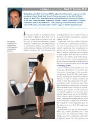
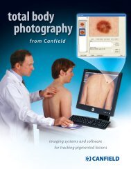
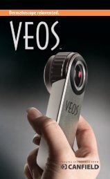
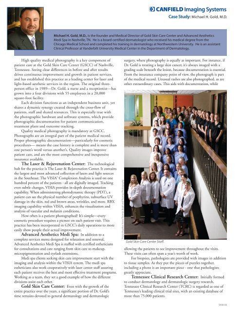
Case Study: <strong>Michael</strong> H. <strong>Gold</strong>, M.D.<strong>Michael</strong> H. <strong>Gold</strong>, M.D., is the founder and Medical Director of <strong>Gold</strong> Skin Care Center and Advanced AestheticsMedi Spa in Nashville, TN. He is a board certified dermatologist who received his medical degree from theChicago Medical School and completed his training in dermatology at Northwestern University. He is an assistantClinical Professor at Vanderbilt University Medical Center in the Department of Dermatology.High quality medical photography is a key component ofpatient care at the <strong>Gold</strong> Skin Care Center (GSCC) of Nashville,Tennessee. Seeing clear differences in before and after resultsdrives continuous improvement and growth in patient services,and has established this practice as a leading center for laser andlight-based aesthetic services in the region. The original threepersonoffice in 1989—Dr. <strong>Gold</strong>, a nurse and a receptionist—hasgrown into a four divisions with 55 employees in a 20,000square-foot facility.Each division functions as an independent business unit, yetshares a dynamic synergy created through the cross-flow ofpatients, staff and shared resources. This is especially true withthe photographic hardware and software systems, which providephotographic documentation for patient communication,treatment plans and outcome tracking.Quality medical photography is mandatory at GSCC.Photographs are an integral part of the patient medical record.Proper photographic documentation—particularly for cosmeticprocedures— means the case history is complete and is more thanone person’s word versus another’s. Quality images improvepatient care, and are the most comprehensive and inexpensiveinsurance available.The Laser & Rejuvenation Center: The technologicalhub for the practice is The Laser & Rejuvenation Center. It containsthe largest and most advanced collection of lasers and light sourcesin the Southeast. The VISIA ® Complexion Analysis is used on onehundred percent of the patients - all are digitally imaged. Trackingeven subtle changes, VISIA provides in-depth documentationcapability. When administering photodynamic therapy (PDT), apatient can see the physical number of porphyrins, subsurface UVdamage in the skin, red and brown areas, wrinkles, and more. RBXimaging capability within VISIA, enhances the visualization andanalysis of vascular and melanin conditions.How often is a patient photographed? It’s simple—everycosmetic procedure requires a picture on each patient visit. Thispractice has been incorporated in GSCC’s daily operations to moreeasily show people their actual improvement.Advanced Aesthetics Medi Spa: In addition to acomplete services menu designed for relaxation and renewal,Advanced Aesthetics Medi Spa is staffed with certified estheticiansfor consultations and care ranging from skin care to makeup,micropigmentation and eyelash extensions.Medi spa clients seeking skin care improvement start with theimaging and analysis within the VISIA system. The medi spaestheticians also work cooperatively with laser center staff assuringeach patient receives the best and most effective treatment programs.Working as a team, they set a good example of how the differentdivisions assist each other.<strong>Gold</strong> Skin Care Center: Even with the growth of theentire practice over the years, a significant portion of Dr. <strong>Gold</strong>’stime remains devoted to general dermatology and dermatologicsurgery, where photography is equally as important. For instance, ifDr. <strong>Gold</strong> is treating a large skin cancer, it’s always imaged with agrading scale beneath the lesion, because documentation is essential.From the insurance company point of view, the photograph is partof the medical record. Unusual rashes are also photographed, as areother extraordinary cases. This aids with documentation, whileallowing the patients to see improvement throughout the visits.These visits can often span a year’s worth of work.For biopsies, pathologists are provided with images in additionto tissue samples. As they put the pieces of puzzles together,including a photo is an important piece - one that pathologistsgreatly appreciate.Tennessee Clinical Research Center: Initially formedto conduct dermatology and dermatologic surgery research,Tennessee Clinical Research Center (TCRC) is regarded as one ofTennessee’s leading clinical trial sites, with an existing database ofmore than 75,000 patients.0908-08
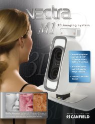
Case Study: <strong>Michael</strong> H. <strong>Gold</strong>, M.D.The Research Center has two rooms dedicated to researchphotography. This provides the consistent environment required toproduce consistent photographs. These photography rooms alsopermit total body imaging.Among its many clinical photography systems, TCRC has twoOMNIA ® Facial Imaging Systems. The OMNIA is great system forpositioning the patient’s head and then rotating the camera system90° to either the left or right side to document change. OMNIAhas the ability to overlay photos from session to session, making itone of the best devices on the market for picture consistency. Itproduces excellent patient images in a minimal amount of time.Imaging systems support: With thousands of imagesgenerated among <strong>Gold</strong> Skin Care Center’s four divisions, a masterimaging database for maintenance and access is a necessity, andthey have chosen to work with Canfield’s Mirror ® software. Thissoftware is interfaced with many of the campus computers but isuser specific to assist in confidentiality.GSCC has people in each department assigned to download andlabel pictures. It was a conscious decision to do so - with consistencycomes reproducible and usable pictures. Fewer people handling thephotos means they are able to maintain patient confidentiality.Photography becomes part of the medical records, and it is vital thatreliable, dependable pictures be taken on each visit.The next evolution of medical photography: Astreatments continue toward more effective delivery systems andtherapies, the hardware and capabilities for medical photographycontinue to evolve as well. Already, both in research and inmainstream medical photography, there is a new frontier of volumeand form, of height, breadth and depth—medical photography andanalysis in 3-D. This may be seen in the development of theVECTRA 3D system and associated software. This type ofquantitative analysis will greatly impact the ability to make betterpatient assessments, and therefore better diagnoses.-#-w w w.canfieldsci.com / phone (USA) 800.815.4330 / phone +1.973.276.0336 / fax +1.973.276.0339imaging sof tware · digital photography · complexion analysis · 3D solutions · research systems · training0908-08