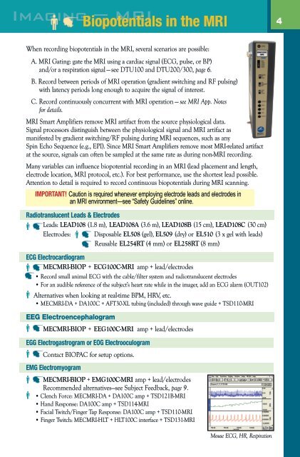
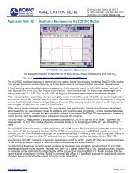
<strong>MRI</strong> Product Line for Human and Animal Protocols<strong>BIOPAC</strong> has expanded its line of specialized <strong>MRI</strong> products. <strong>MRI</strong> Smart Amplifiers and the<strong>MRI</strong> Cable/Filter systems provide isolated and RF-filtered interfacing between the subject/<strong>MRI</strong>chamber and the <strong>MRI</strong> control room to improve signal quality and optimize safety.With <strong>BIOPAC</strong>’s expanded line of <strong>MRI</strong> Smart Amplifiers and compatible transducers, you canrecord physiological signals such as: ECG, EEG, EGG, EMG, EOG, noninvasive blood pressurefor human and animal, pulse, respiration, SpO2, temperature, electrodermal activity, hand gripstrength (dynamometry), finger twitch, and a variety of pressure-based signals.For small animal cardiovascular and neuro studies, use the TSD104A-<strong>MRI</strong> Pressure Transducer ora Micro Pressure Measurement System to record pressure signals such as BP, LVP, and cranialpressure. Measure microvascular blood perfusion with the Laser Doppler Flow System.Specified electrodes, leads, and stimulus options provide safe data acquisition of physiologicalsignals in the <strong>MRI</strong> environment. Caution is required whenever employing electrode leads andelectrodes in an <strong>MRI</strong> environment—see “Safety Guidelines”online.“MR Safe” Products are non-conducting, non-metallic, and non-magnetic items thatpose no known hazards in all <strong>MRI</strong> environments.“MR Conditional” Products pose no known hazards in a specified MR environment withspecified conditions of use, e.g., field strength or lead configuration.Generally considered, if a transducer is MR Safe or MR Conditional, the transducer signal canbe recorded during <strong>MRI</strong> scanning. Transducer signals are typically high level and slow moving.These two features allow the transducer signal to be easily filtered to remove <strong>MRI</strong> artifact, if any.For MR Declarations and Application Notes on connections, analysis tools, and safety whenrecording physiological data in <strong>MRI</strong> or f<strong>MRI</strong>, go to www.biopac.com.<strong>BIOPAC</strong>’s <strong>MRI</strong> solutions include:Airflow & Gas Analysis Gating (Trigger/Synch)Biopotential Signals Laser Doppler FlowBlood PressurePulseElectrodermal Activity RespirationSpO2StimulationSubject FeedbackTemperature<strong>MRI</strong> Cable/Filter System Interface GuideSample isolated RFfilters and cablesMEC<strong>MRI</strong>-BIOP SystemBiopotential AmplifiersECG100C-<strong>MRI</strong> EMG100C-<strong>MRI</strong>EEG100C-<strong>MRI</strong> EGG EOGMEC<strong>MRI</strong>-DA SystemGeneral-purpose Trans. AmplifierTSD104A-<strong>MRI</strong> TSD117-<strong>MRI</strong>TSD121B-<strong>MRI</strong>MEC<strong>MRI</strong>-HLT SystemHigh-level Transducer AmplifierTSD131-<strong>MRI</strong> TSD115-<strong>MRI</strong>MEC<strong>MRI</strong>-TRANS SystemTransducer AmplifiersEDA100C-<strong>MRI</strong> RSP100CSKT100C PPG100C-<strong>MRI</strong>MEC<strong>MRI</strong>-STMISO SystemSTMISOC/D/E to STM100CCBL207 to STM200PNEUMATIC LINESNo electrical <strong>MRI</strong> Cable/Filterrequired—use DA100C.TSD110-<strong>MRI</strong> TSD137 seriesTSD114-<strong>MRI</strong> TSD237 seriesTSD221-<strong>MRI</strong>
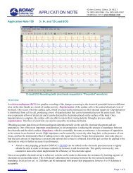
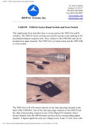
When recording biopotentials in the <strong>MRI</strong>, several scenarios are possible:A. <strong>MRI</strong> Gating: gate the <strong>MRI</strong> using a cardiac signal (ECG, pulse, or BP)and/or a respiration signal — see DTU100 and DTU200/300, page 6.B. Record between periods of <strong>MRI</strong> operation (gradient switching and RF pulsing)with latency periods long enough to acquire the signal of interest.C. Record continuously concurrent with <strong>MRI</strong> operation — see <strong>MRI</strong> App. Notesfor details.<strong>MRI</strong> Smart Amplifiers remove <strong>MRI</strong> artifact from the source physiological data.Signal processors distinguish between the physiological signal and <strong>MRI</strong> artifact asmanifested by gradient switching/RF pulsing during <strong>MRI</strong> sequences, such as anySpin Echo Sequence (e.g., EPI). Since <strong>MRI</strong> Smart Amplifiers remove most <strong>MRI</strong>-related artifactat the source, signals can often be sampled at the same rate as during non-<strong>MRI</strong> recording.Many variables can influence biopotential recording in an <strong>MRI</strong> (lead placement and length,electrode location, <strong>MRI</strong> protocol, etc.). For best performance, use the shortest lead possible.Attention to detail is required to record continuous biopotentials during <strong>MRI</strong> scanning.IMPORTANT! Caution is required whenever employing electrode leads and electrodes inan <strong>MRI</strong> environment—see “Safety Guidelines” online.Radiotranslucent Leads & ElectrodesLeads: LEAD108 (1.8 m), LEAD108A (3.6 m), LEAD108B (15 cm), LEAD108C (30 cm)Electrodes: Disposable EL508 (gel), EL509 (dry) or EL510 (3 x gel with leads)Reusable EL254RT (4 mm) or EL258RT (8 mm)ECG ElectrocardiogramMEC<strong>MRI</strong>-BIOP + ECG100C-<strong>MRI</strong> amp + lead/electrodes• Record small animal ECG with the cable/filter system and radiotranslucent electrodes• For an audible reference of the subject’s heart rate while in the imager, add an ECG alarm (OUT102)Alternatives when looking at real-time BPM, HRV, etc.• MEC<strong>MRI</strong>-DA + DA100C + AFT30-XL tubing (included) through wave guide + TSD110-<strong>MRI</strong>EEG ElectroencephalogramMEC<strong>MRI</strong>-BIOP + EEG100C-<strong>MRI</strong> amp + lead/electrodesEGG Electrogastrogram or EOG ElectrooculogramContact <strong>BIOPAC</strong> for setup options.EMG ElectromyogramMEC<strong>MRI</strong>-BIOP + EMG100C-<strong>MRI</strong> amp + lead/electrodesRecommended alternatives—see Subject Feedback, page 9.• Clench Force: MEC<strong>MRI</strong>-DA + DA100C amp + TSD121B-<strong>MRI</strong>• Hand Response: DA100C amp + TSD114-<strong>MRI</strong>• Facial Twitch/Finger Tap Response: DA100C amp + TSD110-<strong>MRI</strong>• Finger Twitch: MEC<strong>MRI</strong>-HLT + HLT100C interface + TSD131-<strong>MRI</strong>Mouse ECG, HR, Respiration