Worsening Tricuspid Regurgitation Following Pericardiectomy for ...
Worsening Tricuspid Regurgitation Following Pericardiectomy for ...
Worsening Tricuspid Regurgitation Following Pericardiectomy for ...

Create successful ePaper yourself
Turn your PDF publications into a flip-book with our unique Google optimized e-Paper software.
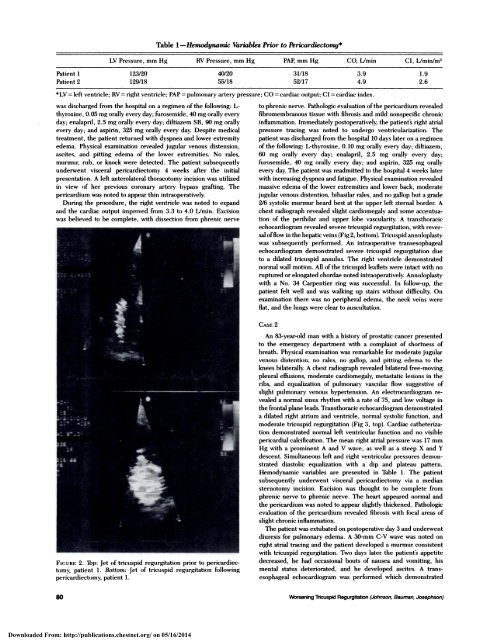
Table 1-Hemodynamw Variables Prior to <strong>Pericardiectomy</strong>*LV Pressure, mm Hg RV Pressure, mm Hg PAP, mm Hg CO. L/min CI, Llmin/m2Patient 1 123/20 40/20 31118 3.9 1.9Patient2 129/18 55/18 52/17 4.9 2.6*LV = left ventricle; RV right ventricle; PAP pulmonary artery pressure; CO cardiac output; CI cardiac index.was discharged from the hospital on a regimen of the following: L-thyroxine, 0.05 mg orally every day; furosemide, 40 mg orally everyday; enalapnil, 2.5 mg orally every day; diltiazem SR, 90 mg orallyevery day; and aspirin, 325 mg orally every day. Despite medicaltreatment, the patient returned with dyspnea and lower extremityedema. Physical examination revealed jugular venous distension,ascites, and pitting edema of the lower extremities. No rales,murmur, rub, or knock were detected. The patient subsequentlyunderwent visceral pericardiectomy 4 weeks after the initialpresentation. A left anterolateral thoracotomy incision was utilizedin view of her previous coronary artery bypass grafting. Thepericardium was noted to appear thin intraoperatively.During the procedure, the right ventricle was noted to expandand the cardiac output improved from 3.3 to 4.0 L/min. Excisionwas believed to be complete, with dissection from phrenic nerve::#{149}:L:: i’..: -t14#{149}1$FIGURE 2. Top: Jet of tricuspid regurgitation prior to pericardiectomy,patient 1 . Bottom: Jet of tricuspid regurgitation followingpericardiectomy, patient 1.to phrenic nerve. Pathologic evaluation ofthe penicardium revealedfibromembranous tissue with fibrosis and mild nonspecific chronicinflammation. Immediately postoperatively, the patient’s right atrialpressure tracing was noted to undergo ventricularization. Thepatient was discharged from the hospital 10 days later on a regimenof the following: L-thyroxine, 0.10 mg orally every day; diltiazem,60 mg orally every day; enalapril, 2.5 mg orally every day;furosemide, 40 mg orally every day; and aspirin, 325 mg orallyevery day. The patient was readmitted to the hospital 4 weeks laterwith increasing dyspnea and fatigue. Physical examination revealedmassive edema of the lower extremities and lower back, moderatejugular venous distention, bibasilar rales, and no gallop but a grade2/6 systolic murmur heard best at the upper left sternal border. Achest radiograph revealed slight cardiomegaly and some accentuationof the perihilar and upper lobe vascularity. A transthoracicechocardiogram revealed severe tricuspid regurgitation, with reversalofflow in the hepatic veins (Fig2, bottom). <strong>Tricuspid</strong> annuloplastywas subsequently per<strong>for</strong>med. An intraoperative transesophagealechocardiogram demonstrated severe tricuspid regurgitation dueto a dilated tricuspid annulus. The right ventricle demonstratednormal wall motion. All of the tricuspid leaflets were intact with noruptured or elongated chordae noted intraoperatively. Annuloplastywith a No. 34 Carpentier ring was successful. In follow-up, thepatient felt well and was walking up stairs without difficulty. Onexamination there was no peripheral edema, the neck veins wereflat, and the lungs were clear to auscultation.CASE 2An 83-year-old man with a history of prostatic cancer presentedto the emergency department with a complaint of shortness ofbreath. Physical examination was remarkable <strong>for</strong> moderate jugularvenous distention, no rales, no gallop, and pitting edema to theknees bilaterally. A chest radiograph revealed bilateral free-movingpleural effusions, moderate cardiomegaly, metastatic lesions in theribs, and equalization of pulmonary vascular flow suggestive ofslight pulmonary venous hypertension. An electrocardiogram revealeda normal sinus rhythm with a rate of 75, and low voltage inthe frontal plane leads. Transthoracic echocardiogram demonstrateda dilated right atrium and ventricle, normal systolic function, andmoderate tricuspid regurgitation (Fig 3, top). Cardiac cathetenizationdemonstrated normal left ventricular function and no visiblepericardial calcification. The mean right atrial pressure was 17 mmHg with a prominent A and V wave, as well as a steep X and Ydescent. Simultaneous left and right ventricular pressures demonstrateddiastolic equalization with a dip and plateau pattern.Hemodynamic variables are presented in Table 1 . The patientsubsequently underwent visceral pericardiectomy via a mediansternotomy incision. Excision was thought to be complete fromphrenic nerve to phrenic nerve. The heart appeared normal andthe pencardium was noted to appear slightly thickened. Pathologicevaluation of the pericardium revealed fibrosis with focal areas ofslight chronic inflammation.The patient was extubated on postoperative day 3 and underwentdiuresis <strong>for</strong> pulmonary edema. A 30-mm C-V wave was noted onright atrial tracing and the patient developed a murmur consistentwith tricuspid regurgitation. Two days later the patient’s appetitedecreased, he had occasional bouts of nausea and vomiting, hismental status deteriorated, and he developed ascites. A transesophagealechocardiogram was per<strong>for</strong>med which demonstrated80 <strong>Worsening</strong> <strong>Tricuspid</strong> <strong>Regurgitation</strong> (Johnson, Bauman, Josephson)Downloaded From: http://publications.chestnet.org/ on 05/16/2014