08Lec-COMPLICATIONS OF PNEUMONIA.pdf - Pediatric Infectious ...
08Lec-COMPLICATIONS OF PNEUMONIA.pdf - Pediatric Infectious ...
08Lec-COMPLICATIONS OF PNEUMONIA.pdf - Pediatric Infectious ...

Create successful ePaper yourself
Turn your PDF publications into a flip-book with our unique Google optimized e-Paper software.
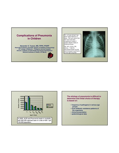
Complications of Pneumoniain ChildrenAlexander O. Tuazon, MD, FPPS, FPAPPAssociate Professor and Head, Section of <strong>Pediatric</strong> PulmonologyUP College of Medicine – Philippine General HospitalDirector, Institute of Child Health and Human DevelopmentNational Institutes of Health, UP ManilaCL, a 5-year-old girl, hasbeen highly febrile for 5days. Her aunt claims thather niece has beencoughing for nearly 3weeks despite intake ofAmbroxol syrup.PE: HR 112/min, RR35/min, T 38.4 0 C;decreased breath soundsand increased vocal fremition the right lung field.Number of Cases14000012000010000080000600004000020000050-6415-49.5-14.1-4Age In YearsMaleFemaleThe etiology of pneumonia is difficult todetermine and initial choice of therapyis based on:• frequency of pathogens in various agegroups• local antibiotic resistance patterns ofthe organisms• clinical presentation• epidemiological dataIn 2005, ALRI and Pneumonia leads in morbiditywith 692,305 reported case or a rate of 830.1 per100,000 population.
Most common pathogens basedon presentationTypicalStreptococcusHemophilusStaphylococcusAtypicalChlamydiaMycoplasmaLegionellaViralNosocomialPseudomonasKlebsiellaE. coliEnterobacterImmunocompromisedPneumocystisSpectrum of pathogens in PCAPBacterial PathogensS pneumoniaeMoraxella catarrhalisHaemophilus influenzaeAtypical PathogensMycoplasma pneumoniae (non-typable)Chlamydia pneumoniaeViral PathogensRespiratory syncitial virusInfluenza A and BAdenovirusRhinovirus, Enterovirus,Human MetapneumovirusPCAP: Incidence of Etiologic Agents by AgeWubble L, et al. Pediatr Infect Dis J 1999; 18:98–104.Adolescents may demonstrate the classicadult presentation of pneumonia, including:1. abrupt onset of symptoms2. high fever3. productive cough4. pleuritic chest pain, and5. possible toxic appearance.The presentation of the younger child withPCAP is often subtle:1. Fever2. Lethargy3. Tachypnea4. Irritability5. Vomiting, diarrhea and poor feeding.
Hypoxemia IrritabilityLethargy RetractionsTachypnea Toxic appearanceVomitingSocial factorsfollow-upPoor home careNeonateProgressionRapid progressionFailed outpatient therapyComplications et al. Prim Care 1996 Latham-Sadler,Leventhal JM. Clin Pediatr 1982Sensitivity and Specificity of Symptoms for PCAPSymptom Sensitivity SpecificityTachypnea 92% 15%Cough 92% 19%Toxic81% 60%appearanceCrackles 44% 80%Retractions 35% 82%Flaring 35% 82%Pallor 35% 87%Grunting 19% 94%A chest radiograph should be obtained if(1) the diagnosis is questionable(2) this is a repeated episode(3) the patient is ill enough to be admitted(4) the child is younger than 3 years and• has a fever > 39°C without a source and• leukocytosis > 15,000 mm 3(5) a complicated pneumonia is suspectedIndications for Admission in PCAPSigns and symptomsDyspnea GruntingInitial Empirical Treatment of PCAP Based onAge and Severity of PneumoniaAgeOutpatients(Mild to Moderate)3–6 mo Amoxicillin with orwithoutclavulanateErythromycin6 mo to5 yrAmoxicillin with orwithoutclavulanateMacrolideInpatients(Moderate)Ceftriaxone orcefotaximeCeftriaxone,Cefotaxime, orCefuroxime ±macrolide5–18 yr Macrolide Ceftriaxone orCefotaxime ±macrolideInpatients(Severe)Ceftriaxone orCefotaxime ±vancomycinCeftriaxone orCefotaxime ±macrolide ±vancomycinCeftriaxone orCefotaxime ±macrolide ±vancomycinHsiao G, PCCU 2001
<strong>Pediatric</strong> CAP<strong>Pediatric</strong> CAPPossible PathogenEmpiric TherapyPossible PathogenEmpiric TherapyMild, Nontoxicpharyngitis,rhinorrhea ordiarrheaProbably viralNoneBilateral, severeLobar or segmentalconsolidation,moderate - severeS pneumoniaeS pyogenesS aureusM pneumoniaeCefuroxime, ceftriaxone,cefotaximeNafcillin, oxacillin,cefazolin, clindamycinMacrolideModerate toxicityno hospitalizationLobar or segmentalconsolidation, mildS pneumoniaeS pyogenesH influenzaeM pneumoniaeInfluenza A and BAmoxicillinCo-amoxiclavCefprozil, Cefdinir,Cefpodoxime,Cefuroxime,CeftriaxoneMacrolideWith pleural fluid,empyema ornecrotizingS pneumoniaeS pyogenesS aureusH influenzaeAnaerobeCeftriaxone, cefotaximeNafcilllin, oxacillin,cefazolin, clindamycinVancomycinMeropenem, imipenemBL-BIBIBradley JS. Pediatr Infec Dis J 2002;21(6):592-598.Bradley JS. Pediatr Infec Dis J 2002;21(6):592-598.Predictors of Mortality in CommunityAcquired Pneumonia in ChildrenMaria Liza B. Zabala and Alexander O. Tuazon. UP-PGH 2001Predictors of Mortality in CommunityAcquired Pneumonia in ChildrenMaria Liza B. Zabala and Alexander O. Tuazon. UP-PGH 2001Variable Survivor Mortality p OR 95% CIPre-existing illness 27/74 32/46 0.0004 3.98 1.68-9.54Temp 38.5 27/74 26/46 0.009 2.26 1-5.15Altered mental status 60/74 45/46 0.007 10.5 1.47-453.64Mechanical ventilation 20/74 46/46 0.0000Variable Survivor Mortality p OR 95% CIHemoglobin (m+SD) 111+ 20 94+ 28 0.0002WBC < 5 x 10 9 /L 1/74 7/43 0.002 15.37 1.9-690.98WBC > 28 x 10 9 /L 6/74 7/43 0.033 3.15 0.94-11.33Platelet < 150x10 6 /mm 3 5/70 15/43 0.0003 6.5 1.98-24.6O2 supplementation 46/74 46/46 0.0000
Comparison of children admitted for Pneumococcal Pneumonia (Jerusalem)Characteristic Pulmonarycomplications(n=43) No pulmonarycomplications(n=68) P valueAge (years) 3.4 +3.24.0 +4.0M/F ratio 1.47 1.76 Background disease 50.0 46.5 NSRespiratory distress 52.3 21.7
Complications Comparison of children admitted for Pneumococcal Pneumonia (USA) ( Complications rose from 14% (1994) to 27% (1999), mostly with strain 1 ) Characteristic Pulmonarycomplications(n=133) No pulmonarycomplications(n=235) P valueAge (mos) 45 27 0.008White race 58 85 Background disease (%) 22.6 48.5 Antibiotics before diagnosis 19 12.8 NSChest pain (%) 29.3 7.7 Fever before diagnosis >3d (%) 65.1 31.4 findings >2 2 lobes (%) 37 CXR consolidation (%) 90.2 41.3 Defervescence >2d (%) 86.1 26.4 Hospitalization days (mean) 17.7 6.45
Lung Abscess• A circumscribed, thick-walled cavity in the lungthat contains purulent material resulting fromsuppuration and necrosis of the involved lungparenchyma.• An unresolved area of pneumonia is the site inwhich an abscess develops most frequently.• Pulmonary aspiration, diminished clearancemechanisms, embolic phenomena,hematogenous spread from septicemia, or localextension from oropharyngeal or abdominalprocesses contribute to abscess development.• Abscess may develop indolently over a fewweeks with tachypnea, cough and fever.2007 Patradoon-HoLung Abscess: OrganismsCommon anaerobes:• Fusobacterium nucleatum• Prevotella melaninogenica• Bacteroides fragilis group• Bacteroides urealyticus group• Peptostreptococcus species• Veilonella species• Microaerophilis streptococci• Porphyromonas• Prevotella oralis groupLung Abscess: OrganismsCommon aerobes:• S.aureus• E. coli• Klebsiella pneumoniae• Pseudomonas aeruginosa• S. pyogenes• Group B StreptococcusLung Abscess: Evaluation• Common Signs and Symptoms:Fever, pleuritic chest pain, cough, hemoptysis,dyspnea, sputum production, weight loss,malaise• Physical Examination:Tachypnea, tachycardia, retractions,decreasedchest movement, decreased breath sounds,dullness to percussion, crackles, bronchialbreathing
(%)Lethargy 31Yen et al(n+23)9187352622Lung abscess in children.Patradoon-Ho P, et al. Paediatr Respir Rev 2007Symptoms (%) Children’sHosp, Sydney (n=23) Tan, et al(n=25) Chan et al(n=27) Yen et al(n=23)Fever 83 84 100 91Cough 65 53 67 87Dyspnea 36 35 19 35Cheat pain 24 22 9Anorexia/ Nauseaand Vomiting 24 20 4 26Malaise and31 11 NR 22Lung Abscess: EvaluationDiagnosis:Chest X-Ray: solitary, thick-walled cavity in thelung with or without air fluid levelUltrasonography and CT scan: to localize thelesion and guide drainage or needle aspiration.Direct percutaneous aspiration is the mostreliable mode of identification of the etiologicagent.Lung Abscess: Antimicrobial Treatment• Overall outcome is good, with mortality rateslower than those in adults.• Up to 90% of patients with lung abscess may beadequately treated with intravenous antibiotictherapy.• The choice of antibiotic is usually empiric basedon the underlying condition of the patient and thepresumed etiologic agent(s).• The duration of parenteral treatment varies from 5days (Patradoon-Ho 2007) to 3 weeks (Tan 1995),followed by oral therapy.2007 Patradoon-HoLung Abscess: Surgical Treatment• Surgical management is considered in cases oflarge lung abscess especially when associatedwith hemoptysis.• Surgical management is indicated if there isclinical deterioration despite appropriateantibiotic therapy.1. Drainage via bronchoscopy2. Percutaneous tube drainage3. Percutaneous needle aspiration4. LobectomyLobectomy or wedge resection should be reservedfor massive expansion of the abscess associatedwith mediastinal shift and attendant symptoms.
Pleural Effusion and EmpyemaMT a 5 y/o male with high-grade fever and dyspnea. 1 month PTA, he developedcough with low grade fever on-and –off. 2 weeks PTA, consulted with a privatephysician and was given Amoxicillin and carbocisteine with no relief. 2 days PTA,fever became high grade w/ progressive dyspnea.PE: HR 120 RR 48 T 39.1 C; (+) multiple CLAD(+) chest lag on the left, (+) decreased breath sounds and vocal fremitus ,left(+) dullness to percussion left, (-) crackles, (-) wheezing• Collection of fluid or pus in the pleural space• Can occur as a complication of pneumonia,tuberculosis or surgical procedures( post-surgical empyema)• Staphylococcus aureus is the single mostcommon pathogen of empyema in infants< 2 years of age• Other common nontuberculous causes ofempyema include H. influenzae type B,S. pyogenes, D. pneumoniae, E. coli,Klebsiella sp, Pseudomonas aeruginosa.Pleural Effusion and Empyema• The diagnosis of empyema include CXR,ultrasound and examination of pleural fluid• Obliteration of the costophrenic sulcus is theearliest radiologic sign of pleural fluidaccumulation• Failure of the liquid to shift from upright todecubitus view indicates loculation as commonlyseen in staphylococcal empyemaPleural Effusion and EmpyemaPhysical examination findings:• Tachypnea• Fever• Chills, Cough• Irritability, Anorexia, Lethargy• Chest pain, Chest tightness• Diminished thoracic excursion• Fullness of the intercostal spaces, Dull orflat percussion• Decreased tactile and vocal fremiti• Displaced trachea and cardiac apex
Pleural Effusion and Empyema• Expectoration of an increasing amount ofpurulent sputum with or without hemoptysis mayherald the onset of bronchopleural fistula andpyopneumothorax• Bronchopleural fistula may be due to rupture ofneglected empyema into the lung or rupture ofpulmonary suppuration into the pleura• Muffling of the heart tones and pericardial rubindicate extension into the pericardiumPleural Effusion and Empyema: Treatment• Outcome is uniformly good, regardless oftreatment option• Treatment is aimed at specific management of theunderlying cause and relief of functionaldisturbances caused by the existing clinicaldisorder, pleural involvement and concurrentcomplications• The basic principle for treatment is to drain theinfected pleural space and allow lung re-expansion• Treatment is medical (high dose intravenousantibiotics) and surgicalPleural Effusion and Empyema: Treatment• General supportive measures:1. Bed rest2. Analgesia3. Fluid replacement4. Supplemental oxygen5. Lying on the affected side• Choice of antimicrobial is based on bacterialepidemiology in the community, clinical data,pharmacologic properties of the drug.• Repeated thoracentesis and eventuallycontinuous chest tube drainage are indicated ifrapid re-accumulation of effusion inducesdyspnea.Pleural Effusion and Empyema: AntibioticsLittle difference in penetration of penicillinsand cephalosporins into empyemas anduninfected parapneumonic fluids.Drugs with excellent pleural penetrationinclude aztreonam, clindamycin,ciprofloxacin, cephalothin and penicillinAminoglycosides may be inactivated orhave poor penetration into empyemas thanuncomplicated parapneumonic effusions.
Pleural Effusion and Empyema: TreatmentIndications for tube thoracostomy:1. Identification of an organism by gram stain2. Positive pleural fluid culture3. Pleural fluid glucose < 40 mg/dl4. Pleural fluid LDH >1000 IU5. Pleural fluid pH 500/mm 3Fibropurulent Fluid is thicker and opaque, orPositive cultureAntibiotics with orwithout chest tubedrainageAntibiotics with chesttube drainageChronic A peel forms around the lung Decortication
Acute 34/42(81%)Fibropurulent 15/17(88%)ChronicClassification Success DecorticationHospitalizationFever afterdrainageTubeinsertion8/42 (19%) 22.4 + 6.6 d 9.2 + 6.6 d 7.6 + 5.6 d2/17 (12%) 30.1 + 11.5 d 10.0 + 4.0 d 12.8 + 9.3 dparapneumonic effusion and empyema in children. Shen YH, et al. J Microbiol Immunol Infect 2006 ComplicatedFC, 3 y/o male w/ 2-weekhistoryof cough & low-gradefever productive ofwhitish phlegm. 9 days agoconsulted at a local hospital,chest x-ray doneshowed pleural effusion left.Given oral Cefuroxime. Fewhours PTAsuddenly became dyspneicand was rushed to the ER.PE: HR 140 RR 50 T 37.9 CTrachea deviated to the right(+) chest lag, left (+)decreasedbreath sounds left lung field,hyperresonanton percussion, left chest;Apical heart sounds heard onthe rightPneumothorax• An accumulation of air in the pleural spaces dueto secondary to free communication of the pleuralspace with the atmosphere either from a chestwall defect through the parietal pleura or fromalveolar rupture• Can be secondary to infection with gas-producingmicroorganisms.3 factors that determine the extent of alveolar rupture:1. Degree of transpulmonary pressure exerted2. Duration of pressure applied3. Ratio of inexpansible to expansible portion ofthe lungPneumothorax• Signs and symptoms may vary according to theextent of lung collapse, degree of intrapleuralpressure, rapidity of onset and age andrespiratory reserve of the patient• PE includes chest bulging on the affected side ifone side is involved, shift of cardiac impulseaway from the site of the pneumothorax,tachypnea, decreased breath sounds on theaffected side,tachycardia• Grunting,retraction and cyanosis occur late in theprogression of the complication
Pneumothorax• Differential diagnosis include lung cyst, lobaremphysema, bullae,diaphragmatic hernia• CXR is crucial in the confirmation of diagnosis• Effective management requires early clinicalrecognition and prompt radiologic investigation• Therapeutic management should take intoaccount clinical severity, presence and nature ofthe underlying lung disease, precipitating eventand history of recurrencePneumothorax• Direct mechanical evacuation of intrapleural airshould be performed unless the size of thepneumothorax is very small, the underlyingdisorder is mild and the clinical status is stable• Close clinical and blood gas monitoring areintegral parts of the management in all situations.SummaryComplications of lung infections such as lungabscess, empyema and pneumothorax require ahigh index of clinical suspicion and confirmationby employing the appropriate diagnostic testing.Management of these infections includesprescription of appropriate antimicrobials andmay require specific drainage procedures and thejudicial use of surgical interventions.