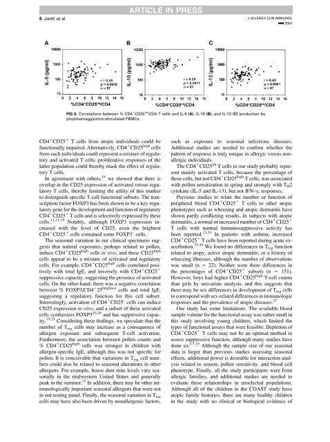
6 Jartti et alARTICLE IN PRESSJ ALLERGY CLIN IMMUNOLnnn 2007FIG 6. Correlations <strong>between</strong> % <strong>CD4</strong> 1 <strong>CD25</strong> <strong>in</strong>t /<strong>CD4</strong> T <strong>cells</strong> <strong>and</strong> IL-5 (A), IL-10 (B), <strong>and</strong> IL-13 (C) production byphytohemagglut<strong>in</strong><strong>in</strong>-stimulated PBMCs.<strong>CD4</strong> 1 <strong>CD25</strong> 1 T <strong>cells</strong> from atopic <strong>in</strong>dividuals could befunctionally impaired. Alternatively, <strong>CD4</strong> 1 <strong>CD25</strong> high <strong>cells</strong>from such <strong>in</strong>dividuals could represent a mixture of regulatory<strong>and</strong> activated T <strong>cells</strong>: proliferative responses of thelatter population could thereby mask the effect of regulatoryT <strong>cells</strong>.In agreement with others, 18 we showed that there isoverlap <strong>in</strong> the <strong>CD25</strong> expression of activated versus regulatoryT <strong>cells</strong>, thereby limit<strong>in</strong>g the utility of this markerto dist<strong>in</strong>guish specific T-cell functional subsets. The transcriptionfactor FOXP3 has been shown to be a key regulatorygene for the development <strong>and</strong> function of regulatory<strong>CD4</strong> 1 <strong>CD25</strong> 1 T <strong>cells</strong> <strong>and</strong> is selectively expressed by these<strong>cells</strong>. 11,12,19 Notably, although FOXP3 expression <strong>in</strong>creasedwith the level of <strong>CD25</strong>, even the brightest<strong>CD4</strong> 1 <strong>CD25</strong> 1 <strong>cells</strong> conta<strong>in</strong>ed some FOXP3 – <strong>cells</strong>.The seasonal variation <strong>in</strong> our cl<strong>in</strong>ical specimens suggeststhat natural exposures, perhaps related to pollen,<strong>in</strong>duce <strong>CD4</strong> 1 <strong>CD25</strong> high <strong>cells</strong> <strong>in</strong> vivo, <strong>and</strong> these <strong>CD25</strong> high<strong>cells</strong> appear to be a mixture of activated <strong>and</strong> regulatory<strong>cells</strong>. For example, <strong>CD4</strong> 1 <strong>CD25</strong> high <strong>cells</strong> correlated positivelywith total IgE, <strong>and</strong> <strong>in</strong>versely with <strong>CD4</strong> 1 <strong>CD25</strong> 1suppressive capacity, suggest<strong>in</strong>g the presence of activated<strong>cells</strong>. On the other h<strong>and</strong>, there was a negative correlation<strong>between</strong> % FOXP3/<strong>CD4</strong> 1 25 brightest <strong>cells</strong> <strong>and</strong> total IgE,suggest<strong>in</strong>g a regulatory function for this cell subset.Interest<strong>in</strong>gly, activation of <strong>CD4</strong> 1 <strong>CD25</strong> – <strong>cells</strong> can <strong>in</strong>duce<strong>CD25</strong> expression <strong>in</strong> vitro, <strong>and</strong> a subset of these activated<strong>cells</strong> synthesizes FOXP3 19,20 <strong>and</strong> has suppressive capacity.19,21 Consider<strong>in</strong>g these f<strong>in</strong>d<strong>in</strong>gs, we speculate that thenumber of T reg <strong>cells</strong> may <strong>in</strong>crease as a consequence ofallergen exposure <strong>and</strong> subsequent T-cell activation.Furthermore, the association <strong>between</strong> pollen counts <strong>and</strong>% <strong>CD4</strong> 1 <strong>CD25</strong> high <strong>cells</strong> was stronger <strong>in</strong> children withallergen-specific IgE, although this was not specific forpollen. It is conceivable that variations <strong>in</strong> T reg cell numberscould also be related to seasonal alterations <strong>in</strong> otherallergens. For example, house dust mite levels vary seasonally<strong>in</strong> the midwestern United States <strong>and</strong> generallypeak <strong>in</strong> the summer. 22 In addition, there may be other immunologicallyimportant seasonal allergens that were not<strong>in</strong> our test<strong>in</strong>g panel. F<strong>in</strong>ally, the seasonal variation <strong>in</strong> T reg<strong>cells</strong> may have also been driven by nonallergenic factors,such as exposure to seasonal <strong>in</strong>fectious diseases.Additional studies are needed to confirm whether thepattern of response is truly unique <strong>in</strong> allergic versus nonallergic<strong>in</strong>dividuals.The <strong>CD4</strong> 1 <strong>CD25</strong> <strong>in</strong>t T <strong>cells</strong> <strong>in</strong> our study probably representma<strong>in</strong>ly activated T <strong>cells</strong>, because the percentage ofthese <strong>cells</strong>, but not <strong>CD4</strong> 1 <strong>CD25</strong> high T <strong>cells</strong>, was associatedwith pollen sensitization <strong>in</strong> spr<strong>in</strong>g <strong>and</strong> strongly with T H 2cytok<strong>in</strong>e (IL-5 <strong>and</strong> IL-13), but not IFN-g, responses.Previous studies to relate the number or function ofperipheral blood <strong>CD4</strong> 1 <strong>CD25</strong> 1 T <strong>cells</strong> to other atopicphenotypes such as wheez<strong>in</strong>g <strong>and</strong> atopic dermatitis haveshown partly conflict<strong>in</strong>g results. In subjects with atopicdermatitis, a normal or <strong>in</strong>creased number of <strong>CD4</strong> 1 <strong>CD25</strong> 1T <strong>cells</strong> with normal immunosuppressive activity hasbeen reported. 23,24 In patients with asthma, <strong>in</strong>creased<strong>CD4</strong> 1 <strong>CD25</strong> 1 T <strong>cells</strong> have been reported dur<strong>in</strong>g acute exacerbation.25,26 We found no differences <strong>in</strong> T reg functionrelated to <strong>atopy</strong>, active atopic dermatitis, or a history ofwheez<strong>in</strong>g illnesses, although the number of observationswas small (n 5 22). Neither were there differences <strong>in</strong>the percentages of <strong>CD4</strong> 1 <strong>CD25</strong> 1 subsets (n 5 151).However, boys had higher <strong>CD4</strong> 1 <strong>CD25</strong> high T-cell countsthan girls by univariate analysis, <strong>and</strong> this suggests thatthere may be sex differences <strong>in</strong> development of T reg <strong>cells</strong>to correspond with sex-related differences <strong>in</strong> immunologicresponses <strong>and</strong> the prevalence of atopic diseases. 27Our study has some limitations. The available bloodsample volume for the functional assay was rather small <strong>in</strong>this study <strong>in</strong>volv<strong>in</strong>g young children, which limited thetypes of functional assays that were feasible. Depletion of<strong>CD4</strong> 1 <strong>CD25</strong> 1 T <strong>cells</strong> may not be an optimal method toassess suppressive function, although many studies havedone so. 3,7,10 Although the sample size of our seasonaldata is larger than previous studies assess<strong>in</strong>g seasonaleffects, additional power is desirable for <strong>in</strong>teraction analysisrelated to season, pollen sensitivity, <strong>and</strong> blood cellphenotype. F<strong>in</strong>ally, all the study participants were fromallergic families, <strong>and</strong> additional studies are needed toevaluate these relationships <strong>in</strong> unselected populations.Although all of the children <strong>in</strong> the COAST study haveatopic family histories, there are many healthy children<strong>in</strong> the study with no cl<strong>in</strong>ical or biological evidence of
J ALLERGY CLIN IMMUNOLVOLUME nnn, NUMBER nnARTICLE IN PRESSJartti et al 7allergic diseases, <strong>and</strong> these serve as our ma<strong>in</strong> controlgroup. We believe that the significant differences <strong>between</strong>atopic <strong>and</strong> nonatopic children <strong>in</strong> COAST are noteworthy<strong>and</strong> significant <strong>in</strong> underst<strong>and</strong><strong>in</strong>g l<strong>in</strong>ks <strong>between</strong> familialpredisposition <strong>and</strong> the development of atopic diseases.Additional studies are be<strong>in</strong>g designed to evaluate theserelationships <strong>in</strong> unselected populations, as well as <strong>in</strong> childrenliv<strong>in</strong>g <strong>in</strong> urban rather than suburban locations.In conclusion, despite previously published suggestionsthat <strong>CD4</strong> 1 <strong>CD25</strong> high T <strong>cells</strong> have high regulatoryactivity, our f<strong>in</strong>d<strong>in</strong>gs <strong>in</strong> children do not support a straightforwardrelationship <strong>between</strong> <strong>CD25</strong> phenotype <strong>and</strong> function.In fact, our f<strong>in</strong>d<strong>in</strong>gs suggest that <strong>CD4</strong> 1 <strong>CD25</strong> highT <strong>cells</strong> are seasonally regulated by environmental factors,<strong>and</strong> are likely to represent a mixture of activated <strong>and</strong> regulatoryT <strong>cells</strong>, especially <strong>in</strong> atopic children.REFERENCES1. Heaton T, Rowe J, Turner S, Aalberse RC, de Klerk N, SuriyaarachchiD, et al. An immunoepidemiological approach to asthma: identificationof <strong>in</strong>-vitro T-cell response patterns associated with different wheez<strong>in</strong>gphenotypes <strong>in</strong> children. Lancet 2005;365:142-9.2. Van Oosterhout AJ, Bloksma N. Regulatory T-lymphocytes <strong>in</strong> asthma.Eur Respir J 2005;26:918-32.3. L<strong>in</strong>g EM, Smith T, Nguyen XD, Pridgeon C, Dallman M, Arbery J, et al.Relation of <strong>CD4</strong>1<strong>CD25</strong>1 regulatory T-cell suppression of allergendrivenT-cell activation to atopic status <strong>and</strong> expression of allergic disease.Lancet 2004;363:608-15.4. Francis JN, Till SJ, Durham SR. Induction of IL-101<strong>CD4</strong>1<strong>CD25</strong>1T <strong>cells</strong> by grass pollen immunotherapy. J Allergy Cl<strong>in</strong> Immunol 2003;111:1255-61.5. Gr<strong>in</strong>debacke H, W<strong>in</strong>g K, Andersson AC, Suri-Payer E, Rak S, Rud<strong>in</strong> A.Defective suppression of Th2 cytok<strong>in</strong>es by <strong>CD4</strong><strong>CD25</strong> regulatory T <strong>cells</strong><strong>in</strong> birch allergics dur<strong>in</strong>g birch pollen season. Cl<strong>in</strong> Exp Allergy 2004;34:1364-72.6. Bell<strong>in</strong>ghausen I, Klostermann B, Knop J, Saloga J. Human<strong>CD4</strong>1<strong>CD25</strong>1 T <strong>cells</strong> derived from the majority of atopic donors areable to suppress TH1 <strong>and</strong> TH2 cytok<strong>in</strong>e production. J Allergy Cl<strong>in</strong>Immunol 2003;111:862-8.7. Jutel M, Akdis M, Budak F, Aebischer-Casaulta C, Wrzyszcz M, BlaserK, et al. IL-10 <strong>and</strong> TGF-beta cooperate <strong>in</strong> the regulatory T cell responseto mucosal allergens <strong>in</strong> normal immunity <strong>and</strong> specific immunotherapy.Eur J Immunol 2003;33:1205-14.8. Smith TR, Alex<strong>and</strong>er C, Kay AB, Larche M, Rob<strong>in</strong>son DS. Cat allergenpeptide immunotherapy reduces <strong>CD4</strong>(1) T cell responses to cat allergenbut does not alter suppression by <strong>CD4</strong>(1) <strong>CD25</strong>(1) T <strong>cells</strong>: a doublebl<strong>in</strong>dplacebo-controlled study. Allergy 2004;59:1097-101.9. Baecher-Allan C, Wolf E, Hafler DA. Functional analysis of highlydef<strong>in</strong>ed, FACS-isolated populations of human regulatory <strong>CD4</strong>1 <strong>CD25</strong>1T <strong>cells</strong>. Cl<strong>in</strong> Immunol 2005;115:10-8.10. Karlsson MR, Rugtveit J, Br<strong>and</strong>tzaeg P. Allergen-responsive<strong>CD4</strong>1<strong>CD25</strong>1 regulatory T <strong>cells</strong> <strong>in</strong> children who have outgrowncow’s milk allergy. J Exp Med 2004;199:1679-88.11. Fontenot JD, Gav<strong>in</strong> MA, Rudensky AY. Foxp3 programs the development<strong>and</strong> function of <strong>CD4</strong>1<strong>CD25</strong>1 regulatory T <strong>cells</strong>. Nat Immunol2003;4:330-6.12. Hori S, Nomura T, Sakaguchi S. Control of regulatory T cell developmentby the transcription factor Foxp3. Science 2003;299:1057-61.13. Lemanske RF Jr. The childhood orig<strong>in</strong>s of asthma (COAST) study.Pediatr Allergy Immunol 2002;13(suppl 15):38-43.14. Taams LS, Vukmanovic-Stejic M, Smith J, Dunne PJ, Fletcher JM,Plunkett FJ, et al. Antigen-specific T cell suppression by human<strong>CD4</strong>1<strong>CD25</strong>1 regulatory T <strong>cells</strong>. Eur J Immunol 2002;32:1621-30.15. Neaville WA, Tisler C, Anklam K, Gilbertson-White S, Hamilton R,Adler K, et al. Developmental cytok<strong>in</strong>e response profiles <strong>and</strong> the cl<strong>in</strong>ical<strong>and</strong> immunologic expression of <strong>atopy</strong> <strong>in</strong> <strong>in</strong>fancy. J Allergy Cl<strong>in</strong> Immunol2003;112:740-6.16. Copenhaver CC, Gern JE, Li Z, Shult PA, Rosenthal LA, Mikus LD, et al.Cytok<strong>in</strong>e response patterns, exposure to viruses, <strong>and</strong> respiratory <strong>in</strong>fections<strong>in</strong> the first year of life. Am J Respir Crit Care Med 2004;170:175-80.17. Smith EG. Sampl<strong>in</strong>g <strong>and</strong> identify<strong>in</strong>g allergenic pollens <strong>and</strong> molds: anillustrated manual for physicians <strong>and</strong> lab technicians. 1st ed. San Antonio(TX): Blewstone Press; 1984.18. Valencia X, Stephens G, Goldbach-Mansky R, Wilson M, Shevach EM,Lipsky PE. TNF downmodulates the function of human <strong>CD4</strong>1<strong>CD25</strong>hiT-regulatory <strong>cells</strong>. Blood 2006;108:253-61.19. Walker MR, Kasprowicz DJ, Gersuk VH, Benard A, Van L<strong>and</strong>eghen M,Buckner JH, et al. Induction of FoxP3 <strong>and</strong> acquisition of T regulatoryactivity by stimulated human <strong>CD4</strong>1. J Cl<strong>in</strong> Invest 2003;112:1437-43.20. Mantel PY, Ouaked N, Ruckert B, Karagiannidis C, Welz R, Blaser K,et al. Molecular mechanisms underly<strong>in</strong>g FOXP3 <strong>in</strong>duction <strong>in</strong> humanT <strong>cells</strong>. J Immunol 2006;176:3593-602.21. Lim HW, Hillsamer P, Banham AH, Kim CH. Cutt<strong>in</strong>g edge: directsuppression of B <strong>cells</strong> by <strong>CD4</strong>1 <strong>CD25</strong>1 regulatory T <strong>cells</strong>. J Immunol2005;175:4180-3.22. Arlian LG, Neal JS, Morgan MS, Vyszenski-Moher DL, Rapp CM,Alex<strong>and</strong>er AK. Reduc<strong>in</strong>g relative humidity is a practical way to controldust mites <strong>and</strong> their allergens <strong>in</strong> homes <strong>in</strong> temperate climates. J AllergyCl<strong>in</strong> Immunol 2001;107:99-104.23. Ou LS, Goleva E, Hall C, Leung DY. T regulatory <strong>cells</strong> <strong>in</strong> atopic dermatitis<strong>and</strong> subversion of their activity by superantigens. J Allergy Cl<strong>in</strong>Immunol 2004;113:756-63.24. Vukmanovic-Stejic M, McQuaid A, Birch KE, Reed JR, Macgregor C,Rust<strong>in</strong> MH, et al. Relative impact of <strong>CD4</strong>1<strong>CD25</strong>1 regulatory T <strong>cells</strong><strong>and</strong> tacrolimus on <strong>in</strong>hibition of T-cell proliferation <strong>in</strong> patients with atopicdermatitis. Br J Dermatol 2005;153:750-7.25. Gemou-Engesaeth V, Bush A, Kay AB, Hamid Q, Corrigan CJ. Inhaledglucocorticoid therapy of childhood asthma is associated with reducedperipheral blood T cell activation <strong>and</strong> ‘‘Th2-type’’ cytok<strong>in</strong>e mRNAexpression. Pediatr 1997;99:695-703.26. Shi HZ, Li S, Xie ZF, Q<strong>in</strong> XJ, Q<strong>in</strong> X, Zhong XN. Regulatory<strong>CD4</strong>1<strong>CD25</strong>1 T lymphocytes <strong>in</strong> peripheral blood from patients withatopic asthma. Cl<strong>in</strong> Immunol 2004;113:172-8.27. Uekert SJ, Akan G, Evans MD, Li Z, Roberg K, Tisler C, et al. Sexrelateddifferences <strong>in</strong> immune development <strong>and</strong> the expression of <strong>atopy</strong><strong>in</strong> early childhood. J Allergy Cl<strong>in</strong> Immunol 2006;118:1375-81.