40 Il Tabacco, 7(2)Sannino et al. (1995), <strong>the</strong> setae according to <strong>the</strong>nomenclature suggested by Hinton (1946). Thedescription <strong>of</strong> <strong>the</strong> surface structure <strong>of</strong> <strong>the</strong> egg(sculpturing) is based upon <strong>the</strong> nomenclatureproposed by Döring (1955).Part <strong>of</strong> <strong>the</strong> examined specimens was prepared- adults pinned and dried, immature stages inethyl alcohol (80 %) or in KAAD solutionslightly modified (see Sannino et al., 1995) - anddeposited in <strong>the</strong> Experimental Tobacco Institutecollection.Description <strong>of</strong> <strong>the</strong> stagesEggIt is <strong>of</strong> an upright type, us usual in this group.Cream white when just laid, turns to dark greywith embryo development. Subspherical, with asomewhat flat base, slightly wider (ca. 0.70 mm)and distinctly ribbed. Some 35-40 vertical ribsextend from <strong>the</strong> base to <strong>the</strong> micropylar area, 10-12 <strong>of</strong> which are shorter, terminating before <strong>the</strong>yreach <strong>the</strong> micropylar area. Finer crossribs (12-16) occur between <strong>the</strong> vertical ribs to form rows<strong>of</strong> rectangular areas between <strong>the</strong>m. Micropylarrosette, consisting <strong>of</strong> 11-13 petal-shaped cells,clearly exceeds in height <strong>the</strong> level <strong>of</strong> adjacentcells. A more thorough examination <strong>of</strong> <strong>the</strong>chorionic structure has been possible with <strong>the</strong> use<strong>of</strong> electron microscopy (Mazzini, 1974; Beck etal., 1993). Eggs appear more or less circular whenviewed from above (Fig. I).Mature larvaHead hypognathous, ochreous or pale brown,darkish reticulate, streaked with two black widestripes along <strong>the</strong> margins <strong>of</strong> <strong>the</strong> adfrontal suturesand two finer ones in <strong>the</strong> stemmatal area.Epicranial notch (= vertical triangle) obtuse andventrally rounded. Upper portion <strong>of</strong> <strong>the</strong> epicranialsuture at least twice longer than <strong>the</strong> portionincluded between <strong>the</strong> adfrontal sutures.Antennae, as usual in this group, quite short andthree-segmented, with <strong>the</strong> first antennal jointblack and <strong>the</strong> last two whitish-yellow (Fig. II,1). Mandible stout, subquadrangular, with 4 teethon cutting edge and 1 outer seta; inner surfacewith 4 simple ridges and a distinct, triangulartooth (Fig. II, 2).Body integument smooth. Backgroundcoloration grayish-ochreous to greyish-brown.Sometimes, ventral surface slightly paler thandorsum. Middorsal line pale, thin, generally littledistinct. A broad dorsal longitudinal band, barelyvisible in <strong>the</strong> pale forms, slightly darker thanground colour, is present on all segments tillabdominal segment 8 (A8), becoming much moreevident along <strong>the</strong> A7-8 lateral margins, wherecouples <strong>of</strong> typical conspicuous subtriangularblack marks stand out. The ones present on A8,are usually joined posteriorly forming a typicalhorseshoe drawing. Laterally, <strong>the</strong>re can be seen<strong>the</strong> lowerspiracular and <strong>the</strong> abovespiracular fascia.The former is ochreous or whitish, with <strong>the</strong>margins shaded; <strong>the</strong> latter is dark, generally moremarked nearby spiracles (Fig. III, 1, 2, 3).Pinacula inconspicuous, dark, non elevatedexcept for L3 (located above <strong>the</strong> base <strong>of</strong> prolegs)much more evident.Spiracles elliptical, white with blackperitreme. Prothoracic and A8 spiracles distinctlylarger than those on A1-7.Prothoracic shield subtrapeziodal, barelydarker than background, on which threelongitudinal, pale and straight stripes stand out:one median with well defined margins and twoon <strong>the</strong> sides, wider (Fig. III, 4).Anal shield subtriangular, darker thanbackground. Anterior margin presents a midslight prominence, while <strong>the</strong> posterior one isstrongly curved (Fig. III, 5).Legs pale. Prolegs present, as usual in thisgroup, on A3-6 and A10. They are concolorouswith background, with an external grayish or darkirregular spot. A3-6 and A10 crochets arrangedin an uniordinal mesoseries, in number <strong>of</strong> 25-28,29-32, 29-33, 32-35 and 35-39, respectively.Some coloured pictures <strong>of</strong> <strong>the</strong> larva may befound in Gómez de Aizpúrua (1985), Sauer(1992) and Porter (1997).Body length 43-50 mm (mean 46.9 ± 0.2. No.= 20); body width 6-7 mm (mean 6.3 ± 0.1. No.= 20); head capsule width 3-3.2 mm.
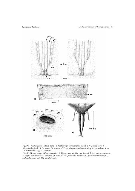
Sannino ed Espinosa <strong>On</strong> <strong>the</strong> <strong>morphology</strong> <strong>of</strong> <strong>Noctua</strong> <strong>comes</strong> 41Fig. IV. - <strong>Noctua</strong> <strong>comes</strong> Hübner, pupa – 1. Ventral view (two different cases). 2. A4, dorsal view. 3.Abdominal spiracle. 4. Cremaster. (A, antenna; FW, forewing or mesothoracic wing; L2, mesothoracic leg;L3, metathoracic leg; MX, maxilla).Fig. IV. - <strong>Noctua</strong> <strong>comes</strong> Hübner, crisalide – 1. Visione ventrale (due casi diversi). 2. A4, visto dorsalmente.3. Stigma addominale. 4. Cremaster. (A, antenna; FW, pteroteche anteriori; L2, podoteche mediane; L3,podoteche posteriori; MX, maxilloteche).