Environmental and Experimental Botany Naphthoquinones as ...
Environmental and Experimental Botany Naphthoquinones as ...
Environmental and Experimental Botany Naphthoquinones as ...

Create successful ePaper yourself
Turn your PDF publications into a flip-book with our unique Google optimized e-Paper software.
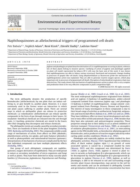
<strong>Environmental</strong> <strong>and</strong> <strong>Experimental</strong> <strong>Botany</strong> 65 (2009) 330–337Contents lists available at ScienceDirect<strong>Environmental</strong> <strong>and</strong> <strong>Experimental</strong> <strong>Botany</strong>journal homepage: www.elsevier.com/locate/envexpbot<strong>Naphthoquinones</strong> <strong>as</strong> allelochemical triggers of programmed cell deathPetr Babula a,∗ , Vojtěch Adam b , René Kizek b , Zdeněk Sladký a , Ladislav Havel ca Department of Natural Drugs, Faculty of Pharmacy, University of Veterinary <strong>and</strong> Pharmaceutical Sciences, Palackeho 1-3, CZ-612 42 Brno, Czech Republicb Department of Chemistry <strong>and</strong> Biochemistry, Mendel University of Agriculture <strong>and</strong> Forestry, Zemedelska 1, CZ-613 00 Brno, Czech Republicc Department of Plant Biology, Faculty of Agronomy, Mendel University of Agriculture <strong>and</strong> Forestry, Zemedelska 1, CZ-613 00 Brno, Czech RepublicarticleinfoabstractArticle history:Received 9 September 2008Received in revised form 23 October 2008Accepted 9 November 2008Keywords:Programmed cell deathJuglonePlumbaginTobacco BY-2Juglone <strong>and</strong> plumbagin are plant bioactive derivatives of 1,4-naphthoquinone occurring in plants, where<strong>as</strong>lots of these plants belong to inv<strong>as</strong>ive species. Clarifying of action of juglone <strong>and</strong> plumbagin appliedon plant cell model represented by tobacco BY-2 cells w<strong>as</strong> the b<strong>as</strong>ic aim of this work. It w<strong>as</strong> shownthat naphthoquinones are able to induce various structural, functional <strong>and</strong> enzymatic changes leadingto processes of apoptic-like cell death. Using dihydroethidium <strong>as</strong> fluorescent probe the mechanism ofnaphthoquinones action w<strong>as</strong> explained. They are able to generate reactive oxygen species, which playimportant role in processes of programmed cell death. Disruption of mitochondrial respiratory chain w<strong>as</strong>detected too. This study shown that mechanism of naphthoquinones action to plant cells is very complex<strong>and</strong> predestine them to be very effective compounds in plant competition fight.© 2008 Elsevier B.V. All rights reserved.1. IntroductionThe term alellopathy denotes the production of specificbiomolecules (alellochemicals) by one plant that can induce sufferingin, or give benefit to, another plant. However, it is mostcommonly used in the former sense—an interaction in whichone plant causes suffering to another plant. There are severalways in which an alellopathic plant can rele<strong>as</strong>e its protectivechemicals: (a) volatilization—alellochemicals can rele<strong>as</strong>e specificcompounds in the form of g<strong>as</strong> through stomata in their leaves, (b)exudation—beneficial chemicals are rele<strong>as</strong>ed into the soil throughroots, (c) leaching—protective chemicals are stored in the leavesafter their dropping they decompose <strong>and</strong> rele<strong>as</strong>e chemicals.The allelopathic events are usually caused by secondary metabolites.There are hundreds of them in the plant kingdom (Einhellig,1995; Barkosky <strong>and</strong> Einhellig, 2003). The compounds exhibit a widerange of mechanisms of action, from affects on DNA (alkaloids),phytohormone activity, ion uptake, <strong>and</strong> water balance (phenolics)<strong>as</strong> well <strong>as</strong> photosynthetic <strong>and</strong> mitochondrial function (quinones).Interpretations of mechanisms of action are complicated by the factthat individual compounds can have multiple phytotoxic effects(Einhellig, 1995).One group of such allelophatic compounds are naphthoquinones,oxygen-derivatives of naphthalene. They are widespreadin nature <strong>as</strong> secondary metabolites of micro-organisms <strong>and</strong> fungi<strong>as</strong> well <strong>as</strong> plants. They have been detected in many v<strong>as</strong>cular plantfamilies (cf. Droseraceae, Jugl<strong>and</strong>aceae, Nepenthaceae <strong>and</strong> Plumbag-∗ Corresponding author.E-mail address: babulap@vfu.cz (P. Babula).inaceae (Binder et al., 1989; Crouch et al., 1990; Lin et al., 2003).The most widespread naphthoquinones originated from shikimicacid are juglone (5-hydroxy-1,4-naphthoquinone, amber-coloredcompound isolated from numerous Juglans spp.) <strong>and</strong> plumbagin(5-hydroxy-2-methyl-1,4-naphthoquinone, orange-colored compoundisolated from Plumbago, Drosera, Dionaea <strong>and</strong> Nepenthes).The interest of many investigators in these compounds is due totheir broad-range of activities <strong>as</strong> antibacterial, fungicidal, antipar<strong>as</strong>itic<strong>and</strong> insecticidal effects (Babula et al., 2009; Lee <strong>and</strong> Lee, 2008).They have inhibitory effect on insect larval development <strong>and</strong> sedativeor toxic effect on fish <strong>and</strong> animals (Higa et al., 1998) besides thatthey have also cytostatic <strong>and</strong> anticarcinogenic properties. Theseproperties emerge due to their ability to act <strong>as</strong> potent inhibitors ofelectron transport (Vennerstrom <strong>and</strong> Eaton, 1988), <strong>as</strong> uncouplersof oxidative phosphorylation (Ferraz et al., 2001), <strong>as</strong> intercalatingagents in the DNA double helix, <strong>as</strong> bioreductive alkylating agents ofbiomolecules, <strong>and</strong> <strong>as</strong> producers of reactive oxygen radicals by redoxcycling under aerobic conditions (Monks et al., 1992).Plumbagin <strong>and</strong> especially juglone are widely studied for theirallelopathic activities. M<strong>as</strong>sey (1925) hypothesized that inhibitoryeffect of black walnut (Juglans nigra L.) on growth of some<strong>as</strong>sociated species is caused by a compound exuded by roots(M<strong>as</strong>sey, 1925) <strong>and</strong> this compound w<strong>as</strong> determined by Davis(1928) <strong>as</strong> juglone (Davis, 1928). Juglone is stored in plant tissuesin vacuoles <strong>as</strong> hydrojuglone--d-glucopyranoside, which canbe decomposed by enzyme hydrojuglone--d-glucopyranosid-glucosid<strong>as</strong>eto juglone (Duroux et al., 1998). Juglone, <strong>as</strong> well <strong>as</strong>plumbagin, is able to be rele<strong>as</strong>ed to soil <strong>and</strong> surrounding troughnecrotic plant tissues decomposition, may persist in the soil forweeks <strong>and</strong> months <strong>and</strong> can be attributed to alellopathy. Growth<strong>and</strong> germination inhibition at micromolar concentration h<strong>as</strong> been0098-8472/$ – see front matter © 2008 Elsevier B.V. All rights reserved.doi:10.1016/j.envexpbot.2008.11.007
P. Babula et al. / <strong>Environmental</strong> <strong>and</strong> <strong>Experimental</strong> <strong>Botany</strong> 65 (2009) 330–337 331reported for numerous plant species (Marion Meyer et al., 2007;Terzi, 2008; Topal et al., 2007). Mechanisms of plant growth depressionby juglone b<strong>as</strong>es in reducing of H + -ATP-<strong>as</strong>e activity, whichw<strong>as</strong> shown at corn <strong>and</strong> soybean root microsomal fraction treatedwith juglone in concentration from 0 to 1000 M. Other effectsare inhibition of p-hydroxyphenylpyruvate dioxygen<strong>as</strong>e, the crucialenzyme of pl<strong>as</strong>toquinone synthesis, decrement photosynthesis inleaf tissues <strong>and</strong> transpiration <strong>and</strong> stomatal conductance inhibition(Hejl <strong>and</strong> Koster, 2004). Important fact is ability of naphthoquinonesto generate active oxygen species that may play a criticalrole in the protection of plants against pathogens. This work w<strong>as</strong>aimed on investigating the way of cell death observed in culturesof tobacco BY-2 cells exposed to various doses of juglone <strong>as</strong> well <strong>as</strong>plumbagin.2. Materials <strong>and</strong> methods2.1. Plant materialNicotina tabacum L. cv. Bright Yellow-2 suspension-cultured cells(BY-2) were grown in liquid medium according Mur<strong>as</strong>hige <strong>and</strong>Skoog (1962), modified by Nagata et al. (1992) with constant shaking(Kühner Shaker LT-W, Adolf Kühner AG, Switzerl<strong>and</strong>, 130 rpm)at 27 ◦ C in the dark in 250 ml Erlenmeyer fl<strong>as</strong>ks.Cells in exponential growth ph<strong>as</strong>e were exposed to naphthoquinonesplumbagin <strong>and</strong> juglone for various times under cultureconditions. <strong>Naphthoquinones</strong> were added to the cell suspension atfinal concentrations between 0 <strong>and</strong> 500 gl −1 . Control <strong>and</strong> treatedcells were collected in the time interval of 0, 24, 48, 72, 96, 120, 144,168, 192, 216 <strong>and</strong> 240 h, respectively.2.2. Cytological observationThe viability of the cells w<strong>as</strong> me<strong>as</strong>ured by the addition of fluoresceindiacetate (FDA, Sigma–Aldrich Chem. Corp., USA) <strong>and</strong>propidium iodide (PI, Sigma–Aldrich Chem. Corp.). The sample ofcell suspend culture w<strong>as</strong> supplemented to volume of 50 l <strong>and</strong> incubated5 min at room temperature with FDA (2.4 mol l −1 ) <strong>and</strong> PI(30 mol l −1 ). PI is the a nucleic acid stain that can only penetratecells with damaged or leaking cell membranes, so PI-positive stainingwill only be observed in dying or dead cells. The percentage ofviable <strong>and</strong> death cells w<strong>as</strong> evaluated by counting using the fluorescentmicroscope (Olympus AX 70) equipped with broad spectrumUV excitation, from each series w<strong>as</strong> evaluated 10 r<strong>and</strong>om fields inthe microscope <strong>and</strong> the viability w<strong>as</strong> determined in triplicates. Thecell density w<strong>as</strong> determined using Fuchs-Rosenthal haemocytometerby cell counting at white light (Olympus AX 70) at 0–240 h afterthe beginning of treatment; from each series w<strong>as</strong> evaluated 10 r<strong>and</strong>omfields in the microscope <strong>and</strong> the cell number w<strong>as</strong> determinedin triplicates.For nuclei observation, cells (4 ml) were treated by adding4 ml PEM-buffer (100 mM PIPES, 10 mM EGTA, 10 mM MgCl 2 ,pH 6.9) containing formaldehyde (4%, w/w). Hoechst 33258(Sigma–Aldrich Chem. Corp.) w<strong>as</strong> used. One thous<strong>and</strong> nuclei ineach preparation were observed using the fluorescent microscope(Olympus AX 70) equipped with broad spectrum UV excitation <strong>and</strong>the numbers of each morphological change were expressed <strong>as</strong> apercentage of the total cells.To determine reactive oxygen (ROS) production <strong>and</strong> intracellularO − 2 levels oxidative fluorescence probe dihydroethidium(Sigma–Aldrich Chem. Corp.) in the combination with MitoTrackerGreen FM (Molecular Probes, Inc., USA) w<strong>as</strong> used. The cells havebeen incubated with MitoTracker Green FM (75 nmol l −1 ) for 40 minat the room temperature <strong>and</strong> dark. After this time, dihydroethidium(10 mol l −1 ) w<strong>as</strong> added <strong>and</strong> the incubation w<strong>as</strong> continued fornext 20 min under the same conditions. Then the cells were twicew<strong>as</strong>hed by fresh cultivation medium; fluorescence w<strong>as</strong> monitoredby using the Olympus AX 70 fluorescent microscope.2.3. Isolation of genomic DNA <strong>and</strong> electrophoresis of DNATotal genomic DNA w<strong>as</strong> isolated from control <strong>as</strong> well <strong>as</strong>treated cells using CTAB method. The frozen tobacco cells(100–500 mg) were mechanically disrupted with a mortar <strong>and</strong> pestlein the presence of liquid nitrogen <strong>and</strong> then lysed by buffer (2%cetyltrimethylamonium bromide—CTAB, 2% polyvinylpyrrolidone,100 mM Tris–HCl, 25 mM EDTA <strong>and</strong> 2 M NaCl, pH 8.0). After thawingthe mixture w<strong>as</strong> incubated for 40 min at 65 ◦ C <strong>and</strong> treatedwith extraction mixture (chloroform:phenol 9:1, w/w) <strong>and</strong> DNAw<strong>as</strong> precipitated by adding 2-propanol (1:1, v/v). After centrifugation(500 × g, 25 ◦ C, 5 min), w<strong>as</strong>hing (70% ethanol, w/w) <strong>and</strong>dissolving in TE buffer (1 mM EDTA, 10 mM Tris, pH 8,0) thesolution w<strong>as</strong> incubated with RNA<strong>as</strong>e (at the final concentration40 gml −1 )for10minat65 ◦ C. Electrophoresis w<strong>as</strong> carried outon 1% agarose gel containing ethidium bromide at the final concentration0.1 gml −1 .1g DNA samples in 5 times diluted buffer(50% glycerol <strong>and</strong> 0.04% bromophenol blue) were electrophoresedat constant voltage (70 V). The gels were observed with a Grab-IT 1.3(UVP, USA).2.4. MTT <strong>and</strong> TTC <strong>as</strong>saysThe dehydrogen<strong>as</strong>e <strong>and</strong> oxidoreduct<strong>as</strong>e activity w<strong>as</strong> evaluatedby me<strong>as</strong>uring the mitochondrial-dependent reductionof 1-(4,5-dimethylthiazol-2-yl)-3,5-diphenyltetrazolium bromide(MTT, Sigma–Aldrich Chem. Corp.) <strong>and</strong> 1,3,5-triphenyltetrazoliumchloride (TTC, Sigma Aldrich Chem. Corp.) to formazan. At theeach point of treatment, the cells were twice w<strong>as</strong>hed (0.05 MNa 2 HPO 4 /KH 2 PO 4 buffer, pH 7.4) <strong>and</strong> incubated (0.6% (w/v)MTT/TTC in 0.05 M Na 2 HPO 4 /KH 2 PO 4 buffer, pH 7.4 with 0.05% (v/v)Tween 80) for 60 min at 28 ◦ C. Cells were than lysed by addition ofthe same volume of propane-2-ol + 0.1 mM HCl. The amount of formazanproduced w<strong>as</strong> proportional to absorbance me<strong>as</strong>ured at 570(MTT) or 490 (TTC) nm. Dehydrogen<strong>as</strong>e <strong>and</strong> oxidoreduct<strong>as</strong>e activityare expressed <strong>as</strong> a percentage of the activity of control tobaccocells, which were taken <strong>as</strong> 100%.3. Results3.1. Effect of naphthoquinones on growth <strong>and</strong> viability of tobaccoBY-2 cellsAs it is shown in Fig. 1, the exposure of tobacco BY-2 cell tonaphthoquinones plumbagin <strong>and</strong> juglone for 0–240 h resulted ina dose-dependent growth deceleration <strong>and</strong> cell viability reductioncompared with control cells.The presence of both naphthoquinones decre<strong>as</strong>ed cell numberin culture at concentration dependent manner. The plumbagin hada stronger influence in all concentration than juglone except thehighest used concentrations (50 <strong>and</strong> 500 gl −1 ). The maximum ofthe cell number w<strong>as</strong> recorded 48 h after juglone <strong>and</strong> plumbaginexposure. The total amount of tobacco BY-2 cells w<strong>as</strong> enhanced atthe lowest juglone concentration (1 gl −1 ) during 240 h cultivation.Compared with control BY-2 cells, plumbagin showed strongcell growth inhibition at all used concentrations. After 240 h l<strong>as</strong>tedtreatment of BY-2 tobacco cells with naphthoquinones in the concentration500 gl −1 the amount of tobacco cell reached only 9.4%(juglone) <strong>and</strong> 13% (plumbagin) of untreated control cell number.The experiment provided evidence of cytotoxicity of plumbagin<strong>as</strong> well <strong>as</strong> juglone above 50 gl −1 , but the toxicity of plumbaginis more significant (Fig. 2A <strong>and</strong> B). At the concentration 50 <strong>and</strong>
332 P. Babula et al. / <strong>Environmental</strong> <strong>and</strong> <strong>Experimental</strong> <strong>Botany</strong> 65 (2009) 330–337Fig. 1. Time-dependent response of tobacco BY-2 cells treated with naphthoquinones on cell number (1) <strong>and</strong> BY-2 cells viability (2). Control tobacco BY-2 cells (); BY-2 cellstreated with naphthoquinones juglone (1A, 2A) <strong>and</strong> plumbagin (1B, 2B) at the concentration 1 gl −1 (♦), 5 gl −1 (△), 50 gl −1 (×) <strong>and</strong> 500 gl −1 (□). Each point representsthe mean of three independent experiments.Fig. 2. Dose-dependent response of tobacco BY-2 cells treated with juglone (1) <strong>and</strong> plumbagin (2). The figure shows the percentage of various types of morphological changesin nuclei after 48 h of treatment. The diagrams represent three independent experiments, where 1000 nuclei have been analysed. A, Normal nucleus; B, irregular nucleus;C, stretched nucleus; D, pre-apoptic nucleus with hyperchrom<strong>as</strong>ia at the periphery; E, pre-apoptic nucleus with chromatin condensation around nucleolus; F, micronucleusformation; G, apoptic nucleus.
P. Babula et al. / <strong>Environmental</strong> <strong>and</strong> <strong>Experimental</strong> <strong>Botany</strong> 65 (2009) 330–337 333500 gl −1 cell viability decre<strong>as</strong>ed with time of treatment: within24 h after the addition of 500 gl −1 naphthoquinone, 29% of cellsare dead in the c<strong>as</strong>e of juglone <strong>and</strong> more than 30% in the c<strong>as</strong>e ofplumbagin where<strong>as</strong> 240 h after treatment, 51% (juglone) <strong>and</strong> morethan 61% (plumbagin) of the cells were dead. Low concentrations ofjuglone <strong>and</strong> plumbagin (1 <strong>and</strong> 5 gl −1 ) had no significant influenceon cell viability.3.2. Characterization of structural changes in tobacco BY-2 cellstreated with naphthoquinonesThe study of morphological structural changes w<strong>as</strong> performedon control tobacco BY-2 cells <strong>and</strong> at 48 h after exposure of0–500 gl −1 juglone <strong>and</strong> plumbagin, respectively. The BY-2 cellsunderwent various structural nuclear changes related to the concentrationof juglone <strong>and</strong> plumbagin. These changes includedchromatin condensation, nucleolar architecture <strong>and</strong> “apoptic-likenuclei” formation, resembling those observed during apoptosisinduction in animal <strong>as</strong> well <strong>as</strong> human cells, described, but not quantifiedpreviously (Babula et al., 2006). Empty cells with completelylysed nuclei were not examined.Six prominent nuclear structural changes were determined <strong>and</strong>quantified 48 h after treatment (Fig. 2). These changes were notvery significant at low concentrations of juglone <strong>and</strong> plumbagin,but with incre<strong>as</strong>ing amount of these naphthoquinones thechanges were more <strong>and</strong> more apparent. The most numerous eventsat the concentration of 500 gl −1 were represented by irregularnuclei (juglone nearly 22%, plumbagin 28.5%), nuclei withchromatin condensation around nucleoli (juglone 23.52%, plumbagin16.23%), nuclei with hyperchrom<strong>as</strong>ia at the periphery (14.51%juglone <strong>and</strong> 20% plumbagin) <strong>and</strong> stretched nuclei (juglone 8.24%,plumbagin 9.34%). Micronuclei (juglone 5.09%, plumbagin 5.21%)<strong>and</strong> “apoptic-like” nuclei (juglone 1.57%, plumbagin 2.15%) werealso observed.The cells presenting appearance of micronuclei, apoptic cells <strong>and</strong>cells with irregular nuclei are still alive, the cells with stretchednuclei or with chromatin condensation around nucleoli were deadwithin 48 h, <strong>as</strong> observed with double staining with fluorescein diacetate<strong>and</strong> propidium iodide. The first dead cells were observed 12 hafter the start of treatment.3.3. Characterization of mitotic changesThe changes in frequency of particular ph<strong>as</strong>es of mitosis <strong>as</strong> well<strong>as</strong> several chromosomal disorders 48 h after addition of juglone<strong>and</strong> plumbagin to the tobacco BY-2 cell suspension culture wereobserved (Fig. 3). The representation of mitotic ph<strong>as</strong>es w<strong>as</strong> moreor less well-balanced (29% proph<strong>as</strong>e, 19.4% metaph<strong>as</strong>e, 25.8%anaph<strong>as</strong>e <strong>and</strong> 22.6% teloph<strong>as</strong>e), less common “chromosomes extrametaph<strong>as</strong>e mitotic spindle” were observed too (3.2%). Metaph<strong>as</strong>eunambiguously predominated after application of both naphthoquinones,frequency of anaph<strong>as</strong>e <strong>and</strong> teloph<strong>as</strong>e w<strong>as</strong> decre<strong>as</strong>ed withnaphthoquinones concentration. Chromosomal disorders including“extra chromosomes” <strong>and</strong> “chromosomal bond” were determinedat low concentrations (from 1 gl −1 , the most expressive at500 gl −1 ). The most numerous chromosomal disorders werechromosomes out of metaph<strong>as</strong>ic mitotic spindle (juglone at concentration500 gl −1 19.04% <strong>and</strong> nearly 12.5% for plumbagin atFig. 3. (1 <strong>and</strong> 2) ph<strong>as</strong>es of mitosis (A–D) <strong>and</strong> chromosomal disorders during mitosis (E, F, <strong>and</strong> G) of tobacco BY-2 cells after 48 h treatment with juglone (1) <strong>and</strong> plumbagin(2); (A) proph<strong>as</strong>e. (B) Metaph<strong>as</strong>e. (C) Anaph<strong>as</strong>e. (D) Teloph<strong>as</strong>e. (3 <strong>and</strong> 4) Time response diagram showing the dependence of mitotic index of BY-2 cells on cultivation time;cells were treated with juglone (3) <strong>and</strong> plumbagin (4).
334 P. Babula et al. / <strong>Environmental</strong> <strong>and</strong> <strong>Experimental</strong> <strong>Botany</strong> 65 (2009) 330–337Fig. 4. Effect of naphthoquinone plumbagin on ROS production using double MitoTracker Green FM, dihydroethidium staining in tobacco BY-2 cells untreated cells (A) <strong>and</strong>BY-2 cells treated with plumbagin at the concentration 50 gl −1 (A) <strong>and</strong> 500 gl −1 24 h after treatment. Incre<strong>as</strong>ing in dihydroethidium staining is evident. Magnification is200×.the same concentration), less “chromosome out of chromosomalmake-up in proph<strong>as</strong>e” (juglone 4.76%, plumbagin 5%) <strong>and</strong>“chromosomal bonds” in anaph<strong>as</strong>e (4 G) (7.5% juglone <strong>and</strong> 14.3%plumbagin).Mitotic index w<strong>as</strong> determined during whole experiment in 24 hintervals. The mitotic index of the control gradually incre<strong>as</strong>ed<strong>and</strong> the maximum reached 96 h after sub cultivation (11.49%);then it gradually decre<strong>as</strong>ed <strong>and</strong> 168 h after sub cultivation w<strong>as</strong>lower than 2%. The decre<strong>as</strong>e of mitotic index w<strong>as</strong> observed afterapplication of both naphthoquinones at all applied concentrationswith an exception in plumbagin where 24 <strong>and</strong> 48 h aftersub cultivation at concentrations 1 <strong>and</strong> 5 gl −1 . The maximalmitotic index w<strong>as</strong> observed after 120 h in cell culture treated withjuglone concentration 1 gl −1 —it w<strong>as</strong> higher than mitotic indexof untreated BY-2 cells. Maxima at other juglone concentrations(from5to500gl −1 ) were reached 72 h after application. Maximaof mitotic indexes after plumbagin treatment were observedat all concentrations 48 h after application. Mitotic index w<strong>as</strong>most reduced by higher naphthoquinone concentrations (50 <strong>and</strong>500 gl −1 ).3.4. Production of ROSProduction of reactive oxygen species w<strong>as</strong> monitored intobacco BY-2 cells exposed to naphthoquinones using thesuperoxide-sensitive fluorophore dihydroethidium that is oxidizedby superoxide anions into fluorescent ethidium. BY-2 cells wereincubated with MitoTracker Green FM for 40 min at the room temperature<strong>and</strong> dark. After this time, dihydroethidium w<strong>as</strong> added <strong>and</strong>incubation w<strong>as</strong> continued for next 20 min under the same conditions.Both naphthoquinones induced incre<strong>as</strong>es in dihydroethidiumstaining in the concentration-dependent doses at time course (notshown). Representative results of dihydroethidium staining of controlBY-2 cells <strong>and</strong> BY-2 cells treated by juglone <strong>and</strong> plumbagin atFig. 5. Time dependent response of tobacco BY-2 cells treated with naphthoquinones juglone <strong>and</strong> plumbagin on dehydrogen<strong>as</strong>e (1) <strong>and</strong> oxidoreduct<strong>as</strong>e (2) activity. 100%represent the highest value of the experiment. Control tobacco BY-2 cells (); BY-2 cells treated with naphthoquinones juglone (1A, 2A) <strong>and</strong> plumbagin (1B, 2B) at theconcentration 1 gl −1 (♦), 5 gl −1 (△), 50 gl −1 (×) <strong>and</strong> 500 gl −1 (□). Each point represents the mean of three independent experiments.
P. Babula et al. / <strong>Environmental</strong> <strong>and</strong> <strong>Experimental</strong> <strong>Botany</strong> 65 (2009) 330–337 335the concentration 50 <strong>and</strong> 500 gl −1 24 h after treatment are shownin Fig. 4.3.5. MTT <strong>and</strong> TTC <strong>as</strong>saysMTT <strong>as</strong>say <strong>as</strong> well <strong>as</strong> TTC <strong>as</strong>say revealed that untreated cellsexhibited continual incre<strong>as</strong>e of dehydrogen<strong>as</strong>e (maximum 96 hafter sub cultivation) <strong>and</strong> oxidoreduct<strong>as</strong>e activities (maximum after240 h of sub cultivation) (Fig. 5). Juglone incre<strong>as</strong>ed dehydrogen<strong>as</strong>eactivity at the concentration 1 gl −1 24 <strong>and</strong> 48 h after treatment<strong>and</strong> at the concentrations 1 <strong>and</strong> 5 gl −1 96 h after treatment incomparison with untreated cells, when dehydrogen<strong>as</strong>e activity ofthis control cells w<strong>as</strong> significantly lower. The lowest percentagew<strong>as</strong> confirmed by the highest concentration of juglone. Plumbaginincre<strong>as</strong>ed dehydrogen<strong>as</strong>e activity to 48 after treatment at all usedconcentrations; this elevation w<strong>as</strong> followed about rapid descentwith the minimum at 72 h after treatment.Oxidoreduct<strong>as</strong>e activity w<strong>as</strong> enhanced after juglone <strong>as</strong> well<strong>as</strong> plumbagin application. The highest point of this activity w<strong>as</strong>obtained 24 h after application, <strong>and</strong> then quickly decre<strong>as</strong>ed. Thesecond maxima of oxidoreduct<strong>as</strong>e activity of cells treated byplumbagin were observed 168 h after treatment, but these pointsdid not reach enzyme activity of control tobacco BY-2 cells. Oxidoreduct<strong>as</strong>eactivity w<strong>as</strong> reduced to a minimum 48 h after application<strong>and</strong> 192 h after plumbagin treatment w<strong>as</strong> not determined.3.6. DNA fragmentation in tobacco BY-2 cells treated with juglone<strong>and</strong> plumbaginThe integrity of DNA w<strong>as</strong> monitored by electrophoresis in 1%agarose gel. Fragmentation of DNA from tobacco BY-2 cells treatedwith naphthoquinones juglone <strong>and</strong> plumbagin w<strong>as</strong> not observable.This fact could correspond to the low appearance of “apoptic-likebodies” in only 1.57% of tobacco cells treated with juglone <strong>and</strong> 2.15%of tobacco cells treated with plumbagin.4. DiscussionTwo studied naphthoquinones in this work, juglone <strong>and</strong>plumbagin, play important role in ecological interactions <strong>as</strong> protectivecompounds. They can protect plants against insectual pests(herbivores) <strong>as</strong> well <strong>as</strong> fungal pathogens. However, what functionsdo naphthoquinones have in the relations between plants,respectively? Necrotic plant tissues can rele<strong>as</strong>e naphthoquinonesto the surroundings <strong>and</strong> than the naphthoquinones can affect otherplants. Previous essays demonstrated that naphthoquinones, especiallyjuglone, affect growth <strong>and</strong> differentiating processes of variousplant species, including herbs, shrubs, deciduous trees (Rietveld,1983) <strong>and</strong> coniferous seedlings (Funk et al., 1979). Significantreductions in relative growth rates of shoots <strong>and</strong> roots of hydroponicallygrown corn <strong>and</strong> soybean after 3 days exposure to jugloneat concentration 10 <strong>and</strong> 100 M were demonstrated (Jose <strong>and</strong>Gillispie, 1998). The mechanism of effect of naphthoquinones onanimal cell models is well known—includes inhibition of purine <strong>and</strong>pyrimidine b<strong>as</strong>es synthesis (Elangovan et al., 1994), inhibition ofdihydroorotate dehydrogen<strong>as</strong>e, enzyme, that catalyses conversionof dihydroorotate to orotate (Jones, 1980; Angermuller <strong>and</strong> Loffler,1995), interactions with DNA <strong>and</strong> topoisomer<strong>as</strong>es I <strong>and</strong> II inhibition(Fujii et al., 1992; Vanni et al., 1998; Krishnan <strong>and</strong> B<strong>as</strong>tow, 2000,2001; Ting et al., 2003) <strong>and</strong> inhibition of electron transport of respiratorychain resulting in cycline-dependent kin<strong>as</strong>e activation <strong>and</strong>apoptosis induction (Sriniv<strong>as</strong> et al., 2004a,b). Juglone disrupts electrontransport function in isolated mitochondria <strong>and</strong> chloropl<strong>as</strong>ts<strong>and</strong> affects processes of photosynthesis <strong>and</strong> respiration (Hejl <strong>and</strong>Koster, 1993). Reduction of transpiration <strong>and</strong> stomatal conductanceafter juglone treatment w<strong>as</strong> observed too (Jose <strong>and</strong> Gillispie, 1998).Later explorations modified these views—juglone does not damageprocesses proceed in chloropl<strong>as</strong>ts, but reduces water uptake by theroots. Reduction of H + -ATP-<strong>as</strong>e activity in corn <strong>and</strong> soybean rootmicrosomal fraction by incre<strong>as</strong>ing amount of juglone in concentrationfrom 0 to 1000 M w<strong>as</strong> investigated too (Hejl <strong>and</strong> Koster, 2004).Detailed essays of naphthoquinones effects on plant cell models areabsent. Because of these facts, this work w<strong>as</strong> aimed at investigatingof naphthoquinones effects in cultures of plant BY-2 cells. Plant cellswere treated using different naphthoquinones concentrations (1, 5,50 <strong>and</strong> 500 gl −1 ) with the aim on investigation of growth parameters,viability <strong>and</strong> enzymatic changes of BY-2 cells during 240 hcultivation in 24 h intervals. In this study, we determined <strong>and</strong> especiallyquantified changes in cell <strong>and</strong> nuclear architecture, mitosis<strong>and</strong> DNA fragmentations.It w<strong>as</strong> shown that naphthoquinones applied to BY-2 tobaccocells induced various anomalous cell processes that were closelydependent on the amount <strong>and</strong> type of applied naphthoquinone.Already low levels of applied naphthoquinones significantly influencedgrowth curves of tobacco BY-2 cells without important effecton cell viability – it means that these compounds can interferewith processes of mitosis <strong>and</strong> slow the number of “new” cells– so they retard cell cycle of BY-2 cells. Studies on animal cellmodels demonstrated mitotic abnormalities, such <strong>as</strong> decre<strong>as</strong>e inthe percentage of mitotic figures with accumulation of cells inmetaph<strong>as</strong>e—the authors concluded that juglone inhibits the enteringof cells to mitosis (Okada et al., 1967; Santhakumari et al., 1980).These data concur with our results—in our study the accumulationof cells in metaph<strong>as</strong>e <strong>and</strong> decre<strong>as</strong>e of other mitotic ph<strong>as</strong>es(especially anaph<strong>as</strong>e <strong>and</strong> teloph<strong>as</strong>e) <strong>as</strong> well <strong>as</strong> mitotic indexes weredemonstrated too. In higher concentrations of applied naphthoquinones(50 <strong>and</strong> 500 gl −1 ) the cell viability w<strong>as</strong> significantlydecre<strong>as</strong>ed <strong>as</strong> well <strong>as</strong> number of cells; amount of cells in metaph<strong>as</strong>ew<strong>as</strong> incre<strong>as</strong>ed. Mitotic indexes were decre<strong>as</strong>ed by both naphthoquinonesexcepting plumbagin in the concentrations 1 <strong>and</strong> 5 gl −1 ,where cells in mitosis were markedly accumulated in metaph<strong>as</strong>e.Mitotic abnormalities were determined too, such <strong>as</strong> chromosomesout of metaph<strong>as</strong>ic mitotic spindle <strong>and</strong> chromosomal bonds inanaph<strong>as</strong>e according to naphthoquinones levels with the maximumat the concentration 500 gl −1 . Cells in mitosis were stillalive—this fact means that chromosomes out of mitotic spindleformed micronuclei that were observed too, particularly in longerperiod after application.Programmed cell death (PCD) in plants <strong>as</strong> physiological celldeath process of unwanted cells (Ellis et al., 1991), plays importantrole in a cell <strong>and</strong> tissue homeost<strong>as</strong>is <strong>and</strong> specialization, tissuesculpting <strong>and</strong> dise<strong>as</strong>es. Damaged cells or cells unable to functioncorrectly can undergo PCD. Cell death can occur in the cortex ofroot <strong>and</strong> stem b<strong>as</strong>e in response to hypoxia (Armstrong, 1979) <strong>and</strong>in plant–pathogen interactions (Dangl et al., 1996). The observationof various cell death processes dependent on the amount ofapplied juglone or plumbagin w<strong>as</strong> the most considerable resultin our experiments. All major hallmarks of apoptosis followingthe treatment of tobacco BY-2 cells with juglone <strong>as</strong> well <strong>as</strong>plumbagin were observed. Nuclei reported characteristic marksof chromatin condensation, namely how in the peripheries of thenuclei, so around nucleoli. Well perceptible “apoptic-like bodies”were identified using epifluorescence microscopy. Necroticcells with distinctive cell disorganization were not observed. Similarresults were obtained experimentally in tobacco BY-2 cellstreated with hydrogen peroxide, camptothecin, okaidic <strong>and</strong> salicylicacid (OǐBrien et al., 1998). Very important it is <strong>as</strong>signmentthat changes in architecture of chromatin <strong>and</strong> chromatin condensationoriginated by naphthoquinones juglone <strong>and</strong> plumbagin arevery analogous to chromatin changes in studies, where BY-2 cellswere treated by hydrogen peroxide (Houot et al., 2001). The only
336 P. Babula et al. / <strong>Environmental</strong> <strong>and</strong> <strong>Experimental</strong> <strong>Botany</strong> 65 (2009) 330–337significant difference is absence of cleavage of nuclear DNA intonucleosomal fragments. This may be caused by low percentage ofcell in the state of apoptosis induced by naphthoquinones (only near2%, while after application of 12.5 mM hydrogen peroxide about20%). Mechanism of incurrence of PCD by naphthoquinones staysquestion. Some works demonstrated that hypersensitive cell deathis a process <strong>as</strong>sociated with the accumulation of O 2 − <strong>and</strong> H 2 O 2 leadingto the protein kin<strong>as</strong>e-mediated cell death process that is similarto PCD (Mehdy, 1994; Van Breusegem <strong>and</strong> Dat, 2006); in many systems,sublethal oxidative stress induced by xenobiotics w<strong>as</strong> foundto be involved either directly or indirectly in PCD processes (Lamb<strong>and</strong> Dixon, 1997; Mylona et al., 2007). Ability of generating of reactiveoxygen species (ROS) by naphthoquinones w<strong>as</strong> recovered inbacterial cellular models (Imlay <strong>and</strong> Fridovich, 1992). The ability ofjuglone <strong>and</strong> plumbagin to generate ROS on plant cell model w<strong>as</strong>investigated using fluorescent probe dihydroethidium in our study.Our study shows that ROS generating by investigated naphthoquinonesis dependent on their concentration. This mechanism wellexplains the induction of PCD by naphthoquinones—it means thattreatment of cultured tobacco BY-2 cells by juglone <strong>and</strong> plumbagininduces generation of ROS <strong>and</strong> subsequently initiation of steps leadingto apoptic-like cell death processes. This theory is supported bystudies on BY-2 cells using hydrogen peroxide, where very similarnuclear changes were observed. Ourselves finding morphological<strong>and</strong> biochemical events are <strong>as</strong>sociated with PCD during the hypersensitiveresponse of tobacco plants infected with tobacco mosaicvirus too; cleavage of nuclear DNA w<strong>as</strong> observed, but in contr<strong>as</strong>t,chromatin condensation <strong>and</strong> apoptic bodies were not observed(Mittler et al., 1997). The mechanism of PCD observed in our studymay be very similar to PCD described in animal cell models thatincludes nuclear condensation <strong>and</strong> especially DNA fragmentationinto small fragments. We did not observe fragmentation of genomicDNA from tobacco BY-2 cells treated with juglone <strong>and</strong> plumbaginbecause of very low level of “apoptic-like” cells.Changes in enzymatic activities of dehydrogen<strong>as</strong>es <strong>and</strong> oxidoreduct<strong>as</strong>esof BY-2 cells treated by naphthoquinones wereinvestigated too. These methods are b<strong>as</strong>ed on reduction of tetrazoliumsalts to intensely colored formazans (Nineham, 1955).Tetrazolium salts are able to accept electrons of respiratory chain<strong>and</strong> are also found to be able to uncouple phosphorylation fromelectron transport (Palmer <strong>and</strong> Kalina, 1967), but they can serve<strong>as</strong> substrates to elucidate damage to the mitochondrial electrontransport of cells (Musser <strong>and</strong> Oseroff, 1994). Inhibition of MTTby different compounds w<strong>as</strong> explained by their entrance to complexI, inhibition of TTC by entrance to complex IV of mitochondrialelectron transport chain (Musser <strong>and</strong> Oseroff, 1994). The formerexperiments on animal cell models demonstrate that naphthoquinonescan perform <strong>as</strong> acceptors of electrons (Muto et al., 1987;Sriniv<strong>as</strong> et al., 2004a,b) <strong>and</strong> our experiments on plant cell modelconfirm this fact. This fact means that juglone <strong>as</strong> well <strong>as</strong> plumbaginare compounds capable to p<strong>as</strong>s through cell walls <strong>and</strong> participatein various places of mitochondrial electron transport, whereoperate <strong>as</strong> acceptors of electrons <strong>and</strong> disrupt electron transport.Very intense incre<strong>as</strong>e of oxidoreduct<strong>as</strong>e activity <strong>and</strong> less markedlyincre<strong>as</strong>e of dehydrogen<strong>as</strong>e activity of BY-2 cells 24 h after treatmentfollowing by expressive depression is very interesting. These datacan be explained by 1. Disruption of mitochondrial electron transport<strong>and</strong> after it rele<strong>as</strong>e of electrons that are able to reduce TTC <strong>as</strong>substrate, or 2. Metabolize of naphthoquinones by oxidoreduct<strong>as</strong>es.Ascertained data demonstrate very heterogeneous <strong>and</strong> complexeffect of naphthoquinones on plant cells. They affect processes ofmitosis with accumulation of mitotic cells in metaph<strong>as</strong>e <strong>as</strong> well<strong>as</strong> they cause consequential disorders of mitosis. Mitochondria arethe next place of naphthoquinone activities: they disrupt electrontransport in various places of respiratory chain. Very importantfact is the ability of naphthoquinones to generate reactive oxygenspecies leading to PCD processes. However, how can naphthoquinonesaffect plants? They may be rele<strong>as</strong>ed from necrotic plantparts containing naphthoquinones <strong>and</strong> after it they can be reuptakeby roots of other plant species, where can start sequence ofprocesses leading to negative affection of plant condition or plantdeath. These interactions are undoubtedly an important factor indetermining species distribution <strong>and</strong> abundance within some plantcommunities. Allelopathic interactions are also thought to be animportant factor in the success of many inv<strong>as</strong>ive plants, for specificexamples, see species of genera Impatiens (Maci<strong>as</strong> et al., 2008). Dueto these naphthoquinones juglone <strong>and</strong> plumbagin properties, theycan be very effective compounds of plant competition fight.5. Conclusion<strong>Naphthoquinones</strong>, the natural occurring phenolic compoundsderived from naphthalene, were tested for cytotoxic propertiesusing plant cell suspension culture of tobacco BY-2 <strong>as</strong>. Our resultsdemonstrate that cytotoxic effect of naphthoquinones plumbagin<strong>and</strong> juglone is caused by their ability to generate reactive oxygenspecies (ROS) <strong>as</strong> well <strong>as</strong> DNA damage <strong>and</strong> disturbance of mitosis.All these plumbagin <strong>and</strong> juglone features lead to programmed celldeath that w<strong>as</strong> observable <strong>as</strong> chromatine condensation <strong>as</strong> well <strong>as</strong>apoptic nuclei. Our work confirms the utilization of tobacco BY-2 suspension culture <strong>as</strong> suitable model for cytotoxic properties ofvarious compounds testing.AcknowledgmentThis work w<strong>as</strong> supported by grant 1M06030.ReferencesAngermuller, S., Loffler, M., 1995. Location of dihydroorotate oxid<strong>as</strong>e in myocardium<strong>and</strong> kidney cortex of the rat. An electron microscopic study using the ceriumtechnique. Histochemistry 103, 287–292.Armstrong, W., 1979. Aeration in higher plants. Adv. Bot. Res. 7, 225–332.Babula, P., Huska, D., Hanustiak, P., Baloun, J., Krizkova, S., Adam, V., Havel, L., Zemlicka,M., Horna, A., Beklova, M., Kizek, R., 2006. Flow injection analysis coupledwith carbon electrodes <strong>as</strong> the tool for analysis of napthoquinones with respectto their content <strong>and</strong> functions in biological samples. Sensors 6, 1466–1482.Babula, P., Adam, V., Havel, L., Kizel, R., 2009. Noteworthy secondary metabolitesnaphthoquinones—their occurrence, pharmacological properties <strong>and</strong> analysis.Curr. Pharm. Anal. 5 (1).Barkosky, R.R., Einhellig, F.A., 2003. Allelopathic interference of plant-water relationshipsby parahydroxybenzoic acid. Bot. Bull. 44, 53–58.Binder, R.G., Benson, M.E., Flath, R.A., 1989. Eight 1,4-naphthoquinones from juglans.Phytochemistry 28, 2799–2801.Crouch, I.J., Finnie, J.F., vanStaden, J., 1990. Studies on the isolation of plumbagin fromin vitro <strong>and</strong> in vivo grown Drosera species. Plant Cell Tiss. Org. Cult. 21, 79–82.Dangl, J.L., Dietrich, R.A., Richberg, M.H., 1996. Death donǐt have no mercy: cell deathprograms in plant–microbe interactions. Plant Cell 8, 1793–1807.Davis, E.F., 1928. Toxic principle of Fuglans nigra <strong>as</strong> identified with synthetic juglone,<strong>and</strong> its toxic effect on tomato <strong>and</strong> alfalfa plants. Am. J. Bot. 15, 620.Duroux, L., Delmotte, F.M., Lancelin, J.M., Keravis, G., Jay-Allem<strong>and</strong>, C., 1998. Insightinto naphthoquinone metabolism: beta-glucosid<strong>as</strong>e-catalysed hydrolysis ofhydrojuglone beta-d-glucopyranoside. Biochem. J. 333, 275–283.Einhellig, F.A., 1995. Mechanism of action of allelochemicals in allelopathy. AllelopathyACS Symp. Series 582, 96–106.Elangovan, V., Ramamoorthy, N., Bal<strong>as</strong>ubramanian, S., Sekar, N., Govindsamy,S., 1994. Studies of antiproliferative effect of some naturally occurringbioflavonoidal compounds against human carcinoma of larinx <strong>and</strong> sarcoma—180cell lines. Ind. J. Pharm. 26, 266–269.Ellis, R.E., Yuan, J., Horvitz, H.R., 1991. The mechanisms <strong>and</strong> functions of cell death.Ann. Rev. Cell Biol. 7, 663–698.Ferraz, P.A.L., de Abreu, F.C., Pinto, A.V., Glezer, V., Tonholo, J., Goulart, M.O.F., 2001.Electrochemical <strong>as</strong>pects of the reduction of biologically active 2-hydroxy-3-alkyl-1,4-naphthoquinones. J. Electroanal. Chem. 507, 275–286.Fujii, N., Yam<strong>as</strong>hita, Y., Arima, Y., Nag<strong>as</strong>hima, M., Nakano, H., 1992. Induction of topoisomer<strong>as</strong>eII-mediated DNA cleavage by the plant naphthoquinones plumbagin<strong>and</strong> shikonin. Antimicrob. Agents Chemother. 36, 2589–2594.Funk, D.T., C<strong>as</strong>e, P.J., Rietveld, W.J., Phares, R.E., 1979. Effects of juglone on the growthof coniferous seedling. Forest Sci. 25, 452–454.Hejl, A.M., Koster, K.L., 1993. Effect of juglone on growth, photosynthesis, <strong>and</strong> respiration.J. Chem. Ecol. 19, 559–568.
P. Babula et al. / <strong>Environmental</strong> <strong>and</strong> <strong>Experimental</strong> <strong>Botany</strong> 65 (2009) 330–337 337Hejl, A.M., Koster, K.L., 2004. Juglone disrupts root pl<strong>as</strong>ma membrane H+-ATP<strong>as</strong>eActivity <strong>and</strong> impairs water uptake, root respiration, <strong>and</strong> growth in soybean(Glycine max) <strong>and</strong> corn (Zea mays). J. Chem. Ecol. 30, 453–471.Higa, M., Ogihara, K., Yogi, S., 1998. Bioactive naphthoquinone derivates from Diospyrosmaritima Blume. Chem. Pharm. Bull. 46, 1189–1193.Houot, V., Etienne, P., Petitot, A.S., Barbier, S., Blein, J.P., Suty, L., 2001. Hydrogenperoxide induces programmed cell death features in cultured tobacco BY-2 cells,in a dose-dependent manner. J. Exp. Bot. 52, 1721–1730.Imlay, J., Fridovich, I., 1992. Exogenous quinones directly inhibit the respiratoryNADH dehydrogen<strong>as</strong>e in Escherichia coli. Arch. Biochem. Biophys. 296, 337–346.Jones, M.E., 1980. Pyrimidine nucleotide biosynthesis in animals: genes, enzymes<strong>and</strong> regulation of UMP biosynthesis. Ann. Rev. Biochem. 49, 253–279.Jose, S., Gillispie, A.R., 1998. Allelopathy in black walnut (Juglans nigra L.) alleycropping. II. Effect of juglone on hydroponically grown corn (Zea mays L.) <strong>and</strong>soybean (Glycine max L.) growth <strong>and</strong> physiology. Plant Soil Environ. 203, 199–205.Krishnan, P., B<strong>as</strong>tow, K.F., 2000. Novel mechanisms of DNA topoisomer<strong>as</strong>e II inhibitionby pyranonaphthoquinone derivatives—eleutherin, alpha lapachone, <strong>and</strong>beta lapachone. Biochem. Pharm. 60, 1367–1379.Krishnan, P., B<strong>as</strong>tow, K.F., 2001. Novel mechanism of cellular DNA topoisomer<strong>as</strong>e IIinhibition by the pyranonaphthoquinone derivatives alpha-lapachone <strong>and</strong> betalapachone.Cancer Chem. Pharm. 47, 187–198.Lamb, C., Dixon, R.A., 1997. Die <strong>and</strong> let li-programmed cell death in plants. Annu.Rev. Plant Physiol. Plant Mol. Biol. 48, 251–275.Lee, C.H., Lee, H.S., 2008. Acaricidal activity <strong>and</strong> function of mite indicator usingplumbagin <strong>and</strong> its derivates isolated from Diospyros kaki Thunb. roots (Ebenaceae).J. Microbiol. Biotech. 18, 314–321.Lin, L.C., Yang, L.L., Chou, C.J., 2003. Cytotoxic naphthoquinones <strong>and</strong> plumbagic acidglucosides from Plumbago zeylanica. Phytochemistry 62, 619–622.Maci<strong>as</strong>, F.A., Oliveros-B<strong>as</strong>tid<strong>as</strong>, A., Marin, D., Carrera, C., Chinchilla, N., Molinillo,J.M.G., 2008. Plant biocommunicators: their phytotoxicity, degradation studies<strong>and</strong> potential use <strong>as</strong> herbicide models. Phytochem. Rev. 7, 179–194.Marion Meyer, J.J., Van der Kooy, F., Joubert, A., 2007. Identification of plumbaginepoxide <strong>as</strong> a germination inhibitory compound through a rapid bio<strong>as</strong>say on TLC.South Afr. J. Bot. 73, 654–656.M<strong>as</strong>sey, A.B., 1925. Antagonism of the walnuts (Juglans nigra L. <strong>and</strong> Juglans cinereaL.) in certain plant <strong>as</strong>sociations. Phytopathology 15, 773.Mehdy, M.C., 1994. Active oxygen species in plant defense against pathogens. PlantPhysiol. 105, 467–472.Mittler, R., Simon, L., Lam, E., 1997. Pathogen-induced programmed cell death. J. CellSci. 110, 1333–1344.Monks, T.J., Hanzlik, R.P., Cohen, G.M., Ross, D., Graham, D.G., 1992. Quinone chemistry<strong>and</strong> toxicity. Toxicol. Appl. Pharm. 112, 2–16.Mur<strong>as</strong>hige, T., Skoog, F., 1962. A revised medium for rapid growth <strong>and</strong> bio<strong>as</strong>says withtobacco tissue cultures. Phys. Plant Pathol. 15, 473–497.Musser, D.A., Oseroff, A.R., 1994. The use of tetrazolium salts to determine sitesof damage to the mitochondrial electron transport chain in intact cells followingin vitro photodynamic therapy with Photofrin II. Photochem. Photobiol. 59,621–626.Muto, N., Inouye, K., Inada, A., Nakanishi, T., Tan, L., 1987. Inhibition of cytochromeP-450-linked monooxygen<strong>as</strong>e systems by naphthoquinones. Biochem. Biophys.Res. Commun. 146, 487–494.Mylona, P.V., Polidoros, A.N., Sc<strong>and</strong>alios, J.G., 2007. Antioxidant gene responses toROS-generating xenobiotics in developing <strong>and</strong> germinated scutella of maize. J.Exp. Bot. 58, 1301–1312.Nagata, T., Nemoto, Y., H<strong>as</strong>etawa, S., 1992. Tobacco BY-2 cell-line <strong>as</strong> the HeLa-cell inthe cell biology of higher plants. Int. Rev. Cytol. Surv. Cell Biol. 132, 1–30.Nineham, A., 1955. The chemistry of formazans <strong>and</strong> tetrazolium salts. Chem. Rev. 55,355–483.OǐBrien, I., Baguley, B., Murray, B., Morris, B., Ferguson, I., 1998. Early stages of theapoptic pathway in plant cells are reversible. Plant J. 13, 803–814.Okada, T.A., Roberts, E., Brodie, A.F., 1967. Mitotic abnormalities produced by juglonein Ehrlich <strong>as</strong>cities tumor cells. Proc. Soc. Exp. Biol. Med. 126, 583–588.Palmer, J.M., Kalina, M., 1967. Some effect of tetrazolium salt on the metabolism ofmitochondria isolated from Jerusalem artichoke tubers. Plant 78, 358–365.Rietveld, W.J., 1983. Allelopathic effects of juglone on germination <strong>and</strong> growth ofseveral herbaceous <strong>and</strong> woody species. J. Chem. Ecol. 9, 295–307.Santhakumari, G., Saralamma, P., Radkakrishnan, N., 1980. Effects of plumbagin oncell growth <strong>and</strong> mitosis. Ind. J. Exp. Biol. 18, 215–218.Sriniv<strong>as</strong>, G., Annab, L.A., Gopinath, G., Banerji, A., Sriniv<strong>as</strong>, P., 2004a. Antisense blockingof BRCA1 enhances sensitivity to plumbagin but not tamoxifen in BG-1ovarian cancer cells. Mol. Carcinog. 39, 15–25.Sriniv<strong>as</strong>, P., Gopinath, G., Banerji, A., Dinakar, A., Sriniv<strong>as</strong>, G., 2004b. Plumbagininduces reactive oxygen species, which mediate apoptosis in human cervicalcancer cells. Mol. Carcinog. 40, 201–211.Terzi, I., 2008. Allelopathic effects of juglone <strong>and</strong> decomposed walnut leaf juice onmuskmelon <strong>and</strong> cucumber seed germination <strong>and</strong> seedling growth. African J.Biotech. 7, 1870–1874.Ting, C.Y., Hsu, C.T., Hsu, H.T., Su, J.S., Chen, T.Y., Tarn, W.Y., Kuo, Y.H., Whang-Peng, J.,Liu, L.F., Hwang, J.L., 2003. Isodiospyrin <strong>as</strong> a novel human DNA topoisomer<strong>as</strong>e Iinhibitor. Biochem. Pharmacol. 66, 1981–1991.Topal, S., Kocacaliskan, I., Arslan, O., Tel, A.Z., 2007. Herbicidal effects of juglone <strong>as</strong>an allelochemical. Phyton Ann. Rei Bot. 46, 259–269.Van Breusegem, F., Dat, J.F., 2006. Reactive oxygen species in plant cell death. PlantPhysiol. 141, 384–390.Vanni, A., Fiore, M., de Salvia, R., Cundari, E., Ricordy, R., Ceccarelli, R., Degr<strong>as</strong>si,F., 1998. DNA damage <strong>and</strong> cytotoxicity induced by beta-lapachone: relation topoly(ADP-ribose) polymer<strong>as</strong>e inhibition. Mutat. Res. 401, 55–63.Vennerstrom, J.L., Eaton, J.W., 1988. Oxidants, oxidant drugs <strong>and</strong> malaria. J. Med.Chem. 31, 1269–1277.