J. Virol. - Uniformed Services University of the Health Sciences
J. Virol. - Uniformed Services University of the Health Sciences
J. Virol. - Uniformed Services University of the Health Sciences

Create successful ePaper yourself
Turn your PDF publications into a flip-book with our unique Google optimized e-Paper software.
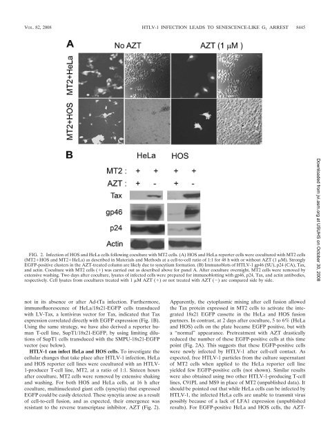
VOL. 82, 2008 HTLV-1 INFECTION LEADS TO SENESCENCE-LIKE G 1 ARREST 8445FIG. 2. Infection <strong>of</strong> HOS and HeLa cells following coculture with MT2 cells. (A) HOS and HeLa reporter cells were cocultured with MT2 cells(MT2HOS and MT2HeLa) as described in Materials and Methods at a cell-to-cell ratio <strong>of</strong> 1:1 for 48 h with or without AZT (1 M). StronglyEGFP-positive clusters in <strong>the</strong> AZT-treated column are likely due to syncytium formation. (B) Immunoblots <strong>of</strong> HTLV-1 gp46 (SU), p24 (CA), Tax,and actin. Coculture with MT2 cells () was carried out as described above for panel A. After coculture overnight, MT2 cells were removed byextensive washing. Two days after coculture, lysates <strong>of</strong> infected cells were prepared for immunoblotting with gp46, p24, Tax, and actin antibodies,respectively. Cell lysates from cocultures treated with 1 M AZT () or not treated with AZT () are compared side by side.Downloaded from jvi.asm.org at USUHS on October 30, 2008not in its absence or after Ad-tTa infection. Fur<strong>the</strong>rmore,immun<strong>of</strong>luorescence <strong>of</strong> HeLa/18x21-EGFP cells transducedwith LV-Tax, a lentivirus vector for Tax, indicated that Taxexpression correlated directly with EGFP expression (Fig. 1B).Using <strong>the</strong> same strategy, we have also derived a reporter humanT-cell line, SupT1/18x21-EGFP, by using limiting dilutions<strong>of</strong> SupT1 cells transduced with <strong>the</strong> SMPU-18x21-EGFPvector (see below).HTLV-1 can infect HeLa and HOS cells. To investigate <strong>the</strong>cellular changes that take place after HTLV-1 infection, HeLaand HOS reporter cell lines were cocultured with an HTLV-1-producer T-cell line, MT2, at a ratio <strong>of</strong> 1:1. Sixteen hoursafter coculture, MT2 cells were removed by extensive shakingand washing. For both HOS and HeLa cells, at 16 h aftercoculture, multinucleated giant cells (syncytia) that expressedEGFP could be easily detected. These syncytia arose as a result<strong>of</strong> cell-to-cell fusion, and as expected, <strong>the</strong>ir emergence wasresistant to <strong>the</strong> reverse transcriptase inhibitor, AZT (Fig. 2).Apparently, <strong>the</strong> cytoplasmic mixing after cell fusion allowed<strong>the</strong> Tax protein expressed in MT2 cells to activate <strong>the</strong> integrated18x21 EGFP cassette in <strong>the</strong> HeLa and HOS fusionpartners. In contrast, at 2 days after coculture, 5 to 6% (HeLaand HOS) cells on <strong>the</strong> plate became EGFP positive, but witha “normal” appearance. Pretreatment with AZT drasticallyreduced <strong>the</strong> number <strong>of</strong> <strong>the</strong>se EGFP-positive cells at this timepoint (Fig. 2A). This suggests that <strong>the</strong>se EGFP-positive cellswere newly infected by HTLV-1 after cell-cell contact. Asexpected, free HTLV-1 particles from <strong>the</strong> culture supernatant<strong>of</strong> MT2 cells when applied to <strong>the</strong> HeLa reporter cell lineyielded few EGFP-positive cells (not shown). Similar resultswere also obtained using two o<strong>the</strong>r HTLV-1-producing T-celllines, C91PL and MS9 in place <strong>of</strong> MT2 (unpublished data). Itshould be pointed out that while HeLa cells can be infected byHTLV-1, <strong>the</strong> infected HeLa cells are unable to transmit viruspossibly because <strong>of</strong> a lack <strong>of</strong> LFA1 expression (unpublishedresults). For EGFP-positive HeLa and HOS cells, <strong>the</strong> AZT-