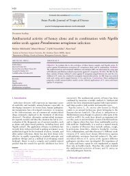
S966Poppy M. Lintong et al ./Asian Paicfic Journal of Tropical Disease (2012)S965-S968symptoms according to a study conducted in Baghdad,which concludes that B. <strong>hominis</strong> is a pathogen <strong>with</strong>inpatients <strong>with</strong> symptomatically diarrheal syndromes[10]. As ahuman parasite, B. <strong>hominis</strong> causes abdominal disturbances,manifested as anorexia, diarrhea and flatus[11].Biopsies from those who are infected by the parasitemostly show normal appearances in the intestinal mucosa.The abnormal appearances only demonstrate the infiltrationof mild and unspecific inflammatory cells. Hardly ever, theparasite provokes mucosal destruction; as well, the parasitegenerally does not penetrate nor invade the tissues[1,8].Moreover, through endoscopic procedure, the intestinalmucosa can be seen normally[1]. B. <strong>hominis</strong> can also bediagnosed by cytological examination from intestinalflushing matter[1].B. <strong>hominis</strong> infection is also associated <strong>with</strong> ulcerativecolitis, terminal ileitis and enteritis that can be cured bymetronidazole[1]. What is more, B. <strong>hominis</strong> infection hasbeen reported to be found in a four year child who showeddiarrheal symptom <strong>with</strong> fever and bloody feces. Throughcolonoscopy, the patient’s colon demonstrated superficialulcer <strong>with</strong> pseudo-membrane throughout the colon; also,through feces examination, B. <strong>hominis</strong> was found. Accordingto the histological examination from the biopsy tissue of thepatient, inflammatory cells <strong>with</strong>in the mucosa of the largeintestinewere found. Also, <strong>with</strong>in the ulcer of the samepatient, spherical or oval forms of B. <strong>hominis</strong> <strong>with</strong> centralgranulated vacuole and single nucleolus were found along<strong>with</strong> the infiltration of inflammatory cells[2].More than 34.7% of B. <strong>hominis</strong> may be present <strong>with</strong>inindividuals who are clinically asymptomatic. On the otherhand, those who show symptomatic signs are mostly markedby abdominal pain, watery diarrhea, constipation, anorexia,nausea, flatulence, and weight losses; such symptomsand signs may demonstrate more than two weeks[1,2].Some authors have reported that B. <strong>hominis</strong> is one of thediarrheal-causal agents[12]. B. <strong>hominis</strong> infection has alsoreported <strong>with</strong>in patients <strong>with</strong> irritable bowel syndrome (IBS)[13]. Likewise, the same circumstance has also been foundin the patients <strong>with</strong> inflammatory bowel disease (IBD), andchronic diarrheas[14].Furthermore, B. <strong>hominis</strong> has also been described as anopportunistic pathogen, found among immunosuppressiveand immunocompromise patients. Clinical symptoms areassociated <strong>with</strong> both the severity of the infection and thevirulence of B. <strong>hominis</strong> strains[2].Conventionally, diagnosis of B. <strong>hominis</strong> is establishedthrough direct examination beneath microscope from thefeces-flushing matter of the patients processed by trichromstain and Kinyoun acid fast technique. Usually, throughthe procedure positive result is marked by the discovery ofvacuolar form of the parasite. The result is considered to besignificant if we discover more than 5 parasites per 400 暳high-power field[2]. What is more, B. <strong>hominis</strong> can also bedetected by polymerase chain reaction technique[15].<strong>Acute</strong> <strong>appendicitis</strong> in human may occur in every stageof life span;however, the disease mostly occurs among theadults and young adults and men (7%) are more affected thanwomen[16]. At first, acute <strong>appendicitis</strong> appears because ofthe increase of intra-luminal pressure that disturb venouscirculation. For about 50%-80% cases are associated <strong>with</strong> theobstruction of appendix’s lumen, usually due to the fecesmass resembling a little stone, called feces stone (fecolite).The rarer etiologies are bladder stones, tumors, or massesfrom helminthes (Oxyuris vermicularis). Ischemic injuryand static condition from the lumen’s contents facilitatebacterial proliferation and precipitate inflammation process,tissue edema and polymorphonuclear infiltration from thelumen unto muscular wall and peri-appendiculary softtissue. At the first stage of the acute <strong>appendicitis</strong> there areedema and congestion of sub serous layer and infiltrationof polymorphonuclear cells throughout all layers of theappendix’s wall[16].Diagnosis of acute <strong>appendicitis</strong> can be established ifthere is infiltration of polymorphonuclear cells penetratingmuscular layers. In severe cases, inflammatory exudates andpolymorphonuclear cells may cause fibrinopurulent reaction,and if the process develops progressively, focal abscess<strong>with</strong>in appendix’s wall can be called acute <strong>suppurative</strong><strong>appendicitis</strong>[16].Initially, clinical appearance of <strong>appendicitis</strong> is markedby a pain <strong>with</strong>in peri-umbilical region, which subsequentlyshifts to the lower-right quadrant. Pressure pain <strong>with</strong>inthe lower-right quadrant is termed Mc Burney’s point.Another symptoms are nausea, vomiting or both; whereas theabdomen is palpated tightly, accompanied <strong>with</strong> mild feverand the increase of peripheral blood leukocytes[16].2. Case reportaFigure 1. Macroscopic appearance of the patient’s appendix.a) Long section of the appendix’s mass <strong>with</strong> 7 cm in length; b)Transversal section of both distal and proximal parts of the appendix;at distal, the section is lack of mucosa layer whilst at proximal,mucosal part remains exist shown as gelatin.a b c d eFigure 2. Transversal section of the patient’s appendix from proximalto distal parts.a, b show the proximal fraction of the appendix: all layers of theappendix (from mucosal up to serous layers); c, d, e show the distalfraction of the appendix: the wall becomes thin, mucosa disappears,and lumen is filled by gelatinous mass.b
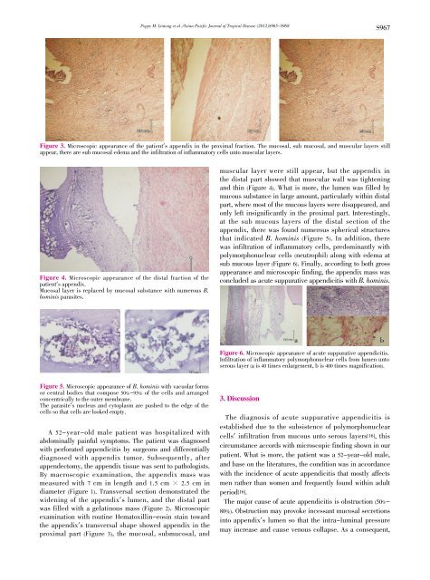
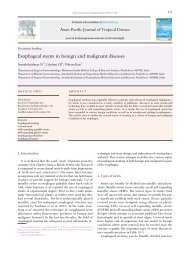
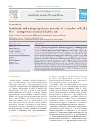
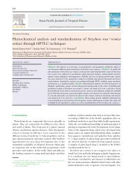
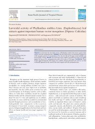
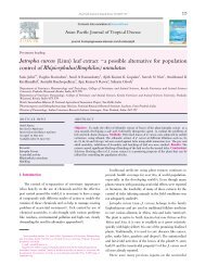
Poppy M. Lintong et al ./Asian Paicfic Journal of Tropical Disease (2012)S965-S968S967Figure 3. Microscopic appearance of the patient’s appendix in the proximal fraction. The mucosal, sub mucosal, and muscular layers stillappear, there are sub mucosal edema and the infiltration of inflammatory cells unto muscular layers.Figure 4. Microscopic appearance of the distal fraction of thepatient’s appendix.Mucosal layer is replaced by mucosal substance <strong>with</strong> numerous B.<strong>hominis</strong> parasites.muscular layer were still appear, but the appendix inthe distal part showed that muscular wall was tighteningand thin (Figure 4). What is more, the lumen was filled bymucous substance in large amount, particularly <strong>with</strong>in distalpart, where most of the mucous layers were disappeared, andonly left insignificantly in the proximal part. Interestingly,at the sub mucous layers of the distal section of theappendix, there was found numerous spherical structuresthat indicated B. <strong>hominis</strong> (Figure 5). In addition, therewas infiltration of inflammatory cells, predominantly <strong>with</strong>polymorphonuclear cells (neutrophil) along <strong>with</strong> edema atsub mucous layer (Figure 6). Finally, according to both grossappearance and microscopic finding, the appendix mass wasconcluded as acute <strong>suppurative</strong> <strong>appendicitis</strong> <strong>with</strong> B. <strong>hominis</strong>.aFigure 6. Microscopic appearance of acute <strong>suppurative</strong> <strong>appendicitis</strong>.Infiltration of inflammatory polymorphonuclear cells from lumen untoserous layer (a is 40 times enlargement, b is 400 times magnification).bFigure 5. Microscopic appearance of B. <strong>hominis</strong> <strong>with</strong> vacuolar formsor central bodies that compose 50%-95% of the cells and arrangedconcentrically to the outer membrane.The parasite’s nucleus and cytoplasm are pushed to the edge of thecells so that cells are looked empty.A 52-year-old male patient was hospitalized <strong>with</strong>abdominally painful symptoms. The patient was diagnosed<strong>with</strong> perforated <strong>appendicitis</strong> by surgeons and differentiallydiagnosed <strong>with</strong> appendix tumor. Subsequently, afterappendectomy, the appendix tissue was sent to pathologists.By macroscopic examination, the appendix mass wasmeasured <strong>with</strong> 7 cm in length and 1.5 cm 暳 2.5 cm indiameter (Figure 1). Transversal section demonstrated thewidening of the appendix’s lumen, and the distal partwas filled <strong>with</strong> a gelatinous mass (Figure 2). Microscopicexamination <strong>with</strong> routine Hematoxillin-eosin stain towardthe appendix’s transversal shape showed appendix in theproximal part (Figure 3), the mucosal, submucosal, and3. DiscussionThe diagnosis of acute <strong>suppurative</strong> <strong>appendicitis</strong> isestablished due to the subsistence of polymorphonuclearcells’ infiltration from mucous unto serous layers[16], thiscircumstance accords <strong>with</strong> microscopic finding shown in ourpatient. What is more, the patient was a 52-year-old male,and base on the literatures, the condition was in accordance<strong>with</strong> the incidence of acute <strong>appendicitis</strong> that mostly affectsmen rather than women and frequently found <strong>with</strong>in adultperiod[16].The major cause of acute <strong>appendicitis</strong> is obstruction (50%-80%). Obstruction may provoke incessant mucosal secretionsinto appendix’s lumen so that the intra-luminal pressuremay increase and cause venous collapse. As a consequent,