âThe evolution of the classification of nephrotic syndrome and the ...
âThe evolution of the classification of nephrotic syndrome and the ...
âThe evolution of the classification of nephrotic syndrome and the ...

Create successful ePaper yourself
Turn your PDF publications into a flip-book with our unique Google optimized e-Paper software.
“The <strong>evolution</strong> <strong>of</strong> <strong>the</strong> <strong>classification</strong> <strong>of</strong><strong>nephrotic</strong> <strong>syndrome</strong> <strong>and</strong> <strong>the</strong> newtaxonomy for <strong>the</strong> podocytopathies”Laura Barisoni, MDDepartment <strong>of</strong> Pathology <strong>and</strong>Medicine, Division <strong>of</strong> NephrologyNew York University
Old <strong>classification</strong> schemes:Proteinuria <strong>and</strong><strong>nephrotic</strong> <strong>syndrome</strong>MCDFSGSGood prognosis <strong>and</strong>Response to steroidTherapyPoor prognosis <strong>and</strong>Poor Response toSteroid <strong>the</strong>rapy
Nephrotic <strong>syndrome</strong> - <strong>the</strong> 80’s <strong>and</strong> 90’s• While <strong>the</strong> definition <strong>of</strong> minimal change disease did not change over <strong>the</strong> years, in <strong>the</strong> mid80’s o<strong>the</strong>r patterns <strong>of</strong> glomerular damage have became part <strong>of</strong> <strong>the</strong> FSGS spectrum.• Collapsing glomerulopathy:- first description in 1978 as “ malignant FSGS” (Brown Clin Nephrol 1978)- 1980’s frequent diagnosis during HIV p<strong>and</strong>emic (HIV-AN)- first described in non-HIV pts in 1986 (Weiss et al AJKD 1986) – “collapsingglomerulopathy” – new clinical-pathologic entity.- in mid 90’s became “idiopathic collapsing FSGS”(Detwiler et al KI 1994 & Valeri et al JASN 1996)• Cellular lesion:- Term used first by Schwarz <strong>and</strong> colleagues to indicate a group <strong>of</strong> lesions wi<strong>the</strong>ndocapillary <strong>and</strong>/or extracapillary increased cellularity.- O<strong>the</strong>r authors used <strong>the</strong> term cellular to indicate intracapillary cellularity only.• Tip lesion:- Howie et al described tip lesion as a well-defined <strong>and</strong> specific pathological entitywith clinical similarity to MCD. (J Pathol 1984)- Tip lesions are also seen in associations with o<strong>the</strong>r glomerular diseases such asdiabetic nephropathy or membranous glomerulopathy.
Relatively recent <strong>classification</strong> schemes:Columbia <strong>classification</strong> - FSGS variantsPerihilar NOS TipCellularCollapsing
Limitations <strong>of</strong> <strong>the</strong> morphologic<strong>classification</strong>• Various histopathologic lesions are listed under“focal segmental glomerulosclerosis” regardless <strong>the</strong>presence or absence <strong>of</strong> segmental sclerosis.• Lack <strong>of</strong> correlation with pathogenetic mechanisms<strong>and</strong> etiology.• Lack <strong>of</strong> correlation with treatment
Proteinuria <strong>and</strong> <strong>nephrotic</strong> <strong>syndrome</strong> in<strong>the</strong> 21 st century• The attention <strong>of</strong> scientists, nephrologists <strong>and</strong> pathologistshas been recently focused on <strong>the</strong> role <strong>of</strong> podocytes ascause <strong>of</strong> proteinuria• In <strong>the</strong> last 10 years lot <strong>of</strong> progress has been made in <strong>the</strong>underst<strong>and</strong>ing <strong>the</strong> biology <strong>of</strong> podocytes, how <strong>the</strong>y function<strong>and</strong> how <strong>the</strong>y are injured.“Taxonomy <strong>of</strong> <strong>the</strong> podocytopathies”where morphologic diagnosis are integrated with etiology(Barisoni, Schnaper, Kopp, CJASN 2007)
PodocytopathiesDEFINITION: Proteinuric diseases in which pathologicprocesses arise from intrinsic or extrinsic “primary”podocyte injury <strong>and</strong> where <strong>the</strong> podocyte genotype/phenotypeis altered.
Taxonomyταξισ + νοµιαarrangement or divisionlaw or methodA taxonomy is organized into multiple levels, each <strong>of</strong> which represents a taxon with one ormore elements (taxa), which are mutually exclusive, unambiguous, <strong>and</strong> all-encompassingcategories.Taxonomies provide <strong>classification</strong> <strong>and</strong> conceptual framework for analysis, discussion, <strong>and</strong>hypo<strong>the</strong>sis generation.Taxonomy <strong>of</strong> PodocytopathiesΤαξονHISTOPATHOLOGY- pattern <strong>of</strong> glom injury- podocyte numberETHIOLOGYidiopathic genetic reactiveΤαξα
Podocytopathies:4 morphologic patterns <strong>of</strong> glomerular injuryNormalHistologySegmentalSclerosisMesangialSclerosisCollapse <strong>of</strong><strong>the</strong> GBMMCNFSGSDMSCG
Causes <strong>of</strong> foot process effacement1. Impaired formation <strong>of</strong> <strong>the</strong> slitdiaphragm complex2. Abnormalities <strong>of</strong> <strong>the</strong> adhesiveinteraction between podocytes<strong>and</strong> GBM3. Alterations <strong>of</strong> transcription factors4. Abnormalities <strong>of</strong> <strong>the</strong> actin-basedcytoskeleton5. Alterations <strong>of</strong> <strong>the</strong> apical domain <strong>of</strong>podocytes6. Mitochondria abnormalities7. Abnormalities <strong>of</strong> cell metabolism8. Mechanical stress9. Viral infection10. Acute ischemic injury11. Toxic / metabolic effect12. Immunologic stimuli
Hypo<strong>the</strong>sis #1:Injured podocytes can take different pathwaysPodocyte injuryAlteredphenotypeEngagement <strong>of</strong>apoptotic pathwaysDevelopmentalarrestDe-differentiationNo change inpodocyte numberCell deathProliferation(low)Proliferation(high)No changeSegmentalsclerosisMesangialsclerosisCollapseMCNFSGSDMSCG
The role <strong>of</strong> <strong>the</strong> renopoietic systemHierarchical distribution <strong>of</strong> CD133+CD24+PDX- <strong>and</strong>CD133+CD24+PDX+ cells within human glomeruliRonconi, E. et al. J Am Soc Nephrol 2009;20:322-332Copyright ©2009 American Society <strong>of</strong> Nephrology
Hypo<strong>the</strong>sis #2The role <strong>of</strong> CD24+CD133+ renal progenitors in FSGS & CG.Podocyte injuryAlteredphenotypePodocytedeathDevelopmentalarrestPodocytedeathNo change inpodocyte numberCD24+CD133+Repair activityProliferation(low)ExuberantCD24+CD133+ActivityNo changeSegmentalsclerosisMesangialsclerosispseudocrescentsMCNFSGSDMSCG
MINIMAL CHANGENEPHROPATHY
Minimal Change NephropathyDEFINITIONNormal histology.Extensive foot process effacement, but preserved number <strong>of</strong>podocytes.ETIOLOGY AND CLINICAL ASSOCIATION• Idiopathic• Inherited- Non-Syndromic (NPHS1, NPHS2)- Syndromic (DYSF)• Reactive- drug-induced(NSAID, pamidronate, interferon, o<strong>the</strong>rs)- dysregulation <strong>of</strong> <strong>the</strong> immune system- hematologic malignancy
Minimal change nephropathy• Reversible – Steroid sensitive- idiopathic- reactive (secondary)- drug-induced (NSAID,pamidronate, interferon, o<strong>the</strong>rs)- dysregulation <strong>of</strong> <strong>the</strong>immune system- hematologic malignancy• Irreversible - Steroid resistant- idiopathic- genetically determined- NPHS2- DYSF
Can pathologists discriminatebetween steroid sensitive <strong>and</strong>steroid-resistant MCN?
Glomerular expression <strong>of</strong> dystroglycans isreduced in MCD but not in FSGSRegele JASN11:403-412, 2000α-dystroglycan β-dystroglycan β 1 -integrinNormalkidneyFSGSMCD
DG staining in steroid sensitive <strong>and</strong> steroid resistant MCNLaura De Petris, David Thomas, Helen Liapis, Laura BarisoniIHC stainingCFig 1negativepositiveFSGS Ctrl SR-MCN SS-MCN MCN (no f-up)
aPodocin: controlbPodocin: steroid-resistant MCNcd
FOCAL SEGMENTALGLOMERULOSCLEROSIS
FSGSDEFINITIONSegmental solidification <strong>of</strong> <strong>the</strong> tuft accompanied by sinechiae.Hyalinosis <strong>and</strong> foam cells can also be present. Low number <strong>of</strong>podocytes (podocytopenia).ETIOLOGY AND CLINICAL ASSOCIATION• Idiopathic• Inherited- syndromic- non-syndromic• Reactive- hyperfiltration-mediatednormal renal massreduced renal mass- medication-induced- permeability factor (?)
Idiopathic FSGSIs idiopathic really idiopathic?MYH9 is a major-effect risk gene for FSGS.(Kopp et al. Nat Genet. 2008)MYH9 risk alleles are more frequent in AA. MYH9 protective alleles are more frequent in EA.
Genetic forms <strong>of</strong> FSGS• Associated with o<strong>the</strong>r organ abnormalities (syndromic):– Freiser Syndrome (WT-1).– Nail-patella <strong>syndrome</strong> (LMX1B)– Renal-coloboma <strong>syndrome</strong> with oligomeganephronia (PAX2)– Alport’s disease (COL4A3, A4, A5)– Metabolic disorders (GLA – Fabry’s)– Mitochondriopathies (mtDNA tRNA Leu <strong>and</strong> tRNA Tyr ,CoQ2 NP, CoQ6 NP)• Limited to <strong>the</strong> kidney (non-syndromic):- NPHS1 – nephrin – autosomal recessive- NPHS2 – podocin – autosomal recessive- NPHS3 – phospholipase Cε1 – autosomal recessive- CD2AP – susceptibility to FSGS- MYH9 – susceptibility to FSGS- ACTN4 – α-actinin-4 - autosomal dominant- INF2 – autosomal dominant- TRPC6 – Transient Receptor Potential channel 6 - autosomal dominant- WT1 – sporadic/isolated FSGS
Reactive forms:Hyperfiltration-mediated FSGSglomerulomegaly in pt with single kidneySegmental sclerosislarge non-sclerotic glomerulus
Which is <strong>the</strong> relationshipbetween glomerulomegaly<strong>and</strong> FSGS?
FSGS: From podocyte hypertrophy to podocytopenia.Wiggins et al JASN 2005.In response to increased glomerular volume, podocytes undergo hypertrophythough 5 stages.•Stage 1, normal podocyte;•Stage 2, non-stressed hypertrophy;•Stage 3, "adaptive" hypertrophy: changes in syn<strong>the</strong>sis <strong>of</strong> structural componentsbut maintenance <strong>of</strong> normal function;•Stage 4, "de-compensated" hypertrophy- reduced production <strong>of</strong> proteins necessary for normal podocyte function.- widened foot processes <strong>and</strong> decreased filter efficiency (proteinuria);•Stage 5, podocyte numbers decrease.Dr Kriz’s model
DIFFUSE MESANGIALSCLEROSIS
DMSDEFINITION:Diffuse increase <strong>of</strong> mesangial matrix accompaniedby mild proliferation <strong>of</strong> hypertrophic podocytes.ETIOLOGY:• Idiopathic• Genetic- Non-syndromic- WT1- NPHS1- NPHS2- NPHS3- COQ6- Syndromic- LAMB2 (Pierson S.)- WT-1 (Denys-Drash S.)
WT-1 associated DMS•Reduced or dysfunctional expression <strong>of</strong> WT-1, a podocyte transcription factor.•Increased expression <strong>of</strong> growth-promoting molecules (Pax-2, Ki-67).•Podocyte entry into <strong>the</strong> cell cycle.•Preservation <strong>of</strong> o<strong>the</strong>r podocyte markers (nephrin, synaptopodin, a-actinin-4).
NPHS3-PLCE1Chromosome 10q23.32-q24.1 = phospholipase Cε1.Truncating mutationsnon-truncating missensemutationsDevelopmental arrestPodocytopeniaDMSearly onset <strong>of</strong> severe NS <strong>and</strong>rapid progression to renal failure.Of Note: 2 pts responded tosteroid <strong>and</strong> Cyclosporin A.FSGSLater onset <strong>of</strong> NS <strong>and</strong> slowerprogression to renal failure.Hinkes et al. Nat Genetic 2006
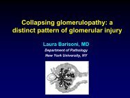
COLLAPSINGGLOMERULOPATHY
CG Definition: GBM collapse <strong>and</strong>pseudocrescent formationHIV
CG: etiology <strong>and</strong> clinical associations• Idiopathic• Genetic• Syndromic - action myoclonus renal failure• Non-Syndormic - CoQ2 NP• Reactive• Virus associated- HIV- parvovirus B19- CMV• Infections - filariasis- leishmania- TB• Autoimmune - Still’s disease- lupus like- RA- mixed connective tissue• Malignancy (myeloma, AML)• Medications - pamidronate- interferon- valproic acid• Vascular insult - TMA• Permeability factor
TAXONOMY OF PODOCYTOPATHIESidiopathic genetic reactiveMCNIdiopathic•Steroid-sensitive•Steroid-resistantNon-syndromic•NPHS1•NPHS2Syndromic•DYSFClinical association(immunologic stimuli, Tumors)Medications(NSAID,gold, penicillamine, lithium,IF, pamidronate)FSGSIdiopathic•Steroid-sensitive•Steroid-resistantNon-syndromicITGB4, NPSH2, NPHS3, NPHS1 +NPHS2, COQ2, MHY9, ACTN4,CD2AP, TRCP6, WT-1, SYNPO, INF2SyndromicMtDNA, WT1, PAX2, COQ6,COL4,GLA, LMBX1Post-adaptive•nephron mass•Initially normal nephron massMedications(tacrolimus, lithium, IF, pamidronate)DMS Idiopathic Non-syndromicWT1, NPHS1, NPSH2, NPHS3,LAMB2,SyndromicWT1, LAMB2, COQ6,CG Idiopathic Non-syndromicCOQ2MHY9SyndromicSCARBInfections(viruses, TB, o<strong>the</strong>rs)Clinical association•Autoimmune, TMA, tumorsMedications(IF, pamidronate, valproic acid)
Proteinuria <strong>and</strong> <strong>nephrotic</strong> <strong>syndrome</strong> – <strong>the</strong> present‣ MCN, FSGS, DMS <strong>and</strong> CG are patterns <strong>of</strong> glomerular damage where <strong>the</strong> commondenominator is podocyte injury .‣ Morphologic <strong>classification</strong>s alone are insufficient to capture <strong>the</strong> complexity <strong>and</strong>heterogeneity <strong>of</strong> diseases presenting with NS.- multiple specific disease processes can present with indistinguishable histopatholology- a specific monogenetic disorder can present with more than one form <strong>of</strong> histopathologic pattern <strong>of</strong>glomerular damage.‣ Final diagnosis <strong>of</strong> <strong>the</strong> podocytopathies should occur in 3 steps:a. clinical evaluationb. morphologic evaluationc. additional clinical tests, such as genetic or serology for evidence <strong>of</strong> infections,or o<strong>the</strong>rs, when indicated.Proteinuria <strong>and</strong> <strong>nephrotic</strong> <strong>syndrome</strong> – <strong>the</strong> futureNEPTUNE – international effort with <strong>the</strong> following major goals:Determination <strong>of</strong> rates <strong>and</strong> predictors <strong>of</strong> clinical remission or progression in NSIdentification <strong>of</strong> gene expression pr<strong>of</strong>ilesIdentification <strong>of</strong> patient specific molecular signaturesClinically useful <strong>classification</strong> based on morphologic & molecular phenotype