A New Species of Clistobothrium - University of Connecticut
A New Species of Clistobothrium - University of Connecticut
A New Species of Clistobothrium - University of Connecticut

You also want an ePaper? Increase the reach of your titles
YUMPU automatically turns print PDFs into web optimized ePapers that Google loves.
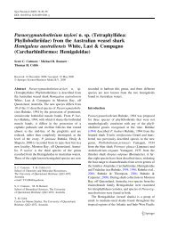
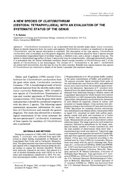
J. Parasitol., 79(1), 1993, p. 37-43? American Society <strong>of</strong> Parasitologists 1993A NEW SPECIES OF CLISTOBOTHRIUM(CESTODA: TETRAPHYLLIDEA), WITH AN EVALUATION OF THESYSTEMATIC STATUS OF THE GENUST. R. RuhnkeDepartment <strong>of</strong> Ecology and Evolutionary Biology, <strong>University</strong> <strong>of</strong> <strong>Connecticut</strong>, 314 TLS,Storrs, <strong>Connecticut</strong> 06269-3043ABSTRACT: <strong>Clistobothrium</strong> montaukensis n. sp. is described from the shortfin mako shark, Isurus oxyrinchus.Based on shared characters from the scolex and segment, Phyllobothrium tumidum is transferred to the genus<strong>Clistobothrium</strong>, and the species description is emended. The description <strong>of</strong> the type species, <strong>Clistobothrium</strong>carcharodoni, also is emended, as is the generic diagnosis. Derived characters shared by these 3 species includea dorsomedian longitudinal band <strong>of</strong> strobilar muscles, a bilobed ovary, and a dorsal L-shaped cirrus sac. Thespined or mammilated egg surface <strong>of</strong> these 3 species also may be synapomorphic. Although phenetically similar,it is postulated that the foliose bothridial condition shared among members <strong>of</strong> Phyllobothrium and 2 <strong>of</strong> thespecies <strong>of</strong> <strong>Clistobothrium</strong> is not homologous. The cirruses <strong>of</strong> C. montaukensis n. sp. and C. carcharodoniare armed with microtriches, but this may be true for other cestodes. Reliable host reports indicate that species<strong>of</strong> <strong>Clistobothrium</strong> are restricted to sharks <strong>of</strong> the family Lamnidae (the mackerel sharks).Dailey and Vogelbein (1990) erected <strong>Clistobothrium</strong>for <strong>Clistobothrium</strong> carcharodoni fromthe great white shark, Carcharodon carchariasLinnaeus, 1758. A morphological study <strong>of</strong> newlycollected material from the shortfin mako shark,Isurus oxyrinchus Rafinesque, 1809, revealed anew species <strong>of</strong> <strong>Clistobothrium</strong>. Examination <strong>of</strong>type and voucher specimens <strong>of</strong> Phyllobothriumtumidum Linton, 1922, from the great white sharkindicated that it should be considered congenericwith the above 2 species. The following reportsummarizes taxonomic information for members<strong>of</strong> <strong>Clistobothrium</strong> and emends the genericdiagnosis. The study also allowed for a morphologicalexamination <strong>of</strong> the cirrus armature forthe new species and C. carcharodoni. A summary<strong>of</strong> host reports for members <strong>of</strong> <strong>Clistobothrium</strong>also is presented.MATERIALS AND METHODSDuring the summers <strong>of</strong> 1988-1990, 5 shortfin makosharks, I. oxyrinchus, were collected from sport fishermenat shark fishing tournaments in Montauk onLong Island, <strong>New</strong> York, and in Yarmouth, Massachusetts.The spiral intestine was removed from each sharkand opened with a longitudinal incision along the dorsalsurface to expose the internal chambers. Wormswere removed, relaxed in sea water, then placed in avial <strong>of</strong> alcohol-formalin-acetic acid (AFA) or in hotwater and immediately transferred to AFA. For electronmicroscopical studies, worms were fixed in coldReceived 30 August 1991; revised 14 August 1992;accepted 26 August 1992.372.5% glutaraldehyde in 0.1 M cacodylate buffer, washedin the same concentration <strong>of</strong> buffer, and postfixed in1% osmium tetroxide. Spiral intestines from some <strong>of</strong>the individuals also were fixed in AFA, and additionalworms were removed from the intestines upon returningto the laboratory. Specimens <strong>of</strong> P. tumidum wereobtained from the spiral intestine <strong>of</strong> a great white sharkobtained from fisherman fishing in Atlantic waters <strong>of</strong>fLong Island. Specimens for whole mounts were stainedwith Ehrlich's or Gill's hematoxylin, flattened between2 glass slides, dehydrated in an alcohol series, destainedin acid ethanol (if required), cleared in xylene, andmounted in Canada balsam. Cross sections were preparedaccording to the methods <strong>of</strong> Caira and Gavarrino(1990). Specimens for scanning electron microscopywere dehydrated in an ethanol series, critical point driedin liquid CO2, mounted on stubs with carbon paint,sputter coated with gold, and examined in a Coatesand Welter field emission scanning electron microscope.Measurements are given in micrometers unlessotherwise stated. The range is given for each numericalcharacter, followed in parentheses by the mean, standarddeviation, number <strong>of</strong> worms examined (n), andthe total number <strong>of</strong> observations when more than 1measurement per worm was taken (n). Tapeworm eggswere measured from segments mounted in Canada balsamand from segments opened and examined in 70%ethanol. Cirrus sacs were measured according to themethod used by Caira (1990) for trematodes. Illustrationswere made with the aid <strong>of</strong> a drawing tube. Paratypes<strong>of</strong> C. carcharodoni (USNM no. 80985 and HWMLno. 31397) were borrowed from the U.S. National MuseumHelminthological Collection, Beltsville, Maryland,and the H. W. Manter Laboratory, <strong>University</strong> <strong>of</strong>Nebraska State Museum. Type specimens <strong>of</strong> P. tumidum(USNM nos. 2630 and 2631) were borrowed fromthe U.S. National Museum Helminthological Collectionin Beltsville, Maryland. A voucher specimen <strong>of</strong>P. tumidum (USNM no. 35802) was borrowed fromthe U.S. National Museum Helminthological Collection.
38 THE JOURNAL OF PARASITOLOGY, VOL. 79, NO. 1, FEBRUARY 19937X4 0 Ii- : ':;'0FIGURES 1-8. Line drawings <strong>of</strong> <strong>Clistobothrium</strong> carcharodoni, <strong>Clistobothrium</strong> tumidum, and <strong>Clistobothrium</strong>montaukensis. 1. Cirrus microtriche from partially everted cirrus <strong>of</strong> paratype (HWML no. 31397) <strong>of</strong> C carcharodoni.2. Cirrus sac from end segment <strong>of</strong> lectotype (USNM no. 7631) <strong>of</strong> C. tumidum n. comb. 3. Scolex <strong>of</strong>holotype (USNM no. 82490) <strong>of</strong> C. montaukensis. 4. Mature segment, ventral view <strong>of</strong> paratype (USNM no.82490) <strong>of</strong> C. montaukensis. 5. Dorsomedian muscle band <strong>of</strong> paratype (USNM no. 82490) <strong>of</strong> C. montaukensis.6. Cross section through segment posterior to cirrus sac and anterior to ovary <strong>of</strong> paratype (HWML no. 35289)<strong>of</strong> C. montaukensis. 7. Cross section through segment at ovary <strong>of</strong> paratype (HWML no. 35289) <strong>of</strong> C. montaukensis.8. Cirrus microtriche from cross section through cirrus <strong>of</strong> voucher (HWML no. 35289) <strong>of</strong> C. montaukensis.DE, dorsal excretory duct; MB, dorsomedian longitudinal muscle band; NC, nerve cord; 0, ovary; T, testes; U,uterus; V, vagina; VE, ventral excretory duct.
RUHNKE-NEW SPECIES OF CLISTOBOTHRIUM 39DESCRIPTIONS<strong>Clistobothrium</strong> carcharodoniDailey and Vogelbein, 1990(Fig. 1)The following information from 4 paratype specimensis provided to emend the description <strong>of</strong> Daileyand Vogelbein (1990) for C. carcharodoni. Only characteristicsrequiring emendation are given: Wormsapolytic. Maximum width 1,500-1,950 (1,733 + 225;n = 3) at scolex. Strobila with a distinct band <strong>of</strong> dorsomedianlongitudinal muscles 70-85 (76 ? 6; n = 4)wide in anterior and 50-73 (63 ? 11; n = 4) wide inposterior segments. Cirrus sac L-shaped, 408-670 (564? 91; n = 4; n = 13) long by 145-229 (198 ? 37; n =4; n = 13) wide, containing cirrus. Cirrus coiled, expandedproximally, and armed with spiniform microtriches(Fig. 1). Genital pores lateral, irregularly alternating,positioned in the anterior 60-68% (65 ? 3; n= 5; n = 13) <strong>of</strong> mature segments; genital atrium present.Vagina medial, extending from ovary anteriorly andcrossing ventrally the proximal portion <strong>of</strong> cirrus sac,extending laterally to genital atrium, 30-135 (72 ? 34;n = 5; n = 10) wide above cirrus sac in mature segments.Ovary posterior to testes, H-shaped, 210-420(295 + 68; n = 5; n = 11) long by 320-500 (425 +63; n = 5; n = 11) wide. Vitelline follicles 12-30 (21+ 6; n = 4; n = 8) long by 40-80 (59 + 15; n = 4; n=8) wide. Eggs round, 21-24 (23 ? 1; n = 4; n = 9)in diameter, found in terminal and free segments.Taxonomic summaryType host: Carcharodon carcharias Linnaeus, 1758,the great white shark.Type locality: Off Pt. Dume, Los Angeles County,California, 33?55'N, 118?48'W.Site <strong>of</strong> infection: Spiral intestine.Specimens examined: Paratype specimens USNMno. 80895 and HWML no. 31397.RemarksThe description <strong>of</strong> C. carcharodoni is emended forseveral reasons. Apparently, Dailey and Vogelbein(1990) were mistaken in their estimation <strong>of</strong> the size <strong>of</strong>the egg and the width <strong>of</strong> the scolex in this species.Quantitative values have been added for some <strong>of</strong> themorphological features <strong>of</strong> this species and informationalso has been added concerning the dorsomedian band<strong>of</strong> longitudinal muscles extending throughout the length<strong>of</strong> the strobila. The length <strong>of</strong> the cirrus sac has beenremeasured according to the methods used by Caira(1990) to reflect the total length <strong>of</strong> the structure. Inaddition, Dailey and Vogelbein (1990) described C.carcharodoni as anapolytic. However, an apolytic (free)segment was observed in the paratype series, and theposterior segments <strong>of</strong> some <strong>of</strong> the specimens, althoughcontaining eggs, were not completely gravid. Thus, myobservations indicate that the species should be consideredapolytic.<strong>Clistobothrium</strong> tumidum(Linton, 1922) n. comb.(Fig. 2)The following information is provided to emendLinton's (1922) decription <strong>of</strong> this species: Wormsslightly craspedote, apolytic. Scolex with dome-shapedapical region and dorsal and ventral pairs <strong>of</strong>bothridia.Each bothridium with a single anterior apical sucker,measuring 280-360 (307 ? 35; n = 4; n = 12) and asingle foliose posterior loculus. Strobila with more than100 segments. Strobila with a distinct dorsomedianmuscle band 75-120 (98 ? 32; n = 2) wide in anteriorsegments, 33-62 (49 ? 12; n = 4) wide in posteriorsegments. Mature segments 1,625-1,925 (1,761 ? 106;n = 4; n = 7) long by 1,025-1,800 (1,457 ? 231; n =4; n = 7) wide. Testes 234-307 (282 ? 33; n = 4) innumber in mature segments in dorsal or ventral view,distributed in 2 irregular fields, intravascular. Maturesegments with thin-walled, L-shaped, dorsal cirrus sac(Fig. 2), 316-411 (376 ? 41; n = 4) long by 105-168(126 ? 30; n = 4) wide, opening into genital atrium.Cirrus armed, coiled inside cirrus sac, expanded proximally.Genital pores lateral, irregularly alternating,positioned in the anterior 61-76% (67 ? 4; n = 5; n= 9) <strong>of</strong> mature segments. Vagina medial, extendinganteriorly, then laterally and anteriorly over proximalportion <strong>of</strong> cirrus sac (Fig. 2) to genital atrium. Ovaryposterior, H-shaped, 420-520 (477 ? 37; n = 3; n =6) long by 470-600 (538 + 47; n = 3; n = 6) wide inmature segments, bilobed in cross section. Uterus ventral,extending to posterior margin <strong>of</strong> cirrus sac in maturesegments, extending to anterior margin <strong>of</strong> cirrussac in gravid free segments. Some free segments witha midventral uterine pore. Eggs round, 20-27 (24 + 2;n = 3; n = 8) in diameter in free segments, surfacemammilated.Taxonomy summarySynonym: Phyllobothrium tumidum Linton, 1922.Type host: Carcharodon carcharias Linnaeus, 1758,the great white shark.Type locality: Woods Hole, Massachusetts.Additional locality: Moriches, Long Island, <strong>New</strong> York.Site <strong>of</strong> infection: Spiral intestine.Specimens examined: Lectotype (USNM no. 7631);paralectotypes (USNM nos. 7630 and 7631); voucherspecimen (USNM no. 35802).Specimens deposited: Three voucher specimens(USNM no. 82491) deposited in the U.S. NationalMuseum Helminthological Collection in Beltsville,Maryland, and 3 voucher specimens (HWML no.35290) deposited in the Harold W. Manter Laboratory<strong>of</strong> Parasitology, Lincoln, Nebraska.RemarksLinton (1922) described this species as P. tumidumfrom the great white shark. He included an additionalspecimen from the shortfin mako shark in the typeseries (USNM no. 7632), but the specimen is immatureand its conspecificity with C. tumidum cannot be determined.A holotype was not designated by Linton(1922) for this species. I hereby designate as the lectotype1 <strong>of</strong> the partial worms on the "type" slide cataloguedUSNM no. 7631. This strobila was drawn byLinton and most clearly depicts the segment structure<strong>of</strong> the specimens in the type series. The remainingspecimens on slides catalogued USNM nos. 7630 and7631 should be considered paralectotypes. <strong>Clistobothrium</strong>tumidum differs from C. carcharodoni in the shape<strong>of</strong> the posterior loculus, diameter <strong>of</strong> the apical sucker,
40 THE JOURNAL OF PARASITOLOGY, VOL. 79, NO. 1, FEBRUARY 1993and number <strong>of</strong> testes in mature segments. The 2 speciesalso differ in the length <strong>of</strong> the cirrus sac and in theposition <strong>of</strong> the proximal portion <strong>of</strong> the cirrus sac relativeto the vagina. The description is emended t<strong>of</strong>acilitate future comparisons with this species. A morecomplete description <strong>of</strong> this species must await additionalcollections from the type host, C. carcharias.<strong>Clistobothrium</strong> montaukensis n. sp.(Figs. 3-15)The description is based on 4 whole worms, 13 partialworms, 2 critically point dried scolices with strobilae,eggs from 3 critically point dried free segments,5 free segments, and eggs from 5 free segments: Worms38.5-119.5 mm (73.5 + 34.3; n = 4) long, maximumwidth 2,475-3,750 (3,000 + 522; n = 7) at scolex,apolytic, slightly craspedote. Scolex with large, domeshapedapical region (Figs. 3, 9), apical region coveredwith filiform microtriches (Fig. 10). Scolex 2,100-3,650(3,000 + 544; n = 7) long by 2,475-3,750 (3,000 +522; n = 7) wide, with 2 ventral and 2 dorsal large,foliose bothridia (Fig. 11) each with 1 circular, muscular,anterior accessory sucker 310-500 (372 + 52; n= 9; n = 18) in diameter. Bothridia 1,750-2,125 (1,970+ 159; n = 5) long by 1,350-1,500 (1,465 + 89; n =5) wide. Proximal surface <strong>of</strong> bothridia covered withslender spiniform microtriches (Fig. 12); distal bothridialsurface not observed. Neck short, 950-3,700(2,110 + 1,125; n = 5) long, covered with filiformmicrotriches (Fig. 13). Strobila with more than 100segments, with distinct dorsomedian muscle band (Figs.4, 5, 14), 63-160 (91 + 33; n = 8) wide in anteriorsegments, 30-58 (45 + 8; n = 9) wide in posteriorsegments. Anterior segments much wider than long,immature segments at midworm 525-800 (669 + 86;n = 4; n = 8) long by 1,050-1,300 (1,191 + 92; n -4; n = 8) wide, mature segments 1,400-3,200 (2,122+ 569; n = 6; n = 13) long by 865-1,212 (1,035 +127; n = 6; n = 13) wide, generally twice as long aswide, with a dorsal and ventral pair <strong>of</strong> lateral excretoryducts and a pair <strong>of</strong> lateral nerve cords in cross section(Figs. 6, 7). Testes round, 33-72 (56 + 12; n = 5; n =17) in diameter, numbering 198-263 (242 + 28; n =6; n = 8) in dorsal or ventral view, distributed in 2irregular fields between the ventral excretory ducts incross section (Fig. 6). Vas deferens coiled, medial, inanterior third <strong>of</strong> segment. Cirrus sac dorsal, L-shaped,proximal portion <strong>of</strong> cirrus sac immediately posteriorto vas deferens (Fig. 4), 519-737 (638 + 66; n = 4; n10) long by 123-251 (173 ? 39; n = 4; n = 10) wide,opening into a genital atrium. Cirrus long, coiled insidecirrus sac, expanded proximally, covered with slenderspiniform microtriches (Fig. 8). Genital pores lateral,irregularly alternating, positioned in the anterior 57-64% (61 + 3; n = 8; n = 14) <strong>of</strong> mature segments. Vaginamedial, extending from ovary anteriorly and crossingproximal portion <strong>of</strong> cirrus sac ventrally and extendinglaterally to genital atrium, 28-170 (78 + 45; n = 6; n=12) wide anterior to cirrus sac. Ovary posterior,H-shaped with conspicuous lateral lobes (Fig. 4), positionedbetween ventral excretory ducts, 330-550 (409+ 83; n = 3; n = 5) long by 375-500 (429 + 48; n =3; n = 5) wide; bilobed and dorsal in cross section (Fig.7). Mehlis' gland posterior to ovary.Uterus ventral (Fig.6), beginning anterior to ovary and extending to pos-terior margin <strong>of</strong> cirrus sac in mature segments; no uterineduct observed. Uterus extending to anterior margin<strong>of</strong> cirrus sac in free segments. Free segments 6-7 mm(6.7 + 0.6; n = 5) long by 2-3 mm (2.5 + 0.4; n = 5)wide. Eggs round 21-28 (26 + 1.4; n = 6; n = 35) indiameter, surface covered with small spinose projections(Fig. 15), found only in free segments. Vitellariafollicular, distributed dorsally and ventrally, extendingto median third <strong>of</strong> segment in cross section (Figs. 6,7), follicles 20-30 (26 + 3; n = 4; n = 11) long by 22-42 (31 + 5; n = 4; n = 11) wide, interrupted by thecirrus sac.Taxonomic summaryType host: Isurus oxyrinchus Rafinesque, 1809, theshortfin mako shark.Type locality: Montauk, Long Island, <strong>New</strong> York.Additional locality: Yarmouth, Massachusetts.Site <strong>of</strong> infection: Spiral intestine.Specimens deposited: Holotype (USNM no. 82489)and 8 paratype specimens (USNM no. 82490) depositedin the U.S. National Museum HelminthologicalCollection, Beltsville, Maryland. Additional 9 paratypespecimens (HWML no. 35289) deposited in the HaroldW. Manter Laboratory, <strong>University</strong> <strong>of</strong> Nebraska StateMuseum, Lincoln, Nebraska. Remaining paratypespecimens are in the collection <strong>of</strong> the author.Etymology: The species is named for the type locality.Remarks<strong>Clistobothrium</strong> montaukensis is most similar to C.tumidum in testes number and anterior and posteriorwidth <strong>of</strong> the dorsomedian band <strong>of</strong> strobilar muscles.Both species have foliose bothridia. <strong>Clistobothrium</strong>montaukensis differs from C. tumidum in having alonger cirrus sac (x = 638 vs. x = 376, respectively).In C. montaukensis and C. carcharodoni, the proximalportion <strong>of</strong> the cirrus sac crosses and lies dorsal to theanterior portion <strong>of</strong> the vagina, but the cirrus sac doesnot cross the vagina in C. tumidum (Figs. 2, 4). <strong>Clistobothrium</strong>montaukensis differs from C. carcharodoniin testes number (x = 236 vs. x = 107) and in thefoliose versus lappetlike nature <strong>of</strong> the posterior bothridialloculus.<strong>Clistobothrium</strong> Dailey and Vogelbein, 1990Emended generic diagnosis: Phyllobothriidae. Wormsapolytic. Scolex with 2 dorsal and 2 ventral pedunculatebothridia and dome-shaped or cruciform apicalregion. Myzorhynchus absent. Each bothridium withsingle apical, muscular, round sucker. Posterior loculusfoliose or folding flap <strong>of</strong> tissue. Neck short; immaturesegments wider than long; mature segments at leasttwice as long as wide. Strobila with distinct longitudinaldorsomedian band <strong>of</strong> muscles. Testes numerous, intervascular,in 2 irregular fields. Cirrus sac extends mediallywith proximal portion directed anteriorly (Lshaped).Cirrus armed with spiniform microtriches.Genital atrium present. Vagina ventral to, and openinganterior to, cirrus sac. Ovary posterior, bilobed in crosssection. Uterus ventral, reaching only to posterior margin<strong>of</strong> cirrus sac in mature segments; reaching anterior
RUHNKE-NEW SPECIES OF CLISTOBOTHRIUM 41,r *. I., " I;'6t ~ X .. ,v .IB.*:"r"?->^K>FIGURES 9-15. Scanning electron micrographs <strong>of</strong> <strong>Clistobothrium</strong> montaukensis. 9. Apical region. 10. Enlargedview <strong>of</strong> region indicated by triangle in Figure 9. 11. Two bothridia in lateral contraction. 12. Enlarged view <strong>of</strong>region indicated by circle <strong>of</strong> Figure 11. 13. Enlarged view <strong>of</strong> region indicated by square <strong>of</strong> Figure 11. 14. Crosssection <strong>of</strong> strobila. 15. Egg. ED, excretory duct; LMB, dorsomedian longitudinal muscle band. Scale bar in Figure9, 250 Aum; scale bar in Figure 11, 500 ,im; scale bars in Figures 12 and 13, 2 um; scale bars in Figures 10 and15, 5 ium; scale bar in Figure 14, 100 Aim.
42 THE JOURNAL OF PARASITOLOGY, VOL. 79, NO. 1, FEBRUARY 1993margin <strong>of</strong> cirrus sac in gravid segments. Egg surfacemammilated or spinose. Parasites <strong>of</strong> elasmobranchs.RemarksDailey and Vogelbein (1990) considered <strong>Clistobothrium</strong>to be different from all phyllobothriid genera basedprimarily on its unique scolex structure. The genericdiagnosis is expanded here to include the foliose bothridia<strong>of</strong> C. montaukensis and C. tumidum. <strong>Clistobothrium</strong>differs from all other phyllobothriid genera in itspossession <strong>of</strong> a longitudinal band <strong>of</strong>dorsomedian musclesextending throughout the strobila. The L-shapedcirrus sac <strong>of</strong> these species is also apparently uniqueamong the phyllobothriids.DISCUSSIONOne <strong>of</strong> the primary aims <strong>of</strong> systematics is toproduce a taxonomy that is consistent with thephylogenetic relationships <strong>of</strong> the organisms inquestion, such that taxa are monophyletic sensuHennig (1966). The morphological evidence presentedhere suggests that C. carcharodoni, C.montaukensis, and C. tumidum are members <strong>of</strong>such a monophyletic group. These species possessa dorsomedian band <strong>of</strong> muscles that runslongitudinally throughout the strobila. This conditionhas not been described in other tetraphyllideanspecies and is therefore hypothesizedto be synapomorphic for these 3 species. In additionat least 8 other features support the grouping<strong>of</strong> these species. The worms are <strong>of</strong> similarsize, have cirrus sacs <strong>of</strong> similar size and shape(L-shaped), have genital pores similar in positionand eggs <strong>of</strong> similar size and surface structure,testes in more than 1 dorsoventral field (in crosssection), bilobed ovaries in cross section, and theuterus in these 3 species extends only to the posteriorregion <strong>of</strong> the cirrus sac in mature, strobilatedsegments. Although the bothridia <strong>of</strong> C.tumidum and C. montaukensis are similar pheneticallyto the type species <strong>of</strong> Phyllobothrium,Phyllobothrium lactuca Van Beneden, 1850 (thebothridia are extensively foliose), the structureand nature <strong>of</strong> the folding and contraction <strong>of</strong> thebothridia are different. In P. lactuca, the posteriorportions <strong>of</strong> the bothridia contract anteriorlyfrom a relaxed condition in which the bothridiaare posteriorly bifid (see Euzet, 1959: figs. 46,49). The bothridia <strong>of</strong> <strong>Clistobothrium</strong> species contractlaterally (Figs. 3, 11) in a fashion that coversmost <strong>of</strong> the distal surface <strong>of</strong> the bothridium, obscuringthe view <strong>of</strong> the apical sucker, and thebothridia in these 2 species are not posteriorlybifid. Hence, the foliose bothridia <strong>of</strong> C. tumidumand C. montaukensis cannot be considered homologousto those in species <strong>of</strong> Phyllobothrium.The cirruses <strong>of</strong> C. carcharodoni and C. montaukensisare armed with structures that are morphologicallyconsistent with the definition <strong>of</strong> amicrotriche (Figs. 1, 8), which is an extension <strong>of</strong>the cestode syncytial tegument consisting <strong>of</strong> acytoplasmic base and a distal electron-dense shaftseparated from the base by a base plate (Thompsonet al., 1980). Previously, tapeworm cirruseshave been described as being spinose or armedwith spines (e.g., Euzet, 1959). Microtriches havebeen described with a bladelike electron-denseshaft that extends proximally to the tegumentarysurface <strong>of</strong> the worm with no observable cytoplasmicbase. For example, Thompson et al.(1980) described microtriche types ranging fromspiniform to filiform in Proteocephalus tidswelli(Johnston, 1909). These spiniform microtrichesmay appear to be spines at the light microscopicallevel. Future transmission electron microscopicalinvestigations will be needed to confirmwhether microtriches are a general feature <strong>of</strong> thetapeworm cirrus.With the exception <strong>of</strong> Dailey and Vogelbein's(1990) description <strong>of</strong> C. carcharodoni and thedescription <strong>of</strong> C. montaukensis given here, allother reports <strong>of</strong> the genus have been <strong>of</strong> C. tumidum,reported as P. tumidum. This species hasbeen reported from 9 species <strong>of</strong> elasmobranchsin 3 orders (see Williams, 1968). I believe thereports <strong>of</strong> <strong>Clistobothrium</strong> species from lamniformsharks are corroborated because the accountswere accompanied by original drawingsor descriptions <strong>of</strong> considerable detail (Linton,1922; Euzet, 1959; Williams, 1968; Dailey andVogelbein, 1990), all <strong>of</strong> which seem to be consistentwith the diagnosis <strong>of</strong> <strong>Clistobothrium</strong>. Reports<strong>of</strong> <strong>Clistobothrium</strong> species from other elasmobranchspecies may refer to other tapewormspecies. For example, the reports <strong>of</strong> C. tumidumfrom Mustelus manazo Bleeker, 1854 (reportedas Cynias manazo) and Triakis scyllium Miillerand Henle, 1839, by Hsii (1935) probably referto P. lactuca and Phyllobothrium serratum Yamaguti,1952, respectively. These 2 species aresubstantially similar phenetically in bothridialstructure to C. tumidum. Thus, additional species<strong>of</strong> <strong>Clistobothrium</strong>, if in existence, are likelyto be found in sharks <strong>of</strong> the family Lamnidae(the mackerel sharks).ACKNOWLEDGMENTSI thank J. Ralph Lichtenfels and Mary HansonPritchard for lending type specimens. I also thankJack Casey <strong>of</strong> the National Marine Fisheries Ser-
RUHNKE-NEW SPECIES OF CLISTOBOTHRIUM 43vice for help in procuring hosts. I thank BobConklin <strong>of</strong> Riverhead, Long Island, <strong>New</strong> York,for a rare collection <strong>of</strong> a fixed great white sharkspiral intestine. Ken Barber, Scott Snyder, andJanine Caira also contributed to collection <strong>of</strong>specimens. Dr. Caira also provided critical commentson this manuscript. This study was supportedin part by NSF grant BRS-9007613 toJanine Caira.LITERATURE CITEDCAIRA, J. N. 1990. A revision <strong>of</strong> the North Americanpapillose Allocreadiidae (Digenea) with independentcladistic analyses <strong>of</strong> larval and adult forms.Bulletin <strong>of</strong> the <strong>University</strong> <strong>of</strong> Nebraska State Museum11: 1-58.,AND M. M. GAVARRINO. 1990. Grillotia similis(Linton, 1908) comb. n. (Cestoda: Trypanorhyncha)from nurse sharks in the Florida Keys.Journal <strong>of</strong> the Helminthological Society <strong>of</strong> Washington57: 15-20.DAILEY, M. D., AND W. VOGELBEIN. 1990. <strong>Clistobothrium</strong>carcharodoni gen. et sp. n. (Cestoda: Tetraphyllidea)from the spiral valve <strong>of</strong> the great whiteshark (Carcharodon carcharias). Journal <strong>of</strong> theHelminthological Society <strong>of</strong> Washington 57: 113-119.EUZET, L. 1959. Recherches sur les cestodes tetraphyllidesdes selaciens des cotes France. Thesesde Ph.D. Faculte des Sciences, Universite deMontpellier, Montpellier, 263 p.HENNIG, W. 1966. Phylogenetic systematics. <strong>University</strong><strong>of</strong> Illinois Press, Urbana, Illinois, 263 p.HsO, H. F. 1935. Contributions a l'tude des cestodesde Chine. Revue Suisse de Zoologie 42: 477-570.LINTON, E. 1922. A new cestode from the maneaterand mackerel sharks. Proceedings <strong>of</strong> the UnitedStates National Museum 64: 1-16.THOMPSON, R. C. A., A. R. HAYTON, AND L. P. JUESUE. 1980. An ultrastructural study <strong>of</strong> the microtriches<strong>of</strong> adult Proteocephalus tidswelli (Cestoda:Proteocephalidae). Zeitschrift fir Parasitenkunde64: 95-111.WILLIAMS, H. H. 1968. The taxonomy, ecology, andhost-specificity <strong>of</strong> some Phyllobothriidae (Cestoda:Tetraphyllidea), a critical revision <strong>of</strong> PhyllobothriumBeneden, 1849, and comments on someallied genera. Philosophical Transactions <strong>of</strong> theRoyal Society <strong>of</strong> London, Series B 253: 231-307.