to View In Vivo Study - universaldental.com.pk
to View In Vivo Study - universaldental.com.pk
to View In Vivo Study - universaldental.com.pk
- No tags were found...

You also want an ePaper? Increase the reach of your titles
YUMPU automatically turns print PDFs into web optimized ePapers that Google loves.
WJD<br />
A Comparative Evaluation of Noninstrumentation Endodontic Techniques with Conventional ZOE Pulpec<strong>to</strong>my in Deciduous Molars<br />
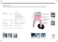
surgical bur, endo bur, diamond pear shaped bur and paste filler)<br />
procedure. 10<br />
After preoperative assessment, local anesthesia was given<br />
along with rubber dam placement. Roof of pulp chamber was<br />
removed using surgical bur after which vital pulp was excised<br />
from the chamber by means of endo bur. Shape the pulp chamber<br />
using diamond pear shaped bur. Pulpotec liquid and powder<br />
were blended <strong>to</strong> obtain thick, creamy consistency of the paste<br />
which was inserted in<strong>to</strong> the pulp chamber using paste filler.<br />
The cavity was then sealed with temporary ZOE cement. The<br />
patient was then asked <strong>to</strong> bite progressively but firmly on the<br />
cot<strong>to</strong>n placed between the two dental arches, so that the paste<br />
clings <strong>to</strong> the walls of the pulp cavity as well as <strong>to</strong> the root canal<br />
orifices. Excess cement was eliminated and pos<strong>to</strong>perative<br />
IOPAR taken after <strong>com</strong>pletion of procedure. Once the initial<br />
set has occurred (after 7 hours), second session was undertaken<br />
<strong>to</strong> <strong>com</strong>plete the treatment by seating stainless steel crown.<br />
Group 3 (Fig. 3)<br />
Experimental<br />
Twenty primary mandibular molars were treated with ‘lesion<br />
sterilization and tissue repair therapy’ (LSTR-3 Mix-MP, mixture<br />
of drug <strong>com</strong>bination of ciprofloxacin 500, metronidazole 400<br />
and minocycline 100 in a ratio of 1:3:3 prepared with macrogol<br />
and propylene glycogol in ointment form) procedure. 15<br />
Once the preoperative assessment was made, local<br />
anesthesia administration and rubber dam application were<br />
done. Access cavity was then prepared using no. 56 fissure bur<br />
in high speed NSK airo<strong>to</strong>r handpiece. 5% NaOCl immersed in<br />
cot<strong>to</strong>n was applied <strong>to</strong> control the hemorrhage, followed by<br />
application of the antibiotic paste <strong>to</strong> the pulpal floor. The <strong>to</strong>oth<br />
was then sealed with GIC and pos<strong>to</strong>perative IOPAR was taken<br />
after <strong>com</strong>pletion of procedure. Permanent res<strong>to</strong>ration was done<br />
by cementing stainless steel crown in second appointment after<br />
24 hours.<br />
The children were recalled for clinical evaluation at the<br />
interval of 1 month; clinical and radiographic 3, 6 and 12<br />
months.<br />
Scoring Criteria 17<br />
Scoring for clinical success of teeth:<br />
0. Failure<br />
1. No pain symp<strong>to</strong>ms<br />
2. No tenderness <strong>to</strong> percussion<br />
3. No swelling<br />
4. No fistula<br />
5. No pathologic mobility.<br />
Scoring for radiographic success of teeth:<br />
0. Failure<br />
1. No radicular radiolucency<br />
2. No internal/external root resorption<br />
3. No periodontal ligament space widening.<br />
[Failure (0): Defined as presence of any one or more of the<br />
above clinical and/or radiographic signs and symp<strong>to</strong>ms].<br />
RESULTS<br />
A <strong>to</strong>tal of 60 mandibular primary molars, 26 first molars and<br />
34 second molars in 34 children (18 males, 16 females) were<br />
endodontically treated in group I (ZOE) and in group 2<br />
(Pulpotec) and 3 (lesion sterilization and tissue repair) where<br />
noninstrumentation directed endodontic treatment was carried<br />
out. The symp<strong>to</strong>ms of the patients were recorded regarding the<br />
status of the pulp and single sitting roof canal procedure was<br />
carried out (Table 1).<br />
Statistical Analysis was done using Fisher’s exact test <strong>to</strong><br />
<strong>com</strong>pare the proportion of failure/success (Tables 2 <strong>to</strong> 5) in the<br />
three groups at different time intervals (Graphs 1 <strong>to</strong> 4). Null<br />
hypothesis H0 taken was that there is no significant difference<br />
in the proportion of failures among the three groups, i.e.<br />
p1 = p2 = p3 and the alternate hypothesis H1 was that at least<br />
one pair of the groups differ with level of significance being<br />
Table 1: Distribution of teeth according <strong>to</strong> presenting symp<strong>to</strong>ms<br />
Group Teeth with Acute symp<strong>to</strong>ms Abscess Asymp<strong>to</strong>matic<br />
deep carious<br />
lesions and<br />
exposed pulp<br />
1 20 13 — 7<br />
2 20 12 1 7<br />
3 20 13 — 7<br />
Table 2: Cumulative distribution of failure and success in each<br />
group at 1 month<br />
Group Failures Success p-value (Fisher’s test)<br />
Fig. 3: LSTR (3 Mix-MP placed over the pulp stump)<br />
1 (n = 16) 0 16 (100%) 1.000<br />
2 (n = 20) 0 20 (100%)<br />
1 (n = 16) 0 16 (100%) 0.024<br />
3 (n = 20) 6 (30%) 14 (70%)<br />
2 (n = 20) 0 20 (100%) 0.020<br />
3 (n = 20) 6 (30%) 14 (70%)<br />
World Journal of Dentistry, July-September 2011;2(3):187-192 189