going
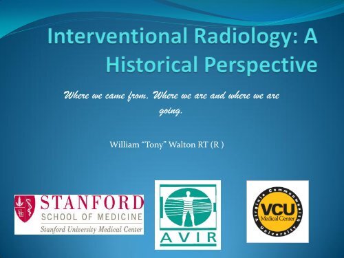
Interventional Radiology: A Historical Perspective - Executive Insight
Interventional Radiology: A Historical Perspective - Executive Insight
- No tags were found...

Create successful ePaper yourself
Turn your PDF publications into a flip-book with our unique Google optimized e-Paper software.
Where we came from, Where we are and where we are<br />
<strong>going</strong>.<br />
William “Tony” Walton RT (R )
Disclosure:<br />
• I have not been compensated in any form for<br />
mentioning people,products, or places.<br />
• This presentation, while being correct historically, is<br />
really meant to present you with the ideas and<br />
backgrounds of those who laid the foundation for<br />
Interventional Radiology.<br />
• There will always be opinions debated over who, or<br />
what happened first. This is my egg.
CE Information<br />
• This course awards the attendee 1.0 A credit.<br />
• Registrants who provided an email address will be sent<br />
a certificate of attendance from the AVIR with the CE#<br />
and title/date. This is only offered for the live course.<br />
• Those with groups of technologists participating, and<br />
registered under one individual should send an email<br />
with name, email, ARRT# and location to :<br />
membership@avir.org
Objectives<br />
• Present a extrospective review of the advent and<br />
progress of technology through 5 eras.<br />
• Review the impact of technology and course of change<br />
that evolved in those time periods.<br />
• Examine the inventions that formed the foundation of<br />
Interventional Radiology and the procedures that<br />
arose from them.
13 Jan 1983
• Five eras:<br />
History of Angiography<br />
A. Pre-clinical: 1895 – 1927 (before clinical<br />
application)<br />
B. Pre-Seldinger: 1928 – 1952<br />
C. Post – Seldinger: 1953 –1964<br />
D. Dotter Era 1964- 2000<br />
E. New millennium: 2000 - present
Timelines…..<br />
•Radiation Technology- Rise of the<br />
Machine<br />
•Radiology Practice- Pioneers in<br />
Medicine<br />
•Device Development-<br />
Interventional Inventions
• 1880-1900 The Second Industrial Revolution & The<br />
Gilded Age.<br />
• 1895: The discovery of X-Rays<br />
• 1914 WW1<br />
“Copy from one, it's plagiarism; copy from two, it's research.”<br />
Wilson Mizner<br />
Credit should be given to the erudites of the age, a majority which resided<br />
in Europe.
Or Do We?<br />
•Julius Plucker (1801-1868) - In 1858, published a paper relating the effects of magnetism on electrical current in rarified gases.<br />
•Johann Hienrich Geissler (1814-1879) Worked with Plucker to create the Geissler tube, later to become the foundation of what<br />
we know as neon lights.<br />
Johann. Hittorf (1824-1914)- In 1869, discovered that varied gases and pressures, produced different types of fluorescence in the<br />
vacuum tube.<br />
Sir William Crookes (1832-1919) Around 1875, invented the first CRT or Crookes tube, this led to the electron being studied<br />
further at a subatomic level<br />
Eugen Goldstein (1850-1931) – Credited with naming Cathode Rays (1876), and eventually discovered Anode rays (1886)<br />
Heinrich Hertz (1857-1894) In 1892, demonstrated Cathode Rays could penetrate very thin metal foil ( aluminum)<br />
Philipp Eduard Anton von Lenard (1862-1947)- A student of Hertz and awarded the 1905 Nobel Prize in Physics for the Cathode<br />
Ray Tube, demonstrated penetration through other materials, , Lenard did not realize he was producing X-rays.<br />
Heinrich Ruhmkorff‘ (1851) and Nicholas Callan (1836) invented / patented the induction coil , which led to the early formation<br />
of transformers which literally empowered the gas tube discoveries
Recognized early developments from :<br />
Hauksbee- gas discharged lamps 1705<br />
Farraday- Voltaic cells 1810<br />
J. Nollet/ Franklin- Egg vs Jar 1853<br />
A. Goodspeed- UPenn 1890
Wilhelm Roentgen, Nov 8 1896, “accidentally”started the<br />
revolutionary science of Radiology as we know it today. He was<br />
awarded the first Nobel Prize in Physics in ?.<br />
Anna Bertha’s Hand
Wilhelm<br />
Conrad<br />
Roentgen<br />
Discovered X-Rays<br />
on a Friday evening<br />
in Warzburg.<br />
50 days later, presents a paper to<br />
the Medical Society of Warzburg.<br />
28 December 1895.
The 1901 Nobel Prize for Physics<br />
Lenard and Roentgen were<br />
colleagues leading up to the 1896<br />
discovery.<br />
Lived within 200 miles of each<br />
other.<br />
1845-<br />
1923
Other Advancements Which Affected<br />
Technology<br />
From the 1830’s – transatlantic ship crossings accounted for all<br />
commerce and communication between US and Europe<br />
Telegraph -1866 First TA communication- Cyrus West Field ( ATC)<br />
Wireless TA Radio Communication- 1901-1903 Marconi, Tesla, Bose<br />
Air- 1910 First Dirigible /Airship Transatlantic Flight – Walter Wellman<br />
Air- First Solo Nonstop TA Flight ( May 1927) - Charles Lindbergh
Within 2 months after Roentgen’s discovery, January 17, 1896, Eduard Haschek<br />
and Otto Lindenthal produce first arterial map of an amputated hand, with a<br />
solution of iodine, chalk, cinnabar, and parrafin. And a 57 minute exposure!!<br />
Yikes!!!
• 1880-1900 The Second Industrial Revolution & The Gilded Age.<br />
• 1895: The discovery of X-Rays<br />
• 1896- “Angiogram” of amputated hand
German Beginnings<br />
• Siemens was the first company to patent the x ray<br />
tube in 1896. German patent.<br />
• Roentgen reportedly refused to patent the first tube.<br />
• Moritz Jastrowicz , Neuropsychiatrist<br />
• As early as January 12, 1896, first reported use ( Spies)<br />
on a 4 y/o FB.<br />
• “obviously important for medicine” 30 Jan 1896.
30 Jan 1896<br />
Jastrowicz<br />
article from<br />
Deutsches<br />
Medizinische<br />
Wochen schrift
Other Notable Firsts<br />
• First medical litigation involving malpractice<br />
occurred in England in the summer of 1896. Cuboid<br />
fx.<br />
• First radiation burn of a hand reported by Leppin in<br />
1896, later to contribute to the ICRP .<br />
• First pioneer to die from radiation exposure was C.M.<br />
Dally While Experimenting with X-rays. Worker with<br />
Edison For Seven Years. 1904
Other Radiation Related Notable Events<br />
1896 January 3 - X-Ray report made public<br />
• February - Discovery of Radioactivity -----H. Becquerel<br />
• March 3 - First reports of possible x-ray injury; damage to eyes -----T.A. Edison, W.J.<br />
Morton<br />
• March 14 - Concern expressed over possibility of x-ray injury -----F. Battelli<br />
• April 10 - Epilation noted from x-ray exposure -----J. Daniel<br />
• April 18 - Skin effects first noted -----L. G. Stevens<br />
• July - First x-ray protective device: a heavy glass plate to protect the eyes during<br />
dental radiography -----W.H. Rollins<br />
• Reports of accidental injury (burns) -----H.D. Hawks<br />
• November 18 - Deliberately induced experimental injury (burns) -----E. Thomson<br />
• Gold leaf electroscope used to make ionization measurements -----L. Benoist<br />
1897 Air thermometer used to measure energy transfer by x-rays -----E. Dorn<br />
1898 January - Aluminum filter used as protective device -----E. Thomson<br />
• May - Dark adaptation prior to fluoroscopy suggested -----F.H. Williams<br />
• July - Leaded x-ray tub housing; collimators -----W.H. Rollins<br />
• July - Word "radioactivity" coined -----P. & M. Curie<br />
• December - Radium discovered -----P. & M. Curie<br />
• Gamma rays discovered -----P. Villard<br />
1899 April - Radiographer licensure recommended to protect public -----J. Dennis<br />
• May - Malpractice award for x-ray burns
Monument to Radiation workers<br />
at the Allgemein Krakenhaus St.<br />
George in Hamburg, Germany.<br />
Erected in 1936, by the German<br />
Roentgen Society.<br />
136 Individuals from 15<br />
countries<br />
71 German<br />
15 Britain<br />
“They were heroic pioneers for a safe<br />
and successful application of x-rays to<br />
medicine. The fame of their deeds are<br />
immortal.”
•Ronald A. Fessenden<br />
Applied August 7 , 1899 , Issued May 1, 1900 (
William Andrews<br />
General Electric Company<br />
Filed January 20, 1898<br />
Issued September 4, 1900
Humble Beginnings in the U.S.<br />
• Victor Electric plunged into the x-ray business and by<br />
1896 (one year after Roentgen discovery) were making<br />
x-ray machines.<br />
• The business grew rapidly and so, in 1896, moved into<br />
new premises three times the original size, but this did<br />
not solve the space problems and the company made 3<br />
moves by 1899.<br />
• Later bought by GE, by 1913 became sole<br />
manufacturer of Coolidge tubes in the US. GE Origins<br />
as Edison Electric 1876
Samuel Puffer<br />
Applied February 27,1898, Awarded August 8,1899
A. Pre-clinical Era<br />
• 1895: X-rays discovered<br />
• 1896: “Angiogram” of amputated hand<br />
• 1898: visualization of pellets “floating” in heart<br />
• 1900: First Diagnostic Apparatus<br />
• Still Radiography<br />
• Cavaderic Studies<br />
• Isolated Silos of Inventors
Gallot- Pilon France 1910-1920’s<br />
Courtesy BRM
Other contributors throughout the<br />
era …<br />
William Coolidge – (GE) First X-Ray tube with a focal spot (Nov 1913) First transformer<br />
feed x ray apparatus (1920), allowed consistent timed high voltage exposures.<br />
Albert Hall (GE) – Filter for monochromatic X-Ray photographic imaging<br />
(1921).<br />
Samuel Shepperd (Kodak)- First intensifying screen technology (1922)-<br />
meant lower exposures .<br />
James Haste (Kodak)- First cellulose based film for radiography (1922)
Add Fluoroscopy to the mix…….<br />
Snook and Kelly 1914 in<br />
Philadelphia<br />
Utilized mirrors to visualize a<br />
fluoroscopic screen
1933 Flouroscopes just<br />
prior to image<br />
intensification.<br />
Room would have been<br />
darkened…
Angiography<br />
• Angiography dates to 1895<br />
Angiography from Greek<br />
• Angeion = vessel<br />
• Graphein = to record<br />
• Discovery of X-rays: Wilhelm Conrad Rontgen<br />
• Pre-Seldinger Era begins with Moniz<br />
• Involves the evolving of the x ray apparatus<br />
• The evolution of image capture<br />
• Add the contribution of contrast media
Angiographic Techniques<br />
• Interventional Radiology origins came from early<br />
pioneers in angiography<br />
António Egas Moniz<br />
Reynoldo Dos Santos<br />
Moniz formulated<br />
strontium bromide<br />
65% and sodium<br />
iodine contrast 65%><br />
25%.<br />
Thorotrast (thorium<br />
dioxide) was used later<br />
in the 1930’s .<br />
28 June 1927<br />
January 1929
Cerebral Angiography<br />
• 1927: Egas Moniz (Portugal) radiologic demonstration of<br />
cerebral vessels (25% NaBr)<br />
• Direct exposure of CA<br />
• 1931: Moniz: 25% of Thorium dioxide (Thorotrast)<br />
• Thorotrast: “no pain”<br />
• 1950s: Thorotrast: angiosarcomas
Early Aortography<br />
• 1929: Dos Santos (Portugal) – 1 st desciption of TLA for<br />
Drug delivery and dx<br />
• 1937: Dos Santos > 1000 TLA<br />
• “No suitable contrast material”<br />
• 1960 – 1970s: TLA “popular”<br />
• 2000: Technique unchanged
Technology of the time<br />
• Bill Hogan (1920”s)- Modest Electrician- Most<br />
influential of non- physicians of his time. Started as a<br />
repairman, then moved to installing the then modern<br />
day systems for Standard X-Ray Company, Chicago, Ill.<br />
• Influenced technology at G.E., Westinghouse, and<br />
Picker.<br />
• In 1932,Started the Franklin X-Ray Company in<br />
Philadelphia designed the franklin head unit.<br />
• Franklin Biplane Angiographic Machine –developed<br />
for Dr. Edward Chamberlain ( Temple); also cinefluoro<br />
camera apparatii.
Sure they invented it, but how did they do it?<br />
• All the early cerebral arteriograms were done using the cut-down<br />
technique, exposing the CA, occluding flow, injecting the<br />
contrast, and resuming flow while taking 3 serial radiographs.<br />
• Saito,Makato-A Japanese pioneer, in 1930, cited over 100 cerebral<br />
arteriograms injecting sodium bromide via direct puncture of<br />
the superior thyroid artery.<br />
• Dr. Werner Forssmann- In 1929, Catheterized his own heart<br />
(venous ) via antecubital vein, self-experimentation led him to<br />
quit cardiology and become a urologist. 1956 Nobel Prize.
B. Pre – Seldinger Era<br />
• 1923: Berberich: arteriograms and venograms with 10 –<br />
20% solutions strontium bromide<br />
• 1923: Siccard & Forester: no untoward reaction with iodized<br />
oil in canine A and V<br />
• 1924:Brooks: sodium iodide injection in surgically exposed<br />
SFA<br />
• 1927: Post Moniz-Carnett: “arteriograms” of LE with iodized<br />
oil<br />
• 1930: Saito in Japan: “fine iodized oil”
Angiography Timeline<br />
• 1930s: “direct” puncture of TA<br />
• 1939: Robb and Steinberg: injection of CM in vein to visualize heart<br />
chambers -Harvard<br />
• 1947: Radner: cut down in radial artery, catheter advanced into aorta: TA<br />
Britain<br />
• 1949: Johnson: TA by direct puncture of CA -U Penn<br />
• Opacification of TA and coronary A, Mapping potential for CEA<br />
• CAROTID ANGIOGRAPHY A CLINICAL EVALUATION OF 200 CONSECUTIVE<br />
CASES DWIGHT PARKINSON, M.D.* and A. E. CHILDE, M.D.,t Winnipeg,<br />
Man. 1950- 3 fatalies, few side effects<br />
• 1950: IV injections of large volume CM to opacify TA and AA
Pre- Seldinger Techniques<br />
• (a) Percutaneous trans-lumbar needle puncture<br />
of the aorta as introduced by Dos Santos, Lamas<br />
and Pereira Caldas (1929).<br />
• (b) ' Cut down ' or modified ' cut down ' transfemoral<br />
arterial catheterization of the aorta as<br />
advocated by Farinas (I946) and Lindgren (I953).<br />
• (c) Percutaneous transfemoral retrograde aortography<br />
• as introduced by Seldinger (1953).
Pulmonary Arteriography by Direct<br />
Puncture
Technology Timeline….<br />
• Caldas “Carousel” 1931- Allowed up to six exposures,<br />
manual. Developed in Portugual. Allowed 6 exposures in<br />
6 seconds.<br />
• Sanchez- Perez Film Changer – up to 6 films, manual<br />
crank, then electrified, 1 film every 0.7 sec ( late 1940s).
Sanchez-Perez. First Patent.
If only ……..<br />
Life could be this simple!!!!
The Franklin Biplane Multi Film<br />
Radiographic Device<br />
Developed in the early<br />
1940’s, 3 built in first<br />
year<br />
Temple, UPenn Chicago<br />
Veterans Hospital<br />
Synchonized exposures<br />
Same 9 ½ film format<br />
Still purely serial<br />
angiographs…
Fairchild<br />
• Fairchild Roll Film Changer –<br />
75ft of<br />
9 ½ inch film, 2 f/s capability ,<br />
late 1940’s.<br />
• “Engineered” by C.T. Dotter<br />
• Origins came from WW2, aerial<br />
camera technology
Injectors in the 1940’s. Your basic<br />
design.
Pre- Seldinger<br />
• Up until 1952, arteriography /angiography was<br />
typically performed by surgical exposure of the artery<br />
or a direct pucture by a needle or trocar.<br />
• Trocar/needles were often larger than the catheter<br />
being inserted.<br />
• In 1952, Seldinger reported his technique
Seldinger Technique<br />
• Contrast could be injected at any level<br />
• Less risk of extravisation<br />
• Very simple technique<br />
• Patient can be in any position<br />
• Catheter could be left in place for longer periods of<br />
time
Post-Seldinger Period<br />
• From the advent of the percutaneous technique,<br />
angiography and the disciplines that arose over the<br />
next 3 decades would shape IR as we know it today.<br />
• Technology advancements in imaging and devices<br />
would generate new diagnosis/therapies.<br />
• The developments are as unique as the minds that<br />
shaped them.
Selective Angiography<br />
1954: Seldinger: parathyroid<br />
1956: Odman: radiopaque polyethylene selective catheter<br />
Odman, P.: Percutaneous Selective Angiography<br />
of the Main Branches of the Aorta.<br />
Acta Radiol. (Stockh.), 45:1, 1956.<br />
•Polyethylene can be reshaped by steam and cooled<br />
•Holes can be made<br />
•Different sizes and diameters<br />
1966: Boijsen: abdominal arteriography<br />
1969: Reuter: pancreatic arteriography<br />
1965: Viamonte: selective arteriography<br />
1969: Rösh: superselective arteriography
The Modern Day injector.<br />
Developed in the late 1950’s, first “automatic” “safe”<br />
injector by Williamson (Cordis)
Elema- Schonander AOT-S<br />
Late 1950 through 1970’s<br />
Became the standard in High Tech Labs of the time period.<br />
Programmable up to 4 f/s.<br />
Magazine held 30 14”x<br />
14” films , separated by<br />
wire looms- Very heavy<br />
Early units had dial selectors<br />
for frame rates and intervalsup<br />
to four<br />
Advanced to use key punch<br />
cards to program exposure<br />
sequences
Long Leg Film Changers by C. Medical Manufacturing,Canada<br />
Held 6 Long cassettes – 14” x 51” tri-fold film<br />
Required 84-112” focal distance, usually a ceiling<br />
mounted tube configuration.<br />
Patient had to be positioned on or<br />
over a separate radiographic<br />
table.<br />
Visualize the entire LE vasculature<br />
Cassettes were heavy and<br />
cumbersome, to say the least.
With Cut Film , comes subtraction!!<br />
The technique is not cited in journals or<br />
patents until the late 1970’s, Kodak/Fuji
Subtraction Procedure<br />
(Cerebral angiogram)<br />
1. Before angiogram, take scout<br />
skull x-ray on angio equipment<br />
that will be used – looks like<br />
regular skull film (this is for<br />
technique only , zero image is<br />
first radiograph taken during the<br />
angiogram!)<br />
1. Place subtraction mask film in<br />
copier with original (zero)<br />
lateral skull film - expose and<br />
process<br />
• (original x-ray (negative)<br />
converts into “positive” )<br />
Zero (base, scout)<br />
Subtraction mask
Creating Image free of background –The Art of<br />
Subtraction<br />
+<br />
=<br />
Mask-series<br />
combination<br />
Mask<br />
(Inverse of<br />
original)<br />
Series<br />
(Like original but<br />
with Contrast)<br />
Vessels appear<br />
white on black<br />
background<br />
+<br />
Subtraction<br />
copy film<br />
(Reverses<br />
tones)<br />
=<br />
Finished product<br />
Vessels appear<br />
black,<br />
background clear
First in Film