

images
1TEZSjF
1TEZSjF

You also want an ePaper? Increase the reach of your titles
YUMPU automatically turns print PDFs into web optimized ePapers that Google loves.
clinical and research news<br />
Liver PET/MRI<br />
Thomas Hope, MD, Miguel Pampaloni, MD, PhD, Michael Ohliger, MD, Spencer Behr, MD, Vahid Ravanfar, RT, Henry VanBrocklin, PhD,<br />
James Slater, RPh, PhD, Carina Mari Aparici, MD, Judy Yee, MD, Eric Nakakura, MD, Emily Bergsland, MD, Carlos Corvera, MD,<br />
Daniel Vigneron, PhD, and Sharmila Majumdar, PhD<br />
Hepatic imaging benefits greatly from the use of MRI compared<br />
to CT. In addition to contrast dynamics you can investigate<br />
T2 signal intensity and diffusion weighted imaging that<br />
can help detect and characterize hepatic lesions in ways<br />
that are not possible on CT. PET/MRI promises to provide<br />
a significant advance in clinical staging compared to PET/<br />
CT for hepatic lesions.<br />
Imaging using FDG<br />
Fluorodeoxyglucose (FDG) is the most common PET<br />
radiotracer used in clinical medicine. There are numerous<br />
clinical indications for imaging hepatic metastatic disease,<br />
although FDG PET is used infrequently to evaluate primary<br />
liver malignancy. Combining focused liver MRI has<br />
demonstrated an ability to increase the detection of small<br />
hepatic metastases that can change patient care. This is<br />
particularly relevant in patients with colorectal cancer being<br />
treated with curative resection. In addition to metastatic<br />
disease, there may be a role in the setting of hepatocellular<br />
carcinoma. Well-differentiated HCCs are typically not<br />
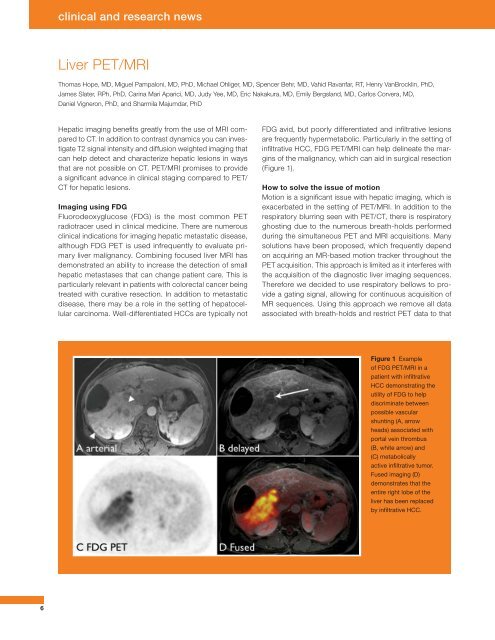
FDG avid, but poorly differentiated and infiltrative lesions<br />
are frequently hypermetabolic. Particularly in the setting of<br />
infiltrative HCC, FDG PET/MRI can help delineate the margins<br />
of the malignancy, which can aid in surgical resection<br />
(Figure 1).<br />
How to solve the issue of motion<br />
Motion is a significant issue with hepatic imaging, which is<br />
exacerbated in the setting of PET/MRI. In addition to the<br />
respiratory blurring seen with PET/CT, there is respiratory<br />
ghosting due to the numerous breath-holds performed<br />
during the simultaneous PET and MRI acquisitions. Many<br />
solutions have been proposed, which frequently depend<br />
on acquiring an MR-based motion tracker throughout the<br />
PET acquisition. This approach is limited as it interferes with<br />
the acquisition of the diagnostic liver imaging sequences.<br />
Therefore we decided to use respiratory bellows to provide<br />
a gating signal, allowing for continuous acquisition of<br />
MR sequences. Using this approach we remove all data<br />
associated with breath-holds and restrict PET data to that<br />
Figure 1 Example<br />
of FDG PET/MRI in a<br />
patient with infiltrative<br />
HCC demonstrating the<br />
utility of FDG to help<br />
discriminate between<br />
possible vascular<br />
shunting (A, arrow<br />
heads) associated with<br />
portal vein thrombus<br />
(B, white arrow) and<br />
(C) metabolically<br />
active infiltrative tumor.<br />
Fused imaging (D)<br />
demonstrates that the<br />
entire right lobe of the<br />
liver has been replaced<br />
by infiltrative HCC.<br />
6