PANCREATIC NEUROENDOCRINE TUMORS

Create successful ePaper yourself
Turn your PDF publications into a flip-book with our unique Google optimized e-Paper software.
<strong>PANCREATIC</strong> <strong>NEUROENDOCRINE</strong> <strong>TUMORS</strong><br />
INFORMATION<br />
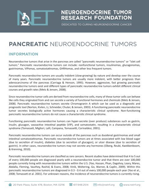
Neuroendocrine tumors that arise in the pancreas are called “pancreatic neuroendocrine tumors” or “islet cell<br />
tumors.” Pancreatic neuroendocrine tumors can include: nonfunctional tumors, insulinomas, glucagonomas,<br />
gastrinomas, VIPomas, somatostatinomas, GHRHomas, and other less frequent tumors.<br />
Pancreatic neuroendocrine tumors are usually indolent (slow-growing) by nature and develop over the course<br />
of many years. Pancreatic neuroendocrine tumors are usually more indolent, with better prognosis than<br />
adenocarcinoma of the pancreas (Carriaga & Henson, 1995). However, aggressive, fast growing pancreatic<br />
neuroendocrine tumors exist and different types of pancreatic neuroendocrine tumors exhibit different clinical<br />
courses and growth rates (Metz & Jensen, 2008).<br />
Since neuroendocrine tumor cells are derived from neuroendocrine cells, many of these tumor cells can behave<br />
like cells they originated from and can secrete a variety of functional hormones and chemicals (Metz & Jensen,<br />
2008). Pancreatic neuroendocrine tumors secrete Chromogranin A which can be used as a diagnostic and<br />
prognostic tool (Norton, Kivlen, Li, Schneider, Chuter, & Jensen, 2003). A functioning pancreatic neuroendocrine<br />
tumor secretes biologically active hormones causing a characteristic clinical syndrome. Non-functioning<br />
pancreatic neuroendocrine tumors do not cause a characteristic clinical syndrome.<br />
Functioning pancreatic neuroendocrine tumors can hyper-secrete (over produce) substances such as gastrin,<br />
insulin, glucagon, vasoactive intestinal peptide (VIP), and somatostatin, resulting in a characteristic clinical<br />
syndrome (Tomasseti, Migliori, Lalli, Campana, Tomassetti, Corinaldesi, 2001).<br />
Pancreatic neuroendocrine tumors can occur outside of the pancreas such as duodenal gastrinomas and small<br />
intestinal somatostatinomas. Pancreatic neuroendocrine tumors are at times associated with low blood sugar<br />
(due to secretion of insulin), diabetes (due to secretion of glucagon), or ulcer disease (due to secretion of<br />
gastrin). In other cases, neuroendocrine tumors may not secrete any hormones (Oberg, Reubi, Kwekkenboom,<br />
& Krenning, 2010).<br />
Pancreatic neuroendocrine tumors are classified as rare cancers. Recent studies have determined that 4 to 5 out<br />
of every 100,000 people are diagnosed yearly with a neuroendocrine tumor and that there are over 100,000<br />
people currently living with neuroendocrine tumors within the U.S. (Yao, Hassan, Phan, Dagohoy, Leary, Mares,<br />
Abdalla, Fleming, Vauthey, Rashid, & Evans, 2008; Vinik, Woltering, Go, Warner, & Caplin, 2009). Within this<br />
pancreatic neuroendocrine tumors are diagnosed in 0.3 - 0.4 out of every 100,000 people each year (Yao et al.,<br />
2008; Tomasseti et al. 2001). For unknown reasons, the incidence of neuroendocrine tumors is currently rising.<br />
20 Park Plaza, Suite 478, Boston, MA 02116 617.948.2514 info@netrf.org www.netrf.org
Pancreatic neuroendocrine can be difficult to diagnosis with the average time between tumor development and<br />
diagnosis being between 5 and 10 years (Vinik, Feliberti, Perry & Nakave, 2008; Vinik et al., 2009). Survival rates<br />
for individuals with pancreatic neuroendocrine tumors vary and depend upon tumor type, the location of the<br />
tumors, the size of the tumors, the extent and growth rate of liver and bone metastases, proliferative indices,<br />
presence of clinical syndromes and many other factors (Metz & Jensen, 2008). Currently, surgery is the only<br />
option that offers hope for a cure (Ramage, Ahmed, Ardill, Bax, Breen, Caplin, Corrie, Davar, Davies, Lewington,<br />
Meyer, Newell-Price, Poston, Reed, Rockall, Steward, Thakker, Toubanakis, Valle, Verbeke, Grossman, and UK and<br />
Ireland Neuroendocrine Tumor Society, 2012; Metz & Jensen, 2008).<br />
Pancreatic neuroendocrine tumors can be associated with genetic syndromes such as Multiple Endocrine<br />
Neoplasia Type 1 (MEN1), Von Hippel-Lindau Disease (VHL),Tuberous Sclerosis Complex and Neurofibromatosis<br />
Type 1 (NF1) (Metz & Jensen, 2008). MEN1 is the most significant genetic syndrome - over 80% of patients<br />
with MEN1 develop pancreatic neuroendocrine tumors, over 40% of patients develop gastrinomas and smaller<br />
percentages develop other types of pancreatic neuroendocrine tumors (Metz & Jensen, 2008; Gibril & Jensen,<br />
2004; Brandi et al., 2002).<br />
What is a Tumor?<br />
Cells are the building blocks of all life. All cells have highly specific functions, but not all cells have the same<br />
function. When cells that have similar functions are grouped together they form a tissue. Tissues when grouped<br />
together to perform a specific function are called organs. All cells of the human body have the same DNA (genetic<br />
language). Cell growth and replication is highly controlled and is encoded in each cell’s DNA. However if there are<br />
enough mutations (changes) within a cell’s DNA, a cell can grow and replicate uncontrollably.<br />
A tumor is a mass formed by an abnormal growth of cells within the body. A tumor can be non-cancerous<br />
(benign) or cancerous (malignant). A tumor is considered cancerous when it has uncontrolled proliferation<br />
(abnormal growth) and can invade and destroy surrounding tissue. Malignant tumors can also have the ability to<br />
metastasize (spread to other organs of the body).<br />
What is a Neuroendocrine Tumor?<br />
The neuroendocrine system consists of highly specialized neuroendocrine cells which act as an interface or<br />
junction between the nervous system and the endocrine system. The endocrine system is made up of cells whose<br />
function is to produce and secrete hormones into the bloodstream. Hormones are biochemical messengers that<br />
help to regulate many different processes within the body. The nervous system is composed of specialized cells<br />
(neurons) that control the activities of all body parts. A neuroendocrine cell is a cell which receives neuronal<br />
input (a signal from a nerve cell) and releases hormones in response to this signal.<br />
A neuroendocrine tumor can develop anywhere there are neuroendocrine cells. The most common sites from<br />
which neuroendocrine tumors arise are the lungs, appendix, small intestine, rectum and pancreas (Yao, Hassan,<br />
Phan, Dagohoy, Leary, Mares, Abdalla, Fleming, Vauthey, Rashid, & Evans, 2008). Neuroendocrine tumors that<br />
arise in the pancreas are called pancreatic neuroendocrine tumors or islet cell tumors. When neuroendocrine<br />
tumors originate in other areas, they are often classified as carcinoid tumors.<br />
20 Park Plaza, Suite 478, Boston, MA 02116 617.948.2514 info@netrf.org www.netrf.org
Since neuroendocrine tumor cells are derived from neuroendocrine cells, many of these tumor cells can behave<br />
like cells they originated from and can secrete a variety of hormones. A functioning neuroendocrine tumor is one<br />
that secretes biologically active hormones causing a clinical syndrome. Non-functioning neuroendocrine tumors<br />
do not cause clinical syndromes.<br />
Carcinoid tumors and pancreatic neuroendocrine tumors share similarities including often indolent behavior,<br />
ability to secrete biologically active hormones, and well-differentiated histology (Reidy, Tang & Saltz, 2009).<br />
INFORMATION REFERENCES<br />
• Carriaga, M. and Henson, D. (1995). Liver, gallbladder, extrahepatic bile ducts, and pancreas. Cancer, 75, 171-190.<br />
• Jensen, R., Berna, M., Bingham, D. and Norton, J. (2008). Inherited pancreatic endocrine tumor syndromes:<br />
advances in molecular pathogenesis, diagnosis, management, and controversies. Cancer, 113(7), 1807-1843.<br />
Retrieved from: http://www.ncbi.nlm.nih.gov/pubmed/18798544.<br />
• Metz, D. and Jensen, R. (2008). Gastrointestinal neuroendocrine tumors, pancreatic endocrine tumors.<br />
Gastroenterology, 135(5), 1469-1492. Retrieved from: http://www.ncbi.nlm.nih.gov/pubmed/18703061.<br />
• Norton, J., Kivlen, M., Li, M., Schneider, D., Chuter, T., and Jensen, R. (2003). Morbidity and mortality of<br />
aggressive resection in patients with advanced neuroendocrine tumors. Archives of Surgery, 138(8), 859-<br />
866. Retrieved from: http://www.ncbi.nlm.nih.gov/pubmed/12912744.<br />
• Oberg, K. E., Reubi, J. C., Kwekkeboom, D. J., Krenning, E. P. (2010) Role of somatostatins in gastroenteropancreatic<br />
neuroendocrine tumor development and therapy. Gastroenterology, 139(3), 742-753. Retrieved from: http://<br />
www.ncbi.nlm.nih.gov/pubmed/20637207.<br />
• Ramage, J., Ahmed, A., Ardill, J., Bax, N., Breen, D.J, Caplin, M.E., Corrie, P., Davar, J., Davies, A.H., Lewington,<br />
V., Meyer, T., Newell-Price, J., Poston, G., Reed, N., Rockall, A., Steward, W., Thakker, R.V., Toubanakis, C., Valle,<br />
J., Verbeke, C., and Grossman, A.B., and UK and Ireland Neuroendocrine Tumor Society (2012) . Guidelines<br />
for the management of gastroenteropancreatic neuroendocrine (including carcinoid) tumours (NETs). Gut,<br />
61(1):6-32. Retrieved from: http://www.ncbi.nlm.nih.gov/pubmed/22052063.<br />
• Tomasseti, P., Migliori, M., Lalli, S., Campana, D., Tomassetti, V., Corinaldesi, R. (2001). Epidemiology, clinical<br />
features and diagnosis of gastroenteropancreatic endocrine tumours. Annals of Oncology, 12(2), S95-S99.<br />
Retrieved from: http://www.ncbi.nlm.nih.gov/pubmed/11762360.<br />
• Vinik, A. I., Feliberti, E., Perry, R., Nakave, A. (2008). Diffuse Hormonal Systems and Endocrine Tumor<br />
Syndromes. Retrieved from: http://www.endotext.org/guthormones/index.htm/.<br />
• Vinik, A. I., Silva, M. P., Woltering, E. A., Go, V., Warner, R., Caplin, M. (2009). Biochemical Testing for<br />
Neuroendocrine Tumors. Pancreas, 38(8), 876-899. Retrieved from: http://www.ncbi.nlm.nih.gov/<br />
pubmed/19855234.<br />
• Yao J. C., Hassan, M., Phan, A., Dagohoy, C., Leary, C., Mares, J. E., Abdalla, E. K., Fleming, J. B., Vauthey, J. N.,<br />
Rashid, A., Evans, D. B. (2008). One hundred years after “carcinoid”: epidemiology of and prognostic factors<br />
for neuroendocrine tumors in 35,825 cases in the United States. Journal of Clinical Oncology, 20(26), 3063-<br />
3072. Retrieved from: http://www.ncbi.nlm.nih.gov/pubmed/18565894.<br />
20 Park Plaza, Suite 478, Boston, MA 02116 617.948.2514 info@netrf.org www.netrf.org
LOCATIONS & CLASSIFICATIONS<br />
Pancreatic Neuroendocrine Tumor Histological Features<br />
Since neuroendocrine tumors, including pancreatic neuroendocrine tumors, represent a heterogeneous group<br />
of tumors, the World Health Organization in 2000 updated the classification system for them based upon their<br />
clinical pathological criteria (Brandi, Gagel, Angeli, Bilezikian, Beck-Peccoz, Bordi, Conte-Devolx, Falchetti, Gheri,<br />
Libroia, Lips, Lombardi, Mannelli, Pacicni, Ponder, Raue, Skogseid, Tamburrano, Thakker, Thompson, Tomassetti,<br />
Tonelli, Wells, and Marx, 2002). Each category includes both functioning and non-functioning tumors.<br />
• Well-differentiated endocrine tumors, with benign or uncertain behavior.<br />
• Well-differentiated endocrine carcinomas, with low-grade malignant behavior.<br />
• Poorly differentiated endocrine carcinomas, with high-grade malignant behavior.<br />
• Endocrine/exocrine carcinomas with characteristic of both endocrine and exocrine tumors.<br />
Carcinoid tumors and pancreatic neuroendocrine tumors fall within the classification of well-differentiated<br />
endocrine tumors. Tumors classified as poorly differentiated endocrine carcinoma or endocrine/exocrine<br />
carcinoma with characteristics of both endocrine and exocrine tumors are not covered on the NET Research<br />
Foundation’s website. For these types of tumors please contact your physician for more information.<br />
For our purposes, the terms carcinoid tumors and pancreatic neuroendocrine tumors refer to well-differentiated<br />
endocrine tumors and the information provided should not be applied to any tumors not characterized as welldifferentiated.<br />
Presence of Clinical Syndrome<br />
Pancreatic neuroendocrine tumors are classified as functioning or non-functioning.<br />
Functioning<br />
A functioning pancreatic neuroendocrine tumor secretes biochemically active substances such as hormones<br />
which cause specific clinical syndromes such as Carcinoid Syndrome, Zollinger-Ellison Syndrome, or symptoms<br />
from hormone over secretion such as insulin or glucagons.<br />
Non-functioning<br />
A non-functioning pancreatic neuroendocrine tumor secretes specific substances but these substances are<br />
either inactive and/or do not cause any clinical syndrome.<br />
Pancreatic Neuroendocrine Tumor Classification<br />
Pancreatic neuroendocrine tumors vary in clinical course, location, and hormone secretion. Pancreatic<br />
neuroendocrine tumors can occur outside of the pancreas such as the duodenum and small intestine.<br />
20 Park Plaza, Suite 478, Boston, MA 02116 617.948.2514 info@netrf.org www.netrf.org
Functioning Pancreatic Neuroendocrine Tumors<br />
Insulinoma<br />
characterized by excess secretion of insulin and pro-insulin which causes confusion, sweating, dizziness,<br />
weakness and unconsciousness (Ramage, Ahmed, Ardill, Bax, Breen, Caplin, Corrie, Davar, Davies, Lewington,<br />
Meyer, Newell-Price, Poston, Reed, Rockall, Steward, Thakker, Toubanakis, Valle, Verbeke, Grossman, and UK<br />
and Ireland Neuroendocrine Tumor Society, 2012; Tomasseti, Migliori, Lalli, Campana, Tomassetti, Corinaldesi,<br />
2001). Prolonged hypoglycemia (low blood sugar) can have permanent impact on brain function. Insulinomas<br />
tend to be small and rarely metastasize (Metz & Jensen, 2008; Modlin, Oberg, Chung, Jensen, de Herder,<br />
Thakker, Caplin, Delle Fave, Kaltsas, Krenning, Moss, Nilsson, Rindi, Salazar, Rusziniewsku, & Sundin, 2008; Kulke,<br />
Anthony, Bushnell, de Herder, Goldsmith, Klimstra, Marx, Pasieka, Pommier, Yao, and Jensen, 2010), many report<br />
metastases in about 10% of patients (Ramage et al., 2012). Insulinomas can be associated with MEN1. (Tomasetti,<br />
et al., 2001).<br />
Gastrinoma<br />
Characterized by Zollinger-Ellison Syndrome caused by excess secretion of gastrin (Tomasetti et al., 2001).<br />
Zollinger-Ellison syndrome causes diarrhea and peptic-ulcers. Patients with Zollinger-Ellison syndrome may also<br />
develop gastric carcinoid as a result of prolonged gastrin hypersecretion (over production). Patients frequently<br />
have liver and lymph node metastasis at diagnosis (Tomasetti et al., 2001). Gastrinomas occur mainly in the<br />
duodenum and pancreas (Tomasetti et al., 2001). When Gastrinomas occur in the duodenum they are frequently<br />
associated with MEN1. (Tomasetti et al., 2001; Ramage et al., 2012). All patients with Zollinger-Ellison Syndrome<br />
should be screened for MEN1 (Mamikunian, Vinik, O’Dorisio, Woltering, & Go, 2009).<br />
Glucagonoma<br />
Characterized by hypersecretion of glucagon which causes skin rash, diabetes, and weight loss (Tomasetti et<br />
al., 2001; Ramage et al., 2012; Metz & Jensen, 2008). Glucagonomas occur mainly in the pancreas, are typically<br />
large, and are highly metastatic. Glucagonoma is rarely associated with MEN1 (Tomasetti et al., 2001).<br />
VIPoma<br />
Characterized by hypersecretion of Vasoactive Intestinal Peptide (VIP) which causes Verner-Morrison Syndrome.<br />
Symptoms of Verner-Morrison Syndrome include severe watery diarrhea, which can be life threatening.<br />
Somatostatinoma –Somatostatinomas are primarily or exclusively composed of somatostatin producing cells.<br />
Somatostatinomas are found in the pancreas and duodenum. (Tomasetti et al., 2001; Metz & Jensen, 2008;<br />
Mamikunian, 2009). Somatostatinomas may not provoke clinical symptoms or they may be characterized by<br />
clinical symptoms of gallbladder stones, diabetes, weight loss, and diarrhea (Metz & Jensen, 2008).<br />
20 Park Plaza, Suite 478, Boston, MA 02116 617.948.2514 info@netrf.org www.netrf.org
Non-Functioning Pancreatic Neuroendocrine Tumors<br />
Non-functioning pancreatic neuroendocrine tumors are not associated with characteristic clinical symptoms<br />
from secretion of hormones. Non-functioning pancreatic neuroendocrine tumors occur in the pancreas. They do<br />
not cause hormonal symptoms but can cause pain, weight loss and jaundice. Because there is no characteristic<br />
clinical syndrome associated with non-functioning pancreatic neuroendocrine tumors they can be difficult to<br />
diagnose and are often diagnosed after presenting with large tumors and liver metastases (Metz & Jensen, 2008;<br />
Kulke et al., 2010).<br />
Inherited Versus Sporadic<br />
Pancreatic neuroendocrine tumors can be sporadic or familial, when the patient has genetically inherited<br />
mutations, which predispose development of tumors.<br />
Sporadic<br />
Cancer causing mutation arise randomly<br />
Inherited<br />
Cancer causing mutations are inherited such in the case of MEN-I.<br />
Pancreatic neuroendocrine tumors can be associated with genetic syndromes such as Multiple Endocrine<br />
Neoplasia Type 1 (MEN1), Von Hippel-Lindau Disease (VHL),Tuberous Sclerosis Complex and Neurofibromatosis<br />
Type 1 (NF1) (Metz & Jensen, 2008). MEN1 is the most significant genetic syndrome - over 80% of patients<br />
with MEN1 develop pancreatic neuroendocrine tumors, over 40% of patients develop gastrinomas and smaller<br />
percentages develop other types of pancreatic neuroendocrine tumors (Metz & Jensen, 2008; Gibril & Jensen,<br />
2004; Brandi et al., 2002).<br />
MEN1 is an autosomal dominant inherited syndrome characterized by multiple endocrine and non-endocrine<br />
tumors (Brandi et al., 2001). The tumors most frequently observed in patients with MEN1 include parathyroid<br />
adenomas, pituitary adenomas, and pancreatic endocrine tumors.<br />
VHL is an autosomal dominant inherited syndrome caused by a mutation in the VHL gene.<br />
Pancreatic neuroendocrine tumor patients should always have a clinical examination including family history to<br />
exclude genetic syndromes like MEN1 (Ramage et al., 2012). Presence of MEN1 or another clinical syndrome<br />
may change the optimal treatment course and so it is important to rule out. If a patient has a genetic syndrome<br />
it may be advised that certain family members are tested as well.<br />
20 Park Plaza, Suite 478, Boston, MA 02116 617.948.2514 info@netrf.org www.netrf.org
LOCATIONS & CLASSIFICATIONS REFERENCES<br />
• Brandi, M., Gagel, R., Angeli, A., Bilezikian, J., Beck-Peccoz, P., Bordi, C., Conte-Devolx, B., Falchetti, A., Gheri,<br />
R., Libroia, A., Lips, C., Lombardi, G., Mannelli, M., Pacicni, F., Ponder, B., Raue, F., Skogseid, B., Tamburrano,<br />
G., Thakker, R., Thompson, N., Tomassetti, P., Tonelli, F., Wells, S., and Marx, J. (2002). Guidelines for diagnosis<br />
and therapy of MEN type 1 and type 2. The Journal of Clinical Endocrinology and Metabolism, 86(12): 5658-<br />
5671. Retrieved from: http://www.ncbi.nlm.nih.gov/pubmed/11739416<br />
• Gibril, F. and Jensen, R. (2004). Zollinger-Ellison syndrome revisited: diagnosis, biologic markers, associated<br />
with inherited disorders, and acid hypersecretion. Current Gastroenterology Reports, 6(6), 454-463. Retrieved<br />
from: http://www.ncbi.nlm.nih.gov/pubmed/15527675.<br />
• Kulke, M., Anthony, L., Bushnell, D., de Herder, W., Goldsmith, S., Klimstra, D., Marx, S., Pasieka, J., Pommier,<br />
R., Yao, J., and Jensen, R. (2010). NANETS Treatment Guidelines: Well-Differentiated Neuroendocrine Tumors<br />
of the Stomach and Pancreas. Pancreas, 39, 735-752.<br />
• Mamikunian, G., Vinik, A., O’Dorisio, T., Woltering, E., and Go, V. (2009). Neuroendocrine Tumors: A<br />
Comprehensive Guide to Diagnosis and Management, Fourth Edition. Retrieved from: http://www.<br />
interscienceinstitute.com/docs/Neuroendocrine-Tumors-4th-Edition.pdf.<br />
• Metz, D. and Jensen, R. (2008). Gastrointestinal neuroendocrine tumors, pancreatic endocrine tumors.<br />
Gastroenterology, 135(5), 1469-1492. Retrieved from: http://www.ncbi.nlm.nih.gov/pubmed/18703061.<br />
• Modlin, I., Oberg, K., Chung, D., Jensen, R., de Herder, W., Thakker, R., Caplin, M., Delle Fave, G.,<br />
Kaltsas, G., Krenning, E., Moss, S., Nilsson, O., Rindi, G., Salazar, R., Rusziniewski, P., Sundin, A. (2008).<br />
Gastroenteropancreatic neuroendocrine tumors. Lancet Oncology, 9(3), 203. Retrieved from: http://www.<br />
ncbi.nlm.nih.gov/pubmed/18177818.<br />
• Ramage, J., Ahmed, A., Ardill, J., Bax, N., Breen, D.J, Caplin, M.E., Corrie, P., Davar, J., Davies, A.H., Lewington,<br />
V., Meyer, T., Newell-Price, J., Poston, G., Reed, N., Rockall, A., Steward, W., Thakker, R.V., Toubanakis, C., Valle,<br />
J., Verbeke, C., and Grossman, A.B., and UK and Ireland Neuroendocrine Tumor Society (2012) . Guidelines<br />
for the management of gastroenteropancreatic neuroendocrine (including carcinoid) tumours (NETs). Gut,<br />
61(1):6-32. Retrieved from: http://www.ncbi.nlm.nih.gov/pubmed/22052063.<br />
• Tomasseti, P., Migliori, M., Lalli, S., Campana, D., Tomassetti, V., Corinaldesi, R. (2001). Epidemiology, clinical<br />
features and diagnosis of gastroenteropancreatic endocrine tumours. Annals of Oncology, 12(2), S95-S99.<br />
Retrieved from: http://www.ncbi.nlm.nih.gov/pubmed/11762360.<br />
20 Park Plaza, Suite 478, Boston, MA 02116 617.948.2514 info@netrf.org www.netrf.org
SYMPTOMS AND SIDE EFFECTS<br />
Pancreatic neuroendocrine tumors can cause life-threatening symptoms from both hormone hypersecretion<br />
(over production) as well as tumor growth and invasion. They may also be asymptomatic, however: as the tumors<br />
grow they can cause obstructive symptoms or symptoms from growth and invasion of surrounding tissue.<br />
Obstructive Symptoms<br />
Individuals with pancreatic neuroendocrine tumors may experience symptoms such as abdominal pain, nausea,<br />
and vomiting, even though diagnostic scanning shows nothing. Many individuals diagnosed with liver metastases<br />
have reported having undiagnosed abdominal pain for several years prior to their diagnosis.<br />
Carcinoid Syndrome<br />
Pancreatic neuroendocrine tumors can secrete a variety of hormones which can cause many clinical symptoms<br />
such as flushing and diarrhea. Symptoms occurring together may be classified as a syndrome.<br />
Typical Carcinoid Syndrome<br />
Typical Carcinoid Syndrome is the most common form of Carcinoid Syndrome and is most often caused by<br />
midgut carcinoids that have metastasized to the liver. Excess serotonin is the hormone most frequently related<br />
to Carcinoid Syndrome. The syndrome is characterized by brief episodes of flushing, diarrhea, cough, wheezing,<br />
shortness of breath, heart disease, and in rare cases, pellagra. Flushing and diarrhea are the two main symptoms<br />
that are associated with Carcinoid Syndrome. Diarrhea can be mild to severe which may lead to weight loss and<br />
life style changes. The flushing may be light pink to a deep red and occurs in the face and in the nipple-line. It<br />
may be triggered by stress, alcohol, exercise and certain types of foods.<br />
Carcinoid Crisis<br />
Individuals with Carcinoid Syndrome can also experience Carcinoid Crisis which can occur spontaneously or be<br />
stress induced. A Carcinoid Crisis can be a life-threatening event that requires careful monitoring. Symptoms of<br />
a Carcinoid Crisis may include severe hypotension or hypertension, irregular and/or rapid heartbeat, wheezing,<br />
prolonged flushing, severe dyspnea (shortness of breath), and peripheral cyanosis (lack of oxygenated blood).<br />
Carcinoid Heart Disease<br />
Pancreatic neuroendocrine tumors can secrete a variety of hormones and vasoactive substances such as<br />
serotonin. When these substances are released from liver metastases, the right side of the heart is exposed to<br />
them. As a result, patients may experience Carcinoid Heart Disease characterized by plaque lesions in the right<br />
side of the heart. Carcinoid Heart Disease can cause right-sided heart failure (Moller, Connolly, Rubin, Seward,<br />
Modesto, & Pellikka, 2003). Carcinoid Heart Disease is most common on the right side of the heart but can<br />
also occur on the left side (Tomassetti, Migliori, Lalli, Campana, Tomasetti, Cordinaldesi, 2001). While serotonin<br />
production is related to development of Carcinoid Heart Disease, there is evidence of increased cardiac lesions<br />
during somatostatin analog therapy (Moller et al., 2003). All neuroendocrine tumor patients should be familiar<br />
with Carcinoid Heart Disease and discuss appropriate monitoring with their physician.<br />
20 Park Plaza, Suite 478, Boston, MA 02116 617.948.2514 info@netrf.org www.netrf.org
Cushing’s Syndrome<br />
Certain pancreatic neuroendocrine tumors can secrete adrenocorticotropic hormone (ACTH) causing Cushing’s<br />
Syndrome. Cushing’s Syndrome is characterized by excessive upper body weight gain, skin disorders (bruising<br />
and poor healing), baldness, and psychological disorders such as depression and anxiety.<br />
Zollinger-Ellison Syndrome<br />
Gastrinomas hypersecrete (over produce) gastrin causing Zollinger-Ellison Syndrome. Symptoms of Zollinger-<br />
Ellison Syndrome include diarrhea and peptic-ulcers. Patients with Zollinger-Ellison Syndrome may also develop<br />
gastric carcinoid as a result of prolonged gastrin hypersecretion.<br />
Verner Morrison Syndrome<br />
VIPomas hypersecrete Vasoactive Intestinal Peptide (VIP) which causes Verner-Morrison Syndrome. Symptoms<br />
of Verner-Morrison Syndrome include severe watery diarrhea, which can be life threatening.<br />
Insulinoma Syndrome<br />
Insulinomas hypersecrete insulin and pro-insulin which causes confusion, sweating, dizziness, weakness and<br />
unconsciousness (Ramage, Ahmed, Ardill, Bax, Breen, Caplin, Corrie, Davar, Davies, Lewington, Meyer, Newell-<br />
Price, Poston, Reed, Rockall, Steward, Thakker, Toubanakis, Valle, Verbeke, Grossman, and UK and Ireland<br />
Neuroendocrine Tumor Society, 2012; Tomassetti et al., 2001). Prolonged hypoglycemia (low blood sugar) can<br />
have permanent impact on brain function.<br />
Glucagonoma Syndrome<br />
Glucagonomas hypersecrete glucagon which causes skin rash, diabetes, and weight loss.<br />
SYMPTOMS AND SIDE EFFECTS REFERENCES<br />
• Kulke, M. H. (2007). Clinical Presentation and Management of Carcinoid Tumors. Hematology/Oncology<br />
Clinics of North America, 21, 433-455. Retrieved from: http://www.ncbi.nlm.nih.gov/pubmed/17548033.<br />
• Moller, J. E., Connolly, H. M., Rubin, J., Seward, J. B., Modesto, K., Pellikka, P. A. (2003). Factors associated<br />
with progression of carcinoid heart disease. New England Journal of Medicine, 13(348), 1005-1015. Retrieved<br />
from: http://www.ncbi.nlm.nih.gov/pubmed/12637610.<br />
• Ramage, J., Ahmed, A., Ardill, J., Bax, N., Breen, D.J, Caplin, M.E., Corrie, P., Davar, J., Davies, A.H., Lewington,<br />
V., Meyer, T., Newell-Price, J., Poston, G., Reed, N., Rockall, A., Steward, W., Thakker, R.V., Toubanakis, C., Valle,<br />
J., Verbeke, C., and Grossman, A.B., and UK and Ireland Neuroendocrine Tumor Society (2012) . Guidelines<br />
for the management of gastroenteropancreatic neuroendocrine (including carcinoid) tumours (NETs). Gut,<br />
61(1):6-32. Retrieved from: www.ncbi.nlm.nih.gov/pubmed/22052063.<br />
• Tomasseti, P., Migliori, M., Lalli, S., Campana, D., Tomassetti, V., Corinaldesi, R. (2001). Epidemiology, clinical<br />
features and diagnosis of gastroenteropancreatic endocrine tumours. Annals of Oncology, 12(2), S95-S99.<br />
Retrieved from: http://www.ncbi.nlm.nih.gov/pubmed/11762360.<br />
20 Park Plaza, Suite 478, Boston, MA 02116 617.948.2514 info@netrf.org www.netrf.org
DIAGNOSIS<br />
Pancreatic neuroendocrine tumors, like many neuroendocrine tumors, can be very difficult to diagnose. It is<br />
common for individuals with pancreatic neuroendocrine tumors to remain asymptomatic until the tumors have<br />
metastasized or grow large enough to affect normal bodily functions. After an individual develops symptoms,<br />
diagnosis can be problematic since the symptoms of neuroendocrine tumors can mimic other diseases.<br />
If your physician suspects you have a pancreatic neuroendocrine tumor, there are specific biochemical tests which<br />
measure tumor markers and imagining tests that can help confirm a diagnosis or pancreatic neuroendocrine<br />
tumor and potentially determine the tumor type, location, load and prognosis. A tissue biopsy of a suspected<br />
tumor is, in most cases, the only definitive test to diagnosis a pancreatic neuroendocrine tumor.<br />
If you have already been diagnosed with a pancreatic neuroendocrine tumor, biochemical and imaging tests are<br />
very important tools for disease staging and clinical management.<br />
Pancreatic Neuroendocrine Tumor Biochemical Testing<br />
Pancreatic neuroendocrine tumors produce a variety of substances which include hormones, proteins, and<br />
biogenic amines. Some tumors are termed functioning since they are able to secrete an active form of these<br />
substances, which can cause a characteristic clinical syndrome such as Carcinoid Syndrome, Zollinger-Ellison<br />
Syndrome, and Cushing’s Syndrome. Biochemical tests are necessary to diagnosis a functional pancreatic<br />
neuroendocrine tumor (Vinik, Woltering, Warner, Caplin, O’Dorisio, Wiseman, Coppola, Go, 2010). However<br />
most pancreatic neuroendocrine tumors are non-functioning and are not associated with a characteristic<br />
syndrome either because the substances secreted are biologically inactive or because they do not cause any<br />
specific symptoms (Metz & Jensen, 2008).<br />
The substances secreted by a pancreatic neuroendocrine tumor can be measured by biochemical tumor markers.<br />
Biochemical tumor markers can be divided into two categories: those which are specific to a particular type of<br />
pancreatic neuroendocrine tumor and those which are general. The most common tumor marker for pancreatic<br />
neuroendocrine tumors is Chromogranin A (CgA).<br />
Chromogranin A is a secretory protein that is common to most neuroendocrine tumor cells, and is a general tumor<br />
marker for neuroendocrine tumors. CgA is a useful marker for diagnosing, staging and monitoring pancreatic<br />
neuroendocrine tumors (Metz & Jensen, 2008; Tomasetti, Migliori, Lalli, Campana, Tomassetti, & Corinaldesi,<br />
2001). Since it is secreted into the blood stream it can be measured by a simple blood test. Blood plasma levels of<br />
CgA have been shown to relate to prognosis ( Kulke, 2007a). Somatostatin analogs can reduce CgA levels and so<br />
CgA should be used with caution among patients using somatostatin analogs (Vinik, Woltering, Warner, Caplin,<br />
O’Dorisio, Wiseman, Coppola, Go, 2010).<br />
Drugs such as PPI’s can also affect CgA levels, please discuss medications that may affect CgA levels with your physician.<br />
For patients with confirmed or suspected Zollinger-Ellison Syndrome there can be risks to discontinuing PPI’s and<br />
patients should speak with a physician regarding appropriate use of PPI’s prior to CgA testing (Vinik et al., 2010).<br />
20 Park Plaza, Suite 478, Boston, MA 02116 617.948.2514 info@netrf.org www.netrf.org
Other general markers for pancreatic neuroendocrine tumors include neuron specific enolase, pancreatic<br />
polypeptide, and pancreastatin (Metz & Jensen, 2008). Specific tumor markers for functioning pancreatic<br />
neuroendocrine tumors include: gastrin, insulin, pancreatic polypeptide, glucagon, somatostatin, and vasoactive<br />
intestinal peptide. (Tomasetti, Migliori, Lalli, Campana, Tomassetti, & Corinaldesi, 2001)<br />
All neuroendocrine tumors, including pancreatic neuroendocrine tumors, secrete hormones. However, what<br />
they secrete depends upon the type of tumor and the tumor location. The following table depicts pancreatic<br />
neuroendocrine tumors by type, secreted substances, and symptoms associated with secreted substances.<br />
TUMOR<br />
TYPE<br />
All pancreatic<br />
neuroendocrine tumors<br />
TUMOR<br />
MARKER<br />
CgA<br />
CHARACTERISTIC<br />
SYMPTOMS<br />
None<br />
Non-functioning CgA, Pancreatic Polypeptide Abdominal pain, weight loss,<br />
jaundice<br />
Gastrinoma CgA, Gastrin Zollinger-Ellison Syndrome<br />
Insulinoma<br />
CgA, Insulin, Pro-insulin,<br />
C-peptide<br />
Hypoglycemia<br />
Glucagonoma Cga, Glucagon Rash, diabetes, weight loss<br />
VIPoma<br />
Somatostatinoma<br />
Pancreatic neuroendocrine<br />
tumor causing Carcinoid<br />
Syndrome<br />
Pancreatic neuroendocrine<br />
tumor causing Cushing’s<br />
Syndrome<br />
Cga, Vasoactive Intestinal<br />
Peptide<br />
CgA, Somatostain<br />
Gall-bladder stones,<br />
diabetes, diarrhea,<br />
steatorrhea, anemia<br />
CgA<br />
CgA, ACTH<br />
Verner Morrison syndrome<br />
Carcinoid Syndrome<br />
Cushing’s Syndrome<br />
(Tomasetti et al., 2001; Metz & Jensen, 2008; Ramage et al., 2012; Vinik, Strodel, Eckhauser, Moattari, & Lloyd,<br />
1987; Kulke, Anthony, Bushnell, de Herder, Goldsmith, Klimstra, Marx, Pasieka, Pommier, Yao, Jensen, 2010)<br />
Depending upon the tumor markers your physician is testing for there are certain foods and medications to avoid<br />
prior to testing. Speak with your physician to discuss foods and medications to discontinue prior to testing.<br />
20 Park Plaza, Suite 478, Boston, MA 02116 617.948.2514 info@netrf.org www.netrf.org
Pancreatic Neuroendocrine Tumor Imaging<br />
Along with biochemical testing, there are several imaging techniques which are useful to help determine a<br />
tumor(s) location, size, and extent of metastases. The imagining technique used and the combination thereof<br />
depend upon the primary tumor type, location, presence or absence of hormonal symptoms (functioning vs. nonfunctioning),<br />
and extent of the disease (Sundin, Garske, Orlefors, 2007). Imaging is especially important when<br />
liver metastases are suspected because liver function tests can be an unreliable predictor of liver metastases<br />
(Kulke, 2007a; Kulke, 2007b).<br />
Computed Tomography (CT) and Magnetic Resonance Imaging (MRI)<br />
Computed Tomography (CT) is an imaging technique that uses a highly specialized X-Ray machine and computers<br />
to create multiple cross sectional images of the body. CT can generate images of different body tissues as well<br />
as help detect tumors.<br />
Magnetic Resonance Imaging (MRI) uses radio waves, a powerful magnetic field and a computer to generate<br />
detailed (2 or 3 dimensional) images of the body. These images are very useful in contrasting different types of<br />
tissue as well as detecting abnormal growths such as tumors within the body. MRI can create better images than<br />
CT, but is less commonly used.(Rockall & Reznek, 2007)<br />
CT/MRI can be useful to detect both functioning and non-functioning tumors in the pancreas and to define<br />
the extent of metastasis (particularly liver and lymph node metastasis). Effectiveness of CT/MRI for pancreatic<br />
neuroendocrine tumors is dependent upon tumor size and location with difficulties visualizing tumors smaller<br />
than 3 centimeters, and tumors in the tail of the pancreas and duodenum. (Rockall & Reznek, 2007). As<br />
gastrinomas, insulinomas, and duodenenal somatostinomas are frequently small they may be missed with CT/<br />
MRI. (Metz & Jensen, 2008)<br />
Both CT and MRI are useful to define liver metastasis in pancreatic neuroendocrine tumors and some studies<br />
have shown that these modalities have better results in imaging liver metastases than Somatostatin Receptor<br />
Scintography. (Dromain, de Baere, Caillet, Laplanche, Boige, Ducreux, Duvillard, Elias, Schlumberger, Sigal, &<br />
Baudin, 2005).<br />
Somatostatin Receptor Scintigraphy (SRS) or Octreotide Scan<br />
Somatostatin Receptor Scintigraphy (SRS) is a type of radionuclide scan that uses the radionuclide (radioactive<br />
substance) Octreotide (111-In-DTPA-octreotide) and a highly specialized machine to detect Neuroendocrine<br />
tumors. When octreotide is injected into a patient’s vein, it can travel through the bloodstream and bind to<br />
pancreatic neuroendocrine tumors.<br />
Octreotide is a synthetic (man-made), radiolabled analogue of the naturally occurring hormone somatostatin.<br />
Over 90% (Kulke 2007a) of all neuroendocrine tumor cells have receptors for somatostatin (Reubi, Kvols, Waser,<br />
Nagorney, Heitz, Charboneau, Reading, & Moertel, 1990). Different types of pancreatic neuroendocrine tumors<br />
express different levels of somatostatin receptors. Insulinomas less frequently express somatostatin receptors<br />
than other types of pancreatic neuroendocrine tumors (Oberg, 2010). Octreotide, like somatostatin, is able to<br />
20 Park Plaza, Suite 478, Boston, MA 02116 617.948.2514 info@netrf.org www.netrf.org
ind to two of the five receptors (receptors two and five) on pancreatic neuroendocrine tumors (Oberg, 2009).<br />
SRS is used to find the tumors which bind octreotide. If the octreotide binds to the tumors, doctors can visualize<br />
them through the use of an imaging machine. Scans can be done at different intervals following an octreotide<br />
injection: 4 hours, 24 hours and 48 hours. However a scan at 24 hours after octreotide injection is preferred<br />
(Sundin, Garske, & Orlefors, 2007; Mamikunian, Vinik, O’Dorisio, Woltering, & Go, 2009). SRS can be used to<br />
detect neuroendocrine tumors that bind octreotide and are greater than 1- 1.5 cm (Dromain, de Baere, Caillet,<br />
Laplanche, Boige, Ducreux, Duvillard, Elias, Schlumberger, Sigal, & Baudin, 2005). SRS can be used to find primary<br />
pancreatic neuroendocrine tumors as well as metastases to the liver, lungs and bones (Metz & Jensen, 2008).<br />
Patients who are being treated with a somatostatin analogue such as Sandostatin or Lanreotide are strongly<br />
encouraged to temporarily discontinue treatment before undergoing SRS because somatostatin analogues used for<br />
treatment and for the scan compete for the same receptor (Sundin, Garske, & Orlefors, 2007). Patients should speak<br />
with their physicians to determine when and for how long they should discontinue treatment to maximize SRS.<br />
SRS has varying effectiveness in detecting different types of pancreatic neuroendocrine tumors. SRS is useful<br />
in detecting gastrinomas, non-functioning tumors, somatostatinomas, and VIPomas. SRS is not as effective in<br />
detecting insulinomas because insulinomas express lower levels of somatostatin receptors and are frequently small.<br />
SRS is not only useful in imaging tumors but because it is a functional imaging technique SRS is also commonly used<br />
to predict response to somatostatin analogue therapy as well as peptide receptor radionuclide therapy (PRRT)<br />
(Kulke, Anthony, Bushnell, de Herder, Goldsmith, Klimstra, Stephen, Pasieka, Pommier, Yao, & Jensen, 2010).<br />
Positron Emission Tomography (PET)<br />
Positron Emission Tomography (PET) is another form of radionuclide scan that uses a radioactive material and<br />
a special scanning device to detect cancerous tumors. Most commonly, the radionuclide 18F-labelled deoxyglucose<br />
(FDG) is used to detect many forms of cancer. However FDG is not effective in detecting most tumors with<br />
the exception of tumors with high proliferative activity and low differentiation (Adams, Baum, Rink, Schumm-<br />
Drager, Usadel, & Hor, 1998). Instead, 68Ga-DOTA-TOC is the radionuclide that is most commonly used with PET<br />
to detect pancreatic neuroendocrine tumors (Sundin, Garske, & Orlefors, 2007).<br />
PET with 68Ga-DOTA-TOC works in a similar fashion to octreotide in that like octreotide,68Ga-DOTA-TOC is able<br />
to bind to specific receptors on pancreatic neuroendocrine tumors tumors. Once bound, the tumors can be<br />
visualized with a PET scan. However,68Ga can be accumulated much faster by pancreatic neuroendocrine tumors<br />
and so the scan for the tumors can be done approximately one hour after the 68Ga has been administered<br />
(Sundin, Garske, & Orlefors, 2007) .68Ga-DOTA-TOC has been effective in detecting pancreatic neuroendocrine<br />
tumors that are greater in size than 0.5 cm (Sundin, Garske, & Orlefors, 2007) 68 GA DOTA-TOC is not currently<br />
FDA approved for use in the United States. Other radionuclides that are used with PET are: 11C-labelled<br />
L-dihydroxyphenylalanine,18F L-dihydroxyphenylalanine, and 5-Hydroxly-L-tryptophan.<br />
20 Park Plaza, Suite 478, Boston, MA 02116 617.948.2514 info@netrf.org www.netrf.org
Endoscopy<br />
Endoscopy is a medical procedure that uses an endoscope to view the lining of multiple organs and tracts of<br />
the body. An endoscope is a flexible or rigid tube that has imaging capabilities and can enable small surgical<br />
procedures. Endoscopic ultrasound can be used to locate small tumors in the pancreas and duodenum including<br />
those such as small gastrinomas and insulinomas that are often missed by CT, MRI, and SRS (Metz & Jensen,<br />
2008).<br />
DIAGNOSIS REFERENCES<br />
• Adams, S., Baum, R., Rink, T., Schumm-Drager, P., Usadel, K., and Hor, G. (1998). Limited value of fluorine-18<br />
fluorodeoxkyglucose positron emission tomography for the imaging of neuroendocrine tumours. European<br />
Journal of Nuclear Medicine, 25(1), 79-83. Retrieved from: http://www.ncbi.nlm.nih.gov/pubmed/9396878.<br />
• Dromain, C., de Baere, T., Caillet, H. Laplanche, A., Boige, V., Ducreux, M., Duvillard, P., Elias, D., Schlumberger,<br />
M. Sigal, R., Baudin, E. (2005). Detection of liver metastases from endocrine tumors: a prospective comparison<br />
of somatostatin receptor scintigraphy, computed tomography, and magnetic resonance imaging. Journal of<br />
Clinical Oncology, 23(1), 70-78. Retrieved from: http://www.ncbi.nlm.nih.gov/pubmed/15625361.<br />
• Kulke, M. H. (2007). Clinical Presentation and Management of Carcinoid Tumors. Hematology/Oncology<br />
Clinics of North America, 21, 433-455. Retrieved from: http://www.ncbi.nlm.nih.gov/pubmed/17548033.<br />
• Kulke, M. H., (2007). Gastrointestinal neuroendocrine tumors: a role for targeted therapies? Endocrine<br />
Related Cancer, 14(2), 207-219. Retrieved from: http://www.ncbi.nlm.nih.gov/pubmed/17639038.<br />
• Kulke, M. H., Anthony, L., Bushnell, D., de Herder, W., Goldsmith, S., Klimstra, D., Marx, S., Pasieka, J., Pommier,<br />
R., Yao, J., Jensen, R. (2010). NANETS Treament Guidelines: Well-Differentiated Tumors of the Stomach and<br />
Pancreas. Pancreas, 39(6), 735-752.<br />
• Kvols, L. K., Brown, M. I., O’Connor, M. K., Hung, J.C., Hayostek, R. J., Reubi, J. C., Lamberts, S. W. J. (1993). Evaluation<br />
of a Radiolabeled Somatostain Analog (I-123 Octreotide) in the Detection and Localization of Carcinoid and Islet<br />
Cell Tumors. Radiology, 187(1), 129-133. Retrieved from: http://www.ncbi.nlm.nih.gov/pubmed/8383865.<br />
• Mamikunian, G., Vinik, A., O’Dorisio, T., Woltering, E., Go, V. (2009). Neuroendocrine Tumors: A Comprehensive<br />
Guide to Diagnosis and Management. InterScience Institute, Fourth Edition. Retrieved from: http://www.<br />
interscienceinstitute.com/docs/Neuroendocrine-Tumors-4th-Edition.pdf.<br />
• Metz, D. and Jensen, R. (2008). Gastrointestinal neuroendocrine tumors, pancreatic endocrine tumors.<br />
Gastroenterology, 135(5), 1469-1492. Retrieved from: http://www.ncbi.nlm.nih.gov/pubmed/18703061.<br />
• Oberg, K. (2009). Is it time to widen the use of somatostatin analogs in neuroendocrine tumors? Journal of<br />
Clinical Oncology, 27(28), 4635-4636. Retrieved from: http://www.ncbi.nlm.nih.gov/pubmed/19704053.<br />
• Oberg, K. E., Reubi, J. C., Kwekkeboom, D. J., Krenning, E. P. (2010) Role of somatostatins in gastroenteropancreatic<br />
neuroendocrine tumor development and therapy. Gastroenterology, 139(3), 742-753. Retrieved from: http://<br />
www.ncbi.nlm.nih.gov/pubmed/20637207.<br />
• Ramage, J., Ahmed, A., Ardill, J., Bax, N., Breen, D.J, Caplin, M.E., Corrie, P., Davar, J., Davies, A.H., Lewington,<br />
V., Meyer, T., Newell-Price, J., Poston, G., Reed, N., Rockall, A., Steward, W., Thakker, R.V., Toubanakis, C., Valle,<br />
J., Verbeke, C., and Grossman, A.B., and UK and Ireland Neuroendocrine Tumor Society (2012) . Guidelines<br />
for the management of gastroenteropancreatic neuroendocrine (including carcinoid) tumours (NETs). Gut,<br />
61(1):6-32. Retrieved from: http://www.ncbi.nlm.nih.gov/pubmed/22052063.<br />
20 Park Plaza, Suite 478, Boston, MA 02116 617.948.2514 info@netrf.org www.netrf.org
• Rockall, A.G., Reznek, R.H. (2007). Imaging of neuroendocrine tumours (CT/MR/US). Best Pract Res Clin<br />
Endocrinol Metab. 21(1):43-68. Retrieved from: http://www.ncbi.nlm.nih.gov/pubmed/17382265<br />
• Reidy, D. L., Tang, L. H., Saltz, L. B. (2009). Treatment of advanced disease in patients with well-differentiated<br />
neuroendocrine tumors. Nature Clinical Practice, Oncology, 6(3), 143-152. Retrieved from: http://www.ncbi.<br />
nlm.nih.gov/pubmed/19190591.<br />
• Reubi, J., Kvolz, L., Waser, B., Nagorney, D., Heitz, P., Chaboneau, J., Reading, C., Moertel, C. (1990). Detection of<br />
somatostatin receptors in surgical percutaneous needle biopsy samples of carcinoids and islet cell carcinomas.<br />
Cancer Research, 50(18), 5969-5977. Retrieved from: http://www.ncbi.nlm.nih.gov/pubmed/2168286.<br />
• Sundin, A., Garske, U., Orlefors, H. (2007). Nuclear imaging of neuroendocrine tumours. Best Practice &<br />
Research, Clinical Endocrinology & Metabolism, 21(1), 69-85. Retrieved from: http://www.ncbi.nlm.nih.gov/<br />
pubmed/17382266.<br />
• Tomasseti, P., Migliori, M., Lalli, S., Campana, D., Tomassetti, V., Corinaldesi, R. (2001). Epidemiology, clinical<br />
features and diagnosis of gastroenteropancreatic endocrine tumours. Annals of Oncology, 12(2), S95-S99.<br />
Retrieved from: http://www.ncbi.nlm.nih.gov/pubmed/11762360.<br />
• Vinik, A., Strodel, W., Eckhauser, F., Moattari, A., Lloyd, R. (1987). Somatostatinomas, PPomas, neurotensinomas.<br />
Seminars in Oncology, 14(3), 263-281. Retrieved from: http://www.ncbi.nlm.nih.gov/pubmed/2820062.<br />
• Vinik, A., Woltering, E., Warner, R., Caplin, M., O’Dorisio, T., Wiseman, G., Coppola, D., Go, V. (2010). NANETS<br />
Consensus Guidelines for the Diagnosis of Neuroendocrine Tumor. Pancreas, 39(6), 713-734.<br />
TREATMENT<br />
Pancreatic Neuroendocrine Tumor Treatment<br />
Pancreatic neuroendocrine tumors can be very difficult to treat. Pancreatic neuroendocrine tumors can be benign<br />
to highly malignant, indolent (slow growing) to very aggressive in development, and range from asymptomatic to<br />
causing debilitating syndromes. As a result, a multi-disciplinary team consisting of specialist physicians in NETs<br />
(gastroenterologists, oncologists, and endocrinologists), surgeons, radiologists, nuclear medicine specialists,<br />
histopathlogists, and clinical nurse specialists is often recommended (Ramage, Ahmed, Ardill, Bax, Breen, Caplin,<br />
Corrie, Davar, Davies, Lewington, Meyer, Newell-Price, Poston, Reed, Rockall, Steward, Thakker, Toubanakis,<br />
Valle, Verbeke, Grossman, and UK and Ireland Neuroendocrine Tumor Society, 2012).<br />
Pancreatic Neuroendocrine Tumor treatment must be tailored to each patient’s tumor burden and symptoms.<br />
Treatments may be focused on inhibiting tumor growth or symptom relief. Often, this means that any given<br />
treatment plan may consist of a combination and/or series of several treatments. Be sure to discuss treatment<br />
options thoroughly with your physician(s). Ultimately, all treatment decisions should be made by the patient.<br />
Pancreatic Neuroendocrine Tumor Surgery<br />
The surgical treatment of pancreatic neuroendocrine tumors depends on the tumor type, location, extent of<br />
metastases, as well as other factors. For individuals who have metastases, surgery can often increase survival<br />
and provide palliative care depending on tumor size and location (Steinmuller, Kianmanesh, Falconi, Scarpa,<br />
Taal, Kwekkeboom, Lopes, Perren, Nikou, Yao, Dell Fave, O’Toole, & Frascati Consensus Conference participants,<br />
20 Park Plaza, Suite 478, Boston, MA 02116 617.948.2514 info@netrf.org www.netrf.org
2008). For both local and metastatic tumors surgery can be helpful for reducing symptoms from tumor secretion<br />
(Akerstrom, Hellman, Hessman, & Osmak, 2005). Surgical resection of a functioning pancreatic neuroendocrine<br />
tumor should be considered when possible (Kulke, Anthony, Bushnell, de Herder, Goldsmith, Klimstra, Marx,<br />
Pasieka, Pommier, Yao, & Jensen, 2010). A multimodal approach combining surgery with embolization or other<br />
treatment methods may also be possible for patients with liver metastases (Steinmuller et al., 2005). For all<br />
patients who undergo surgery, continued and extensive follow up is recommended.<br />
Insulinomas<br />
Insulinomas are frequently small with indolent behavior. In these cases treatment is indicated to relieve the<br />
characteristic syndrome from hormone hypersecretion. The type and extent of the surgery depends on the<br />
nature, location and size of the tumor(s). For small benign insulinomas enucleation can be preformed. Pancreatic<br />
enucleation is an operation when the tumor is removed from the pancreas without removing any pancreatic<br />
tissue. For cases where tumors are larger than 1 or 2 cm, where aggressive behavior is suspected, or where there<br />
is suspected local, liver, or lymph node invasion pancreatoduodenectomy or aggressive pancreatic resection may<br />
be indicated. Pancreatic-duodectonomy also known as a Whipple Procedure is an extensive surgical operation of<br />
the pancreas, duodenum and other organs.<br />
Gastrinoma<br />
Since the advent of medications such as Proton Pump Inhibitor’s (PPI’s) to control symptoms surgery for patients<br />
with gastrinoma has been considered controversial and different opinions exist. In certain cases removal of<br />
these tumors can help to decrease hormonal syndrome, alleviate pain, prevent future metastases and increase<br />
survival (Norton, Fraker, Alexander, Gibril, Liewehr, Venzon, & Jensen, 2006; Kulke et al., 2010).<br />
The type and extent of the surgery depends on the nature, location and size of the tumor(s). Depending upon<br />
location and metastatic behavior gastrinoma may be removed by pancreatic enucleation, or more invasive<br />
pancreaticoduodenectomy. Pancreatic enucleation is an operation when the tumor is removed from the<br />
pancreas without removing any pancreatic tissue. Pancreaticduodectonomy also known as a Whipple Procedure<br />
is an extensive surgical operation of the pancreas, duodenum and other organs.<br />
Glucagonoma<br />
Glucagonomas are often large with metastasis to the liver. The type and extent of the surgery depends on<br />
the nature, location and size of the tumor(s). Surgical treatment may require distal pancreatectomy. A distal<br />
pancreatectomy is removal of the body and tail of the pancreas leaving the head intact.<br />
VIPoma<br />
The type and extent of the surgery depends on the nature, location and size of the tumor(s). Surgical treatment<br />
may require distal pancreatectomy. A distal pancreatectomy is removal of the body and tail of the pancreas<br />
leaving the head intact.<br />
20 Park Plaza, Suite 478, Boston, MA 02116 617.948.2514 info@netrf.org www.netrf.org
Non-functioning Pancreatic Neuroendocrine Tumors<br />
These tumors do not causes characteristic hormonal symptoms but surgery may be indicated to prevent<br />
obstructive symptoms, manage symptoms of pain, jaundice, and weight loss or inhibit tumor growth. Because<br />
non-functioning neuroendocrine tumors are difficult to diagnosis they are usually large at diagnosis and may<br />
require pancreaticduodenectomy. Pancreaticduodectonomy also known as a Whipple Procedure is an extensive<br />
surgical operation of the pancreas, duodenum and other organs.<br />
Liver Metastasis<br />
The liver is the most common site for pancreatic neuroendocrine tumors to metastasize but it is rare for the<br />
liver to be the primary site of neuroendocrine tumor development (Reidy, Tang, & Saltz, 2009). The type and<br />
extent of surgery for liver metastasis is contingent upon tumor type, size, location, disease progression, site of<br />
origin and other factors. Liver resection, the surgical removal of part of the liver, is a common treatment protocol<br />
for individuals for whom a complete resection is possible (Norton, Warren, Kelly, Zuraek, & Jensen, 2003; Cho,<br />
Labow, Tang, Klimstra, Loeffler, Leverson, Fong, Jarnagin, D’Angelica, Weber, Blumgart, & Dematteo, 2008).<br />
For individuals for whom a complete resection is not possible, surgery, in combination with other treatment<br />
modalities, may be used to debulk (decrease) tumor burden. Resection and debulking (for individuals for whom<br />
the majority of tumor burden is removed) have resulted in increased survival and a decrease in disease symptoms<br />
(Norton et al., 2003).<br />
Presence of liver metastasis is a major prognostic factor with presence of liver metastasis indicating worse<br />
outcome (Metz & Jenson, 2008). In certain cases, a two-stage surgical resection can be done for patients with<br />
extensive liver metastases. The first phase of a two-stage resection involves the radical resection of a portion of<br />
the left side of the liver with right portal vein ligation to encourage the left side of the liver to regenerate. After<br />
the liver is allowed to regenerate, the right side of the liver is then removed.<br />
In a very small group of individuals with neuroendocrine tumor liver metastases, orthotopic liver transplantation<br />
(OLT) has been used. OLT is the process in which the diseased liver is completely removed and replaced with a<br />
healthy, donor liver (Steinmuller et al., 2008).<br />
Currently, there is little clinical evidence on the results of radical, two-part liver resections and orthotopic liver<br />
transplantation. Due to the lack of clinical evidence, the benefit of these procedures, in particular OLT, has yet<br />
to be determined. (Lang, Oldhafer, Weimann, Schlitt, Scheumann, Flemming, Ringe, & Pichlmayr, 1997; van<br />
Vilsteren, Baskin-Bey, Nagorney, Sanderson, Kremers, Rosen, Gores, & Hobday, 2006; Blonski, Reddy, Shaked,<br />
Siegelman, & Metz, 2005)<br />
Lymph Nodes<br />
Lymph nodes are often the site of neuroendocrine tumor metastases. When an individual is diagnosed with a<br />
pancreatic neuroendocrine tumor and is a surgical candidate, the lymph nodes surrounding the affected area<br />
should be examined for metastases and removed if affected. A lymphadenectomy is the surgical removal of one<br />
or more groups of lymph nodes (Akerstrom et al., 2005).<br />
20 Park Plaza, Suite 478, Boston, MA 02116 617.948.2514 info@netrf.org www.netrf.org
Non-Surgical Therapies<br />
If curative surgery is not possible, other treatment options are available to individuals with pancreatic<br />
neuroendocrine tumors. Currently, there is no non-surgical curative treatment, but there are several non-surgical<br />
treatment options which can result in decreasing tumor bulk, halting tumor progression, and/or managing tumor<br />
symptoms. The type of treatment used is determined by tumor type, size, location, disease progression, as well<br />
as many other factors.<br />
While there is currently no non-surgical curative treatment, progress in pancreatic neuroendocrine tumor<br />
treatment is being made through clinical trials.<br />
Somatostatin Analogues<br />
The excess of hormones produced and secreted into the body by pancreatic neuroendocrine tumors can<br />
cause characteristic syndromes from hormonal hypersecretion. Most neuroendocrine tumors, have five highly<br />
specialized receptors for the naturally occurring hormone somatostatin. (Reubi, Kvolz, Waser, Nagorney, Heitz,<br />
Chaboneau, Reading, & Moertel, 1990). When somatostatin is bound to these receptors, especially receptors<br />
two and five, it inhibits the release of the various hormones that cause many of the symptoms associated with<br />
hormonal hypersecretion (Oberg, 2009). Synthetic analogues (man-made versions) of somatostatin can mimic<br />
somatostatin by binding to receptors two and five and inhibiting hormone secretion. Currently, there are two<br />
synthetic somatostatin analogue products available: octreotide (Sandostatin) and lanreotide (Somatuline Depot)<br />
(Oberg, 2009). These somatostatin analogues have been proven to control, decrease and prevent symptoms<br />
associated with pancreatic neuroendocrine tumors (Oberg, 2009). Somatostatin analogs can be used for initial<br />
management of some patients with glucagonomas, VIPomas, and somatistatinomas (Kulke et al., 2010).<br />
Somatostatin analogs may be used to manage hormone secretion from pancreatic neuroendocrine tumors and<br />
is approved for use in patients with VIPomas (Metz & Jensen, 2008). In patients with VIPomas somatostatin<br />
analogs can improve diarrhea but dose escalation may be necessary to maintain benefit (Metz & Jensen, 2008).<br />
Somatostatin analogs may be used to manage patients with insulinoma but patients should be monitored as<br />
somatostatin analogs can worsen symptoms of hypoglycemia. Somatostatin analogs can decrease glucagon<br />
levels and improve skin rash in patients with glucagonoma.<br />
For patients with symptoms that are unresponsive to somatostatin analogs breakthrough medication, increased<br />
dosage or more frequent dosage can be considered (Kulke et al., 2010).<br />
Side effects from somatostatin analogs include diarrhea, nausea, gallstones and glucose intolerance. In a recent<br />
study, octreotide also demonstrated possible antitumor effects when compared to a placebo in patients with<br />
well-differentiated carcinoid tumors of midgut origin, limited hepatic tumor mass and a resected primary tumor.<br />
Similar studies do not yet exist for pancreatic neuroendocrine tumors.<br />
Novel somatostatin analogs such as pasireteotide are currently in clinical trials to determine their role in<br />
treating characteristic syndromes from neuroendocrine tumors and/or for antitumor effects. Pasireotide is one<br />
somatostatin analog in clinical development which binds to somatostatin receptors one, two, three and five.<br />
20 Park Plaza, Suite 478, Boston, MA 02116 617.948.2514 info@netrf.org www.netrf.org
Proton Pump Inhibitors (PPIs)<br />
Gastrinomas secrete excess gastrin causing Zollinger-Ellison Syndrome. Proton Pump Inhibitors can effectively<br />
control the clinical symptoms of Zollinger-Ellison Syndrome and is the preferred non-surgical treatment (Metz &<br />
Jensen, 2008). Proton Pump Inhibitors (PPIs) are a group of drugs used to reduce production of gastric acid by<br />
blocking an enzyme in the wall of the stomach. All patients with Zollinger-Ellison Syndrome should work with a<br />
physician to manage PPI usage – treating symptoms may not be sufficient to neutralize the gastrin hypersecretion<br />
and a physician should follow acid output (Metz & Jensen, 2008). PPIs are most commonly given as oral tablets<br />
but can be given intravenously in patients who cannot tolerate oral therapy (Metz & Jensen, 2008).<br />
In animal studies long-term exposure to high doses of PPIs has been shown to lead to development of gastric<br />
carcinoid tumors. However, in humans, there is no evidence of increased rates of gastric carcinoid tumors among<br />
patients with Zollinger-Ellison Syndrome on chronic PPI treatment (Metz & Jensen, 2008).<br />
Diazoxide<br />
Insulinomas can secrete insulin causing hypoglycemia. Diazoxide can be used to treat hypoglycemia associated<br />
with insulinomas. Diazoxide is a benzothiadiazide that inhibits insulin release. Side effects include sodium/fluid<br />
retention and nausea.<br />
Interferon-α<br />
Interferons are naturally occurring proteins that are secreted by specialized cells in the body to activate the<br />
body’s natural protective response to harmful substances including some tumors. There are many types of<br />
interferon produced by the body. A synthetic version of one type, interferon-α, can be used to stabilize tumor<br />
growth in pancreatic neuroendocrine tumors. (Metz & Jensen, 2008). However, interferon-α can have severe<br />
side effects, such as myelosuppression (the decrease in bone marrow activity resulting in lower blood cell levels),<br />
fatigue, depression and changes in thyroid function.<br />
Cytotoxic Chemotherapy<br />
Cytotoxic chemotherapy is the use of anticancer drugs that target and kill rapidly proliferating (dividing)<br />
cells. Evidence has shown that well-differentiated pancreatic neuroendocrine tumors are more responsive to<br />
chemotherapeutic drugs than well-differentiated carcinoid tumors. Poorly differentiated neuroendocrine tumors<br />
have been shown to respond to chemotherapeutic drugs, especially the combination of cisplatin and etoposide<br />
(Moertel, Kvols, O’Connell, & Rubin, 1991; Metz & Jensen, 2008; Grabowski & Baum).<br />
Pancreatic neuroendocrine tumors may respond to chemotherapeutics such as streptozocin. Streptozocin is<br />
approved by the Food and Drug Administration (FDA) to treat pancreatic neuroendocrine tumors. Temozolomide<br />
has also shown effectiveness in treatment pancreatic neuroendocrine tumors; in particular among patients<br />
with deficiency of the MGMT gene (0^6 methylguanine DNA methyl transferace deficiency and respnse<br />
to temozolomide based therapies in patients with neuroendocrine tumors (Kulke, Hornick, Frauenhoffer,<br />
Hooshmand, yan, Enzinger, Meyerhardt, Clark, Stuart, Fuchs, & Redston, 2009). MGMT is a DNA repair enzyme.<br />
Several chemotherapeutics are currently being investigated in combination with other chemotherapeutics and/<br />
or targeted agents to determine their effects on neuroendocrine tumors.<br />
20 Park Plaza, Suite 478, Boston, MA 02116 617.948.2514 info@netrf.org www.netrf.org
Ablative Therapies<br />
Hepatic Artery Embolization<br />
All cells require an adequate blood supply to survive. The human liver has two main sources of blood: the portal<br />
vein and hepatic artery. The portal vein supplies blood to most liver cells while tumor cells mostly depend on<br />
the hepatic artery for their blood supply. A hepatic embolization is a non-surgical procedure which involves the<br />
blockage of selective branches of the hepatic artery that supply tumor cells with blood. This blockage is made<br />
possible by the injection of embolic particles (specialized particles that cause a blockage) which travel to and<br />
cut off tumor blood supply. There are two types of embolization of the hepatic arteries: 1) bland embolization<br />
– the injection of just embolic particles, and 2) chemoembolization – the injection of embolic particles and<br />
chemotherapeutic agent (drug).<br />
Individuals with liver metastases may be considered candidates for hepatic embolization or hepatic<br />
chemoembolization if they have non-resectable liver metastases, uncontrolled growth of liver metastases and/<br />
or uncontrolled symptoms (Reidy, Tang, & Saltz, 2009). However, other factors such as physical health and the<br />
extent of tumor growth must also be taken into consideration. These procedures can have very positive but<br />
short-term results of: a decrease in tumor size, a decrease in tumor symptoms, and a halt in tumor progression.<br />
Duration of response is highly variable (Reidy, Tang, & Saltz, 2009). Individuals who are candidates may undergo<br />
more than one embolization.<br />
Common side-effects of either procedure can include fever, fatigue, abdominal pain, nausea and vomiting. The<br />
severity of these varies for each individual. For liver-directed therapies the order of treatments may matter and<br />
the decision to have one therapy may affect treatment options down the road (Kulke et al., 2010). Please discuss<br />
any limitations that embolizations may place on future treatment options with your physician.<br />
Radioembolization<br />
Radioembolization is a form of selective internal radiation therapy (SIRT). It is a minimally invasive procedure<br />
that combines embolization and radiation therapy to target liver metastases. Radioembolization involves the<br />
injection of millions of radioactive microspheres (microscopic beads) into a branch of the hepatic artery which<br />
supplies blood to the tumor. From there, the microspheres travel to the tumor site where they inhibit the blood<br />
supply to the tumor and emit radiation effectively killing tumor cells.<br />
Currently, there are two radioactive microsphere products available for patients with metastatic tumors to the<br />
liver, one made of glass and the other resin. Both products use Yttrium-90 (90Y), a beta emitting radionuclide.<br />
Individuals with liver metastases may be considered candidates for hepatic embolization or hepatic<br />
chemoembolization if they have non-resectable liver metastases, uncontrolled growth of liver metastases and/<br />
or uncontrolled symptoms (Saxena, Chua, Bester, Kokandi, & Morris, 2010). Other factors such as physical health,<br />
extent of tumor burden and prior treatment therapies must also be taken into consideration. These procedures<br />
can have very positive but short-term results of: a decrease in tumor size, a decrease in tumor symptoms, and<br />
a halt in tumor progression. Currently, the role of radioembolization in combination with other therapies is not<br />
well understood.<br />
20 Park Plaza, Suite 478, Boston, MA 02116 617.948.2514 info@netrf.org www.netrf.org
Common side-effects of radioembolization can include fever, abdominal pain, fatigue, nausea and vomiting. The<br />
severity of these varies for each individual. For liver-directed therapies the order of treatments may matter and<br />
the decision to have one therapy may affect treatment options down the road (Kulke et al., 2010). Please discuss<br />
any limitations that these procedures may place on future treatment options with your physician.<br />
Radiofrequency Ablation (RFA)<br />
Radiofrequency ablation (RFA) is a minimally invasive procedure that uses a high frequency electrical current<br />
to destroy tumor cells. RFA involves placing a small probe into a tumor. Electrical currents (which are at the<br />
same range of radiofrequency) are sent through the probe. This effectively raises the temperature of the tumor<br />
tissue and destroys it. RFA can be done laparoscopically but is more commonly done in combination with liver<br />
resection.<br />
Individuals with inoperable neuroendocrine tumors may be candidates for RFA. RFA has been shown to<br />
temporarily decrease tumor burden, stall tumor progression and temporarily relieve tumor symptoms. There are<br />
many limitations to RFA, including tumor size and tumor location. Tumors that are greater in diameter than 3 cm<br />
are difficult to eradicate and RFA cannot be used in tumors that are greater in diameter than 5 cm (Steinmuller<br />
et al., 2005).<br />
Peptide Receptor Radionuclide Therapy (PRRT)<br />
Most neuroendocrine tumors, including functioning and non-functioning pancreatic neuroendocrine tumors,<br />
have five highly specialized receptors that bind to the naturally occurring hormone somatostatin. Octreotide is a<br />
synthetic analogue (a man-made version) of somatostatin that is able to attach to two of these five somatostatin<br />
receptors.<br />
Peptide receptor radionuclide therapy (PRRT) combines octreotide with a radionuclide (a radioactive substance)<br />
to form highly specialized molecules called radiolabeled somatostatin analogues or radiopeptides. These<br />
radiopeptides can be injected into a patient and will travel throughout the body binding to carcinoid tumor cells<br />
that have receptors for them. Once bound, these radiopeptides emit radiation and kill the tumor cells they are<br />
bound to.<br />
There are three radionuclides that are attached to octreotide to create radiopeptides: indium 111 (111In), yttrium<br />
90 (90Y) and lutetium 177 (177Lu). These radiopeptides differ in the type of radiation they emit as well as the<br />
depth of tissue into which they penetrate (Kwekkeboom, D., de Herder, W., van Eijck, C., Kam, B., van Essen, M.,<br />
Teunissen, J., Krenning, E., 2010). Tissue penetration is an important factor since a certain range of radiation is<br />
necessary to kill tumor cells but not damage surrounding, healthy tissues.111In emits both Auger electrons and<br />
γ-radiation and has the shortest range of tissue penetration (10 µm), 90Y emits β-radiation and has a range of 12<br />
mm, and 177Lu emits both β-radiation and γ-radiation and has a range of 2 mm (Grabowski & Baum).<br />
Studies have shown that in certain individuals, the short-term results of PRRT with 177Lu and 90Y (and 111In to<br />
a much lesser degree) are: a decrease in tumor size, a decrease in symptoms, and a halt in tumor progression<br />
(Bushnell, O’Dorisio, O’Dorisio, menda, Hicks, Van cutsem, Baulieu, Borson-Chazot, Anthony, Benson, Oberg,<br />
20 Park Plaza, Suite 478, Boston, MA 02116 617.948.2514 info@netrf.org www.netrf.org
Grossman, Connolly, Bouterfra, Li, Kacena, LaFrance, & Pauwels, 2010). Among pancreatic neuroendocrine<br />
tumors Gastrinomas and glucagonomas seem to have the best results (Grabowski, & Baum).<br />
Common side effects of radiopeptide therapy are nausea, vomiting and abdominal pain. Other less common<br />
side-effects are bone, liver and kidney toxicity, and mild hair loss (Bushnell et al., 2010).<br />
Individuals whose tumors can be visualized by somatostatin receptor scintigraphy (SRS) or 68 GA –DOTATE PET/<br />
CT and have inoperable neuroendocrine tumors that are growing or individuals whose symptoms are not well<br />
managed by somatostatin analogues may be candidates for PRRT (Bushnell et al., 2010). However, the extent of<br />
tumor growth, kidney function, liver function, prior treatments, and many other factors must also be considered.<br />
Molecular Targeted Therapies to Treat Pancreatic Neuroendocrine Tumors<br />
Pancreatic neuroendocrine tumors are formed by an abnormal growth of cells within the body. Normally, the<br />
growth and replication of all cells within the body is strictly regulated at a molecular and genetic level. However,<br />
tumors are made up of cells that have undergone multiple mutations in their genetic code, which allow them<br />
to grow and replicate without the normal controls. By understanding what molecular and genetic mutations<br />
have occurred, scientists can develop drug therapies that target these mutations (targeted therapies) effectively<br />
stopping tumor cell growth and even promoting tumor cell death. At this time, there are two molecular pathways<br />
for which novel targeted therapies are being developed.<br />
Vascular Endothelial Growth Factor (VEGF) Inhibitors<br />
All cells require an adequate blood supply to survive. Cancer cells, since they tend to replicate faster than<br />
normal cells, require an even greater blood supply. In order to achieve this, many tumors, including pancreatic<br />
neuroendocrine tumors, undergo angiogenesis, the development of new blood vessels. Vascular endothelial<br />
growth factor (VEGF) is a highly specialized chemical signal that cells produce in order to stimulate new blood<br />
vessel growth. In pancreatic neuroendocrine tumors, this signal is over expressed (Kulke, 2007). Several targeted<br />
therapies called angiogenic inhibitors are currently being investigated to see if they can effectively suppress<br />
VEGF in pancreatic neuroendocrine tumors or inhibit pathways that would disrupt its production or effects.<br />
One angiogenic inhibitor, Sunitinib malate (SUTENT), is approved by the US FDA for patients with progressive,<br />
well-differentiated pancreatic neuroendocrine tumors that are unresectable locally advanced, or metastatic<br />
(Raymond, 2011). The approval was based on a double-blind, randomized, placebo-controlled, phase III trial with<br />
progression free survival as the primary endpoint. This phase III trial was conducted at institutions worldwide and<br />
the results were published in the New England Journal of Medicine. Common side effects of sunitinib included<br />
diarrhea, nausea, asthenia (weakness), vomiting and fatigue (Raymond, 2011).<br />
SUTENT is only the second therapy approved by the FDA to treat pancreatic neuroendocrine tumor patients<br />
since 1982.<br />
20 Park Plaza, Suite 478, Boston, MA 02116 617.948.2514 info@netrf.org www.netrf.org
Mammalian Target of Rapamycin (mTOR) Inhibitors<br />
Normally, cells that have unfixable mutations in the genetic code will undergo apoptosis (programmed cell<br />
death). Neuroendocrine tumors cells, like other cancer cells, do not do this. Instead, their growth and death<br />
is unregulated (Kulke, 1999). The mammalian target of rapamycin (mTOR) is a protein that is involved in many<br />
cellular pathways including cell growth and death (Reidy, 2005). In neuroendocrine tumors, mTOR is not regulated<br />
and consequently promotes tumor cell growth. Targeted therapies called mTOR inhibitors deactivate mTOR and<br />
prevent cellular growth and replication. Everolimus (Afinitor) is drug therapy that inhibits mTOR.<br />
NET Research Foundation-funded scientists at Johns Hopkins University identified mutations in the mTOR<br />
signaling pathway among patients with non-functioning pancreatic neuroendocrine tumors (Jiao, 2011).<br />
Everolimus tablets (Afinitor) are approved by the US FDA to treat patients with progressive pancreatic<br />
neuroendocrine tumors that are unresectable, locally advanced or metastatic. The approval was based on a doubleblind,<br />
randomized, placebo-controlled, phase III trial with progression free survival as the primary endpoint (Yao,<br />
2011). This phase III trial was conducted at institutions worldwide and the results were published in the New<br />
England Journal of Medicine here. Common side effects of everolimus included stomatitis (inflammation in the<br />
mouth), rash, diarrhea, fatigue, and infections (Yao, 2011). The year 2011 approval marks the first FDA approval<br />
for pancreatic neuroendocrine tumor patients in nearly 30 years. Everolimus’ effects on other neuroendocrine<br />
tumors are being investigated.<br />
TREATMENT REFERENCES<br />
• Akerstrom, G., Hellman, P., Hessman, O., and Osmak, L. (2005). Management of midgut carcinoids. Journal of<br />
Surgical Oncology, 1(89), 161-169. Retrieved from: http://www.ncbi.nlm.nih.gov/pubmed/15719373.<br />
• Blonski, W., Reddy, K., Shaked, A., Siegelman, E., Metz, D. (2005) Liver transplantation for metastatic<br />
neuroendocrine tumor: a case report and review of the literature. World Journal of Gastroenterology, 11(48),<br />
7676-7683. Retrieved from: http://www.ncbi.nlm.nih.gov/pubmed/16437698.<br />
• Bushnell, D., O’Dorisio, T., O’Dorisio, M., Menda, Y., Hicks, R., Van Cutsem, E., Baulieu, J., Borson-Chazot,<br />
F., Anthony, L., Benson, A., Oberg, K., Grossman, A., Connolly, M., Bouterfa, H., Li, Y., Kacena, K., LaFrance,<br />
N., Pauwels, S. (2010). 90Y-edotreotide for metastatic carcinoid refractory to octreotide. Journal of Clinical<br />
Oncology, 28(10), 1652-1659. Retrieved from: http://www.ncbi.nlm.nih.gov/pubmed/20194865.<br />
• Cho, C., Labow, D., Tang, L., Klimstra, D., Loeffler, A., Leverson, G., Fong, Y., Jarnagin, W., D’Angelica, M.,<br />
Weber, S., Blumgart, L., and Dematteo, R. (2008). Histologic grade is correlated with outcome after resection<br />
of hepatic neuroendocrine neoplasms. Cancer, 113(1), 126-134. Retrieved from: http://www.ncbi.nlm.nih.<br />
gov/pubmed/18457323.<br />
• Grabowski, P. & Baum, R. Differential Therapy for Neuroendocrine Tumors. Retrieved from: http://www.rhoenklinikum-ag.com/rka/cms/zbb_2/deu/download/Differential_Therapy_for_Neuroendocrine_Tumors.pdf.<br />
• Jiao, Y., Shi, C., Edil, B., de Wilde, R., Klimstra, D., Maitra, A., Schulick, R., Tang, L., Wolfgang, C., Choti, M.,<br />
Velculescu, V., Diaz, L., Vogelstein, B., Kinzler, K., Hruban, R., and Papadopoulos, N. (2011). AXX/ATRX, MEN1,<br />
and mTOR Pathway Genes Are Frequently Altered in Pancreatic Neuroendocrine Tumors. Science, 331(6021),<br />
1199-1203. Retrieved from: http://www.sciencemag.org/content/331/6021/1199.<br />
20 Park Plaza, Suite 478, Boston, MA 02116 617.948.2514 info@netrf.org www.netrf.org
• Kulke, M. H., Mayer, R. J. (1999). Carcinoid Tumors. New England Journal of Medicine, 340(11), 858-868.<br />
Retrieved from: http://www.ncbi.nlm.nih.gov/pubmed/10080850.<br />
• Kulke, M. H. (2007). Clinical Presentation and Management of Carcinoid Tumors. Hematology/Oncology<br />
Clinics of North America, 21, 433-455. Retrieved from: http://www.ncbi.nlm.nih.gov/pubmed/17548033.<br />
• Kulke, M., Lenz, H., Meropol, N., Posey, J., Ryan, D., Picus, J., Bergsland, E., Stuart, K., Tye, L., Huang, X., Li, J.,<br />
Baum, C., and Fuchs, C. (2008). Activity of sunitinib in patients with advanced neuroendocrine tumors. Journal<br />
of Clinical Oncology, 26(20), 3403-3410. Retrieved from: http://www.ncbi.nlm.nih.gov/pubmed/18612155.<br />
• Kulke, M., Hornick, J., Frauenhoffer, C., Hooshmand, S., Ryan, D., Enzinger, P., Meyerhardt, J., Clark, J., Stuart,<br />
K., Fuchs, C., & Redston, M. (2009). O6-methylguanine DNA methyltransferase deficiency and response to<br />
temozolomide-based therapy in patients with neuroendocrine tumors. Clinical Cancer Research, 15(1), 338-<br />
345. Retrieved from: http://www.ncbi.nlm.nih.gov/pubmed/19118063.<br />
• Kulke, M. H., Anthony, L., Bushnell, D., de Herder, W., Goldsmith, S., Klimstra, D., Marx, S., Pasieka, J., Pommier,<br />
R., Yao, J., Jensen, R. (2010). NANETS Treatment Guidelines: Well-Differentiated Tumors of the Stomach and<br />
Pancreas. Pancreas, 39(6), 735-752.<br />
• Kwekkeboom, D., de Herder, W., van Eijck, C., Kam, B., van Essen, M., Teunissen, J., Krenning, E. (2010). Peptide<br />
receptor radionuclide therapy in patients with gastroenteropancreatic neuroendocrine tumors. Seminars in<br />
Nuclear Medicine, 40(2), 78-88. Retrieved from: http://www.ncbi.nlm.nih.gov/pubmed/20113677.<br />
• Lang, H., Oldhafer, K., Weimann, A., Schlitt, H., Scheumann, G., Flemming, P. Ringe, B., and Pichlmayr, R.<br />
(1997). Liver transplantation for metastatic neuroendocrine tumors. Annals of Surgery, 225(4), 347-354.<br />
Retrieved from: http://www.ncbi.nlm.nih.gov/pubmed/9114792.<br />
• Moertel, C, Kvols, L, O’Connell, M., & Rubin, J. (1991). Treatment of neuroendocrine carcinomas with<br />
combined etoposide and cisplatin. Evidence of major therapeutic activity in the analplastic variants of these<br />
neoplasms. Cancer, 68(2), 227-232. Retrieved from: http://www.ncbi.nlm.nih.gov/pubmed/1712661.<br />
• Metz, D. and Jensen, R. (2008). Gastrointestinal neuroendocrine tumors, pancreatic endocrine tumors.<br />
Gastroenterology, 135(5), 1469-1492. Retrieved from: http://www.ncbi.nlm.nih.gov/pubmed/18703061.<br />
• Norton, J., Warren, R., Kelly, M., Zuraek, M., Jensen, R. (2003). Aggressive surgery for metastatic liver<br />
neuroendocrine tumors. Surgery, 134(6), 1057-1063. Retrieved from: http://www.ncbi.nlm.nih.gov/<br />
pubmed/14668741.<br />
• Norton, J., Fraker, D., Alexander, H., Gibril, F., Liewehr, D., Venzon, D., and Jensen, R. (2006). Surgery increases<br />
survival in patients with gastrinoma. Annals of Surgery, 244(3), 410-419. Retrieved from: http://www.ncbi.<br />
nlm.nih.gov/pubmed/16926567.<br />
• Oberg, K. (2009). Is it time to widen the use of somatostatin analogs in neuroendocrine tumors? Journal of<br />
Clinical Oncology, 27(28), 4635-4636. Retrieved from: http://www.ncbi.nlm.nih.gov/pubmed/19704053.<br />
• Ramage, J., Ahmed, A., Ardill, J., Bax, N., Breen, D.J, Caplin, M.E., Corrie, P., Davar, J., Davies, A.H., Lewington,<br />
V., Meyer, T., Newell-Price, J., Poston, G., Reed, N., Rockall, A., Steward, W., Thakker, R.V., Toubanakis, C., Valle,<br />
J., Verbeke, C., and Grossman, A.B., and UK and Ireland Neuroendocrine Tumor Society (2012) . Guidelines<br />
for the management of gastroenteropancreatic neuroendocrine (including carcinoid) tumours (NETs). Gut,<br />
61(1):6-32. Retrieved from: http://www.ncbi.nlm.nih.gov/pubmed/22052063.<br />
• Raymond E., Dahan L., Raoul, J., Bang Y., Borbath I., Lombard-Bohas C., Valle J., Metrakos P., Smith D., Vinik<br />
A., Chen J., Hörsch D., Hammel P., Wiedenmann B., Van Cutsem E., Patyna S., Lu D., Blanckmeister C., Chao<br />
20 Park Plaza, Suite 478, Boston, MA 02116 617.948.2514 info@netrf.org www.netrf.org
R., Ruszniewski P. (2011) Sunitinib Malate for the Treatment of Pancreatic. New England Journal of Medicine.<br />
Retrieved from: http://www.nejm.org/doi/full/10.1056/NEJMoa1003825#t=references<br />
• Reidy, D. L., Tang, L. H., Saltz, L. B. (2009). Treatment of advanced disease in patients with well-differentiated<br />
neuroendocrine tumors. Nature Clinical Practice, Oncology, 6(3), 143-152. Retrieved from: http://www.ncbi.<br />
nlm.nih.gov/pubmed/19190591.<br />
• Reubi, J., Kvolz, L., Waser, B., Nagorney, D., Heitz, P., Chaboneau, J., Reading, C., Moertel, C. (1990). Detection of<br />
somatostatin receptors in surgical percutaneous needle biopsy samples of carcinoids and islet cell carcinomas.<br />
Cancer Research, 50(18), 5969-5977. Retrieved from: http://www.ncbi.nlm.nih.gov/pubmed/2168286.<br />
• Saxena, A., Chua, T., Bester, L., Kokandi, A., Morris, D. (2010). Factors predicting response and survival<br />
after yttrium-90 radioembolization of unresectable neuroendocrine tumor liver metastases: a critical<br />
appraisal of 48 cases. Annals of Surgery, 251(5), 910-916. Retrieved from: http://www.ncbi.nlm.nih.gov/<br />
pubmed/20395859.<br />
• Steinmuller, T., Kianmanesh, R., Falconi, M., Scarpa, A., Taal, B., Kwekkeboom, D., Lopes, J., Perren, A.,<br />
Nikou, G., Yao, J., Dell Fave, G., O’Toole, D., Frascati Consensus Conference participants. (2008). Consensus<br />
guidelines for the management of patients with liver metastases from digestive (neuro)endocrine tumors:<br />
foregut, midgut, hindgut, and unknown primary. Neuroendocrinology, 87(1), 47-62. Retrieved from: http://<br />
www.ncbi.nlm.nih.gov/pubmed/18097131/.<br />
• Vignot, S., Faivre, S., Aguirre, D., Raymond, E. (2005). mTOR-targeted therapy of cancer with rapamycin<br />
derivatives. Annals of Oncology, 16(4), 525-537. Retrieved from: http://www.ncbi.nlm.nih.gov/<br />
pubmed/15728109.<br />
• van Vilsteren, F., Baskin-Bey, E., Nagorney, D., Sanderson, S., Kremers, W., Rosen, C., Gores, G., Hobday, T.<br />
(2006). Liver transplantation for gasteroenteropancreatic neuroendocrine cancers: Defining selection criteria<br />
to improve survival. Liver Transplantation, 12(3), 448-456. Retrieved from: http://www.ncbi.nlm.nih.gov/<br />
pubmed/16498656.<br />
20 Park Plaza, Suite 478, Boston, MA 02116 617.948.2514 info@netrf.org www.netrf.org