micro-examination of dental samples to enable the quality ...

Create successful ePaper yourself
Turn your PDF publications into a flip-book with our unique Google optimized e-Paper software.
MICRO-EXAMINATION OF DENTAL SAMPLES TO ENABLE THE<br />
QUALITY CHARACTERISTICS REQUIRED BY THE CLINICAL<br />
EXPERIENCE USING BIOMEDICAL METROLOGY<br />
M.N. Durakbasa 1 , P. Demircioglu 2 , G. Bas 1 , W. Pirker 3<br />
1 Vienna University <strong>of</strong> Technology, Institute for Production Engineering and Laser<br />
Technology, Department <strong>of</strong> Interchangeable Manufacturing and Industrial<br />
Metrology, Karlsplatz 13/3113 1040 Wien, Austria,<br />
durakbasa@mail.ift.tuwien.ac.at<br />
2 Adnan Menderes University, Engineering Faculty, Department <strong>of</strong> Mechanical<br />
Engineering, Aydin, Turkey<br />
3 Face Your Face Tageschirurgie, Wien, Austria<br />
Abstract<br />
In <strong>the</strong> study <strong>of</strong> <strong>dental</strong> implants, <strong>quality</strong> characteristics required by <strong>the</strong> clinical<br />
experience is <strong>the</strong> basis for <strong>the</strong> improvement and optimization <strong>of</strong> future work in<br />
biomedical engineering. Replacement <strong>of</strong> human tissues and organs with artificial<br />
human parts is a vital discipline in <strong>the</strong> biomedical engineering that needs <strong>the</strong><br />
application <strong>of</strong> modern computerized measurement techniques and utilization <strong>of</strong><br />
measuring devices with new technologies. It is presupposed that artificial <strong>dental</strong><br />
parts must be constructed according <strong>to</strong> defined geometry and kinematic regularities<br />
o<strong>the</strong>rwise it would it would not be possible that <strong>the</strong>se parts will stay in correct<br />
function during <strong>the</strong> whole lifetime.<br />
In this study, <strong>the</strong> advanced measurement and evaluation <strong>of</strong> <strong>the</strong> <strong>dental</strong> structures<br />
have been described and <strong>the</strong> measurements have been compared using a contactstylus<br />
pr<strong>of</strong>ilometer, a digital <strong>micro</strong>scope, a 3D laser scanning system and a scanning<br />
type confocal laser <strong>micro</strong>scope. The quantitative results <strong>of</strong> <strong>the</strong> measurements were<br />
used <strong>to</strong> characterize <strong>the</strong> roughness levels <strong>of</strong> <strong>the</strong> surfaces that describe <strong>the</strong> effect<br />
activity <strong>of</strong> <strong>the</strong> <strong>samples</strong>. The model representation <strong>of</strong> <strong>to</strong>oth surfaces can give <strong>the</strong><br />
basis for <strong>quality</strong> assurance <strong>of</strong> <strong>to</strong>oth preparations and res<strong>to</strong>rations and also <strong>the</strong><br />
production <strong>of</strong> artificial teeth.<br />
Keywords: <strong>dental</strong> sample, <strong>quality</strong>, metrology, biomedical, surface measurement<br />
1. INTRODUCTION<br />
There exists rapid development in biomedical engineering and this demands <strong>the</strong><br />
application <strong>of</strong> novel measurement techniques and utilization <strong>of</strong> measuring devices<br />
with new technologies. The <strong>dental</strong> applications affect <strong>the</strong> life <strong>quality</strong> <strong>of</strong> patients<br />
assured by <strong>the</strong> fac<strong>to</strong>rs <strong>of</strong> longer lifetime and stable integration <strong>of</strong> <strong>the</strong> implant.<br />
In this study, <strong>the</strong> surface roughness is a critical parameter <strong>to</strong> be measured and<br />
evaluated in cooperation with <strong>the</strong> clinical experience. In <strong>the</strong> study <strong>of</strong> medical<br />
implants, not only <strong>the</strong> macroscopic structural shape and material but also <strong>the</strong><br />
<strong>micro</strong>scopic structure such as <strong>the</strong> contribution <strong>of</strong> surface roughness and precision
are absolute necessary determinants for a long-term successful enhanced<br />
biocompatibility providing <strong>quality</strong> criteria.<br />
The <strong>quality</strong> characteristics and <strong>the</strong> application reliability are <strong>the</strong> main<br />
requirements <strong>of</strong> <strong>to</strong>day's biomedical engineering. The basis for <strong>the</strong> improvement and<br />
optimization <strong>of</strong> <strong>the</strong> biomedical engineering must be also considered from <strong>the</strong> point<br />
<strong>of</strong> view <strong>of</strong> international standards dealing with <strong>quality</strong> management and <strong>quality</strong><br />
assurance [1, 2].<br />
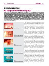
The <strong>dental</strong> implants are characterized for every individual <strong>to</strong>oth. The process<br />
flow <strong>of</strong> <strong>the</strong> implantation and manufacturing <strong>of</strong> <strong>the</strong> implants consist <strong>of</strong> handmade<br />
modeling as presented in <strong>the</strong> Fig. 1. The implants are sandblasted using Aluminum<br />
oxide pearl powder by a range <strong>of</strong> pressure between 1.5 and 3 bar.<br />
a<br />
b<br />
Fig.1. The preparation <strong>of</strong> two <strong>dental</strong> implants by hand (a) Wax model (b) Sintered<br />
milled Zircon dioxide implants.<br />
The measurement and evaluation <strong>of</strong> <strong>the</strong> <strong>dental</strong> <strong>samples</strong> require precise and<br />
accurate metrology techniques. At <strong>the</strong> time being biomedical metrology is a very<br />
important <strong>to</strong>ol for solving various problems in especially in <strong>the</strong> case <strong>of</strong> high<br />
flexibility and high accuracy are demanded [3].<br />
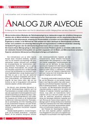
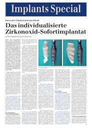
In this study, <strong>the</strong> target is <strong>to</strong> achieve <strong>the</strong> best application method <strong>to</strong> be used in<br />
clinical praxis by following <strong>the</strong> patients conditions (Fig.2)<br />
a<br />
b<br />
Fig. 2. (a) X-ray <strong>of</strong> <strong>the</strong> partial mouth <strong>of</strong> a patient before <strong>the</strong> implant treatments. (b)<br />
X-ray <strong>of</strong> <strong>the</strong> partial mouth <strong>of</strong> a patient at 1-year follow up control after <strong>the</strong> implant<br />
treatments [4].
2. QUANTITATIVE CHARACTERISATION OF NON-TECHNICAL<br />
STRUCTURES FOR MEDICAL IMPLANTS<br />
Clinical results achieved from successful placement <strong>of</strong> <strong>dental</strong> implants have a<br />
critical determinant called osseointegration. It is maintained by es<strong>the</strong>tic and<br />
functional stability <strong>of</strong> <strong>the</strong> implantation without any complications [4].<br />
The ana<strong>to</strong>mically specific macro retentions and <strong>micro</strong> retentions <strong>of</strong> <strong>the</strong> analogue<br />
<strong>to</strong>oth is <strong>the</strong> key <strong>to</strong> <strong>the</strong> successful osseointegration that must be individually<br />
measured, analysed and evaluated due <strong>to</strong> specific ana<strong>to</strong>mic properties. The reason<br />
<strong>of</strong> choosing <strong>the</strong> <strong>dental</strong> surfaces was <strong>to</strong> see <strong>the</strong> differences between <strong>the</strong> methods,<br />
which are tactile and optical. Hence <strong>the</strong> differences are significant particularly when<br />
such non-technical surfaces are being measured.<br />
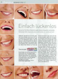
Stylus pr<strong>of</strong>ilometer and digital <strong>micro</strong>scope are commonly used instruments in <strong>the</strong><br />
field <strong>of</strong> precision metrology. The contact-stylus pr<strong>of</strong>ilometer while measuring a<br />
<strong>dental</strong> sample is represented in Fig. 3.<br />
Fig.3. Schematic diagram illustrating Form Talysurf Intra 50 pr<strong>of</strong>ilograph during<br />
<strong>the</strong> contact measurement <strong>of</strong> <strong>dental</strong> sample used in this study<br />
Talysurf Intra 50 pr<strong>of</strong>ilograph [5] with µltra s<strong>of</strong>tware (FTS Iµ) represented in <strong>the</strong><br />
Fig.3 according <strong>to</strong> <strong>the</strong> ISO 4287 and ISO 4288 [6, 7]. In <strong>the</strong> measurements <strong>of</strong> <strong>the</strong><br />
stylus pr<strong>of</strong>ilometer, 60 mm stylus arm length, 2 µm radius conisphere diamond<br />
stylus tip size and 1 mN force (speed=1 mm/s) were selected. Table 1 denotes <strong>the</strong><br />
specifications <strong>of</strong> <strong>the</strong> contact stylus pr<strong>of</strong>ilometer.<br />
Table 1. The specifications <strong>of</strong> contact stylus type pr<strong>of</strong>ilometer<br />
Measurement Method Spatial resolution Z Resolution Range Z<br />
Stylus Pr<strong>of</strong>ilometer (SP) 1-2 µm 3 -16 nm 0.2-1 mm<br />
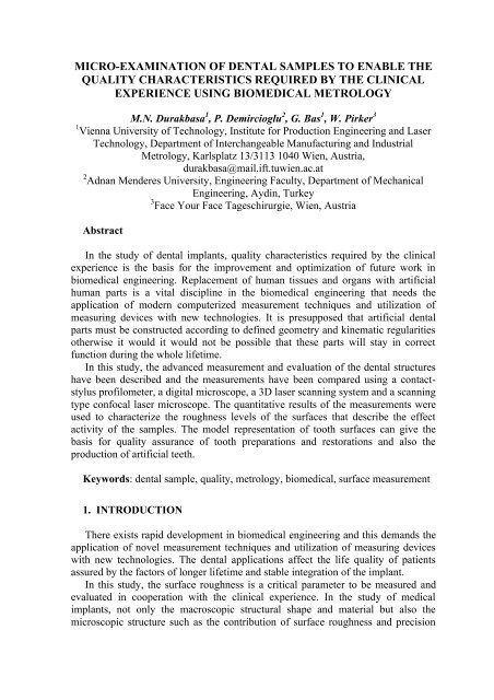
The digital <strong>micro</strong>scope (Keyence VHX-1000) and 3D laser scanning <strong>micro</strong>scope<br />
was used <strong>to</strong> observe, measure and record stabilized, fully-focused, uniformly<br />
illuminated image <strong>of</strong> <strong>dental</strong> <strong>samples</strong> (Fig.4) [8] . In this study, <strong>the</strong> technical<br />
structures are investigated using a digital <strong>micro</strong>scope (Keyence VHX-1000) with a<br />
high resolution CCD camera based system with a high intensity halogen lamp that<br />
integrates observation, recording, and measurement functions.
Fig.4. The original <strong>to</strong>oth sample surface <strong>to</strong>pography using a 3D laser scanning<br />
<strong>micro</strong>scope<br />
The Olympus LEXT4000, <strong>the</strong> scanning type confocal laser <strong>micro</strong>scope, is used<br />
<strong>to</strong> evaluate <strong>the</strong> surface <strong>to</strong>pography as presented in <strong>the</strong> Fig.5 captured using a<br />
collimated laser beam with 408 nm laser diode (LD) laser and white light emitting<br />
diode (LED) illumination [9]. The measured area is 0.933192 mm2 and <strong>the</strong> radius<br />
is 0.545mm.<br />
Fig.5. The surface <strong>to</strong>pography <strong>of</strong> <strong>the</strong> <strong>dental</strong> implant using a scanning type<br />
confocal laser <strong>micro</strong>scope (Scan-mode: XYZ-Feinscan, 1024x1024Pixel,<br />
1280x1280µm, Zoom:1x)
3. MEASUREMENT RESULTS<br />
In this research, <strong>the</strong> surface roughness measurements <strong>of</strong> three original teeth and<br />
four medical implants manufactured by different pressures were performed with a<br />
contact-stylus pr<strong>of</strong>ilometer. The outcome was compared with each o<strong>the</strong>r and it is<br />
found that <strong>the</strong> difference between <strong>the</strong>m is significant.<br />
Table 2. R a values belonging <strong>to</strong> three different teeth<br />
Roughness Measurement R a (µm) Mean Std.Dev.<br />
Original Tooth 1 6,1356 6,8910 7,8193 7,9923 8,0371 7,3751 0,8347<br />
Original Tooth 2 7,1169 7,6722 8,4380 7,5274 8,4416 7,8392 0,5849<br />
Original Tooth 3 7,0023 5,5159 6,5012 7,5831 6,1739 6,5553 0,7882<br />
Fig.6. The roughness values belonging <strong>to</strong> three different teeth taken from <strong>the</strong> stylus<br />
pr<strong>of</strong>ilometer in terms <strong>of</strong> <strong>the</strong> parameter R a .<br />
As seen in Fig. 6, <strong>the</strong> results for three different teeth characterize analogous<br />
surface <strong>to</strong>pography in terms <strong>of</strong> R a values. R a values are around 6,5 µm <strong>to</strong> 7,8 µm<br />
for <strong>the</strong> original teeth <strong>samples</strong>.<br />
Table 3. R a values belonging <strong>to</strong> four medical implants manufactured by different<br />
pressures<br />
Roughness Measurement R a (µm) Mean Std. Dev.<br />
Implant 1 6,1169 5,8697 5,8274 5,9571 6,1674 5,9877 0,1496<br />
Implant 2 9,4074 9,5551 9,8005 9,7558 10,2517 9,7541 0,3200<br />
Implant 3 13,2140 13,6919 12,6261 14,0001 14,0685 13,5201 0,6028<br />
Implant 4 20,5499 19,3169 19,6167 21,2405 20,9839 20,3416 0,8425
Fig.7. The roughness values belonging <strong>to</strong> four medical implants manufactured by<br />
different pressures taken from <strong>the</strong> stylus pr<strong>of</strong>ilometer in terms <strong>of</strong> <strong>the</strong> parameter <strong>the</strong><br />
parameter R a . (Implant1 : 1,5 bar, Implant2 : 2 bar, Implant3: 2,5 bar, Implant4 : 3<br />
bar)<br />
The R a values <strong>of</strong> <strong>the</strong> implant <strong>samples</strong> are measured <strong>to</strong> be in <strong>the</strong> range <strong>of</strong> 5,9 µm<br />
<strong>to</strong> 20,3 µm as presented in <strong>the</strong> Fig.7. The roughness values resulting different<br />
surface <strong>to</strong>pography are caused by different pressure values while manufacturing.<br />
4. CONCLUSION<br />
In this study, <strong>the</strong> measurement and evaluation <strong>of</strong> <strong>the</strong> non-technical <strong>dental</strong><br />
structures have been investigated and <strong>the</strong> measurements have been evaluated using<br />
a contact-stylus pr<strong>of</strong>ilometer, a digital <strong>micro</strong>scope, a 3D laser scanning system and<br />
a scanning type confocal laser <strong>micro</strong>scope. The quantitative results <strong>of</strong> <strong>the</strong><br />
measurements were used <strong>to</strong> characterize <strong>the</strong> roughness levels <strong>of</strong> <strong>the</strong> surfaces that<br />
describe <strong>the</strong> effect activity <strong>of</strong> <strong>the</strong> <strong>samples</strong>.<br />
The R a values are measured in <strong>the</strong> range <strong>of</strong> 6,5 µm <strong>to</strong> 7,8 µm for <strong>the</strong> original<br />
teeth <strong>samples</strong>. Whereas R a values <strong>of</strong> <strong>the</strong> implant <strong>samples</strong> are in <strong>the</strong> range <strong>of</strong> 5,9 µm<br />
<strong>to</strong> 20,3 µm. The roughness values resulting different surface <strong>to</strong>pography are caused<br />
by different pressure values while manufacturing. The best solution is sought by <strong>the</strong><br />
clinical experience <strong>to</strong> advance <strong>the</strong> health <strong>quality</strong> <strong>of</strong> <strong>the</strong> patients using this study in a<br />
long term project.
REFERENCES<br />
[1] EN ISO 9001: 2008: Quality management systems – Requirements. 2008.<br />
[2] EN ISO 13485: 2003 (AC:2009) Medical devices - Quality management<br />
systems - Requirements for regula<strong>to</strong>ry purposes.<br />
[3] DURAKBASA M. N.: Koordinatenmesstechnik für Biomedizinische<br />
Anwendungen; e&i, 117 (2000), 4; S. 269 – 272.<br />
[4]PIRKER, W., KOCHER, A.: Immediate, non-submerged, root-analogue<br />
zirconia implant in single <strong>to</strong>oth replacement.Int. J. Oral Maxill<strong>of</strong>acial<br />
Surgery 2008; Vol. 37, pp. 293-295.<br />
[5] FORM TALYSURF INTRA: Opera<strong>to</strong>r’s Handbook, http://taylor-hobson.com<br />
[6] EN ISO 4287:2009: Geometrical Product Specifications (GPS) - Surface<br />
texture: Pr<strong>of</strong>ile method - Terms, definitions and surface texture<br />
parameters (ISO 4287:1997 + Cor 1: 1998 + Cor 2: 2005 + Amd 1: 2009)<br />
(includes Corrigendum AC:2008 and Amendment A1:2009).<br />
[7] EN ISO 4288:1998-04: Geometrical Product Specifications (GPS) - Surface<br />
texture: Pr<strong>of</strong>ile method - Rules and procedures for <strong>the</strong> assessment <strong>of</strong><br />
surface texture<br />
[8] PRODUCT CATALOGUE, FEATURES, SPECIFICATIONS: 3D Laser<br />
Scanning Microscope, Digital Micrsocope VHX-1000,<br />
http://de.keyence.eu/products/<strong>micro</strong>scope/<strong>micro</strong>scope/<strong>micro</strong>scope.php<br />
[9] OLYMPUS LEXT OLS4000 – EIN NEUES INSTRUMENT FÜR DIE<br />
OPTISCHE MESSTECHNIK:<br />
http://www.netinform.net/GW/files/pdf/LEXT_OLS400.pdf