Dental-Microscopes - Kaps Optik GmbH
Dental-Microscopes - Kaps Optik GmbH
Dental-Microscopes - Kaps Optik GmbH

Create successful ePaper yourself
Turn your PDF publications into a flip-book with our unique Google optimized e-Paper software.
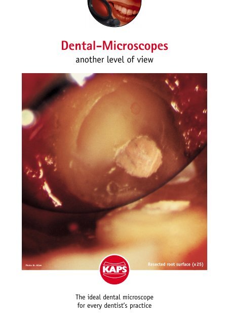
Photo: Dr. Allam<br />
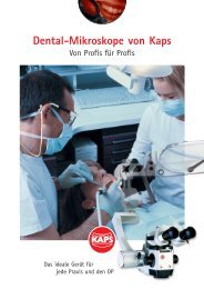
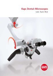
<strong>Dental</strong>-<strong>Microscopes</strong><br />
another level of view<br />
The ideal dental microscope<br />
for every dentist’s practice<br />
Resected root surface (x25)
Photos: Prof. Pecora<br />
<strong>Dental</strong> Microscope:<br />
The ideal start<br />
The more you use, the more you will want to use it<br />
The need to maintain patients dentition for a longer period of time may<br />
lead to the use of advanced procedures:<br />
the micro-endodontic retreatment, the conventional and surgical<br />
endodontic management.<br />
Training, practice, and technology have improved the quality of care<br />
provided to the population. Change your life and save your teeth, use<br />
dental microscopes ... but choose the best.<br />
View of a IRM retrofilling<br />
on a retro minor (x16)<br />
(Screen-Photo: Dr. Allam)<br />
Hex top of implant covered by soft tissue (16x)<br />
Fitting of crown of upper central incisor<br />
on tooth (26x)
The ML canal on a resected root of<br />
mandibular first molar (x 25)<br />
Retromirror view of retropreparation<br />
of MB and ML canal on a mandibular<br />
first molar (x 16)<br />
Isthmus between the two canals (x 25)<br />
Retromirror view of leakage on MB<br />
canal on a resected root of mandibular<br />
first molar (x 25)<br />
“Exact therapy requires<br />
exact vision.”<br />
Perfectly prepared canal with<br />
a ultrasonic tip (x25)<br />
Large magnification of a IRM<br />
retrofilling (x25)<br />
Photos: Dr. Allam
Layout: Grips Design, Rinker & Weber, Wetzlar<br />
Due to continual advances in technology, features and specifications are subject to change without notice.<br />
Leakage is shown on the resected root<br />
surface (x16)<br />
Photos: Dr. Allam<br />
Clinical view of an apicectomy on a<br />
mandibular first molar<br />
Microscope reduces distance:<br />
It provides the optimum magnification range for every application from x4 to x26 or more.<br />
“I can not practice without a<br />
microscope because it is a part<br />
of my visual sensory system.”<br />
Dr. Charbel ALLAM DCD, DESE<br />
Department of Endodontics<br />
School of <strong>Dental</strong> Medicine<br />
St. Joseph University<br />
Beirut–Lebanon<br />
View of the root end filling under the<br />
microscope (x25)<br />
Endodontic Surgery after root end filling<br />
“Light and magnification<br />
have given a better standard<br />
of quality in dentistry.”<br />
Karl <strong>Kaps</strong> <strong>GmbH</strong> & Co.KG P. O. Box 12 25 35608 Asslar/Wetzlar<br />
Fon (+49) 64 41 8 07 04-0 Fax (+49) 64 41 8 59 85<br />
www.kaps-optik.de e-mail: info@kaps-optik.de<br />
Prof. Gabriele PECORA<br />
Associate Professor, Dep. Endodontics,<br />
University of Pennsylvania<br />
Co-Director, Endodontic Microsurgery Training<br />
Center, Suny-University of Buffalo