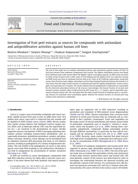
Investigation of fruit peel extracts as sources for ... - ThaiScience
Investigation of fruit peel extracts as sources for ... - ThaiScience
Investigation of fruit peel extracts as sources for ... - ThaiScience

Create successful ePaper yourself
Turn your PDF publications into a flip-book with our unique Google optimized e-Paper software.
<strong>Investigation</strong> <strong>of</strong> <strong>fruit</strong> <strong>peel</strong> <strong>extracts</strong> <strong>as</strong> <strong>sources</strong> <strong>for</strong> compounds with antioxidant<br />
and antiproliferative activities against human cell lines<br />
Ruttiros Khonkarn a , Siriporn Okonogi a, *, Chadarat Amp<strong>as</strong>avate a , Songyot Anuchapreeda b<br />
a Department <strong>of</strong> Pharmaceutical Sciences, Faculty <strong>of</strong> Pharmacy, Chiang Mai University, Chiang Mai 50200, Thailand<br />
b Faculty <strong>of</strong> Associated Medical Sciences, Chiang Mai University, Chiang Mai 50200, Thailand<br />
article info<br />
Article history:<br />
Received 6 February 2010<br />
Accepted 7 May 2010<br />
Keywords:<br />
Cytotoxicity<br />
Human cell lines, antioxidant, rambutan<br />
Coconut<br />
Mangosteen<br />
1. Introduction<br />
abstract<br />
Cancer is a major cause <strong>of</strong> mortality worldwide and cancer incidents<br />
rapidly incre<strong>as</strong>e from year to year. In 2000, there were 10.4<br />
million new cancer c<strong>as</strong>es and it is expected that this number will<br />
be doubled in 2030 (Global cancer control, 2008). Recent studies<br />
have shown strong evidence that biological reactive oxygen species<br />
(ROS) such <strong>as</strong> hydroxyl radicals (OH ) and the superoxide anion<br />
(O2 ) are involved in the development <strong>of</strong> cancer. Aerobic<br />
organisms possess mechanisms <strong>of</strong> ROS scavenging exploiting antioxidant<br />
systems including enzyme-b<strong>as</strong>ed antioxidants such <strong>as</strong><br />
superoxide dismut<strong>as</strong>e, glutathione peroxid<strong>as</strong>e, catal<strong>as</strong>e, and glutathione<br />
reduct<strong>as</strong>e. However, tissue damage can be developed from<br />
an imbalance between ROS-generating and these in vivo defense<br />
systems, resulting in pathogenesis <strong>of</strong> a variety <strong>of</strong> degenerative disorders.<br />
The role <strong>of</strong> ROS in the pathogenesis <strong>of</strong> cancer is that these<br />
species react with DNA resulting in cell malfunction. Ultimately it<br />
can lead to malignancies by accumulation <strong>of</strong> somatic mutations<br />
(Hursting et al., 1999).<br />
As ROS are involved in cancer development, compounds with<br />
high ROS reduction activity are likely able to prevent cancer incidence.<br />
It h<strong>as</strong> been shown that both synthetic and natural antioxi-<br />
* Corresponding author. Tel.: +66 53 944311; fax: +66 53 278708.<br />
E-mail address: sirioko@chiangmai.ac.th (S. Okonogi).<br />
0278-6915/$ - see front matter Ó 2010 Elsevier Ltd. All rights reserved.<br />
doi:10.1016/j.fct.2010.05.014<br />
Food and Chemical Toxicology 48 (2010) 2122–2129<br />
Contents lists available at ScienceDirect<br />
Food and Chemical Toxicology<br />
journal homepage: www.elsevier.com/locate/foodchemtox<br />
The aim <strong>of</strong> this study w<strong>as</strong> to evaluate antioxidant activity and cytotoxicity against human cell lines <strong>of</strong><br />
<strong>fruit</strong> <strong>peel</strong> <strong>extracts</strong> from rambutan, mangosteen and coconut. The highest antioxidant activity w<strong>as</strong> found<br />
from rambutan <strong>peel</strong> crude extract where the highest radical scavenging capacity via ABTS <strong>as</strong>say w<strong>as</strong> from<br />
its ethyl acetate fraction with a TEAC value <strong>of</strong> 23.0 mM/mg and the highest ferric ion reduction activity<br />
via FRAP <strong>as</strong>say w<strong>as</strong> from its methanol fraction with an EC value <strong>of</strong> 20.2 mM/mg. Importantly, using both<br />
<strong>as</strong>says, these fractions had a higher antioxidant activity than butylated hydroxyl toluene and vitamin E. It<br />
w<strong>as</strong> shown that the ethyl acetate fraction <strong>of</strong> rambutan <strong>peel</strong> had the highest polyphenolic content with a<br />
gallic acid equivalent <strong>of</strong> 2.3 mg/mL. The results indicate that the polyphenolic compounds are responsible<br />
<strong>for</strong> the observed antioxidant activity <strong>of</strong> the <strong>extracts</strong>. Interestingly, the hexane fraction <strong>of</strong> coconut <strong>peel</strong><br />
showed a potent cytotoxic effect on KB cell line by MTT <strong>as</strong>say (IC50 = 7.7 lg/mL), and no detectable cytotoxicity<br />
toward normal cells. We concluded that the ethyl acetate fraction <strong>of</strong> rambutan <strong>peel</strong> is a promising<br />
resource <strong>for</strong> potential novel antioxidant agents where<strong>as</strong> the hexane fraction <strong>of</strong> coconut <strong>peel</strong> may<br />
contain novel anticancer compounds.<br />
Ó 2010 Elsevier Ltd. All rights reserved.<br />
dants play an important role in ROS reduction, resulting in<br />
prevention and treatment <strong>of</strong> ROS-induced tissue degenerations<br />
(Shukla and Pal, 2004). Natural antioxidants have received much<br />
attention in recent years because they are relatively safe <strong>as</strong> compared<br />
to their synthetic counterparts. Fruits and vegetables are<br />
the major source <strong>of</strong> natural antioxidants and contain various kinds<br />
<strong>of</strong> antioxidant compounds such <strong>as</strong> vitamin C, vitamin E, carotenoids,<br />
lutein and lycopene (Cai et al., 2004). Among these compounds,<br />
polyphenolic compounds display antioxidant activity<br />
through different mechanisms, in particular by free radical scavenging<br />
and by chelating <strong>of</strong> metal ions (Du et al., 2009; Jacob<br />
et al., 2008). Recently, many studies reported that plant <strong>extracts</strong><br />
with high levels <strong>of</strong> polyphenolic compounds have anticancer activity<br />
due to neutralization <strong>of</strong> ROS (Moongkarndi et al., 2004; Russo<br />
et al., 2005).<br />
The <strong>extracts</strong> <strong>of</strong> rambutan, mangosteen, and coconut <strong>peel</strong>s were<br />
reported to possess high antioxidant potential (Esquenazi et al.,<br />
2002; Palanisamy et al., 2008; Yu et al., 2007). The aim <strong>of</strong> the present<br />
study w<strong>as</strong> to evaluate the antioxidant activity and cytotoxicity<br />
<strong>of</strong> these plant <strong>extracts</strong> toward both cancer and normal cells. The<br />
total phenolic content <strong>of</strong> the <strong>extracts</strong> w<strong>as</strong> determined in order to<br />
investigate the relationship between the observed activities and<br />
the phenolic content <strong>of</strong> the <strong>extracts</strong>. For cytotoxicity tests, KB (human<br />
oral squamous carcinoma cell), and Caco-2 (human colon adenocarcinoma<br />
cell) cancer cell lines were used <strong>as</strong> cancer cell models.
Moreover, peripheral blood mononuclear cells (PBMC) were also<br />
used <strong>as</strong> human normal cells <strong>for</strong> cytotoxic evaluation <strong>of</strong> the plant<br />
<strong>extracts</strong>.<br />
2. Materials and methods<br />
2.1. Chemicals<br />
Ethanol, hexane, ethyl acetate, acetone, methanol, and hydrochloric acid were<br />
purch<strong>as</strong>ed from Merck (Darmstadt, Germany). Folin–Ciocalteu reagent, 2,2 0 -azinobis-(3-ethylbenzothiazoline-6-sulfonic<br />
acid) diammonium salt (ABTS), 2,4,6tri(2-pyridyl)-S-triazine<br />
(TPTZ), sodium carbonate, pot<strong>as</strong>sium persulfate, ferrous<br />
sulfate (FeSO4 7H2O), ferric chloride (FeCl3 6H2O), gallic acid, butylated hydroxytoluene<br />
(BHT), vitamin E, dimethyl sulfoxide (DMSO), Histopaque Ò -1077, and 3-(4,5dimethylthiazolyl-2)-2,5-diphenyl<br />
tetrazolium bromide (MTT) were purch<strong>as</strong>ed<br />
from Sigma–Aldrich (St. Louis, MO, USA). Trolox w<strong>as</strong> obtained from Aldrich Chemical<br />
Company (Steinheim, Germany). Fetal bovine serum (FBS) w<strong>as</strong> obtained from<br />
Biochrom AG (Berlin, Germany). Eagle’s minimal essential medium (EMEM), RPMI<br />
1640, trypsin–EDTA, and penicillin–streptomycin were purch<strong>as</strong>ed from GIBCO<br />
Invitrogen (Grand Island, NY, USA). All other chemicals were <strong>of</strong> the highest purity<br />
available.<br />
2.2. Plant materials<br />
Ripe <strong>fruit</strong>s <strong>of</strong> rambutan (Nephelium lappaceum), mangosteen (Garcinia mangostana),<br />
and coconut (Cocos nucifera) were purch<strong>as</strong>ed fresh from markets in Chiang<br />
Mai, Thailand.<br />
2.3. Preparation <strong>of</strong> plant <strong>extracts</strong><br />
Fruit <strong>peel</strong>s <strong>of</strong> rambutan, mangosteen, and coconut were w<strong>as</strong>hed with clean<br />
water and subsequently cut into small pieces and placed in a circulating oven at<br />
45 °C until complete dryness. The stalk <strong>of</strong> mangosteen <strong>peel</strong>s w<strong>as</strong> removed and only<br />
the mesocarp part <strong>of</strong> the coconut <strong>peel</strong> w<strong>as</strong> used in the study. The dried plant materials<br />
were ground into fine powders.<br />
2.3.1. Crude <strong>extracts</strong><br />
The dried powders <strong>of</strong> each plant material ( 100 g) were macerated with 95%<br />
ethanol ( 500 mL, 48 h 3) at room temperature. The <strong>extracts</strong> were combined, filtered<br />
and the solvent w<strong>as</strong> removed under vacuum using a rotary evaporator at<br />
45 °C. The obtained crude <strong>extracts</strong> were kept in a vacuum desiccator at 4 °C until<br />
further use.<br />
2.3.2. Fractionated <strong>extracts</strong><br />
Four solvents <strong>of</strong> different polarity (hexane, ethyl acetate, butanol, and methanol)<br />
were used to consecutively extract the powders <strong>of</strong> the selected plants. Firstly,<br />
the dried powder ( 100 g) w<strong>as</strong> extracted with hexane through maceration<br />
( 500 mL, 48 h 3) at room temperature. The residue after the third extraction<br />
w<strong>as</strong> dried <strong>for</strong> 24 h at room temperature in order to remove hexane. Next, the dried<br />
residue w<strong>as</strong> further extracted by maceration in ethyl acetate, butanol, and methanol,<br />
respectively, in the same way <strong>as</strong> described <strong>for</strong> hexane. Extracts <strong>of</strong> the same solvent<br />
were pooled and filtered. The solvent w<strong>as</strong> removed under reduced pressure<br />
using a rotary evaporator at 45 °C. The <strong>extracts</strong> obtained were stored in a desiccator<br />
at 4 °C <strong>for</strong> further studies.<br />
2.4. Determination <strong>of</strong> total phenolic content<br />
The total content <strong>of</strong> phenolic compounds <strong>of</strong> the different <strong>extracts</strong> w<strong>as</strong> determined<br />
by the colorimetric Folin–Ciocalteu method with slight adaptation <strong>as</strong> described<br />
by Sato et al. (1996). Briefly, samples <strong>of</strong> the obtained <strong>extracts</strong> were<br />
diluted in ethanol and pipetted into 96-well plates. Next, Folin–Ciocalteu reagent<br />
and sodium carbonate were added to each well. The mixture w<strong>as</strong> incubated <strong>for</strong><br />
2 h at room temperature in the dark. The blue color that developed w<strong>as</strong> me<strong>as</strong>ured<br />
using a microtitre plate reader. Gallic acid w<strong>as</strong> used <strong>for</strong> calibration. The phenolic<br />
content <strong>of</strong> the <strong>extracts</strong> is expressed in gallic acid equivalent (GAE) concentration<br />
(mg/mL).<br />
2.5. Antioxidant activity <strong>as</strong>says<br />
2.5.1. ABTS <strong>as</strong>say<br />
This <strong>as</strong>say me<strong>as</strong>ures the free radical scavenging activity <strong>of</strong> agents b<strong>as</strong>ed on the<br />
decolorization <strong>of</strong> ABTS free radicals. The <strong>as</strong>say w<strong>as</strong> done in the same manner <strong>as</strong> described<br />
previously (Tachakittirungrod et al., 2007). Briefly, ABTS radical cations<br />
were generated by reaction <strong>of</strong> ABTS and pot<strong>as</strong>sium persulfate (K 2S 2O 8) in water.<br />
The ABTS free radical solution w<strong>as</strong> diluted to an absorbance <strong>of</strong> 0.7 ± 0.2 at<br />
750 nm and stored in the dark <strong>for</strong> 12 h. Next, <strong>extracts</strong> in ethanol were added to<br />
the ABTS free radical solution. The absorbance <strong>of</strong> the mixture w<strong>as</strong> me<strong>as</strong>ured <strong>for</strong><br />
R. Khonkarn et al. / Food and Chemical Toxicology 48 (2010) 2122–2129 2123<br />
5 min using a microtitre plate reader. Vitamin E and BHT were used <strong>as</strong> positive controls<br />
and trolox w<strong>as</strong> used <strong>for</strong> calibration. The antioxidant potential is expressed <strong>as</strong><br />
trolox equivalent antioxidant capacity (TEAC) value.<br />
2.5.2. FRAP <strong>as</strong>say<br />
This <strong>as</strong>say me<strong>as</strong>ures the reducing properties <strong>of</strong> antioxidants b<strong>as</strong>ed on the<br />
reduction <strong>of</strong> the ferric ion. The me<strong>as</strong>urements were done in the same manner <strong>as</strong> described<br />
previously (Okonogi et al., 2007). In short, FRAP reagent w<strong>as</strong> prepared by<br />
mixing TPTZ and FeCl3 solution in acetate buffer. The <strong>extracts</strong> dissolved in ethanol<br />
were mixed with the FRAP reagent in 96-well plates and allowed to react <strong>for</strong> 5 min<br />
at room temperature. Vitamin E and BHT were used <strong>as</strong> positive controls and ferrous<br />
sulfate w<strong>as</strong> used <strong>for</strong> calibration. A microtitre plate reader w<strong>as</strong> used to me<strong>as</strong>ure the<br />
absorbance <strong>of</strong> the resulting mixture. The antioxidant activity <strong>of</strong> the <strong>extracts</strong> is expressed<br />
<strong>as</strong> equivalent capacity (EC) using ferrous sulfate <strong>for</strong> calibration. A higher value<br />
<strong>of</strong> EC implies a higher reducing activity <strong>of</strong> the <strong>extracts</strong>.<br />
2.6. Cytotoxicity <strong>as</strong>says<br />
KB (human epidermal carcinoma <strong>of</strong> the mouth with HeLa contaminant) and<br />
Caco-2 (human colorectal adenocarcinoma) cells were purch<strong>as</strong>ed from the American<br />
Type Culture Collection (ATCC) and cultured in Eagle’s minimal essential medium<br />
(EMEM) supplemented with 10% fetal bovine serum (FBS), 100 U/mL <strong>of</strong><br />
penicillin and 0.1 mg/mL <strong>of</strong> streptomycin. Cells were cultured in a humidified<br />
atmosphere at 37 °Cin5%CO 2. The culture medium w<strong>as</strong> renewed on alternate days.<br />
When the cells had reached confluency, they were subcultured at a split ratio <strong>of</strong> one<br />
to four by trypsinizing with 0.25% trypsin–EDTA in phosphate-buffered saline (PBS).<br />
The cytotoxicity study w<strong>as</strong> per<strong>for</strong>med when the cells reached 80–90% confluence.<br />
Caco-2 and KB cells were seeded into the 96-well plates at a density <strong>of</strong><br />
8 10 3 cells/well and 3 10 3 cells/well, respectively.<br />
Blood samples from healthy volunteers were collected by venipuncture and<br />
transferred into heparin-coated, 15 mL test tubes. Blood w<strong>as</strong> diluted at 1:1 ratio<br />
with PBS, layered onto Histopaque Ò -1077 at a volume ratio <strong>of</strong> 3:1 and centrifuged<br />
at 1000g <strong>for</strong> 30 min. With this procedure, the PBMCs were separated from the<br />
erythrocytes, granulocytes, and platelets. The PBMC layer w<strong>as</strong> removed and then<br />
w<strong>as</strong>hed twice with PBS. Subsequently, the cells were resuspended in RPMI 1640<br />
culture medium supplemented with 10% FBS, 100 U/mL <strong>of</strong> penicillin, 0.1 mg/mL<br />
<strong>of</strong> streptomycin. Cell viability w<strong>as</strong> determined by using the trypan-blue dye exclusion<br />
method (Tennant, 1964). The cell density <strong>of</strong> PBMCs used in the cytotoxicity<br />
study w<strong>as</strong> 1 10 5 cells/well <strong>of</strong> the 96-well tissue culture plates.<br />
The cytotoxicity <strong>of</strong> the different <strong>extracts</strong> w<strong>as</strong> tested by using a standard MTT <strong>as</strong>say<br />
described by Alley et al. (1988) with a minor modification. Briefly, KB cells suspended<br />
in 100 lL <strong>of</strong> medium were plated into 96-well tissue culture plates. After<br />
24 h incubation, 100 lL fresh medium containing various concentrations <strong>of</strong> plant<br />
<strong>extracts</strong> (5–100 lg/mL) were added to each well, and incubated <strong>for</strong> another 48 h.<br />
Next, 100 lL <strong>of</strong> the medium w<strong>as</strong> removed and 15 lL <strong>of</strong> 5 mg/mL MTT dye in PBS<br />
w<strong>as</strong> added to the wells. Plates were incubated at 37 °C <strong>for</strong> 4 h in humidified 5%<br />
CO 2 atmosphere. Subsequently, the MTT solution w<strong>as</strong> removed, 200 lL <strong>of</strong> DMSO<br />
w<strong>as</strong> added and mixed thoroughly to dissolve the dye crystals. The absorbance at<br />
570 nm w<strong>as</strong> me<strong>as</strong>ured using a microtitre plate reader at 570 nm with a reference<br />
wavelength <strong>of</strong> 630 nm. The cell viability w<strong>as</strong> determined by the following <strong>for</strong>mula:<br />
Mean absorbance in test wells<br />
%Cell viability ¼<br />
Mean absorbance in control wells<br />
Dose–response curves <strong>of</strong> percentages <strong>of</strong> cell viability plotted against concentrations<br />
<strong>of</strong> the <strong>extracts</strong> were constructed. The 50% inhibitory concentration (IC 50) in<br />
c<strong>as</strong>e <strong>of</strong> cell inhibition, or effective dose (ED 50) in c<strong>as</strong>e <strong>of</strong> cell stimulation w<strong>as</strong> determined<br />
from the obtained plots.<br />
2.7. Statistical analysis<br />
The experiments were done in the triplicate and the results are expressed <strong>as</strong><br />
mean ± SD. Statistical analysis w<strong>as</strong> done using one-way ANOVA and P value at a level<br />
<strong>of</strong> 95% confidence limit.<br />
Table 1<br />
Yields <strong>of</strong> the ethanolic <strong>extracts</strong> (%, w/w) and total phenolic content from the <strong>fruit</strong><br />
<strong>peel</strong>s <strong>of</strong> three plants.<br />
Plant names Sample code Yield (%, w/w) GAE (mg/mL)<br />
Common<br />
name<br />
Scientific<br />
name<br />
Coconut Cocos<br />
nucifera<br />
Rambutan Nephelium<br />
lappaceum<br />
Mangosteen Garcinia<br />
mangostana<br />
100<br />
CP 5.8 0.63 ± 0.03<br />
RP 10.7 0.72 ± 0.05<br />
MP 7.2 0.68 ± 0.06
2124 R. Khonkarn et al. / Food and Chemical Toxicology 48 (2010) 2122–2129<br />
3. Results and discussion<br />
3.1. Extraction yields and total phenolic contents<br />
Table 1 shows the results <strong>of</strong> the extraction <strong>of</strong> the three plants<br />
(Cocos nucifera, G. mangostana, and N. lappaceum) with ethanol.<br />
The highest yield (10.7%) w<strong>as</strong> obtained from rambutan <strong>fruit</strong> <strong>peel</strong>s<br />
while the lowest yield (5.8%) w<strong>as</strong> from coconut <strong>fruit</strong> <strong>peel</strong>s. The total<br />
phenolic contents <strong>as</strong> me<strong>as</strong>ured using Folin–Ciocalteu <strong>as</strong>say ranged<br />
from 0.63 to 0.72 mg/mL. It is noted that the <strong>fruit</strong> <strong>peel</strong>s <strong>of</strong><br />
Table 2<br />
Yields and total phenolic content <strong>of</strong> fractionated plant <strong>peel</strong> <strong>extracts</strong>.<br />
Plant Solvent Sample code Yield (%, w/w) GAE (mg/mL)<br />
Coconut Hexane HCP 0.6 0.30 ± 0.01<br />
Ethyl acetate ECP 3.7 1.31 ± 0.01<br />
Butanol BCP 4.1 1.06 ± 0.04<br />
Methanol MCP 15.9 0.67 ± 0.07<br />
Rambutan Hexane HRP 0.6 0.29 ± 0.01<br />
Ethyl acetate ERP 3.0 2.28 ± 0.02<br />
Butanol BRP 7.4 1.73 ± 0.03<br />
Methanol MRP 25.5 2.05 ± 0.11<br />
Mangosteen Hexane HMP 0.9 0.09 ± 0.01<br />
Ethyl acetate EMP 5.5 1.16 ± 0.17<br />
Butanol BMP 11.6 0.49 ± 0.05<br />
Methanol MMP 18.8 1.64 ± 0.06<br />
rambutan and mangosteen demonstrated significantly higher total<br />
phenolic content than that <strong>of</strong> coconut <strong>peel</strong>s.<br />
The plant powders were consecutively fractionated with organic<br />
solvents with incre<strong>as</strong>ing polarity. The yields <strong>of</strong> these extractions<br />
and the total phenolic contents are reported in Table 2. It w<strong>as</strong><br />
found that extraction <strong>of</strong> the different plants with methanol gave<br />
the highest yields (16–26%), where<strong>as</strong> the yields obtained after<br />
extraction with hexane were the lowest (0.6–0.9%). This is in line<br />
with the findings <strong>of</strong> Singh et al. (2002) who reported that extraction<br />
<strong>of</strong> pomegranates with methanol gave the highest yield. Extraction<br />
<strong>of</strong> the different plants with ethyl acetate and butanol gave<br />
intermediate yields (ranging from 3% to 12%). The hexane <strong>extracts</strong><br />
not only gave low yields, but also contained the lowest levels <strong>of</strong><br />
phenolic compounds (0.1–0.3 mg/mL). The <strong>extracts</strong> <strong>of</strong> the more<br />
polar solvents (ethyl acetate, butanol, and methanol) contained<br />
substantially higher levels <strong>of</strong> phenolic compounds (ranging from<br />
0.7 to 2.3 mg/mL) and comparable with those found in the<br />
ethanolic <strong>extracts</strong> (Table 1). Also in other studies, <strong>extracts</strong> obtained<br />
with relatively polar solvents (methanol or aqueous methanol mixtures)<br />
had the highest content <strong>of</strong> polyphenolic compounds (Dufour<br />
et al., 2007).<br />
3.2. Antioxidant activities <strong>of</strong> the different plant <strong>extracts</strong><br />
Many methods have been used to determine the antioxidant<br />
activity <strong>of</strong> natural products (Antolovich et al., 2002). No single <strong>as</strong>-<br />
Fig. 1. Antioxidant activity <strong>of</strong> the ethanolic <strong>extracts</strong> (Table 1) from the <strong>fruit</strong> <strong>peel</strong>s <strong>of</strong> three plants using ABTS (A) and FRAP (B) <strong>as</strong>says. Vitamin E and BHT were used <strong>as</strong><br />
standards. Calibration w<strong>as</strong> done with trolox (ABTS <strong>as</strong>say) and ferrous sulfate (FRAP <strong>as</strong>say).<br />
Fig. 2. Antioxidant activity <strong>of</strong> fractionated <strong>extracts</strong> (Table 2) using ABTS <strong>as</strong>say. Vitamin E and BHT were used <strong>as</strong> standards.
Fig. 3. Antioxidant activity <strong>of</strong> extract fractions (Table 2) using FRAP <strong>as</strong>say. Vitamin E and BHT were used <strong>as</strong> standards.<br />
say however can establish the full antioxidant potential <strong>of</strong> compounds,<br />
because multiple reactions and mechanisms are involved<br />
in antioxidative processes (Chu et al., 2000). There<strong>for</strong>e, at le<strong>as</strong>t<br />
two different methods should be applied in order to evaluate the<br />
antioxidant capacity <strong>of</strong> <strong>extracts</strong> <strong>of</strong> <strong>fruit</strong>s, vegetables, and other<br />
plant products (Du et al., 2009). In the present study, the antioxidant<br />
potency <strong>of</strong> our <strong>extracts</strong> w<strong>as</strong> evaluated using the ABTS <strong>as</strong>say,<br />
which determines the free radical scavenging activity, and the<br />
FRAP <strong>as</strong>say, which me<strong>as</strong>ures the ferric ion reducing ability. Fig. 1<br />
shows the results obtained with the ethanolic <strong>extracts</strong>.<br />
Fig. 1A shows that the TEAC values <strong>of</strong> the ethanolic <strong>extracts</strong> <strong>of</strong><br />
the different plants were between 13.8 and 15.6 mM/mg. The<br />
antioxidant activity <strong>of</strong> the <strong>extracts</strong> w<strong>as</strong> around the same <strong>as</strong> that<br />
<strong>of</strong> BHT but significantly higher than that <strong>of</strong> vitamin E. The crude<br />
coconut <strong>peel</strong> extract had higher TEAC activity than vitamin E but<br />
lower than that <strong>of</strong> BHT. Fig. 1B demonstrates that the antioxidant<br />
activities <strong>of</strong> the ethanolic <strong>extracts</strong> were about the same <strong>as</strong> that <strong>of</strong><br />
vitamin E but around three-fold higher than that <strong>of</strong> BHT. It is<br />
important to mention that the <strong>extracts</strong> <strong>of</strong> the plant <strong>peel</strong>s have<br />
both excellent radical scavenging (Fig. 1A) and ferric ion reducing<br />
capacities (Fig. 1B). Because <strong>of</strong> the high antioxidant activity and<br />
polyphenolic content <strong>of</strong> the crude ethanolic <strong>extracts</strong>, we fractionated<br />
the plants using solvents <strong>of</strong> incre<strong>as</strong>ing polarity (hexane,<br />
ethyl acetate, butanol, and methanol) in order to investigate<br />
whether fraction(s) with higher antioxidant levels could be obtained.<br />
The results are shown in Fig. 2 (ABTS <strong>as</strong>say) and Fig. 3<br />
(FRAP <strong>as</strong>say). Fig. 2 shows that by far the lowest antioxidant<br />
activity, <strong>as</strong> determined by the ABTS <strong>as</strong>say, w<strong>as</strong> found in the hexane<br />
fractions (TEAC values ranging from 0.6 to 2.1 mM/mg) which<br />
EC values (mM/mg)<br />
25<br />
20<br />
15<br />
10<br />
5<br />
0<br />
R 2 = 0.979<br />
0 5 10 15 20 25<br />
TEAC values (mM/mg)<br />
Fig. 4. Correlation between TEAC and EC values <strong>of</strong> the different fractionated<br />
<strong>extracts</strong> <strong>of</strong> three <strong>fruit</strong> <strong>peel</strong>s.<br />
R. Khonkarn et al. / Food and Chemical Toxicology 48 (2010) 2122–2129 2125<br />
can be <strong>as</strong>cribed to their very low polyphenolic contents (Table 2).<br />
The <strong>extracts</strong> <strong>of</strong> the more polar solvents had substantially higher<br />
activity (ranging from 6 to 23 mM/mg) which is corresponding<br />
to their high polyphenolic levels (Table 2). The ERP fraction even<br />
had 1.5 and 2.5 higher activity than that <strong>of</strong> BHT and vitamin E,<br />
respectively.<br />
Fig. 3 indicates in line with the data <strong>of</strong> Fig. 2 that the hexane<br />
fractions had low antioxidant activity <strong>as</strong> determined by the FRAP<br />
<strong>as</strong>say (EC values ranging from 0.6 to 1.0 mM/mg). The <strong>extracts</strong> obtained<br />
with the more polar solvents had higher activities (EC val-<br />
GAE values (mg/mL)<br />
GAE values (mg/mL)<br />
2.5<br />
2.0<br />
1.5<br />
1.0<br />
0.5<br />
2.5<br />
2.0<br />
1.5<br />
1.0<br />
0.5<br />
0.0<br />
R 2 = 0.897<br />
0.0<br />
0 5 10 15 20 25<br />
TEAC values (mM/mg)<br />
Fig. 5. Correlation between TEAC values and total phenolic contents <strong>of</strong> the different<br />
<strong>extracts</strong>.<br />
R 2 = 0.886<br />
0 5 10 15 20 25<br />
EC values (mM/mg)<br />
Fig. 6. Correlation <strong>of</strong> EC values and total phenolic contents <strong>of</strong> the different <strong>extracts</strong>.
2126 R. Khonkarn et al. / Food and Chemical Toxicology 48 (2010) 2122–2129<br />
ues from 2.1 to 20.2 mM/mg). The highest activity found in the<br />
MRP fraction exceeded the activity <strong>of</strong> vitamin E and BHT by a factor<br />
<strong>of</strong> 2 and 7, respectively. These results show that by using solvents<br />
with different polarity, fractions were obtained with higher antioxidant<br />
activity than that <strong>of</strong> the crude ethanolic <strong>extracts</strong>. Taken together,<br />
it should be emph<strong>as</strong>ized that the ERP and MRP fractions<br />
had significantly better antioxidant activity in both <strong>as</strong>says than<br />
the controls, BHT and vitamin E (p < 0.05).<br />
The correlation <strong>of</strong> TEAC and EC values is reported in Fig. 4<br />
which shows that there is a linear relationship (R 2 = 0.979)<br />
between the values obtained with both <strong>as</strong>says. It cannot be concluded,<br />
yet, whether one or more compounds in the same <strong>extracts</strong><br />
h<strong>as</strong> both radical scavenging activity and ferric ion<br />
A<br />
% Cell viability<br />
B<br />
% Cell viability<br />
C<br />
% Cell viability<br />
250<br />
200<br />
150<br />
100<br />
50<br />
0<br />
200<br />
150<br />
100<br />
50<br />
0<br />
reduction capacity, or that different compounds are responsible<br />
<strong>for</strong> the different activities. There<strong>for</strong>e, further fractionation <strong>of</strong> the<br />
<strong>extracts</strong> is necessary.<br />
3.3. The relationship between the total phenol contents and<br />
antioxidant activities<br />
Figs. 5 and 6 show the relationship between the total phenolic<br />
contents and the TEAC (R 2 = 0.897), and the EC (R 2 = 0.886) values,<br />
respectively. This good linear relationship indicates that the<br />
antioxidant activity in the different fractionated <strong>extracts</strong> is, <strong>as</strong><br />
anticipated, due to polyphenolic compounds.<br />
0 10 20 30 40 50 60 70 80 90 100<br />
Concentration (µg/mL)<br />
250<br />
250<br />
200<br />
150<br />
100<br />
50<br />
0<br />
0 10 20 30 40 50 60 70 80 90 100<br />
Concentration (µg/mL)<br />
0 10 20 30 40 50 60 70 80 90 100<br />
Concentration (µg/mL)<br />
Fig. 7. Dose–response curves <strong>of</strong> viability <strong>of</strong> KB cell line (A), Caco-2 cell line (B), and PBMC cells (C) and concentration <strong>of</strong> fractionated <strong>peel</strong> <strong>extracts</strong> <strong>of</strong> coconut.
3.4. Cytotoxicity test<br />
Figs. 7–9 show the dose–response curve <strong>of</strong> the tested fractions<br />
against KB, Caco-2, and PBMCs, respectively. Table 3 summarizes<br />
the IC50 and EC50 values <strong>as</strong> derived from these figures. It w<strong>as</strong><br />
found that the ECP and BCP <strong>extracts</strong> stimulate the proliferation<br />
<strong>of</strong> these cells (ED50 values ranging from 13 to 82 lg/mL). This<br />
suggests that the application <strong>of</strong> this extract, <strong>as</strong> a natural antioxidant<br />
in food or drug products <strong>for</strong> humans, should be done with<br />
caution. Interestingly, the HCP extract shows a potent antiproliferative<br />
effect on the KB tumor cell line (IC50 = 7.7 lg/mL), but<br />
A<br />
% Cell viability<br />
B<br />
% Cell viability<br />
C<br />
% Cell viability<br />
250<br />
200<br />
150<br />
100<br />
50<br />
0<br />
250<br />
200<br />
150<br />
100<br />
50<br />
0<br />
250<br />
200<br />
150<br />
100<br />
50<br />
0<br />
R. Khonkarn et al. / Food and Chemical Toxicology 48 (2010) 2122–2129 2127<br />
h<strong>as</strong> no antiproliferative effect on normal cells (PBMCs). There<strong>for</strong>e<br />
this fraction is an interesting natural source <strong>for</strong> a potential novel<br />
antiproliferative agent inhibitory to cancer cell growth, but that is<br />
nontoxic to healthy human cells. Combined with the results <strong>of</strong><br />
Figs. 2 and 3 it can be concluded that the antiproliferative activity<br />
<strong>of</strong> the HCP extract is not related to its antioxidant activity, which<br />
suggests that the responsible compounds are not polyphenolic.<br />
The fractions obtained by extractions with the more polar solvent<br />
(ECP, BCP, and MCP) did not have antiproliferative activities. Neither<br />
did the fractions <strong>of</strong> rambutan <strong>peel</strong> show strong cell growth<br />
inhibition nor stimulation except BRP and MRP which showed<br />
0 10 20 30 40 50 60 70 80 90 100<br />
Concentration (µg/mL)<br />
0 10 20 30 40 50 60 70 80 90 100<br />
Concentration (µg/mL)<br />
0 10 20 30 40 50 60 70 80 90 100<br />
Concentration (µg/mL)<br />
Fig. 8. Dose–response curves <strong>of</strong> viability <strong>of</strong> KB cell line (A), Caco-2 cell line (B), and PBMC cells (C) and concentration <strong>of</strong> fractionated <strong>peel</strong> <strong>extracts</strong> <strong>of</strong> rambutan.
2128 R. Khonkarn et al. / Food and Chemical Toxicology 48 (2010) 2122–2129<br />
some stimulation on KB cell (ED50 values <strong>of</strong> 15.7 and 9.8 lg/mL,<br />
respectively). Although ERP and MRP showed high antioxidant<br />
activity, ERP is preferred <strong>as</strong> antioxidant because MRP may induce<br />
cancer cell growth.<br />
The HMP, EMP, and BMP <strong>extracts</strong> showed potent antiproliferative<br />
activity on KB and Caco-2 cell line (IC 50 values ranging from 8<br />
to 97 lg/mL). This is in line with the results reported by Moongkarndi<br />
et al. (2004) who found that the crude methanolic extract<br />
from the pericarp <strong>of</strong> mangosteen showed strong antiproliferation<br />
and induction <strong>of</strong> apoptosis on SKBR3 human bre<strong>as</strong>t cancer cells.<br />
Moreover, the ethanolic extract from mangosteen <strong>peel</strong>s had strong<br />
cytotoxic effects on K562, U937, and Molt4 cell lines with the IC 50<br />
A<br />
% Cell viability<br />
B<br />
% Cell viability<br />
C<br />
% Cell viability<br />
250<br />
200<br />
150<br />
100<br />
50<br />
0<br />
250<br />
200<br />
150<br />
100<br />
50<br />
0<br />
250<br />
200<br />
150<br />
100<br />
50<br />
0<br />
values <strong>of</strong> 23.6, 4.5, and 10.1 lg/mL (Amp<strong>as</strong>avate et al., 2010). Mangosteen<br />
<strong>fruit</strong> is a rich source <strong>of</strong> phenolic compounds such <strong>as</strong> xanthone,<br />
a-mangostin, and tannin (Zadernowski et al., 2009). It h<strong>as</strong><br />
been reported that six xanthones from pericarps <strong>of</strong> mangosteen<br />
had potent cytotoxic activity against human leukemia HL60 cells<br />
(Matsumoto et al., 2003) and a-mangostin induced cell cycle arrest<br />
and apoptosis in human colon cancer DLD-1 cells (Nakagawa et al.,<br />
2007). Table 3 shows that all mangosteen <strong>peel</strong> fractionated <strong>extracts</strong><br />
had cytotoxic action on PBMCs with IC50 values ranging from<br />
6to24lg/mL. This limits the application <strong>of</strong> this fraction <strong>as</strong> a<br />
source <strong>of</strong> novel anticancer agents. In this respect the HCP extract<br />
h<strong>as</strong> the highest potential <strong>as</strong> a resource <strong>for</strong> antiproliferative agents.<br />
0 10 20 30 40 50 60 70 80 90 100<br />
Concentration (µg/mL)<br />
0 10 20 30 40 50 60 70 80 90 100<br />
Concentration (µg/mL)<br />
0 10 20 30 40 50 60 70 80 90 100<br />
Concentration (µg/mL)<br />
Fig. 9. Dose–response curves <strong>of</strong> viability <strong>of</strong> KB cell line (A), Caco-2 cell line (B), and PBMC cells (C) and concentration <strong>of</strong> fractionated <strong>peel</strong> <strong>extracts</strong> <strong>of</strong> mangosteen.
Table 3<br />
IC 50 and ED 50 values <strong>of</strong> the fractionated <strong>peel</strong> <strong>extracts</strong> <strong>for</strong> KB cell line, Caco-2 cell line and PBMC normal cells.<br />
Fractionated <strong>extracts</strong> KB Caco-2 PBMC<br />
IC50 (lg/mL) ED50 (lg/mL) IC50 (lg/mL) ED50 (lg/mL) IC50 (lg/mL) ED50 (lg/mL)<br />
HCP 7.7 ± 0.5 ND ND >100 >100 ND<br />
ECP ND 13.4 ± 5.6 ND 48.3 ± 2.7 ND >100<br />
BCP ND 33.5 ± 1.3 ND >100 ND 82.3 ± 11.4<br />
MCP >100 ND ND >100 ND >100<br />
HRP ND >100 >100 ND ND >100<br />
ERP >100 ND >100 ND ND >100<br />
BRP ND 15.7 ± 6.9 >100 ND ND >100<br />
MRP ND 9.8 ± 3.4 >100 ND ND >100<br />
HMP 97.4 ± 17.7 ND 13.0 ± 3.8 ND 13.4 ± 1.8 ND<br />
EMP 24.3 ± 0.3 ND 8.1 ± 0.1 ND 5.9 ± 0.2 ND<br />
BMP 8.2 ± 1.6 ND 33.7 ± 2.8 ND 24.2 ± 1.2 ND<br />
MMP ND >100 98.8 ± 2.8 ND 11.3 ± 0.2 ND<br />
Tamoxifen ND ND 4.0 ± 0.4 ND 4.0 ± 0.7 ND<br />
ND = Not detectable.<br />
4. Conclusion<br />
In this study, we have shown that rambutan <strong>peel</strong> <strong>extracts</strong>, particularly<br />
the ERP and MRP fractions possess strong radical scavenging<br />
activity and ferric ion reduction capacity. It w<strong>as</strong> shown that<br />
polyphenolic compounds in <strong>extracts</strong> are most likely responsible<br />
<strong>for</strong> the observed antioxidant action. Interestingly, HCP showed a<br />
high cytotoxic effect towards KB cells, but possessed no detectable<br />
cytotoxicity towards PBMCs. In future studies, it is our aim to<br />
establish the chemical structures and biological activities <strong>of</strong> the<br />
compounds present in ERP responsible <strong>for</strong> the antioxidant activity<br />
and present in HCP responsible <strong>for</strong> the antiproliferative activity.<br />
Conflict <strong>of</strong> Interest<br />
The authors declare that there are no conflicts <strong>of</strong> interest.<br />
Acknowledgements<br />
The authors are grateful <strong>for</strong> the financial support <strong>of</strong> the Royal<br />
Golden Jubilee (RGJ) Grant by the Thailand Research Fund (TRF).<br />
We also thank Pr<strong>of</strong>essor Dr. Wim E. Hennink <strong>of</strong> the Faculty <strong>of</strong> Science,<br />
Utrecht University, the Netherlands, <strong>for</strong> valuable discussions.<br />
References<br />
Alley, M.C., Scudiero, D.A., Monks, A., 1988. Fe<strong>as</strong>ibility <strong>of</strong> drug screening with panels<br />
<strong>of</strong> human tumour cell lines using a microculture tetrazolium <strong>as</strong>say. Cancer<br />
Research 48, 589–601.<br />
Amp<strong>as</strong>avate, C., Okonogi, S., Anuchapreeda, S., 2010. Cytotoxicity from <strong>fruit</strong> plants<br />
against leukemic cell lines. African Journal <strong>of</strong> Pharmacy and Pharmacology 4 (1),<br />
013–021.<br />
Antolovich, M., Prenzler, P.D., Patsalides, E., McDonald, S., Robards, K., 2002.<br />
Methods <strong>for</strong> testing antioxidant activity. The Analyst 127 (1), 183–198.<br />
Cai, Y., Luo, Q., Sun, M., Corke, H., 2004. Antioxidant activity and phenolic<br />
compounds <strong>of</strong> 112 traditional Chinese medicinal plants <strong>as</strong>sociated with<br />
anticancer. Life Sciences 74 (17), 2157–2184.<br />
Chu, Y.H., Chang, C.L., Hsu, H.F., 2000. Flavonoid content <strong>of</strong> several vegetables and<br />
their antioxidant activity. Journal <strong>of</strong> the Science <strong>of</strong> Food and Agriculture 80 (5),<br />
561–566.<br />
Du, G., Li, M., Ma, F., Liang, D., 2009. Antioxidant capacity and the relationship with<br />
polyphenol and vitamin C in Actinidia <strong>fruit</strong>s. Food Chemistry 113 (2), 557–562.<br />
Dufour, D., Pichette, A., Mshvildadze, V., Bradette-Hehert, M.E., Lavoie, S., Longtin,<br />
A., Laprise, C., Legault, J., 2007. Antioxidant, anti-inflammatory and anticancer<br />
activities <strong>of</strong> methanolic <strong>extracts</strong> from Ledum groenlandicum Retzius. Journal <strong>of</strong><br />
Ethnopharmacology 111 (1), 22–28.<br />
R. Khonkarn et al. / Food and Chemical Toxicology 48 (2010) 2122–2129 2129<br />
Esquenazi, D., Wigg, M.D., Miranda, M.M.F.S., Rodrigues, H.M., Tostes, J.B.F.,<br />
Rozental, S., da Silva, A.J.R., Alviano, C.S., 2002. Antimicrobial and antiviral<br />
activities <strong>of</strong> polyphenolics from Cocos nucifera Linn. (Palmae) husk fiber extract.<br />
Research in Microbiology 153 (10), 647–652.<br />
Global cancer control, 2008. A Call to Action from the Global Cancer Community.<br />
Draft World Cancer Declaration.<br />
Hursting, S.D., Slaga, T.J., Fischer, S.M., DiGiovanni, J., Phang, J.M., 1999. Mechanismb<strong>as</strong>ed<br />
cancer prevention approaches: targets, examples, and the use <strong>of</strong><br />
transgenic mice. Journal <strong>of</strong> the National Cancer Institute 91 (3), 215–224.<br />
Jacob, J.K., Hakimuddin, F., Paliyath, G., Fisher, H., 2008. Antioxidant and<br />
antiproliferative activity <strong>of</strong> polyphenols in novel high-polyphenol grape lines.<br />
Food Research International 41 (4), 419–428.<br />
Matsumoto, K., Akao, Y., Kobay<strong>as</strong>hi, E., Ohguchi, K., Ito, T., Tanaka, T., Iinuma,<br />
M., Nozawa, Y., 2003. Induction <strong>of</strong> apoptosis by xanthones from mangosteen<br />
in human leukemia cell lines. Journal <strong>of</strong> Natural Products 66 (8), 1124–<br />
1127.<br />
Moongkarndi, P., Kosem, N., K<strong>as</strong>lungka, S., Luanratana, O., Pongpan, N., Neungton,<br />
N., 2004. Antiproliferation, antioxidation and induction <strong>of</strong> apoptosis by Garcinia<br />
mangostana (mangosteen) on SKBR3 human bre<strong>as</strong>t cancer cell line. Journal <strong>of</strong><br />
Ethnopharmacology 90 (1), 161–166.<br />
Nakagawa, Y., Iinuma, M., Naoe, T., Nozawa, Y., Akao, Y., 2007. Characterized<br />
mechanism <strong>of</strong> [alpha]-mangostin-induced cell death: c<strong>as</strong>p<strong>as</strong>e-independent<br />
apoptosis with rele<strong>as</strong>e <strong>of</strong> endonucle<strong>as</strong>e-G from mitochondria and incre<strong>as</strong>ed<br />
miR-143 expression in human colorectal cancer DLD-1 cells. Bioorganic and<br />
Medicinal Chemistry 15 (16), 5620–5628.<br />
Okonogi, S., Duangrat, C., Anuchapreeda, S., Tachakittirungrod, S.,<br />
Chowwanapoonpohn, S., 2007. Comparison <strong>of</strong> antioxidant capacities and<br />
cytotoxicities <strong>of</strong> certain <strong>fruit</strong> <strong>peel</strong>s. Food Chemistry 103 (3), 839–846.<br />
Palanisamy, U., Cheng, H.M., M<strong>as</strong>ilamani, T., Subramaniam, T., Ling, L.T.,<br />
Radhakrishnan, A.K., 2008. Rind <strong>of</strong> the rambutan, Nephelium lappaceum, a<br />
potential source <strong>of</strong> natural antioxidants. Food Chemistry 109 (1), 54–63.<br />
Russo, A., Cardile, V., Lombardo, L., Vanella, L., Vanella, A., Garbarino, J.A., 2005.<br />
Antioxidant activity and antiproliferative action <strong>of</strong> methanolic extract <strong>of</strong> Geum<br />
quellyon sweet roots in human tumor cell lines. Journal <strong>of</strong> Ethnopharmacology<br />
100 (3), 323–332.<br />
Sato, M., Ramarathnam, N., Suzuki, Y., Ohkubo, T., Takeuchi, M., Ochi, H., 1996.<br />
Varietal differences in the phenolic content and superoxide radical scavenging<br />
potential <strong>of</strong> wines from different <strong>sources</strong>. Journal <strong>of</strong> Agricultural and Food<br />
Chemistry 44 (1), 37–41.<br />
Shukla, Y., Pal, S.K., 2004. Dietary cancer chemoprevention: an overview. The<br />
International Journal <strong>of</strong> Human Genetics 4 (4), 265–276.<br />
Singh, R.P., Murthy, K.N.C., Jayaprak<strong>as</strong>ha, G.K., 2002. Studies on the antioxidant<br />
activity <strong>of</strong> pomegranate (Punica granatum) <strong>peel</strong> and seed <strong>extracts</strong> using in vitro<br />
models. Journal <strong>of</strong> Agricultural and Food Chemistry 50 (1), 81–86.<br />
Tachakittirungrod, S., Okonogi, S., Chowwanapoonpohn, S., 2007. Study on<br />
antioxidant activity <strong>of</strong> certain plants in Thailand: mechanism <strong>of</strong> antioxidant<br />
action <strong>of</strong> guava leaf extract. Food Chemistry 103 (2), 381–388.<br />
Tennant, J.R., 1964. Evaluation <strong>of</strong> the trypan blue technique <strong>for</strong> determination <strong>of</strong> cell<br />
viability. Transplantation 2 (6), 685–694.<br />
Yu, L., Zhao, M., Yang, B., Zhao, Q., Jiang, Y., 2007. Phenolics from hull <strong>of</strong> Garcinia<br />
mangostana <strong>fruit</strong> and their antioxidant activities. Food Chemistry 104 (1), 176–<br />
181.<br />
Zadernowski, R., Czaplicki, S., Naczk, M., 2009. Phenolic acid pr<strong>of</strong>iles <strong>of</strong> mangosteen<br />
<strong>fruit</strong>s (Garcinia mangostana). Food Chemistry 112 (3), 685–689.