Ergonom 4000-3/4 Research Microscope - Grayfield Optical Inc
Ergonom 4000-3/4 Research Microscope - Grayfield Optical Inc
Ergonom 4000-3/4 Research Microscope - Grayfield Optical Inc

Create successful ePaper yourself
Turn your PDF publications into a flip-book with our unique Google optimized e-Paper software.
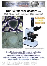
http://www.grayfieldoptical.com – info@grayfieldoptical.com – USA: (702) 425-7775 – UK: (020) 8133 4321 – EU: +49 221 20046970<br />
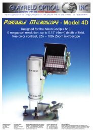
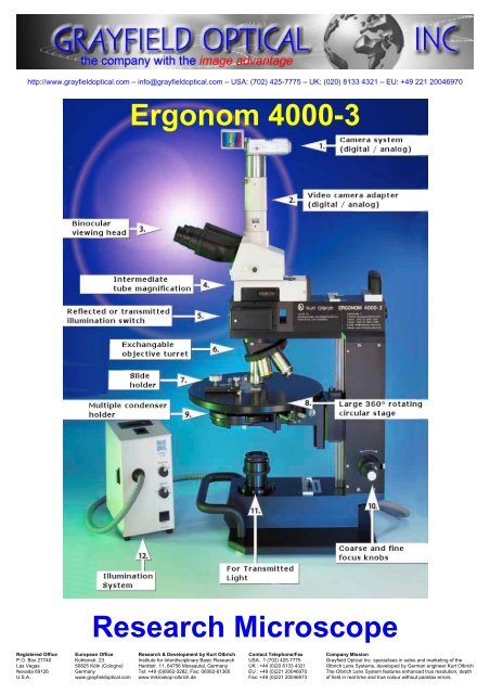
<strong>Ergonom</strong> <strong>4000</strong>-3<br />
<strong>Research</strong> <strong>Microscope</strong><br />
Registered Office European Office <strong>Research</strong> & Development by Kurt Olbrich Contact Telephone/Fax Company Mission<br />
P.O. Box 27740 Kohlenstr. 23 Institute for Interdisciplinary Basic <strong>Research</strong> USA: 1 (702) 425-7775 <strong>Grayfield</strong> <strong>Optical</strong> <strong>Inc</strong>. specialises in sales and marketing of the<br />
Las Vegas 50825 Köln (Cologne) Hardtstr. 11, 64756 Mossautal, Germany UK : +44 (0)20 8133 4321 Olbrich Lens Systems, developed by German engineer Kurt Olbrich<br />
Nevada 89126 Germany Tel: +49 (0)6062-3282, Fax: 06062-61360 EU : +49 (0)221 20046970 The Olbrich Lens System features enhanced true resolution, depth<br />
U.S.A. www.grayfieldoptical.com www.mikroskop-olbrich.de Fax: +49 (0)221 20046973 of field in real time and true colour without parallax errors.
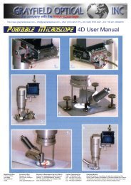
<strong>Ergonom</strong> <strong>4000</strong>-3/4 <strong>Research</strong> <strong>Microscope</strong><br />
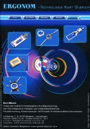
Kurt Olbrich<br />
<strong>Ergonom</strong> 500<br />
The <strong>Ergonom</strong> Technology<br />
The <strong>Ergonom</strong> Series of <strong>Microscope</strong>s<br />
In 1972, Kurt Olbrich investigated why the resolution and depth of field, of existing light microscopes,<br />
is so limited. In 1976, he discovered a unique new way of building microscopes where by using a<br />
different approach to optics and a new mathematical approach, he could build microscopes with a<br />
large "cylinder of sharpness" (depth of field) and a true resolution better than 100nm, while<br />
maintaining full contour sharpness and true colours without the need for staining, oil immersion, etc.<br />
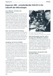
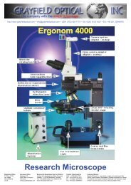
The <strong>Ergonom</strong> <strong>4000</strong>-3 is our top<br />
model and a direct successor of<br />
the <strong>Ergonom</strong> 500, with the same<br />
high resolution, depth-of-field,<br />
etc, yet is now even easier to<br />
use. It can be separated into 3<br />
major parts for transport, and the<br />
illumination system has been<br />
further improved with the choice<br />
of a separate halogen lamp<br />
system, or an internal special<br />
LED light source. The microscope<br />
is built entirely to our own<br />
specifications in order to guarantee the use of top quality components.<br />
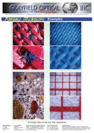
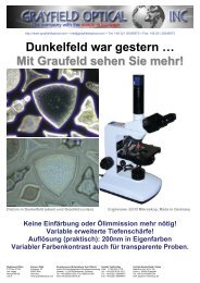
Depth of Field: All <strong>Ergonom</strong> microscopes feature variable depth of field where the true colours and<br />
contour sharpness remain clearly discernable, even with ever increasing magnifications comparable with that<br />
of mid-range scanning electron microscopes. This makes it possible to vary the depth of field independently of<br />
magnification, which also allows much more detail to be seen (see image on right), live and in real time!<br />
No Staining, No Oil: The unique optical system provides so much contrast that staining is not required.<br />
This allows you to observe the specimens, under a white light source, in their true (living) state, in vivid contrast<br />
and true colours even at the highest magnifications. All objectives remain dry as oil immersion is not required.<br />
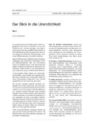
<strong>Grayfield</strong>: An entirely new method in optical microscopy is the <strong>Grayfield</strong> method, developed by Kurt Olbrich.<br />
This method allows you to see detailed structures that are otherwise not even visible with conventional phase<br />
contrast microscopes. For example, the <strong>Grayfield</strong> method allows you to observe the in vitro decomposition<br />
processes of blood. During this transitional phase new viruses and structures arise, which tend to decay and<br />
could previously not be made visible due to the lack of suitable microscope techniques.<br />
Phase Contrast: The phase contrast method is used for all thin-layered structures including fibres and<br />
textiles. With conventional methods, focusing is limited by overlapping layers and the structures also have the<br />
same fractal index causing the structures to become blurred. The <strong>Ergonom</strong> technology removes these<br />
limitations and provides clear and sharp contours.<br />
The <strong>Ergonom</strong> <strong>4000</strong>-3 is the top model offering both incident (