
You also want an ePaper? Increase the reach of your titles
YUMPU automatically turns print PDFs into web optimized ePapers that Google loves.
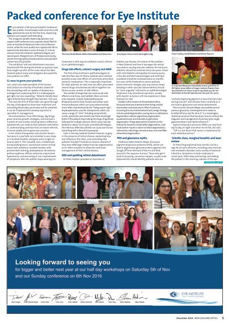
Packed conference for Eye Institute<br />
Eye Institute’s 11th Annual Scientific Conference<br />
was another record-breaker with more than 320<br />
optometrists and, for the first time, dispensing<br />
opticians and support staff attending.<br />
The inaugural, parallel dispensing opticians and<br />
support staff conference (see separate story) filled its<br />
room at Auckland’s Waipuna Hotel and Conference<br />
Centre, while the main auditorium rippled with life as<br />
optometrists attended a series of sharp, 15-minute<br />
sessions from the Institute’s ophthalmologists and<br />
special guest, Margaret Lam, of theeyecarecompany,<br />
award-winning Sydney-based practices and specialist<br />
contact lens (CL) provider.<br />
The exhibitors’ hall and refreshment area also<br />
teemed with life during the breaks as sponsors (see<br />
box) caught up with all the news about the New<br />
Zealand optical scene and delegates discussed the<br />
new products on offer.<br />
CL nous to grow your practice<br />
Lam, who’s also state president of the Cornea<br />
and Contact Lens Society of Australia, kicked off<br />
the proceedings with an update on keratoconus<br />
management, explaining that it is challenging to<br />
get right, but also rewarding. “Patients literally have<br />
sight restored they thought they had lost,” she said.<br />
This was the first of three talks Lam gave through<br />
the day, all designed to show how important and<br />
rewarding (financially, personally and for patients)<br />
it can be if optometrists develop expertise in<br />
speciality CL management.<br />
Her presentation, From little things, big things<br />
grow–practice growth strategies, summarised a<br />
number of case studies showing what a difference<br />
optometrists can make to their patients with better<br />
CL understanding and how that can boost referrals,<br />
increase loyalty and so grow your practice.<br />
In her Ocular therapeutics and contact lenses–<br />
two peas in a pod talk, Lam provided a case study<br />
on microbial keratitis, and gave some insight on<br />
how to spot it. This included: sore, irritated eyes<br />
during waking hours; round focal central corneal<br />
lesion with indistinct, roundish borders with<br />
positive NaFl staining; photophobia; lid oedema;<br />
profuse epiphora; diffuse extensive conjunctival<br />
hyperaemia; and worsening or non-improvement<br />
of symptoms after the patient stops wearing CLs.<br />
Tina Gao, Renita Martis, Nafisa Slaimankhel and Teresa Hsu<br />
Treatment is with topical antibiotic and/or referral<br />
to an ophthalmologist.<br />
Drugs side effects, cataract surgery and AMD<br />
The first of Eye Institute’s ophthalmologists to<br />
take the floor was Dr Shanu Subbiah who outlined<br />
possible ocular side effects of commonly prescribed<br />
systemic medications. This is especially important<br />
for older patients, he said, who are often prescribed<br />
several drugs simultaneously which together can<br />
throw up any number of side-effects.<br />
The number of drugs that can cause ocular side<br />
effects is enormous, said Subbiah. More common<br />
ones, however, include: hydroxychloroquine<br />
(Plaquenil) used to treat malaria and certain autoimmune<br />
diseases, which can cause retinal toxicity<br />
and is often characterised by the “flying saucer sign”;<br />
bisphosphonates, used for osteoporosis, which can<br />
cause a range of “-itis’s” including conjunctivitis,<br />
uveitis, episcleritis and scleritis, but these should get<br />
better if the patient stops taking the drugs; fingolimod<br />
(Gilenya) for multiple sclerosis, which cause macular<br />
oedema in about 1% of cases; and sildenafil (Viagra),<br />
the infamous little blue pill that can cause users to see<br />
everything with a blue tint (cyanopsia).<br />
Later in the day Subbiah tackled Cataract surgery<br />
in the presence of retinal disease, explaining how<br />
one influences the other, and concluding that<br />
patients shouldn’t hesitate to remove cataracts if<br />
they have AMD (age-related macular degeneration)<br />
as it’s often essential to allow the continued<br />
management of their retinal disease.<br />
DED and spotting retinal detachment<br />
Dr Peter Hadden provided an overview of<br />
Amy Royal, Chloe Lovell and Angeline Ng<br />
diabetic eye disease, the extent of the problem<br />
in New Zealand and how it damages the retinal<br />
vasculature causing macular oedema. At minimum,<br />
diabetics should be screened every two years; those<br />
with mild diabetic retinopathy (microaneurysms,<br />
a few dot and blot haemorrhages and mild lipid<br />
exudates) should be reviewed every six months<br />
to a year, while moderate to severe patients<br />
(more extensive changes, plus any venous loops,<br />
beading or other vascular abnormalities) should<br />
be “semi-urgently” referred to an ophthalmologist.<br />
Treatment is by intravitreal injections, usually<br />
with Avastin as Eylea is still too expensive in New<br />
Zealand, he said.<br />
Hadden’s other session on the peripheral retina,<br />
discussed what was important when doing a retinal<br />
examination and what wasn’t. When it comes to<br />
increasing the risk of retinal detachment, benign findings<br />
include cystoid degeneration, paving stone or cobblestone<br />
degeneration, reticular pigmentary degeneration,<br />
equatorial drusen and choroidal or pigmentary<br />
degeneration. Things optometrists should be on the<br />
lookout for, however, include lattice degeneration, snail<br />
tracks (which may be a variant on lattice degeneration),<br />
retinoschisis, retinal tags, horseshoe tears and snowflake<br />
vitreoretinal degeneration.<br />
PDS and glaucoma myths<br />
Professor Helen Danesh-Meyer discussed<br />
pigment dispersion syndrome (PDS), which can<br />
lead to pigmentary glaucoma when pigment cells<br />
slough off from the back of the iris and float<br />
around in the aqueous humour. Those who get it<br />
tend to be young, caucasian myopes, usually male.<br />
Optometrists should identify patients who are<br />
Gary Crowley, David Roberts and Diane Pearson<br />
A surprised Maryanne Dransfield, former publisher of<br />
NZ Optics (now editor-at-large), receives flowers from<br />
Eye Institute’s Dr Trevor Gray to say thank you for her<br />
dedication to the NZ industry over the past 35+ years<br />
“actively dispersing pigment or have blurred vision<br />
during exercise” and monitor them carefully as it<br />
can lead to glaucoma and retinal detachment.<br />
Prior to lunch, Danesh-Meyer also tackled normal<br />
pressure glaucoma, stressing the term needs to<br />
be killed off once and for all as it “is a meaningless<br />
statistical construct that has done more to confuse the<br />
diagnosis and management of primary open-angle<br />
glaucoma than it ever did to enhance it.”<br />
Typical intraocular pressures (IOPs) can vary from<br />
day to day, at night and when we lie flat, she said.<br />
“IOP is a risk factor that varies in importance for<br />
each individual person.”<br />
Scleritis clues, marginal keratitis and laser<br />
review<br />
Dr Peter Ring explained how scleritis can be a<br />
sign for all sorts ailments, including auto-immune<br />
and metabolic disorders and a variety of bacterial<br />
infections. Symptoms include moderate to<br />
severe pain, often deep and boring, and waking<br />
the patient in the morning; redness of the eye;<br />
CONTINUED ON P18<br />
Looking forward to seeing you<br />
for bigger and better next year at our half day workshops on Saturday 5th of Nov<br />
and our Sunday conference on 6th Nov 2016<br />
<strong>Dec</strong>ember <strong>2015</strong><br />
NEW ZEALAND OPTICS<br />
5<br />
<strong>Dec</strong>ember <strong>2015</strong>.indd 5<br />
19-Nov-15 3:26:53 PM