(C. Berkland ,2002).

You also want an ePaper? Increase the reach of your titles
YUMPU automatically turns print PDFs into web optimized ePapers that Google loves.
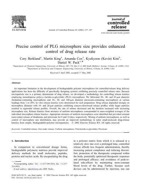
Journal of Controlled Release 82 (<strong>2002</strong>) 137–147<br />
www.elsevier.com/locate/jconrel<br />
Precise control of PLG microsphere size provides enhanced<br />
control of drug release rate<br />
a a a b<br />
Cory <strong>Berkland</strong> , Martin King , Amanda Cox , Kyekyoon (Kevin) Kim ,<br />
Daniel W. Pack a , *<br />
a<br />
Department of Chemical Engineering, University of Illinois, MC-712, Box C-3, 600 South Mathews Avenue, Urbana, IL 61801, USA<br />
b<br />
Department of Electrical and Computer Engineering, University of Illinois, Urbana, IL 61801, USA<br />
Received 6 April <strong>2002</strong>; accepted 17 May <strong>2002</strong><br />
Abstract<br />
An important limitation in the development of biodegradable polymer microspheres for controlled-release drug delivery<br />
applications has been the difficulty of specifically designing systems exhibiting precisely controlled release rates. Because<br />
microparticle size is a primary determinant of drug release, we developed a methodology for controlling release kinetics<br />
employing monodisperse poly(D,L-lactide-co-glycolide) (PLG) microspheres. We fabricated 20-, 40- and 65-mm diameter<br />
rhodamine-containing microspheres and 10-, 50- and 100-mm diameter piroxicam-containing microspheres at various<br />
loadings from 1 to 20%. In vitro release kinetics were determined for each preparation. Drug release depended strongly on<br />
microsphere diameter with 10- and 20-mm particles exhibiting concave-downward release profiles while larger particles<br />
resulted in sigmoidal release profiles. Overall, the rate of release decreased and the duration increased with increasing<br />
microsphere size. Release kinetics from mixtures of uniform microspheres corresponded to mass-weighted averages of the<br />
individual microsphere release kinetics. Appropriate mixtures of uniform microspheres were identified that provided constant<br />
(zero-order) release of rhodamine and piroxicam for 8 and 14 days, respectively. Mixing of uniform microspheres, as well as<br />
control of microsphere size distribution, may provide an improved methodology to tailor small-molecule drug-release<br />
kinetics from simple, biodegradable-polymer microparticles. © <strong>2002</strong> Elsevier Science B.V. All rights reserved.<br />
Keywords: Controlled release; Zero-order release; Uniform microspheres; Poly(lactide-co-glycolide); Piroxicam<br />
1. Introduction in a polymer matrix from which it is released at a<br />
relatively slow rate over a prolonged time, controlled<br />
In comparison to conventional dosage forms, release affords less frequent administration, thereby<br />
biodegradable polymeric matrices provide improved increasing patient compliance and reducing discomdelivery<br />
methods for small molecules, peptides,<br />
proteins and nucleic acids. By encapsulating the drug<br />
*Corresponding author. Tel.: 11-217-244-2816; fax: 11-217-<br />
333-5052.<br />
E-mail address: dpack@uiuc.edu (D.W. Pack).<br />
fort; protection of the therapeutic compound within<br />
the body; potentially optimized therapeutic responses<br />
and prolonged efficacy; and avoidance of peak-related<br />
side-effects by maintaining more-constant<br />
blood levels of the drug. Further, because such<br />
devices can be administered by injection, one can<br />
0168-3659/02/$ – see front matter © <strong>2002</strong> Elsevier Science B.V. All rights reserved.<br />
PII: S0168-3659(02)00136-0
138 C. <strong>Berkland</strong> et al. / Journal of Controlled Release 82 (<strong>2002</strong>) 137 –147<br />
also achieve localized drug delivery and high local release rates. Larger spheres generally release enconcentrations.<br />
capsulated compounds more slowly and over longer<br />
The large and growing variety of pharmaceuticals time periods, other properties (polymer molecular<br />
on the market and in development require versatile weight, initial porosity, drug distribution within the<br />
delivery systems that can adapt to the needs of sphere, etc.) being equal. Thus, controlling sphere<br />
particular applications [1], especially the capacity to size provides an opportunity for control of release<br />
generate the required delivery rates and, perhaps, kinetics. Numerous studies have been conducted to<br />
variation of delivery rate over time. For example, determine the effects of sphere size on drug release<br />
many therapeutics require a constant release rate for [3,10,11,13,20,21]. However, due to a limited ability<br />
varying durations from several days to several weeks to control microsphere size, this approach to modu-<br />
[2–6]. Such ‘zero-order’ release is a long-sought lating release rates has been relatively unexplored.<br />
goal of controlled-release drug delivery, but has been We have devised a methodology for precisely<br />
difficult to achieve for many pharmaceuticals. In controlling microsphere size and size distribution<br />
contrast, variable drug release rates can be beneficial [20]. Our spraying technology is capable of generatfor<br />
many important indications [7]. Intermittent high ing uniform PLG microspheres ranging in size from<br />
doses of antibiotics may alleviate evolution of resist- about 1 to .500 mm. For example, we recently<br />
ance in bacteria, and discontinuous administration of reported fabrication of microspheres with diameters<br />
vaccines often enhances the immune response of |5–80 mm, wherein 95% of the particles had a<br />
[2,8,9]. diameter within 1.0–1.5 mm of the average [20].<br />
Microparticle drug delivery systems may provide Furthermore, the methodology allows fabrication of<br />
the needed versatility. Drug release rates can be novel, continuously varying size distributions of any<br />
controlled through the choice of polymer chemistry desired shape. We hypothesized that the ability to<br />
[10,11] (e.g. polymer composition, co-monomer control particle size afforded by our system would<br />
ratios, molecular weight, etc.) or variation of the lead to enhanced control of drug release kinetics.<br />
microparticle formulation parameters, and thus the Here we report release kinetics for a model comphysical<br />
characteristics of the resulting particles pound, rhodamine B, and the non-steroidal anti-<br />
[12,13]. Nevertheless, the ability to tailor drug inflammatory drug (NSAID) piroxicam from unirelease<br />
kinetics is limited. For typical small-molecule form PLG microspheres. While piroxicam is similar<br />
therapeutics, as well as some proteins [11,14–17], in molecular weight to rhodamine, these two comdrug<br />
release often exhibits an initial ‘burst’ phase pounds were chosen to represent water-soluble<br />
during which a significant fraction (typically 5– (rhodamine, 7.8 mg/ml) and -insoluble (piroxicam,<br />
50%) of the encapsulated compound is released in a 53.3 mg/ml at pH|7) drugs [22]. We demonstrate<br />
short time (,24 h). The burst is usually undesirable that the release kinetics of both compounds are<br />
because the drug that is released in this phase is not indeed variable depending on the microsphere size,<br />
available for prolonged release, and more important- as expected. Further, we show that mixtures of<br />
ly for potent therapeutics or drugs with a narrow uniform microspheres exhibit release kinetics that are<br />
therapeutic window, this initial bolus may result in weighted averages of the individual microsphere<br />
toxicity or other side-effects. The burst may be release kinetics. Based on this finding, we chose<br />
followed by a lag phase exhibiting negligible release appropriate mixtures to generate zero-order release,<br />
and, more typically, a phase in which the release rate without an initial burst phase, for both rhodamine<br />
decreases with time due to a decreasing driving force and piroxicam.<br />
as drug is depleted from the matrix. Various strategies<br />
for reducing or eliminating the initial burst have<br />
been studied including chemistry (block copolymers 2. Materials and methods<br />
with hydrophilic regions) [10], variation of microsphere<br />
formation parameters [12,13], coating of 2.1. Materials<br />
microspheres (microencapsulated microspheres) [18]<br />
and conjugation of drug to the polymer matrix [19]. Poly(D,L-lactide-co-glycolide) (50:50 lactic<br />
Microsphere size is a primary determinant of drug acid:glycolic acid; i.v.50.20–24 dl/g corresponding
C. <strong>Berkland</strong> et al. / Journal of Controlled Release 82 (<strong>2002</strong>) 137 –147 139<br />
to Mw<br />
10,000–15,000) was obtained from Birming- nm (Varian Cary 50) in a quartz cuvette and subtract-<br />
ham Polymers. Poly(vinyl alcohol) (PVA; 88% hy- ing absorbance values for the blank microspheres.<br />
drolyzed) was obtained from Polysciences.<br />
Rhodamine B chloride was obtained from Sigma. 2.4. In vitro drug release<br />
Piroxicam free base was a gift from Dongwha<br />
Pharmaceuticals (Seoul, Korea). HPLC grade di- Rhodamine release was determined by resuspendchloromethane<br />
(DCM), dimethylsulfoxide and so- ing a known mass of microspheres encapsulating<br />
dium hydroxide were purchased from Fisher Scientific.<br />
2.2. Preparation of microspheres<br />
rhodamine B in 2 ml of phosphate-buffered saline<br />
(PBS, pH 7.4) containing 0.5% Tween. The suspensions<br />
were continuously agitated by inversion (at<br />
|10 rpm) in a 37 8C incubator. At regular intervals<br />
the samples were centrifuged, the supernatant was<br />
removed, and the spheres were resuspended in fresh<br />
Microspheres were prepared as described previ-<br />
PBS. Concentration of rhodamine B in the supernaously<br />
[20]. Briefly, PLG solutions (5% (w/v) in<br />
tant was determined using the spectrophotometer as<br />
DCM) containing rhodamine B or piroxicam at the<br />
described above. The amount of rhodamine in each<br />
various concentrations indicated were pumped<br />
sample was summed with the amounts at each<br />
through a small glass nozzle at various flow rates,<br />
previous time point, and the total divided by the<br />
while an ultrasonic transducer (Branson Ultrasonics)<br />
amount of rhodamine in the microspheres (excontrolled<br />
by a frequency generator (Hewlett Pacperimental<br />
loading3mass of microspheres), to arrive<br />
kard model 3325A) disrupted the stream into uniat<br />
the ‘cumulative percent released’.<br />
form droplets. A carrier stream (1% (w/v) PVA in<br />
Piroxicam release was determined by resuspending<br />
distilled water) flowed around the emerging PLG<br />
|5 mg of microspheres in 1.3 ml of PBS containing<br />
stream. The streams flowed into a beaker containing<br />
0.5% Tween. Conditions during drug release were<br />
|500 ml of 1% PVA, and the particles were stirred at<br />
the same as described above for rhodamine. After<br />
room temperature for 3 h, filtered, and rinsed with<br />
centrifugation, the concentration of piroxicam in the<br />
distilled water. The microspheres were lyophilized<br />
supernatant was determined by measuring the ab-<br />
(Labconco benchtop model) for a minimum of 48 h<br />
sorbance at 276 nm as described. Average absorand<br />
were stored at 220 8C under desiccant.<br />
bance of the supernatant from tubes containing blank<br />
microspheres treated identically was subtracted from<br />
2.3. Determination of drug loading all measurements.<br />
The initial loading of rhodamine B was deter- 2.5. Scanning electron microscopy<br />
mined as follows. A known mass (|2–5 mg) of<br />
microspheres was dissolved in 50 ml dimethylsulfox- Microsphere surface structure and porosity were<br />
ide. PBS (500 ml) was added and precipitated investigated by scanning electron microscopy<br />
polymer was removed by centrifugation at 12,000 (Hitachi S-4700). Samples were prepared by placing<br />
rpm for 10 min. Rhodamine B concentration in the a droplet of an aqueous microsphere suspension onto<br />
supernatant was determined by measuring the ab- a silicon stub. The samples were dried overnight and<br />
sorbance at 550 nm in a multi-well plate spec- were sputter coated with gold prior to imaging at<br />
trophotometer (Molecular Devices Spectra Max 2–10 eV.<br />
340PC).<br />
To determine piroxicam loading, a known mass 2.6. Particle size distribution<br />
(|5 mg) of microspheres containing piroxicam was<br />
dissolved in 1 ml of 0.25 M sodium hydroxide at A Coulter Multisizer 3 (Beckman Coulter)<br />
room temperature for 5 min. Blank (piroxicam free) equipped with a 100- or 280-mm aperture was used<br />
microspheres of the same size were treated identical- to determine the size distribution of the various<br />
ly. Piroxicam concentration in the resulting solution sphere preparations. The lyophilized particles were<br />
was determined by measuring the absorbance at 276 resuspended in Isoton electrolyte and a type I-A
140 C. <strong>Berkland</strong> et al. / Journal of Controlled Release 82 (<strong>2002</strong>) 137 –147<br />
dispersant was used to prevent microsphere aggregation.<br />
A minimum of 5000 microspheres was analyzed<br />
for each sample.<br />
3. Results<br />
3.1. Microsphere fabrication and characterization<br />
taining particles of 10, 50 and 100 mm with 5, 10,<br />
15, and 20% loading. Rhodamine and piroxicam overlap.<br />
loading and encapsulation efficiency (e.e.) are reported<br />
in Tables 1 and 2, respectively. (While drug<br />
loading is less than theoretical, e.e.510–60%, for<br />
simplicity we will refer to the various samples by the<br />
theoretical loading.)<br />
The microspheres were very uniform, typically<br />
Uniform PLG microspheres were fabricated employing<br />
the spraying apparatus described previously<br />
[20]. The model drug compounds, rhodamine B and<br />
piroxicam (free base form), were encapsulated by<br />
co-dissolving the drug with the PLG in DCM. In<br />
order to examine the effect of microsphere diameter<br />
on drug release kinetics, we fabricated rhodaminecontaining<br />
particles of 20, 40 and 65 mm, at theoret-<br />
ical loadings of 1, 3, and 5%, and piroxicam-con-<br />
Fig. 1. Typical size distributions of uniform microspheres loaded<br />
with rhodamine (20-, 45- and 75-mm diameter) and piroxicam<br />
(10- and 55-mm diameter). All distributions are normalized by<br />
total area under the curve. Thus, the peak height is also an<br />
indication of the relative particle uniformity. Each distribution is<br />
colored with a different shade of gray to distinguish where they<br />
having .90% of the particles within 2-mm ofthe<br />
average diameter (Fig. 1). Microsphere homogeneity<br />
is also evident in scanning electron micrographs of<br />
the various microsphere preparations (Fig. 2). The<br />
Table 1<br />
Characterization of rhodamine-loaded PLG microspheres<br />
20 mm 40 mm 65 mm<br />
Theoretical 1 3 5 1 3 5 1 3 5<br />
loading (%)<br />
Experimental 0.63 1.80 2.50 0.37 1.05 1.75 0.61 1.29 3.00<br />
loading (%)<br />
Encapsulation 63 60 50 37 35 35 61 43 60<br />
efficiency (%)<br />
Table 2<br />
Characterization of piroxicam-loaded PLG microspheres<br />
10 mm 50 mm 100 mm<br />
Theoretical f5 10 15 20 5 10 15 20 5 10 15 20<br />
loading (%)<br />
Experimental 3.0 4.6 5.6 5.8 1.0 1.0 1.5 3.6 1.0 3.1 3.0 5.8<br />
loading (%)<br />
Encapsulation 59 46 37 29 19 10 10 18 20 31 20 29<br />
efficiency (%)
C. <strong>Berkland</strong> et al. / Journal of Controlled Release 82 (<strong>2002</strong>) 137 –147 141<br />
Fig. 2. Scanning electron micrographs of (A) 65- (B) 40- and (C) 20-mm rhodamine-loaded microspheres and (D) 100- (E) 50- and (F)<br />
10-mm piroxicam-loaded microspheres. Scale bar represents 100 mm.<br />
particles exhibit a smooth, slightly porous surface<br />
and dense polymer interior similar to microspheres<br />
produced using conventional emulsion techniques<br />
(Fig. 3) [16,17,23,24]. The average sizes determined<br />
by the Coulter counter are |5–10% larger than the<br />
sizes obtained from SEM, but the uniformity is<br />
readily apparent. The larger size may be the result of<br />
swelling due to water uptake. We refer to the<br />
particles according to the smaller sizes obtained from<br />
SEM.<br />
3.2. In vitro release from uniform microspheres<br />
To examine the effect of microsphere size and size
142 C. <strong>Berkland</strong> et al. / Journal of Controlled Release 82 (<strong>2002</strong>) 137 –147<br />
Fig. 3. Scanning electron micrographs of a fractured PLG microsphere<br />
depicting the dense polymer matrix of drug-encapsulating<br />
microspheres. Scale bars represents 10 mm.<br />
uniformity on drug release kinetics, we measured<br />
release profiles for both drugs from spheres of all<br />
loadings and sizes. Rhodamine release profiles are<br />
shown in Fig. 4. As expected, 20-mm microspheres<br />
exhibited a faster initial release than 65-mm micro-<br />
Fig. 4. Effect of microsphere size and drug loading on rhodamine<br />
spheres, likely due to the increased surface-to-volrelease<br />
rates: (A) 1%, (B) 3% and (C) 5% theoretical loading.<br />
ume ratio of the smaller particles. Further, as drug<br />
loading increased, the initial rate of drug release<br />
increased. An interesting concave-upward (i.e. sigmoidal)<br />
profile was observed with the 65-mm par- drug release profiles. The smallest microspheres (10-<br />
ticles and to a lesser extent with the 45-mm particles, mm diameter) exhibited a rapid initial rate of release,<br />
wherein drug release was initially slow, then pro- with 40–60% of encapsulated piroxicam released<br />
gressed to a more rapid release phase before leveling within the first 24 h. Initial release rates decreased<br />
off [25,26].<br />
with increasing microsphere diameter for all drug<br />
Piroxicam release profiles show similar trends loadings examined. Further, the initial release rate<br />
(Fig. 5). Samples of 10-, 50- and 100-mm micro- decreased with increasing drug loading. Interestingly,<br />
spheres were studied. The microspheres span a the 50- and 100-mm particles exhibited sigmoidal<br />
broader size range than the rhodamine-loaded par- release profiles similar to rhodamine release from<br />
ticles, resulting in a more pronounced difference in 65-mm microspheres.
C. <strong>Berkland</strong> et al. / Journal of Controlled Release 82 (<strong>2002</strong>) 137 –147 143<br />
and 3:1, w/w) of 20-mm/5% and 65-mm/5%<br />
rhodamine-containing microspheres. The release profiles<br />
for the microsphere mixtures were intermediate<br />
between the two individual microsphere release<br />
profiles (Fig. 6A). Further, the mixture release<br />
profiles corresponded to a mass-weighted linear<br />
combination of the individual release profiles (see<br />
dotted lines in Fig. 6A).<br />
Given the agreement between predicted release<br />
profiles (i.e. linear combinations) and the experimental<br />
data, various linear combinations of individualmicrosphere<br />
release profiles, based on the nine<br />
preparations of varying microsphere size and<br />
rhodamine loading, were examined to identify a<br />
combination of uniform microspheres that might<br />
Fig. 5. Effect of microsphere size and drug loading on piroxicam<br />
release rates: (A) 5%, (B) 10% and (C) 15% theoretical loading.<br />
3.3. In vitro release from mixtures of uniform<br />
microspheres<br />
Based on the different shapes of the uniform<br />
microsphere release profiles, and given the reproducibility<br />
of our methodology for uniform microsphere<br />
fabrication, we reasoned that it may be<br />
possible to modulate release kinetics in a desired<br />
fashion by mixing appropriate proportions of two or<br />
more uniform microsphere preparations [3,10]. To<br />
test this hypothesis, we mixed known ratios (1:3, 1:1<br />
Fig. 6. (A) Rhodamine release from 20-mm/5% microspheres,<br />
65-mm/5% microspheres and 3:1, 1:1 and 1:3 (w/w) mixtures.<br />
(B) Rhodamine release from 20-mm/5% microspheres, 65-mm/<br />
3% microspheres and 1:4, 1:9 and 1:24 (w/w) mixtures. Filled<br />
symbols: experimental data points for individual microspheres.<br />
Open symbols: experimental data points for mixtures. Dotted line:<br />
weighted average of individual microsphere experimental release<br />
data (predicted release). Error bars, typical of those shown in Fig.<br />
4, were removed for clarity.
144 C. <strong>Berkland</strong> et al. / Journal of Controlled Release 82 (<strong>2002</strong>) 137 –147<br />
result in zero-order release kinetics. Based on the tion combined 10-mm/15% and 50-mm/15% micropredicted<br />
release profiles, we chose to mix 20-mm/ spheres in ratios of 3:1, 1:1, and 1:3 (w/w). This<br />
5% and 65-mm/3% microspheres in various ratios formulation resulted in slightly concave downward<br />
and performed in vitro release studies for comparison release profiles for the 3:1 and 1:1 ratios and a linear<br />
to predicted release profiles (Fig. 6B). Again, the drug release profile for the 1:3 (w/w) mixture (Fig.<br />
experimental release data coincided precisely with 7A). The second formulation comprised 10-mm/20%<br />
the predicted release profile, and the 1:4 (w/w) and 50-mm/10% microspheres in three ratios, 1:6.1,<br />
mixture provided constant release for 8 days.<br />
1:11.5, and 1:39 (w/w). The 1:6.1 (w/w) formulation<br />
To investigate the generality of this approach for produced near linear release over the course of the<br />
modulating drug release kinetics, we performed a experiment (Fig. 7B).<br />
similar set of experiments to generate zero-order<br />
release of the clinically relevant NSAID, piroxicam.<br />
Multiple linear combinations of 10-, 50- and 100-mm 4. Discussion<br />
piroxicam-containing microspheres at various drug<br />
loadings were examined computationally to identify Long-term zero-order release of small-molecule<br />
a combination resulting in linear drug release. Two therapeutics from biodegradable microspheres has<br />
possible formulations were found. The first formula- been difficult to achieve. Release of model compounds<br />
similar to rhodamine B is often rapid and<br />
diffusion controlled [27,28]. Similarly, release of<br />
NSAIDs, especially piroxicam, encapsulated in polymeric<br />
particles typically occurs within 24 h and is<br />
dominated by a large initial rate of release (or<br />
‘burst’), offering little advantage over conventional<br />
oral dosage forms [16,17,23,24,29,30]. In contrast,<br />
our results show that simple molecules can be<br />
released in a controlled manner over significant<br />
durations of time.<br />
The initial release rates of both rhodamine and<br />
piroxicam decreased with increasing sphere diameter.<br />
This is expected due to the decrease in surface<br />
area/volume ratio with increasing size. Furthermore,<br />
we observed that the release rates (as percent of total<br />
drug released vs. time) from rhodamine-containing<br />
microspheres increased slightly with increasing loading<br />
for all sphere sizes. For purely diffusion-controlled<br />
release, no dependence on drug loading is<br />
expected. Other researchers have reported faster<br />
release with increased loading, especially for cases in<br />
which the drug is phase-separated from the polymer<br />
matrix and release can occur through aqueous mesoand<br />
macropores created by the drug [31,32]. Such a<br />
mechanism may explain the effect of rhodamine<br />
loading on release rates. In contrast, we observed<br />
that the release rate of piroxicam decreased with<br />
Fig. 7. (A) Piroxicam release from 3:1, 1:1 and 1:3 (w/w) increasing loading. Possible explanations for this<br />
mixtures of 10-mm/15% microspheres and 50-mm/15% microsurprising<br />
result may be that the piroxicam, with pK a<br />
spheres. (B) Piroxicam release from 1:6.1, 1:11.5 and 1:39 (w/w)<br />
mixtures of 10-mm/20% microspheres and 50-mm/10% microspheres.<br />
intrapolymer environment caused by accumulation<br />
of 5.07 and 2.33, is buffering acidification of the<br />
of
C. <strong>Berkland</strong> et al. / Journal of Controlled Release 82 (<strong>2002</strong>) 137 –147 145<br />
polymer degradation products, or that the hydro- aqueous pores in the polymer (also potentially leadphobic<br />
drug is slowing water uptake. Another possi- ing to an increase in the effective diffusivity).<br />
bility is that, due to the low solubility of piroxicam, Additional studies directly addressing this hypothesis<br />
‘sink’ conditions were not achieved. The highest are in progress.<br />
piroxicam concentrations measured were |100 mg/ Constant release is highly desirable for many drug<br />
ml while the solubility of piroxicam in the release delivery applications. Because there is a transition<br />
media at 37 8C is 1.3 mg/ml (the relatively high from the concave downward to sigmoidal release<br />
solubility is due to the presence of Tween 20 at a profiles as sphere size increases, it appears that<br />
concentration, 0.5%, above its critical micelle con- nearly linear release may be achieved at a certain<br />
centration). Because the maximum piroxicam con- size. For example, between 10 and 50 mm, a<br />
centration was less than one tenth of its solubility, microsphere size may exist that would provide zerodrug<br />
concentration in the release media likely had order piroxicam release over a 4- to 8-day duration<br />
little effect on the release kinetics.<br />
(cf. Figs. 4 and 5). Others report linear or near-linear<br />
Small and large particles resulted in qualitatively release profiles achieved with microspheres of simidifferent<br />
release profiles. The smallest microspheres lar size, |30–50 mm in diameter [4,5,10]. For<br />
(10- and 20-mm diameter, encapsulating piroxicam example, Woo et al. formulated a leuprolide delivery<br />
and rhodamine, respectively) exhibited concave system using PLA microspheres with an average<br />
downward release profiles typical of diffusion-con- diameter of 51.7 mm achieving near-linear peptide<br />
trolled release from PLG microspheres. However, the release for 135 days following a 15-day period of<br />
larger microspheres exhibited a sigmoidal release ‘diffusion-controlled release’ [5]. Our hypothesis<br />
profile in which the release rate initially increased suggests that the early phase of release results from<br />
with time. Similar release profiles have been ob- the portion of the microspheres in this formulation<br />
served previously. For example, Sansdrap and Moes under |35 mm, which would be expected to release<br />
reported a sigmoidal release profile for a low solu- drug more rapidly. Further, Bezemer et al. used a<br />
bility (10 mg/ml in water) drug, nifedipine [21] poly(ethylene glycol)-poly(butylene terephthalate)<br />
using 80-mm (630 mm) PLG microspheres, while (PEG-PBT) block copolymer to test the effects of<br />
Guiziou et al. found sigmoidal release from poly(lac- microsphere size on drug release [10]. They also<br />
tic acid) (PLA) microspheres loaded with the NSAID discovered that decreasing the average microsphere<br />
indomethacin [24]. Further, Ravivarapu et al. re- size from 108 to 29 mm causes the release kinetics to<br />
ported a similar release profile for leuprolide acetate- change gradually from zero-order release to release<br />
loaded PLG microspheres [33].<br />
controlled by Fickian diffusion. The microspheres<br />
The sigmoidal release profiles may result from used for these experiments were not uniform, but the<br />
several mechanisms. Guiziou et al. attributed the trends are indicative of the trends we observed for<br />
upward bending release profile to the microspheres uniform PLG microspheres.<br />
having a more dense polymer matrix with increased Other researchers have suggested that drug delivstability<br />
as compared to microspheres fabricated ery rates may be controlled by mixing microspheres<br />
using lower molecular weight PLA which released in of varying sizes or characteristics. For example,<br />
a more diffusion-controlled manner [24]. Alterna- Ravivarapu et al. mixed microspheres comprising<br />
tively, water uptake, resulting in increased solubiliza- 8.6- or 28.3-kDa PLG encapsulating leuprolide acetion<br />
of the drug and swelling of the polymer matrix tate [33]. The low-molecular-weight polymer re-<br />
[21], may cause an increasing release rate. However, sulted in porous, quickly releasing microspheres<br />
we propose that the sigmoidal shape results from while the high-molecular-weight formulation resulted<br />
polymer degradation. Erosion was suggested as the in dense microspheres and produced a sigmoidal<br />
cause of sigmoidal leuprolide release profiles de- release profile. By mixing microspheres comprising<br />
scribed above [33]. Polymer hydrolysis, with or the two polymers, release rates could be tailored, and<br />
without the subsequent mass loss, is expected to the resulting profiles were linear combinations of<br />
cause both increasing diffusivity of drug through the those resulting from individual microspheres.<br />
polymer matrix and an increase in the size of Bezemer et al. produced linear lysozyme release over
146 C. <strong>Berkland</strong> et al. / Journal of Controlled Release 82 (<strong>2002</strong>) 137 –147<br />
25 days from PEG-PBT microspheres having a Acknowledgements<br />
bimodal size distribution dominated by 50- and 110-<br />
mm particles (in essence a combination of two sizes) We wish to thank Young Bin Choy for improve-<br />
[10]. Finally, Narayani and Panduranga Rao com- ments to the microparticle fabrication apparatus and<br />
bined gelatin microspheres of various size ranges Dong Wha Pharmaceuticals (Korea) for providing<br />
producing zero-order release of methotrexate [3]. piroxicam.<br />
Because release kinetics from uniform spheres are<br />
very predictable and reproducible, our ability to<br />
fabricate uniform microspheres enhances such a References<br />
technique. We found that upon mixing uniform<br />
microsphere preparations, the resulting release pro- [1] D.M. Schachter, J. Kohn, A synthetic polymer matrix for the<br />
file is a mass-weighted average of the release profiles delayed or pulsatile release of water-soluble peptides, J.<br />
of the individual microspheres. This demonstrates<br />
Controlled Release 78 (<strong>2002</strong>) 143–153.<br />
that the microspheres release their payload indepentunities<br />
and challenges in the design of implantable bio-<br />
[2] D.L. Wise, D.J. Trantolo, R.T. Marino, J.P. Kitchell, Oppordently;<br />
there is no interaction between the particles.<br />
degradable polymeric systems for the delivery of anti-micro-<br />
In these experiments, the shapes of the rhodamine bial agents and vaccines, Adv. Drug Deliv. Rev. 1 (1987)<br />
and piroxicam release profiles were such that it was 19–39.<br />
possible to choose appropriate microsphere mixtures [3] R. Narayani, K. Panduranga Rao, Gelatin microsphere<br />
that provided zero-order release kinetics (Figs. 6 and<br />
cocktails of different sizes for the controlled release of<br />
anticancer drugs, Int. J. Pharm. 143 (1996) 255–258.<br />
7). However, it may not always be possible to [4] R.T. Liggins, S. D’Amours, J.S. Demetrick, L.S. Machan,<br />
generate a desired release profile from mixtures of<br />
H.M. Burt, Paclitaxel loaded poly(L-lactic acid) microonly<br />
two microsphere sizes. Depending on the spheres for the prevention of intraperitoneal carcinomatosis<br />
desired profile and the shape of the individual release<br />
after a surgical repair and tumor cell spill, Biomaterials 21<br />
curves, one may need to mix multiple microsphere<br />
(2000) 1959–1969.<br />
[5] B.H. Woo, J.W. Kostanski, S. Gebrekidan, B.A. Dani, B.C.<br />
samples or to fabricate complex microsphere size<br />
Tahanoo, P.P. DeLuca, Preparation, characterization and in<br />
distributions. Because the reported fabrication meth-<br />
vivo evaluation of 120-day poly(DL-lactide) leuprolide microod<br />
provides a unique ability to generate predefined spheres, J. Controlled Release 75 (2001) 307–315.<br />
microsphere sizes [20], this technology may lead to [6] A.M. Radder, H. Leenders, C.A. van Blitterswijk, Applica-<br />
enhanced control of release rates.<br />
tion of PEO/PBT copolymers for bone replacement, J.<br />
Biomed. Mater. Res. 30 (1996) 341–351.<br />
[7] B. Lemmer, in: R. Gurny, H. Junginger, N. Peppas (Eds.),<br />
Why Are So Many Biological Systems Periodic, Wissenschaftliche<br />
Verlagsgesellschaft, Stuttgart, 1992, pp. 11–<br />
5. Conclusions<br />
24.<br />
[8] J.W. Shiver, T.-M. Fu, L. Chen et al., Replication-incompetent<br />
adenoviral vaccine vector elicits effective anti-immuno-<br />
Microsphere size is a primary determinant of drug deficiency-virus immunity, Nature 415 (<strong>2002</strong>) 331–335.<br />
release kinetics. Release of model small-molecule [9] J.L. Cleland, Single-administration vaccines: controlled-redrugs<br />
can be varied from typical diffusion-controlled<br />
lease technology to mimic repeated immunizations, TIBtech<br />
profiles to slower, sigmoidal profiles as microsphere<br />
17 (1999) 25–29.<br />
diameter is increased in the range of 10–100 mm. [10] J.M. Bezemer, R. Radersma, D.W. Grijpma, P.J. Dijkstra,<br />
C.A. van Blitterswijk, J. Feijen, Microspheres for protein<br />
Drug release from mixtures of uniform microspheres<br />
delivery prepared from amphiphilic multiblock copolymers<br />
corresponds to a weighted average of the release 2. Modulation of release rate, J. Controlled Release 67<br />
from individual uniform microspheres. As a result, it (2000) 249–260.<br />
is possible to choose appropriate mixtures to gener- [11] Y. Men, C. Thomasin, H.P. Merkle, B. Gander, G. Corradin,<br />
ate desired release rate profiles, in particular constant<br />
A single administration of tetanus toxoid in biodegradable<br />
microspheres elicits T cell and antibody responses similar or<br />
release. Thus, microsphere mixtures with well-de-<br />
superior to those obtained with aluminum hydroxide, Vaccine<br />
fined size distributions may provide a general meth- 13 (1995) 683–689.<br />
odology for controlling drug release rates.<br />
[12] S. Cohen, T. Yoshika, M. Lucarelli, L.H. Hwang, R. Langer,
C. <strong>Berkland</strong> et al. / Journal of Controlled Release 82 (<strong>2002</strong>) 137 –147 147<br />
Controlled delivery systems for proteins based on poly(lac- [23] J.K. Lalla, K. Sapna, Biodegradable microspheres of polytic/glycolic<br />
acid) microspheres, Pharm. Res. 8 (1991) 713–<br />
(DL-lactic acid) containing piroxicam as a model drug for<br />
720. controlled release via the parenteral route, J. Microencapsul.<br />
[13] J. Akbuga, Effect of microsphere size and formulation 10 (1993) 449–460.<br />
factors on drug release from controlled-release furosemide [24] B. Guiziou, D.J. Armstrong, P.N.C. Elliot, J.L. Ford, C.<br />
microspheres, Drug Dev. Ind. Pharm. 17 (1991) 593–607.<br />
Rostron, Investigation of in-vitro release characteristics of<br />
[14] Y. Ozalp, N. Ozdemir, S. Kocagoz, V. Hasirci, Controlled NSAID-loaded polylactic acid microspheres, J. Microencaprelease<br />
of vancomycin from biodegradable microcapsules, J. sul. 13 (1996) 701–708.<br />
Microencapsul. 18 (2001) 89–110.<br />
[25] Y. Cheng, L. Illum, S.S. Davis, A poly(DL-lactide-co-gly-<br />
[15] S. Cohen, T. Yoshioka, M. Lucarelli, L.H. Hwang, R. colide) microsphere depot system for delivery of haloperidol,<br />
Langer, Controlled drug delivery systems for proteins based J. Controlled Release 55 (1998) 203–212.<br />
on poly(lactic/glycolic acid) microspheres, Pharm. Res. 8 [26] K. Suzuki, J.C. Price, Microencapsulation and dissolution<br />
(1991) 713–720. properties of a neuroleptic in a biodegradable polymer<br />
[16] M. Tuncay, S. Calis, H.S. Kas, M.T. Ercan, I. Peksoy, A.A. poly(D,L-lactide), J. Pharm. Sci. 74 (1985) 21–24.<br />
Hincal, Diclofenac sodium incorporated PLGA (50:50) [27] B.G. Jones, P.A. Dickinson, M. Gumbleton, I.W. Kellaway,<br />
microspheres: formulation considerations and in vitro/in<br />
The inhibition of phagocytosis of respirable microspheres by<br />
vivo evaluation, Int. J. Pharm. 195 (2000) 179–188. alveolar and peritoneal macrophages, Int. J. Pharm. 236<br />
[17] S. Bozdag, S. Calis, H.S. Kas, M.T. Ercan, I. Peksoy, A.A. (<strong>2002</strong>) 65–79.<br />
Kincal, In vitro evaluation and intra-articular administration [28] D.C. Bibby, N.M. Davies, I.G. Tucker, Poly(acrylic acid)<br />
of biodegradable microspheres containing naproxen sodium,<br />
microspheres containing beta-cyclodextrin: loading and in<br />
J. Microencapsul. 18 (2001) 443–456. vitro release of two dyes, Int. J. Pharm. 187 (1999) 243–250.<br />
[18] A. Gopferich, ¨ M.J. Alonso, R. Langer, Development and [29] B.W. Wagenaar, B.W. Muller, Piroxicam release from spraycharacterization<br />
of microencapsulated microspheres, Pharm. dried biodegradable microspheres, Biomaterials 15 (1994)<br />
Res. 11 (1994) 1568–1574. 49–54.<br />
[19] J.E. Oh, Y.S. Nam, K.H. Lee, T.G. Park, Conjugation of drug [30] D. Perumal, C.M. Dangor, R.S. Alcock, N. Hurbans, K.R.<br />
to poly(DL-lactic-co-glycolic acid) for controlled release from<br />
Moopanar, Effect of formulation variable on in vitro drug<br />
biodegradable microspheres, J. Controlled Release 57 (1999)<br />
release and micromeritic properties of modified release<br />
269–280. ibuprofen microspheres, J. Microencapsul. 16 (1999) 475–<br />
[20] C. <strong>Berkland</strong>, K. Kim, D.W. Pack, Fabrication of PLG 487.<br />
microspheres with precisely controlled and monodisperse [31] W.M. Saltzman, S.H. Pasternak, R. Langer, Microstructural<br />
size distributions, J. Controlled Release 73 (2001) 59–74.<br />
models for diffusive transport in porous polymers, ACS<br />
[21] P. Sansdrap, A.J. Moes, In vitro evaluation of the hydrolytic Symp. Ser. 348 (1987) 16–33.<br />
degradation of dispersed and aggregated poly(DL-lactide-co- [32] W.M. Saltzman, R. Langer, Transport rates of proteins in<br />
glycolide) microspheres, J. Controlled Release 43 (1997) porous polymers with known microgeometry, Biophys. J. 55<br />
47–58. (1989) 163–171.<br />
[22] J. Hadgraft, J. du Plessis, C. Goosen, The selection of [33] H.B. Ravivarapu, K. Burton, P.P. DeLuca, Polymer and<br />
non-steroidal anti-inflammatory agents for dermal delivery,<br />
microsphere blending to alter the release of a peptide from<br />
Int. J. Pharm. 207 (2000) 31–37. PLGA microspheres, Eur. J. Pharm. 50 (2000) 263–270.