FM JANUARY 2019 - digital edition

You also want an ePaper? Increase the reach of your titles
YUMPU automatically turns print PDFs into web optimized ePapers that Google loves.
₹ 250.00<br />
VOL 5 | ISSUE 9<br />
PAGES 100<br />
<strong>JANUARY</strong> <strong>2019</strong><br />
FUTUREMEDICINEINDIA.COM<br />
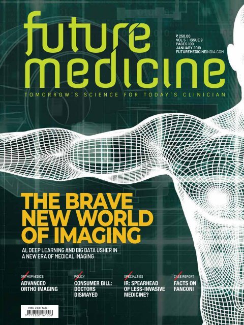
THE BRAVE<br />
NEW WORLD<br />
OF IMAGING<br />
AI, DEEP LEARNING AND BIG DATA USHER IN<br />
A NEW ERA OF MEDICAL IMAGING<br />
ORTHOPAEDICS POLICY SPECIALTIES CASE REPORT<br />
ADVANCED<br />
ORTHO IMAGING<br />
CONSUMER BILL:<br />
DOCTORS<br />
DISMAYED<br />
IR: SPEARHEAD<br />
OF LESS-INVASIVE<br />
MEDICINE?<br />
FACTS ON<br />
FANCONI
THE VASTNESS OF THE SKY... INSIDE<br />
The most sought-after feature<br />
in any architecture is a visual<br />
connection to nature. When<br />
building design or location<br />
precludes this life-supporting<br />
feature, a biophilic illusion of<br />
nature restores this essential<br />
wellness benefit.<br />
Generate Wellness<br />
Our therapeutic installations engage and<br />
relax patients, dramatically altering clinical<br />
interiors and facilitating medical treatments<br />
and procedures.<br />
Evidence-based<br />
Illusions of Nature<br />
Neural research on Sky Factory’s Open Sky<br />
Compositions has received international<br />
recognition for its exceptional quality and<br />
practice-based application.<br />
APPLICATIONS<br />
IMAGING & ONCOLOGY ENVIRONMENTS, PATIENT & EMERGENCY<br />
ROOMS, TREATMENT ROOMS, RECEPTION & WAITING ROOMS...<br />
www.skyfactoryindia.com<br />
EXCLUSIVE INDIAN PARTNER<br />
UNIT NO. G-157, DREAMS MALL,<br />
L.B.S. MARG, BHANDUP (W), MUMBAI - 400078<br />
TELEPHONE: +91 22 21660060<br />
MOBILE: +91 98211 43366
editor’s note<br />
Dear Doctor,<br />
<strong>JANUARY</strong> AUGUST 2018 <strong>2019</strong> / / Vol: Vol. 55 // Issue: 49<br />
Founder & Editor<br />
CH Unnikrishnan<br />
Executive Editor<br />
S Harachand<br />
Science Editor<br />
Dr Rajanikant Vangala<br />
Founder & Editor<br />
Consulting Editors<br />
CH Unnikrishnan<br />
Dr Shivanee Shah<br />
Jeetha Executive D’Silva Editor<br />
Dr S Harachand Sumit Ghoshal<br />
Copy Science Editor<br />
Sreejiraj<br />
Dr Rajanikant<br />
Eluvangal<br />
Vangala<br />
Curator-cum-Correspondent<br />
Divya Copy Editor Choyikutty<br />
Sreejiraj Eluvangal<br />
Photo Editor<br />
Umesh Consulting Goswami Editors<br />
Design Dr Shivanee Editor Shah<br />
Gopakumar Dr Sumit Ghoshal K<br />
Illustrator<br />
Photo Editor<br />
Mathewkutty Umesh Goswami J Mattam<br />
Advisory Illustrator Board<br />
Dr Mathewkutty Devi Shetty J Mattam<br />
Dr B S Ajaikumar<br />
Dr Advisory Shashank Board Joshi<br />
Dr Devi Prof. Shetty Arumugam S<br />
Dr B I C S Ajaikumar Verma<br />
Dr Shashank N K Warrier Joshi<br />
Dr Prof. Indira Arumugam HindujaS<br />
Dr<br />
Dr I<br />
Sekar<br />
C Varma<br />
Seshagiri<br />
Mr Rajesh R Nair<br />
Dr N K Warrier<br />
Knowledge Dr Sekar Seshagiri Partner<br />
SGRF<br />
Knowledge Partner<br />
National Business Head<br />
SGRF<br />
Shiny Thomas<br />
Phone: Business +91 Head 9821435120<br />
e-mail: Tushar Kanchan shiny@futuremedicineindia.com<br />
Manager- Circulation & Business Subscription Development<br />
Manager<br />
Jyotsna Budhiraja<br />
S Sanjeev Nair<br />
Phone: +91 8586094582<br />
e-mail: Design & jyotsna@futuremedicineindia.com<br />
Graphics<br />
Chief Blackboard Consultant Kochi<br />
(Circulation Editorial Officesand Market Development)<br />
Rajesh BANGALORE A Shah (MediaCafe)<br />
Phone: +91 9594 625 231<br />
Ground Floor, JP Tower, Whitefield, Bangaluru.<br />
e-mail: rajesh@futuremedicineindia.com<br />
MUMBAI<br />
Editorial<br />
M9B, Press Enclave,<br />
Offices<br />
Prateeksha Nagar, Sion East Mumbai.<br />
BANGALORE<br />
KOCHI<br />
Ground Floor, JP Classic, Corporate Block,<br />
EPIP 3Rd Floor, Zone, Kurian Whitefield, Towers, Banerji Bengaluru Road - 560066<br />
Ernakulam - 682 018.<br />
MUMBAI<br />
5 Printed 101, Wework and Published Zenia, by<br />
Central Ravi DeeCee, Circle, DC Books Hiranandani Business Park,<br />
Off. Ghodbunder Road, Thane,<br />
Printed at<br />
Mumbai, MH- 400 607.<br />
Spenta Multimedia Pvt Ltd.<br />
KOCHI<br />
3rd<br />
Lower<br />
Floor,<br />
Parel<br />
Kurian<br />
(W), Mumbai<br />
Towers,<br />
400 013.<br />
Banerji Road<br />
Ernakulam - 682 018<br />
Printed and Published by<br />
The publishers regret that they cannot accept liability for errors or omissions<br />
Ravi DeeCee, DC Books<br />
contained in this publication, however caused. The opinions and views contained<br />
Printed this publication are not necessarily those of the publishers. Readers are advised<br />
Spenta to seek specialist Multimedia advice before Pvt acting Ltd. on information contained in this publication,<br />
Ambernath which is provided (West), for general Thane use and 421 may not 505. be appropriate for the readers’<br />
particular circumstances. The ownership of trademarks is acknowledged. No part of<br />
The this publishers publication regret or any part that of they the cannot contents accept thereof liability may be for reproduced, errors or stored omissions in a<br />
contained retrieval system in this or publication, transmitted however in any form caused. without The opinions the permission and views of the contained publishers in<br />
this publication are not necessarily those of the publishers. Readers are advised to<br />
seek<br />
in writing.<br />
specialist<br />
An exemption<br />
advice before<br />
is hereby<br />
acting<br />
granted<br />
on information<br />
for extracts<br />
contained<br />
used for<br />
in<br />
the<br />
this<br />
purpose<br />
publication,<br />
of fair<br />
which review. is provided for general use and may not be appropriate for the readers’<br />
particular circumstances. The ownership of trademarks is acknowledged. No part of<br />
this Printed publication and Published or any part by Ravi of the Dee contents Cee, DC thereof Books, may D C be Kizhakkemuri reproduced, Edam, stored Good in a<br />
retrieval Shephered system Street, or transmitted Kottayam, Kerala in any on form behalf without of NextGen the permission Science of Media the publishers Pvt. Ltd,<br />
in writing. An exemption is hereby granted for extracts used for the purpose of fair<br />
printed at Spenta Multimedia Pvt, Lower Parel (West), Mumbai-400 013,India and<br />
review.<br />
Printed<br />
published<br />
and<br />
at<br />
Published<br />
DC Books,<br />
by<br />
D C<br />
Ravi<br />
Kizhakkemuri<br />
Dee Cee, DC<br />
Edam,<br />
Books,<br />
Good<br />
D C<br />
Shephered<br />
Kizhakkemuri<br />
Street,<br />
Edam,<br />
Kottayam,<br />
Good<br />
Shephered Kerala Street, Kottayam, Kerala on behalf of NextGen Science Media Pvt. Ltd,<br />
printed at Spenta Multimedia Pvt, Lower Parel (West), Mumbai-400 013,India and<br />
published © 2018 NextGen at DC Books, Science D Media C Kizhakkemuri Pvt. Ltd, RNI Edam, Number Good KERENG/2012/44529<br />
Shephered Street, Kottayam,<br />
Kerala<br />
© 2018 NextGen Science Media Pvt. Ltd, RNI Number KERENG/2012/44529<br />
Very happy new year!<br />
Dear<br />
As you<br />
Doctor<br />
know, technology is predicted to completely change the way that<br />
diseases are detected and treated in the next five years. This is a natural<br />
progression We know you of are science busy. that It is makes always things reassuring more precise that the and trust conclusive. and faith With of<br />
systems hundreds like of super-speed patients in your internet, healing cloud touch computing, keeps you deep busy machine this learning noble<br />
and profession. artificial In intelligence, the hectic this practice, process it’s has quite just natural got faster that and you more might targeted. miss<br />
out In on all some this, diagnostics of the latest is the developments area that will in witness emerging the medicine. most accelerated In this era<br />
change. of innovation, Within medical this, radiology science has is getting already redefined seen the paradigm almost by shifting. the day. The Old<br />
answer technologies as to why are being radiology replaced is simple: by the It is new the branch in the blink of medicine of an eye. which Robots will<br />
form and artificial the foundation intelligence of precision are taking healthcare, over a good which part is where of the the procedures, world wants to<br />
move while now. genomics and molecular science unveil the mysteries of life further.<br />
We This are has fortunate been the to motivation have such for breakthroughs this <strong>edition</strong> that as they takes help you specialists to the brave like new<br />
world you rise of above radiology the and expectations medical imaging. of today’s As we informed look at patient. these developments<br />
closely, it is obvious that technology has actually taken the practice of radiology<br />
far Similarly, ahead, it compared is also a time to the when way India it is practised witnessing in this revolutionary part of the world. growth These in<br />
new healthcare changes industry, range from especially technologies in the private that make sector, the wherein patient experience an increasing more<br />
friendly, number to of scans doctors that are can taking 3D-print up multiple models of roles internal of clinician, organs researcher that are accurate and<br />
enough entrepreneur. for clinical This judgements.<br />
requires expansion of your focus to a wider canvas. In<br />
this While, context, molecular it becomes imaging important has enabled how accurate a busy professional assessment like of drug you can<br />
response keep pace in with cancer these treatment, latest developments interventional in radiology a quick (IR) and provides easy way. an imageguided<br />
approach to diagnose and treat diseases. Similarly, radiomics— AI<br />
enabled At Future radiology Medicine, -- which takes the is conceived world much and closer crafted to precision by a team medicine. of senior<br />
journalists, The other scientists most exciting and doctors, read for you our in aim this issue to help is Dr you Indira do just Hinduja’s that. We<br />
tireless are equipped journey to from bring India’s you the IVF baby latest revolution from the science three decades of care ago from to across her latest<br />
research the world that in an can interesting eventually and produce convenient India’s way, first ‘unibaby’, supplemented in Straight by the Talk. best<br />
of Our views series and on analyses India’s First from & the Most masters Unique in on each DY field. Patil Medical We present Simulation you this<br />
Lab specialised in this issue, knowledge along with vehicle other that features plugs you and into columns, the emerging will make world it a unique of<br />
reading care seamlessly. experience. Come, let’s join hands in this information journey.<br />
Happy CH Unnikrishnan reading,<br />
editor@futuremedicineindia.com<br />
C H Unnikrishnan<br />
editor@futuremedicineindia.com<br />
www.futuremedicineindia.com futuremedicineindia FutureMedIndia<br />
AUGUST 2018/ FUTURE MEDICINE / 3
ORTHOPAEDICS POLICY SPECIALTIES CASE REPORT<br />
Vol 5 Issue 9<br />
January <strong>2019</strong><br />
₹ 250.00<br />
VOL 5 | ISSUE 9<br />
PAGES 100<br />
<strong>JANUARY</strong> <strong>2019</strong><br />
FUTUREMEDICINEINDIA.COM<br />
THE BRAVE<br />
NEW WORLD<br />
OF IMAGING<br />
AI, DEEP LEARNING AND BIG DATA USHER IN<br />
A NEW ERA OF MEDICAL IMAGING<br />
ADVANCED<br />
ORTHO IMAGING<br />
CONSUMER BILL:<br />
DOCTORS<br />
DISMAYED<br />
IR: SPEARHEAD<br />
OF LESS-INVASIVE<br />
MEDICINE?<br />
FACTS ON<br />
FANCONI<br />
28<br />
RADIOLOGY<br />
NEXTGEN RADIOLOGY<br />
POWERED BY AI<br />
REGULAR FEATURES<br />
06 Letters<br />
08 News updates<br />
32 Drug approvals<br />
46 Research snippets<br />
56 Hospital news<br />
60 Orthopaedics<br />
64 Diagnostics<br />
68 Drug delivery<br />
72 Guidelines<br />
76 Devices&gadgets<br />
88 Events<br />
96 Calendar<br />
97 Book review<br />
98 Holy grail<br />
Columns<br />
14 THE CATALYST<br />
Muralidharan Nair<br />
48 THE CELLVIEW<br />
Dr Rajani Kanth Vangala<br />
66 TRIALOMICS<br />
Dr Arun Bhatt<br />
12<br />
POLICY<br />
THE CONSUMER<br />
PROTECTION BILL 2018<br />
DOCTORS<br />
DISMAYED<br />
Medical practitioners will<br />
have to pay dearly for<br />
medical negligence<br />
36<br />
STRAIGHT TALK<br />
‘UNIBABY’ IS NEXT<br />
IN FOCUS FOR<br />
MOTHER OF<br />
INDIAN IVF<br />
Dr Indira Hinduja<br />
Celebrated gynaecologist<br />
and IVF pioneer
50<br />
SPECIALTIES<br />
SPEARHEAD OF<br />
LESS-INVASIVE<br />
MEDICINE?<br />
Interventional radiology<br />
seeks to maximize<br />
benefit through<br />
quick and bloodless<br />
procedures<br />
40<br />
CASE REPORT<br />
WHAT IT<br />
MEANS TO<br />
HAVE CYSTIC<br />
FIBROSIS<br />
A type of mucopolysaccharidosis<br />
can be easily misdiagnosed<br />
as ADHD or autism<br />
84<br />
D Y PATIL<br />
UNIVERSITY<br />
MEDICAL<br />
SIMULATION LAB<br />
Radiology is not<br />
restricted to<br />
simple diagnostic<br />
tests anymore.<br />
It has become<br />
more complex.<br />
Today, a CT of the<br />
abdomen can be<br />
done in a hundred<br />
different ways.<br />
Dr Rajendran<br />
Vilvendhan<br />
Section Chief<br />
Interventional<br />
Radiology<br />
University of Boston<br />
USA<br />
16<br />
COVER STORY<br />
THE BRAVE<br />
NEW WORLD<br />
OF MEDICAL<br />
IMAGING<br />
Application of artificial intelligence<br />
and big data accelerates medical<br />
imaging technology at an<br />
unprecedented pace
letters to the editor<br />
Look forward to<br />
reading more<br />
Dear Sir<br />
I recently read your article<br />
regarding centre for sports<br />
science, Chennai. It was wellwritten<br />
and as Dr Arumugam<br />
said we needed a sports<br />
medicine specialty centre for<br />
our athletes. The coverage<br />
on use of bNAbs and the<br />
current drug development<br />
claiming for cure of HIV was<br />
a really insighful discussion. I<br />
look forward to reading your<br />
next informative work. Thank<br />
you.<br />
Regards<br />
Dr Philips Varghese,<br />
Pune<br />
Really interesting<br />
Hello,<br />
The story on CSS, Chennai in<br />
the current <strong>edition</strong> was really<br />
interesting.<br />
Pratyusha<br />
Researcher, Bengaluru<br />
Nice coverage<br />
Hi,<br />
I get the monthly subscription<br />
of this magazine. In fact, the<br />
coverage on Immunotherapy<br />
in NSCLC is nice in the<br />
November <strong>edition</strong>.<br />
Regards<br />
Swapnil<br />
Bristol-Myers Squibb<br />
Immunotherapy<br />
enlightening<br />
Hi,<br />
Thank you for the new<br />
magazine. Subscribed recently.<br />
I was interested in the cover<br />
story on immunotherapy<br />
of November issue. The<br />
explanation behind subsets<br />
of cancers and the working of<br />
immunotherapy was basic yet<br />
enlightening. Expecting more.<br />
Best Regards<br />
Dr M G Shiva<br />
Coimbatore<br />
Updates on education<br />
Dear Sir<br />
I am a PG student. As a book<br />
reader I find this magazine<br />
a real good start. It helps<br />
me keep in touch with the<br />
latest happenings in the field.<br />
Updates on the education<br />
system is also helpful.<br />
Thanks<br />
Saranya Vivek,<br />
Mangalore<br />
Keep up the good work<br />
Hello Sir<br />
Subscribed to Future Medicine<br />
recently. The story covered on<br />
liquid biopsy in the first issue<br />
and immunotherapy in the<br />
previous issue was really nice.<br />
Best regards,<br />
Shivalik Bhowmik,<br />
Agartala<br />
& GET 30 %<br />
NOW<br />
Please send me my subscription of FUTURE MEDICINE for (Select your plan)<br />
SUBSCRIBE<br />
One year Rs. 2,100/- Two years Rs. 4,200/- Three years Rs. 6,300/-<br />
OFF<br />
NAME<br />
ADDRESS<br />
CITY<br />
POSTAL CODE<br />
E-MAIL<br />
PHONE<br />
Fill complete details and send it along with the cheque/DD in favour of ‘NEXTGEN SCIENCE MEDIA (P.) LTD.’ to Future Medicine,<br />
5th floor, Wework Zenia, Zenia Building, Hiranandani Business Park, Off. Ghodbunder Road, Thane, Mumbai, MH- 400 607<br />
For NEFT/RTGS : Account No. 50200032001372, IFSC:- HDFC0000684 Name: NextGen Science Media Pvt Ltd, HDFC Bank Ltd,<br />
Kakkanad Branch, Cochin • For more details call - 9594 625 231 or mail - subscribe@futuremedicineindia.com
A medical science and news magazine for every new-age<br />
clinician. It empowers doctors with the most relevant updates,<br />
trends, case studies, expert views, knowledge exchange,<br />
hospital management and latest breakthroughs in medical<br />
science. To be relevant in the future of care, subscribe today.<br />
AUGUST 2018/ FUTURE MEDICINE / 59
news updates<br />
Karnataka starts<br />
DNB courses at<br />
govt hospitals<br />
K<br />
arnataka has started to<br />
offer Diplomate of National<br />
Board (DNB) courses at<br />
seven government hospitals<br />
as a measure to tackle<br />
shortage specialists across<br />
several public hospitals in the<br />
southern Indian state.<br />
The government hospitals<br />
in the districts of Bagalkot,<br />
Chitradurga, Dharwad, Kolar,<br />
Tumkuru, Vijayapura and<br />
Ballari, besides two general<br />
hospitals— KC General and<br />
Jayanagar in Bengaluru- will<br />
now offer DNB programme in<br />
41 specialities, equivalent to<br />
Doctor of Medicine and Master<br />
of Surgery, under the National<br />
Health Mission and the<br />
National Board of Education,<br />
according to reports.<br />
In October 2017, the state<br />
government passed orders<br />
to start DNB courses at six<br />
hospitals to cover 35 primary<br />
seats and 25 secondary seats.<br />
The NBE approved the course<br />
only recently.<br />
Candidates are selected<br />
through the national<br />
eligibility cum entrance test<br />
(NEET-PG).<br />
A bond of three years has<br />
been given to the students<br />
of July 2018 session to make<br />
them serve in government<br />
hospitals after the successful<br />
completion of the course.<br />
India to ban commercial<br />
surrogacy<br />
Lok Sabha, the lower house of the<br />
Indian parliament, passed the<br />
Surrogacy (Regulation) Bill 2016, which<br />
aims to ban commercial surrogacy to<br />
protect women from exploitation.<br />
Surrogacy, an arrangement where<br />
a woman agrees to carry a pregnancy<br />
for another person, is a legally accepted<br />
practice in many parts of the world for<br />
childless couples.<br />
According to the bill, only childless<br />
couples, legally married for at least<br />
five years, are allowed to commission<br />
surrogacy, and that too, only from a<br />
woman who is a “close relative” of the<br />
couple.<br />
The blood relative should be married<br />
and must have herself borne a child. The<br />
woman can become a surrogate only<br />
once in a lifetime. NRIs and foreigners<br />
cannot hire surrogate mothers in India.<br />
Couples who do not have a large<br />
“close” family — or members who might<br />
be willing to be surrogates for them —<br />
cannot have a baby through surrogacy.<br />
The only available option for them would<br />
be adoption.<br />
The bill makes the provision of<br />
surrogacy exclusively for Indian citizens<br />
and prohibits foreign nationals from<br />
applying for surrogacy in India.<br />
Singles or those in a homosexual<br />
relationship cannot apply for surrogacy.<br />
The child, thus born, will be deemed to<br />
be the legal offspring of the intended<br />
couple.<br />
It was on August 24, 2017, that the<br />
Union Cabinet approved the Surrogacy<br />
(Regulation) Bill 2016. The bill was<br />
introduced in Lok Sabha in November<br />
2016 and was later referred to a<br />
parliamentary standing committee on<br />
Health and Family Welfare in January<br />
2017.<br />
8 / FUTURE MEDICINE / <strong>JANUARY</strong> <strong>2019</strong>
AYUSH practitioners and dentists can use ‘Dr’<br />
All the practitioners of<br />
modern allopathic<br />
medicine, Indian systems of<br />
medicines, as well as dentists<br />
who are recognised by the<br />
central government can use<br />
the prefix “Dr” in the country,<br />
Minister of State (Health),<br />
Anupriya Patel said in the<br />
parliament recently.<br />
The minister was<br />
responding to a question<br />
raised in the parliament asking<br />
whether the persons holding<br />
BAMS, BHMS, BUMS, BSMS,<br />
BDS, BYNS and MBBS degrees<br />
are allowed to prefix ‘Dr’<br />
before their name.<br />
Clarifying the position,<br />
the minister said the Union<br />
government had constituted a<br />
standing committee of experts<br />
under the chairmanship<br />
of Director General, Indian<br />
Council of Medical Research<br />
to consider and give its<br />
recommendations on the<br />
efficacy and merits of various<br />
streams of alternative<br />
medicine.<br />
Among the various<br />
recommendations, the expert<br />
committee suggested that<br />
the term ‘doctor’ should be<br />
used only by practitioners of a<br />
system of medicine recognized<br />
by the government of India.<br />
The recommendations were<br />
accepted by the government.<br />
The Clause 1.4.2 of<br />
the Indian Medical Council<br />
(Professional Conduct,<br />
Etiquette and Ethics)<br />
Regulations, 2002 which is<br />
applicable for the medical<br />
practitioners, provides that<br />
the physicians shall display<br />
as suffix to their names only<br />
recognized medical degrees or<br />
such certificates/diplomas and<br />
membership/honours which<br />
confer professional knowledge<br />
or recognizes any exemplary<br />
qualification/achievements.<br />
Similar provisions are also<br />
available under the Revised<br />
Dentists (Code of Ethics)<br />
Regulations, 2014 applicable<br />
for the Dentists, according to<br />
the minister.<br />
Earlier, a parliamentary<br />
committee recommended<br />
that the AYUSH Practitioners<br />
should be called Vaidya,<br />
Vaidyaraj, Hakim etc. but not<br />
“doctors”.<br />
Oxygen IP in<br />
portable cans<br />
launched in<br />
Delhi<br />
Oxygen I.P. in portable<br />
cans is now available<br />
in India, announced Gupta<br />
Oxygen Pvt Ltd, an industrial,<br />
medical and refrigeration<br />
gases firm.<br />
Measuring 5.9 liters at<br />
1200 kilopascal, the portable<br />
can (MyOxy) is a seamless<br />
aluminum disposable can with<br />
inbuilt mask allowing up to<br />
100-150 inhalations per can.<br />
The cans can be bought<br />
from leading pharmacies<br />
including Apollo pharmacy,<br />
medical stores across Delhi/<br />
NCR and online pharmacies<br />
such as Netmeds at a price of<br />
INR 399 per can.<br />
Suitable for all age<br />
groups, the canned oxygen<br />
is fit for use by children,<br />
expecting and lactating<br />
mothers and elderly people.<br />
At >99% pure oxygen, it helps<br />
supplement low oxygen levels<br />
in the body caused due to air<br />
pollution, high altitude and<br />
breathlessness due to<br />
various reasons such as<br />
stale air, intense workout,<br />
alcohol consumption, jet lag,<br />
stress etc, said a company<br />
release.<br />
The portable oxygen can<br />
is meant to help people in<br />
polluted cities like Delhi/NCR<br />
realize the benefits of fresh<br />
oxygen and make it a part of<br />
their everyday life<br />
for their safety. The can has<br />
been designed in an easy<br />
to use, compact packaging<br />
that is lightweight, weighing<br />
lesser than the mobile<br />
phones, and can be stored<br />
at room temperature, the<br />
release said.<br />
TN and<br />
Telangana to get<br />
new AIIMS<br />
Two new All India Institute<br />
of Medical Sciences (AIIMS)<br />
will soon come up in the<br />
southern Indian states of<br />
Tamil Nadu and Telangana.<br />
The Union Cabinet<br />
<strong>JANUARY</strong> <strong>2019</strong> / FUTURE MEDICINE / 9
Fluoroquinolones can lead to aortic rupture: USFDA<br />
Fluoroquinolone antibiotics<br />
can increase the<br />
occurrence of rare but serious<br />
events of ruptures or tears<br />
in the aorta, a review of US<br />
FDA found. These aortic<br />
dissections or ruptures of an<br />
aortic aneurysm can lead to<br />
dangerous bleeding or even<br />
death. They can occur with<br />
fluoroquinolones for systemic<br />
use given by mouth or<br />
through an injection.<br />
Fluoroquinolones should<br />
not be used in patients at<br />
increased risk unless there<br />
are no other treatment<br />
options available. People at<br />
increased risk include those<br />
with a history of blockages<br />
or aneurysms of the aorta<br />
or other blood vessels, high<br />
blood pressure, certain<br />
genetic disorders that involve<br />
blood vessel changes such as<br />
Marfan syndrome and Ehlers-<br />
Danlos syndrome, as well as<br />
the elderly.<br />
The US regulatory agency<br />
said a new warning about this<br />
risk was required to be added<br />
to the prescribing information<br />
approved the establishment<br />
of two AIIMS at Madurai,<br />
Tamil Nadu and Bibinagar,<br />
Telangana under the central<br />
scheme Pradhan Mantri<br />
Swasthya Suraksha Yojana<br />
(PMSSY).<br />
The proposed institutions<br />
will have a hospital with a<br />
capacity of 750 beds which<br />
will include emergency/<br />
trauma beds, AYUSH Beds,<br />
private beds and ICU specialty<br />
and super specialty beds.<br />
In addition, there will be a<br />
medical college, Ayush Block,<br />
auditorium, night shelter, guest<br />
house, hostels, and residential<br />
facilities, reports said.<br />
Both Madurai and<br />
Bibinagar AIIMS are expected<br />
to be completed in 45<br />
months. Cost of construction<br />
and the day-to-day<br />
management of the new<br />
AIIMS would be met under<br />
PMSSY.<br />
As per data of current<br />
functional AIIMS, it is expected<br />
that each new AIIMS would<br />
cater to around 1,500 outdoor<br />
patients per day and around<br />
1,000 indoor patients per<br />
month.<br />
The Union government<br />
has the plan to set up around<br />
22 new AIIMS across India.<br />
The ruling Bharatiya Janata<br />
Party (BJP) announced the<br />
establishment of two new<br />
AIIMS in Jharkhand and<br />
Gujarat in 2017-18 and the<br />
setting up of 20 new “AIIMSlike”<br />
hospitals.<br />
DCGI clears apomorphine<br />
for PD patients<br />
The Drug Controller General<br />
of India, the country’s top<br />
drug regulator, has approved<br />
apomorphine hydrochloride<br />
infusion for Parkinson’s Disease<br />
patients. The drug approval,<br />
which was long awaited in<br />
India as there were only limited<br />
options of treatment available<br />
for the patients in the country,<br />
through the drug developed<br />
by UK-based Britannia<br />
Pharmaceuticals has been in<br />
the Western markets for long.<br />
“So far the country<br />
had only two options of<br />
treatment for Parkinson’s<br />
disease — levodopa oral<br />
medication and Deep Brain<br />
Stimulation (DBS) surgery.<br />
Both these options have<br />
their limitations. For example,<br />
levodopa has its side effects<br />
when the diseases progress<br />
after the initial stage and<br />
DBS is expensive, and Indian<br />
patients are typically averse to<br />
surgery and chip implantation<br />
in the brain,” said Dr Anil<br />
Venkat, senior neurologist at<br />
Nanavati Hospital, Mumbai,<br />
which launched apomorphine<br />
treatment in association with<br />
Kings College in London in<br />
December.<br />
Though levodopa is still<br />
the gold standard treatment<br />
to manage Parkinson’s<br />
disease, there are other issues<br />
associated with the oral<br />
medication for late-stage and<br />
elderly patients. This includes<br />
difficulty in swallowing and<br />
decreased movement of the<br />
stomach, called gastroparesis,<br />
Dr Venkat said in an interview<br />
with Future Medicine.<br />
Though apomorphine<br />
treatment was approved in<br />
the West long ago, approval<br />
in India was pending for<br />
long. The drug, a dopamine<br />
receptor agonist and a highly<br />
selective dopamine receptor<br />
stimulator, is administered<br />
through an infusion pump with<br />
a subcutaneous needle as per<br />
the dosage requirement of the<br />
patient.<br />
“Typically, a single injection<br />
of apomorphine lasts for 100<br />
minutes, which is short acting.<br />
So, in the West, early stage<br />
patients are normally given an<br />
10 / FUTURE MEDICINE / <strong>JANUARY</strong> <strong>2019</strong>
and patient medication guide<br />
for all fluoroquinolones.<br />
Fluoroquinolone<br />
antibiotics are approved<br />
to treat certain bacterial<br />
infections and have been used<br />
for more than 30 years. They<br />
work by killing or stopping the<br />
growth of bacteria.<br />
FDA arrived at the<br />
conclusion following a review<br />
of cases reported to the<br />
agency and four published<br />
observational studies that<br />
showed an increased risk<br />
of aortic aneurysm or<br />
dissection associated with<br />
fluoroquinolone use.<br />
The results of all four<br />
studies provide consistent<br />
FDA-APPROVED SYSTEMIC<br />
FLUOROQUINOLONES<br />
Moxifloxacin<br />
Delafloxacin<br />
Ciprofloxacin<br />
Gemifloxacin<br />
Levofloxacin<br />
Ofloxacin<br />
evidence of an association<br />
between fluoroquinolone<br />
use and aortic aneurysm or<br />
dissection. The underlying<br />
mechanism for this risk<br />
cannot be determined<br />
from these studies, and the<br />
background risk of aortic<br />
aneurysm can vary depending<br />
on the population.<br />
The background risk has<br />
been estimated from nine<br />
aortic aneurysm events per<br />
100,000 people per year in<br />
the general population to<br />
300 aortic aneurysm events<br />
per 100,000 people per<br />
year in individuals at the<br />
highest risk. Because multiple<br />
studies showed higher rates<br />
of about twice the risk of<br />
aortic aneurysm rupture<br />
and dissection in those<br />
taking fluoroquinolones, FDA<br />
determined the warnings<br />
were warranted to alert<br />
health care professionals and<br />
patients.<br />
US FDA approved<br />
changes to the labels of<br />
fluoroquinolone antibacterial<br />
drugs for systemic use in 2016<br />
finding that these medicines<br />
are associated with disabling<br />
and potentially permanent<br />
side effects of the tendons,<br />
muscles, joints, nerves, and<br />
central nervous system that<br />
can occur together in the<br />
same patient.<br />
Though levodopa is<br />
still the gold standard<br />
treatment to manage<br />
Parkinson’s disease,<br />
there are other issues<br />
associated with the<br />
oral medication for<br />
late-stage and elderly<br />
patients.<br />
Dr Anil Venkat<br />
Senior Neurologist<br />
injection in the early morning<br />
when they wake up very rigid<br />
and stiff and they can’t take<br />
any oral medication. This<br />
shot will help them start the<br />
day and move on. But this is<br />
only in the initial stage, and<br />
they would require repeated<br />
injections as the disease<br />
progresses, and they start<br />
to freeze and not be able to<br />
move. Then, patients would<br />
require a continued release<br />
of the drug into the body<br />
using an infusion pump just<br />
like an insulin pump. That is<br />
the point of transition from<br />
injection to infusion,” says Dr<br />
Venkat.<br />
“With apomorphine, we<br />
have the advantage of giving<br />
it with a pen as well as an<br />
infusion pump, with regulated<br />
release of doses as per the<br />
requirement of the patient.<br />
And the other most important<br />
advantage with apomorphine<br />
is that the result is pretty<br />
obvious as you will know if the<br />
patient is responding to the<br />
medication or not,” he said.<br />
Parkinson’s disease is a<br />
progressive nervous system<br />
disorder that affects movement.<br />
Symptoms start gradually,<br />
sometimes starting with a<br />
barely noticeable tremor in<br />
just one hand. Tremors are<br />
common, but the disorder also<br />
commonly causes stiffness or<br />
slowing of movement. Although<br />
a complete cure is not possible<br />
as of now, the disease can be<br />
managed with medications with<br />
significant improvement in the<br />
symptoms.<br />
<strong>JANUARY</strong> <strong>2019</strong> / FUTURE MEDICINE / 11
policy<br />
THE CONSUMER PROTECTION BILL 2018<br />
DOCTORS DISMAYED<br />
Medical practitioners will have to pay dearly for medical negligence<br />
With the Lok Sabha passing<br />
The Consumer Protection Bill<br />
2018, medical practitioners in<br />
the country are concerned over various<br />
provisions in the bill.<br />
The bill, which is sent to Rajya Sabha<br />
for passage, will replace the threedecade-old<br />
Consumer Protection Act of<br />
1986. Medical practitioners fear that if<br />
Rajya Sabha too passes the bill in the<br />
same form, it will trigger several issues<br />
in the medical field.<br />
“The Indian Medical Association, the<br />
umbrella organisation of all modern<br />
medical practitioners in the country, is<br />
very much concerned about many of the<br />
provisions of the Consumer Protection<br />
Bill 2018 and we feel that this move<br />
will cause further increase in treatment<br />
costs, make healthcare unaffordable<br />
and inaccessible to weaker sections of<br />
the society, promote corporatisation of<br />
healthcare, eliminating smaller hospitals,<br />
and will make implementation of public<br />
funded health programmes difficult,”<br />
said Dr Jayakrishnan A. V., Chairman, IMA<br />
Hospital Board of India, Kerala Chapter.<br />
Though the Consumer Protection Act<br />
of 1986 passed by the parliament didn’t<br />
bring the medical profession under its<br />
purview, it was brought under the act<br />
following the verdict of Supreme Court<br />
in Indian Medical Association vs V. P.<br />
Shanta and Ors. Since then, the topic<br />
has sparked off numerous discussions.<br />
It has once again become a topic of<br />
discussion among medical fraternity and<br />
consumer activists with the Lok Sabha<br />
passing the bill.<br />
Hefty penalty<br />
The bill proposes consumer disputes<br />
redressal commissions at national, state<br />
and district levels to deal with consumer<br />
THE BILL EMPOWERS THE<br />
DISTRICT LEVEL CONSUMER<br />
DISPUTES REDRESSAL<br />
COMMISSIONS TO AWARD<br />
MONETARY COMPENSATION<br />
OF UP TO RS 1 CRORE<br />
complaints. The district level body will<br />
comprise a president and at least two<br />
members and the state and national<br />
level bodies will have a president and<br />
at least four members. The president<br />
and members will be appointed by<br />
the central government as per the bill.<br />
A major highlight of the bill is that it<br />
empowers the district level consumer<br />
disputes redressal commissions to<br />
award monetary compensation of up<br />
to Rs 1 crore against Rs 20 lakh in The<br />
Consumer Protection Act 1986. The<br />
state-level body can award up to Rs<br />
20 crore against previous Rs 1 crore.<br />
As per the bill, not only individuals but<br />
associations and other bodies can also<br />
file a complaint with the consumer<br />
forums.<br />
Even though IMA proposed certain<br />
suggestions regarding the draft bill<br />
2015 that was put in the public domain,<br />
it was not considered in the 2018<br />
bill. Now, the body has approached<br />
Rajya Sabha members to consider<br />
the suggestions made by them in the<br />
bill. The suggestions made by IMA<br />
include seeking expert opinion before<br />
taking up a case of medical negligence<br />
by consumer fora. According to the<br />
association, it has been emphasized by<br />
the Supreme Court in Martin F. D’ Souza<br />
vs Mohd. Ishfaq case.<br />
No judicial concept?<br />
The association feels that the very<br />
high compensation proposed in<br />
the bill may result in an increase in<br />
frivolous litigations. “The medical<br />
profession will have to bear the brunt<br />
of higher compensation proposed in<br />
the bill. The compensation awards<br />
in medical negligence cases need to<br />
be capped,” said Dr Jayakrishnan.<br />
Medical practitioners also demanded<br />
that litigations against the medical<br />
BILL HIGHLIGHTS<br />
The salient points in the<br />
Consumer Protection Bill<br />
2018 passed by Lok Sabha on<br />
20/12/18 which will have an<br />
impact on the health sector<br />
are:<br />
• District consumer<br />
redressal fora also named<br />
as commission, jurisdiction<br />
increased from Rs 20 L to Rs<br />
1 crore.<br />
• District, state and national<br />
fora do not require judicial<br />
members.<br />
• Jurisdiction of State<br />
Consumer Commission<br />
increased from Rs 1 crore to<br />
Rs 20 crore<br />
• Not only individuals but<br />
associations and other bodies<br />
can complain to consumer<br />
fora<br />
• Consumer Mediation<br />
Cells at district, state and<br />
national level<br />
• District, state and national<br />
councils which are advisory in<br />
nature<br />
• Central Consumer Authority<br />
which has judicial powers can<br />
conduct investigations, search<br />
and make judgements<br />
12 / FUTURE MEDICINE / <strong>JANUARY</strong> <strong>2019</strong>
professionals by organisations or<br />
associations should not be allowed. They<br />
further demand that representatives of<br />
IMA have to be included in the consumer<br />
mediation cells, and district, state and<br />
national consumer councils. “At present,<br />
consumer forums comprise judicial<br />
members. But the new bill does not have<br />
the judicial concept. We are not against<br />
judicial scrutiny, but it should be done by<br />
civil courts,” he added. Another demand<br />
of the medical practitioners is for<br />
imposing sufficient penalty for frivolous<br />
complaints against medical professionals.<br />
Meanwhile, consumer rights<br />
protection activists feel that the bill<br />
will make the medical profession more<br />
accountable. “The provisions in the bill<br />
will enable people to approach district<br />
forums within 48 hours in cases of<br />
medical negligence by accessing medical<br />
records under Right to Information,”<br />
said activist and advocate D. B. Vinu.<br />
However, he said that some of<br />
the concerns raised by medical<br />
practitioners are justifiable. “Now<br />
the district forums can award<br />
compensation up to Rs 1<br />
crore. But the question<br />
is whether the forum is<br />
professionally capable<br />
to deal with such<br />
cases,” he said.<br />
WHAT IMA WANTS<br />
TO INCLUDE*<br />
1. Expert opinion should be sought<br />
before taking up a case of medical<br />
negligence by consumer fora. This has<br />
been emphasized in the judgement by<br />
Martin D’Souza Vs Mohammed Ishaq<br />
read in 2009(3) SCC-1<br />
2. Very high compensation awards in<br />
some cases have given rise to a greater<br />
number of frivolous litigations and hence<br />
the compensation awards in medical<br />
negligence cases to be capped<br />
3. Litigations against medical<br />
professionals by organisations or<br />
associations should not be allowed.<br />
4. Representatives of Indian Medical<br />
Association have to be included in the<br />
consumer mediation cells and district,<br />
state and national consumer councils.<br />
5. Provisions for imposing sufficient<br />
penalty for frivolous complaints against<br />
medical profession to be introduced.<br />
*The Indian Medical Association placed<br />
certain suggestions regarding the draft<br />
bill 2015 which were not considered in<br />
the 2018 bill.<br />
<strong>JANUARY</strong> <strong>2019</strong> / FUTURE MEDICINE / 13
column<br />
the catalyst<br />
2018 – A year of hope<br />
and apprehension<br />
The foregone year generated a great deal of aspirations,<br />
while also creating a considerable degree of anxiety<br />
MURALIDHARAN NAIR<br />
The year 2018 was an extraordinary year<br />
for the Indian healthcare industry for<br />
the attention it received from both the<br />
highest levels of the government as well as<br />
the regular public, and for the aspirations,<br />
apprehensions, anxiety and even the agony<br />
it created among different stakeholders.<br />
Undoubtedly, healthcare has never been in<br />
so much limelight in the past, and no one<br />
remained untouched by the heat and the<br />
hope it generated. The most defining feature<br />
of last year’s developments, for me, was<br />
the irreverence with which the agenda of<br />
affordable healthcare was being pushed by<br />
governments (at the centre and the states).<br />
Naturally, this meant striking a blow to the<br />
status quo and the prevailing order. This<br />
naturally resulted a diversity of perspectives<br />
among different stakeholders, depending on<br />
their assessment of themselves as victims or<br />
beneficiaries from the intended change. To<br />
sum up my thoughts within the limits of this<br />
column space, I have picked what I believe are<br />
a few good and some not-so-good aspects of<br />
what happened during the year 2018.<br />
The Good<br />
1. Scope and Political will behind Ayushman<br />
Bharat: I have always believed that providing<br />
government-sponsored health for BPL<br />
population is necessary, though far from<br />
sufficient, in the Indian context, where there<br />
is a large segment of the population above<br />
the poverty line for whom the prevailing<br />
healthcare services are prohibitively expensive.<br />
Hence, the intent of AB to progressively cover<br />
up to 70 percent of the population, starting<br />
with approx. 40 percent, and its focus on rural<br />
areas is most appropriate. So is the coverage<br />
amount of Rs. 5 lakh per family. But what is<br />
truly unprecedented is the political capital<br />
invested behind the scheme by a highly<br />
image-conscious and the most popular leader<br />
that India has seen in several decades. This<br />
has, in no small measure, helped the adoption<br />
of the scheme by states and smoothened<br />
WHAT IS TRULY UNPRECEDENTED<br />
IS THE POLITICAL CAPITAL<br />
INVESTED BEHIND THE SCHEME<br />
BY THE MOST POPULAR LEADER<br />
THAT INDIA HAS SEEN IN<br />
SEVERAL DECADES<br />
its roll out, though we still have a long way<br />
to go. More importantly, this has led to an<br />
unprecedented focus among private players<br />
on non-urban expansion and on evolving<br />
appropriate affordable healthcare models<br />
with much greater urgency and seriousness<br />
than before<br />
2. Changing mindset towards “Health”care<br />
from “Sick”care: I am most happy to see a<br />
real and tangible increase in people pursuing<br />
good health, more than ever in the past.<br />
Importantly, this is seen across all age groups.<br />
A simple survey among your friends and<br />
neighbours will reveal the increasing number<br />
of people adopting yoga, exercise regimens,<br />
smart fitness trackers and a generally<br />
more proactive approach towards health<br />
management. This is a very welcome and<br />
significant change. Of course, this attitude<br />
14 / FUTURE MEDICINE / <strong>JANUARY</strong> <strong>2019</strong>
is getting further catalysed by technology<br />
products that have made measuring and<br />
monitoring much easier, leading to more<br />
effective health management.<br />
3. Coming of age of the Indian medical device:<br />
Except in pharmaceuticals, where Indian<br />
companies have built a robust capability<br />
that has significantly contributed to cost<br />
competitive and MNC-equivalent products,<br />
Indian companies have been marginal players<br />
in other medical categories like implants,<br />
medical equipment and consumables, with<br />
a reputation for manufacturing inferior<br />
products. Hence, the conclusions of a<br />
10-year study by a German cardiologist<br />
that rated Indian stents on par with their<br />
higher-branded global counterparts is a<br />
very significant development which has the<br />
potential to unleash unprecedented aspiration<br />
and investment into developing the local<br />
medical device industry along the lines of the<br />
Indian pharmaceutical industry, seen today as<br />
the pharmacy of the world.<br />
The Not-So-Good<br />
1. Populism overshadowing patient safety:<br />
The flip side of the political relevance that<br />
affordable healthcare is gaining is the<br />
unbridled populism guiding health policies.<br />
An overwhelming focus on cost, without<br />
an appropriate framework for defining and<br />
enforcing quality standards, has the potential<br />
to compromise the quality of the healthcare<br />
provided. In a country where clinical pathways<br />
can be highly subjective and the regulatory<br />
regime does not assure equivalence between<br />
equally qualified medical products, such<br />
populism is a serious threat to patient safety.<br />
2. Health of corporate hospital chains:<br />
Private sector healthcare has contributed<br />
to 70 percent of the capacity growth in<br />
the last decade, with a vast majority of the<br />
population depending on them for highend<br />
care. However, the financial health of<br />
the corporate chains is under severe stress,<br />
with leverage moving north of 5 times the<br />
EBITDA for several chains. This is owing to<br />
challenges in growth from traditional markets<br />
and pricing pressure, coupled with increasing<br />
costs and interest burden. I see this as a<br />
part of transient turbulence accompanying<br />
any transformational change. I am sure the<br />
industry will focus on efficiency and redesign<br />
their operating models to come out stronger<br />
and bigger with time.<br />
3. Patient Voice is still fragmented and<br />
powerless: I am a firm believer that more than<br />
regulations, it will be the voice of the patients<br />
that will transform the industry in the times<br />
to come. Something like a TripAdvisor for<br />
representing the patient voice is a crying need<br />
of the hour, but I am yet to see any initiative<br />
of great promise in this regard.<br />
The Bad<br />
1. Budget for AB: I think it was unfortunate to<br />
see the government and policy makers quote<br />
completely irrational and inadequate numbers<br />
as the required expenditure for a project of<br />
such significance. Very unfortunate. Period.<br />
2. Make in India: There was a lot of hope that<br />
the “Make in India” programme will unleash<br />
an environment of much-needed localization<br />
of medical products, particularly medical<br />
devices and equipment. With four years of<br />
the current regime already over, the sad<br />
truth is that Make in India has not delivered<br />
anything at all for the industry in the absence<br />
of a compelling value proposition.<br />
3. State of public healthcare: The less said<br />
about this, the better. Barring a few states in<br />
the South, and perhaps with the exception<br />
of Delhi which has witnessed some degree<br />
of improvement, public healthcare in the<br />
rest of the country continues to be in a state<br />
of apology. With the inherent dependence<br />
on public health for the success of the AB<br />
scheme, this is a very serious concern.<br />
The author has long-standing association with<br />
EY India but the views are strictly personal.<br />
<strong>JANUARY</strong> <strong>2019</strong> / FUTURE MEDICINE / 15
cover story<br />
THE<br />
BRAVE<br />
NEW<br />
WORLD<br />
OF MEDICAL<br />
IMAGING<br />
Application of artificial intelligence and big data accelerates<br />
medical imaging technology at an unprecedented pace<br />
16 / FUTURE MEDICINE / <strong>JANUARY</strong> <strong>2019</strong>
111010101100010<br />
0 10011101010110001001011101000011000010 1<br />
01101001101110110010001000101111<br />
10000110000101110111100001011111011111001000010110100101011110000101101010001010111111101111010111000011101001001<br />
1110101011000100100111010101100010010111010000110000101011010011011101100011000100100111010100110001001001110<br />
10000010001001010100000010111111001111111110001111101111011111011001111001<br />
S HARACHAND<br />
Medical imaging is on the threshold of a new era,<br />
thanks to new boundary-pushing tools such as<br />
artificial intelligence, machine learning and<br />
big data.<br />
Accelerated processing speed is essential for creating<br />
high-quality images. Even as new imaging techniques<br />
provide greater anatomical and clinical details on the one<br />
hand, radiologists, oncologists and other diagnosticians<br />
also get faster access to imaging reports. This is one of the<br />
places where deep learning and artificial intelligence play<br />
crucial roles. These tools help to bring relevant information<br />
out of the electronic medical record and present it in<br />
a meaningful way, facilitating better-informed clinical<br />
judgment. Incorporating this information directly into the<br />
report can really add value as radiologists use deep learning.<br />
It will not only streamline workflows, but also be a major<br />
step towards more personalized medicine in radiology,<br />
experts in the field say.<br />
Besides, incorporating AI and deep learning into<br />
<strong>JANUARY</strong> <strong>2019</strong> / FUTURE MEDICINE / 17
operating systems helps to automate workflow.<br />
Automation is especially important for measurementintensive<br />
procedures in specialties like cardiology and<br />
ob-gyn.<br />
Diving deep down images<br />
It was in late 2000 that the industry started taking<br />
notice of the high relevance and applicability of AI to<br />
medical imaging. The first system with embedded AI<br />
capabilities, the Logiq E10 by GE Healthcare ultrasound,<br />
secured approval from USFDA a decade later. This<br />
followed the CAD platform for evaluation of breast<br />
abnormalities, software meant for detection of diabetic<br />
retinopathy (IDx), AI triage software for stroke detection<br />
(Viz.AI), wrist fracture CADx software (Imagen Tech)<br />
etc. The USFDA decisions represented major drivers to<br />
global market development.<br />
Since then, there has been an explosion of<br />
technologies that led to automation, acceleration,<br />
augmentation, on-demand access, intelligent machines<br />
and cognitive workflow applications.<br />
Other than accelerating basic imaging exams, AIbased<br />
image analysis is employed in diverse scenarios<br />
such as high-volume routine imaging, time-sensitive<br />
imaging (especially in trauma cases) as well as for<br />
enhancing complex investigations.<br />
CT, MRI and X-ray are the most preferred modalities<br />
for AI imaging companies. As many as 41 companies are<br />
tracking these types of images first, according to<br />
the report Artificial Intelligence for Medical Image<br />
Analysis — Companies-to-Action, 2018 by Frost and<br />
Sullivan, a global market research firm. Image analysis<br />
using deep learning facilities has been the first and the<br />
foremost use case for AI.<br />
Oncology is currently the hottest area for clinical<br />
PHOTO: UMESH GOSWAMI/ COURTESY: RELIANCE HOSPITAL<br />
IMAGING MODALITIES:<br />
PREFERENCE<br />
CT is the most preferred<br />
imaging modality, followed by<br />
MRI and X-ray. Globally<br />
41 companies are tracking<br />
CT images first<br />
41<br />
32<br />
23<br />
COMPUTED TOMOGRAPHY<br />
MAGNETIC RESONANCE<br />
IMAGING<br />
X-RAY<br />
IMAGING<br />
18 / FUTURE MEDICINE / <strong>JANUARY</strong> <strong>2019</strong>
application of medical imaging AI. The<br />
availability of a large amount of imaging<br />
data, coupled with the rising incidence<br />
of cancer, is fuelling demand in oncology.<br />
Presently, 31 companies are focusing on<br />
covering brain tumours, oesophageal,<br />
colorectal, liver, lung and prostate cancer,<br />
shows the report.<br />
Among the target disease areas, lung<br />
cancer, breast cancer, cardiovascular diseases,<br />
stroke and neurodegenerative diseases are<br />
of prime focus. Paediatrics and orthopaedics<br />
are the emerging areas.<br />
In terms of organs, the brain is the most<br />
focused for medical imaging AI solutions.<br />
Lungs and breast follow.<br />
However, abdominal organs like kidney,<br />
liver and prostate have very few solutions<br />
focusing on them, says the report.<br />
Applications are predicted to move<br />
beyond the current core-imaging modalities<br />
and key clinical areas to more challenging,<br />
niche and underserved imaging areas in the<br />
future.<br />
Big data analytics has gained prominence<br />
in the medical imaging arena, critically<br />
contributing to the care continuum, along<br />
with other electronic health record (EHR)<br />
data. The imaging algorithms are capable<br />
of deriving metrics using intensive analysis<br />
of patterns in a given <strong>digital</strong> image and<br />
detecting specific patterns identified with<br />
a specific pathology. Data analytics has<br />
been extensively used to complement<br />
the analyses made by the radiologist. The<br />
9<br />
MAMMOGRAPHY<br />
ULTRA SOUND<br />
ECHOCARDIOGRAPHY<br />
FUNDUS<br />
IMAGING<br />
6 4<br />
8 7<br />
3D BREAST<br />
TOMOSYNTHESIS<br />
GENERAL<br />
ULTRASOUND<br />
1<br />
POSITRON EMISSION<br />
TOMOGRAPHY<br />
SOURCE: FROST & SULLIVAN<br />
INNOVATIVE APPROACHES<br />
TO IMPROVE PATIENT<br />
EXPERIENCE<br />
Leaders in the segment are experimenting<br />
with novel approaches to make imaging<br />
sessions more easy and patient-friendly.<br />
Recently, Siemens Healthineers has introduced<br />
Magnetom Altea 1.5T MR Scanner which<br />
features the Innovision in-bore infotainment<br />
system, which is designed to travel with the<br />
scanner table while immersing the patient in a<br />
unique exam experience. In addition to creating<br />
the illusion of an enlarged bore, it is designed<br />
to provide a video experience with excellent<br />
sound quality and display the remaining scan<br />
time to improve patient satisfaction in MR<br />
exams.<br />
The system completely transforms the<br />
patient experience from its wide 70-cm bore<br />
to its lightweight, flexible coils and new speed<br />
applications that enable the provider to get the<br />
patient on and off the table faster, Siemens said<br />
in news release.<br />
In December last, GE Healthcare came out<br />
with the Invenia automated breast ultrasound<br />
(ABUS) 2.0 system, the first USFDA approved<br />
ultrasound supplemental breast screening<br />
technology, according to GE, specifically<br />
designed for detecting cancer in dense breast<br />
tissue. Both cancer and dense tissue appear<br />
white on a mammogram, so looking for<br />
tumours in women with dense breasts can<br />
be like looking for a snowball in a snowstorm.<br />
Because of this, tumours are often unseen on<br />
mammography exams.<br />
The device has the Reverse Curve<br />
transducer that follows the natural contour of<br />
the breast, providing patient comfort, thorough<br />
contact and helping ensure comprehensive<br />
coverage. Since no two women are identical,<br />
exams can be customized with programmable<br />
scan protocols, adjustable scan depths and<br />
compression levels to improve the patient<br />
experience.<br />
<strong>JANUARY</strong> <strong>2019</strong> / FUTURE MEDICINE / 19
future of analytics in diagnostic imaging data may rely on<br />
radiology information system (RIS) and picture archiving and<br />
communication system (PACS) systems, especially those in<br />
the cloud.<br />
Molecular imaging to predict drug response<br />
The advent of molecular medicine has fundamentally<br />
changed the treatment of cancer. Molecular imaging has a<br />
bigger role to play as personalized medicine seeks<br />
more effective ways to assess tumour response, given the<br />
current imaging standards are often inadequate to reliably<br />
quantify the changes in the tumour microenvironment.<br />
Recent advances in imaging science have made it now<br />
possible to visualize previously inaccessible and even<br />
unrecognized biological phenomena in cells and tissues in<br />
vivo.<br />
The AI space is still mostly focused on whether a machine<br />
can recognize a disease condition such as identifying a<br />
nodule. However, radiologists are trying to determine what AI<br />
can offer and whether AI can make a diagnosis.<br />
Radiomics, which deals with the extraction of data<br />
from medical images using algorithms to uncover disease<br />
characteristics, is generating a lot of interest as a field of<br />
medical study. Studies are underway to find ways to make<br />
cancer prognosis and to predict the response to therapy on<br />
the basis of imaging.<br />
Molecular brain imaging, using new PET technologies such<br />
as non-fluorodeoxyglucose (FDG) PET tracers, is also being<br />
explored.<br />
Ultra-high-speed, low dose CTs<br />
Along with developing faster CT scanners, efforts are also<br />
ongoing to limit the radiation load to the patients as the CT<br />
technology is based on ionizing radiation.<br />
Dual and multi-energy CT enhances image quality by<br />
improving material differentiation. Both provide functional<br />
information above and beyond CT imaging of morphology<br />
alone. Dual-energy CT imaging has several clinical<br />
applications.<br />
Siemens Healthineers Somatom Drive CT system is a dual<br />
source scanner designed to drive precision in diagnostic<br />
imaging with potential to reduce examination time,<br />
preparation and follow-up care.<br />
Allowing for more targeted beam focusing, the technology<br />
enables CT lung scans at an extremely low dose. It is also<br />
useful for spinal diagnostics and orthopaedic examinations.<br />
The scanner’s dual energy mode can help clinicians accurately<br />
differentiate between tissue and bone. With the system’s<br />
ultra-fast scanning speed, the patient’s heart and lung<br />
movement do not compromise diagnostic imaging quality,<br />
according to a Siemens release.<br />
Multi-energy CT imaging improved clinical differentiations,<br />
such as distinguishing blood from calcification and<br />
calcification from iodinated contrast. It can also create<br />
Dr Hemant Patel<br />
PHOTO: RAVI KUMAR<br />
20 / FUTURE MEDICINE / <strong>JANUARY</strong> <strong>2019</strong>
“Challenges of education research<br />
are multiplied in radiology”<br />
Dr Hemant Patel is President Elect,<br />
Indian Radiological & Imaging<br />
Association (IRIA) — a national<br />
body promoting the study and the<br />
practice of diagnostic, radiological<br />
and imaging modalities. A recipient<br />
of molecular imaging award and<br />
prestigious cum laude award from the<br />
Radiological Society of North America<br />
(RSNA), Dr Patel has done extensive<br />
work in MRI, CT scan and fusion<br />
imaging. A renowned academician,<br />
entrepreneur and professional<br />
sportsman, he is currently working as<br />
a Consultant Radiologist in Gujarat<br />
Imaging Centre. Excerpts from his<br />
conversation with <strong>FM</strong><br />
The role of radiologists is getting<br />
bigger by the day with the integration<br />
of advanced technology into clinical<br />
practice. How do you see the situation<br />
evolving in India?<br />
Radiology has come a long way from<br />
the era of radiographs and conventional<br />
barium procedures to state-of-the-art<br />
cross-sectional imaging modalities<br />
with increased spatial and temporal<br />
resolutions and digitization with ‘filmless’<br />
technology.<br />
Despite the immense advancements<br />
in imaging technology, we still have<br />
a large population in rural India that<br />
does not have access to these newer<br />
advancements. Progressively, imaging<br />
modalities through refurbished<br />
machines or old-generation scanners<br />
are being installed and facilities are<br />
coming up in these parts of the country.<br />
Telemedicine and web-based remote<br />
viewing systems are bringing them<br />
a step forward, facilitating access to<br />
expert opinions in remote locations<br />
and reducing expenses, thus improving<br />
patient care and management.<br />
Some radiologists say that the<br />
decision as to what imaging modality<br />
is required for a particular case should<br />
be taken by the radiologist and not<br />
the referring clinician. What is your<br />
comment?<br />
I believe that referring clinicians<br />
and surgeons ask the right questions<br />
to which they need answers from the<br />
radiologist to help in the management<br />
of the patient and can rely on the<br />
radiologists to correctly use the optimal<br />
imaging modality to provide the most<br />
satisfying answers, thus benefiting the<br />
patient.<br />
THE TIME IS NOT FAR<br />
WHEN CATHETER-BASED<br />
DELIVERY OF TAGGED STEM<br />
CELLS TO TARGET LESIONS<br />
BECOMES AN ESTABLISHED<br />
PROCEDURE<br />
Only through continuous liaising<br />
and communication with our referring<br />
colleagues can we keep pace with<br />
the progressive understanding of the<br />
science, learn and find answers to the<br />
ever-increasing demands and thus<br />
provide better and higher standard of<br />
care to our patients.<br />
Interventional radiology is coming<br />
up as a subspecialty to radiology.<br />
What is the current status of IR<br />
training in India?<br />
IR definitely has a promising future<br />
with the continuing advances in the<br />
emerging field – namely clinical gene<br />
therapy – which is rapidly developing<br />
and a promising therapeutic modality.<br />
Interventional radiologists should<br />
also recognize and be aware of the<br />
ongoing advances in the development<br />
of novel treatment technologies,<br />
commonly used targeted tracers and<br />
probes, and of the visualization tools<br />
employed to analyze targeted therapy.<br />
The time is not far when catheterbased<br />
delivery of tagged stem cells to<br />
target lesions becomes an established<br />
procedure.<br />
As president-elect of IRIA, what<br />
areas of the profession and the<br />
practice do you think deserve<br />
immediate consideration?<br />
The radiological community needs to<br />
invest in the future through education,<br />
participation and by creating relevant<br />
research facilities. I have taken a lead<br />
and started with IRIA Research and<br />
Education Foundation. Today, medical<br />
education research is not as well<br />
understood or established in India.<br />
Compared with medicine in general,<br />
these challenges are multiplied in<br />
radiology, where there are relatively less<br />
extramural research money available<br />
and fewer skilled investigators to carry<br />
out radiology education research.<br />
IRIA Research & Education<br />
Foundation’s mission is to improve<br />
radiology education, training, orations,<br />
fellowships, journals and research<br />
projects within India.<br />
But it is not just research – clinical<br />
radiologists have to be aware of the<br />
ongoing innovations in the profession,<br />
and support the training and education<br />
of the next generation. We need to<br />
make vigorous efforts for the motivation<br />
of students and radiology residents in all<br />
the leading Indian medical institutions so<br />
that we can create the next generation<br />
of super-specialized diagnostic and<br />
interventional radiologists who can<br />
gradually step into the shoes of the<br />
current practitioners, as is happening in<br />
many other countries.<br />
<strong>JANUARY</strong> <strong>2019</strong> / FUTURE MEDICINE / 21
COMBINED<br />
MODALITIES<br />
TO OBTAIN<br />
BETTER<br />
ACCURACY<br />
The rapid development of positron<br />
emission tomography (PET) and MR<br />
technology in the last few decades has<br />
led to multimodality imaging devices.<br />
Combining PET and CT scanning,<br />
radiologists can better detect changes in<br />
lesions over time. This can also provide<br />
information on the nature of growth. The<br />
clinician will be able to know whether the<br />
growth is stable, quick, and if the patient<br />
is not responding to treatment.<br />
A new generation of hybrid scanners<br />
with integrated PET and single-photon<br />
emission computed tomography (SPECT)<br />
with CT is a considered option for<br />
patients with recurrent ovarian cancer.<br />
PET/CT detects more lesions than PET<br />
or CT alone. PET/CT permits the exact<br />
anatomical localisation of pathologic<br />
tracer uptake. This will help the oncologist<br />
to direct treatment to the precise site of<br />
tumour recurrence. Hence, PET/CT should<br />
be considered for follow-up of patients<br />
with ovarian cancer.<br />
Adding CT perfusion (CTP) imaging<br />
is a safe and powerful tool to improve<br />
the accuracy and the positive predictive<br />
value of coronary computed tomography<br />
angiography (CTA) alone, shows current<br />
evidence. The combination of CTP<br />
with CTA not only provides anatomic<br />
information concerning luminal stenosis,<br />
plaque morphology, and total plaque<br />
burden but also provides data on<br />
myocardial tissue hemodynamics.<br />
Different acquisition protocols for CTP<br />
imaging can assess myocardial perfusion<br />
in a qualitative, semiquantitative, or<br />
quantitative manner. It holds immense<br />
potential to evaluate almost every aspect<br />
of the broad spectra of ischemic heart<br />
disease with the possibility of guiding<br />
treatment decisions for a patient on an<br />
individual basis, researchers say.<br />
Recently, cardiovascular PET-CT has<br />
emerged as an imaging technology<br />
with the potential to simultaneously<br />
describe both anatomical structures and<br />
physiological processes in vivo. PET-CT<br />
has tremendous clinical application.<br />
Studies are underway to explore these<br />
possibilities.<br />
“Fusion imaging is now catching<br />
up very fast among various clinical<br />
disciplines,” says Dr Shanmugham, a<br />
consultant radiologist from Tuticorin, Tamil<br />
Nadu. “Almost all the imaging modalities<br />
have their own limitations. Combining<br />
different technologies can provide better<br />
characterisation and clarity in many<br />
clinical situations where an assessment<br />
becomes challenging.”<br />
In some cases, for example in a bone<br />
tumour, CT/MRI can be preferred. “It<br />
harnesses the strength of both CT and<br />
MRI. While one modality helps determine<br />
the status of bone marrow, the other<br />
gives a better view of the bone tissue.<br />
Combining both, the clinician will get a<br />
highly detailed picture of the condition,”<br />
virtual mono-energetic images for clinical<br />
evaluation. Multi-energy CT expands on<br />
spectral imaging.<br />
Infervision, a big data and AI company,<br />
announced the launch of InferRead<br />
CT Chest, a product that detects four<br />
different conditions with just one set of chest<br />
scans.<br />
InferRead CT Chest will allow a doctor<br />
to review an image only once to perform<br />
multiple disease screenings in the chest,<br />
including lung nodule, chest fractures, bone<br />
metastases and bone tumor, chronic lung<br />
disease (such as emphysema) and cardiac<br />
calcification. The lung nodule screening has<br />
been enhanced to provide a complete view<br />
of the nodule, including volume and density.<br />
This product can automatically compare<br />
similar cases from a case report bank to<br />
Elastography can<br />
detect liver cancer<br />
even in the very early<br />
stages.<br />
Dr A Anbarasu<br />
Consultant Radiologist<br />
and Imaging Specialist<br />
provide further information and diagnostic<br />
information to physicians.<br />
Tracer PETs<br />
In PET-CT, positron emission tomography<br />
combines information about the metabolic<br />
function with that of the body’s anatomy<br />
captured by CT in a single session to provide<br />
a more detailed picture of cancerous tissues<br />
than either test does alone, with a high level<br />
of accuracy.<br />
PET scanning using the tracer fluorine-18<br />
(F-18) fluorodeoxyglucose (FDG), called FDG-<br />
PET, is widely used in clinical oncology. PET<br />
is also used to diagnose some degenerative<br />
brain diseases. Continued development<br />
of new radiotracers will lead to a growth<br />
in clinical applications for PET/CT both<br />
in the field of oncology and in functional<br />
22 / FUTURE MEDICINE / <strong>JANUARY</strong> <strong>2019</strong>
CT<br />
MRI<br />
informs Dr A Anbarasu, a consultant<br />
radiologist and imaging specialist from<br />
Coimbatore, Tamil Nadu and the author<br />
of Oxford Handbook of Head and Neck<br />
Imaging - an introduction to the theory<br />
and practice of head and neck radiology.<br />
Ultrasound has its limitations in<br />
poorly echogenic patients or organs.<br />
Such difficult-to-visualize lesion can<br />
be rendered visible by Smart Fusion,<br />
a technique that combines imaging<br />
modalities. Smart Navigation, a needle<br />
tracking technology, allows for oblique<br />
needle paths and real-time monitoring at<br />
the patient’s bedside without exposing<br />
him or her to ionizing radiation. Both<br />
Smart Fusion and Smart Navigation are<br />
more accessible and less expensive than<br />
a procedure performed under CT or MRI<br />
guidance. This modality is especially<br />
useful in interventional radiology settings.<br />
PET/MR imaging can be beneficial to<br />
improve early and differential diagnosis<br />
of Alzheimer’s disease, studies show.<br />
Through combining the metabolic and<br />
molecular information from PET, and the<br />
structural and functional information<br />
from MR imaging, PET/MR imaging is<br />
considered to have the potential to<br />
broaden diagnostic power. With new<br />
emerging PET radiotracers and MR<br />
imaging techniques, it could become<br />
a conventional first-line one-stop-shop<br />
clinical imaging tool to improve the<br />
current understanding of AD<br />
neuroimaging such as amyloid PET screening for Alzheimer’s<br />
disease etc..<br />
PET-MRI scanners are also being tested on an<br />
experimental basis in the clinical setting. PET/MRIs lead to<br />
lower radiation exposure compared to a PET-CT. PET-MRIs,<br />
however, come with a much higher price tag than PET/CTs.<br />
Some studies show that PET/MRI scans of the brain can<br />
detect abnormal findings that PET/CT misses in more than<br />
50% of patients scanned.<br />
MRI: Defining pathology<br />
Magnetic resonance imaging (MRI) can detect diseases and<br />
pathological tissue. The superior soft tissue contrast in this<br />
cutting-edge imaging modality allows better definition of the<br />
pathology.<br />
MRI is also increasingly used for guiding, monitoring<br />
and controlling percutaneous procedures and surgery. It is<br />
billed a as faster and more accurate method of imaging.<br />
More demanding interventional radiology procedures utilize<br />
the MRI approach.<br />
Magnetic resonance angiography (MRA), an MRI technique<br />
which specifically looks at blood vessels, has been used to<br />
image cerebral and renal arteries and other vessels in the<br />
head and neck, the aorta and its branches, etc.<br />
Recent MRI allows scans of the lungs. Traditionally, MR<br />
imaging has not been possible in the lung since the lungs<br />
are filled with air and there is a low density of the hydrogen<br />
atoms required to create MR images. Now, Ultrashort Echo<br />
Time (UTE) sequence for dedicated pulmonary MRI has been<br />
introduced for clinicians to view high susceptibility regions of<br />
the lung tissue where signals generally disappear too quickly.<br />
The simultaneous multi-slice application software enables<br />
acquisition of MR images simultaneously as opposed to<br />
sequentially. With the use of the new software, physicians<br />
can bring down the length of MRI brain examinations<br />
considerably, which usually vary significantly.<br />
Similarly, cardiac MRI has been made simple with the<br />
introduction of new technology. The new MRI software helps<br />
<strong>JANUARY</strong> <strong>2019</strong> / FUTURE MEDICINE / 23
automate the image sequences to perform a full 3D chest<br />
volume scan, including the full motion of the myocardium<br />
during the cardiac cycle and blood flow. It also speeds the<br />
imaging time from 70 minutes to about 10 minutes using a<br />
single, free-breathing exam, according to reports.<br />
Quantitative diffusion-weighted imaging (DWI) and<br />
diffusion tensor imaging (DTI), which offer visualization of<br />
the exact location of tumours, is found to be beneficial in the<br />
neurosurgical planning and postoperative assessment.<br />
Quantitative imaging is becoming more and more<br />
important in clinical practice today, comments Dr C Kesavadas,<br />
Professor of Radiology, Sree Chitra Thirunal Institute of<br />
Medical Sciences and Technology, Thiruvananthapuram, India.<br />
“Differentiation between an infection and a tumour is possible<br />
through perfusion technology neuro imaging. Infections and<br />
granuloma have mostly low perfusions.”<br />
Clinical researchers have recently demonstrated restingstate<br />
functional MRI (fMRI) to develop a prognostic index<br />
of clinical response to antipsychotic drug treatment in a<br />
cohort of schizophrenia patients. Since clinical response to<br />
antipsychotic drug treatment is highly variable, prognostic<br />
information can serve as a potential biomarker of treatment<br />
response.<br />
The USFDA cleared the first 7 Tesla (7T) MRI device toward<br />
the end of 2017. The Magnetom Terra from Siemens more<br />
than doubles the static magnetic field strength available. This<br />
advanced ultra-high-field scanner is intended for patients<br />
over 66 pounds of body weight. The scanner has two coils<br />
optimized for clinical neuro and knee imaging. It also features<br />
the hyper-fast image reconstruction technology for speeds<br />
that are up to 20 times faster than previous generations of 7T<br />
research scanners.<br />
New frontiers in real-time US<br />
The use of real-time 3D US imaging has been expanded<br />
in scope with improvements in acquisition techniques,<br />
24 / FUTURE MEDICINE / <strong>JANUARY</strong> <strong>2019</strong>
TARGET ORGANS<br />
Brain, lungs and breast tend<br />
to be targeted the most, with<br />
33 companies focusing on the<br />
brain, presently.<br />
Eyes<br />
9<br />
Brain<br />
33<br />
Heart<br />
14<br />
Breast<br />
18<br />
Liver<br />
7<br />
Lungs<br />
28<br />
Bones<br />
10<br />
MRI TECHNIQUE TO<br />
OBVIATE BIOPSY?<br />
Joints<br />
2<br />
Kidney<br />
1<br />
Prostate<br />
2<br />
Cancer<br />
of<br />
all types<br />
6<br />
SOURCE: FROST & SULLIVAN<br />
new MRI technique that could<br />
A potentially obviate the need for<br />
biopsies to identify composition and<br />
aggressiveness of a tumour.<br />
A recent study using multiparametric<br />
MRI or mpMRI involving a standardized<br />
diagnostic algorithm evaluated specific<br />
MRI images of a targeted renal mass.<br />
A cohort of 110 patients, who<br />
underwent MRI and partial or radical<br />
nephrectomy, showed 50% of the lesions<br />
with malignant clear cell histology. The<br />
MRI images evaluated factors including<br />
the presence of microscopic fat in<br />
a tumour and signal intensity in T2-<br />
weighted imaging.<br />
The multiple types of images<br />
obtained from the renal mass could<br />
reveal more about the histology of<br />
the tissue with 80% specificity, aiding<br />
in their risk stratification. Though the<br />
research does not suggest that mpMRI<br />
will replace all biopsies, it does say<br />
that it could help avoid unnecessary or<br />
painful examinations done to determine<br />
malignancy of tumours to a large extent.<br />
<strong>JANUARY</strong> <strong>2019</strong> / FUTURE MEDICINE / 25
econstruction algorithms, rendering methods and<br />
computer GPU acceleration approaches. In obstetrics,<br />
abnormalities of the foetal face, rib anomalies and fluid<br />
accumulation can be detected. The technology is also<br />
used to quantitatively measure left ventricular volume,<br />
to diagnose ischemic and congenital heart disease<br />
(cardiology), bone erosions in small joints, enthesitis<br />
and partial tear of tendons (rheumatology), bladder<br />
cancer recurrence and as an alternative to voiding<br />
cystourethrography (VCUG) (urology) as well as for<br />
surgical guidance and vascular imaging<br />
Foetal HQ heart and vascular software from GE<br />
Healthcare for foetal ultrasound now helps evaluate the<br />
foetal heart shape, size and contractibility. Radiant Flow<br />
shows the blood flow in a 3D view and image slow-flow<br />
blood such as neuro-vascular circulation.<br />
Elastography for early detection<br />
Elastography is a newer technique that is based on study<br />
findings that a pathological process alters the elastic<br />
properties of the involved tissue. When ultrasound<br />
is used to assess elastography, it is termed sonoelastography.<br />
MRI-elastography uses shear waves to<br />
assess tissue displacement in all directions, making it<br />
more precise than sonoelastography. The modality is<br />
widely used in cases of liver fibrosis, where larger lesions<br />
can be easily assessed even in the presence of ascites.<br />
Elastography is very useful in detecting liver cancer.<br />
Even the very early changes in the liver tissue, before<br />
it starts getting hardened, can be pictured using this<br />
imaging modality, said Dr Anbarasu, a consultant<br />
radiologist and imaging specialist from Tuticorin, Tamil<br />
Nadu.<br />
It is also used to differentiate malignant and benign<br />
neoplasms in the breast and in identifying early traumatic<br />
changes in muscles and tendons.<br />
X-rays: Yet untapped?<br />
X-ray technology has advanced toward reducing<br />
radiation doses and time in the acquisition of scan.<br />
The advent of automatic exposure detection (AED)<br />
has transformed radiographic image capture. With<br />
the help of AED, <strong>digital</strong> radiography can be adapted<br />
to computed radiography. Wireless DR detector offers<br />
advanced image quality, greater reliability, and faster<br />
capture speeds.<br />
Tomosynthesis is an advanced application of DR<br />
where the X-ray tube sweeps across the patient to<br />
get a series of exposures during the pass. The<br />
imaging computer draws out a 3D image from the<br />
views acquired. Tomosynthesis is presently used in<br />
mammography.<br />
Dual-energy imaging is another advance in X-ray<br />
technology which is becoming popular. This imaging<br />
TOP CLINICAL APPLICATION AREAS<br />
Oncology is the dominant clinical area<br />
for imaging followed by breast care<br />
Breast care<br />
Stroke & trauma<br />
Lung care<br />
Neurology*<br />
Cardiac care<br />
Oncology<br />
20.2%<br />
20.2%<br />
19.1%<br />
18%<br />
15.7%<br />
9%<br />
9%<br />
6.7%<br />
3.4%<br />
3.4%<br />
Opthalmology<br />
Orthopedics<br />
Abdominal<br />
Pediatrics<br />
Vascular<br />
3.4%<br />
2.2%<br />
2.2%<br />
1.1%<br />
Radiation Therapy<br />
Gynecology<br />
Urology<br />
Metabolic<br />
34.8%<br />
*Excluding stroke<br />
SOURCE: FROST & SULLIVAN<br />
26 / FUTURE MEDICINE / <strong>JANUARY</strong> <strong>2019</strong>
makes it possible to get three images<br />
produced by subtractive software: Just<br />
the bones, or just the soft tissues and<br />
internal organs, or both together.<br />
Experts say that DR is yet an<br />
untapped opportunity and X-ray will<br />
find new avenues of clinical usefulness<br />
in the future.<br />
“Digital radiology platform has<br />
dramatically changed the reporting<br />
scenario. Today, images can be sent<br />
to the physician sitting anywhere in<br />
the world for assessment and further<br />
treatment course,” says Dr Anbarasu,<br />
consultant radiologist and imaging<br />
EXPERTS SAY THAT DR<br />
IS YET AN UNTAPPED<br />
OPPORTUNITY AND X-RAY<br />
WILL FIND NEW AVENUES<br />
OF CLINICAL USEFULNESS<br />
IN THE FUTURE<br />
specialist, Aran Diagnostic Imaging,<br />
Coimbatore, Chennai. Teleradiology can<br />
be effectively utilized for the people<br />
dwelling in the remote areas of India,<br />
he added.<br />
INDIA-MADE PORTABLE MRI<br />
Tata Trusts’ Foundation<br />
for Innovation and Social<br />
Entrepreneurship (FISE)<br />
launched a portable MRI<br />
scanner in India in June<br />
2018.<br />
The scanner is<br />
developed by VoxelGrids, a<br />
Bengaluru based Magnetic<br />
Resonance Imaging<br />
(MRI) technology start-up<br />
supported by FISE.<br />
The MRI machine can<br />
be mounted on a truck<br />
and transported to the<br />
remotest villages. The 1.5<br />
Tesla whole-body scanner<br />
is three to four times faster<br />
than other MRI machines,<br />
said Tata Trust in a press<br />
release.<br />
The machine cuts down<br />
the cost of MRI significantly<br />
besides enabling<br />
exceptional acceleration of<br />
the scans. 2D scans can be<br />
accelerated by factors of<br />
6-12 while 3D scans can be<br />
accelerated by factors of 18<br />
or more.<br />
The two-hour switch-on<br />
feature of the made-in-<br />
India portable MRI scanner<br />
is also an added attraction.<br />
MRI scanners usually take a<br />
week to be made functional<br />
if fully turned off as they<br />
lose all their liquid helium.<br />
This makes the turn-off<br />
and turn-on process quite<br />
expensive and timeconsuming.<br />
The current MRI<br />
scanners are heavy.<br />
They are also expensive<br />
and unaffordable to<br />
many people. Moreover,<br />
these MRI machines have<br />
limited ability to image<br />
moving organs like the<br />
human heart, which<br />
results in failure to<br />
diagnose heart diseases.<br />
This MRI scanner addresses<br />
these limitations, according<br />
to VoxelGrids.<br />
Adding dimensions<br />
Although CTs and MRIs can provide a<br />
treasure of information, the planning<br />
of the resection line may be difficult<br />
when relying on conventional twodimensional<br />
images. 3D imaging<br />
techniques are of great value, especially<br />
in liver resections. Three-dimensional<br />
appearances of liver structures may<br />
further improve the results of curative<br />
liver surgery. More studies are still<br />
needed before 3D becomes a routine<br />
clinical procedure.3D models are<br />
likely to play an important role in the<br />
preoperative planning in many surgical<br />
procedures.<br />
New 3D and 4D software increase<br />
the contrast of soft tissues and reduce<br />
the visibility of metal artifacts compared<br />
to traditional CT images.<br />
<strong>JANUARY</strong> <strong>2019</strong> / FUTURE MEDICINE / 27
adiology<br />
NEXTGEN<br />
RADIOLOGY<br />
POWERED BY AI<br />
Radiomics, which integrates AI into radiology, offers<br />
great promise to accelerate precision medicine<br />
DR RAJANI KANTH VANGALA<br />
Radiomics, the application of<br />
artificial intelligence (AI) to<br />
radiology, may well be the trail<br />
blazer that the rest of the specializations<br />
in healthcare have been waiting for. In<br />
November 2018 M*Modal announced<br />
a cloud-based version of its radiology<br />
reporting solution designed with the<br />
help of Microsoft and Aligned Imaging<br />
Solutions, a radiology company<br />
focused on X-rays. In March 2018, GE<br />
Healthcare introduced the LOGIQ E10,<br />
its next-generation radiology ultrasound<br />
technology. This <strong>digital</strong> system integrates<br />
artificial intelligence, cloud connectivity<br />
and advanced algorithms to gather and<br />
reconstruct imaging data faster than<br />
ever before.<br />
The progress of radiology since<br />
Wilhelm Roentgen’s discovery of X-rays<br />
in 1895 can now be propelled into<br />
the next century if we can use AI with<br />
good practice guidelines and validated<br />
biomarkers. Radiologists are not new<br />
to the concept of AI, as there has been<br />
pioneering work in this field since<br />
1985 (Krupinski, Elizabeth A, Academic<br />
Radiology, 2003), when several symbolic<br />
interpretations of medical images<br />
based on human decisions were used<br />
for high-level assessments (Matsuyama<br />
T, Comput Vision Graph, 1989). This<br />
approach involved simple processes;<br />
for example, binarising / thresholding<br />
geometric structures in an image<br />
and evolving a set of logical rules for<br />
further diagnosis. This approach had<br />
a strong human involvement as the<br />
decision is taken based on human<br />
medical knowledge. However, it did<br />
not prove to be a successful decision<br />
support system. The second approach<br />
‘RADIOMICS’ IS A DATA-<br />
DRIVEN APPROACH, WHERE<br />
A SET OF CHARACTERISTIC<br />
LABELED OR UNLABELLED<br />
APPEARANCES OF ORGANS<br />
ARE USED FOR TRAINING<br />
of probabilistic interpretation of<br />
medical images was driven by models<br />
which used combinatorial systems.<br />
This statistical approach depended<br />
on human decision-making expertise<br />
along with labeled parameters from the<br />
reference data set using probabilistic<br />
methods that are likely to determine<br />
the best solutions. This approach has<br />
numerous strengths, like aggregation of<br />
information across populations, expert<br />
knowledge and human-understandable<br />
models. However, the<br />
choices of the statistical<br />
methods and the process of<br />
building appropriate models which<br />
successfully form a reference data-set<br />
have become huge challenges.<br />
Data-driven approach<br />
The limitations of the above methods<br />
lie in the requirement for expert human<br />
knowledge. Moreover, converting<br />
this into a model system can be<br />
challenging, especially when the said<br />
expertise/knowledge is incomplete.<br />
‘Radiomics’ is a data-driven / modelfree<br />
approach, where a set of<br />
characteristic labeled (supervised) or<br />
unlabelled (unsupervised) appearances/<br />
representations of organs are used<br />
28 / FUTURE MEDICINE / <strong>JANUARY</strong> <strong>2019</strong>
for training. In both learning methods,<br />
large datasets of image features are<br />
automatically extracted from each<br />
data point/image. By using these<br />
approaches of machine learning —<br />
along with statistical tools like logistic<br />
regression, support vector machine and<br />
decision trees — a better, feature-based<br />
separation between normal and disease<br />
conditions are achieved (Cortes C,<br />
Vapnik V. Mach Learn 1995).<br />
In radiology, the data-driven<br />
approaches work by using specific<br />
features designed to reflect the<br />
properties of data, such as density,<br />
heterogeneity of tumours, shape etc.<br />
Newer approaches are being developed<br />
using deep learning (Chartrand G,<br />
et al, Radiographics 2017), which<br />
are improving the feature-based<br />
methods by using artificial neural<br />
networks (ANNs). These ANNs work by<br />
introducing a hierarchy of non-linear,<br />
multi-layer data nodes including the<br />
pixel values in an image. Thousands of<br />
these nodes with millions of networks<br />
become the best way of training the<br />
algorithms to respond to the new<br />
inputs for diagnostics. This approach<br />
moves ways from a hypothesis-based<br />
approach to a data-driven model, which<br />
is more powerful and leads to novel<br />
discoveries. The first sets of features,<br />
called engineered features, are specific<br />
characteristics of disease tissues which<br />
are used by domain-specific experts. In<br />
case of scarcity of data, a pre-trained<br />
network can be used to perform<br />
transfer learning. For any deep learning<br />
approach, data normalisation is<br />
an essential preprocessing<br />
step. This ensures better<br />
numerical stability<br />
and quicker<br />
<strong>JANUARY</strong> <strong>2019</strong> / FUTURE MEDICINE / 29
convergence into the required output.<br />
This step could be achieved by principal<br />
component analysis (PCA) or a sample<br />
wise, feature-wise normalisation, making<br />
sure that “internal covariance shift” is<br />
understood. This shift can be mitigated<br />
using batch (Ioffe s and Szegedy C.<br />
Ithaca (NY): Cornell University 2015) or<br />
layer normalisation (Ba JL et al, Ithaca<br />
(NY): Cornell University 2016 ).<br />
Overcoming overfitting<br />
In deep learning, prediction<br />
performance can be influenced by<br />
several parameters and architectures like<br />
dimensionality and feature extraction.<br />
The selection of architecture may<br />
depend on the data size, statistical<br />
properties of data and the underlying<br />
scope of the analysis. One of the major<br />
challenges in deep learning is overfitting,<br />
which can influence the network’s<br />
ability to generalise unseen data. This<br />
kind of black box treatment and not<br />
giving enough attention to technical<br />
requirements can lead to undue<br />
complications. However, using shallower<br />
networks to avoid overfitting may<br />
result in underfitting due to insufficient<br />
learning of the training algorithm. In<br />
such scenarios, data augmentation<br />
by transformations and other<br />
regularisations, such as dropout, can<br />
be used, which reduces the individual<br />
parameter/node weights and hence<br />
increases the robustness of the network.<br />
Similarly, penalising large parametric<br />
weights can also enable better network<br />
generalisations (Pereira F et al, Advances<br />
in neural information processing systems<br />
2012 and Srivastava N et al SIGKDD<br />
Explor Newsl 2007). For identifying<br />
the best biomarkers along with<br />
above steps, a cross-validation-based<br />
early-stopping approach can help in<br />
reducing overfitting (Orr GB et al. Neural<br />
networks: tricks of the trade. Berlin/<br />
Heidelberg (Germany): Springer; 1998).<br />
Biomarker validation using<br />
machine learning and deep learning<br />
models needs more than just avoiding<br />
overfitting and data leakages. The<br />
approaches must include locked<br />
validation cohorts and blinding them<br />
BIOMARKER VALIDATION<br />
USING MACHINE LEARNING<br />
AND DEEP LEARNING<br />
MODELS NEEDS MORE<br />
THAN JUST AVOIDING<br />
OVERFITTING AND DATA<br />
LEAKAGES<br />
during the training and hyperparameter<br />
tuning. Important accuracy classifiers<br />
must be accurately evaluated in multiple<br />
performance metrics such as AUC (area<br />
under the curve), sensitivity, specificity,<br />
positive predictive value, negative<br />
predictive value etc. Along with these<br />
aspects, when we perform multiple<br />
testing or testing hundreds of features,<br />
corrections like Bonferroni (Bonferroni<br />
CE. Pubblicazioni del R Istituto Superiore<br />
di Scienze Economiche e Commericiali<br />
di Firenze 1936) and Benjamini and<br />
Hochberg (Benjamini Y, Hochberg Y. J R<br />
Stat Soc Series B Stat Methodol 1995)<br />
must be applied. To understand the true<br />
clinical value of biomarkers, it is also<br />
important to statistically compare them<br />
to present standard markers for the<br />
additive increase in the computational<br />
models.<br />
Data science and big data are<br />
going to play a major role in healthcare<br />
applications and will have an impact<br />
globally at industrial as well as academic<br />
settings. The radiology committees from<br />
professional colleges and societies must<br />
take such data and knowledge base to<br />
create frameworks and define required<br />
advance steps. Individual radiologists<br />
play a very pivotal role as the integration<br />
of machine learning workflows will<br />
need their undivided attention and<br />
can help them in clinical outcomes. A<br />
mindful approach of radiomic analysis of<br />
imaging data can lead to patient-specific<br />
data, leading to precision medicine.<br />
The author is medical<br />
scientist and former<br />
director of SGRF,<br />
Bangalore<br />
30 / FUTURE MEDICINE / <strong>JANUARY</strong> <strong>2019</strong>
drug approvals<br />
Sanofi and MSD’s hexavalent<br />
vaccine in US<br />
US FDA has approved diphtheria and<br />
tetanus toxoids and acellular<br />
pertussis adsorbed, inactivated poliovirus,<br />
haemophilus b conjugate and hepatitis B<br />
vaccine (Vaxelis) for use in children from 6<br />
weeks through 4 years of age.<br />
The combination vaccine indicated for<br />
active immunization to prevent diphtheria,<br />
tetanus, pertussis, poliomyelitis, hepatitis B,<br />
and invasive disease due<br />
to haemophilus influenzae type b. It is<br />
approved for<br />
use as a 3-dose series in children 6 weeks<br />
through 4 years of age (prior to the 5th<br />
birthday).<br />
Sanofi and MSD are working to<br />
maximize production of Vaxelis to allow for<br />
a sustainable supply to meet anticipated<br />
US demand. Commercial supply will not be<br />
available in the U.S. prior to 2020.<br />
The hexavalent vaccine was developed<br />
as part of a joint venture established in<br />
1991 between Merck & Co, Inc and Sanofi<br />
Pasteur, the vaccines unit of Sanofi<br />
significant improvement<br />
in motor function at the<br />
Week 12 visit, as measured<br />
by a reduction in Unified<br />
Parkinson’s Disease Rating<br />
Scale (UPDRS) Part III score<br />
for Inbrija 84 mg compared to<br />
placebo at 30 minutes postdose.<br />
The onset of action was<br />
seen as early as 10 minutes.<br />
The drug was also studied<br />
in a Phase 3 long-term,<br />
active-controlled, randomized,<br />
open-label study assessing<br />
safety and tolerability over<br />
one year. This study showed<br />
the average reduction in FEV1<br />
(forced expiratory volume in<br />
1 second) from baseline was<br />
the same for the Inbrija and<br />
observational cohorts.<br />
Inbrija is expected to be<br />
commercially available by<br />
prescription in the U.S. in the<br />
first quarter of <strong>2019</strong>.<br />
Levadopa<br />
inhalation<br />
powder for<br />
Parkinson’s<br />
Levodopa inhalation powder<br />
(Inbrija) has been granted<br />
marketing approval by US FDA<br />
for intermittent treatment of<br />
OFF episodes in people with<br />
Parkinson’s disease, Acorda<br />
Therapeutics, Inc announced.<br />
OFF episodes, also known<br />
as OFF periods, are defined<br />
as the return of Parkinson’s<br />
symptoms that result from<br />
low levels of dopamine<br />
between doses of oral<br />
carbidopa/levodopa,<br />
the standard oral<br />
baseline Parkinson’s<br />
treatment.<br />
FDA<br />
approval of the inhalable<br />
levadopa was based on a<br />
clinical programme that<br />
included approximately 900<br />
people with Parkinson’s on a<br />
carbidopa/levodopa regimen<br />
experiencing OFF periods.<br />
The Phase 3 pivotal<br />
efficacy trial – SPANSM-PD –<br />
was a 12-week, randomized,<br />
placebo controlled, double<br />
blind study evaluating the<br />
effectiveness of INBRIJA<br />
in patients with mild to<br />
moderate Parkinson’s<br />
experiencing OFF periods.<br />
The SPAN-PD trial met<br />
its primary endpoint, with<br />
patients showing a statistically<br />
Ravulizumab<br />
injection for<br />
blood disorder<br />
R<br />
avulizumab (Ultomiris)<br />
injection secured approval<br />
from the US FDA for the<br />
treatment of adult patients<br />
with paroxysmal nocturnal<br />
hemoglobinuria (PNH).<br />
Ravulizumab is a longacting<br />
complement inhibitor<br />
that prevents haemolysis in<br />
the life-threatening blood<br />
disease.<br />
The FDA approval was<br />
backed by a clinical trial of<br />
246 treatment naïve patients,<br />
who were randomized to<br />
be treated with ravulizumab<br />
or eculizumab, the current<br />
standard of care for PNH.<br />
The results of the<br />
trial demonstrated that<br />
ravulizumab had similar<br />
results to eculizumab.<br />
The patients did not<br />
receive a transfusion and<br />
had similar incidence of<br />
haemolyses measured by<br />
the normalization of lactate<br />
dehydrogenase (LDH)<br />
levels in patients’ blood.<br />
In addition,<br />
32 / FUTURE MEDICINE / <strong>JANUARY</strong> <strong>2019</strong>
avulizumab was studied<br />
in a second clinical trial<br />
of 195 patients with PNH<br />
who were clinically stable<br />
after having been treated<br />
with eculizumab for at least<br />
the past six months. These<br />
patients were randomly<br />
selected to be treated with<br />
ravulizumab or to continue<br />
eculizumab. Ravulizumab<br />
again demonstrated similar<br />
effects to eculizumab based<br />
on several clinical measures<br />
including haemolysis and<br />
avoiding transfusion.<br />
The FDA granted the<br />
approval of ravulizumab to<br />
Alexion Pharmaceuticals.<br />
Calaspargase<br />
pegol for ALL<br />
The US FDA has cleared<br />
calaspargase pegol-mknl<br />
for acute lymphoblastic<br />
leukaemia as part of a<br />
multiagent chemotherapeutic<br />
regimen for paediatric and<br />
young adult patients.<br />
Calaspargase pegol-mknl<br />
(Asparlas) is an asparagine<br />
specific enzyme that is proven<br />
to have a longer interval<br />
between doses compared with<br />
other approved pegaspargase<br />
products. Cal-PEG would<br />
provide a treatment that has<br />
an extended shelf life beyond<br />
that of the current PEGylated<br />
asparaginase treatment,<br />
helping ensure availability to<br />
patients.<br />
The corresponding study<br />
that supported the approval<br />
analysed the drug with<br />
multiagent chemotherapy<br />
in 124 patients with B-cell<br />
lineage ALL.<br />
Olaparib to treat<br />
BRCA-mutated<br />
ovarian cancer<br />
Olaparib has been given<br />
the nod by US FDA for use<br />
as maintenance treatment of<br />
adult patients with deleterious<br />
germline or somatic BRCAmutated<br />
(gBRCAm or<br />
sBRCAm) advanced epithelial<br />
ovarian, fallopian tube or<br />
primary peritoneal cancer.<br />
Patients with gBRCAm<br />
advanced epithelial ovarian,<br />
fallopian tube or primary<br />
peritoneal cancer are selected<br />
for therapy based on an<br />
FDA-approved companion<br />
diagnostic for olaparib.<br />
This is the first regulatory<br />
approval for a PARP inhibitor<br />
in a first-line maintenance<br />
setting for BRCAm advanced<br />
ovarian cancer, according to<br />
AstraZeneca and MSD.<br />
The approval was based<br />
on positive results from the<br />
pivotal Phase 3 SOLO-1 trial<br />
in which olaparib (Lynparza)<br />
reduced the risk of disease<br />
progression or death by<br />
70 percent in patients with<br />
BRCAm advanced ovarian<br />
cancer who were in complete<br />
or partial response to<br />
platinum-based chemotherapy<br />
compared to placebo.<br />
In the SOLO-1 trial,<br />
with median of 41 months<br />
of follow-up, the median<br />
progression-free survival for<br />
patients treated with olaparib<br />
was not reached compared<br />
to 13.8 months for patients<br />
treated with placebo. In the<br />
trial, 60 percent of patients<br />
receiving olaparib remained<br />
progression-free at three<br />
years, compared to 27 percent<br />
of patients receiving placebo.<br />
Fast track status<br />
to lupus drug<br />
Baricitinib, which is<br />
being studied for the<br />
treatment of systemic lupus<br />
erythematosus (SLE),has been<br />
granted fast-track designation.<br />
Eli Lilly is currently<br />
studying two doses of<br />
baricitinib in phase 3 SLE<br />
trials. Additionally, Lilly is<br />
investigating baricitinib as<br />
a potential treatment for<br />
moderate to severe atopic<br />
dermatitis, a serious form of<br />
eczema, with Phase 3 results<br />
projected to be shared during<br />
the first half of <strong>2019</strong>.<br />
Baricitinib is approved in<br />
over 50 countries globally as<br />
Olumiant for the treatment<br />
of adults with rheumatoid<br />
arthritis<br />
Baricitinib 2 mg is<br />
indicated for the treatment<br />
of adult patients with<br />
moderately to<br />
severely active<br />
rheumatoid<br />
arthritis who<br />
have had an<br />
inadequate<br />
response to one or<br />
Oxcarbazepine as monotherapy for seizure<br />
Supernus Pharmaceuticals said<br />
USFDA has approved the company’s<br />
supplemental new drug application<br />
(sNDA) for the anti-seizure drug<br />
oxcarbazepine extended release.<br />
Currently, oxcarbazepine is indicated<br />
as adjunctive therapy in the treatment<br />
of partial-onset seizures in adults and in<br />
children 6 to 17 years of age.<br />
Oxcarbazepine (Oxtellar XR) is an<br />
oral once-daily formulation.<br />
Supernus currently markets<br />
extended-release topiramate<br />
(Trokendi XR) for the prophylaxis of<br />
a migraine and the treatment of<br />
epilepsy besides Oxtellar XR. It is also<br />
developing<br />
several product candidates including<br />
SPN-810 for the treatment of impulsive<br />
aggression in ADHD patients, SPN-812<br />
for the treatment of ADHD, and SPN-604<br />
for the treatment of bipolar disorder.<br />
<strong>JANUARY</strong> <strong>2019</strong> / FUTURE MEDICINE / 33
more tumour necrosis factor<br />
(TNF) antagonist therapies.<br />
Baricitinib is a oncedaily,<br />
oral JAK inhibitor for<br />
the treatment of rheumatoid<br />
arthritis who have had an<br />
inadequate response to<br />
one or more TNF inhibitor<br />
therapies. Olumiant has shown<br />
in non-clinical studies greater<br />
inhibitory potency at JAK1,<br />
JAK2 and TYK2 relative to<br />
JAK3, the four known JAK<br />
enzymes dependent cytokines<br />
have been implicated in the<br />
pathogenesis of a number of<br />
inflammatory and autoimmune<br />
diseases.<br />
CF drug for<br />
children in<br />
Canada<br />
Health Canada has granted<br />
market authorization for<br />
lumacaftor/ivacaftor (Orkambi)<br />
to include use in children ages<br />
2 through 5 years with cystic<br />
fibrosis (CF) who have two<br />
copies of the F508del CFTR<br />
mutation.<br />
Approval is based on a<br />
phase 3 open-label safety<br />
study in 60 patients that<br />
showed treatment with<br />
lumacaftor/ivacaftor was<br />
generally well tolerated for 24<br />
weeks, with a safety profile<br />
similar to that in patients ages<br />
6-11 years. Improvements in<br />
sweat chloride, a secondary<br />
endpoint, were observed at<br />
week 24.<br />
The phase 3 study also<br />
demonstrated changes in<br />
key growth parameters,<br />
which were also secondary<br />
endpoints in the study.<br />
Lumacaftor is designed<br />
to increase the amount of<br />
mature protein at the cell<br />
surface by targeting the<br />
processing and trafficking<br />
defect of the F508del CFTR<br />
protein, and ivacaftor, which<br />
is meant to enhance the<br />
function of the CFTR protein<br />
once it reaches the cell<br />
surface.<br />
Orkambi was approved by<br />
Omalizumab pre-filled syringes in EU<br />
European Commission (EC)<br />
has approved omalizumab<br />
(Xolair) prefilled syringe for<br />
self-administration, allowing<br />
patients with severe allergic<br />
asthma (SAA) and chronic<br />
spontaneous urticaria (CSU)<br />
to administer their own<br />
treatment.<br />
Omalizumab (Xolair) is<br />
the first biologic to offer the<br />
option of self-administration<br />
for SAA and CSU.<br />
Omalizumab, which targets<br />
immunoglobulin E (IgE), is the<br />
first and only biologic to be<br />
approved in the EU, Iceland,<br />
Norway, and Liechtenstein<br />
for self-administration for the<br />
treatment of SAA in patients<br />
6 years of age and older that<br />
have difficulty in controlling<br />
the US FDA in August of last<br />
year for use in patients ages<br />
2 to 5 years who have two<br />
copies of the F508del CFTR<br />
mutation.<br />
Orkambi is marketed by<br />
Vertex Pharmaceuticals Inc.<br />
Gilteritinib to<br />
treat AML with<br />
FLT3 mutation<br />
Gilteritinib<br />
(Xospata)<br />
has been granted<br />
approval for the<br />
treatment of adult<br />
patients who have<br />
relapsed or refractory<br />
acute myeloid<br />
their asthma symptoms<br />
and for CSU in patients 12<br />
years of age and older who<br />
continue to have hives that<br />
are not controlled by H1<br />
antihistamines.<br />
Studies in severe<br />
allergic asthma and chronic<br />
spontaneous urticaria have<br />
shown that appropriately<br />
trained patients can effectively<br />
self-administer omalizumab at<br />
home.<br />
The efficacy of Xolair<br />
has been demonstrated in<br />
large-scale clinical trials and<br />
real-world studies. Xolair<br />
has been shown to reduce<br />
severe exacerbations and<br />
corticosteroid use in SAA,<br />
as well as rapidly reduce<br />
symptoms in CSU.<br />
leukemia (AML) with a FLT3<br />
mutation.<br />
The US FDA also approved<br />
an expanded indication<br />
for LeukoStrat CDx FLT3<br />
Mutation Assay, developed by<br />
Invivoscribe Technologies, Inc.<br />
and used to detect the FLT3<br />
mutation in patients with AML.<br />
The efficiency of gilteritinib<br />
(Xospata) was studied in a<br />
clinical trial of 138 patients<br />
with relapsed or refractory<br />
AML having a confirmed FLT3<br />
mutation.<br />
Twenty-one percent of<br />
patients achieved a complete<br />
remission or complete<br />
remission with partial<br />
hematologic recovery with<br />
treatment. Of the 106 patients<br />
who required red blood cell<br />
or platelet transfusions at<br />
the start of treatment with<br />
gilteritinib, 31 percent became<br />
transfusion-free for at least<br />
56 days.<br />
The FDA granted this<br />
application Fast Track and<br />
Priority Review designation.<br />
Gilteritinib also received<br />
Orphan Drug designation.<br />
Xospata is marketed by<br />
Astellas Pharma.<br />
Plitidepsin to<br />
treat multiple<br />
myeloma in<br />
Australia<br />
T<br />
he Australian Regulatory<br />
Agency (TGA) has given<br />
approval plitidepsin (Aplidin)<br />
for the treatment of multiple<br />
myeloma in combination with<br />
dexamethasone.<br />
The indication has<br />
been approved for the<br />
treatment of patients that<br />
relapse after three lines<br />
of treatment, including<br />
proteasome inhibitors or<br />
immunomodulators. It can<br />
also be administered as<br />
3rd line treatment when<br />
the patient has already<br />
received two prior lines and<br />
is refractory or intolerant<br />
to proteasome inhibitors or<br />
immunomodulators.<br />
Plitidepsin is an anticancer<br />
agent of marine origin,<br />
originally obtained from the<br />
ascidian Aplidium albicans.<br />
It specifically binds to the<br />
eEF1A2 and targets the non<br />
-canonical role of this protein,<br />
resulting in tumour cell death<br />
34 / FUTURE MEDICINE / <strong>JANUARY</strong> <strong>2019</strong>
via apoptosis. Plitidepsin<br />
has received orphan drug<br />
designation in the EU and the<br />
US.<br />
Glasdegib for<br />
adult patients<br />
with leukemia<br />
The USFDA approved<br />
glasdegib (Daurismo),<br />
a once-daily oral medicine,<br />
for the treatment of newlydiagnosed<br />
acute myeloid<br />
leukemia (AML) in adult<br />
patients who are 75 years or<br />
older.<br />
Glasdegib is taken in<br />
combination with low-dose<br />
cytarabine (LDAC), a type of<br />
chemotherapy.<br />
Pfizer said glasdegib<br />
is the first and only FDAapproved<br />
Hedgehog pathway<br />
inhibitor for AML. The<br />
Hedgehog signalling pathway<br />
plays an essential role in<br />
embryogenesis.<br />
In the pivotal, randomized,<br />
international Phase 2 BRIGHT<br />
1003 trial, 115 patients<br />
with newly diagnosed<br />
AML were randomized 2:1<br />
to receive glasdegib plus<br />
LDAC or LDAC alone. Of the<br />
77 patients treated with<br />
glasdegib plus LDAC, more<br />
than half had secondary<br />
AML, or AML that develops<br />
as a result of prior blood/<br />
bone marrow conditions or<br />
previous anticancer therapy.<br />
Eleven of the 39 patients<br />
with secondary AML received<br />
prior treatment with a<br />
hypomethylating agent.<br />
Historically, the prognosis is<br />
poor for these patients and<br />
treatment options have been<br />
limited to clinical trials or<br />
palliative care.<br />
Emapalumab for<br />
primary HLH<br />
Emapalumab has been<br />
cleared for the treatment<br />
of primary haemophagocytic<br />
lymphohistiocytosis (HLH).<br />
Emapalumab (Gamifant)<br />
is the first and only treatment<br />
for HLH, according to Swedish<br />
Orphan Biovitrum AB and<br />
Novimmune SA, a Swiss<br />
biotech company.<br />
Emapalumab-lzsg is an<br />
interferon gamma blocking<br />
antibody for the treatment<br />
of paediatric and adult<br />
patients with HLH. The drug is<br />
a monoclonal antibody (mAb)<br />
that binds to and neutralises<br />
interferon gamma (IFNy).<br />
Primary HLH is an<br />
ultra-rare syndrome of<br />
hyperinflammation with high<br />
morbidity and mortality and<br />
for which there was previously<br />
no approved drug.<br />
The FDA approval is based<br />
on data from the pivotal<br />
phase 2/3 study<br />
which enrolled patients with<br />
primary HLH. The study’s<br />
primary endpoint in patients<br />
with either refractory,<br />
recurrent, or progressive<br />
disease during conventional<br />
HLH therapy or who were<br />
intolerant of conventional<br />
HLH therapy was achieved,<br />
with a clinically meaningful<br />
and statistically significant<br />
proportion of patients<br />
demonstrating an overall<br />
response at the end of<br />
treatment. In addition, 70<br />
percent of patients proceeded<br />
to haematopoietic stem-cell<br />
transplantation (HSCT).<br />
reSET-O app for opioid use disorder<br />
The US FDA has cleared reSET-O<br />
to treat opioid use disorder,<br />
Sandoz Inc and Pear Therapeutics, Inc<br />
announced.<br />
reSET-O is intended to increase<br />
retention of patients with opioid<br />
use disorder (OUD) in outpatient<br />
treatment. It provides cognitive<br />
behavioural therapy (CBT), as an<br />
adjunct to outpatient treatment that<br />
includes transmucosal buprenorphine<br />
medication-assisted-treatment (MAT).<br />
reSET-O is a 12-week interval<br />
prescription <strong>digital</strong> therapeutic<br />
(PDT) for OUD. reSET-O is modelled<br />
on the community reinforcement<br />
approach (CRA) and engineered to<br />
deliver CBT for patients with OUD.<br />
reSET-O delivers CRA therapy as a<br />
series of interactive therapy lessons.<br />
Each therapy lesson is comprised<br />
of a cognitive behavioural therapy<br />
component and skill-building<br />
exercises. Therapy lesson content is<br />
delivered primarily via text or audio,<br />
and may include videos, animations,<br />
and graphics.<br />
reSET-O is intended as an adjunct<br />
to standard of care for patients<br />
with OUD. It is limited to persons<br />
with a valid prescription from their<br />
licensed provider. reSET-O supports<br />
clinician-patient communication<br />
between visits, by providing a means<br />
for patients to self-report cravings<br />
and triggers, and buprenorphine<br />
use/non-use. reSET-O reinforces the<br />
importance buprenorphine use.<br />
<strong>JANUARY</strong> <strong>2019</strong> / FUTURE MEDICINE / 35
straight talk<br />
‘UNIBABY’ IS NEXT IN FOCUS<br />
FOR MOTHER OF INDIAN IVF<br />
India’s first scientifically documented<br />
IVF baby Harsha Chawda is 33 years<br />
old now and is the mother of two —a<br />
boy and a girl, who were also born<br />
in the same hands that gave birth<br />
to their mother three decades ago.<br />
While Harsha’s birth was a medical<br />
wonder in India then, the magic hand<br />
that made it happen, DR INDIRA<br />
HINDUJA, is still active in her tireless<br />
journey of creating such breakthroughs<br />
in the field of assisted reproductive<br />
techniques. Dr Hinduja is the country’s<br />
most celebrated gynaecologist and the<br />
mother of India’s IVF, who again made<br />
history with India’s first GIFT (Gamete<br />
intrafallopian transfer) baby in 1988<br />
and the country’s first baby through<br />
Oocyte Donation technique in 1991.<br />
Now, Dr Hinduja is part of another<br />
ambitious research project. It aims to<br />
create eggs and sperm from human<br />
skin cells — a much bigger dream that<br />
the world of biomedical research is<br />
currently chasing in the field of assisted<br />
reproduction. In this <strong>edition</strong>’s Straight<br />
Talk with CH UNNIKRISHNAN, Dr<br />
Hinduja says our country has got the<br />
brains that can create even greater<br />
wonders with a little more support and<br />
hard work. Edited excerpts:<br />
You pioneered IVF in India in the eighties. It has become<br />
a big industry now and many players are there to provide<br />
treatment. Although many childless couples have benefited<br />
from it, do you think it is being over-exploited by the<br />
industry with a commercial motive, and the passion for<br />
further research is lost among the providers?<br />
The ART segment has grown big in India today and there<br />
are also research initiatives happening side-by-side, though<br />
it is not in proportion to the big ‘business’. But, as it is in any<br />
other industry, there are black sheep in this field as well and<br />
patients have been exploited with no proper treatments,<br />
overcharging and other unethical practices. However, with<br />
the new set of guidelines laid out by ICMR, I believe most<br />
organised players follow them.<br />
But, when I look at my own experience at the time,<br />
it was sheer passion to pursue research that led to such<br />
breakthroughs in a developing country like India where we<br />
didn’t have proper infrastructure, money or motivation. I got<br />
interested in it when I was doing my MD. That was the time<br />
when the world’s first test-tube baby was born and there<br />
were media reports all around. I used to read such reports<br />
with much curiosity and followed every development taking<br />
place in this area across the world. I remember IVF was then<br />
perceived as a miracle and the doctors who were involved in<br />
it were considered like Gods. So, I used to always think, is it<br />
possible?<br />
How was the beginning?<br />
I tried to read all available literature related to earlier<br />
research, especially how they did it in animals like rats,<br />
mice etc.. At that time, India had no infrastructure and<br />
opportunities for one to pursue research in this field. So, I<br />
thought I will go to the UK once I finish my MD, to pursue<br />
this. It was at that time that I happened to notice that there<br />
is an ICMR Institute of Research in Reproduction near the bus<br />
stand close to KEM Hospital in Mumbai, where I was doing<br />
my MD. Though it excited me, I had no guts to approach it as<br />
it was a government organisation and there the regulations<br />
are different. One day I went and met Dr Peter at the institute<br />
who was doing reproduction in animals, in hamster etc., as<br />
part of his Ph.D. I asked him if I can know exactly how he is<br />
doing it and why it’s not done in humans. He was excited<br />
when I offered that, since we do sterilisation in women at<br />
KEM and sometimes we find ovulation and collect the egg,<br />
we could give it to him to be used in his research. That was<br />
36 / FUTURE MEDICINE / <strong>JANUARY</strong> <strong>2019</strong>
pursue the same project with Dr Peter as he<br />
left Mumbai when I was back from the US.<br />
But later, I had to register for Ph D under<br />
Dr Anant Kumar from ICMR to continue<br />
the project and literally struggled between<br />
my working hours in KEM for MD and the<br />
additional time at ICMR institute for Ph.D.<br />
Those days were tough but exciting.<br />
I would collect the ovulation fluid from<br />
hospital and come running to the lab to<br />
keep it in the incubator and again come<br />
in the afternoon to screen the egg. This<br />
is how I started, with a grant from ICMR<br />
and also with active support from KEM. I<br />
actually went through a lot of struggle. And,<br />
when I got the first pregnancy in vitro, I just<br />
couldn’t believe it and I came running to<br />
Kusum (my friend) and to my hospital to<br />
say that I got it positive. The pregnancy test<br />
[result] was announced and flashed in the<br />
media. At the same time, I got a firing from<br />
my team at ICMR for giving it to the media,<br />
though it all happened accidentally after a<br />
media person picked up the news at a KEM<br />
meeting. Shortly after that, I got the second<br />
result, and another day, three conceptions<br />
together. Without any infrastructure, I<br />
used to distil and autoclave the water and<br />
prepare the media, though all these are<br />
readily available today.<br />
the beginning. It was in early 80s, and we designed a long<br />
spiral needle to collect eggs, though many of my attempts<br />
to collect the human egg failed. Finally, I managed one and<br />
we tried to see the difference between the hamster egg and<br />
human egg and if they required the same medium to grow<br />
etc. Since that was a breakthrough, we wanted to present it<br />
in a Boston conference. I had no money to go to Boston and<br />
one of my friends bought a ticket for me. While we presented<br />
the paper, there was overwhelming response as we were<br />
there from a developing country. But unfortunately, I couldn’t<br />
Dr Indira Hinduja<br />
PHOTO: UMESH GOSWAMI<br />
IVF has revolutionized the field of<br />
reproductive technology, providing hope,<br />
faith and a chance at parenthood to<br />
childless couples suffering from a variety<br />
of causes including male factor, who<br />
struggle with various medical and social<br />
aspects of infertility. Could you elaborate<br />
the process that you followed in this<br />
revolution?<br />
Infertility is a multifactorial disease<br />
in which couples are unable to achieve<br />
a pregnancy even after being sexually<br />
active and not using any contraception. In<br />
vitro fertility in India was inspired by the<br />
success of Dr Patrick Steptoe and Dr Robert<br />
Edwards — the scientific team that created<br />
the world’s first ‘test tube baby’ — Louise<br />
Brown — in 1978. We, under the aegis of<br />
the Institute of Research in Reproduction<br />
of the Indian Council of Medical Research<br />
(ICMR) and Kings Edward Memorial<br />
Hospital (KEM) in Mumbai, started testing<br />
the therapeutic and surgical procedures<br />
<strong>JANUARY</strong> <strong>2019</strong> / FUTURE MEDICINE / 37
initially on laboratory animals. Success in<br />
animals was followed by extensive work<br />
for an attempt to achieve the same in<br />
humans. From August to December 1985,<br />
a number of cycles were taken up for IVF-<br />
ET. Each cycle of failure taught us what<br />
could be further improved in our methods<br />
and techniques. The patient, Maniben, was<br />
then 23 years old and married, and whose<br />
fallopian tubes had been damaged due<br />
to a tuberculous infection and subsequent<br />
surgery. Consolidating the lessons that we<br />
learnt from previous patients, we used the<br />
following procedure.<br />
Ovaries of women contain millions of<br />
eggs since birth, and a number of eggs start<br />
growing in each menstrual [cycle], one egg<br />
matures in a single cycle and releases eggs<br />
(oocytes) for the purpose of fertilisation.<br />
For in-vitro fertilisation, we needed that<br />
the ovaries should release more than one<br />
egg. Hence, the ovaries were stimulated by<br />
giving Oral medication namely, (clomiphene<br />
citrate 100mg) from day 3 to day 7 of the<br />
menstrual cycle. The oral medication was<br />
supported by administering an injection of<br />
human menopausal gonadotropin, which<br />
was also given intramuscular daily from<br />
day 5 to day 10 of the cycle in a dose of<br />
75 IU per day. The number and size of the<br />
growing follicles was monitored by doing<br />
transabdominal sonography. The growth of<br />
these follicles suggested that multiple eggs<br />
were maturing in the ovary. We correlated<br />
this with levels of blood oestradiol, a<br />
hormone released by growing follicles.<br />
An increase in the level of this hormone<br />
means that ovaries are responding to<br />
treatment and the eggs are growing. In this<br />
manner, we were able to see at least four<br />
growing follicles in each of the ovaries of<br />
the patient.<br />
When the follicles were adequately<br />
grown, an injection of Human Chorionic<br />
gonadotropin (hCG) 10,000 IU was<br />
administered to the patient on day 13 to<br />
mature the egg within the follicle. This<br />
was in order to make the egg ready for<br />
fertilization. Thirty-four hours after hCG, the<br />
eggs were retrieved. Using an abdominal<br />
approach, the ovaries were seen, and a<br />
needle was inserted in each follicle to<br />
remove the fluid collected in the follicles.<br />
This fluid was screened under microscope<br />
for the presence of oocytes / eggs. In this way, we found 5<br />
mature eggs and 3 immature eggs.<br />
Simultaneously, the semen of the husband was taken,<br />
washed and centrifuged. This helped us to isolate the<br />
best and most rapidly motile sperm from the sample for<br />
fertilisation. The eggs that were retrieved were combined in<br />
a laboratory dish with her husband’s sperm. The eggs were<br />
seen after 24 hrs for penetration of sperm in it and it was<br />
checked after 48 hours and 72 hours for further growth, i.e.,<br />
2-4 cells and 6-8 cells. Shortly, (on November 30, 1985), we<br />
transferred the embryos into the patient Maniben’s uterus. On<br />
December 18, we did BhCG testing, which indicated a positive<br />
pregnancy test and subsequently confirmed the pregnancy by<br />
redoing the BhCG test on 26 December 1985. Ultrasound was<br />
done on January 6, 1986 which showed a healthy growing<br />
pregnancy.<br />
Have you ever wondered why none attempted this<br />
before you in India even as the world’s first test tube baby<br />
was born in 1978?<br />
I think there were many reasons, including the lack of<br />
infrastructure and a focused effort from the larger institutions,<br />
as well as the controversies surrounding artificial reproduction<br />
in humans. We all remember the strong opposition and the<br />
uproar against artificial reproductive techniques and IVF in<br />
1978 from key religious bodies like the Vatican Church and<br />
some Muslim organizations.<br />
What about the other ART breakthroughs that came your<br />
way after IVF?<br />
The later projects, such as the first case of GIFT in 1988<br />
and the first case of egg donation in 1990, enabled the<br />
use of somebody else’s egg in women whose ovaries can’t<br />
produce eggs for natural fertilisation. In the case of GIFT, the<br />
egg and the sperm are inserted in the fallopian tube of the<br />
mother before fertilisation. Whereas, under the egg donation,<br />
a woman’s ovaries are stimulated to produce multiple mature<br />
eggs, which are then harvested and donated, usually for the<br />
purpose of assisted or third-party reproduction. These eggs<br />
may also be frozen for later use or for in vitro fertilisation.<br />
There are apprehensions, even among gynaecologists,<br />
about defective or multiple pregnancy in IVF and future<br />
health issues in ART children. Any comments?<br />
Such fears are baseless. In IVF, there are rare possibilities of<br />
pregnancy happening in the tube, which you cannot prevent,<br />
because the embryo that is inserted into the uterus may<br />
migrate into the fallopian tube. This happens in the natural<br />
process as well, and it has nothing to do with IVF in particular.<br />
In order to ensure the success rate in ART, we put in multiple<br />
eggs, where chances of multiple pregnancy are more. But<br />
mostly, the patients do accept it or rather they wanted it.<br />
The concerns about future health issues in both ART as well<br />
as natural pregnancy are mostly addressed by the latest<br />
38 / FUTURE MEDICINE / <strong>JANUARY</strong> <strong>2019</strong>
PHOTO: UMESH GOSWAMI<br />
NGS technologies and parental and neonatal<br />
screening and tests.<br />
What is next in focus?<br />
While stem cell treatment to rejuvenate<br />
ovaries to produce eggs is one of the latest<br />
focus areas in the field of biomedical research,<br />
another revolutionary concept that is currently<br />
being worked on in the world, including<br />
India, is to create eggs and sperm from<br />
human somatic cells like the skin, fertilise it<br />
and preserve it. It has been proved in animal<br />
studies. We have created in our lab stem cell<br />
from the skin, but we haven’t differentiated it<br />
into egg and sperm. We will start that work<br />
once we get the required permissions. There<br />
are additional processes required to extract<br />
stem cells from the skin and then stimulate it<br />
in such a way that they differentiate into egg<br />
and sperm, which has been proved. We have<br />
already got a grant from BARC to create a<br />
We have created<br />
in our lab stem<br />
cells from the skin,<br />
but we haven’t<br />
differentiated it<br />
into egg and<br />
sperm. We will<br />
start that work<br />
once we get<br />
the required<br />
permissions.<br />
stem cell from women’s skin.<br />
Stem cells are the first cells to come from<br />
the embryo. Before the embryo becomes a<br />
baby, you can create organs using the stem<br />
cells in it. Now, from the skin, you can create<br />
tissues similar to the embryo, and from this,<br />
stem cells can be sourced to create any<br />
organ such as the heart or the liver, and<br />
now, eggs and sperm. This can overcome<br />
the ethical issues surrounding embryonic<br />
stem cells. We have already created stem<br />
cells from skin biopsy. This revolutionary<br />
technique offers help to couples eager to<br />
have a child that is genetically theirs, but<br />
who lack the eggs or sperm to make it. This<br />
project is inspired by the path-breaking<br />
finding of a Japanese research team led by<br />
biologists Shinya Yamanaka and Kazutoshi<br />
Takahashi. It could also result in any one of<br />
the couple providing both the sperm and the<br />
egg, creating a ‘unibaby’.<br />
<strong>JANUARY</strong> <strong>2019</strong> / FUTURE MEDICINE / 39
case reports<br />
WHAT IT MEANS TO HAVE<br />
CYSTIC FIBROSIS<br />
An inherited autosomal recessive disorder, CF has few treatment options<br />
Gurvinder Kaur and Devender Singh gave birth to<br />
a normal 3.5 kg baby boy, Angad Singh, on 2 Nov<br />
2011 in Raipur. While everything was normal at birth,<br />
Angad Singh did not pass stools in the 24-48-hour window<br />
as expected. Angad’s abdomen was soon distended and<br />
he started vomiting green colored fluids. Based on these<br />
symptoms, Angad was thought to have Hirschsprung’s<br />
disease and he was referred to a paediatric surgeon who<br />
performed a colostomy and biopsy of the intestine. After the<br />
surgery, Angad now had a problem of loose stools and within<br />
2 months of age, he had lost considerable weight and was<br />
at 1.8 kgs. He was then presented to Dr K. N. Shah, a senior<br />
consulting paediatrician at Lilavati Hospital and Research<br />
Centre, Mumbai.<br />
At Lilavati, Angad underwent another biopsy, which<br />
turned out to be normal, and Hirschsprung’s disease was<br />
ruled out. Based on his history and the normal biopsy<br />
results, Dr Shah suspected that the child had meconium<br />
ileus, which is a bowel obstruction due to<br />
a sticky meconium in the intestine. The<br />
symptoms for meconium ileus are similar to<br />
that of Hirschsprung’s disease – distended<br />
abdomen, green vomit and no stools.<br />
Meconium ileus is most often associated<br />
with cystic fibrosis, and Gurvinder Kaur was<br />
counselled to have Angad undergo genetic<br />
testing to confirm the case.<br />
ABERRANT CFTR RESULTS IN<br />
AN ION MISBALANCE WHICH<br />
CAUSES THE FORMATION OF A<br />
THICK AND VISCOUS MUCOUS.<br />
SUCH MUCOUS CAN AFFECT<br />
THE FUNCTIONING OF THE<br />
PANCREAS AND LIVER, CAUSING<br />
DIGESTIVE SYMPTOMS<br />
Cystic fibrosis is an inherited autosomal<br />
recessive disorder caused due to a<br />
homozygous mutation in the gene for the<br />
cystic fibrosis transmembrane conductance<br />
regulator (CFTR). The most common gene<br />
mutation is the deletion mutation, ΔF508,<br />
which results in a non-functional CFTR<br />
protein. CFTR is a chloride ion channel<br />
required for creating sweat, digestive juices<br />
and mucous. Aberrant CFTR results in an ion<br />
misbalance which causes the formation of a<br />
thick and viscous mucous. Such mucous can<br />
affect the functioning of the pancreas and<br />
liver, causing digestive symptoms, or allow<br />
bacteria to hide from the immune system<br />
in the lungs, causing repeated respiratory<br />
infections. People with cystic fibrosis also<br />
40 / FUTURE MEDICINE / <strong>JANUARY</strong> <strong>2019</strong>
have salty skin due to high levels of salt in their sweat.<br />
Mucous buildup results in blocking of passages of affected<br />
organs. In the pancreas, it results in the accumulation of<br />
digestive enzymes and prevents proper digestion of food,<br />
causing diarrhea and consequently weight loss, as was<br />
observed for Angad. Pancreatic enzyme replacement therapy<br />
has been shown to be helpful in improving absorption<br />
of nutrients. Genetic testing confirmed that Angad was<br />
homozygous for ΔF508. Since Angad’s condition had to<br />
do with digestive symptoms, he was started on pancreatic<br />
enzyme replacement therapy. The diarrhea stopped, and<br />
Angad started gaining weight within 1 month of starting<br />
therapy.<br />
At 3 years of age, Angad started showing symptoms<br />
of lung dysfunction with a sticky sputum that was difficult<br />
to come out. This was part 2 of cystic fibrosis that was<br />
now affecting the lungs. Thick mucus in the lungs results in<br />
repeated respiratory infections and repeated pneumonia with<br />
foul smelling sputum and may even lead to lung collapse. For<br />
lung involvement, treatment revolves around administration<br />
of antibiotics — oral, intravenous as well as via inhalation.<br />
Typically, the patient will recover temporarily, but will again<br />
succumb to new infections. The patient starts losing weight<br />
because of poor appetite due to infections. Currently, there is<br />
no cure for cystic fibrosis. When the condition worsens, the<br />
only option is a lung transplantation.<br />
Gurvinder Kaur and<br />
Devender Singh also<br />
underwent the same<br />
genetic testing and<br />
were found to be<br />
heterozygous for the<br />
ΔF508 and hence<br />
carriers of cystic fibrosis.<br />
When they decided<br />
to conceive again,<br />
extra vigilance was<br />
demanded, and it was<br />
mandatory that the<br />
foetus also underwent<br />
ANTIBIOTICS ARE HIS FRIENDS<br />
AND ENEMIES AT THE SAME<br />
TIME. UNTIL NEW AFFORDABLE<br />
TREATMENTS HIT THE<br />
MARKET, HIS CONDITION IS<br />
LIKELY TO CONTINUE FOR THE<br />
REST OF HIS LIFE<br />
genetic testing for the CFTR gene mutation. This test was<br />
carried out after 20 weeks of pregnancy and as fortune<br />
would have it, the second child was normal. If the foetus<br />
would have been carrying a homozygous mutation, it would<br />
have to be aborted. Even the knowledge of a heterozygous<br />
mutation would be an important aspect for the child, as the<br />
child would be a carrier. Gurvinder Kaur and Devender Singh<br />
were however much relieved with the results and proceeded<br />
to have a healthy baby girl.<br />
While Angad is doing much better now, he is a constant<br />
visitor at the AIIMS, Raipur. He has just come home from<br />
another respiratory infection where he was diagnosed with a<br />
double infection of Staphylococcus and Klebsiella. Antibiotics<br />
are his friends and enemies at the same time. His doctors<br />
have learnt the hard way that he is allergic<br />
to both vancomycin as well as lizonide. Until<br />
new affordable treatments hit the market,<br />
his condition is likely to continue for the rest<br />
of his life. Meanwhile, there are some new<br />
drugs in the market. Orkambi is a lumacaftor/<br />
ivacaftor combination drug which has<br />
recently been approved in Europe. However,<br />
the drug is extremely expensive and<br />
practically unaffordable for patients in India.<br />
Interestingly, Gurvinder Kaur is now<br />
so involved in everything to do with cystic<br />
fibrosis that she is researching on her own<br />
and trying to stay abreast of the current<br />
treatment options for cystic fibrosis in the<br />
hope of being able to tackle her son’s<br />
disorder. She has found a Facebook page for<br />
other cystic fibrosis patients in Delhi and has<br />
recently been added to a WhatsApp group<br />
with about 115 patients from all over India.<br />
She believes that more such support groups<br />
are needed in India, especially for families<br />
who may find a genetic disorder such as<br />
cystic fibrosis a drain on their financial<br />
situation. “The government should look<br />
after such patients, and genetic disorders<br />
should be covered by insurance companies,”<br />
advocates Gurvinder Kaur.<br />
DR SHIVANEE SHAH<br />
42 / FUTURE MEDICINE / <strong>JANUARY</strong> <strong>2019</strong>
case reports<br />
FACTS ON FANCONI<br />
How bone marrow transplant gave a new lease of life<br />
to a rare case of aplastic anaemia<br />
Two-year-old Aarav was constantly getting fever and<br />
appeared tired and lethargic for over a month. His<br />
parents were worried about him and finally took him<br />
to Lilavati Hospital and Research Center in Mumbai. The<br />
pediatrician referred the patient to Dr Swati Kanakia, a<br />
paediatric haemato-oncologist.<br />
On clinical examination, Aarav had fever with low<br />
hemoglobin, low WBC and low platelets, which were initially<br />
attributed to an infection, which Aarav did have at the time.<br />
While there were no other obvious abnormalities, Aarav<br />
showed pallor and had macrocytosis (large RBC), slightly<br />
smaller stature, darkness around the lips and 4 Café au<br />
lait spots on his chest and back. In combination with these<br />
observations and the pancytopenia, Dr Kanakia ordered a<br />
hemoglobin chromatography. The results showed very high<br />
Hb F levels, which are indicative of stressed erythropoiesis<br />
and of bone marrow failure or aplastic anaemia. Aplastic<br />
anaemia can be inherited or acquired following infections.<br />
A bone marrow biopsy was done to confirm bone marrow<br />
failure. Bone marrow results also showed a pancytopenia<br />
and the aplastic anemia was likely to be inherited. Further,<br />
placing the family history in this light, Aarav was born to<br />
a 3rd degree consanguineous marriage, and an inherited<br />
syndrome was suspected.<br />
Inherited bone marrow failure syndromes are a group<br />
of heterogeneous disorders involving bone marrow failure.<br />
These are often presented during childhood and are<br />
associated with somatic abnormalities. Fanconi anaemia<br />
is one of the most frequently reported of such inherited<br />
bone marrow failure syndromes and it typically manifests<br />
before the age of 10. However, it is still very rare and only<br />
1 out of 1.3 million children are diagnosed with Fanconi<br />
anaemia. Classical features include short stature, abnormal<br />
thumbs and hyperpigmentation of the skin. About 30% of<br />
Fanconi anaemia patients have no somatic abnormalities,<br />
while about 80% develop bone marrow failure by age 20.<br />
The primary signs of hematologic origin are petechiae and<br />
bruises, which may graduate to a pale appearance and<br />
infections.<br />
Fanconi anaemia is an autosomal recessive genetic<br />
disorder, requiring 2 copies of the mutant gene. At least 17<br />
genes have been identified to carry mutations that result<br />
in Fanconi anaemia. These genes encode for a cluster of<br />
proteins important for DNA repair. Mutant genes result in<br />
abnormal proteins, such that the repair<br />
of the DNA is less effective and it is easily<br />
damaged by cross-linking agents. Bone<br />
marrow is particularly sensitive to such<br />
defects.<br />
To determine the type of possible bone<br />
marrow failure and to see if Aarav had<br />
Fanconi anaemia, a cytogenetic stress test<br />
was done. Diepoxybutane analysis is the<br />
preferred test for Fanconi anaemia. As part<br />
of the test, chromosomes are exposed<br />
FANCONI ANAEMIA<br />
CAN BE TREATED WITH<br />
ANDROGEN THERAPY OR<br />
AN ADMINISTRATION OF<br />
GROWTH FACTORS<br />
to a stress agent such as diepoxybutane<br />
or mitomycin C and the number of<br />
chromosome breakages are assessed.<br />
The test showed that Aarav had a 7- to<br />
14-fold increase in the number of induced<br />
breaks compared to the control, and<br />
Aarav was suspected to have Fanconi<br />
anaemia. This was then confirmed by<br />
genetic testing, which showed that he was<br />
homozygous for a mutation in the FANCL<br />
gene, one of the most common mutations<br />
in India.<br />
Fanconi anaemia can be treated with<br />
androgen therapy or an administration of<br />
growth factors. Androgen therapy helps<br />
improve blood counts, but can result in<br />
a long list of side effects ranging from<br />
vomiting, and behavioral changes to liver<br />
toxicity. Growth factors such as G-Colony<br />
Stimulating Factor or GM- Colony Stimulating<br />
Factor can temporarily improve white blood<br />
44 / FUTURE MEDICINE / <strong>JANUARY</strong> <strong>2019</strong>
cell counts. Neither androgen<br />
therapy nor growth hormone<br />
administration are curative<br />
options. The only known cure<br />
is a stem cell transplant or<br />
bone marrow transplant. The<br />
success of a bone marrow<br />
transplant is dependent on a<br />
close match with the donor;<br />
with the best donors being<br />
healthy siblings. In addition,<br />
Fanconi anaemia makes<br />
FANCONI ANAEMIA MAKES<br />
INDIVIDUALS SENSITIVE<br />
TO CHEMOTHERAPY AND<br />
RADIATION THERAPY<br />
REQUIRED AS PART OF<br />
THE BONE MARROW<br />
TRANSPLANT<br />
individuals sensitive to chemotherapy and radiation therapy<br />
required as part of the bone marrow transplant. Thus,<br />
bone marrow transplant may not be suitable for all Fanconi<br />
anaemia patients. Several reduced intensity conditioning<br />
options have been standardized to aid bone marrow<br />
transplant in Fanconi anaemia patients.<br />
In Aarav’s case, his parents and 2 siblings were<br />
screened for the mutation and all 4 were found to be<br />
heterozygous. Since Aarav needed immediate treatment<br />
and one of his siblings was found to be a good match, a<br />
bone marrow transplant was performed<br />
from the matched sibling at a specialized<br />
centre in Pune. Aarav is doing very well<br />
now. However, he will need to be under<br />
surveillance to ensure that the transplant is<br />
not rejected.<br />
Dr Kanakia has major hope for bone<br />
marrow transplantation in India. “Bone<br />
marrow transplant is the way forward<br />
for many malignant and non-malignant<br />
conditions. More and more centres for bone<br />
marrow transplant for paediatric patients<br />
are coming up and that is the need for the<br />
hour.” As the technique becomes more<br />
affordable and popular in coming decades,<br />
it may become as simple as a blood<br />
transfusion and is likely to be a commonplace<br />
treatment for many different types of<br />
conditions.<br />
DR SHIVANEE SHAH<br />
<strong>JANUARY</strong> <strong>2019</strong> / FUTURE MEDICINE / 45
esearch snippets<br />
Lateral OFC stimulation<br />
found effective in treating<br />
mood disorders<br />
Rao et al unveiled an effective<br />
target in the brain for<br />
electrical stimulation to improve<br />
mood in people suffering<br />
from depression. Using direct<br />
neurophysiological recordings, the<br />
scientists revealed that unilateral<br />
stimulation of the lateral<br />
orbitofrontal cortex<br />
(OFC) produced acute, dosedependent<br />
improvement<br />
among patients who had<br />
baseline depression. The<br />
study was performed in a<br />
small cohort of 25 individuals<br />
who suffered from epilepsy.<br />
Intracranial electrodes were<br />
implanted in the participants<br />
for seizure localization by<br />
subjecting to continuous<br />
electroencephalography<br />
(iEEG). Mood-state changes<br />
were assessed with the iEEG<br />
recordings which showed<br />
acute improvement in the<br />
mood of the patients. The<br />
study showed lateral OFC as<br />
a promising new stimulation<br />
target with therapeutic potential<br />
suggesting to set the stage for<br />
next generation of stimulation<br />
therapies.<br />
Source: Current Biology https://<br />
doi.org/10.1016/j.cub.2018.10.026<br />
VOLUME 28, ISSUE 24, P3893-3902.E4,<br />
DECEMBER 17, 2018<br />
New radiomics method<br />
may reduce need for<br />
breast biopsies<br />
Karen Drukker et al developed a novel<br />
technology that uses mammography<br />
to determine the biological tissue<br />
composition of a tumour that could help<br />
reduce unnecessary breast biopsies.<br />
The new technique, called threecompartment<br />
breast (3CB) imaging,<br />
measures the water, lipid and protein<br />
tissue composition characterising the<br />
tumour. The researchers acquired dualenergy<br />
mammograms from 109 women<br />
with Breast Imaging Reporting and Data<br />
System category 4 or 5 breast masses.<br />
The 3CB images were combined with<br />
mammography radiomics which showed<br />
positive predictive value in large breast<br />
masses. The combined method improved<br />
positive predictive value from 32% for<br />
visual interpretation to almost 50% with<br />
a reduction in biopsies to 36%. The 3CBradiomics<br />
method missed one of the 35<br />
cancers for a 97% sensitivity rate. The<br />
research suggests the need for further<br />
progress in studying how the combined<br />
approach would help radiologists in<br />
making their final determinations.<br />
Source: Radiology.rsna.org n Radiology: Volume<br />
00: Number 0—<strong>2019</strong>https://pubs.rsna.org/<br />
doi/10.1148/radiol.2018180608 Dec 11 2018<br />
Cancer DNA’s affinity to<br />
gold paves way for new<br />
detection test<br />
Abu Ali Ibn Sina et al have found a<br />
novel method for detecting cancer<br />
46 / FUTURE MEDICINE / <strong>JANUARY</strong> <strong>2019</strong>
y exploiting the methylscape biomarker<br />
found in cancer types, which allows<br />
higher DNA-gold affinity compared to the<br />
normal genome. The foundation of the<br />
research was based on the difference<br />
in solvation property of normal and<br />
cancer epigenomes. Methylcytosines<br />
found in cancer epigenomes give them<br />
a methylation landscape (methylscape).<br />
These methylcytosines have a higher<br />
affinity towards gold than regular<br />
cytosines which tend to aggregate in<br />
aqueous solution due to hydrophobic<br />
property of methylcytosines. The study<br />
was performed on a large cohort of<br />
over 200 human samples from celllines<br />
representing various cancer types.<br />
Using electrochemical and colorimetric<br />
techniques, the simplified test could<br />
enable detection of the biomarker<br />
through naked eyes. The test<br />
claims to detect cancer in less<br />
than 10 minutes as a non-invasive<br />
cancer detection method from plasma<br />
with high specificity. Researchers<br />
proclaim that methylscape in its current<br />
form is only able to determine the<br />
presence of disease and a detailed<br />
analysis is required<br />
to fully understand the type, stage<br />
and disease recurrence.<br />
Source: Nature Communications volume<br />
9, Article number: 4915 (2018) https://<br />
www.nature.com/articles/s41467-018-<br />
07214-w 04 December 2018<br />
Impaired clearance<br />
of mucin linked to<br />
lung fibrosis<br />
Laura A. Hancock et al found evidence<br />
suggesting that impaired clearance<br />
of mucus in lungs due to overexpression<br />
of MUC5B gene may lead to pulmonary<br />
fibrosis, identifying it as a potential<br />
therapeutic target. Researchers reveal<br />
that the MUC5B, a mucin thought to<br />
be restricted to conducting airways, is<br />
co-expressed with surfactant protein C<br />
(SFTPC) in the type-2 alveolar epithelium<br />
in humans, indicating their role in<br />
developing idiopathic pulmonary fibrosis<br />
(IPF). Overexpression of Muc5b in a<br />
fibrosis induced mice model showed<br />
a reversal effect in the presence of<br />
mucolytic agent P-2119. With mucolytic<br />
agent, the mucociliary clearance (MCC)<br />
was restored with acute clearance of<br />
inflammatory cells from the lungs. The<br />
findings suggest that targeting MUC5B<br />
in the terminal airways of patients with<br />
preclinical stages of interstitial lung<br />
disease represents a rational strategy to<br />
prevent the progression of pulmonary<br />
fibrosis.<br />
Source: Nature Communications volume 9, Article<br />
number: 5363 (2018) https://www.nature.com/<br />
articles/s41467-018-07768-9#Sec6 18 December<br />
2018<br />
New gene target found for acute myeloid leukemia<br />
Konstantinos Tzelepis et al<br />
demonstrated a plausible therapeutic<br />
potential of splicing kinase gene (SRPK1)<br />
in treating acute myeloid leukemia (AML).<br />
Scientists found that inhibition of the<br />
SRPK1 altered various gene isoforms,<br />
including BRD4 specific to mixed lineage<br />
leukemia (MLL), rearranged AML. BRD4,<br />
which is thought to be involved in antileukemogenesis,<br />
is already a validated<br />
drug target for leukemia. The study found<br />
the effect of a specific agent SPHINX31 to<br />
have a remarkable effect in the inhibition<br />
of SRPK1. The effect on BRD4 isoform<br />
suggests that it acts as a key mediator of<br />
the anti-leukemic effects brought about<br />
by SPHINX31. This was demonstrated<br />
in immunocompromised mice by<br />
transplanting the patient-derived<br />
human MLL rearranged AML cells.<br />
Treatment with SPHINX31 was shown to<br />
prolong the survival of mice, which was<br />
brought about by cell cycle arrest of the<br />
leukemic cells without any noticeable<br />
toxicity to healthy cells. The scientists<br />
propose that inhibition of SRPK1 should<br />
be investigated in the treatment of<br />
other malignancies where BRD4 isoform<br />
balance plays a role as in metastatic<br />
breast cancer.<br />
Source: Nature Communications volume 9, Article<br />
number: 5378 (2018) https://www.nature.com/<br />
articles/s41467-018-07620-0<br />
—Compiled by Divya Choyikutty<br />
<strong>JANUARY</strong> <strong>2019</strong> / FUTURE MEDICINE / 47
column<br />
the cellview<br />
Can India take AI road to<br />
excellence in radiology?<br />
To enable true implementation of AI in radiology, it is<br />
important to develop a multi-stakeholder plan<br />
DR RAJANI KANTH<br />
VANGALA<br />
The author is medical<br />
scientist and former<br />
director of SGRF,<br />
Bangalore<br />
Artificial intelligence (AI) has made<br />
several strides in recent years, allowing<br />
machines to interpret complex data<br />
leading to advances like self-driving vehicles<br />
to natural language processing.<br />
AI is the much-needed tool to make<br />
healthcare reach all segments in India as it<br />
can reduce costs, improve reach and clinical<br />
outcomes.<br />
New applications like big data analysis in<br />
DNA and RNA sequencing will help improve<br />
healthcare and India’s computational<br />
capabilities can certainly make it a hub<br />
of AI in healthcare. Imaging in fields like<br />
pathology, dermatology and ophthalmology<br />
have already started benefiting through the<br />
implementation of AI. The application of AI<br />
in radiology is called radiomics, and it must<br />
be prioritised in India due to the fact that we<br />
have a skewed doctor-to-patient ratio and an<br />
ever-growing population.<br />
There is an urgent need to leverage<br />
technology to face this huge task of adoption<br />
of <strong>digital</strong> healthcare in day-to-day clinical<br />
decision-making, which will make patient care<br />
more affordable. By combining teleradiology<br />
and AI in India, we can provide better<br />
approaches in reaching out to rural areas.<br />
According to a recent publication by CIS (The<br />
Centre for Internet and Society), AI could<br />
potentially add $957 billion (15% of current<br />
gross value added) to the Indian economy by<br />
2035.<br />
One of the main obstacles in<br />
implementing AI in Indian healthcare is not<br />
technological, but related to data access.<br />
Other obstacles include consent and ensuring<br />
clean, uniform and digitized data. As hospitals<br />
are typically only contact centres for illness,<br />
and care is often delivered through multiple<br />
physicians, it makes AI less penetrative<br />
despite India being relatively data-dense.<br />
Furthermore, even though India started<br />
adopting electronic healthcare records<br />
(EHR) policy, the same is not robust enough<br />
and is in fact highly inconsistent and not<br />
harmonized, leading to several difficulties.<br />
Along with the above problems, radiologists<br />
are overloaded with work and left with no<br />
time to do research on incorporating AI<br />
into their practice, which in turn results in<br />
industries not having enough backing and<br />
support from clinical practitioners. Another<br />
aspect of the adoption of AI in radiology<br />
is that startup companies are often forced<br />
to show proof of concept in the form of<br />
clinical trials/studies and this takes enormous<br />
investments of both money and time. A<br />
possible solution to this problem could be<br />
for the industry to have strong collaborations<br />
or partnerships with radiologists to perform<br />
clinical trials and studies.<br />
In order to enable true implementation<br />
of AI in radiology, it is important to develop<br />
a multi-stakeholder plan incorporating all<br />
relevant sectors to formulate comprehensive<br />
guidelines. It is also imperative to encourage<br />
digitization and open data systems, and<br />
set standards for data collection, privacy<br />
and safety. This can encourage research<br />
and development in AI applications, while<br />
promoting public-private partnerships. India<br />
is presently in a unique position to be in the<br />
driver seat in radiomics and <strong>digital</strong> healthcare<br />
in general with a flourishing startup<br />
community and a realisation by clinicians,<br />
scientists, policymakers and the general<br />
population about the importance of AI. We<br />
have joined the revolution of AI and will have<br />
to clear each obstacle as we move forward.<br />
This can be sustainable only with robust<br />
skill development and the establishment of<br />
an ecosystem of interdisciplinary research<br />
continuum of AI in healthcare.<br />
48 / FUTURE MEDICINE / <strong>JANUARY</strong> <strong>2019</strong>
specialties<br />
interventional radiology<br />
SPEARHEAD OF<br />
LESS-INVASIVE<br />
MEDICINE?<br />
Interventional radiology seeks to<br />
maximize benefit through<br />
quick and bloodless procedures<br />
S HARACHAND<br />
Interventional radiology (IR), which<br />
is emerging as a subspecialty to<br />
radiology, provides image-guided<br />
interventions to diagnose and treat<br />
diseases. Practitioners of the new<br />
discipline see the specialty at the<br />
spearhead of modern medicine,<br />
providing maximum benefit to the<br />
patient with minimal invasion.<br />
“IRs are the real innovators in<br />
medicine, because they have found<br />
techniques to treat several tough-totreat<br />
conditions,” says Dr Lijesh Kumar,<br />
Consultant Interventional Radiologist,<br />
PVS Memorial Hospital, Ernakulam.<br />
“Many of the techniques practiced by<br />
cardiologists or urologists are offshoots<br />
of what IR discovered.” Making use of<br />
imaging techniques such as X-rays, MRIs,<br />
fluoroscopy, CT and ultrasounds, IRs<br />
perform a broad range of diagnostic as<br />
well as treatment procedures, including<br />
taking organ biopsies and placing stents.<br />
They can also treat tumours by inserting<br />
tiny instruments and catheters into the<br />
body via a percutaneous route. Since<br />
images are used to guide the catheters<br />
and instruments to the exact location<br />
where the procedure or treatment is to<br />
be performed, it obviates the need for<br />
open or even keyhole surgery.<br />
IR is an innovative field where<br />
50 / FUTURE MEDICINE / <strong>JANUARY</strong> <strong>2019</strong>
practitioners play multiple roles,<br />
comments Dr Amar Mukund, Additional<br />
Professor, Institute of Liver and Biliary<br />
Sciences, Delhi. An IR specialist may be<br />
able to block an internal bleeding simply<br />
by putting a needle directly into the<br />
bleeding site without an open surgery.<br />
Similarly, there are cases tackled by IRs<br />
where patients start bleeding postsurgery.<br />
“In fact, IRs can help bridge<br />
the gap in between... It is a subject that<br />
helps all clinicians when they are in a<br />
difficult situation where they can’t offer<br />
anything,” he explains.<br />
From inoperable to operable<br />
Patients with certain liver conditions<br />
are not good surgical candidates. If<br />
surgery is done on such patients, there<br />
IR IS A SUBJECT THAT HELPS<br />
ALL CLINICIANS WHEN<br />
THEY ARE IN A DIFFICULT<br />
SITUATION WHERE THEY<br />
CAN’T OFFER ANYTHING<br />
is a high risk of complications. These<br />
patients have to be treated either with<br />
endoscopy or interventional radiology.<br />
Again, in situations where a patient has<br />
to undergo organ transplantation but<br />
cannot do the surgery immediately, IRs<br />
can offer him some treatment in the<br />
interim so that he can buy time and plan<br />
for the transplant.<br />
Earlier, many patients had just one<br />
option — surgery. Now, IR provides<br />
multi-optional situations, prolonging the<br />
quality time of the patient.<br />
“Many a time, IRs can make an<br />
inoperable patient operable,” says Dr<br />
Hemant Patel, president-elect, Indian<br />
Radiological & Imaging Association<br />
(IRIA). “The role of IRs will increase day<br />
by day.”<br />
For patients referred by clinicians,<br />
IRs do interventions on the brain,<br />
interventions for the abdomen and for<br />
peripheral arteries. Unlike earlier days<br />
EMBOLISATION<br />
AN INDISPENSABLE OPTION<br />
Embolotherapy, which is used<br />
for inoperable haemorrhage<br />
or pre-operative management<br />
of highly vascular neoplasms,<br />
has become an indispensable<br />
treatment option for a variety of<br />
conditions and an integral part of<br />
IR practice.<br />
Radioembolisation or Selective<br />
Internal Radiation Therapy (SIRT),<br />
which combines embolisation<br />
with radiation therapy, is an<br />
endovascular treatment for<br />
primary and secondary liver<br />
tumours<br />
Radioembolisation has<br />
increasingly been considered as a<br />
treatment modality in the West,<br />
especially in Europe.<br />
Data on the safety and<br />
efficacy of this procedure for<br />
the treatment of primary and<br />
secondary liver tumours is<br />
available.<br />
In chemoembolsation,<br />
chemotherapy along with<br />
embolic agents are injected, in<br />
combination, into the tumour.<br />
Currently, the Cardiovascular<br />
and Interventional Radiological<br />
Society of Europe (CIRSE) is<br />
conducting a Europe-wide,<br />
prospective, observational study<br />
to gather data on transarterial<br />
chemoembolisation (TACE)<br />
using LifePearl Microspheres<br />
loaded with irinotecan (LP-IRI) in<br />
colorectal cancer (CRC) patients<br />
with liver metastatic disease.<br />
Called CIREL (CIRSE Registry<br />
for LifePearl Microspheres), it<br />
is expected to provide more<br />
data on the appropriate use of<br />
drug-eluting beads (DEB-TACE)<br />
loaded with the chemo agent<br />
irinotecan (DEB-IRI), helping<br />
avoid unnecessary standard<br />
chemotherapy where possible.<br />
Also, the study may bring in<br />
evidence for applying TACE with<br />
drug-eluting beads earlier in the<br />
disease, besides giving some idea<br />
about the potential indications to<br />
treat patients with TACE.<br />
Already, many clinicians in<br />
the West have started to use<br />
DEB-TACE with irinotecan to<br />
treat metastatic liver tumours.<br />
DEB-IRI is mainly considered as<br />
a palliative option for patients<br />
who have preserved liver function<br />
and performance status with<br />
unresectable chemotherapyresistant<br />
lesions, liver metastases,<br />
beyond second-line treatment.<br />
IRs expect that DEB-TACE<br />
has a substantial potential to<br />
become a standard treatment<br />
in metastatic colorectal cancer<br />
(mCRC), as well.<br />
<strong>JANUARY</strong> <strong>2019</strong> / FUTURE MEDICINE / 51
when catheters were the mainstay of<br />
interventional procedures, IRs currently<br />
employ a variety of devices and<br />
techniques to tackle complex anatomies<br />
like the brain. IR itself is getting further<br />
diversified into subspecialties focusing<br />
on areas like the brain or abdomen<br />
exclusively.<br />
IR, presently, allows procedures<br />
including angioplasty, endovascular<br />
aneurysm repair, embolisations,<br />
ablations, biliary intervention,<br />
placement of central venous catheters,<br />
nephrostomy, pleural aspiration and<br />
vertebroplasty. The list of indications<br />
is likely to expand with advances in<br />
technology.<br />
In situations where a patient is<br />
contraindicated for a surgical procedure<br />
due to a highly complicated condition,<br />
the intervention by an IR can often make<br />
a dramatic difference.<br />
Even though percutaneous<br />
interventions usually represent a safer<br />
approach compared with traditional<br />
surgical alternatives, IR procedures<br />
can lead to iatrogenic interventional<br />
complications. Inadvertent injury to<br />
blood vessels represents one of the<br />
most common among them. Such<br />
complications can range from minor to<br />
catastrophic.<br />
Biopsy to<br />
diagnose cancer<br />
• Angioplasty and stent<br />
insertion<br />
• Ascitic tap<br />
• Biliary drainage<br />
• Bursal injection<br />
• Carotid stenting<br />
• Carpal tunnel ultrasound<br />
and injection<br />
• Image-guided cervical<br />
nerve root sleeve<br />
corticosteroid injection<br />
• Image-guided liver biopsy<br />
• Image-guided lumbar<br />
epidural corticosteroid<br />
COMMON IR PROCEDURES<br />
Treatment of<br />
Primary Tumors<br />
injection<br />
• Image-guided lumbar<br />
nerve root sleeve injection<br />
• Nephrostomy<br />
• Pleural aspiration<br />
• Radiofrequency ablation<br />
• Radioembolisation<br />
• Cryoablation<br />
• Joint injection<br />
• Thyroid fine needle<br />
aspiration (FNA)<br />
• Inferior vena cava filters<br />
• Uterine fibroid<br />
embolisation<br />
Treatment of<br />
symptoms such as<br />
pain, infection of<br />
swelling caused from<br />
spread of cancer<br />
Placement of devices<br />
such as catheters,<br />
feeding tubes and<br />
ports to deliver<br />
medications<br />
• Varicose vein ablation<br />
• Vascular closure devices<br />
• Venous access<br />
• Vertebroplasty<br />
• Subarachnoid<br />
hemorrhage vasospasm<br />
endovascular treatment<br />
• Selective internal radiation<br />
therapy (SIRT)<br />
• Spinal cord embolisation<br />
(AVM/DAVF)<br />
• Transarterial<br />
chemoembolisation (TACE)<br />
ABLATING<br />
UNRESECTABLE<br />
TUMOURS<br />
RFA is more effective than any<br />
other modality for<br />
unresectable liver cancer<br />
Radio Frequency Ablation (RFA)<br />
has been found highly useful in<br />
liver tumours. With the help of this<br />
outpatient procedure, clinicians can<br />
change the inoperable status of a<br />
patient to operable in many cases.<br />
Its effectiveness, however, greatly<br />
depends upon the size of the growth.<br />
“The size of the liver tumour is an<br />
important determining criterion as far<br />
the outcome of RFA is concerned,’’<br />
said Dr S Kalpana, Professor at Barnard<br />
Institute of Radiology, Chennai, making<br />
a presentation titled “RFA for Liver<br />
Tumours” at the recent TNPY IRIA,<br />
at Chennai. According to her, RFA is<br />
a safe, cost-effective and promising<br />
option for recurrent, unresectable<br />
hepatocellular carcinoma (HCC). The<br />
procedure is more effective than other<br />
modalities in HCC.<br />
Microwave tumour ablation<br />
provides a potentially curative<br />
treatment option for various<br />
neoplasms. Tried and tested for over<br />
15 years, the safety and efficacy of<br />
colorectal liver metastasis (CRLM)<br />
ablation has been demonstrated in<br />
52 / FUTURE MEDICINE / <strong>JANUARY</strong> <strong>2019</strong>
Lacking in awareness;<br />
dearth of expertise<br />
Radiology is not restricted to simple<br />
diagnostic tests anymore. It has become<br />
more complex. Today, a CT of the<br />
abdomen can be done in a hundred<br />
different ways, opines Dr Rajendran<br />
Vilvendhan, Section Chief, Interventional<br />
Radiology, University of Boston, USA.<br />
In US hospitals, IR is an integral part<br />
of healthcare delivery. “If IR is taken<br />
out of the Boston hospital, the hospital<br />
will collapse,” points out Dr Vilvendhan.<br />
It is part of the “decision tree”. The<br />
intervention of an IR expert helps make<br />
a risky emergency surgery into elective<br />
surgery. This happens only when there<br />
is a multi-disciplinary approach. In India,<br />
such practices are not common. There<br />
is no protocol. Patients go to different<br />
doctors and hospitals. There are so<br />
many small hospitals and many scanning<br />
centres. Scanning is a lot cheaper here.<br />
The clinicians, on the other hand, are<br />
also very “protective” of their patients.<br />
They don’t want to lose patients and<br />
revenue.<br />
IR is also highly capital-intensive.<br />
Most of the hospitals in India are not<br />
equipped with IR departments because<br />
of the high cost of the devices. Lack<br />
of expertise is another impeding<br />
If IR is taken out of<br />
the Boston hospital,<br />
the hospital will<br />
collapse.<br />
Dr Rajendran<br />
Vilvendhan<br />
Section Chief<br />
Interventional Radiology<br />
University of Boston, USA<br />
factor. Currently, the country has only<br />
a handful of well-trained IR experts. It<br />
is not the technology, but the humans<br />
that are doing the actual job. So,<br />
there’s no point in having technology<br />
without trained human resources to<br />
make use of it. Human capital is more<br />
important than technology, observes Dr<br />
Vilvendhan.<br />
It is a fact that India is lacking in<br />
experts who can impart training in<br />
IR, concurs Dr Gireesh Warawdekar,<br />
Consultant Interventional Radiologist,<br />
Mumbai. A lot of modalities are<br />
available in bigger cities. However,<br />
there’s a lacuna as far as smaller cities<br />
are concerned. “IR is the way to go. The<br />
more IR experts are available, the better<br />
the results for the patients. It is not<br />
going to replace any other specialty,”<br />
he adds.<br />
Despite the huge promise imageguided<br />
treatment interventions hold,<br />
the availability of such treatment<br />
facilities poses an issue. The awareness<br />
about IR is still relatively low in many<br />
regions of the world. Not just the<br />
public, but many family physicians and<br />
specialist physicians do not know the<br />
capabilities of IR and that there are<br />
many IR procedures available to help<br />
their respective patients.<br />
randomised trials.<br />
The use of microwave technology<br />
can improve liver ablation outcomes. -<br />
IR has minimally invasive solutions<br />
to many of the conditions affecting<br />
the kidneys, said Dr Mukuntharajan,<br />
head, Department of Imaging and<br />
Interventional Radiology, Meenakshi<br />
Mission Hospital & Research Centre<br />
at Madurai, deliberating on the topic<br />
“Renal Interventions” at TNPY IRIA<br />
meet.<br />
Ablation treatment of lung<br />
tumours as an alternative to surgery<br />
and radiation treatment is growing in<br />
acceptance.<br />
Similarly, the treatment of<br />
metastatic pulmonary disease has<br />
demonstrated good treatment<br />
outcomes. Oligometastatic treatment<br />
is also a developing option for some<br />
patients with metastatic renal cancer<br />
and prostate cancer.<br />
Advanced interventional MRbased<br />
ablation is being tried on an<br />
experimental basis for new indications,<br />
including focally recurrent prostate<br />
cancer and for vascular malformation<br />
treatment, according to CIRSE.<br />
In hepatic cell carcinoma (HCC), an<br />
approach combining immunotherapy<br />
with local ablative treatments is being<br />
evaluated currently.<br />
However, large-scale studies are<br />
still needed to establish the safety of<br />
microwave ablation therapy.<br />
<strong>JANUARY</strong> <strong>2019</strong> / FUTURE MEDICINE / 53
slug<br />
Creation of awareness about IR<br />
becomes the top priority among<br />
the practitioners of this emerging<br />
subspecialty. “We want the radiologist<br />
to take up interventional radiology<br />
and spread the message that IR can<br />
really be helpful in a lot of places that<br />
people are not aware of,” says Dr Lijesh<br />
Kumar. The need to create awareness<br />
is especially greater in changing<br />
disease scenarios. There has been an<br />
exponential rise in liver diseases owing<br />
to lifestyle changes.<br />
Towards longitudinal care<br />
Further, there is a need for a proper<br />
referral system like multi-disciplinary<br />
board meetings in hospitals. Experts<br />
from various specialties should sit<br />
together to decide what is the best<br />
treatment course to be followed for a<br />
patient, says Dr Amar Mukund. Multidisciplinary<br />
decision making, which<br />
is common in oncological settings, is<br />
the need of the hour, because all the<br />
specialists may not always know what is<br />
happening in other fields. ”So, if we sit<br />
together, we’ll be able to narrow down<br />
on the best tailor-made approach to the<br />
patient. Probably in difficult situations,<br />
we may give options that surgical<br />
therapy is better in a certain case, or we<br />
What surgery,<br />
laparoscopy and<br />
endoscopy cannot<br />
do, can be done by<br />
interventional<br />
radiology. The<br />
problem we face<br />
with IR is the lack of<br />
big infrastructure<br />
and more trained<br />
personnel.<br />
Dr T S Chandrashekar<br />
Gastroenterologist and<br />
Founder Chairman<br />
Medindia Hospitals<br />
Chennai<br />
may feel that neither surgical nor IR is<br />
needed, only medicinal therapy will do,”<br />
he explains.<br />
Indications are that things are<br />
gradually moving toward that direction,<br />
believe many IR experts. “There’s a<br />
slow shift towards a group interactive<br />
approach,” argues Dr Mukunthan, “and<br />
we [radiologists] need to become<br />
clinical radiologists rather than [imaging]<br />
radiologists.”<br />
As a safe, quick, cost-effective and<br />
less invasive solution in many clinical<br />
conditions, IR holds much promise for<br />
the future. As awareness grows, IR<br />
technology should be accepted as a part<br />
of the clinical management workflow.<br />
Furthermore, IRs are globally<br />
becoming more clinical, providing a<br />
longitudinal care model by seeing<br />
patients in the clinic before and after<br />
procedures to ensure that proper<br />
treatment has occurred. This model<br />
will provide more momentum to the<br />
subspecialty to emerge as a leader in<br />
the future.<br />
IR clinicians feel that the technology<br />
will grow faster in the future, and that its<br />
presence should have more impact in the<br />
practice of medicine going forward.<br />
—With inputs from Divya Choyikutty, Kochi<br />
54 / FUTURE MEDICINE / <strong>JANUARY</strong> <strong>2019</strong>
Ultrasound<br />
All-in-one imaging.<br />
Complete solution.<br />
Breast cancer is the most common cancer among women, early detection is key<br />
to saving lives. Introducing eL18-4, a single probe solution for breast assessment.<br />
It allows clinicians to effectively assess, monitor and treat breasts diseases;<br />
PureWave crystal technology on<br />
eL18-4 enables transducer to achieve<br />
superb clinical performance across<br />
various patients including technically<br />
difficult patients.<br />
Complete Elastography solutions for<br />
rapid assessment of whole array of<br />
breasts lesions with enhanced<br />
confidence.<br />
Anatomical intelligence facilitates<br />
breast exam while preserving superb<br />
image quality.<br />
Assess<br />
Monitor<br />
Treat<br />
New precision biopsy capabilities<br />
reduce needle blind zone and<br />
ensures enhanced display of needle<br />
reflections.<br />
For more information, please visit:<br />
www.philips.co.in/healthcare or call 1800-419-8844 (Toll Free)
hospital news<br />
Radiant Life buys majority stake in Max Healthcare<br />
Radiant Life Care Private Limited, an<br />
Indian hospital management company<br />
promoted by Abhay Soi and backed by<br />
global investment firm KKR, will acquire a<br />
majority stake in Max Healthcare Institute<br />
Limited.<br />
The acquisition will be undertaken<br />
through a series of transactions, including<br />
Radiant’s purchase of a 49.7% stake in<br />
Max Healthcare from South Africa-based<br />
hospital operator Life Healthcare in an<br />
all-cash deal, followed by the demerger<br />
of Radiant’s healthcare assets into Max<br />
Healthcare, the company said in a press<br />
release.<br />
The merged entity will operate over<br />
3,200 beds throughout 16 hospitals<br />
across India, including tertiary and<br />
quaternary care facilities offering high end<br />
critical and super specialty care supported<br />
by local brands such as BLK Hospital,<br />
Max Saket Hospital, Max Smart Hospital,<br />
Max Patparganj Hospital, and Nanavati<br />
Hospital.<br />
“Radiant has achieved significant<br />
growth and expansion during a time of<br />
rapid industry consolidation, and the<br />
proposed acquisition of a majority stake<br />
in Max Healthcare marks an exciting<br />
step forward in our strategy to increase<br />
scale by merging with a leading and<br />
complementary hospital network,” said<br />
Abhay Soi, chairman and managing<br />
director of Radiant. Soi will lead the<br />
combined company as its chairman.<br />
With an equity valuation of INR 7,242<br />
crores, the combination of Radiant and<br />
Max Healthcare is expected to create the<br />
largest hospital network in North India,<br />
which will become among the top three<br />
hospital networks in India by revenue<br />
and the fourth largest in India in terms of<br />
operating beds, according to the release.<br />
Portea to deliver critical care at home<br />
Portea Medical, a home healthcare<br />
firm, has expanded its services to<br />
include a range of specialized home<br />
care.<br />
Called Portea Intensive &<br />
Specialized Care, the offerings include<br />
critical care at home, respiratory<br />
services such as home ventilation,<br />
sleep apnoea care, palliative care,<br />
cancer support services, post-trauma/<br />
accident care, and specialized rehab<br />
services including pulmonary, neuro,<br />
and cardiac rehabilitation and speech<br />
therapy. The suite of services also<br />
includes end of life care for terminally ill<br />
patients.<br />
Portea has currently introduced<br />
these services in Delhi NCR, Mumbai,<br />
Pune, Bengaluru, Kolkata, Hyderabad<br />
and Chennai. All the services will be<br />
backed by Portea’s connected care<br />
platform, making remote monitoring<br />
possible and driving availability of<br />
hospital-like care at home.<br />
Portea also plans to include<br />
home haemodialysis and home<br />
chemotherapy; and remote<br />
monitoring and management of<br />
chronic diseases.<br />
Portea’s Critical Care encompasses<br />
customized care plans in consultation<br />
with the treating physician; detailed<br />
clinical assessment of the patient;<br />
regular clinical audits; presence of<br />
highly trained and expert ICU care<br />
providers; remote patient support/<br />
monitoring; and 24x7 clinical<br />
supervision by ICU doctors.<br />
Texila and Yashoda<br />
to offer cardiology<br />
programme<br />
Texila American University (TAU), a<br />
provider of medical education, has<br />
joined hands with Yashoda Hospital &<br />
Research Centre, Ghaziabad to launch a<br />
fellowship programme in non-invasive<br />
cardiology in India.<br />
Non-invasive cardiology involves the<br />
detection and treatment of heart disease,<br />
using external tests.<br />
Yashoda Hospital will introduce the<br />
fellowship programme in non- invasive<br />
cardiology. As a specialisation, noninvasive<br />
cardiology is much in demand<br />
across the globe as heart-related ailment<br />
have increased many- folds.<br />
According to recent statistics, on<br />
an average, a non-invasive cardiologist<br />
receives about 25-30 patients per day, said<br />
Saju Bhaskar, founder and president, Texila<br />
American University.<br />
56 / FUTURE MEDICINE / <strong>JANUARY</strong> <strong>2019</strong>
hospital news<br />
WORST OVER FOR<br />
HOSPITAL SECTOR: ICRA<br />
Regulatory pressure impacted Indian hospitals in 2017,<br />
but worst likely over, says rating agency<br />
The hospital industry in the country<br />
has seen its muted performance,<br />
which started in 2017, continuing<br />
through the quarter ended September<br />
owing to pressure from regulatory<br />
actions by both the centre and the<br />
states, said a report by the credit rating<br />
agency ICRA.<br />
However, the impact of these<br />
factors has likely peaked and conditions<br />
can only improve going forward in<br />
the absence of additional regulatory<br />
setbacks.<br />
ICRA’s sample set of hospital<br />
operators - Apollo Hospitals Enterprise<br />
Limited, Fortis Healthcare Limited,<br />
Narayana Hrudayalaya Limited,<br />
Healthcare Global Enterprises Limited,<br />
Max India Limited and Shalby Limited -<br />
reported a seven percent year-on-year<br />
drop in its earnings before interest, tax,<br />
depreciation and amortisation (EBITDA)<br />
to Rs 516-crore in Q2 FY<strong>2019</strong> from Rs<br />
556-crore in Q2 FY2018. Operating<br />
margin declined from 15% to 13.3%,<br />
while aggregate revenue grew by 5%<br />
to Rs 3,889 crore. This is in line with<br />
the subdued performance in FY2018,<br />
a year that witnessed the first fall in<br />
aggregate EBITDA of the sample set<br />
in over six years. The year also saw<br />
operating profitability hitting a multiyear<br />
low, dropping to 11.4% in FY2018<br />
from a peak profitability of 15.7%<br />
during the six-year period.<br />
DECLINING MARGINS<br />
Leading hospitals reported a seven<br />
percent year-on-year drop in their<br />
earnings<br />
₹556<br />
crore<br />
₹516<br />
crore<br />
GST impact<br />
The hospital sector has been<br />
witnessing a decline in performance<br />
since the beginning of 2017 due to<br />
Q2 FY2018<br />
Q2 FY<strong>2019</strong><br />
58 / FUTURE MEDICINE / <strong>JANUARY</strong> <strong>2019</strong>
several factors that have adversely<br />
affected profitability. These include<br />
the implementation of the Goods and<br />
Services Tax (GST), after which there<br />
was a hike in the indirect tax rate on<br />
services. GST on certain medical inputs<br />
has adversely impacted the hospitals,<br />
since hospitals could not claim input<br />
credits because they are exempt<br />
from GST. Also, they could not do<br />
commensurate tariff increases to offset<br />
the full impact of higher tax burden,<br />
which impacted margins, as per ICRA<br />
report.<br />
Other factors that impacted the<br />
sector include the cap on prices of<br />
stents and knee implants by the<br />
National Pharmaceutical Pricing<br />
BEDS vs OCCUPANCY<br />
The aggregate number of<br />
operational beds has gone<br />
up but occupancy level has<br />
dropped.<br />
20,665<br />
21,551<br />
Beds<br />
65.6% 63.9%<br />
Occupancy<br />
Q2 FY2018<br />
Q2 FY<strong>2019</strong><br />
GST ON CERTAIN MEDICAL<br />
INPUTS HAS ADVERSELY<br />
IMPACTED THE HOSPITALS,<br />
SINCE HOSPITALS COULD<br />
NOT CLAIM INPUT<br />
CREDITS THEY ARE<br />
EXEMPT FROM GST<br />
Authority (NPPA) and stiff regulatory<br />
action by certain states, including<br />
restrictions on procedure rates and the<br />
imposition of penalties and operational<br />
limitations on erring hospitals. Owing<br />
to these factors, the Average Revenue<br />
Per Occupied Bed (ARPOB) of the<br />
sample set of hospitals has grown by<br />
a muted two per cent in the second<br />
quarter of the financial year on a yearover-year<br />
basis,well below the fiveyear<br />
compounded annual growth rate<br />
(CAGR) of around 7.2%. The intense<br />
competition in some of the key markets<br />
of operations of these players has also<br />
resulted in sub-optimal operating and<br />
financial parameters.<br />
On account of the increase in debt,<br />
the rise in financial expenses and the<br />
pressure on operating margins, the<br />
interest coverage ratio of the hospitals<br />
under study has dropped from 2.79<br />
times in the first half of financial year<br />
2017-18 to 2.05 times in the same<br />
period of year 2018-19. The net<br />
debt/EBITDA of these hospitals<br />
has jumped from 2.78 times as on<br />
September 30, 2017 to 3.14 times<br />
during the same quarter this year. The<br />
aggregate debt coverage indicators of<br />
these companies include the impact<br />
of non-hospital businesses as well,<br />
such as standalone pharmacies and<br />
diagnostics services, which have fared<br />
much better.<br />
Outlook good in long run<br />
The regulatory environment continues<br />
to be a challenge for the hospital<br />
sector, as wide-ranging regulatory<br />
restrictions from multiple authorities<br />
continue to suppress the ARPOB of<br />
the players. This has had its impact<br />
during the quarter, resulting in a subpar<br />
growth in ARPOB and a drop in the<br />
profit margin.<br />
“The aggregate number of<br />
operational beds has gone up by<br />
4% from 20,665 beds in Q2 FY2018<br />
to 21,551 beds in Q2 FY<strong>2019</strong>, but<br />
the aggregate occupancy level has<br />
dropped during this period, from<br />
65.6% to 63.9%. This is due to capacity<br />
expansion, the recalibration of the<br />
payee-mix and the specialty-mix at<br />
some of the facilities and a sharp drop<br />
in occupancy rates at one of the largest<br />
players due to internal challenges,<br />
including corporate governance and<br />
liquidity issues. Consequently, the<br />
occupied bed nights grew by a modest<br />
2%,” Shubham Jain, Group Head and<br />
Vice President, ICRA said.<br />
However, Jain added, the impact of<br />
these factors has likely peaked and if<br />
there are no additional measures, the<br />
worst is behind for the sector. Besides,<br />
the significant capex in the last four<br />
years will start showing marked results<br />
going forward and start-up costs of<br />
new hospitals will be much lower.<br />
Structurally, in the long term, underlying<br />
fundamentals continue to favour the<br />
sector, he added. This is because of<br />
the significant shortage of beds in the<br />
country, the increase in disease burden<br />
and an ageing demographic profile. The<br />
demand for quality healthcare will be<br />
supported by rising per capita income,<br />
increasing penetration of medical<br />
insurance and double-digit growth in<br />
medical tourism, ICRA added.<br />
<strong>JANUARY</strong> <strong>2019</strong> / FUTURE MEDICINE / 59
orthopaedics<br />
ADVANCED<br />
ORTHO IMAGING<br />
Newer imaging modalities are set to transform<br />
the management of skeletal diseases<br />
DR VINOD KUMAR B P<br />
X-rays revolutionized orthopedic<br />
management in fundamental<br />
ways, especially in the diagnosis<br />
of conditions and their management.<br />
However, there are limitations to<br />
X-rays that have paved the way for the<br />
further advancements in this field.<br />
An analysis of the limitations<br />
reveals that X-rays cannot visualise<br />
cartilages, occult fractures and stress<br />
fractures. Also, early detection of<br />
osteomyelitis, tumours with marrow<br />
involvement and micrometastasis or<br />
soft tissue component are not possible.<br />
Many hospitals nowadays<br />
utilise PACS (Picture Archival and<br />
Communicating System). PACS is an<br />
imaging technology which transmits<br />
images and reports <strong>digital</strong>ly in the<br />
universal format DICOM (Digital<br />
Imaging and Communications in<br />
Medicine). The major components of<br />
PACS include the imaging modalities<br />
THE MAJOR COMPONENTS<br />
OF PACS INCLUDE THE<br />
IMAGING MODALITIES<br />
A SECURED NETWORK<br />
AND WORKSTATIONS FOR<br />
INTERPRETING THE IMAGES<br />
(like X-ray, CT, MRI), a secured network,<br />
and workstations for interpreting the<br />
images, and archives for the storage<br />
and retrieval of images and reports.<br />
The system offers some major benefits.<br />
A soft copy of X-ray films replaces the<br />
costlier hard copy film. In teleradiology<br />
and telediagnosis, it facilitates the<br />
effective and efficient management<br />
of various medical conditions. It<br />
also allows for electronic image<br />
integration with Hospital Information<br />
System, Radiology Information<br />
System, Electronic Medical Record,<br />
thus enabling a seamless flow of<br />
information across the system.<br />
MRIs for occult fractures<br />
Since the development of MRI back<br />
60 / FUTURE MEDICINE / <strong>JANUARY</strong> <strong>2019</strong>
RECONSTRUCTION CT IN PREOPERATIVE PLANNING<br />
A CT, particularly a 3D reconstruction CT, is helpful in determining the number<br />
of fracture lines and their orientation, fragments, displacements, rotation,<br />
impaction and incarcerated fragments, particularly in the hip joint. This<br />
information makes the CT very helpful in pre-operative planning.<br />
The advantages of reconstruction CT can be illustrated with the help of<br />
a few case reports.<br />
1<br />
2<br />
3<br />
CASE 1: ELBOW INJURY TRIAD<br />
A 64-year-old lady presented in our out-patient clinic with an unexplained left<br />
elbow pain. She had a history of fall at her work place 5 years back. Now, she has<br />
sustained another fall at her home. The case is referred from a local hospital. Note<br />
that a proper diagnosis is clearly not possible from the ordinary X-ray (Pic 1 & 2).<br />
The reconstruction CT (Pic 3) clearly shows a displaced fracture head of radius and<br />
a small fracture of coronoid process of ulna, a terrible triad of elbow injury.<br />
in the 1970s, it has become the<br />
best choice for diagnostic medicine<br />
and biomedical research. With<br />
rapid advancements in this imaging<br />
technique, it has become the 1st choice<br />
in detecting occult fractures, stress<br />
fractures, osteomyelitis, tumours with<br />
marrow involvement, micrometastasis<br />
or soft tissue component. MRI studies<br />
are useful in evaluating the internal<br />
structure and surface of the meniscus,<br />
peripheral and inferior surface tears of<br />
the meniscus and multiple tears. MRI<br />
should be done in every patient of<br />
suspected ligamentous injury posted<br />
for arthroscopy to prevent unwanted<br />
arthroscopies. Many anatomic variants<br />
can erroneously look like tears on<br />
MRI. MRIs are always advised before<br />
1 2<br />
3<br />
CASE 2: TRANS SCAPHOID PERI LUNAR DISLOCATION<br />
A case of trans-scaphoid peri lunar dislocation in a 32 years old man sustained<br />
following fall from bike at home.<br />
Pic 1: Digital X-ray. Pic 2 & 3: 3D CT where the fracture scaphoid and orientation<br />
of kunate are clearly visualised. Pic 4: Open reduction and internal fixation with<br />
K-wire.<br />
<strong>JANUARY</strong> <strong>2019</strong> / FUTURE MEDICINE / 61
slug<br />
CASE 3: SUBTALAR ARTHRITIS<br />
Digitalised X-ray showing joints more accurately.<br />
Ordinary X-ray showing joints around foot and ankle.<br />
3D reconstruction CT showing the fracture orientation which helps the<br />
surgeon to do subtalar arthrodesis.<br />
Subtalar arthrodesis for subtalar arthritis when the person<br />
presented with severe pain and not able to walk.<br />
surgery or biopsy, because it reveals the<br />
extent of a tumour better than X- ray.<br />
For example, in enchondroma, X-rays<br />
may not appreciate the involvement<br />
of the shaft fully. Surgery based on<br />
X-rays alone would not be adequate.<br />
Contrast in MRI is used to identify<br />
cystic or necrotic areas from where we<br />
do not take a representative biopsy;<br />
viable areas appear bright. Contrast<br />
can also help to differentiate between<br />
edema and a tumour, as the latter<br />
takes contrast intensely. A dynamic<br />
contrast MRI can also be used to assess<br />
the response to treatment given. It<br />
IN DICOM FORMAT, WE<br />
CAN TAKE MEASUREMENTS<br />
TO PLAN THE SURGERIES,<br />
PARTICULARLY LIMB<br />
SALVAGE PROCEDURES<br />
is always advisable to get a scale for<br />
measurement while doing CT. In DICOM<br />
format, we can take measurements<br />
to plan the surgeries, particularly limb<br />
salvage procedures.<br />
Orthopedicians experience great<br />
challenge in interpreting the MRI of<br />
people with implants in situ. With<br />
newer MRI sequences with MARS<br />
(Metal Artifact Reducing Sequences),<br />
this traditional problem of imaging<br />
can be overcome. MAVRIC (Multi<br />
Acquisition Variable Resonance Image<br />
Combination) and SEMAC (Slice<br />
Encoding for Metal Artifact Correction)<br />
and the use of materials that produce<br />
less artifacts, like titanium, have<br />
overcome the limitations of MRI to a<br />
certain extent. Interventional radiology,<br />
62 / FUTURE MEDICINE / <strong>JANUARY</strong> <strong>2019</strong>
CASE 4: TEMPLATING BEFORE TOTAL<br />
HIP REPLACEMENT<br />
The complications that can arise while performing<br />
total hip replacement are:<br />
1. Femoral neck fracture 2. Limb length<br />
discrepancies 3. Instability 4. Offset abnormalities 5.<br />
Failure in bone in-growth.<br />
These can be prevented by proper pre-operative<br />
planning which include templating. The method of<br />
templating involves combining the use of <strong>digital</strong><br />
radiographs and standard acetate templates.<br />
CASE 5: ACETABULAR FRACTURES<br />
Most of the acetabular fractures can be identified by AP and Judet view<br />
X-rays (Pic 1). But the reconstruction views help a lot to get fracture<br />
orientation beautifully.<br />
Acetabular fracture direction, orientation and displacements are well<br />
explained in reconstruction CT (Pic 2 & Pic 3). Treated with open reduction<br />
and internal fixation with plates and screws (Pic 4).<br />
1<br />
2<br />
4<br />
3<br />
like RFA (Radiofrequency ablation),<br />
is used to treat osteoid osteoma,<br />
metastasis etc.<br />
PET choice<br />
When F18 Fluro Deoxy Glucose is<br />
injected into a patient, a PET (Positron<br />
Emission Tomography) scanner can form<br />
a 2D or 3D image of its distribution in<br />
the whole body. Its uptake is analysed<br />
by SUV (Standard Uptake Value). PET<br />
can be utilized for diagnosing Hodgkin’s<br />
disease, Non-Hodgkin’s lymphoma,<br />
colorectal cancer, breast cancer,<br />
melanoma, lung cancer and Alzheimer’s<br />
disease. PET provides functional<br />
information of tissue metabolism,<br />
helping in initial evaluation and to<br />
measure the response to treatment.<br />
PET, CT/MRI combines functional<br />
and spatial resolution. PET-CT is more<br />
useful than a bone scan as it can<br />
identify primary and is more specific<br />
for tumour cells. But it has its own<br />
limitations when it comes to osteoblastic<br />
lesions, which have limited uptake on<br />
PET, and a bone scan may be more<br />
appropriate in such cases. A Tc-labelled<br />
bone scan can pick up osteoblastic<br />
activity; methylene diphosphonate is<br />
taken up by osteoblasts on scanning<br />
the whole skeleton. However, in multiple<br />
metastases, a PET scan is the first choice.<br />
Similarly, ultrasound-guided<br />
biopsy and CT-guided biopsy have<br />
revolutionized the ease of diagnosis<br />
even in a routine set up.<br />
The author is additional<br />
professor in Orthopedics,<br />
Govt. Medical College,<br />
Kollam, India.<br />
<strong>JANUARY</strong> <strong>2019</strong> / FUTURE MEDICINE / 63
diagnostics<br />
METROPOLIS<br />
LAUNCHES<br />
HISTOXPERT TO<br />
ADVANCE TISSUE<br />
DIAGNOSTICS<br />
A dedicated histopathology lab service for<br />
providing tailor-made services to hospitals<br />
India’s leading diagnostic chain<br />
Metropolis Healthcare Ltd has<br />
launched a unique subsidiary—<br />
Metropolis HistoXpert Digital Services<br />
(P) Ltd, to offer advanced tissue<br />
investigation services in India. The<br />
new unit, which will collaborate with<br />
hospitals across the country to provide<br />
integrated services with more subspecialties<br />
and <strong>digital</strong> pathology,<br />
was formed on the concept of reflex<br />
testing. This helps avoiding the hassle<br />
of having to order more tests to reach<br />
a final diagnosis.<br />
HistoXpert will leverage Metropolis’<br />
existing capabilities of a highly skilled<br />
team and a panel of sub-specialty<br />
experts to provide these services<br />
using latest technologies such as<br />
pathology slide scanner, image<br />
management system and a display.<br />
This diagnostic solution is also<br />
complemented by software tools<br />
to manage the scanning, storage,<br />
presentation, reviewing and sharing<br />
of information to enable a complete<br />
<strong>digital</strong> work flow.<br />
The concept of a dedicated<br />
PHOTO: UMESH GOSWAMI<br />
“HistoXpert model will<br />
make better commercial<br />
sense for users”<br />
AMEERA SHAH<br />
Managing director, Metropolis Healthcare<br />
What prompted you to come up with this concept?<br />
Metropolis, as a company, has always believed in constantly<br />
focusing on innovative models that can benefit both the<br />
patients as well as the organisation. That has made us different<br />
in an industry that has been stodgy and traditional. But now,<br />
healthcare industry is witnessing vibrancy both in India and<br />
internationally. While it is in terms of accessibility and<br />
affordability in India, technology is the main driver<br />
internationally. I am sure, in another five years’ time,<br />
technology is going to change the full paradigm of<br />
healthcare globally. So, our basic idea was to adopt<br />
innovative business models to see how we can<br />
combine these changes in medicine, science and<br />
technology to meet the healthcare needs in India,<br />
64 / FUTURE MEDICINE / <strong>JANUARY</strong> <strong>2019</strong>
histopathology lab service for<br />
providing tailor-made services to<br />
hospitals is being introduced for the<br />
first time in India. It helps even smaller<br />
hospitals avail the whole series of<br />
tests irrespective of the volume that<br />
they get, without maintaining such<br />
large infrastructure and huge panel of<br />
experts.<br />
“We at Metropolis understand that<br />
running an in-house histopathology set<br />
up is not a viable option for hospitals<br />
with less than 500-600 samples<br />
a month. Therefore, Metropolis<br />
HistoXpert is our end to end<br />
automated processing and digitization<br />
solutions to all hospitals,” says Sanjeev<br />
Nair, chief executive officer, Metropolis<br />
HistoXpert, in an interview with Future<br />
Medicine.<br />
At the same time, it is necessary<br />
for every hospital to maintain a<br />
complete and accurate pathology<br />
report for getting a precise diagnosis<br />
and deciding on the best treatment<br />
plan for the patient, he added.<br />
“And the other major innovation<br />
that we are going introduce with<br />
Sanjeev Nair, CEO, Metropolis HistoXpert<br />
PHOTO: SANJAY BORADE<br />
METROPOLIS UNDERSTANDS<br />
THAT RUNNING AN<br />
IN-HOUSE HISTOPATHOLOGY<br />
SET UP IS NOT A VIABLE<br />
OPTION FOR HOSPITALS<br />
WITH LESS THAN 500-600<br />
SAMPLES A MONTH<br />
the launch of HistoXpert is <strong>digital</strong><br />
pathology, which marks a key<br />
milestone for innovation in pathology.<br />
This will also increase accuracy and<br />
efficiency in patient care,” Sanjeev Nair<br />
said.<br />
With 106 clinical laboratories and<br />
1,130 patient touch points, Metropolis<br />
is currently one of the leading<br />
diagnostic chains in India. Founded<br />
by Dr Sushil Kanubhai Shah in 1980,<br />
it offers a broad range of some<br />
3,480 clinical laboratory tests and<br />
524 profiles. Its systems and services<br />
broadly include clinical research,<br />
laboratory in hospital, lab on lease,<br />
home collection services, preventive<br />
health check-ups and corporate and<br />
wellness solutions.<br />
During the financial year 2018,<br />
Metropolis conducted approximately<br />
16.0 million tests from approximately<br />
7.7 million patient visits. The global<br />
reference laboratory is accredited by<br />
the National Accreditation Board for<br />
Testing and Calibration Laboratories<br />
(NABL) and the College of American<br />
Pathologists (CAP).<br />
so that everything doesn’t have to be done in a traditional<br />
way anymore. That’s really the mindset with which we are<br />
approaching our entire business and HistoXpert is an<br />
example of that, taking the sub-specialties up. And the<br />
other thing is that histopathology has been quite a generalised<br />
space, though in reality, every pathologist has been specialised<br />
in their respective areas like lung biopsy, skin biopsy etc.<br />
Since every aspect in medicine is now being focused as<br />
specialised areas, it is time to give credence to this sub-specialty<br />
as well and create opportunities to put the best people for the<br />
best job.<br />
How is it going to make a real impact to the user, means,<br />
hospitals in India?<br />
Although histopathology has been traditionally followed<br />
as a diagnostics segment in the industry, for any individual<br />
player or individual hospital, it has been largely a fragmented<br />
market. Every hospital, even the big ones, will have a maximum<br />
of 10 to 15 samples a day. When it happens at such small scale,<br />
they can never afford to have the best technologies and the<br />
best set of people. Thus, mostly everything is done in a very<br />
substandard way. That’s why we thought: Let’s find a way<br />
that we will be able to use technology in a centralised<br />
manner by which we will be able to provide the best personal<br />
service and better quality of reports and also leverage the<br />
best technology. We believe that it will not only make better<br />
commercial sense for the users, we will also be able to<br />
maintain better quality in the reports and also help them<br />
aspire to and move toward higher technologies. Going forward,<br />
digitisation in histopathology will take the service to a much<br />
higher level wherein many more subspecialties and specialists<br />
from different parts of the world can also get connected for<br />
better analysis of results. Integrated with Al kind of processes,<br />
this will also enable crowd sourcing and large-scale analysis of<br />
big data.<br />
What is the service model that you are going to follow in<br />
this? Will you have centres in your partner hospitals or make it<br />
centralised?<br />
We will have both models and it will be different for different<br />
partners according to their requirement. In some cases, we will<br />
just pick up the samples and bring it to our central lab and for<br />
some we will digitise it and send it. We will have direct labs in<br />
some hospitals and will pick up samples from a few others. Also,<br />
we will actually have wet labs in some hospitals plus digitisation<br />
and interpretation. So, we have created broadly three models<br />
to make it flexible for us to be able to serve the needs of every<br />
partner.<br />
<strong>JANUARY</strong> <strong>2019</strong> / FUTURE MEDICINE / 65
column<br />
trialomics<br />
Evidence-based herbal<br />
medicines<br />
The path of developing a modern drug from herbal therapy<br />
is long and arduous<br />
DR ARUN BHATT<br />
Writer is a consultant<br />
on clinical research &<br />
development from<br />
Mumbai<br />
arun_dbhatt@hotmail.com<br />
Globally, herbal medicine has become a<br />
popular therapy. Many modern drugs,<br />
e.g. aspirin or anticancer paclitaxel<br />
and vinblastine, are derived from plants.<br />
Nevertheless, the path of developing a modern<br />
drug from herbal therapy is long and arduous.<br />
The alternative of generating clinical evidence<br />
through the conduct of randomized placebocontrolled<br />
trials is also fraught with scientific<br />
challenges, some of which are discussed here.<br />
Selection of herbal medicine: In traditional<br />
practice, many herbs are used for one<br />
indication and each herb has multiple uses.<br />
This is compounded by 2 plant species having<br />
the same name e.g. Bacopa monnieri and<br />
Centella asiatica are both called Brahmi. Many<br />
herbal medicines in practice are a mixture of<br />
multiple plants. Choosing such a polyherbal<br />
medicine for a clinical trial will make design<br />
and quality control complex and demanding.<br />
Herbal medicines with long documentation of<br />
safety in human use can undergo pilot clinical<br />
trials in a small number of subjects, without<br />
undergoing pre-clinical animal studies.<br />
Preparation of herbal extract: Hot water<br />
extracts of Artemisia annua – the plant<br />
containing artemisinin – were ineffective<br />
against mice infected with malarial parasite.<br />
But cold ethereal extracts were active. Hence<br />
preparation of the herbal formulation of an<br />
active aqueous or ethanol extract is a must<br />
before initiating a clinical trial.<br />
Deciding dose: Traditional textbooks<br />
recommend the use of the fresh plant in high<br />
doses – grams. However, the herbal medicine<br />
formulations are extracts of the whole plant.<br />
The relationship between these doses may be<br />
difficult to establish. Phase 2 proof-of-concept<br />
studies using biomarker-based efficacy<br />
endpoints would be useful in dose searching.<br />
Placebo control: Some herbs have a distinct<br />
taste, odour and appearance, which makes the<br />
creation of matching placebo difficult. In such<br />
situations, bias could be reduced by blinding<br />
the assessor and having endpoints which are<br />
objective.<br />
Duration of treatment: A short-term trial for<br />
a chronic indication e.g., rheumatoid arthritis,<br />
cannot be conclusive for long-term benefits.<br />
Duration of clinical trials in such conditions<br />
could be 12-24 weeks, to obtain meaningful<br />
conclusions.<br />
Safety issues: Herbal medicines are usually<br />
considered safer than synthetic drugs.<br />
But there are reports of serious adverse<br />
drug reactions e.g. hepatotoxicity of kava,<br />
nephrotoxicity of plants containing aristolochic<br />
acid and adverse drug interactions, e.g.<br />
increase clotting time with garlic in patients<br />
on warfarin. Hence the assessment of safety<br />
by recording adverse events, frequent clinical<br />
examination and laboratory tests should be<br />
included in the clinical trial.<br />
Quality control: For the safe and effective use<br />
of herbal drugs, quality control is essential to<br />
ensure consistency in composition and biologic<br />
activity and the reduction of contamination<br />
and adulteration with toxic metals,<br />
microorganisms, microbial toxins, pesticides<br />
and fumigation agents. Hence, before<br />
initiating clinical trials, quality specifications<br />
and tests should be available for raw herb,<br />
the formulation process for extraction and<br />
the subsequent fractionation and purification,<br />
as well as the manufacturing process. The<br />
formulation should be stable for the duration<br />
of the clinical trial.<br />
If herbal drugs have to compete and<br />
complement modern drugs, the herbal<br />
industry and medical experts should<br />
collaborate to develop robust evidence, based<br />
on randomized controlled clinical trials.<br />
66 / FUTURE MEDICINE / <strong>JANUARY</strong> <strong>2019</strong>
drug delivery<br />
NEUROGLIA AS TARGETS<br />
Non-neuronal cells can be targeted for effective drug delivery to the brain even<br />
though reaching out to them remains a challenge<br />
One of the major challenges in the<br />
treatment of neuropathological<br />
conditions is getting the drug<br />
molecule to cross the blood-brain<br />
barrier. The second challenge is getting<br />
the drug to work only on the tissue/<br />
cells that are affected. With the advent<br />
of targeted drug delivery systems,<br />
much attention has been given on the<br />
subject of specifically targeting neurons.<br />
Though targeting neurons is essential, it<br />
is now becoming clear that therapeutic<br />
targeting of the non-neuronal glial cells<br />
(neuroglia) in the brain might also have<br />
important clinical benefits. Neuroglia<br />
encompass the non-neuronal cells in<br />
the brain, and have been shown to<br />
play major pathophysiological roles<br />
in almost all neurological disorders. In<br />
an article published in February 2017,<br />
Madhusudanan et al. review the current<br />
literature in “Neuroglia as targets<br />
for drug delivery systems: A review”.<br />
Nanomedicine. 2017 Feb;13(2):667-<br />
679.<br />
Why neuroglia?<br />
Glial cells were thought to hold the<br />
nervous system together, forming the<br />
matrix that protects and facilitates<br />
the functioning of neurons. The<br />
word ‘glia’ means ‘glue’ in Greek and<br />
thereby came the name ‘neuroglia’.<br />
It was much later that different cell<br />
types were identified within the neural<br />
parenchyma. Considering their close<br />
proximity to neurons, neuroglia are now<br />
understood to be closely intertwined<br />
with the functioning of neurons,<br />
and consequently<br />
involved in the<br />
majority of neurological disorders.<br />
Neuroglia include astrocytes,<br />
oligodendrocytes, microglia, NG2-glia,<br />
and ependymal cells in the central<br />
nervous system, as well as Schwann<br />
cells and satellite glial cells in the<br />
peripheral nervous system.<br />
Astrocytes are some of the most<br />
studied neuroglial cells, and they<br />
outnumber neurons 10 to 1. They are<br />
NEUROGLIA HAVE<br />
BEEN IMPLICATED IN<br />
AMYOTROPHIC LATERAL<br />
SCLEROSIS, ALZHEIMER’S<br />
DISEASE, PARKINSON’S<br />
DISEASE, STROKE AND<br />
CEREBROVASCULAR<br />
DISEASE<br />
important for neurotransmitter reuptake<br />
and recycling, and play a critical role<br />
in mediating neuronal homeostasis.<br />
Astrocytes also communicate with<br />
the blood-brain barrier, releasing<br />
various vasoactive mediators to<br />
regulate cerebrovascular flow. They<br />
have been implicated in amyotrophic<br />
lateral sclerosis, Alzheimer’s disease,<br />
Parkinson’s disease, stroke<br />
and cerebrovascular<br />
disease, as well as in epilepsy,<br />
neuropathic pain and migraine.<br />
Microglia are resident macrophages<br />
that form the first line of defense<br />
in the brain. They play critical<br />
immunomodulatory roles in the CNS,<br />
and are responsible for clearing<br />
damaged cells and for active<br />
communication between neurons and<br />
surrounding glia. Activated microglia<br />
are implicated in Alzheimer’s disease,<br />
Parkinson’s disease, schizophrenia as<br />
well as in neuroinflammation.<br />
Oligodendrocytes are specialized<br />
cells important for neuronal myelination<br />
and production of trophic factors<br />
important for neuronal function.<br />
They are implicated in multiple<br />
sclerosis, amyotrophic lateral sclerosis,<br />
Alzheimer’s disease and Parkinson’s<br />
disease. NG-2 cells, precursor cells for<br />
oligodendrocytes, also share a close<br />
relationship with neurons. They carry<br />
Na+, K+ and Ca+ channels, in addition<br />
to GABA and glutamate receptors.<br />
However, they have not yet been fully<br />
characterized and only a few studies<br />
have been carried out for targeting<br />
them.<br />
In the peripheral nervous<br />
system, the Schwann<br />
Liposome<br />
polymeric<br />
nanoparticles<br />
68 / FUTURE MEDICINE / <strong>JANUARY</strong> <strong>2019</strong>
cells perform similar roles as the<br />
CNS-resident oligodendrocytes. They<br />
form the myelin sheath and provide<br />
trophic support to the peripheral<br />
axons. They have been implicated<br />
in the onset and development of<br />
peripheral nerve inflammatory diseases,<br />
polyneuropathies and neuropathic pain<br />
conditions.<br />
Satellite glial cells wrap sensory<br />
neurons present within the dorsal<br />
root ganglion and become activated<br />
upon injury. They have been implicated<br />
in severe pain conditions. However,<br />
their physiological roles are yet to be<br />
completely elucidated.<br />
Delivery approaches<br />
Numerous drug delivery systems have<br />
been studied extensively to deliver<br />
different drugs targeting specific<br />
affected areas in case of neurological<br />
disorders. Drug delivery systems to<br />
the brain present multiple challenges.<br />
Such systems first need to cross the<br />
blood-brain barrier. Strategies involving<br />
modulation of vascular permeability,<br />
increasing endothelial fenestration<br />
and inducing selective uptake by brain<br />
endothelial cells have been used.<br />
The second challenge is selectively<br />
delivering the drug in appropriate<br />
concentrations and in a controlled<br />
manner to the affected tissue or cells.<br />
This is key to avoid adverse effects,<br />
while enhancing drug effectiveness.<br />
Microglia<br />
NG-2 cells<br />
Astrocytes<br />
Oligodendrocytes<br />
Schwann cell<br />
Satellite cells<br />
Metallic<br />
nanoparticles<br />
BLOOD-BRAIN<br />
BARRIER<br />
<strong>JANUARY</strong> <strong>2019</strong> / FUTURE MEDICINE / 69
Common strategies in this area involve<br />
encapsulating the drug of interest in<br />
slowly degrading polymeric matrices<br />
and nanoparticles and attaching them<br />
to specific proteins that will specifically<br />
bind to certain cells. It is also key that<br />
the drug delivery systems themselves<br />
are not toxic to the cells. Several<br />
systems have been shown to be taken<br />
up by neuroglia and some can affect the<br />
survival or functional capabilities of the<br />
neuroglia. It is also known that microglia<br />
sequester nanoparticles and prevent<br />
them from reaching their target sites.<br />
To understand the true benefits of drug<br />
delivery systems, it is therefore essential<br />
to perform extensive in vivo studies<br />
in addition to starting with primary<br />
in vitro studies. Polymeric systems,<br />
nanoparticles and liposomes have been,<br />
and continue to be, investigated in vitro<br />
and in vivo as potential therapeutic<br />
options for delivering proteins, drugs<br />
and even siRNA.<br />
Polymeric systems consisting of<br />
polymeric matrices in the form of<br />
microparticles and nanoparticles allow<br />
for both encapsulating the drug as also<br />
for their slow release. Biodegradable<br />
matrices that can specifically target<br />
non-neuronal cell types such as<br />
astrocytes may offer a targeted<br />
therapeutic option for neurological<br />
disorders in which astrocytes have been<br />
implicated. Polymers such as polylacticco-glycolic<br />
acid (PLGA), poly(epsiloncaprolactone)<br />
(PCL) and poly(L-lactic<br />
acid) or PLLA have received regulatory<br />
approval for use in humans, and are<br />
being studied specifically for targeting<br />
astrocytes. PLGA nanoparticles coated<br />
with transferrin protein or bovine<br />
serum albumin are shown to be nontoxic<br />
to astrocytes. PCL and PLGA<br />
microspheres can diminish astrocytic<br />
response in acute traumatic brain<br />
injury. Drugs such as tacrolimus FK506,<br />
paclitaxel and resveratrol flavopiridol<br />
have been attempted to be delivered to<br />
astrocytes in this manner. Nimodipine,<br />
along with PCL and polyethylene glycol<br />
(PEG), has been delivered to microglial<br />
cells to delay ischemic neurological<br />
disorders. PEG and PCL polymersome<br />
nanoparticles with an NGF-derived<br />
peptide have been used to target<br />
neurotrophin receptors on Schwann<br />
cells and may be a potential therapeutic<br />
option for schwannoma, which causes<br />
deafness. Such biodegradable polymers<br />
are likely to have fewer long-term<br />
adverse effects. However, data is still<br />
preliminary in this area.<br />
Drugs can be bound to metallic<br />
nanoparticles such as zinc oxide,<br />
silver, gold or titanium nanoparticles<br />
for creating effective drug delivery<br />
systems. Zinc oxide nanoparticles have<br />
been documented to be taken up<br />
by astrocytes. They, however, cause<br />
oxidative stress and dose-dependent<br />
toxicity. On the other hand, silver<br />
nanoparticles are better tolerated<br />
POLYMERIC SYSTEMS,<br />
NANOPARTICLES AND<br />
LIPOSOMES HAVE BEEN<br />
INVESTIGATED AS<br />
POTENTIAL THERAPEUTIC<br />
OPTIONS FOR DELIVERING<br />
PROTEINS AND DRUGS<br />
by astrocytes. Titanium dioxide<br />
nanoparticles activate microglia to<br />
become pro-inflammatory; silver, silicon<br />
dioxide or iron oxide nanoparticles<br />
have been reported to cause oxidative<br />
stress and induce microglia to secrete<br />
elevated pro-inflammatory cytokines like<br />
TNF-α, IL-1β, and IL-6 that play a vital<br />
role in inflammatory conditions leading<br />
to neurological disorders. However, the<br />
benefits of targeting microglia with<br />
iron oxide nanoparticles have been<br />
reported in imaging techniques for<br />
detection by MRI, especially in case of<br />
glioma. Likewise, they also accumulate<br />
in Schwann cells and can be effectively<br />
used for in vivo labeling and detected<br />
via MRI. Iron oxide nanoparticles<br />
conjugated to three neurotrophic<br />
factors – β nerve growth factor (β NGF),<br />
glial-cell derived factor (GDNF) and<br />
basic fibroblast growth factor (FGF-2)<br />
— have also been reported to promote<br />
peripheral nerve regeneration as well as<br />
myelination. Thus, metallic nanoparticles<br />
have another beneficial role in imaging,<br />
even though there are toxicity concerns<br />
associated with using them as drug<br />
delivery systems.<br />
Liposomes are another category<br />
of drug delivery systems that have<br />
been used widely in attempts to<br />
deliver therapeutic agents to nonneuronal<br />
cells. They have been<br />
used to downregulate certain<br />
key functional pathways within<br />
astrocytes. Liposomes made<br />
ofdioleoylphosphatidylethanolamine/<br />
cholesteryl hemisuccinate (DOPE/<br />
CHEMS) have been used to deliver<br />
antisense oligonucleotides against<br />
sodium-myo-inositol co-transporter,<br />
which is upregulated in bipolar diseases.<br />
Liposomes containing sulfocerebroside,<br />
a lipid derived from the myelin sheath,<br />
have also shown promise for treating<br />
demyelinating disorders, and those<br />
made of phosphatidylserine and<br />
phosphatidylcholine have been reported<br />
to significantly decrease production<br />
of amyloid β and IFN-γ-induced proinflammatory<br />
cytokines and free radicals<br />
in microglial cultures, with potential<br />
to treat Alzheimer’s disease. Inhibiting<br />
colony stimulating factor-1 receptor,<br />
a surface protein found on microglia,<br />
has been reported to reduce microglial<br />
activation in an animal model of<br />
Alzheimer’s disease.<br />
Although promising in in vitro<br />
experiments, a major challenge for<br />
neuroglia-targeted therapy has been<br />
crossing the blood-brain barrier in vivo,<br />
and several of the above drug delivery<br />
systems have not been successful in in<br />
vivo studies and several strategies may<br />
need to be combined for effectively<br />
targeting cells within the central nervous<br />
system. However, in vitro studies show<br />
the proof of concept that neuroglia<br />
can be specifically targeted, and once<br />
targeted, may be used as an effective<br />
treatment option for many neurological<br />
disorders that cause inflammation and<br />
oxidative stress to the local milieu.<br />
70 / FUTURE MEDICINE / <strong>JANUARY</strong> <strong>2019</strong>
guidelines<br />
MAMMO NOT MUST FOR<br />
WOMEN AGED 40-49 YEARS<br />
There is a higher risk of potential harms, including over diagnosis,<br />
says new Canadian guidline<br />
72 / FUTURE MEDICINE / <strong>JANUARY</strong> <strong>2019</strong>
The Canadian Task Force on<br />
Preventive Health Care has released<br />
an updated breast cancer screening<br />
guideline placing emphasis on shared<br />
decision-making between women and<br />
their health care providers. This is meant<br />
to support women to decide whether to<br />
undergo screening or not based on their<br />
own values and preferences.<br />
In Canada, most women 50 years<br />
and older who are not at elevated risk<br />
are invited for mammography screening<br />
for breast cancer and face a decision<br />
about whether to participate, according<br />
to Dr Ainsley Moore Vice-Chair of the<br />
Task Force.<br />
“A review conducted for this<br />
guideline on women’s values and<br />
preferences about breast cancer<br />
screening suggests that many women<br />
aged 40 to 49 years would choose not<br />
to be screened if they were aware of<br />
outcomes for their age group,” said Dr<br />
Moore in a press release.<br />
On the other hand, many women<br />
aged 50 years and older would choose<br />
BREAST CANCER SCREENING<br />
USING MAMMOGRAMS MAY<br />
ALSO LEADS TO KNOWN<br />
HARMS INCLUDING<br />
FALSE-POSITIVE RESULTS<br />
to screen given the more favourable<br />
balance of benefits and harms. Some<br />
women of this age may choose not to be<br />
screened based on their individual values<br />
and preferences around the benefits and<br />
harms of screening.<br />
Breast cancer screening using<br />
mammograms identifies breast cancer<br />
earlier and leads to a reduction in the<br />
risk of breast cancer mortality; however,<br />
it also leads to known harms including<br />
false-positive results, further testing<br />
and possible breast biopsy, as well as<br />
overdiagnosis resulting in unnecessary<br />
treatment and potential associated<br />
complications.<br />
CANADIAN TASK FORCE GUIDELINES ON BREAST CANCER<br />
RECOMMENDATIONS<br />
While the Task Force has not changed<br />
the direction of its recommendations<br />
from its 2011 guideline, the new<br />
guideline clarifies recommendations<br />
as being conditional upon a woman’s<br />
personal priorities around harms and<br />
benefits of screening.<br />
The most important harm of screening<br />
is overdiagnosis which occurs when<br />
a woman is diagnosed with ‘breast<br />
cancer’ but cancer would not have<br />
resulted in symptoms or harm in the<br />
woman’s lifetime. However, since<br />
doctors can’t tell which cancers will<br />
progress and which will not, the<br />
tendency is to treat them all. All cancer<br />
treatments (e.g., chemotherapy,<br />
radiation therapy, surgery) come with<br />
serious harms.<br />
The Task Force recommends against<br />
screening women aged 40 to 49 years<br />
old; the recommendation is conditional<br />
on the relative value a woman places<br />
on possible benefits and harms from<br />
screening. In situations where women of<br />
this age wish to be screened, they are<br />
encouraged to discuss options with their<br />
health care provider.<br />
It recommends in favour of screening<br />
women aged 50 to 74 years with<br />
mammography every 2-3 years. The<br />
decision to undergo screening is<br />
conditional on the relative value that<br />
a woman places on possible benefits<br />
and harms from screening. Clinicians<br />
are encouraged to engage in shared<br />
decision-making to support women to<br />
make an informed decision aligned with<br />
their priorities.<br />
EVIDENCE<br />
Current evidence continues to show<br />
a close balance between potential<br />
benefits and harms of breast cancer<br />
screening; this balance appears to be<br />
less favourable for women under 50<br />
years of age. Individual women may<br />
differ in how they value these harms<br />
and benefits.<br />
For women aged 40 to 49 years who<br />
are not at increased risk of breast<br />
cancer, low-certainty evidence seems<br />
to suggest a small reduction in the risk<br />
of breast cancer death. On the other<br />
hand, these women have a higher risk<br />
of potential harms, including falsepositive<br />
results, leading to further<br />
testing, possible breast biopsy as well as<br />
overdiagnosis resulting in unnecessary<br />
treatment and associated complications.<br />
For women aged 50 to 74 years who<br />
are not at increased risk of breast<br />
cancer, very low-certainty evidence<br />
suggests a modest reduction in the risk<br />
of breast cancer death. While potential<br />
harms of screening are lower for<br />
younger women, they remain a concern.<br />
<strong>JANUARY</strong> <strong>2019</strong> / FUTURE MEDICINE / 73
The most recent evidence examined<br />
by the Task Force continues to show a<br />
close balance between these potential<br />
benefits and harms.<br />
The Guideline reflects the growing<br />
importance of shared decision-making<br />
between patients and physicians in<br />
preventive health screening, especially<br />
in situations like this where the balance<br />
between potential benefits and harms is<br />
not certain.<br />
Meanwhile, screening is<br />
recommended in women aged 50-74.<br />
The Guideline, developed by the<br />
Task Force, an independent body of<br />
primary care and prevention experts, has<br />
been published in the Canadian Medical<br />
Association Journal (CMAJ).<br />
Other screening modalities<br />
The Task Force recommends not using<br />
magnetic resonance imaging (MRI),<br />
tomosynthesis or ultrasound to screen<br />
for breast cancer in women not at<br />
increased risk. (Strong recommendation;<br />
no evidence)<br />
THERE IS EVIDENCE THAT<br />
PERFORMING BREAST<br />
SELF-EXAMINATION HAS<br />
NO IMPACT ON BREAST<br />
CANCER MORTALITY<br />
We recommend not performing<br />
clinical breast examinations to<br />
screen for breast cancer. (Conditional<br />
recommendation; no evidence)<br />
It recommends not advising women<br />
to practice breast self-examination to<br />
screen for breast cancer. (Conditional<br />
recommendation; low-certainty<br />
evidence)<br />
There was an absence of evidence<br />
on clinical outcomes of screening by<br />
magnetic resonance imaging, ultrasound,<br />
<strong>digital</strong> breast tomosynthesis or clinical<br />
breast examination, and there was<br />
evidence that performing breast selfexamination<br />
has no impact on breast<br />
cancer mortality.<br />
However, the Task Force noted that<br />
better-quality evidence is needed on the<br />
impact of breast cancer screening for<br />
women of all ages. Additional studies<br />
on Canadian women’s values and<br />
preferences for screening that are based<br />
on accurate estimates of both benefits<br />
and harms, conducted in a transparent<br />
and easily comparable manner, would<br />
help guide future recommendations.<br />
This clinical practice guideline<br />
has been endorsed by the Nurse<br />
Practitioners’ Association of Canada<br />
(NPAC).<br />
The Canadian Task Force on<br />
Preventive Health Care was established<br />
to develop clinical practice guidelines<br />
that support primary care providers<br />
in delivering preventive health care.<br />
The mandate of the Task Force is to<br />
develop and disseminate clinical practice<br />
guidelines for primary and preventive<br />
care, based on a systematic analysis of<br />
scientific evidence.<br />
74 / FUTURE MEDICINE / <strong>JANUARY</strong> <strong>2019</strong>
devices&gadgets<br />
Sapien 3 Ultra for TAVR<br />
gets US FDA nod<br />
The latest generation Sapien 3 device<br />
for transcatheter aortic valve<br />
replacement (TAVR), Ultra, got clearance<br />
from the US FDA, Edwards Lifesciences<br />
announced.<br />
The Ultra model comes in 20, 23<br />
and 26 mm sizes. New features include<br />
a heightened outer skirt to reduce<br />
paravalvular leak as well as a new<br />
delivery system and 14-French Axela<br />
expandable sheath with an “on balloon”<br />
design that makes valve alignment<br />
unnecessary.<br />
The device was cleared for use in<br />
Europe in November last year for severe,<br />
symptomatic aortic stenosis patients.<br />
However, a patent dispute with CoreValve<br />
maker Boston Scientific has prevented a<br />
launch in Germany.<br />
The prior generation of Sapien 3 device<br />
was approved in 2015. The original Sapien<br />
valve was first approved in 2011.<br />
Hologic launches<br />
TempSure<br />
Surgical RF tech<br />
in North America<br />
Hologic, Inc has launched<br />
TempSure Surgical RF<br />
technology in North America.<br />
The TempSure<br />
radiofrequency (RF) platform<br />
provides clinicians the ability<br />
to perform both surgical<br />
and non-surgical aesthetic<br />
procedures across a variety of<br />
specialties, on a single device.<br />
TempSure Surgical<br />
RF technology harnesses<br />
a 300-watt and 4-MHz<br />
radiofrequency platform that<br />
enables precise incisions<br />
with minimal lateral thermal<br />
damage to the surrounding<br />
tissues. The resulting highquality<br />
coagulation lessens<br />
sparking and charring during<br />
procedures, which promotes<br />
quicker recovery and better<br />
healing for patients.<br />
TempSure Surgical RF<br />
technology includes a variety<br />
of electrodes that integrate<br />
seamlessly with the main<br />
TempSure unit. In January<br />
2018, Cynosure, a Hologic<br />
arm, launched the TempSure<br />
radiofrequency platform<br />
with TempSure Envi, a device<br />
for treating facial fine lines<br />
and wrinkles, tightening<br />
the skin through soft tissue<br />
coagulation, and temporarily<br />
reducing the appearance of<br />
cellulite.<br />
FFRangio for PCI<br />
assessment gets<br />
US FDA clearance<br />
FFRangio System, a noninvasive<br />
device for use<br />
during percutaneous<br />
coronary intervention (PCI)<br />
assessment, received approval<br />
in the US.<br />
The FFRangio system<br />
demonstrated accuracy versus<br />
the invasive FFR wire in a<br />
blinded comparative study,<br />
FAST-FFR. The results of the<br />
FAST-FFR pivotal study were<br />
used to establish substantial<br />
equivalence of the FFRangio<br />
system.<br />
The CathWorks FFRangio<br />
System quickly and precisely<br />
delivers the objective FFR<br />
guidance needed to<br />
optimize PCI therapy<br />
decisions, said Cathworks in<br />
a press release.<br />
FFRangio is derived<br />
from routine X-rays acquired<br />
during a diagnostic angiogram<br />
procedure, is non-invasive<br />
and performed intraprocedurally<br />
during coronary<br />
76 / FUTURE MEDICINE / <strong>JANUARY</strong> <strong>2019</strong>
angiography, eliminating<br />
additional clinical risk, time<br />
and cost associated with<br />
invasive FFR.<br />
FFRangio provides a 3D<br />
reconstruction of the entire<br />
coronary tree with FFR values<br />
along each vessel.<br />
LaparoGuard<br />
to navigate<br />
surgeries<br />
Mariner Endosurgery’s<br />
LaparoGuard Augmented<br />
Surgical Navigation system<br />
has been approved for use in<br />
U.S. operating rooms.<br />
LaparoGuard is an<br />
augmented surgical navigation<br />
system that enables surgeons<br />
to annotate areas of 3D<br />
safety zones during minimally<br />
invasive procedures.<br />
Similar to the headsup<br />
display of a fighter jet,<br />
LaparoGuard allows surgeons<br />
to annotate areas of risk, and<br />
then see 3D renderings of<br />
surgeon-defined safety zones<br />
as a heads-up, real-time<br />
image overlay.<br />
This information from<br />
LaparoGuard is integrated<br />
into the operating room’s<br />
conventional visualization<br />
equipment, and<br />
LaparoGuard’s functionality<br />
provides surgeons with<br />
additional spatial awareness<br />
information.<br />
The navigation system<br />
also provides continuous<br />
tracking of multiple tools<br />
throughout a procedure for<br />
real-time location updates<br />
within the abdominal cavity.<br />
Siemens<br />
launches mobile<br />
C-arm for OR<br />
Siemens Healthineers<br />
has introduced Cios Fit,<br />
a mobile C-arm designed<br />
for operation room and<br />
demanding multidisciplinary<br />
environment, in India.<br />
Cios Fit is designed to<br />
help in improving quality<br />
of care, achieve efficient<br />
workflows, and profit from<br />
high uptime, thereby reducing<br />
avoidable costs, according to<br />
a company release.<br />
It is built for the tough<br />
job in the operation room<br />
(OR) where the equipment<br />
needs to deliver high-quality<br />
imaging, be simplified in its<br />
Exablate Neuro receives CE mark<br />
Exablate Neuro compatible<br />
with Magnetom Skyra<br />
and Prisma Fit scanners<br />
from Siemens Healthineers<br />
received CE mark. Exablate<br />
Neuro uses focused ultrasound<br />
for treatments deep within<br />
the brain with no surgical<br />
incisions. MR imaging provides<br />
a complete anatomical<br />
survey of the treatment area,<br />
patient-specific planning and<br />
real-time outcome monitoring<br />
throughout the treatment.<br />
The CE mark includes<br />
approval for treatment<br />
of medication-refractory<br />
essential tremor, tremordominant<br />
Parkinson’s<br />
disease and neuropathic<br />
pain. FDA approval for the<br />
Exablate Neuro compatibility<br />
with MRI scanners from<br />
usage, and robust to last for<br />
long.<br />
With Cios Fit-powerful<br />
imaging technology, the<br />
surgeons are able to<br />
maintain consistency with<br />
crystal clear images of<br />
anatomical structures,<br />
implants, screws and<br />
devices resulting in accurate<br />
treatment.<br />
Smart radiation dose<br />
Siemens Healthineers to treat<br />
medication-refractory essential<br />
tremor was received on<br />
October 1, 2018.<br />
Clínica Universidad de<br />
Navarra (CUN) in Pamplona,<br />
Spain is the first institution in<br />
Europe to successfully treat<br />
management with dedicated<br />
paediatric mode and<br />
smart power management<br />
for patients with diverse<br />
needs ensure safety in<br />
the OR.<br />
Free movements with<br />
380° C-arm angulation with<br />
no need to lock it after each<br />
movement simplifies the<br />
entire process for the OR<br />
team. Moreover, its small<br />
footprint and lightweight<br />
design ease the Door-to-Door<br />
transfer for the OR staff.<br />
Breakthrough<br />
designation for<br />
Hemopurifier<br />
Aethlon Hemopurifier<br />
for the treatment of<br />
cancer has been granted<br />
breakthrough device<br />
designation from the US FDA.<br />
The Hemopurifier is a first-inclass<br />
technology designed for<br />
the rapid depletion of cancerpromoting<br />
exosomes and lifethreatening<br />
viruses.<br />
FDA’s Breakthrough<br />
patients with Exablate Neuro<br />
compatible with MRI scanners<br />
from Siemens Healthineers.<br />
CUN is a renowned academic<br />
institution with a history of<br />
innovative research and clinical<br />
treatment for neurological<br />
movement disorders.<br />
78 / FUTURE MEDICINE / <strong>JANUARY</strong> <strong>2019</strong> <strong>JANUARY</strong> <strong>2019</strong> / FUTURE MEDICINE / 78
Device programme was<br />
established under the 21st<br />
Century Cures Act to facilitate<br />
more rapid patient access to<br />
breakthrough technologies<br />
with the potential to<br />
address life-threatening<br />
disease conditions for which<br />
no approved or cleared<br />
treatment alternatives exist.<br />
Aethlon has recently<br />
demonstrated the ability of<br />
the Hemopurifier to capture<br />
exosomes underlying several<br />
forms of cancer, including<br />
breast, ovarian and metastatic<br />
melanoma.<br />
The Hemopurifier is a<br />
single-use device indicated for<br />
the treatment of individuals<br />
with advanced or metastatic<br />
cancer who are either<br />
unresponsive to or intolerant<br />
of the standard of care<br />
therapy and with cancer types<br />
in which exosomes have been<br />
shown to participate in the<br />
development or severity of<br />
the disease, according to the<br />
company.<br />
Philips V60 Plus ventilator<br />
receives CE mark<br />
Philips V60 Plus<br />
ventilator has received<br />
CE mark approval, Royal<br />
Philips announced.<br />
The device integrates<br />
non-invasive ventilation<br />
(NIV) with high flow<br />
therapy (HFT) in a single<br />
device. Clinicians can<br />
rapidly adjust therapies<br />
around constantly<br />
changing patient<br />
conditions without having<br />
to switch devices.<br />
Designed for early<br />
intervention in respiratory<br />
failure, the V60 Plus<br />
enables clinicians to<br />
further enhance patient<br />
outcomes with less invasive<br />
respiratory care therapies,<br />
while improving clinician<br />
workflow and maximizing<br />
equipment investments.<br />
The complementary<br />
modes in one device can<br />
simplify both workflow and<br />
device availability.<br />
The V60 Plus has<br />
received CE mark and is<br />
available for sale in Europe,<br />
and the US FDA 510k has<br />
been submitted, according<br />
to Philips.<br />
India’s drug<br />
regulator clears<br />
Meril’s Myval<br />
The Central Drugs Standard<br />
Control Organization<br />
(CDSCO), India’s top drug<br />
regulator, has granted<br />
approval for Myval-<br />
Transcatheter Aortic Heart<br />
Valve Replacement (TAVR)<br />
technology from Meril Life<br />
Sciences.<br />
The device is the first<br />
indigenously developed<br />
and manufactured TAVR<br />
technology.<br />
Headquartered in Vapi,<br />
Gujarat, the company received<br />
approval for the Myval TAVR<br />
technology on the basis of<br />
successful results from clinical<br />
study done in India.<br />
With the launch, Meril Life<br />
will become the first Indian<br />
company to commercially<br />
make this therapy available<br />
in the country. The company<br />
will soon bring the next<br />
generation treatment modality<br />
to thousands of patients<br />
across the country and<br />
globally, Meril said in a press<br />
release.<br />
Spiration valve to<br />
treat emphysema<br />
gets US nod<br />
The US FDA approved<br />
Spiration Valve System<br />
(SVS) for the treatment<br />
of severe emphysema, a<br />
progressive form of Chronic<br />
Obstructive Pulmonary<br />
Disease (COPD), Olympus said.<br />
Placed in targeted airways<br />
of the lung during a short<br />
bronchoscopic procedure, the<br />
Spiration Valve is an umbrellashaped<br />
device that improves<br />
breathing by blocking airflow<br />
to the diseased portion of<br />
the lung. SVS therapy leads<br />
to volume reduction in the<br />
treated part of the lung,<br />
allowing the healthier tissue in<br />
the remaining portion of the<br />
lung to function better.<br />
FDA approval of the<br />
SVS is based on results<br />
of the EMPROVE clinical<br />
trial demonstrating that<br />
patients treated with the SVS<br />
benefited from statistically<br />
80 / FUTURE MEDICINE / <strong>JANUARY</strong> <strong>2019</strong>
Baxter introduces buttressing patch<br />
Baxter has come out with<br />
Peri-Strips Dry with Veritas<br />
(PSDV) Circular Staple Line<br />
Reinforcement with Secure<br />
Grip technology for surgical<br />
buttressing and soft tissue<br />
repair.<br />
PSDV Circular, with the<br />
included cartridge cone, is<br />
designed to allow a circular<br />
surgical stapler to advance<br />
across the incision site with<br />
minimal tissue injury while<br />
protecting the buttress. PSDV<br />
Circular also features Baxter’s<br />
Secure Grip technology, which<br />
provides secure placement of<br />
PSDV onto a circular surgical<br />
stapler. PSDV Circular is easyto-load<br />
through a peel-andstick<br />
configuration and can<br />
be used in a range of surgical<br />
procedures.<br />
During bariatric<br />
procedures, surgeons use<br />
a variety of techniques<br />
to reduce the risk of<br />
significant complications<br />
such as staple line leaks and<br />
bleeds. To reduce this risk, many<br />
surgeons choose to buttress, or<br />
reinforce, the staple line with<br />
biologic buttressing patches like<br />
Baxter’s PSDV Circular product.<br />
significant and clinically<br />
meaningful improvements in<br />
lung function and quality of<br />
life compared to standard of<br />
care medical management.<br />
Further, the results<br />
showed that the SVS offers a<br />
favourable risk-benefit profile,<br />
with a short procedure time,<br />
which may reduce the risk of<br />
adverse events.<br />
Prominent guidelines now<br />
recommend minimally invasive<br />
bronchoscopic lung volume<br />
reduction using endobronchial<br />
valves as an alternative<br />
treatment option for severe<br />
emphysema to more invasive<br />
options, such as open surgery.<br />
The SVS is now approved<br />
for use in emphysema in the<br />
US, EU, Australia and New<br />
Zealand.<br />
Breakthrough<br />
status for CTEPH<br />
device<br />
T<br />
he US FDA granted<br />
breakthrough device<br />
designation to the artificial<br />
intelligence software for<br />
chronic thromboembolic<br />
pulmonary hypertension<br />
(CTEPH) pattern recognition,<br />
which Bayer is currently<br />
developing jointly with MSD.<br />
Development of the<br />
software will rely on using<br />
deep learning methodology<br />
to support radiologists by<br />
identifying signs of CTEPH<br />
in CTPA scans. The software<br />
processes image findings of<br />
cardiovascular, lung perfusion<br />
and pulmonary vessel<br />
analyses in combination<br />
with the patient’s history of<br />
pulmonary embolism.<br />
If successful, the software<br />
could be deployed via Bayer’s<br />
Radimetrics, an informatics<br />
technology platform that<br />
connects contrast medium<br />
with injector and scan<br />
information to provide<br />
important insights.<br />
A rare form of pulmonary<br />
hypertension, CTEPH affects<br />
an estimated 8 to 40 people<br />
per million globally. CTEPH<br />
can be difficult to diagnose<br />
because its symptoms are<br />
similar to those of other lung<br />
diseases.<br />
Tissue Genesis<br />
launches stem<br />
cell isolator<br />
Tissue Genesis LLC has<br />
launched Icellator X, an<br />
advanced system for isolating<br />
stem cells in adipose tissue.<br />
The enhanced enzymederived<br />
stem cell isolation<br />
system offers a greater overall<br />
impact and efficiency in the<br />
field of regenerative medicine<br />
The Icellator X offers<br />
greater cell yields, ease of use,<br />
and a user interface available<br />
in multiple languages.<br />
Capabilities of the Icellator<br />
X include: Input Lipoaspirate<br />
Volume up to 100 mL,<br />
Consistent isolation of high<br />
stromal vascular fraction<br />
(SVF) cell yields, typical 50<br />
million SVF cell recovery per<br />
100 mL of adipose tissue,<br />
cell suspension safe for IV<br />
delivery, approximate 1 hour<br />
run time, touchscreen user<br />
interface available in multiple<br />
languages, sterile, singleuse<br />
disposable cartridge<br />
treatment kit, IEC 60601<br />
compliant, UL listed to IEC<br />
61010.<br />
<strong>JANUARY</strong> <strong>2019</strong> / FUTURE MEDICINE / 81
ADVERTORIAL<br />
VisuMax femtosecond<br />
laser - P150040/S003<br />
Zeiss VisuMax Femtosecond Laser is an<br />
ophthalmic surgical laser indicated for use<br />
in the small-incision lenticule extraction<br />
(SMILE) procedure for the reduction or<br />
elimination of nearsightedness (myopia)<br />
with or without astigmatism. It is designed<br />
to create small incisions in the transparent<br />
layer on the front of the eye (the cornea).<br />
The VisuMax Femtosecond Laser<br />
developed by Carl Zeiss Meditec, Inc. has<br />
received its FDA approval on 4th October<br />
2018.<br />
With its outstanding cutting precision,<br />
unsurpassed speed and gentle treatment<br />
technique VisuMax provides an ideal<br />
platform for therapeutic and refractive<br />
applications of cutting-edge corneal<br />
surgery, including:<br />
Flap - for precise flap cutting for Femto-<br />
LASIK and PRESBYOND Laser Blended<br />
Vision<br />
Keratoplasty - for a broad spectrum of<br />
corneal transplant procedures<br />
Incision for ICR - for tailor-made<br />
intracorneal tunnel segments<br />
ReLEx SMILE - the step towards minimally<br />
invasive laser vision correction<br />
The surgical laser allows the eye surgeon<br />
to cut a disc-shaped piece of corneal tissue<br />
(lenticule), which is thicker in the center<br />
and thinner at the edge. The surgeon<br />
can then remove the tissue with surgical<br />
instruments which flattens the center<br />
of the cornea resulting in the reduction<br />
or elimination of nearsightedness and<br />
astigmatism.<br />
Shaping the world of refractive surgery<br />
VisuMax heralds a paradigm shift in<br />
refractive surgery: minimally invasive laser<br />
vision correction.<br />
The VisuMax Femtosecond Laser<br />
System consists of the following major<br />
components:<br />
Laser Console:<br />
The Laser Console houses the<br />
femtosecond laser source, the scanning<br />
delivery system, the computer and<br />
software-hardware control system, an<br />
uninterruptible electrical power supply,<br />
the power supply distribution electronics,<br />
a visualization system and surgical<br />
microscope, two slit illumination units, the<br />
interface hardware for the Treatment Pack,<br />
user controls and user interface.<br />
The Patient Supporting System (PSS)<br />
Used to support the patient in a supine<br />
position during corneal surgery with the<br />
VisuMax Femtosecond Laser. The PSS is<br />
also used to properly position the patient<br />
with respect to the Treatment Pack affixed<br />
to the treatment objective lens in the Laser<br />
Console. The joystick control on the PSS<br />
is manipulated by the user to position<br />
the patient with respect to the Treatment<br />
Pack, and to applanate and immobilize the<br />
eye of the patient in preparation for laser<br />
treatment.<br />
Accessories - Treatment Pack<br />
The VisuMax Treatment Pack is a<br />
commercially available, pre-sterilized,<br />
single-use disposable accessory to the<br />
VisuMax Femtosecond Laser. It consists<br />
of disposable elements that allow for the<br />
laser beam to be properly coupled onto a<br />
patient’s cornea in a precise and controlled<br />
manner. No cleaning, disinfection or<br />
re-sterilization by the user is required or<br />
permitted.<br />
Features : Maximum cutting precision<br />
High-precision optics provide an<br />
extremely focused laser beam creating<br />
desired depth in the cornea, even with<br />
three-dimensional, curved incisions.<br />
Brilliant visual control<br />
The integrated, high-quality surgical<br />
microscope, includes <strong>digital</strong> video camera<br />
for live recording of the surgical procedure<br />
ensuring precise and complete control of<br />
each treatment step.<br />
Patient Supporting System<br />
The ergonomically pivoting patient<br />
supporting system ensures maximum<br />
comfort. The patient's position is<br />
continuously monitored during treatment<br />
and the sturdy yet comfortable patient<br />
supporting system is automatically<br />
adjusted during surgery.<br />
This is a sponsored article. <strong>FM</strong> editorial holds no responsibility for the information therein.<br />
82 / FUTURE MEDICINE / <strong>JANUARY</strong> <strong>2019</strong>
D Y PATIL<br />
UNIVERSITY<br />
MEDICAL<br />
SIMULATION LAB<br />
India’s first comprehensive<br />
medical simulation facility<br />
to soon get modern<br />
medical simulation<br />
modules such as<br />
augmented reality<br />
Lucina is in the labour room.<br />
Though admitted with normal<br />
pain, the doctors later notice<br />
signs of premature rupture of the<br />
membranes in the patient. Lucina<br />
also developed slight complications,<br />
including symptoms of preeclampsia<br />
84 / FUTURE MEDICINE / <strong>JANUARY</strong> <strong>2019</strong>
INFRASTRUCTURE<br />
There is a total of eight high<br />
fidelity simulators that come<br />
together to form this immersive<br />
learning experience.<br />
The simulation uses one of the<br />
world’s most advanced physiological<br />
modelling system. Built for<br />
anaesthesia, respiratory, emergency<br />
and critical care, it has a true oxygen<br />
and CO2 gas exchange to exhibit the<br />
high-end sophistication in validated<br />
physiology.<br />
It connects to a real patient<br />
monitor and supports mechanical<br />
ventilation to deliver more realism<br />
for immersive learning. Hence, it<br />
enables workshops and modules to<br />
learn, relearn and unlearn the skills<br />
of medical students and practicing<br />
clinicians.<br />
The courses and workshops<br />
at the DY Patil human patient<br />
simulation centre include advanced<br />
cardiac life support and advanced<br />
life support, cardiopulmonary critical<br />
conditions, airway emergencies<br />
and management, disaster medical<br />
readiness, emergency medical<br />
services, foundations of nursing<br />
practice, intensive care emergencies,<br />
preoperative management and rapid<br />
assessment and management.<br />
PHOTOS: UMESH GOSWAMI<br />
and high blood pressure with protein<br />
in the urine. A medical team — doctors,<br />
para medics and a group of medical<br />
students, are keenly observing her<br />
fluctuating BP and other parameters.<br />
Although the doctors suspect abnormal<br />
position and presentation of the foetus,<br />
labour is in progress. Lucia moans<br />
and groans, and the baby’s head is<br />
about to be out. But there is a further<br />
complication with a suspected shoulder<br />
dystocia.<br />
“The labour progressed slower than<br />
expected,” said the trainer and asked<br />
the team to do what is required as<br />
emergency.<br />
Ah... the baby is finally out with a<br />
right 90-degree rotation of its head to<br />
the occipito-anterior position.<br />
The team record every second of<br />
the labor progress meticulously. They<br />
keep monitoring the variation in BP and<br />
the CSE effect. Finally, the trainer takes<br />
the baby out, which makes its first cry,<br />
and puts it over the lactating nipples of<br />
Lucia.<br />
Lucina is a high-fidelity female<br />
mannequin used in various obstetric &<br />
gynaecological workshops in Medical<br />
Simulation Laboratory at D Y Patil<br />
University, Navi Mumbai.<br />
India’s first comprehensive medical<br />
simulation lab is now embarking on<br />
another pioneering project to make it<br />
the most unique medical simulation<br />
lab in the country. Established in 2013,<br />
medical simulation laboratory at DY<br />
Patil University is being expanded to the<br />
country’s largest and the first to have<br />
the most modern medical simulation<br />
modules such as augmented reality and<br />
Microsoft HoloLens platforms.<br />
The Pioneer<br />
The medical simulation laboratory at<br />
the Navi Mumbai campus of DY Patil<br />
University, which has at least half a<br />
dozen high fidelity mannequins to train<br />
<strong>JANUARY</strong> <strong>2019</strong> / FUTURE MEDICINE / 85
medical students and doctors before<br />
they try their hands-on real patients,<br />
was billed as the only such facility in<br />
South East Asia when it was launched<br />
six years ago.<br />
“This facility was started when none<br />
in this part of the world even thought<br />
of having such a comprehensive human<br />
patient simulation lab,” claims Dr<br />
N.Sippy, Associate Professor, DY Patil<br />
University.<br />
A 5,000 sq ft lab is equipped with<br />
high-fidelity mannequins covering most<br />
of the medical specialities , a debriefing<br />
room and a conference room.<br />
“We are now expanding this lab to<br />
a 15,000 sq. ft. facility with the latest<br />
technologies in simulator platforms,<br />
including augmented and mixed reality<br />
studios — again, for first time in India,”<br />
Sippy said.<br />
As part of this innovation, it has<br />
already launched the Microsoft HoloLens<br />
module with CAE just two months<br />
ago. With this, it wants to enhance the<br />
training with the latest technologies<br />
that can interfere in medical simulation,<br />
especially in areas like ultrasound and<br />
gynec procedures among others.<br />
The Beginning<br />
“Since we didn’t have any models<br />
in India to follow, we took time<br />
to understand and analyse the<br />
requirement,” Dr Sippy quipped.<br />
For this lab, mannequins were<br />
supplied by UK’s CAE, one of the best<br />
providers of high-fidelity mannequins<br />
for human patient simulation. Such<br />
mannequins are not available in<br />
India and are imported for the lab,<br />
as there are only three companies<br />
worldwide that have the expertise to<br />
manufacture them.CAE develops end<br />
to end spectrum of simulation solutions<br />
that includes patient ,interventional &<br />
imaging simulation.<br />
“Though the Indian regulatory<br />
system typically mandates the very<br />
minimum modules or programmes,<br />
we wanted to introduce full- fledged<br />
simulation infrastructure and invested<br />
heavily on these high-end mannequins,”<br />
Dr Sippy added.<br />
“We have so far trained at least<br />
25,000 consultants from various<br />
In a medical<br />
simulation lab, it is<br />
not the investment<br />
that is important,<br />
though it is highly<br />
capital intensive. The<br />
most critical aspect<br />
is the utilisation and<br />
training efficiency.<br />
Dr Vijay Patil<br />
President<br />
DY Patil University<br />
86 / FUTURE MEDICINE / <strong>JANUARY</strong> <strong>2019</strong>
hospitals from across India. This<br />
excludes our own UG and PG students,”<br />
says Dr Amit Nagpal, senior trainer at<br />
the Centre.<br />
“India currently lags far behind in the<br />
utilisation of human patient simulation in<br />
medical education as the curriculum in<br />
the country is still based on trials on real<br />
patients,” he added.<br />
Medical students, nurses and doctors<br />
are not allowed touch the real patients<br />
in developed countries before they<br />
complete a specific number of hours of<br />
training in a simulation lab, as human<br />
life is treated differently there.<br />
The concept of simulation labs is<br />
still very nascent in India, though it is<br />
at least 20 years old in the Europe and<br />
US. Medical simulation in India was, till<br />
recently, focussed only on basic things<br />
such as a chest compression test. Even<br />
large government medical colleges,<br />
which do have simulation labs, use them<br />
suboptimally as their importance hasn’t<br />
been fully understood here.<br />
The main reason for this was a<br />
lack of good trainers who can conduct<br />
effective workshops and the absence of<br />
regulation that makes it mandatory.<br />
“In the advanced countries, there<br />
are large patient simulation labs. For<br />
instance, there are labs with much<br />
bigger infrastructure, very high-fidelity<br />
mannequins, simulator ambulances etc.,<br />
to train the students, para medics and<br />
doctors on each and every aspect of the<br />
curriculum, including casualty, ICU and<br />
OT.”<br />
“In a medical simulation lab, it is not<br />
the investment that is important, though<br />
it is highly capital intensive. The most<br />
critical aspect is the utilisation. Achieving<br />
the optimum level of utilisation by<br />
focusing on the efficiency of training<br />
modules and providing the best trainers<br />
are the most important factors,” says Dr<br />
Vijay Patil, President, DY Patil University.<br />
The concept is also not fully<br />
established in India because there is<br />
no standardised way of training that<br />
has been stipulated as per the medical<br />
curriculum.<br />
“There is no standardisation of<br />
training that has been introduced yet<br />
in the country as far as this branch of<br />
medical education is concerned. So, the<br />
student can easily go wrong in assessing<br />
the cause and treatment options or<br />
even get misguided in the way the<br />
procedures are done if he or she is not<br />
being trained the right way.”<br />
“So, we have signed MoUs with at<br />
various universities from the West where<br />
there is a competent and very robust<br />
system in developing the standards for<br />
patient simulation training, for setting<br />
our standard of procedures,” Dr Patil<br />
added.<br />
The world renowned HPS expert<br />
Guisappe Marraro, an Italian clinician<br />
with specialisation in anaesthesia,<br />
intensive care, neonatology and<br />
paediatrics, was instrumental in setting<br />
up the comprehensive human patient<br />
simulation lab at DY Patil University.<br />
Currently Professor Emeritus at the<br />
University, Marraro is still involved in<br />
the development and improvement of<br />
training modules and the standard of<br />
procedures of the lab.<br />
70-year-old Marraro had also<br />
contributed immensely in setting up<br />
medical simulator labs and development<br />
of training standards in several<br />
advanced countries, where this part of<br />
medical education is seriously pursued<br />
and adopted.<br />
The DY Patil centre has also<br />
collaborated with several medical<br />
associations and pharmaceutical<br />
companies in India to conduct<br />
workshops for their members and<br />
consultants. The lab customises the<br />
modules as per the learners of the users<br />
while conducting the workshops.<br />
“These customisations are with an<br />
aim to enhance the training programme<br />
for each set of students as we do not<br />
want to offer a one-fits-all course just<br />
for the sake of giving a certificate.<br />
This is also the reason for not opting<br />
for accreditations that often limit the<br />
possibilities of enhancement in the<br />
quality of workshops,” Dr Sippi added.<br />
The other two medical colleges that<br />
run similar labs in the country are the<br />
Mangalore-based Father Muller Medical<br />
College and Yenepoya Medical College.<br />
But these labs were established much<br />
later.<br />
According to Sippy, the lab at DY<br />
Patil University has been an inspiration<br />
for these later entrants and could also<br />
help them in many ways by sharing the<br />
experience and the learning.<br />
This is part of a series that features India’s<br />
First & Most Unique institutions, facilities,<br />
technologies, products etc in the medical<br />
and healthcare space.<br />
<strong>JANUARY</strong> <strong>2019</strong> / FUTURE MEDICINE / 87
events<br />
TNPY IRIA 2018 calls for constant<br />
practice to stay competitive<br />
Radiology experts from various imaging modalities congregate at the three-day<br />
event of Tamil Nadu and Pondicherry Chapter of IRIA<br />
The 71st Annual Conference of<br />
the Tamil Nadu and Pondicherry<br />
Chapter of the Indian Radiological<br />
and Imaging Association (TNPY IRIA)<br />
called for constant updating and<br />
continuous practice by resident and<br />
practicing radiologists to keep pace with<br />
fast-emerging advances in the field of<br />
medical imaging.<br />
Transformational technologies<br />
are re-drawing the way radiology is<br />
practised with break-neck speed. Older<br />
technologies are getting outdated and<br />
replaced with newer ones faster than<br />
ever, participants pointed out. Keeping<br />
abreast of the latest in each domain<br />
is the only way to be successful in the<br />
profession, they said.<br />
“Today, the practice of radiology<br />
has become a race. And the only way<br />
to win the race is practice, practice,<br />
practice,” said Dr T Mukuntharajan,<br />
president-elect, TNPY IRIA, delivering<br />
the keynote address at the inaugural<br />
ceremony of the three-day conference<br />
at Chennai, which commenced on 14th<br />
of December 2018.If they fail to update<br />
themselves, radiologists are likely to lose<br />
out their turf to others.<br />
Radiologists are the masters of<br />
medicine as they deal with each and<br />
every specialty in their day-to-day<br />
practice, unlike other specialists whose<br />
exposure is usually limited to their<br />
respective areas. In this sense, radiology<br />
is a highly rewarding profession and<br />
every radiologist can be proud of it, he<br />
commented.<br />
The three-year education<br />
programme of post-graduates pursuing<br />
radiology may provide an overview of<br />
radio-imaging, but not necessarily the<br />
kind of exposure required across the<br />
In this conference, we’ve<br />
adopted two approachesregion-specific<br />
and modality<br />
specific, so that participating<br />
doctors get exposure to both<br />
simultaneously.<br />
Dr R Ravi<br />
Organising Chairman<br />
TNPY IRIA 2018<br />
domains. The idea behind organising<br />
the summit was to share the relevant<br />
knowledge and the practical tips to<br />
residents, according to Dr R Ravi,<br />
organising chairman of TNPY IRIA 2018.<br />
“In this conference, we’ve<br />
adopted two approaches- regionspecific<br />
and modality specific, so that<br />
participating doctors get exposure to<br />
both simultaneously. We have experts<br />
specialised in different modalities,”<br />
said Dr Ravi, who is also the director<br />
and professor of Barnard Institute of<br />
Radiology, Chennai, which conducted<br />
the meet under of the auspices of the<br />
Chennai Subchapter of IRIA.<br />
The scientific programme featured<br />
musculoskeletal session, rectal cancer<br />
imaging, geriatric imaging etc.<br />
A customary preconference<br />
workshop was held at the venue on<br />
day one, featuring, for the first time, a<br />
hands-on workstation in select fields<br />
such as advanced neuroimaging, cardiac<br />
CT/MRI, mammography. The secondand<br />
third-day programmes focused on<br />
PHOTOS: RAVI KUMAR<br />
88 / FUTURE MEDICINE / <strong>JANUARY</strong> <strong>2019</strong>
interactive sessions in topics including<br />
the Pearls and Pitfalls in Head and Neck<br />
Imaging; Paediatric Sonography; Pitfalls<br />
in Abdominal Imaging; Post Laparotomy<br />
Complications and USG Assessment of<br />
Abnormal Uterine Bleeding.<br />
Case-based discussions covered<br />
paediatric radiology, commonly missed<br />
musculoskeletal conditions, MRCP, skullbase<br />
lesions, gynaecologic imaging,<br />
interventional radiology and mesentery,<br />
among others.<br />
The 71st state conference had<br />
`Inspiring Minds — Novice to Expert’ as<br />
the theme. Explaining the reason behind<br />
choosing the theme, Dr S Shanmugam,<br />
president, TNPY IRIA said the objective<br />
is to impart the knowledge and<br />
experience to both the residents as well<br />
as the practicing radiologists.<br />
“The whole idea of the CME<br />
sessions was to inspire the young and<br />
experienced radiologists alike by getting<br />
them exposed to the advancements in<br />
medical imaging. There are definitive<br />
takeaways from learning from the<br />
horse’s mouth,” he explained.<br />
Even though the fundamentals<br />
remain the same, the conference<br />
aims to be different every year with<br />
the addition of newer practices and<br />
modalities. This year, for example, it<br />
had a session on elastography. The<br />
technique was earlier limited to breast<br />
imaging. Now elastography’s application<br />
is expanding to various other imaging<br />
modalities such cirrhotic liver etc, Dr<br />
Shanmugan pointed out.<br />
Despite considerable scope in the<br />
profession, Indian radiologists are faced<br />
Today, the practice of<br />
radiology has become a race.<br />
And the only way to win the<br />
race is practice, practice,<br />
practice.<br />
Dr T Mukuntharajan<br />
President-Elect, TNPY IRIA<br />
with challenges like the cost of the<br />
equipment. The machines get outdated<br />
very soon with the advancement of<br />
technology. This necessitates radiologists<br />
to change their imaging systems within<br />
three years. That poses a huge financial<br />
strain to radiologists, especially for those<br />
practising in rural parts of the country.<br />
Any physician, who can afford to buy<br />
an ultrasound device, can start imaging<br />
patients in India. In certain specialties,<br />
such as in gynaecology, the use of<br />
ultrasound is crucial. But it is not always<br />
the case and many others, including<br />
GPs, use them extensively. Now, with the<br />
introduction of the new PNDT Act, the<br />
The whole idea of the CME<br />
sessions was to inspire the<br />
young and experienced<br />
radiologists alike by getting<br />
them exposed to the<br />
advancements in medical<br />
imaging.<br />
Dr S Shanmugam<br />
President, TNPY IRIA<br />
scenario is changing. In fact, the Act is<br />
a blessing in disguise, Dr Shanmugam<br />
added.<br />
Around 500 delegates, including<br />
international faculty, attended the<br />
meet. The conference also featured two<br />
orations. Dr Arthur Daniel Oration by Dr<br />
Krishnakumar on the topic Endovascular<br />
Neurointerventional Surgery and Dr Ida<br />
Scudder Oration by Dr D Karthikeyan on<br />
Cardiac Computed Tomography.<br />
Quantitation CT with Spectral<br />
Imaging, a quiz with audience response<br />
pads, a piano recital by Anil Srinivasan, a<br />
contemporary music festival etc. were the<br />
other highlights of the programme.<br />
<strong>JANUARY</strong> <strong>2019</strong> / FUTURE MEDICINE / 89
events<br />
ISGCON 2018 highlights<br />
transformational GI endoscopy<br />
The four-day session takes a peek into the future of gastroenterology<br />
DIVYA CHOYIKUTTY<br />
The 59th <strong>edition</strong> of the annual<br />
conference, ISGCON, was held<br />
at Kochi on 28th November<br />
2018. The conference discussed<br />
the latest advances in the field of<br />
gastroenterology, hepatology and<br />
gastrointestinal endoscopy.<br />
The four-day event, organized by<br />
the Kerala chapter of Indian Society<br />
of Gastroenterology, saw participation<br />
from over 2,500 delegates from across<br />
the globe.<br />
“The meet is aimed at educating<br />
young gastroenterologists and to give<br />
an update on new technologies and<br />
research happening around the world<br />
through the experts in the field,” said<br />
organizing secretary Dr Mathew Philip,<br />
director of PVS Institute of Digestive<br />
Diseases, Ernakulam, Kerala.<br />
The conference stressed the need<br />
to take up effective strategies to tackle<br />
the gastrointestinal disease burden of<br />
the country, growing by the day.<br />
“The future of gastroenterology<br />
is going to be more interesting,”<br />
mentioned Dr T S Chandrasekhar,<br />
gastroenterologist & founder chairman<br />
of Medindia Hospitals, Chennai.<br />
According to him, capsule<br />
endoscopy, currently used in diagnosis,<br />
can be used for treatment and<br />
screening in near future.<br />
THE FIELD OF<br />
INTERVENTIONAL ENDOSCOPY<br />
IS SLOWLY ENTERING INTO<br />
A NEW ERA OF FLEXIBLE<br />
ENDOSCOPIC SURGERY<br />
“Endoscopy is rapidly changing<br />
from diagnostic to therapeutic,” said<br />
Dr Amit Maydeo, director of Baldota<br />
Institute of Digestive Sciences, Global<br />
Hospital, Mumbai, participating in a<br />
session on third space endoscopy.<br />
“The field of interventional endoscopy<br />
is slowly entering into a new era of<br />
flexible endoscopic surgery.”<br />
Third space endoscopy, commonly<br />
referred to as submucosal endoscopy,<br />
helps assess deeper layers of the<br />
gastrointestinal (GI) tract by tunneling<br />
in the submucosal space without<br />
compromising the integrity of the<br />
overlying mucosa.<br />
Discussing the role of<br />
personalized genomic medicine<br />
in gastroenterology, Dr Douglas<br />
A. Corley, a gastroenterologist at<br />
Kaiser Permanente, San Francisco<br />
Medical Center, said it was possible,<br />
to a limited extent, to predict the<br />
genetic susceptibility of a person<br />
to a particular disease. He said that<br />
the development of chronic diseases<br />
should be viewed as a continuum of<br />
interactions between the individual’s<br />
genetic make-up and environmental<br />
factors such as diet, physical activity<br />
and emotions.<br />
The sessions also emphasized the<br />
lack of gastroenterologists in India.<br />
Parallel sessions by an Asian-EUS<br />
group, giving importance to endoscopic<br />
ultrasound and hands-on workshops on<br />
animal models, were conducted as part<br />
of the event.<br />
90 / FUTURE MEDICINE / <strong>JANUARY</strong> <strong>2019</strong>
AUGUST 2018/ FUTURE MEDICINE / 85
events<br />
Increased medical awareness on MDR<br />
infection need of the hour: HDR Summit<br />
Experts debate critical healthcare issues that pose bigger medical challenges<br />
One of the most critical health<br />
issues that India currently needs<br />
to address by putting its act<br />
together is the increasing risk of drugresistant<br />
infection. MDR — caused by<br />
various factors ranging from community<br />
infected diseases, uncontrolled usage<br />
of antibiotics in the healthcare and<br />
non-healthcare setups and the growthpromotional<br />
use of antibiotics in animals<br />
and plants — is going to be one of<br />
the biggest medical challenges in the<br />
country soon. While non-communicable<br />
diseases such cardiac and hypertension<br />
problems and diabetes and renal<br />
disorders have already created a huge<br />
healthcare burden in the country, it is<br />
soon going to witness an even bigger<br />
crisis caused by bacterial resistance to<br />
a large spectrum of known antibiotics,<br />
cautioned HDR Summit 2018, the multispecialty<br />
medical conference focusing<br />
on hypertension, diabetes and renal<br />
diseases held during December 8 to 9 in<br />
Bengaluru.<br />
Medical experts who spoke at the<br />
conference alerted the community that<br />
it is time to bring in rapid measures<br />
to check the spread of drug-resistant<br />
bacterial infection.<br />
“Besides precautions like strong<br />
implementation of anti-infection<br />
protocols at medical setups, there is<br />
also an urgent need for controlling<br />
overcrowding of population, improving<br />
hygiene and sanitary systems,<br />
vaccination for high-risk people including<br />
immunodeficient patients, discouraging<br />
growth-promotional use of antibiotics<br />
etc.,” said Dr Adbdul Ghafur, an infectious<br />
diseases expert from Chennai, while<br />
making a presentation on the topic of<br />
how to bring down community-acquired<br />
serious infections.<br />
The summit did serve its purpose<br />
of updating the community on<br />
newer challenges and solutions<br />
in the chosen subjects.<br />
Dr Dinesh Kamath<br />
Organising Secretary, HDR Summit 2018<br />
“Colistin resistance in bacteria causes<br />
80% of infection-related deaths in India<br />
and at least 6% of adult population in<br />
the country already carry carbapenemresistant<br />
bacteria in their gut,” he said<br />
referring to recent health surveys.<br />
The conference, which had 18<br />
scientific sessions, also discussed<br />
challenges and the need for new<br />
approaches in disease management in<br />
various disease segments. Presenting<br />
a paper on implications of obesity on<br />
health and its management, senior<br />
endocrinologist Dr Supratik Bhattacharya<br />
from Kolkata said new findings have<br />
proved that there are other factors<br />
such as environmental, lifestyle and<br />
psychological conditions that contribute<br />
significantly to obesity, besides genetic<br />
and food-related causes.<br />
According to Dr Dinesh Kamath,<br />
organising secretary of the Summit,<br />
the topics at the scientific sessions<br />
were chosen on the basis of current<br />
day challenges in the Indian healthcare<br />
scenario and for the purpose of updating<br />
the medical fraternity on several<br />
commonly faced issues.<br />
“The speakers were selected<br />
considering their exposure to such key<br />
issues and their own research work<br />
in the respective areas. I feel that the<br />
conference could fully serve its purpose<br />
of updating the community about newer<br />
challenges and solutions in the chosen<br />
subjects,” Dr Kamath said.<br />
“With 4,200 registered delegates,<br />
this was one of the best conferences in<br />
South India in recent times in terms of<br />
participant response. One of the reasons<br />
for such a positive response from the<br />
fraternity was the selection of current<br />
and topical subjects,” said organising<br />
committee chairman Dr V Shankar.<br />
92 / FUTURE MEDICINE / <strong>JANUARY</strong> <strong>2019</strong>
events<br />
Nanotech-2018 showcases exciting<br />
world of nanomedicine<br />
Seminar focuses on biological applications of various types of nanoparticles<br />
Nanotechnology offers exciting<br />
possibilities in medicine and<br />
medical applications for the<br />
near future. While some of the unique<br />
applications using these possibilities<br />
are still in the conceptual stage, many<br />
are already in trials or being applied in<br />
treatment and disease management.<br />
These promising technologies, involving<br />
applications of nanoparticles or nanorobots<br />
to make repairs at the cellular<br />
level, are predicted to revolutionize the<br />
way the medical world detects and<br />
treats diseases.<br />
Nanotech-2018, the one-day annual<br />
nanotechnology seminar, was jointly<br />
organised by Center for Nanoscience<br />
and Technology, Anna University,<br />
Chennai with Slovak Academy of<br />
Sciences, Slovakia, and MagGenome<br />
Technologies Pvt. Ltd on 7 December in<br />
Chennai. It showcased some of these<br />
techniques, which were only imagined a<br />
few years ago, as they are making their<br />
The seminar mainly<br />
focused on biological<br />
applications of various<br />
types of nanoparticles and<br />
the major emphasis was<br />
given to the development<br />
of novel technologies which<br />
are technically superior and<br />
commercially viable.<br />
Dr C N Ramchand<br />
CEO, MagGenome Technologies<br />
Convenor of Nanotech-2018<br />
way into the market.<br />
Explaining the novel concept of<br />
developing iron oxide nanoparticles<br />
as solid support for affinity and<br />
hydrophobic ligands, Dr Tessy Iype,<br />
scientist at MagGenome Technologies,<br />
said that a novel method of<br />
immobilizing affinity ligands developed<br />
by MagGenome will potentially open<br />
up a whole new avenue for purification<br />
of proteins, especially monoclonal<br />
antibodies using a magnetic system.<br />
“This will provide researchers<br />
a quick and cost-effective method<br />
in reagent or therapeutic antibody<br />
purification, compared to the existing<br />
column-based technology. Our longterm<br />
goal is to provide a unique and<br />
robust automated magnetic system<br />
for effective purification of therapeutic<br />
antibodies,” she added.<br />
While many Indian scientists from<br />
promising research start-ups and<br />
scientific organisations have already<br />
94 / FUTURE MEDICINE / <strong>JANUARY</strong> <strong>2019</strong>
displayed significant breakthroughs in<br />
this field, there are promising large-scale<br />
projects also underway in the world.<br />
A recent study performed by<br />
Institute of Experimental Physics,<br />
Slovak Academy of Sciences showed its<br />
high-potential use of magneto ferritin<br />
in various biomedical applications,<br />
including targeted transport, MRI and<br />
nanocatalytic chemistry.<br />
Ferritin, which is a naturally occurring<br />
iron-storage protein, is essential for iron<br />
homeostasis and is involved in a wide<br />
range of physiologic and pathologic<br />
processes in our body. The magnetic<br />
properties of ferritin nanoparticles play<br />
an important role in nanoengineering<br />
and biomedical applications.<br />
Magnetoferritin biomacromolecule<br />
consist of an apoferritin shell that<br />
surrounds an inorganic core of magnetic<br />
iron oxides, varying in the size according<br />
to the amount of iron ions.<br />
“We have proved that<br />
magnetoferritin plays a significant<br />
biomedical role with its potential<br />
anti-amyloid activity and it helps in<br />
the diagnosis of neurodegenerative<br />
diseases using magneto-optical method.<br />
Magnetoferritin and reconstructed<br />
ferritin is also able to destruct<br />
lysozyme amyloid fibrils, helping<br />
reverse the process of aggregation.<br />
H2O2 accumulation causes oxidative<br />
stress, leading to physiological<br />
conditions of cardiovascular, cancer,<br />
We have proved that<br />
magnetoferritin plays a<br />
significant biomedical<br />
role with its potential<br />
anti-amyloid activity and<br />
it helps in the diagnosis of<br />
neurodegenerative diseases<br />
using magneto-optical<br />
method.<br />
Dr Peter Kopcansky<br />
Director, Institute of Experimental<br />
Science, Slovak Academy of Science<br />
neurodegenerative diseases.<br />
The peroxidase-like activity of<br />
magnetoferritin can provide a model<br />
system of pathological ferritin effect on<br />
H2O2 in vitro,” said Dr Peter Kopcansky,<br />
Director, Institute of Experimental<br />
Science at Slovak Academy of Science, in<br />
his keynote address.<br />
According to Milan Timko, head,<br />
Department of Magnetism at the<br />
PHOTOS: RAVI KUMAR<br />
Institute of Experimental Physics, Slovak<br />
Academy of Sciences, another promising<br />
study has revealed the possibility of<br />
a new innovative thermal-method<br />
coupling magnetic and ultrasonic<br />
hyperthermia as a promising heat<br />
therapy for cancer treatment.<br />
Hyperthermia is a biomedical<br />
application of magnetic nanoparticles.<br />
Mechanical oscillation of magnetic<br />
nanoparticles using ultrasonic waves can<br />
be converted into thermal energy which<br />
increasing temperature in the treatment<br />
of tumour affected zone.<br />
“In Vivo Testing of Magnetic<br />
Hyperthermia showed remarkable<br />
regression of tumours with magnetic<br />
hyperthermia in mice models in previous<br />
studies. This study focussed on the<br />
effect of simultaneous interaction of<br />
ultrasounds and alternate current<br />
(AC) magnetic field with magnetic<br />
nanoparticles (MNPs) with a rise in<br />
temperature in agar phantoms doped<br />
with MNP,” Dr Timko said.<br />
Using magnetic nanoparticles as<br />
a tool for extracting DNA, total protein<br />
and bioanalytical extraction of drugs<br />
was another path-breaking technology<br />
that was discussed at the Seminar. This<br />
research proposed by MagGenome<br />
Technologies offers an alternate method<br />
for bioanalytical extraction of drugs<br />
from human plasma samples using bare<br />
magnetic nanoparticles.<br />
“The seminar mainly focused<br />
on biological applications of various<br />
types of nanoparticles and the major<br />
emphasis was given to the development<br />
of novel technologies which are<br />
technically superior and commercially<br />
viable,” said Dr C N Ramchand,<br />
chief executive officer, MagGenome<br />
Technologies, and convenor of<br />
Nanotech-2018<br />
The other key speakers at the<br />
Seminar included Dr Sailaja Elchuri of<br />
Sankara Nethralaya, Dr Narayana Kalkura<br />
of Anna University, Dr Anima Nanda of<br />
Satyabhama University, Dr P Balakrishna<br />
Murthy of Hiroshima University, Dr<br />
Aniruddha Bhati of MagGenome<br />
Technologies and Dr C. Gopalakrishnan<br />
of SRM University.<br />
<strong>JANUARY</strong> <strong>2019</strong> / FUTURE MEDICINE / 95
calendar<br />
Upcoming conferences<br />
<strong>JANUARY</strong><br />
3-6 PSYCHOTHERAPY<br />
International Conference On<br />
Counselling, Psychotherapy And<br />
Wellness (ICCP)<br />
Bengaluru<br />
PAEDIATRIC<br />
ORTHOPAEDICS<br />
PosiCon<br />
Mumbai<br />
4-6 CLINICAL RESEARCH<br />
Joint International Conference<br />
Ahmedabad<br />
NEUROLOGY<br />
Neuro Updates Conference<br />
Chennai<br />
8-12 OBSTETRICS AND<br />
GYNECOLOGY<br />
62nd All India Congress of<br />
Obstetrics and Gynaecology<br />
(AICOG)<br />
Bengaluru<br />
9-11 MENTAL HEALTH<br />
DYUTI International Symposium<br />
on Evidences in Global Mental<br />
Health<br />
Kakkanad<br />
17-19<br />
VENOUS DISEASES<br />
Vaicon<br />
Hyderabad<br />
17-20 DERMATOLOGY<br />
National Conference of Indian<br />
Association of Dermatologists,<br />
Venereologists & Leprologists<br />
Bengaluru<br />
RADIOLOGY<br />
Annual Conference of the<br />
Indian Radiological and Imaging<br />
Association (IRIA)<br />
Chandigarh<br />
19 GASTROENTEROLOGY<br />
India Fertility Conference and<br />
Awards <strong>2019</strong><br />
New Delhi<br />
23-26 UROLOGY<br />
Annual National Conference of<br />
The Urological Society of India<br />
Bhubaneswar<br />
24-26 GASTRO-ENTEROLOGY<br />
National Conference on Obesity<br />
and Metabolic Surgery Society<br />
of India<br />
Kolkata<br />
24-27 SURGERY<br />
Annual Conference of The<br />
Asociation of Spine Surgeons of<br />
India (ASSICON)<br />
Ahmedabad<br />
25-27 NEUROSURGERY<br />
International Conference on<br />
Complications in Neurosurgery<br />
(ICCN)<br />
Mumbai<br />
30-31<br />
31-<br />
Feb2<br />
ONCOLOGY<br />
International Conference on<br />
Cancer Rehabilitation (CAN-<br />
REHAB)<br />
Mumbai<br />
CRITICAL CARE<br />
Annual National Conference of<br />
Indian Society of Critical Care<br />
Medicine (CRITICARE)<br />
Mumbai<br />
PSYCHIATRY<br />
Annual National Conference of<br />
Indian Psychiatric Society<br />
Lucknow<br />
FEBRUARY<br />
5-9 GYNECOLOGY<br />
All India Congress of Obstetrics<br />
and Gynaecology (AICOG) 2020<br />
Lucknow<br />
6-10 PAEDIATRICS<br />
Illness to Wellness Pedicone<br />
Mumbai<br />
7-9 GASTRO-ENTEROLOGY<br />
Annual Congress of Indian<br />
Association of Gastrointestinal<br />
Endosurgeons (IAGES)<br />
Bhubaneswar<br />
8-9 CLINICAL ANATOMISTS<br />
Society of Clinical Anatomists<br />
Chennai<br />
8-10 PLASTIC SURGERY<br />
Annual Meeting of Indian<br />
Society of Cleft Lip Palate<br />
and Craniofacial Anomalies<br />
(Indocleftcon)<br />
Varanasi<br />
ONCOLOGY<br />
Conference of Society of<br />
Oncologic Imaging India<br />
New Delhi<br />
14-15 NEUROSURGERY<br />
International Conference on<br />
Conjoined Twins (ICCT)<br />
New Delhi<br />
15-17 PHYSIOTHERAPY<br />
Society of Indian Physiotherapist<br />
Annual Conference (Society of<br />
Indian Physiotherapist Annual<br />
Conference)<br />
New Delhi<br />
15-17 NEUROLOGY<br />
Annual Conference of the Indian<br />
Society of Neuroanaesthesiology<br />
and Critical Care (ISNACC)<br />
Gurgaon<br />
HEPATOLOGY<br />
Advanced Institute of Liver<br />
& Biliary Science (AILBS)<br />
International Conference <strong>2019</strong><br />
New Delhi<br />
21-24 CARDIOLOGY<br />
ASCVTS & IACTSCON<br />
Chennai<br />
22-24 CARDIOLOGY<br />
World Congress on Cardiac<br />
Imaging Clinical Cardiology<br />
(WCCICC)<br />
Mumbai<br />
ANAESTHESIOLOGY<br />
Conference of the Indian<br />
Association of Cardiovascular<br />
Thoracic Anaesthesiologists<br />
(IACTACON)<br />
Kolkata<br />
NEUROLOGY AND<br />
PSYCHIATRY<br />
MDSICON<br />
New Delhi<br />
28-3 CARDIOLOGY<br />
India Live Conference<br />
Mumbai<br />
MARCH<br />
1-3 GYNECOLOGY<br />
ISAR Conference<br />
Mumbai<br />
2-3 GASTROENTEROLOGY<br />
ISTH–ILBS Symposium on<br />
Coagulopathy in Liver Disease<br />
<strong>2019</strong><br />
New Delhi<br />
8-10 NEUROLOGY<br />
ISAR Conference<br />
New Delhi<br />
9-10 GYNECOLOGY<br />
India Fertility Show-<strong>2019</strong><br />
Bangalore<br />
11-12 CARDIOLOGY<br />
ICCA Stroke <strong>2019</strong> - Acute Stroke<br />
Interventions and Carotid<br />
Stenting<br />
New Delhi<br />
16-17 NEUROSURGERY<br />
Indo Japan Neurosurgical<br />
Meeting (IJNM)<br />
Secunderabad<br />
The announced dates of the conferences may change<br />
96 / FUTURE MEDICINE / <strong>JANUARY</strong> <strong>2019</strong>
ook review<br />
ON BEHALF OF<br />
SCIENCE<br />
VACCINES DID NOT<br />
CAUSE RACHEL’S<br />
AUTISM:<br />
MY JOURNEY AS A<br />
VACCINE SCIENTIST,<br />
PEDIATRICIAN, AND<br />
AUTISM DAD<br />
By Peter J Hotez<br />
pp 240<br />
JHU Press, 2018<br />
Why should Dr Peter J Hotez be<br />
concerned when anti-vaxxers<br />
portray vaccines as the cause of<br />
autism? Why should he bother about the<br />
vampires who live in the darkest corners<br />
of the Internet, making false claims about<br />
vaccine safety with abstract statistics?<br />
Why should he keep an eye on Dr Andrew<br />
Wakefield, who promulgated the lie about<br />
vaccine link with autism?<br />
Because he is the father of an autistic<br />
girl.<br />
It is also because he is a paediatrician.<br />
Above all, Dr Hotez is a leading vaccine<br />
scientist battling tropical diseases, some of<br />
which is being stalled through vaccines.<br />
In his book: Vaccines Did Not Cause<br />
Rachel›s Autism: My Journey as a Vaccine<br />
Scientist, Pediatrician and Autism Dad, Dr<br />
Hotez takes us through a guided trip through<br />
his own life, his family. He shows you his<br />
daughter Rachel’s life as an autistic child.<br />
After listening to Rachel speaking her<br />
mind about her autism and after knowing<br />
the science of vaccine from the first-hand<br />
experience of Dr Hotez, you will come to<br />
realise why anti-vaccinators are against<br />
this book. Rachel’s autism is not rooted in<br />
vaccines. There is no evidence from current<br />
science that a child is autistic as a result of<br />
vaccination.<br />
Vaccine is a life-saving technology.<br />
Vaccines have prevented the comeback of<br />
many deadly contagions. And they still do.<br />
In the process, they still do maim or kill<br />
some of the children. But vaccine is no<br />
way a cause of autism spectrum disorders.<br />
There is no plausible link between vaccine<br />
and autism. This is what the clinician wants<br />
to convey from his insights and the parent<br />
from his experience.<br />
As the anti-vaccine sentiment spreads<br />
throughout the world without encountering<br />
resistance, a return of measles and other<br />
childhood infections remains a grim<br />
possibility, reversing the declining trend in<br />
child mortality rates seen over the past years,<br />
thanks to UN’s Millennium Development<br />
Goals 2000. This would be nothing less than<br />
a catastrophe, since measles is one of the<br />
most deadly childhood infections, reminds<br />
VACCINE IS NO WAY A CAUSE OF<br />
AUTISM SPECTRUM DISORDERS.<br />
THERE IS NO PLAUSIBLE LINK<br />
BETWEEN VACCINE AND AUTISM<br />
the author.<br />
Some of the best-educated people still<br />
harbour reservations against the safety<br />
of vaccines. Thousands of parents choose<br />
to exempt their kids from vaccination,<br />
potentially contributing to outbreaks<br />
imperilling the lives of many children. What<br />
surprises the author the most is the silence<br />
of the government and authorities that allow<br />
the movement to proceed unopposed.<br />
With the evidence of robust science, the<br />
book provides a counter-narrative against the<br />
growing anti-vaccine movement.<br />
Dr Hotez hopes the book will prove a<br />
clarion call for scientists and clinicians “to<br />
speak out on behalf of science” and against<br />
the false claims and public statements by the<br />
modern vaccine movement which mostly go<br />
unchallenged.<br />
<strong>JANUARY</strong> <strong>2019</strong> / FUTURE MEDICINE / 97
“BE PASSIONATE ABOUT THE<br />
PROFESSION IF YOU WANT TO<br />
SERVE THE PATIENT”<br />
PROF LISELOTTE METTLER M.D, PHD<br />
Consultant of Obstetrics & Gynaecology, Infertility, and Gynaecologic Endoscopic Surgery, Kiel, Germany<br />
Complete involvement in work typically comes<br />
from one’s own interest and not from anyone<br />
else’s compulsion. This has a lot to do with<br />
the profession of a doctor, as every decision that<br />
he or she makes at work can be life-changing in its<br />
impact.<br />
In this profession, there is always an important<br />
element that can never be taught in the classroom<br />
or read in the textbook. That is nothing but your<br />
dedication, which forces you to go the extra mile to<br />
ensure that the patient actually benefits from your<br />
action.<br />
But this dedication won’t come easily unless you<br />
start believing in it from the time you choose the<br />
profession. In other words, it is more important that<br />
one should select his education of medicine out of<br />
his or her passion to serve the patient community<br />
and not as a part of choosing a career option.<br />
I had the opportunity to work as a senior<br />
consultant in several hospitals not only in Germany<br />
but also in other countries, including India. I also<br />
have many students trained under me in all these<br />
places and was fortunate to work with many of<br />
them later when they became senior consultants or<br />
surgeons in many of these big hospitals. At all these<br />
occasions, I could clearly observe how strongly their<br />
attitudinal differences reflected in their performance.<br />
There were students who were forced to choose this<br />
profession and there were others who genuinely<br />
pursued their aspiration to be in it. Here, one can<br />
really make out how that element of passion in the<br />
latter group helps them excel in their work and how<br />
impactful their actions are in the ultimate benefit of<br />
the patients.<br />
So, let your passion choose your profession. It is<br />
the intuition that you get from your soul that often<br />
plays the most crucial role in your performance, and<br />
in turn benefits your community the most.<br />
— As told to CH Unnikrishnan<br />
98 / FUTURE MEDICINE / <strong>JANUARY</strong> <strong>2019</strong>
A New Dawn... A New Hope<br />
With Passion and Expertise<br />
After redefining the healthcare scenario in Middle East Asia,<br />
Visionary Leader & Padmashri, Dr B R Shetty is now focusing on the Indian subcontinent with<br />
his healthcare enterprise<br />
“<br />
With a mission of creating a healthier world by encompassing the values of care, compassion<br />
and clinical excellence, BR Life intends to become the numero uno network of hospitals across<br />
India and the Indian subcontinent.<br />
“<br />
We deliver<br />
Evidence based clinical practice for best patient outcomes<br />
Ergonomically designed state-of-the-art infrastructure<br />
Worldclass affordable healthcare backed with cutting<br />
edge medical technologies<br />
The BR Life Group of Hospitals<br />
BR Life SUT Hospital, Pattom,<br />
Thiruvananthapuram, Kerala<br />
BR Life SSNMC Superspecialty Hospital, R R Nagar,<br />
Bengaluru, Karnataka<br />
BR Life Mother & Child Hospital, Udupi<br />
Karnataka.<br />
BR Life Kalinga Superspecialty Hospital, Gautamnagar<br />
Bhubaneswar, Odisha<br />
<br />
No. 8, Ideal Homes HBCS Layout, Rajarajeshwari Nagar, Bengaluru - 560 098, Karnataka, India. Tel: +91 80 6766 6766
RNI Number KERENG/2012/44529