March 2019 digital v1

Create successful ePaper yourself
Turn your PDF publications into a flip-book with our unique Google optimized e-Paper software.
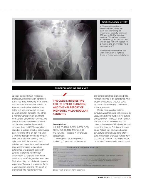
TUBERCULOSIS OF HIP<br />
A 38-year-old gentle man<br />
presented with severe hip<br />
pain, true shortening, all<br />
movements painfully restricted.<br />
ESR was at 70, Mantoux test<br />
positive, CBNAAT test positive,<br />
Histopathology also positive. He<br />
was on bed rest with traction for<br />
2 months and on ATT. Now he is<br />
undergoing ATT.<br />
X-ray pelvis showing both hips<br />
with destruction of right hip<br />
(head of femur and acetabulum).<br />
TUBERCULOSIS OF THE KNEE<br />
32-year-old gentleman, welder by<br />
profession, presented with right knee<br />
pain since 3 yrs. According to his words<br />
the complaint started after a hit to his<br />
knee with an iron bar while working.<br />
In the last one-year period he could<br />
do work only for 4 months (the other<br />
8 months were spent on treatment<br />
from various other health facilities). His<br />
personal history revealed that he has<br />
no diabetes, jaundice, hypertension,<br />
recurrent fever or HIV. The complaint<br />
stated as a sudden onset of pain 3 years<br />
back following hit to an iron rod, with<br />
a swelling disproportionate to the pain.<br />
Now presented with swelling around<br />
right knee. O/E: Patient walks with<br />
antalgic gait, horse shoe swelling around<br />
knee with increased temperature,<br />
patellar tap was present along with<br />
synovial thickening. Fixed flexion<br />
deformity of 20 degrees then flexion<br />
possible up to 90 degrees but with pain.<br />
Clinically a diagnosis of chronic synovitis<br />
was made. The case is interesting for its<br />
3-year duration, and the MRI report of<br />
pigmented villo-nodular synovitis.<br />
THE CASE IS INTERESTING<br />
FOR ITS 3-YEAR DURATION,<br />
AND THE MRI REPORT OF<br />
PIGMENTED VILLO-NODULAR<br />
SYNOVITIS<br />
Investigations<br />
HB: 11.7, TC-4200, P-69%, L-23%, E-6%,<br />
M-2%. ESR-80. RBS: 143mgs, HBS<br />
Ag, HCV, HIV – negative X-ray showed<br />
osteoporosis.<br />
MRI report indicated synovial<br />
thickening, 2 punched out lesions at<br />
Biopsy result of synovectomy specimen.<br />
the femoral condyles, pigmented villo<br />
nodular synovitis to be considered. After<br />
proper preoperative checkup partial<br />
synovectomy and biopsy done under<br />
spinal anesthesia.<br />
Medial para- patellar incision,<br />
synovium was thickened with increase in<br />
vascularity. Synovial fluid sent for culture<br />
and sensitivity - the result after 72 hours<br />
was sterile. Drain removed after 24<br />
hours; collection was 50 ml only. Wound<br />
inspection done on 3rd day which was<br />
clean. Patient was discharged on 3rd<br />
day. Suture removal was done after 14<br />
days. Quadriceps exercises started from<br />
second day onwards. The biopsy report<br />
came after 2 weeks and it was a typical<br />
<strong>March</strong> <strong>2019</strong> / FUTURE MEDICINE / 53