teaching and education - Helmholtz-Zentrum Berlin
teaching and education - Helmholtz-Zentrum Berlin
teaching and education - Helmholtz-Zentrum Berlin

Create successful ePaper yourself
Turn your PDF publications into a flip-book with our unique Google optimized e-Paper software.
ound solvent ions in tetragonal crystals of HEWL using a<br />
longer-wavelength data set <strong>and</strong> (v) the identification of a<br />
weakly bound lig<strong>and</strong> in the active site of HEWL. With these<br />
five experiments some of the most common scenarios<br />
encountered in structural biology using macromolecular<br />
crystallographic techniques were represented in the tutorial.<br />
However, despite its successful use, it became quickly<br />
apparent that the list of experiments was not complete. In<br />
order to remedy that, we have now complemented the first<br />
edition of the tutorial by adding two further experiments, (vi)<br />
a structure determination by single isomorphous replacement<br />
including anomalous scattering (SIRAS) using a gold derivative<br />
of tetragonal HEWL <strong>and</strong> (vii) a structure determination<br />
by ultraviolet radiation damage-induced phasing (UV-RIP) on<br />
the sweet protein thaumatin. The second edition of the tutorial<br />
now consists of seven experiments, which cover the whole<br />
breadth of approaches for structure determination using MX<br />
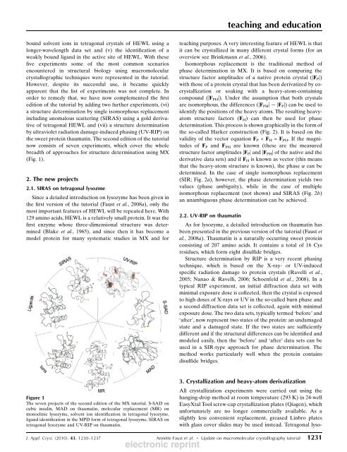
(Fig. 1).<br />
2. The new projects<br />
2.1. SIRAS on tetragonal lysozme<br />
Since a detailed introduction on lysozyme has been given in<br />
the first version of the tutorial (Faust et al., 2008a), only the<br />
most important features of HEWL will be repeated here. With<br />
129 amino acids, HEWL is a relatively small protein. It was the<br />
first enzyme whose three-dimensional structure was determined<br />
(Blake et al., 1965), <strong>and</strong> since then it has become a<br />
model protein for many systematic studies in MX <strong>and</strong> for<br />
Figure 1<br />
The seven projects of the second edition of the MX tutorial. S-SAD on<br />
cubic insulin, MAD on thaumatin, molecular replacement (MR) on<br />
monoclinic lysozyme, solvent ion identification in tetragonal lysozyme,<br />
lig<strong>and</strong> identification in the MPD form of tetragonal lysozyme, SIRAS on<br />
tetragonal lysozyme <strong>and</strong> UV-RIP on thaumatin.<br />
<strong>teaching</strong> <strong>and</strong> <strong>education</strong><br />
<strong>teaching</strong> purposes. A very interesting feature of HEWL is that<br />
it can be crystallized in many different crystal forms (for an<br />
overview see Brinkmann et al., 2006).<br />
Isomorphous replacement is the traditional method of<br />
phase determination in MX. It is based on comparing the<br />
structure factor amplitudes of a native protein crystal (|FP|)<br />
with those of a protein crystal that has been derivatized by cocrystallization<br />
or soaking with a heavy-atom-containing<br />
compound (|F PH|). Under the assumption that both crystals<br />
are isomorphous, the differences (|FPH| |FP|) can be used to<br />
identify the positions of the heavy atoms. The resulting heavyatom<br />
structure factors (FH) can then be used for phase<br />
determination. This process is shown graphically in the form of<br />
the so-called Harker construction (Fig. 2). It is based on the<br />
validity of the vector equation FP + FH = FPH. If the magnitudes<br />
of FP <strong>and</strong> FPH are known (these are the measured<br />
structure factor amplitudes |F P| <strong>and</strong> |F PH| of the native <strong>and</strong> the<br />
derivative data sets) <strong>and</strong> if FH is known as vector (this means<br />
that the heavy-atom structure is known), the phase can be<br />
determined. In the case of single isomorphous replacement<br />
(SIR; Fig. 2a), however, the phase determination yields two<br />
values (phase ambiguity), while in the case of multiple<br />
isomorphous replacement (not shown) <strong>and</strong> SIRAS (Fig. 2b)<br />
an unambiguous phase determination can be achieved.<br />
2.2. UV-RIP on thaumatin<br />
As for lysozyme, a detailed introduction on thaumatin has<br />
been presented in the previous version of the tutorial (Faust et<br />
al., 2008a). Thaumatin is a naturally occurring sweet protein<br />
consisting of 207 amino acids. It contains a total of 16 Cys<br />
residues, which form eight disulfide bridges.<br />
Structure determination by RIP is a very recent phasing<br />
technique, which is based on the X-ray- or UV-induced<br />
specific radiation damage to protein crystals (Ravelli et al.,<br />
2005; Nanao & Ravelli, 2006; Schoenfeld et al., 2008). In a<br />
typical RIP experiment, an initial diffraction data set with<br />
minimal exposure dose is collected, then the crystal is exposed<br />
to high doses of X-rays or UV in the so-called burn phase <strong>and</strong><br />
a second diffraction data set is collected, again with minimal<br />
exposure dose. The two data sets, typically termed ‘before’ <strong>and</strong><br />
‘after’, now represent two states of the protein: an undamaged<br />
state <strong>and</strong> a damaged state. If the two states are sufficiently<br />
different <strong>and</strong> if the structural differences can be identified <strong>and</strong><br />
modeled easily, then the ‘before’ <strong>and</strong> ‘after’ data sets can be<br />
used in a SIR-type approach for phase determination. The<br />
method works particularly well when the protein contains<br />
disulfide bridges.<br />
3. Crystallization <strong>and</strong> heavy-atom derivatization<br />
All crystallization experiments were carried out using the<br />
hanging-drop method at room temperature (293 K) in 24-well<br />
EasyXtal Tool screw-cap crystallization plates (Qiagen), which<br />
unfortunately are no longer commercially available. As a<br />
slightly less convenient replacement, greased Linbro plates<br />
with glass cover slides may be used instead. Tetragonal lyso-<br />
J. Appl. Cryst. (2010). 43, 1230–1237 Annette Faust et al. Update on macromolecular crystallography tutorial 1231<br />
electronic reprint