Susan Wu, MD, FAAP, Editor - American Academy of Pediatrics
Susan Wu, MD, FAAP, Editor - American Academy of Pediatrics
Susan Wu, MD, FAAP, Editor - American Academy of Pediatrics

Create successful ePaper yourself
Turn your PDF publications into a flip-book with our unique Google optimized e-Paper software.
YoU are the hoSPitaLiSt<br />
Case: 6 Year Old Boy With Fever,<br />
Bloody Oral Blisters and Respiratory Distress<br />
Riva Kamat, <strong>MD</strong>, <strong>FAAP</strong>, Pediatric Hospitalist, riva.kamat-nerikar@inova.org<br />
Nora Vish, <strong>MD</strong>, <strong>FAAP</strong>, Pediatric Hospitalist, nora.vish@inova.org<br />
Parag Jain <strong>MD</strong>, Pediatric Resident, drparagjain@gmail.com<br />
Inova Fairfax Hospital for Children, Fairfax, VA 22042<br />
You are the pediatric hospitalist<br />
taking admission calls on a busy<br />
winter day when you accept a<br />
6-year-old boy as a direct admission<br />
from his primary medical doctor (P<strong>MD</strong>).<br />
The P<strong>MD</strong> tells you that the patient<br />
presented to her <strong>of</strong>fice with a 10-day<br />
history <strong>of</strong> fever up to 103°F, croupy<br />
cough, wheezing and a 3-day history <strong>of</strong><br />
stomatitis. Due to the patient’s history<br />
<strong>of</strong> asthma, the patient’s mother began<br />
giving him levalbuterol, prednisone and<br />
his inhaled steroid 2 days ago, without<br />
much improvement. A rash began a<br />
few days earlier which was described as<br />
nonpruritic red macules over the boy’s<br />
chest and abdomen which progressed<br />
over the last 2 days to painful blisters.<br />
The boy has developed redness in both<br />
eyes with some discomfort and painful<br />
sores in his mouth over the last 1-2<br />
days with resultant poor oral intake and<br />
poor urine output. He denies headache,<br />
photophobia, nausea, vomiting, diarrhea,<br />
dysuria. In the <strong>of</strong>fice, the P<strong>MD</strong> noted<br />
bilateral conjunctival injection, mucosal<br />
erosions <strong>of</strong> his lips and mouth as well as<br />
a “crusty” rash on the boys chest and<br />
abdomen. The P<strong>MD</strong> would like to have<br />
the boy admitted for his inadequate oral<br />
intake, and his poorly controlled asthma<br />
exacerbation.<br />
The P<strong>MD</strong> provides the patient’s past<br />
medical history which is significant for<br />
asthma, gastroesophageal reflux and<br />
occasional migraines. The patient is<br />
taking lansoprazole, levalbuterol inhaler,<br />
budesinide inhaler, and monoleukast<br />
sodium. He is fully immunized and has<br />
had no surgical problems. He has not<br />
traveled overseas and there are no recent<br />
significant exposures. He has no sick<br />
contacts, no significant family or social<br />
history, and no known allergies<br />
He is admitted directly to the pediatric<br />
inpatient unit and you immediately<br />
evaluate him when he arrives. On<br />
6 | <strong>American</strong> <strong>Academy</strong> <strong>of</strong> <strong>Pediatrics</strong><br />
examination he is ill appearing but<br />
cooperative and in moderate respiratory<br />
distress. His temperature is 101º F (38.3º<br />
C), with a heart rate <strong>of</strong> 132 beats per<br />
minute, respiratory rate <strong>of</strong> 26 breaths<br />
per minute, and blood pressure <strong>of</strong> 124/57<br />
mm Hg. He is saturating 100% on 3 liters<br />
<strong>of</strong> oxygen (93% on 2 liters). He has<br />
bilateral subconjunctival hemorrhages<br />
without significant discharge or crusting.<br />
There is scant crusty discharge at<br />
the nares. His lips are red, swollen,<br />
bleeding, with tender blisters and<br />
exudates on both upper and lower lips<br />
(Figure 1). The gums have scattered<br />
ulcers which bleed readily. His neck is<br />
supple without significant adenopathy.<br />
Respiratory examination reveals bilateral<br />
wheezing and subcostal retractions with<br />
moderately prolonged expiratory phase,<br />
but no rales. The cardiac examination<br />
is hyperdynamic with tachycardia but<br />
no murmur, gallop or rub. His abdomen<br />
is s<strong>of</strong>t, nontender, nondistended<br />
with hypoactive bowel sounds but no<br />
masses or hepatosplenomegaly. There<br />
is erythema and swelling noted at his<br />
urethral meatus, but no discharge or<br />
bleeding. There are erythematous<br />
papules, coalescent macules and bullae<br />
on his trunk and back. Each bulla is<br />
approximately 1-3 cm in diameter,<br />
and most are intact. The others have<br />
flaccid overlying tissue or are denuded<br />
with sharply circumcised areas that are<br />
tender.<br />
Initial laboratory investigations obtained<br />
by the P<strong>MD</strong> earlier that day revealed<br />
a white cell count <strong>of</strong> 17.9 × 10³/µL<br />
(with 78% neutrophils, 2% bands, 18%<br />
lymphocytes, 2% monocytes). His<br />
hemoglobin, hematocrit and platelets<br />
were within normal limits. His basic<br />
metabolic panel and urinalysis were<br />
normal. A chest radiograph shows a<br />
small right lower lobe consolidation and<br />
bilateral hilar prominence (Figure 2).<br />
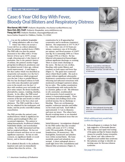
Figure 1: 6 year-old boy with bilateral<br />
subconjunctival hemorrhages, blistering<br />
lesions on mouth and lips, and mild crusting<br />
at both nares.<br />
Figure 2: Chest radiograph showing right<br />
lower lobe consolidation and bilateral hilar<br />
prominence.<br />
Which additional tests would help<br />
confirm the diagnosis?<br />
A. Echocardiogram to evaluate for findings<br />
suggestive <strong>of</strong> Kawasaki disease.<br />
B. Viral cultures and rapid viral testing<br />
<strong>of</strong> the gums and lips for herpes simplex<br />
viruses for possible herpes stomatitis.<br />
C. Eye, throat, and skin cultures for<br />
staphylococcal scalded skin syndrome<br />
D. Mycoplasma serology to identify an<br />
inciting cause for Stevens-Johnson<br />
syndrome<br />
See page 8 for answer.