Proliferation, apoptosis, and manganese superoxide ... - Erbeofficinali
Proliferation, apoptosis, and manganese superoxide ... - Erbeofficinali
Proliferation, apoptosis, and manganese superoxide ... - Erbeofficinali

Create successful ePaper yourself
Turn your PDF publications into a flip-book with our unique Google optimized e-Paper software.
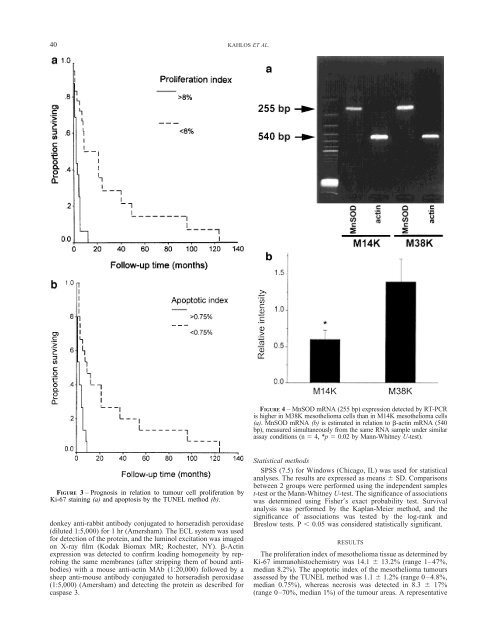
40 KAHLOS ET AL.<br />
FIGURE 3 – Prognosis in relation to tumour cell proliferation by<br />
Ki-67 staining (a) <strong>and</strong> <strong>apoptosis</strong> by the TUNEL method (b).<br />
donkey anti-rabbit antibody conjugated to horseradish peroxidase<br />
(diluted 1:5,000) for 1 hr (Amersham). The ECL system was used<br />
for detection of the protein, <strong>and</strong> the luminol excitation was imaged<br />
on X-ray film (Kodak Biomax MR; Rochester, NY). �-Actin<br />
expression was detected to confirm loading homogeneity by reprobing<br />
the same membranes (after stripping them of bound antibodies)<br />
with a mouse anti-actin MAb (1:20,000) followed by a<br />
sheep anti-mouse antibody conjugated to horseradish peroxidase<br />
(1:5,000) (Amersham) <strong>and</strong> detecting the protein as described for<br />
caspase 3.<br />
FIGURE 4 – MnSOD mRNA (255 bp) expression detected by RT-PCR<br />
is higher in M38K mesothelioma cells than in M14K mesothelioma cells<br />
(a). MnSOD mRNA (b) is estimated in relation to �-actin mRNA (540<br />
bp), measured simultaneously from the same RNA sample under similar<br />
assay conditions (n � 4, *p � 0.02 by Mann-Whitney U-test).<br />
Statistical methods<br />
SPSS (7.5) for Windows (Chicago, IL) was used for statistical<br />
analyses. The results are expressed as means � SD. Comparisons<br />
between 2 groups were performed using the independent samples<br />
t-test or the Mann-Whitney U-test. The significance of associations<br />
was determined using Fisher�s exact probability test. Survival<br />
analysis was performed by the Kaplan-Meier method, <strong>and</strong> the<br />
significance of associations was tested by the log-rank <strong>and</strong><br />
Breslow tests. P � 0.05 was considered statistically significant.<br />
RESULTS<br />
The proliferation index of mesothelioma tissue as determined by<br />
Ki-67 immunohistochemistry was 14.1 � 13.2% (range 1–47%,<br />
median 8.2%). The apoptotic index of the mesothelioma tumours<br />
assessed by the TUNEL method was 1.1 � 1.2% (range 0–4.8%,<br />
median 0.75%), whereas necrosis was detected in 8.3 � 17%<br />
(range 0–70%, median 1%) of the tumour areas. A representative